Efficient singlet oxygen generation upon two-photon ... - IEEE Xplore
Efficient singlet oxygen generation upon two-photon ... - IEEE Xplore
Efficient singlet oxygen generation upon two-photon ... - IEEE Xplore

Create successful ePaper yourself
Turn your PDF publications into a flip-book with our unique Google optimized e-Paper software.
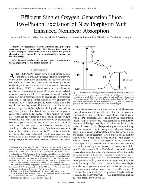
<strong>IEEE</strong> JOURNAL ON SELECTED TOPICS IN QUANTUM ELECTRONICS, VOL. 7, NO. 6, NOVEMBER/DECEMBER 2001 971<br />
<strong>Efficient</strong> Singlet Oxygen Generation Upon<br />
Two-Photon Excitation of New Porphyrin With<br />
Enhanced Nonlinear Absorption<br />
Aliaksandr Karotki, Mikalai Kruk, Mikhail Drobizhev, Aleksander Rebane, Eric Nickel, and Charles W. Spangler<br />
Abstract—We demonstrate efficient <strong>generation</strong> of <strong>singlet</strong> <strong>oxygen</strong><br />
<strong>upon</strong> <strong>two</strong>-<strong>photon</strong> excitation with 150-fs 780-nm laser pulses of<br />
a new porphyrin photosensitizer molecule whose <strong>two</strong>-<strong>photon</strong><br />
absorption cross section has been considerably enhanced by<br />
chemical design.<br />
Index Terms—Photodynamic therapy, porphyrin photosensitizers,<br />
<strong>singlet</strong> <strong>oxygen</strong>, <strong>two</strong>-<strong>photon</strong> absorption.<br />
I. INTRODUCTION<br />
ALONG-STANDING dream in the field of cancer therapy<br />
is the ability to treat subcutaneous tumors noninvasively,<br />
while at the same time eliminating the adverse physical<br />
discomfort associated with traditional chemotherapy and the<br />
debilitating effects of actinic radiation treatments. Photodynamic<br />
therapy (PDT) is gaining acceptance worldwide as<br />
an alternative treatment of tumors [1] as well as age-related<br />
macular de<strong>generation</strong> [2]. PDT employs the special ability of<br />
some porphyrin photosensitizers to accumulate in pathologic<br />
cells and to transfer absorbed <strong>photon</strong> energy efficiently to<br />
extremely active <strong>singlet</strong> <strong>oxygen</strong> molecules, which then wipe<br />
out the surrounding tumor. Unfortunately, the limited penetration<br />
depth by visible light into biological tissue allows<br />
only few types of skin, breast, and certain endoscopically<br />
accessible cancers to be treated in this fashion. To make<br />
PDT more generally applicable, it is crucial to deliver light<br />
deeper into the tissue. This may be achieved by utilizing the<br />
nonlinear-optical effect of <strong>two</strong>-<strong>photon</strong> absorption (TPA) in<br />
which case the illumination is carried out at near-infrared (IR)<br />
wavelengths, where the tissue is significantly more transparent<br />
than in the visible. However, so far TPA of tumor-specific<br />
porphyrins has been notoriously inefficient, rendering the<br />
treatment of deeper tumors impractical. Here, we introduce a<br />
new porphyrin photosensitizer with an enhanced TPA cross<br />
Manuscript received May 22, 2001. This work was supported by the Air<br />
Force Office of Scientific Research under Grant F49620-01-1-0324, Grant<br />
F49620-96-1-0440, Grant F49620-98-1-0422, and Grant F49620-98-1-0157.<br />
A. Karotki and A. Rebane are with the Department of Physics, Montana State<br />
University, Bozeman, MT 59717 USA (e-mail: rebane@physics.montana.edu).<br />
M. Kruk was with the Department of Physics, Montana State University,<br />
Bozeman, MT 59717 USA. He is now with the Institute of Molecular and<br />
Atomic Physics, National Academy of Sciences, 220072 Minsk, Belarus.<br />
M. Drobizhev is with the Department of Physics, Montana State University,<br />
Bozeman, MT 59717 USA and also with the P. N. Lebedev Physical Institute of<br />
the Russian Academy of Sciences, 117942 Moscow, Russia.<br />
E. Nickel is with Synar Technologies, Inc., Atlanta, GA 30303 USA.<br />
C. W. Spangler is with the Department of Chemistry and Biochemistry, Montana<br />
State University, Bozeman, MT 59717 USA.<br />
Publisher Item Identifier S 1077-260X(01)11237-2.<br />
Fig. 1. Schematic of the energy levels for porphyrin photosensitizer (solid<br />
bars) and molecular <strong>oxygen</strong> (open bars). S (g), S (u), S (g), and T represent,<br />
respectively, ground, first <strong>singlet</strong>, ith excited <strong>singlet</strong>, and lowest triplet states<br />
of the photosensitizer. The symbols in the parenthesises denote gerade (g) and<br />
ungerade (u) symmetry of the corresponding states. 6 and 1 denote the<br />
ground and the first excited <strong>singlet</strong> states of molecular <strong>oxygen</strong>.<br />
section, and demonstrate its ability to generate <strong>singlet</strong> <strong>oxygen</strong><br />
<strong>upon</strong> illumination with near-IR light. Injecting a porphyrin<br />
photosensitizer into a patient’s blood stream commences a<br />
typical PDT procedure. After an appropriate time interval<br />
(usually tens of hours), the photosensitizer is activated by<br />
shining a visible light, usually a red color laser beam, at the<br />
tumor’s location [1], [3]. Photophysical processes constituting<br />
PDT are summarized in the energy level diagram shown in<br />
Fig. 1. In its classical implementation, absorption of one visible<br />
<strong>photon</strong> [one-<strong>photon</strong> excitation (OPE)] brings a photosensitizer<br />
molecule into a short-lived excited state with energy of<br />
170–190 kJ mol , which corresponds to an illumination<br />
wavelength of 620–690 nm. After a few nanoseconds,<br />
the porphyrin undergoes an intersystem crossing to a triplet<br />
state with energy of 110–130 kJ mol and with a much<br />
longer life time (on the order of milliseconds). From the triplet<br />
state, the energy is transferred (semicircle arrows in Fig. 1)<br />
to omnipresent <strong>oxygen</strong> molecules by switching them from a<br />
triplet ground state into an excited <strong>singlet</strong> state with<br />
excitation energy of 94 kJ mol . Once in the excited <strong>singlet</strong><br />
state, the <strong>oxygen</strong> presents an extremely reactive species capable<br />
of damaging surrounding tissue in an irreversible manner. A<br />
presence of excited <strong>singlet</strong> <strong>oxygen</strong> molecules ( ) in solution<br />
is usually detected by their<br />
luminescence at about<br />
1270 nm (black arrow in Fig. 1).<br />
1077–260X/01$10.00 © 2001 <strong>IEEE</strong>
972 <strong>IEEE</strong> JOURNAL ON SELECTED TOPICS IN QUANTUM ELECTRONICS, VOL. 7, NO. 6, NOVEMBER/DECEMBER 2001<br />
To be effective, the photosensitizers have to selectively<br />
accumulate in the tumor tissue. Serendipitously, porphyrin<br />
molecules possess this rare feature [3]. The second critical<br />
point is how deep can light penetrate into the body without<br />
being scattered or absorbed by normal tissue. It is well known<br />
that the tissue’s transmission depends critically on the illumination<br />
wavelength [4] and is the largest in the so-called<br />
tissue transparency window at 750–1000 nm. Porphyrins<br />
currently in use for PDT fall short of this transparency window:<br />
their absorption varies from 620 to 690 nm, where<br />
effective penetration in most tissues is no more than just a<br />
few millimeters in depth [5]. Unfortunately, attempts to shift<br />
the one-<strong>photon</strong> absorption band toward longer wavelengths by<br />
chemical modification of the porphyrin structure come into<br />
conflict with the fundamental requirement that the excitation<br />
energy of <strong>singlet</strong> <strong>oxygen</strong> is lower than the energy of the<br />
state. In addition, long-wavelength shift of porphyrin’s energy<br />
levels often aggravates the situation by reducing compound’s<br />
stability.<br />
In the view of these mounting difficulties, a proposal to use<br />
TPA appears as the best alternative way to achieve PDT. TPA<br />
[6] is a third-order nonlinear-optical process and it consists in<br />
simultaneous absorption of <strong>two</strong> <strong>photon</strong>s, so that the illumination<br />
wavelength is twice of that of the actual transition wavelength<br />
[<strong>two</strong>-<strong>photon</strong> excitation (TPE) in Fig. 1]. TPA allows using<br />
near-IR <strong>photon</strong>s in the tissue transparency window and does<br />
not require the red shift of the lowest electronic transition of<br />
the porphyrin. Note that even if TPA takes a molecule into one<br />
of its’ higher excited states , this does not bar the lowest<br />
state to be populated via internal conversion. Once this<br />
state is formed, the subsequent events follow the same path as<br />
described above. In addition, TPA also provides <strong>two</strong> further<br />
advantages: 1) laser-induced hyperthermia is minimized,<br />
since near-IR light has a reduced absorption in tissues and 2)<br />
nonlinear (quadratic) dependence of TPA on laser intensity<br />
provides for higher spatial selectivity of treatment due to the<br />
smaller effective size of the focal spot. The latter is essential<br />
for treatment of sensitive tissues such as those of age-related<br />
wet macular de<strong>generation</strong>. On the downside, because the<br />
probability of TPA with conventional low-power light sources<br />
is vanishing small, laser pulses with high peak intensity are<br />
required for the TPA effect to be pronounced. Fortunately,<br />
the commercialization of femtosecond mode-locked near-IR<br />
Ti : Sapphire lasers has greatly alleviated this last problem.<br />
As one might expect, these useful features have stimulated<br />
an active search for new molecular system for TPA-based PDT<br />
[7]–[11]. Some nonporphyrin-based compounds may indeed<br />
possess an impressively large TPA cross section [8], [9], [12].<br />
However, they either lack the vital property to generate <strong>singlet</strong><br />
<strong>oxygen</strong> or their interaction with biological tissue is not known.<br />
On the other hand, all known porphyrin-based sensitizers were<br />
found to have a very small TPA cross section, typically not<br />
more than few Göppert–Mayer units (GM, 1 GM 10 cm<br />
s <strong>photon</strong> molecule ) [10] and, thus, require unreasonable<br />
prolonged illumination with a tightly focused laser beam [10],<br />
[11]. Our approach lies in special chemical modification of<br />
(b)<br />
(a)<br />
Fig. 2. Molecular structures of porphyrins studied. (a)<br />
5-(4-diphenylaminostilbene), 15-(2, 6-dichlorophenyl)-21H, 23H-porphine.<br />
(b) 5-phenyl, 15-(2, 6-dichlorophenyl)-21H, 23H-porphine. (c) 5, 10, 15,<br />
20-tetraphenyl-21H, 23H-porphine.<br />
porphyrin, which enhances the molecule’s TPA cross section<br />
without jeopardizing its ability to generate <strong>singlet</strong> <strong>oxygen</strong>.<br />
The design was based on structure-property relationships,<br />
known to enhance TPA cross section in organic -conjugated<br />
chromophores [12]–[14]. In particular, introduction of donors<br />
or acceptors of electronic density as substituent groups, extension<br />
of conjugation, and inclusion of donor- -donor<br />
motifs within chromophore have all been previously utilized.<br />
In our particular case, this was achieved by an introduction of a<br />
4-(diphenylaminostilbene)-substituent into the mesoposition of<br />
the tetrapyrrol ring. This results in a 20 times enhancement in<br />
TPA cross section at<br />
nm. In addition, the inclusion<br />
of the 2, 6-dichlorophenyl substituent assists intersystem<br />
crossing via an internal heavy atom effect.<br />
II. EXPERIMENTAL SECTION<br />
Structures of a typical new porphyrin DPASP [5-(4-diphenylaminostilbene),<br />
15-(2, 6-dichlorophenyl)-21H, 23H-porphine]<br />
(I), as well as its nonsubstituted counterpart (II) and commercial<br />
tetraphenylporphyrin (III) are shown in Fig. 2. The last <strong>two</strong><br />
porphyrins were studied for purpose of comparison as model<br />
compounds. Compound I has been synthesized by condensing<br />
10-(2, 6-dichlorophenyl) bilane with (4-diphenylaminostilbene)<br />
aldehyde under acidic conditions followed by oxidation with<br />
2, 3-dichloro-5, 6-cyano-1, 4-benzoquinone. The way of synthesis<br />
of compound II is identical to that of I. Porphyrins I<br />
and II were purified by flash chromatography on silica using<br />
20%–40% dichloromethane/hexane gradient and the structure<br />
of molecules was confirmed by spectroscopic analysis. Compound<br />
III was purchased from Aldrich and used as received.<br />
Our laser system is schematically shown in Fig. 3. It comprised<br />
a Ti : sapphire regenerative amplifier (CPA-1000, Clark<br />
MRX), which was operated at 1-kHz repetition rate and produced<br />
150-fs pulses at 0.8 mJ energy per pulse. These pulses<br />
(c)
KAROTKI et al.: EFFICIENT SINGLET OXYGEN GENERATION UPON TWO-PHOTON EXCITATION 973<br />
Here, indexes 1 and 2 refer to the values measured under OPE<br />
and TPE, respectively, is the fluorescence signal recorded<br />
during time , is the molecule concentration (in molecule<br />
cm ), is the sample thickness (in cm), is the pulse duration<br />
(full width at half maximum in s), is the pulse repetition rate<br />
under TPE, is the average intensity (in W/cm ), is laser frequency<br />
(in hertz), and is the optical density of the sample<br />
at one-<strong>photon</strong> illumination wavelength.<br />
In our experiments, we employed the fundamental of Ti-sapphire<br />
regenerative amplifier for TPE and its second harmonic<br />
for corresponding OPE. In both cases, the beam was slightly<br />
focused with a mm lens (L) into the 1-cm cell with<br />
sample solution through a pinhole (P) placed in front of the lens.<br />
Fluorescence was collected and focused on the entrance slit of<br />
a Jobin–Yvon monochromator with a spherical mirror. Special<br />
care were taken to geometrically eliminate the possible reabsorption<br />
effects.<br />
In experiments with <strong>singlet</strong> <strong>oxygen</strong> <strong>generation</strong>, OPE was carried<br />
out at nm with average intensity of 0.5 W/cm and<br />
TPE was carried out at nm with average intensity of<br />
15 W/cm . The laser beam was slightly focused to give a cylindrical<br />
irradiated volume of 1.5-mm diameter in both cases.<br />
The <strong>singlet</strong> <strong>oxygen</strong> luminescence spectrum was measured with<br />
a nitrogen-cooled Ge detector coupled with monochromator and<br />
lockin amplifier.<br />
Fig. 3. Schematic of experimental setup. For measurement of TPA spectra,<br />
a second harmonic of tunable OPA was used. For measurements of TPA<br />
cross sections and <strong>singlet</strong> <strong>oxygen</strong> <strong>two</strong>-<strong>photon</strong> photo <strong>generation</strong>, the output of<br />
regenerative amplifier was used directly (dashed arrow). For one-<strong>photon</strong> <strong>singlet</strong><br />
<strong>oxygen</strong> photosensitization, the second harmonic of this radiation was used.<br />
were parametrically down-converted in the optical parametric<br />
amplifier (OPA) (TOPAS, Quantronix), which yielded 100-fs<br />
pulses in the range from 1.1 to 1.8 m. TPA spectra were obtained<br />
by tuning the OPA with subsequent second-harmonic<br />
<strong>generation</strong> and registration of the porphyrin fluorescence. Absolute<br />
TPA cross sections were measured by comparing fluorescence<br />
intensity under OPE and TPE (see [15] for details).<br />
For sech temporal profile of excitation pulses and under the assumption<br />
that fluorescence quantum efficiency is the same for<br />
both modes of excitation, the <strong>two</strong>-<strong>photon</strong> cross section writes as<br />
follows:<br />
(1)<br />
III. RESULTS AND DISCUSSION<br />
TPA spectra of the three molecules are shown in Fig. 4. Black<br />
solid lines represent the appropriately scaled one-<strong>photon</strong> absorption.<br />
All three molecules reveal a characteristic strong Soret<br />
band near 415 nm, accompanied by four relatively weak bands<br />
in the visible (not shown).<br />
We observe that the TPA band does not coincide with any of<br />
the bands in the linear absorption spectrum. Quantum-mechanical<br />
calculations [16], [17] show that key spectroscopic properties<br />
of porphyrins such as position and symmetry of main electronic<br />
energy levels are determined by the tetrapyrrol macrocycle<br />
and predict that the lowest excited state of symmetry<br />
should be positioned near the Soret band. According to selection<br />
rules, centrosymmetric molecules should have allowed one<strong>photon</strong><br />
transitions between the states of opposite parity and <strong>two</strong><strong>photon</strong><br />
transitions between the states of same parity. Since the<br />
porphin ground state has symmetry, we assume that the observed<br />
TPA bands correspond to the transitions between the <strong>two</strong><br />
states of parity.<br />
The key point, however, is that DPASP (I) shows a very large<br />
(for porphyrins) TPA cross section with a measured maximum<br />
value of 80 10 GM at about 390 nm ( nm). The<br />
most significant fact is that this excitation wavelength falls well<br />
into the tissue transparency window, which makes the new approach<br />
very promising for future applications of PDT.<br />
Fig. 5 demonstrates that we were able to directly detect<br />
the production of <strong>singlet</strong> <strong>oxygen</strong> <strong>upon</strong> TPA of DPASP (I).<br />
Fig. 5(a) shows <strong>two</strong> luminescence spectra obtained <strong>upon</strong> either<br />
one-<strong>photon</strong> (dash) or <strong>two</strong>-<strong>photon</strong> (solid) excitation of DPASP,<br />
both normalized to unity. Note that the <strong>two</strong> spectra coincide<br />
well within experimental error and show both a characteristic<br />
peak near 1276 nm, which undoubtedly corresponds to the<br />
<strong>singlet</strong> <strong>oxygen</strong> luminescence spectrum [18]. Fig. 5(b) shows<br />
that the intensity of luminescence <strong>upon</strong> TPE increases as a<br />
square of the illumination intensity, which proves that the<br />
porphyrin is indeed excited by absorbing <strong>two</strong> near-IR <strong>photon</strong>s<br />
simultaneously. Furthermore, if the solution was treated with<br />
a nitrogen sparge, the intensity of luminescence decreased<br />
because of the decreased <strong>oxygen</strong> concentration. Note that only<br />
with DPASP (I) could we reliably detect <strong>oxygen</strong> luminescence<br />
<strong>upon</strong> TPA. In the case of II and III, while a strong luminescence<br />
could be obtained <strong>upon</strong> OPE, with infrared excitation the signal<br />
was below the noise level of our detector.<br />
IV. CONCLUSION<br />
We have demonstrated, for the first time to the best of our<br />
knowledge, that <strong>singlet</strong> <strong>oxygen</strong> can be produced efficiently<br />
<strong>upon</strong> TPE of porphyrin photosensitizer with near-IR illumination<br />
within the tissue transparency window. This is achieved<br />
by chemically attaching special moieties to the porphyrin molecule,<br />
which are increasing the TPA cross section in near-IR.<br />
We believe that our results open new possibilities for designing
974 <strong>IEEE</strong> JOURNAL ON SELECTED TOPICS IN QUANTUM ELECTRONICS, VOL. 7, NO. 6, NOVEMBER/DECEMBER 2001<br />
(a) (b) (c)<br />
Fig. 4. TPA spectra (circles) of 10 M toluene solutions of compounds (a) I, (b) II, and (c) III. The spectra could not be measured at wavelength shorter than<br />
shown because the energy of excitation <strong>photon</strong> approaches that of the first excited <strong>singlet</strong> state S and efficient one-<strong>photon</strong> absorption masks TPA. The wavelength<br />
axis corresponds to the transition wavelength, i.e., half the laser wavelength. Vertical axis shows the TPA cross sections in Göppert–Mayer.<br />
Fig. 5. (a) 1 ! 6 luminescence spectra of molecular <strong>oxygen</strong> in air-saturated toluene solution of I. Dashed and solid curves represent, respectively, the<br />
spectra measured with OPE and TPE. Both spectra are normalized to unity. (b) Dependence of the 1 ! 6 <strong>oxygen</strong> luminescence intensity I on the<br />
average illumination intensity, P , <strong>upon</strong> TPE of porphyrin. Experimental data are shown by black squares. Solid curve is the best power-law fit I = aP with<br />
n =2:1 6 0:1.<br />
tumor-specific drugs with a large TPA cross section, which<br />
may eventually lead to PDT-based treatment of various deep<br />
tumors.<br />
ACKNOWLEDGMENT<br />
The authors would like to thank R. Cone for his generous loan<br />
of a Ge-detector.<br />
REFERENCES<br />
[1] B. W. Henderson and T. J. Dougherty, “How does photodynamic therapy<br />
work?,” Photochem. Photobio., vol. 55, no. 1, pp. 147–157, Jan. 1992.<br />
[2] N. Bressler et al., “Photodynamic therapy of subfoveal choroidal neovascularization<br />
in age-related macular de<strong>generation</strong> with verteporfin.<br />
One-year results of 2 randomized clinical trials—TAP Report 1,” Arch.<br />
Ophthalmol., vol. 117, no. 2, pp. 1329–1348, Oct. 1999.<br />
[3] R. Bonnett, “Photosensitizers of the porphyrin and phtalocyanine series<br />
for photodynamic therapy,” Chem. Soc. Rev., vol. 24, no. 1, pp. 19–33,<br />
Feb. 1995.<br />
[4] S. Wan, J. A. Parrish, R. R. Anderson, and M. Madden, “Transmittance<br />
of nonionizing radiation in human tissues,” Photochem. Photobio., vol.<br />
34, pp. 679–681, 1981.<br />
[5] W.-F. Cheong, S. A. Prahl, and A. J Welch, “A review of the optical<br />
properties of biological tissues,” <strong>IEEE</strong> J. Quantum Electron., vol. 26,<br />
pp. 2166–2185, Dec. 1990.<br />
[6] S. Kershaw, “Two-<strong>photon</strong> absorption,” in Characterization Techniques<br />
and Tabulations for Organic Nonlinear Optical Materials, M. G. Kuzyk<br />
and C. W. Dirk, Eds. New York: Marcel Dekker, 1998.<br />
[7] W. G. Fisher, W. R. Partridge, Jr., C. Dees, and E. A. Wachter, “Simultaneous<br />
<strong>two</strong>-<strong>photon</strong> activation of type-I photodynamic therapy agents,”<br />
Photochem. Photobio., vol. 66, no. 2, pp. 1415–155, Aug. 1997.<br />
[8] J. D. Bhawalkar, N. D. Kumar, C.-F. Zhao, and P. N. Prasad, “Two<strong>photon</strong><br />
photodynamic therapy,” J. Clin. Lasers Med. Surg., vol. 15, no.<br />
5, pp. 201–204, Sept.–Oct. 1997.
KAROTKI et al.: EFFICIENT SINGLET OXYGEN GENERATION UPON TWO-PHOTON EXCITATION 975<br />
[9] P. K. Frederiksen and O. P. R. Jørgensen, “Two-<strong>photon</strong> photosensitized<br />
production of <strong>singlet</strong> <strong>oxygen</strong>,” J. Amer. Chem. Soc., vol. 123, no. 6, pp.<br />
1215–1221, Feb. 2001.<br />
[10] R. L. Goyan and D. T. Cramb, “Near-infrared <strong>two</strong>-<strong>photon</strong> excitation of<br />
protoporphyrin IX: Photodynamics and photoproduct <strong>generation</strong>,” Photochem.<br />
Photobio., vol. 72, no. 6, pp. 821–827, Dec. 2000.<br />
[11] K. König, “Multi<strong>photon</strong> microscopy in life sciences,” J. Microsc., pt. 2,<br />
vol. 200, pp. 83–104, Nov. 2000.<br />
[12] M. Albota et al., “Design of organic molecules with large <strong>two</strong>-<strong>photon</strong><br />
absorption cross sections,” Science, vol. 281, no. 5383, pp. 1653–1656,<br />
Sept. 1998.<br />
[13] B. H. Cumpston et al., “Two-<strong>photon</strong> polymerization initiators for<br />
three-dimensional optical data storage and microfabrication,” Nature,<br />
vol. 398, no. 6722, pp. 51–54, Mar. 1999.<br />
[14] C. W. Spangler, “Recent developments in the design of organic materials<br />
for optical power limiting,” J. Mater. Chem., vol. 9, no. 9, pp.<br />
2013–2020, Sept. 1999.<br />
[15] M. Drobizhev, A. Karotki, and A. Rebane, “Persistent spectral hole<br />
burning by simultaneous <strong>two</strong>-<strong>photon</strong> absorption,” Chem. Phys. Lett.,<br />
vol. 334, no. 1–3, pp. 76–82, Feb. 2001.<br />
[16] V. A. Kuz’mitsky and K. N. Solov’ev, “PPDP/S calculation of the porphin<br />
molecule electronic spectra,” J. Appl. Spectr., vol. 27, pp. 724–730,<br />
1977.<br />
[17] D. Sundholm, “Density functional theory study of the electronic absorption<br />
spectrum of Mg-porphyrin and Mg-etioporphyrin—I,” Chem. Phys.<br />
Lett., vol. 317, no. 3–5, pp. 392–399, Feb. 2000.<br />
[18] A. Bromberg and C. S. Foote, “Solvent shift of <strong>singlet</strong> <strong>oxygen</strong> emission<br />
wavelength,” J. Phys. Chem., vol. 93, no. 10, pp. 3968–3969, May 1989.<br />
Aliaksandr Karotki was born in Minsk, Belarus, in<br />
1978. He received the M.S. degree in physics with a<br />
specialization in laser physics and spectroscopy from<br />
the Belarus State University, Minsk, Belarus, in 1998.<br />
He is currently working toward the Ph.D. degree at<br />
Montana State University, Bozeman.<br />
He was with the Institute of Molecular and Atomic<br />
Physics of the Belarus National Academy of Science,<br />
Minsk, Belarus. His current research interests include<br />
nonlinear optics of organic materials and their applications.<br />
Mikalai Kruk was born near Minsk, Belarus, in<br />
1967. He received the M.S. degree in physics from<br />
the Belarussian State University, Minsk, Belarus,<br />
in 1991 and the Ph.D. degree in physics from the<br />
Institute of Molecular and Atomic Physics of the<br />
National Academy of Science of Belarus, Minsk,<br />
Belarus, in 1997.<br />
He was in the Soviet Army and was with the University<br />
of P. and M. Curie from 1997 to 1998. He has<br />
been with the National Academy of Sciences of Belarus<br />
since 1998. He had a short visit with the Montana<br />
State University, Bozeman, in 2001. He has authored or coauthored over 30<br />
papers. His current research interests include the photophysics and photochemistry<br />
of porphyrins and related compounds.<br />
Mikhail Drobizhev was born in Moscow, Russia,<br />
in 1963. He received the M.S. degree in physics<br />
with a specialization in molecular biophysics from<br />
the Moscow Institute of Physics and Technology,<br />
Moscow, Rusia, and Ph.D. degree in physics from<br />
the P. N. Lebedev Physical Institute of the Russian<br />
Academy of Sciences, Moscow, Russia, in 1998.<br />
Since 1986, he has been an Associate Researcher<br />
with the P. N. Lebedev Physical Institute of the<br />
Russian Academy of Sciences and was a Research<br />
Scientist under Prof. Rebane with Montana State<br />
University, Bozeman, in 1999. He has authored or coauthored over 40<br />
papers. His current research interests include selective laser spectroscopy<br />
of porphyrins and other biological molecules and their applications in data<br />
storage, optoelectronics, and laser medicine.<br />
Aleksander Rebane received the M.S. degree in<br />
physics from the Tartu University, Tartu, Estonia,<br />
in 1981 and the Ph.D. degree in physics from the<br />
Estonian Academy of Sciences, Tartu, Estonia, in<br />
1985.<br />
From 1991 to 1997, he was Oberassistent and<br />
Privatdozent with the Institute for Physical Chemistry,<br />
the Swiss Federal Institute of Technology<br />
(ETH), Zurich, Switzerland. He received from the<br />
ETH Habiliation in physical chemistry in 1996<br />
and became an Associate Professor of Physics<br />
with Montana State University, Bozeman, in 1997. His current research<br />
interests include nonlinear optics of organic materials, ultrafast optical<br />
storage, time-and-space-domain holography, ultrafast spectroscopy of organic<br />
molecules, and medical applications of nonlinear optics.<br />
Dr. Rebane received the Intenational Commission for Optics Prize in 1993,<br />
the Leopold Ruzicka Prize in 1996, and the Charles and Nora Wiley Award in<br />
1999.<br />
Eric Nickel, photograph and biography not available at the time of publication.<br />
Charles W. Spangler reeived the B.S. degree<br />
in chemistry from the Massachusetts Institute of<br />
Technology, Cambridge, in 1959, the M.S. degree<br />
in organic chemistry from Northeastern University,<br />
Boston, MA, in 1961, and the Ph.D. degree from the<br />
University of Maryland, College Park, in 1964.<br />
After on year as a Kettering Foundation Teacher-<br />
Intern with Ohio Wesleyan University, he joined the<br />
Faculty of Northern Illinois University as an Assistant<br />
Professor or Organic Chemistry in 1965, where<br />
he became an Associate Professor in 1972, Professor<br />
in 1981, a Presidential Research Professor in 1990, and a Distinguished Research<br />
Professor in 1994. In 1996, he became a Research Professor with the<br />
Department of Chemistry and Biochemistry and the Optical Center, Montana<br />
State University, Bozeman. He has authored or coauthored more than 180 research<br />
papers and has presented at more than 125 national and international<br />
meetings and symposia.