Journal of Luminescence 107 (2004) - Department of Physics ...
Journal of Luminescence 107 (2004) - Department of Physics ...
Journal of Luminescence 107 (2004) - Department of Physics ...

Create successful ePaper yourself
Turn your PDF publications into a flip-book with our unique Google optimized e-Paper software.
ARTICLE IN PRESS<br />
<strong>Journal</strong> <strong>of</strong> <strong>Luminescence</strong> <strong>107</strong> (<strong>2004</strong>) 194–202<br />
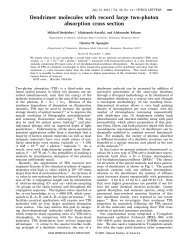
Electron–phonon coupling in two-photon spectral gratings:<br />
role <strong>of</strong> molecular symmetry<br />
M. Drobizhev a,b, *, Yu. Dzenis a , A. Karotki a , A. Rebane a<br />
a <strong>Department</strong> <strong>of</strong> <strong>Physics</strong>, Montana State University, Bozeman, MT 59717-3840, USA<br />
b Lebedev <strong>Physics</strong> Institute, Leninsky pr., 53, Moscow 119991, Russia<br />
Abstract<br />
We studyspectral gratings obtained as a result <strong>of</strong> two-photon excitation <strong>of</strong> the lowest electronic transition <strong>of</strong> several<br />
tetrapyrrole molecules with phase-locked pairs <strong>of</strong> femtosecond pulses. A particular dependence <strong>of</strong> these gratings on<br />
temperature indicates that mirror symmetry is present between two-photon absorption (TPA) and one-photon<br />
fluorescence spectra for non-centrosymmetrical chlorin, but fails for centrosymmetrical naphthalocyanine. The latter<br />
case is best described if the homogeneous TPA spectrum contains phonon sideband, but not the zero-phonon line.<br />
These results are discussed in terms <strong>of</strong> alternative selection rules for one- and two-photon transitions in<br />
centrosymmetrical molecules.<br />
r <strong>2004</strong> Elsevier B.V. All rights reserved.<br />
Keywords: Two-photon absorption; Two-photon coherence; Zero-phonon lines; Spectral hole burning; Herzberg–Teller interaction;<br />
Parityselection rules<br />
1. Introduction<br />
*Corresponding author. <strong>Department</strong> <strong>of</strong> <strong>Physics</strong>, Montana<br />
State University, Bozeman, MT 59717-3840, USA.<br />
E-mail address: drobizhev@physics.montana.edu<br />
(M. Drobizhev).<br />
In our previous papers [1–3], we have demonstrated<br />
the possibility<strong>of</strong> creating spectral gratings<br />
as a result <strong>of</strong> two-photon absorption (TPA) in<br />
inhomogeneouslybroadened media at low temperatures.<br />
Intuitively, this effect implies the presence<br />
<strong>of</strong> narrow zero-phonon lines (ZPL) in TPA<br />
transition. TPA pure electronic ZPLs were shown<br />
for several molecules, including centrosymmetric<br />
[4–10] and non-centrosymmetric ones [11,12].<br />
For non-centrosymmetric molecules, the same<br />
transition is simultaneouslyallowed for onephoton<br />
absorption and TPA and, therefore, the<br />
same pure electronic ZPL can be observed in both<br />
types <strong>of</strong> transition. For example, one can observe<br />
the pure electronic ZPL in one-photon fluorescence<br />
spectrum upon two-photon excitation <strong>of</strong> the<br />
verysame transition. For centrosymmetric molecule<br />
(in centrosymmetric site), selection rules for<br />
two- and one-photon transitions are mutually<br />
exclusive and, therefore, the role <strong>of</strong> vibrations<br />
and matrix phonons becomes decisive.<br />
Similarlyto one-photon case (exemplified by<br />
photon echo and time-domain holography), in<br />
TPA-induced gratings, two fs pulses, delayed by<br />
time Dt; are applied to a material, thus resulting in<br />
interference and interference fringes in frequency<br />
dimension [1–3]. However, in contrast to usual<br />
one-photon case, photons are absorbed bypairs<br />
from each pulse. Two-photon excitation has<br />
0022-2313/$ - see front matter r <strong>2004</strong> Elsevier B.V. All rights reserved.<br />
doi:10.1016/j.jlumin.2003.12.032
ARTICLE IN PRESS<br />
M. Drobizhev et al. / <strong>Journal</strong> <strong>of</strong> <strong>Luminescence</strong> <strong>107</strong> (<strong>2004</strong>) 194–202 195<br />
potential advantage in several applications, including<br />
ultrahigh-densityoptical memory(3D<br />
memory<strong>of</strong> ultrafast temporal processes) and<br />
ultrafast optical computing.<br />
If the homogeneous spectrum <strong>of</strong> impurity<br />
system consists only <strong>of</strong> narrow ZPL, the spectral<br />
response <strong>of</strong> the medium is exactlythe replica <strong>of</strong><br />
power spectrum <strong>of</strong> Fourier transform <strong>of</strong> the<br />
square <strong>of</strong> electric field temporal pr<strong>of</strong>ile [2,3]. For<br />
a series <strong>of</strong> short pulses, the total spectral width is<br />
determined bythe inverse pulse duration. Organic<br />
systems, such as polymer films doped with organic<br />
molecules, present an optimum recording media<br />
for 100 fs duration laser pulses because the<br />
inhomogeneous broadening in these systems is <strong>of</strong><br />
the order <strong>of</strong> the inverse pulse duration. However,<br />
almost all organic systems show considerable<br />
phonon wings (PWs) in their homogeneous spectra<br />
with ZPL–PW separation less than inhomogeneous<br />
width. Therefore, an important problem,<br />
which needs to be considered in connection<br />
with TPA gratings and their practical applications,<br />
is the effect <strong>of</strong> low-frequencyphonons on the<br />
shape <strong>of</strong> TPA-induced spectral gratings. TPA<br />
electron–phonon transitions become dominant<br />
for centrosymmetric molecules, where pure<br />
electronic transition (ZPL) is forbidden by<br />
symmetry.<br />
In this paper we consider TPA gratings created<br />
in different molecular systems, with and without<br />
center <strong>of</strong> inversion. The spectral fringes are excited<br />
with photons <strong>of</strong> energyequal to one-half the first<br />
S 0 S 1 transition energyand monitored in fluorescence<br />
<strong>of</strong> the same S 0 S 1 transition. As compared to<br />
our previous paper [3], here we elaborate a model,<br />
which considers homogeneous TPA spectrum<br />
independently<strong>of</strong> homogeneous fluorescence spectrum,<br />
i.e. we do not restrict ourselves to mirrorsymmetrical<br />
case. We quantitatively simulate the<br />
observed temperature dependence <strong>of</strong> spectral<br />
fringes and show that for centro-symmetric<br />
naphthalocyanine molecule the best result can be<br />
obtained if the Debye–Waller factor <strong>of</strong> homogeneous<br />
TPA spectrum tends to zero. On the other<br />
hand, in non-centrosymmetric chlorin (Chl) molecule<br />
the fringes are described well bya model<br />
where the TPA spectrum is mirror symmetrical to<br />
one-photon fluorescence spectrum.<br />
2. Experimental setup and materials<br />
The details <strong>of</strong> our experimental setup have been<br />
previouslydescribed in Ref. [3]. Briefly, the laser<br />
system comprised a 1 kHz repetition rate Ti:sapphire<br />
regenerative amplifier system (Clark MRX<br />
CPA-1000) seeded bya mode-locked Ti:sapphire<br />
femtosecond laser (Coherent Mira 900). The<br />
amplified pulses had duration <strong>of</strong> 150 fs and energy<br />
<strong>of</strong> 0.8 mJ at 780 nm. TOPAS (Quantronix) optical<br />
parametric amplifier (OPA) was used to convert<br />
780 nm pulses into near-IR pulses tunable in the<br />
range 1100–1600 nm. These pulses were nearly<br />
Fourier-transform limited, had duration 100 fs and<br />
energy0.1–0.2 mJ. Glass color filters were used to<br />
cut <strong>of</strong>f anyresidual visible light from the OPA.<br />
A Michelson interferometer divided the OPA<br />
beam into two spatiallyoverlapped but timedelayed<br />
beams. Time delay between the two pulses<br />
was set Dt ¼ 700 fs. The pulses were focused with<br />
an f ¼ 500 mm lens onto a sample positioned<br />
inside a variable-temperature (2–300 K) helium<br />
bath cryostat.<br />
Fluorescence was collected with a spherical<br />
mirror and focused on the entrance slit <strong>of</strong> a<br />
Jobin-Yvon TRIAX 550 spectrometer equipped<br />
with an N 2 -cooled CCD arraydetector.<br />
We used 0.2-mm thick polyvinylbutyral (PVB)<br />
films activated with tetrapyrrole molecules at a<br />
concentration <strong>of</strong> about 10 4 mol/l. 7,8-dihydroporphyrin<br />
(Chl) was obtained from Swiss Federal<br />
Institute <strong>of</strong> Technology(Z.urich), silicon-2,<br />
3-naphthalocyanine dioctyloxide (SiNc) was purchased<br />
from Aldrich, phthaloanthracenocyanine<br />
(Pc 3 An, Ciba 1009) was obtained from Ciba.<br />
3. Experimental results<br />
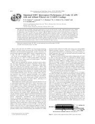
Fig. 1 shows fluorescence spectra <strong>of</strong> the three<br />
molecules in PVB at T ¼ 4 K, recorded in the<br />
region corresponding to the inhomogeneously<br />
broadened 0–0 transition upon excitation with<br />
near-IR pulses <strong>of</strong> wavelength twice the absorption<br />
maximum. One can see that if the sample is<br />
illuminated with pairs <strong>of</strong> phase-locked pulses, the<br />
part <strong>of</strong> the spectrum around the laser carrier<br />
frequencyshows distinct spectral fringes (solid
ARTICLE IN PRESS<br />
196<br />
M. Drobizhev et al. / <strong>Journal</strong> <strong>of</strong> <strong>Luminescence</strong> <strong>107</strong> (<strong>2004</strong>) 194–202<br />
Chl<br />
Pc 3 An SiNc<br />
Fluorescence intensity<br />
(a) 15600<br />
16000 (b) 12400 12800 (c) 12400 12800<br />
Frequency, cm -1<br />
Fig. 1. S 1 2S 0 fluorescence spectra <strong>of</strong> Chl- (a), anthracenophthalocyanine- (b) and Si-naphthalocyanine- (c) doped PVB films obtained<br />
at 4 K upon excitation with pairs <strong>of</strong> near-IR femtosecond pulses. Dashed line in each plot is the best Gaussian fit to the spectrum.<br />
curve). The period <strong>of</strong> fluorescence intensitymodulation<br />
is 40 cm 1 , which corresponds exactlyto<br />
the inverse delaybetween pulses (Dt ¼ 700 fs). A<br />
smooth dashed curve is a Gaussian fit to fluorescence<br />
spectrum, corresponding to the spectrum<br />
excited non-coherently. Fig. 2 shows a typical laser<br />
spectrum coming out <strong>of</strong> Michelson interferometer<br />
and used for TPA excitation. This spectrum is<br />
recorder with a Lambda 900 Perkin-Elmer spectrophotometer<br />
and can be described byFourier<br />
transform <strong>of</strong> two Gaussian pulses separated by Dt:<br />
Intensity L(ν '), arb.u.<br />
20<br />
15<br />
10<br />
5<br />
L(ν') / G( ν')<br />
2.0<br />
1.5<br />
1.0<br />
0.5<br />
0.0<br />
7700 7800 7900<br />
Frequency, cm -1<br />
Lðn 0 Þ<br />
¼ G expð ðn 0 n L Þ 2 =ðdn 0 Þ 2 Þð1 þ b cosð2pn 0 DtÞÞ;<br />
ð1Þ<br />
where G is a constant, n L is the carrier frequency,<br />
dn 0 is the spectral width <strong>of</strong> laser pulse, b is the<br />
modulation depth <strong>of</strong> excitation, which depends on<br />
the relative amplitude <strong>of</strong> the two pulses. If the<br />
amplitudes are the same, b ¼ 1; and bo1 otherwise.<br />
From the plot, presented in inset, we find the<br />
depth <strong>of</strong> excitation modulation b ¼ 0:75:<br />
Returning to Fig. 1, we can note a very<br />
interesting fact that larger one-photon Debye–<br />
Waller factor, a 1 ; does not result in higher contrast<br />
<strong>of</strong> fringes, all other condition being the same.<br />
0<br />
7600 7700 7800 7900 8000 8100<br />
Frequency, cm -1<br />
Fig. 2. Excitation laser spectrum, coming out Michelson<br />
interferometer and recorded with a slow detector <strong>of</strong> spectrophotometer.<br />
Dashed line is the Gaussian envelope <strong>of</strong> this<br />
spectrum, corresponding to the broad envelope in Eq. (1):<br />
Gðn 0 n L Þ¼G expð ðn 0 n L Þ 2 =ðdn 0 Þ 2 Þ: The ratio <strong>of</strong> modulated<br />
spectrum to this Gaussian envelope, 1 þ b cosð2pn 0 DtÞ; is shown<br />
in inset. From this plot we find the depth <strong>of</strong> excitation<br />
modulation b ¼ 0:75:<br />
Indeed, the largest modulation depth, M ¼ 0:32; is<br />
observed for Chl, where a 1 ¼ 0:55 [13]. On the<br />
other hand, SiNc, with much higher a 1 E1:0 [14],
ARTICLE IN PRESS<br />
M. Drobizhev et al. / <strong>Journal</strong> <strong>of</strong> <strong>Luminescence</strong> <strong>107</strong> (<strong>2004</strong>) 194–202 197<br />
shows even smaller modulation depth, M ¼ 0:24:<br />
The lowest M ¼ 0:15 is observed for Pc 3 An, where<br />
the Debye–Waller factor (a 1 ¼ 0:63 [15]) is larger<br />
than that <strong>of</strong> Chl. This first observation can suggest<br />
the important role <strong>of</strong> molecular symmetry in TPAinduced<br />
gratings. More specifically, it seems that<br />
molecules with broken inversion symmetry demonstrate<br />
deeper TPA fringes (sharper homogeneous<br />
TPA spectrum) than molecules possessing<br />
center <strong>of</strong> inversion, cf. Chl and SiNc. The case <strong>of</strong><br />
Pc 3 An can be considered as an intermediate one,<br />
since the main electronic conjugation path in<br />
tetrapyrrolic ring is not broken (cf. chlorin), but<br />
mono-anthraceno substitution can introduce certain<br />
asymmetry. By comparing the modulation<br />
contrast in Fig. 1, we can just qualitatively<br />
estimate at this moment that the sharpness <strong>of</strong><br />
TPA spectrum <strong>of</strong> Pc 3 An is closer to that <strong>of</strong><br />
SiNc.<br />
To get more insight into the role <strong>of</strong> molecular<br />
symmetry in homogeneous TPA spectrum, we<br />
measured the temperature dependence <strong>of</strong> TPA<br />
fringes in two extreme cases, i.e. for symmetrical<br />
SiNc and non-symmetrical Chl. The results are<br />
presented in Fig. 3. In both cases, an increase <strong>of</strong><br />
temperature results in diminishing contrast <strong>of</strong> the<br />
fringes. The striking difference between the two<br />
systems is that in Chl the fringes shift to the red<br />
with increasing temperature, whereas in SiNc this<br />
shift is much less pronounced. Indeed, for Chl the<br />
observed shift <strong>of</strong> the grating constitutes a half <strong>of</strong><br />
its period upon increasing temperature from 4 to<br />
60 K. We checked that this shift was completely<br />
reversible when temperature was lowered down to<br />
4 K again, thus ensuring that it is inherent<br />
property<strong>of</strong> the sample.<br />
In order to quantitativelysimulate the temperature<br />
dependence <strong>of</strong> TPA-induced gratings, presented<br />
in Fig. 3, we have elaborated a simple<br />
model described in the next section.<br />
4. The model<br />
The fluorescence spectrum <strong>of</strong> an inhomogeneouslybroadened<br />
ensemble <strong>of</strong> molecules, excited<br />
bylight with two-photon ‘‘action’’ spectrum Iðn 00 Þ<br />
(proportional to the square <strong>of</strong> the Fourier transform<br />
<strong>of</strong> the electric field squared [2,3]) is<br />
Fðn; TÞ ¼<br />
Z N<br />
N<br />
Nðn 0 Þg 1 ðn n 0 ; TÞ dn 0<br />
Z N<br />
g 2 ðn 00 n 0 ; TÞIðn 00 Þ dn 00 ; ð2Þ<br />
N<br />
Chl<br />
100K<br />
SiNc<br />
120K<br />
Fluorescence intensity<br />
60K<br />
50K<br />
30K<br />
11K<br />
50K<br />
30K<br />
11K<br />
(a)<br />
4K<br />
15500 15750 16000 16250<br />
4.5K<br />
12250 12500 12750 13000<br />
Frequency, cm -1 (b) Frequency, cm -1<br />
Fig. 3. S 1 2S 0 fluorescence spectra <strong>of</strong> Chl- (a) and Si-naphthalocyanine- (b) doped PVB film obtained at different temperatures upon<br />
excitation with pairs <strong>of</strong> femtosecond pulses. Vertical line in each plot correspond to one selected peak in the grating at lowest<br />
temperature. For Chl, a large red shift <strong>of</strong> the grating is observed at increasing temperature, whereas for SiNc this shift is much smaller.
ARTICLE IN PRESS<br />
198<br />
M. Drobizhev et al. / <strong>Journal</strong> <strong>of</strong> <strong>Luminescence</strong> <strong>107</strong> (<strong>2004</strong>) 194–202<br />
where g 2 ðn n 0 ; TÞ is the homogeneous TPA<br />
spectrum, g 1 ðn n 0 ; TÞ is the homogeneous onephoton<br />
fluorescence spectrum with ZPL frequency<br />
n 0 ; and Nðn 0 Þ is the inhomogeneous distribution<br />
function. A slowlyvarying spectral function,<br />
including the excitation spectrum envelope and<br />
the inhomogeneous distribution function, which<br />
are both much broader than the grating period,<br />
ZPL, and PW, can be considered as a constant<br />
and brought out <strong>of</strong> the integral in Eq. (2). In<br />
this approximation, we can write for excitation<br />
spectrum [2,3]<br />
Iðn 00 Þ¼1 þ b cosð2pn 00 DtÞ:<br />
ð3Þ<br />
As usual, we describe the homogeneous (twophoton)<br />
absorption and (one-photon) fluorescence<br />
spectra as a sum <strong>of</strong> ZPL and PW<br />
g 1; 2 ðn n 0 ; TÞ ¼a 1; 2 ðTÞdðn n 0 Þ<br />
þ½1 a 1; 2 ðTÞŠp 1; 2 ðn n 0 Þ; ð4Þ<br />
where a 1; 2 ðTÞ is the Debye–Waller factor, indexes<br />
1 and 2 designate fluorescence and absorption,<br />
respectively, ZPL is represented by delta function,<br />
and PW is described bythe following model<br />
function:<br />
p 1; 2 ðn n 0 Þ<br />
8<br />
><<br />
4<br />
¼ n 3 ðn 0 nÞ 2 exp½8 2<br />
n 1; 2<br />
ðn 0 nÞŠ; 7ðn 0 nÞ > 0;<br />
1; 2<br />
>:<br />
0; 7ðn 0 nÞp0;<br />
ð5Þ<br />
- ∆OD<br />
(b)<br />
- ∆OD<br />
(a)<br />
0.6<br />
0.4<br />
0.2<br />
0.0<br />
0.4<br />
0.3<br />
0.2<br />
0.1<br />
0.0<br />
-20 0 20 40 60 80 100<br />
ν ν<br />
0 - , cm-1<br />
where the upper sign is for fluorescence, the lower<br />
sign is for absorption, n 1; 2 is the maximum <strong>of</strong><br />
spectral distribution <strong>of</strong> PW. The validity<strong>of</strong><br />
approximation (5) is supported bya satisfactory<br />
fit <strong>of</strong> pseudophonon wing to Eq. (5) in standard<br />
hole-burning experiment with Chl and SiNc in<br />
PVB (see Fig. 4). Function (5) includes onlyone<br />
adjustable parameter, n 1; 2 ; which does not depend<br />
on temperature in our model. The best fit (at<br />
T ¼ 4 30 K) gives n 1 ¼ 18 cm 1 for Chl and<br />
n 1 ¼ 14 cm 1 for SiNc, which agrees well with<br />
previous hole-burning [15,16] and Raman scattering<br />
[17] results for PVB. We assume the effect <strong>of</strong><br />
temperature broadening <strong>of</strong> PWs is negligible,<br />
which is justified experimentally [18] in the same<br />
temperature range (4–100 K) as in our experiments.<br />
All the temperature dependence is then<br />
contained in Debye–Waller factor, which we treat<br />
in one-oscillator approximation as<br />
<br />
a 1; 2 ðTÞ ¼exp x 1; 2 cth hn 1;2<br />
2kT<br />
<br />
SiNc in PVB, 4K<br />
Chl in PVB, 12K<br />
-20 0 20 40 60 80 100 120 140<br />
( ν - ν ), cm-1<br />
0<br />
Fig. 4. Spectral hole pr<strong>of</strong>iles for Chl (a) and SiNc (b) in PVB.<br />
Holes are burnt with He–Ne and cw Ti:sapphire lasers,<br />
respectively. A ratio <strong>of</strong> integrated ZPL and PW does not reflect<br />
the Debye–Waller factor, since zero-phonon hole is already<br />
saturated. However, pseudophonon sideband (to the right <strong>of</strong><br />
ZPL) reasonablyreflects the PW pr<strong>of</strong>ile. A fit <strong>of</strong> its pr<strong>of</strong>ile to<br />
function (5) is shown bydashed line on each plot.<br />
<br />
; ð6Þ<br />
where x is the electron–phonon coupling strength.<br />
Substituting Eqs. (5) and (6) into Eq. (4) and<br />
then Eqs. (3) and (4) into Eq. (2) and performing<br />
integration, we get the following expression for
ARTICLE IN PRESS<br />
M. Drobizhev et al. / <strong>Journal</strong> <strong>of</strong> <strong>Luminescence</strong> <strong>107</strong> (<strong>2004</strong>) 194–202 199<br />
fluorescence spectrum:<br />
Fðn; TÞ<br />
¼ KðnÞ½1 þ MðTÞ cosð2pDtn þ DjðTÞÞŠ;<br />
ð7Þ<br />
where the amplitude MðTÞ and the phase shift<br />
DjðTÞ <strong>of</strong> the grating are given by<br />
MðTÞ ¼bf½A þ B cos d 2 þ C cos d 1<br />
þ D cosðd 1 þ d 2 ÞŠ 2<br />
þ½B sin d 2 þ C sin d 1<br />
þ D sinðd 1 þ d 2 ÞŠ 2 g 1=2 ;<br />
ð8Þ<br />
DjðTÞ<br />
B sin d 2 þ C sin d 1 þ D sinðd 1 þ d 2 Þ<br />
¼ arctg<br />
A þ B cos d 2 þ C cos d 1 þ D cosðd 1 þ d 2 Þ :<br />
ð9Þ<br />
Here functions AðTÞ; BðTÞ; CðTÞ; and DðTÞ are<br />
expressed through the model parameters as<br />
follows:<br />
AðTÞ ¼a 1 ðTÞa 2 ðTÞ;<br />
ð10Þ<br />
BðTÞ ¼½1 a 1 ðTÞŠa 2 ðTÞ<br />
f1 þðpDtn 2 Þ 2 g 3=2 ;<br />
CðTÞ ¼a 1 ðTÞ½1 a 2 ðTÞŠ<br />
f1 þðpDtn 1 Þ 2 g 3=2 ;<br />
DðTÞ ¼½1 a 1 ðTÞŠ½1 a 2 ðTÞŠ<br />
f½1 þðpDtn 2 Þ 2 Š½1 þðpDtn 1 Þ 2 Šg 3=2<br />
ð11Þ<br />
ð12Þ<br />
ð13Þ<br />
and<br />
d 1; 2 ¼ 3 arctgðpDtn 1; 2 Þ:<br />
ð14Þ<br />
We note here that in the limiting case a 1 ð0Þ ¼<br />
a 2 ð0Þ ¼1 Eq. (9) does give a quantitativelycorrect<br />
phase shift, Djð0Þ ¼0: However, for example, for<br />
a 1 ¼ a 2 ¼ 0; the correct behavior is reproduced<br />
onlyat small pDtn 1; 2 : Dj ¼ 3pDtðn 1 þ n 2 Þ: This<br />
last value corresponds to a frequencyshift (Stokes<br />
shift) <strong>of</strong> Dn ¼ 3 2 ðn 1 þ n 2 Þ; equal to a difference<br />
between centers <strong>of</strong> gravity<strong>of</strong> PWs (5) in absorption<br />
and fluorescence. In general case, the values <strong>of</strong><br />
Dj allowed byEq. (9) are restricted bydefinition<br />
<strong>of</strong> function arctgðxÞ; and, therefore, Eq. (9) is<br />
unable to describe a wide range <strong>of</strong> other real sets <strong>of</strong><br />
parameters. In our approach we use Eq. (8) for the<br />
simulation <strong>of</strong> the temperature dependence <strong>of</strong> the<br />
amplitude (contrast) <strong>of</strong> the fringes, but we resort<br />
to a simpler model for the simulation <strong>of</strong> the<br />
temperature dependence <strong>of</strong> the phase. We consider<br />
the Stokes shift as a difference between the first<br />
moments (centers <strong>of</strong> gravity) <strong>of</strong> absorption and<br />
fluorescence homogeneous spectra, namely,<br />
DnðTÞ ¼ð1 a 2 ðTÞÞn 2<br />
þð1 a 1 ðTÞÞn 1 ; ð15Þ<br />
and, therefore,<br />
DjðTÞ ¼2pDt½ð1 a 2 ðTÞÞn 2<br />
þð1 a 1 ðTÞÞn 1 Š: ð16Þ<br />
5. Results <strong>of</strong> simulations and discussion<br />
5.1. Chl in PVB<br />
First <strong>of</strong> all, we simulated the temperature<br />
dependence <strong>of</strong> the fringes in Chl:PVB system by<br />
using symmetrical model with x 1 ¼ x 2 ¼ x and<br />
Phase shift, ∆ϕ (rad) Modulation amplitude, M<br />
(a)<br />
(b)<br />
0.3<br />
0.2<br />
0.1<br />
3<br />
2<br />
1<br />
20 40 60 80 100 120<br />
T, K<br />
40 60 80 100<br />
Fig. 5. Temperature dependence <strong>of</strong> the amplitude M (a) and<br />
phase shift Dj (b) <strong>of</strong> spectral grating observed in Chl:PVB and<br />
corresponding fits <strong>of</strong> these data to Eqs. (8) and (16) in mirrorsymmetrical<br />
model.
ARTICLE IN PRESS<br />
200<br />
M. Drobizhev et al. / <strong>Journal</strong> <strong>of</strong> <strong>Luminescence</strong> <strong>107</strong> (<strong>2004</strong>) 194–202<br />
n 1 ¼ n 2 ¼ n m : Byvarying two adjustable parameters,<br />
x and n m ; we obtain the best fit, shown in<br />
Fig. 5a, atx ¼ 0:36; (i.e. a 1 ð0Þ ¼a 2 ð0Þ ¼0:70) and<br />
n m ¼ 25 cm 1 . Substitution <strong>of</strong> these parameters<br />
into Eq. (16) without anyfurther fitting gives a<br />
phase shift, shown in Fig. 5b bysolid line. One can<br />
conclude that the model fits well in this case and<br />
the obtained parameters are reasonable. The value<br />
<strong>of</strong> n m is slightlylarger than the maximum phonon<br />
frequency(18 cm 1 ), but it is veryclose to a center<br />
<strong>of</strong> gravity<strong>of</strong> PW distribution, which can be a real<br />
effective frequencyentering Eq. (6). The limiting<br />
Debye–Waller factor a 1 ð0Þ agrees well with the<br />
value measured at 7 K (a 1 ¼ 0:55) in Ref. [13].<br />
We then performed the fit procedure <strong>of</strong> the same<br />
data but using the asymmetrical model, where all<br />
four parameters x 1 ; x 2 ; n 1 ; and n 2 were varied<br />
independently. The best fit <strong>of</strong> MðTÞ (Fig. 6a), and<br />
DjðTÞ (Fig. 6b), do, in fact, describe the experiment<br />
quite well, but the limiting Debye–Waller<br />
factor a 1 ð0Þ ¼0:86 comes out much larger than the<br />
literature data, cited before. The main conclusion,<br />
which can be drawn from the simulation obtained<br />
for Chl:PVB is that the symmetrical model works<br />
quite well for this system.<br />
5.2. SiNc in PVB<br />
The simulation <strong>of</strong> experimental data for<br />
SiNc:PVB with mirror-symmetrical model is<br />
shown in Figs. 7a and b. Again, the fit procedure<br />
was first accomplished for the amplitude and then<br />
the best parameters were introduced in Eq. (16) to<br />
describe the phase shift. The main inconsistency<br />
between the simulation and experiment is observed<br />
in the phase behavior, because the model tends to<br />
overestimate the rate <strong>of</strong> the shift with temperature.<br />
The obtained best-fit parameters also differ considerablyfrom<br />
literature data: the limiting value <strong>of</strong><br />
Debye–Waller factor ða 1 ð0Þ ¼0:56Þ is waytoo<br />
small as compared to that reported previously<br />
ða 1 ¼ 1:0 at1:5KÞ [14] and the characteristic<br />
Modulation amplitude, M<br />
0.3<br />
0.2<br />
0.1<br />
Modulation amplitude, M<br />
0.2<br />
0.1<br />
(a)<br />
20 40 60 80 100 120<br />
(a)<br />
20 40 60 80 100 120<br />
T, K<br />
∆ϕ (rad)<br />
Phase shift,<br />
(b)<br />
3<br />
2<br />
1<br />
20 40 60 80 100 120<br />
T, K<br />
Fig. 6. Temperature dependence <strong>of</strong> the amplitude M (a) and<br />
phase shift Dj (b) <strong>of</strong> spectral grating observed in Chl:PVB and<br />
corresponding fits <strong>of</strong> these data to Eqs. (8) and (16) in nonmirror-symmetrical<br />
model.<br />
Phase shift, ∆ϕ (rad)<br />
(b)<br />
5<br />
4<br />
3<br />
2<br />
1<br />
20 40 60 80 100 120<br />
Fig. 7. Temperature dependence <strong>of</strong> the amplitude M (a) and<br />
phase shift Dj (b) <strong>of</strong> spectral grating observed in SiNc:PVB and<br />
corresponding fits <strong>of</strong> these data to Eq. (8) and (16) in mirrorsymmetrical<br />
model.
ARTICLE IN PRESS<br />
M. Drobizhev et al. / <strong>Journal</strong> <strong>of</strong> <strong>Luminescence</strong> <strong>107</strong> (<strong>2004</strong>) 194–202 201<br />
Phase shift, ∆ϕ (rad) Modulation amplitude, M<br />
0.2<br />
0.1<br />
(a)<br />
4<br />
3<br />
2<br />
1<br />
20 40 60 80 100 120<br />
Modulation amplitude, M<br />
(a)<br />
Phase shift, ∆ϕ (rad)<br />
0.2<br />
0.1<br />
2<br />
1.5<br />
1<br />
0.5<br />
20 40 60 80 100 120<br />
(b)<br />
20 40 60 80 100 120<br />
T, K<br />
Fig. 8. Temperature dependence <strong>of</strong> the amplitude M (a) and<br />
phase shift Dj (b) <strong>of</strong> spectral grating observed in SiNc:PVB and<br />
corresponding fits <strong>of</strong> these data to Eq. (8) and (16) in nonmirror-symmetrical<br />
model.<br />
phonon frequency n m ¼ 39 cm 1 seems to be<br />
overestimated.<br />
Our next attempt was to simulate the data with<br />
asymmetrical model with four independent parameters<br />
(Figs. 8a and b). One-photon parameters<br />
were fixed to the values, known from hole-burning<br />
spectroscopy: a 1 ð0Þ ¼0:9andn 1 ¼ 17 cm 1 , and x 2<br />
and n 2 were varied. The best fitting values obtained<br />
for these parameters are x 2 ¼ 1:0(a 2 ð0Þ ¼0:37) and<br />
n 2 ¼ 56 cm 1 . It is obvious that this fit fails again<br />
in describing the phase shift <strong>of</strong> Fig. 8b.<br />
We could obtain a reasonablygood fit (Figs. 9a<br />
and b) onlyif we artificiallyset a 2 ðTÞ ¼0ðx 2 ¼<br />
10 000Þ and let other three parameters vary<br />
independently. This implies no ZPL in TPA<br />
spectrum even at the lowest temperature. For the<br />
emission spectrum we obtain reasonable x 1 ¼ 0:46<br />
and n 1 ¼ 27 cm 1 , while the Debye–Waller factor<br />
again occurs smaller than measured in Ref. [14]<br />
(0.63 vs 1.0). Note that the homogeneous TPA<br />
spectrum turns out to be stronglyasymmetric with<br />
(b)<br />
20 40 60 80 100 120<br />
T, K<br />
Fig. 9. Temperature dependence <strong>of</strong> the amplitude M (a) and<br />
phase shift Dj (b) <strong>of</strong> spectral grating observed in SiNc:PVB and<br />
corresponding fits <strong>of</strong> these data to Eq. (8) and (16) in nonmirror-symmetrical<br />
model, where the Debye–Waller factor for<br />
TPA (a 2 ) was set to zero.<br />
respect to homogeneous one-photon emission<br />
spectrum. Not onlydoes this absorption contain<br />
no ZPL (a 2 ð0Þ ¼0), but its PW is shifted byonly<br />
n 2 ¼ 10 cm 1 from the zero-phonon origin.<br />
Several important conclusions can be drawn<br />
from these observations. First <strong>of</strong> all, mirrorsymmetric<br />
spectral model works well for Chl. This<br />
is not verysurprising result because this molecule<br />
does not possesses the center <strong>of</strong> inversion, and,<br />
therefore, selection rules for one- and two-photon<br />
transitions should be the same. On the other hand,<br />
TPA gratings in SiNc:PVB system are best<br />
described bystronglyasymmetrical model in<br />
which ZPL <strong>of</strong> TPA is completelymissing. This<br />
fact can be explained if we consider selection rules<br />
for pure electronic transition in this centrosymmetrical<br />
molecule. Since we are dealing with<br />
gerade2ungerade; S 0 2S 1 one-photon transition,<br />
it must be stronglyforbidden for pure electronic
ARTICLE IN PRESS<br />
202<br />
M. Drobizhev et al. / <strong>Journal</strong> <strong>of</strong> <strong>Luminescence</strong> <strong>107</strong> (<strong>2004</strong>) 194–202<br />
TPA [4–10]. However, TPA with creation <strong>of</strong><br />
phonons (electron–phonon transition, building<br />
up PW in homogeneous TPA spectrum) can be<br />
allowed through Herzberg–Teller mechanism. In<br />
this case, TPA grating can be created on pure<br />
phonon coherences. It is, <strong>of</strong> course, evident that<br />
for the observation <strong>of</strong> the fringes, the phonon<br />
coherence time, T 2 ; must be comparable or longer<br />
than the time delaybetween pulses, DtU It is<br />
known from femtosecond photon echo experiments<br />
[17,18] that the phonon coherence time in<br />
polymer matrices at low temperatures ranges from<br />
several hundred <strong>of</strong> fs to 1 ps. In our experiments<br />
we use Dt ¼ 700 fs, which is a sufficientlysmall<br />
value to produce spectral interference on phonon<br />
coherences, at least in SiNc:PVB system. From the<br />
other side, one cannot reduce Dt to an arbitrarily<br />
small value, since it is limited from below by<br />
inverse inhomogeneous width, which is <strong>of</strong> the<br />
order <strong>of</strong> 100 fs. Therefore, there exists a limited<br />
range <strong>of</strong> delays (DtB10021000 fs), where TPA<br />
gratings can be observed in the case <strong>of</strong> absence <strong>of</strong><br />
ZPLs in a system.<br />
6. Conclusion<br />
Temperature dependence <strong>of</strong> spectral interference<br />
fringes, obtained bytwo-photon excitation <strong>of</strong><br />
the lowest electronic molecular transition with<br />
phase-locked pairs <strong>of</strong> femtosecond pulses is<br />
studied. Mathematical simulation <strong>of</strong> this dependence<br />
demonstrates that Franck–Condon model<br />
with mirror symmetrical TPA and one-photon<br />
fluorescence spectra works well for non-centrosymmetrical<br />
chlorin, but fails for centrosymmetrical<br />
naphthalocyanine. In the later case, the<br />
dependence is best described bya model where<br />
homogeneous TPA spectrum does not contain<br />
ZPL. These results can be explained bysimilar<br />
selection rules for one- and two-photon transitions<br />
in non-centrosymmetrical molecules, but alternative<br />
ones for centrosymmetrical molecules.<br />
Therefore, in the later case onlyelectron–phonon<br />
transitions are allowed and the TPA gratings<br />
can be observed onlyif the time delaybetween<br />
the pulses is still shorter than phonon dephasing<br />
time.<br />
Acknowledgements<br />
This work was supported byAFOSR Grant<br />
F49620-01-0406.<br />
References<br />
[1] M. Drobizhev, A. Karotki, A. Rebane, Chem. Phys. Lett.<br />
334 (2001) 76.<br />
[2] A. Karotki, M. Kruk, M. Drobizhev, A. Rebane, J. Mod.<br />
Opt. 49 (2002) 379.<br />
[3] A. Rebane, M. Drobizhev, A. Karotki, J. Lumin. 98 (2002)<br />
341.<br />
[4] R.M. Hochstrasser, H.N. Sung, J. Chem. Phys. 66 (1977)<br />
3265.<br />
[5] R.M. Hochstrasser, H.N. Sung, J. Chem. Phys. 66 (1977)<br />
3276.<br />
[6] N. Mikami, M. Ito, Chem. Phys. 23 (1977) 141.<br />
[7] M.F. Granville, G.R. Holtom, B.E. Kohler, R.L. Christensen,<br />
K.L. D’Amico, J. Chem. Phys. 70 (1979) 593.<br />
[8] M. Gutmann, P.-F. Schonzart, G. Hohlneicher, Chem.<br />
Phys. Lett. 140 (1990) <strong>107</strong>.<br />
[9] T. Plakhotnik, D. Walser, A. Renn, U.P. Wild, Chem.<br />
Phys. Lett. 262 (1996) 379.<br />
[10] D. Walser, G. Zum<strong>of</strong>en, T. Plakhotnik, J. Chem. Phys. 113<br />
(2000) 8047.<br />
[11] M.C. Edelson, J.M. Hayes, G.J. Small, Chem. Phys. Lett.<br />
60 (1979) 307.<br />
[12] M. Takeda, K. Matsuda, C. Suzuki, S. Saikan, J. Lumin.<br />
86 (2000) 285.<br />
[13] W.-I. Huang, A. Rebane, U.P. Wild, L.W. Johnson,<br />
J. Lumin. 71 (1997) 237.<br />
[14] A.V. Turukhin, A.A. Gorokhovsky, C. Moser, I.V.<br />
Solomatin, D. Psaltis, J. Lumin. 86 (2000) 399.<br />
[15] I. Renge, H. Wolleb, H. Spahni, U.P. Wild, J. Phys. Chem.<br />
A 101 (1997) 6202.<br />
[16] I. Renge, J. Chem. Phys. 106 (1997) 5836.<br />
[17] S. Saikan, J. Lumin. 53 (1992) 147.<br />
[18] A. Rebane, J. Gallus, O. Ollikainen, Laser Phys. 12 (2002)<br />
1126.