Vitamins - sepeap
Vitamins - sepeap
Vitamins - sepeap

Create successful ePaper yourself
Turn your PDF publications into a flip-book with our unique Google optimized e-Paper software.
Article nutrition<br />
<strong>Vitamins</strong><br />
Usha Sethuraman, MD*<br />
Author Disclosure<br />
Dr Sethuraman did<br />
not disclose any<br />
financial relationships<br />
relevant to this<br />
article.<br />
Objectives After completing this article, readers should be able to:<br />
1. Identify the symptoms and signs of deficiency of various vitamins.<br />
2. Characterize the patients at risk for deficiencies and plan their treatment.<br />
3. Discuss the treatment of patients at risk for vitamin deficiencies.<br />
Introduction<br />
<strong>Vitamins</strong> are organic compounds required in small amounts in various cellular metabolisms<br />
that are important for overall health maintenance and normal growth of the organism. First<br />
discovered by Hopkins in 1907, they were named by Funk in 1911. McCollum and Davis<br />
later showed that some vitamins, such as A, D, E, and K, are fat-soluble, and some, such as<br />
B and C, are water-soluble (Table 1).<br />
Vitamin A (Retinol)<br />
Case 1<br />
A 5-year-old boy who recently was adopted from India is brought by his parents for his first<br />
physical examination. They report that he has been doing well except that he seems to bump into<br />
objects frequently, particularly in the evenings. On examination, his height and weight are<br />
below the 5th percentile. He has some silver-colored patches in his conjunctiva, but the rest of the<br />
examination findings are normal. A diagnosis is made clinically and treatment initiated.<br />
Case 2<br />
A 3-year-old girl is brought to the emergency department for irritability. Her mother denies<br />
fever, upper respiratory tract infection symptoms, or trauma, but states that the girl has become<br />
progressively irritable over the past few days. She had been complaining of headache and<br />
nausea but had no emesis. On examination, the child is afebrile and appears irritable. Despite<br />
adequate doses of acetaminophen, she continues to complain of a headache, prompting the<br />
decision to perform a lumbar puncture. Except for an elevated opening pressure, her spinal<br />
fluid appears normal, having no white or red blood cells. Further questioning reveals that the<br />
girl has been taking her brother’s pills three to four times a day to “get healthier”; this leads to<br />
her diagnosis.<br />
Vitamin Function<br />
Vitamin A, derived from pigments in nature called carotenoids (provitamin A), is waterinsoluble<br />
and is destroyed by heat, light, oxidation, and dehydration. Carotenoids and<br />
retinal in the diet are absorbed and subsequently esterified in the intestinal cells to retinyl<br />
esters. The esters are stored in the liver and, when needed, hydrolyzed to retinol,<br />
complexed with retinal-binding protein, and transported to tissues and organs in the body.<br />
Retinol is essential for growth and functional integrity of epithelial cells; retinal, a<br />
component of retinal pigments, is important for normal vision. Retinoic acid is essential for<br />
the synthesis of glycoproteins.<br />
Vitamin A deficiency is the leading cause of preventable blindness in children; it also<br />
increases the risk for severe infections. Malnourished young children and infants in<br />
developing countries are particularly prone to deficiency due to recurrent infections such as<br />
diarrhea and measles. Vitamin A deficiency also can occur due to inadequate absorption, as<br />
seen in celiac disease, hepatic disease, and low-fat and -protein diets.<br />
*Division of Emergency Medicine, Carman and Ann Adams Department of Pediatrics, Children’s Hospital of Michigan, Wayne<br />
State University, Detroit, Mich.<br />
44 Pediatrics in Review Vol.27 No.2 February 2006
Pediatrics in Review Vol.27 No.2 February 2006 45<br />
Table 1. <strong>Vitamins</strong>: Sources, Requirements, Action, Results, Diagnosis, and Treatment<br />
Vitamin<br />
A<br />
B 1 (Thiamine)<br />
Riboflavin (B 2 )<br />
Niacin<br />
(Nicotinic<br />
acid)<br />
Sources<br />
Green leafy<br />
vegetables,<br />
carrots, sweet<br />
potatoes, liver<br />
Liver, pork, milk,<br />
grains<br />
Milk, cheese,<br />
liver, green<br />
leafy<br />
vegetables<br />
Liver, fish,<br />
whole grains,<br />
eggs, milk,<br />
poultry<br />
B 12(Cobalamine) Fish, eggs,<br />
cheese<br />
Daily<br />
Requirement Action Deficiency Excess Diagnosis Treatment<br />
14 yr: 1.3 mg<br />
13 y: 16 mg<br />
0 to 6 mo: 0.4 mcg<br />
6 to 12 mo: 0.5 mcg<br />
1to3y:0.9mcg<br />
4to8y:1.2mcg<br />
9to13y:1.8mcg<br />
>13 y: 2.4 mcg<br />
Vision in dim light,<br />
bone and tooth<br />
growth,<br />
epithelium<br />
maturation<br />
Part of thiamine<br />
pyrophosphate,<br />
which is needed<br />
for oxidative<br />
decarboxylation<br />
Part of<br />
flavoproteins<br />
important for<br />
hydrogen<br />
transfer<br />
Forms NAD and<br />
NADP cofactors<br />
Maturation of red<br />
blood cells<br />
Nyctalopia,<br />
photophobia,<br />
keratomalacia,<br />
blindness,<br />
impaired<br />
growth,<br />
follicular<br />
hyperkeratosis<br />
Beriberi, fatigue,<br />
cardiac failure,<br />
polyneuritis<br />
Blurring of vision,<br />
cheilosis<br />
Pellagra (rash,<br />
diarrhea,<br />
stomatitis,<br />
glossitis,<br />
mental status<br />
changes)<br />
Juvenile<br />
pernicious<br />
anemia<br />
Anorexia, dry skin,<br />
painful joints,<br />
increased<br />
intracranial<br />
pressure, headache,<br />
vomiting<br />
None<br />
None<br />
None<br />
None<br />
Clinical or low<br />
plasma retinol<br />
levels<br />
Response to<br />
thiamine<br />
Urinary riboflavin<br />
of
46 Pediatrics in Review Vol.27 No.2 February 2006<br />
Table 1. <strong>Vitamins</strong>: Sources, Requirements, Action, Results, Diagnosis, and Treatment (continued)<br />
Vitamin Sources<br />
B 6 (Pyridoxine) Meat, liver,<br />
kidneys<br />
Folate<br />
Vitamin D<br />
Vitamin E<br />
(Tocopherol)<br />
Cauliflower,<br />
green leafy<br />
vegetables,<br />
yeast, liver,<br />
kidney<br />
Fortified milk,<br />
liver oils,<br />
sunlight, egg<br />
yolks<br />
Germ oils, green<br />
leafy<br />
vegetables<br />
Daily<br />
Requirement Action Deficiency Excess Diagnosis Treatment<br />
13 y: 1.3 mg<br />
13 y: 100 mcg<br />
Pediatrics in Review Vol.27 No.2 February 2006 47<br />
Table 1. <strong>Vitamins</strong>: Sources, Requirements, Action, Results, Diagnosis, and Treatment (continued)<br />
Vitamin<br />
Vitamin K<br />
Sources<br />
Green leafy<br />
vegetables,<br />
liver<br />
Daily<br />
Requirement Action Deficiency Excess Diagnosis Treatment<br />
nutrition<br />
vitamins<br />
blindness), indicative of vitamin A deficiency. The presence<br />
of Bitot spots in the eyes confirms the diagnosis.<br />
The boy was started on vitamin A, and his symptoms<br />
improved. Delay in diagnosis and treatment would have<br />
led to blindness.<br />
Treatment and Prevention<br />
Breastfeeding is protective against vitamin A deficiency.<br />
Encouraging vitamin A-rich diets, vitamin A supplementation,<br />
and food fortification can prevent and treat this<br />
deficiency.<br />
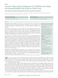
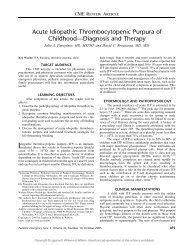
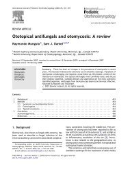
Figure 1. Bitot spots. Reproduced with permission from Indiana<br />
University School of Optometry; pictures by Dr H.D. Riley<br />
Clinical Manifestations of Deficiency<br />
The skin of the extensor surfaces becomes dry and scaly<br />
(follicular hyperkeratosis). Keratinization of the conjunctiva<br />
and lacrimal glands leads to dryness of the eyes and<br />
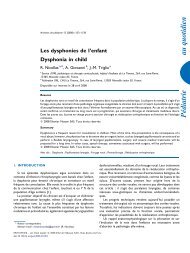
follicular conjunctivitis (xerophthalmia). Bitot spots are<br />
small triangular, silvery, foamlike patches that appear on<br />
the conjunctiva due to keratinization (Figs. 1 and 2).<br />
Night blindness or nyctalopia occurs due to a delay in<br />
resynthesis of rhodopsin. Severe inflammation results in<br />
keratomalacia and blindness. Diagnosis is primarily clinical.<br />
The child in Case 1 was adopted from a developing<br />
country, where malnutrition and vitamin deficiencies are<br />
more common. His height and weight are less than the<br />
5th percentile. The history of bumping into objects in<br />
the evenings is highly suggestive of “nyctalopia” (night<br />
Figure 2. Bitot spots. Reproduced with permission from Indiana<br />
University School of Optometry; pictures by Dr H.D. Riley,<br />
Effects of Excess<br />
The child in Case 2 had been taking her brother’s vitamin<br />
A capsules because her mother thought they would make<br />
the girl stronger. Drowsiness, painful joints, loss of hair,<br />
increased intracranial pressure, and carotenemia are presenting<br />
symptoms of excessive ingestion of vitamin A.<br />
Vitamin D<br />
Case 3<br />
A 2-year-old African-American boy is brought to the pediatrician<br />
for a routine visit. He was born at 30 weeks’<br />
gestation, was exclusively breastfed until 1 year of age, and<br />
has been a poor feeder since then. His height and weight are<br />
less than the 5th percentile. His legs are slightly bowed, and<br />
he has frontal bossing. He has no teeth and is not yet<br />
walking. Laboratory tests reveal decreased phosphorus, decreased<br />
calcium, and elevated alkaline phosphatase concentrations.<br />
Radiography of his wrists documents osteopenia,<br />
with cupping and fraying of the metaphysis. He is diagnosed<br />
as having nutritional rickets and is started on calcium<br />
and vitamin D. His mother is upset and wants to<br />
know the cause.<br />
Case 4<br />
An 8-month-old Caucasian boy is brought to the emergency<br />
department because of complaints of cough, runny nose,<br />
and wheezing for the past 3 days. His mother states that he<br />
has been fussy and has had some redness of his eyes without<br />
discharge. He is breastfed exclusively. His birthweight was<br />
3.2 kg, he was delivered vaginally without complications,<br />
and he has had regular visits with the pediatrician. The<br />
mother reports that the boy has had multiple episodes of<br />
bronchiolitis in the past. On physical examination, the baby<br />
is alert but fussy and has poor muscle tone. He weighs 4.1 kg,<br />
his heart rate is 160 beats/min, his respiratory rate is<br />
approximately 80 breaths/min, and his blood pressure is<br />
normal. His deep tendon reflexes seem to be slightly depressed.<br />
Bronchiolitis with conjunctivitis and failure to<br />
thrive is diagnosed, and the child is admitted to the hospital<br />
48 Pediatrics in Review Vol.27 No.2 February 2006
nutrition<br />
vitamins<br />
on bronchodilators. A chest radiograph reveals a heart of<br />
normal size and multiple areas of atelectasis. Because of a<br />
strong family history of asthma, steroids are added to the<br />
treatment regimen. The laboratory evaluation for the cause<br />
of the failure to thrive reveals anemia, mild indirect hyperbilirubinemia,<br />
a lowered calcium level, and an elevated<br />
alkaline phosphatase level. The radiologist notifies the resident<br />
of what appears to be cupping and fraying of the<br />
humerus ends. Additional tests are performed, the diagnosis<br />
made, and appropriate therapy started.<br />
Vitamin Function<br />
Vitamin D exists as ergosterol in yeast and<br />
7-dehydrocholesterol in human skin. When irradiated<br />
with ultraviolet light, these are converted into ergocalciferol<br />
(vitamin D 2 ) and cholecalciferol (vitamin D 3 ), respectively.<br />
D 2 and D 3 are absorbed readily from the small<br />
intestines. In the liver, D 3 is hydroxylated by 25 hydroxylase<br />
to 25 hydroxyl-D 3 (calciferol). Further hydroxylation<br />
of calciferol by 1-hydroxylase in the kidneys results<br />
in the formation of 1,25 dihydroxy-D 3 , which is the<br />
physiologically active form that regulates calcium and<br />
phosphate metabolisms by releasing calcium from the<br />
bones into blood and increasing calcium reabsorption in<br />
the kidney.<br />
Hypocalcemia causes parathormone to stimulate the<br />
formation of 1,25 dihydroxy-D 3 , while hypophosphatemia<br />
stimulates its formation directly; 1,25<br />
dihydroxy-D 3 , in turn, increases absorption of phosphate<br />
from the intestines. Vitamin D also increases absorption<br />
of calcium and phosphorus from the distal ileum and<br />
promotes endochondral growth of long bones and mineralization<br />
of the zone of provisional calcification (antirachitic<br />
action of vitamin D). Hence, deficiency is characterized<br />
by a defect in mineralization in these areas but<br />
with continued cartilage growth. Exposure to sunlight<br />
produces adequate amounts of vitamin D in the skin.<br />
Rickets can occur in breastfed infants who are not supplemented<br />
with vitamin D and have inadequate exposure<br />
to sunlight; in dark-skinned children due to inadequate<br />
penetration of sunlight; and in children who grow rapidly,<br />
such as very low-birthweight infants and adolescents.<br />
Congenital rickets can occur when the maternal<br />
intake of vitamin D is low. Celiac disease, steatorrhea,<br />
pancreatitis, cystic fibrosis, and medications such as steroids<br />
and anticonvulsants can predispose to vitamin D<br />
deficiency due to malabsorption from the gut.<br />
Clinical Manifestations of Deficiency<br />
Infants present with seizures and tetany due to the hypocalcemia,<br />
hypotonia, failure to thrive, widened cranial<br />
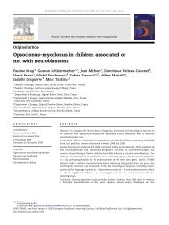
Figure 3. Widened wrists associated with rickets. Reproduced<br />
with permission from Dr Tom Thacher, Jos University Teaching<br />
Hospital, Nigeria.<br />
sutures, frontal bossing, and craniotabes. Older children<br />
may show delayed milestones, potbelly, bowlegs, kyphosis,<br />
pelvic deformities, delayed dentition, and widened<br />
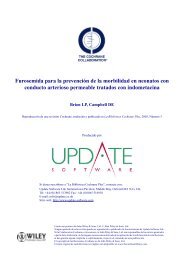
wrists (Fig. 3). Rachitic rosary due to enlarged costochondral<br />
junctions may be present (Fig. 4). Harrison<br />
groove is a horizontal depression along the lower border<br />
of the chest, corresponding to the costal insertion of the<br />
diaphragm. This results from ongoing cartilage growth<br />
and defective bone growth. Deficiency in adults results in<br />
osteomalacia.<br />
Diagnosis<br />
Besides the findings listed in Table 2, a generalized<br />
aminoaciduria can occur. Radiographs of the knees,<br />
Figure 4. Rachitic rosary. Courtesy of Dr Tom Thacher, Jos<br />
University Teaching Hospital, Nigeria.<br />
Pediatrics in Review Vol.27 No.2 February 2006 49
nutrition<br />
vitamins<br />
Table 2. Types of Rickets and Biochemical Findings<br />
Type<br />
Vitamin D-deficient<br />
Vitamin D-dependent<br />
(type 1)<br />
Vitamin D-dependent<br />
(type 2)<br />
Vitamin D-resistant<br />
(X-linked<br />
hypophosphatemia)<br />
Renal disease<br />
Cause<br />
Serum<br />
Calcium<br />
Serum<br />
Phosphate<br />
Alkaline<br />
Phosphate<br />
Parathyroid<br />
Hormone<br />
1,25 dihydroxy-<br />
D 3<br />
Nutrition/poor<br />
ultraviolet<br />
exposure<br />
Decreased Decreased Increased Increased Normal/decreased<br />
Decreased 1-alpha Decreased Decreased Increased Increased Low<br />
hydroxylase<br />
End-organ resistance Decreased Decreased Increased Increased High<br />
dihydroxy-D 3<br />
to 1,25<br />
Defect in tubular Normal Low Increased Normal Normal/decreased<br />
reabsorption of<br />
phosphate<br />
Defect in phosphate<br />
excretion<br />
Mildly<br />
decreased<br />
High Increased Increased Decreased<br />
wrists, and shoulders show widened distal ends with<br />
cupping and fraying, uncalcified larger metaphyses, and<br />
osteopenia (Figs. 5 through 7). A line of preparatory<br />
calcification that is separated from the distal end of the<br />
shaft by a zone of decreased calcification suggests initial<br />
healing.<br />
Nutritional rickets should be distinguished from vitamin<br />
D-resistant rickets (familial hypophosphatemia),<br />
which is due to a defect in the proximal tubular reabsorption<br />
of phosphates and in the conversion of 25<br />
dihydroxy-D 3 to 1,25 dihydroxy-D 3 . It has an X-linked<br />
dominant inheritance. Clinically, legs are bowed, but the<br />
other features of nutritional rickets generally are absent.<br />
Laboratory findings reveal near-normal calcium levels,<br />
lower phosphate concentrations, elevated alkaline phosphatase<br />
values, large urinary phosphate losses, and little<br />
or no evidence of secondary hyperparathyroidism. Vitamin<br />
D-dependent rickets is due to reduced activity of<br />
1-alpha hydroxylase. Levels of calcium and phosphate are<br />
decreased, alkaline phosphatase concentrations are elevated,<br />
and the levels of 1,25 dihydroxy-D 3 are low. Renal<br />
rickets due to the phosphaturia of uremia and secondary<br />
hyperparathyroidism result from renal disease.<br />
Figure 5. Radiographic appearance of rickets in the wrist.<br />
Courtesy of Dr Earl Hartwig, Emergency Medicine, Children’s<br />
Hospital of Michigan.<br />
Figure 6. Radiographic appearance of rickets in the knees.<br />
Courtesy of Dr Earl Hartwig, Emergency Medicine, Children’s<br />
Hospital of Michigan.<br />
50 Pediatrics in Review Vol.27 No.2 February 2006
nutrition<br />
vitamins<br />
Figure 7. Radiographic appearance of rickets in the shoulders.<br />
Courtesy of Dr Earl Hartwig, Emergency Medicine, Children’s<br />
Hospital of Michigan.<br />
Treatment and Prevention<br />
All infants, including those who are exclusively breastfed,<br />
should receive a minimum daily intake of 200 IU of<br />
vitamin D beginning during the first 2 postnatal months.<br />
Formula-fed infants who ingest less than 500 mL/d of<br />
formula or milk also should be supplemented. Studies<br />
have shown a higher bone density in adult women who<br />
were supplemented with vitamin D in infancy. Once<br />
deficiency is diagnosed, treatment must be initiated immediately.<br />
Calcium and phosphorus levels must be corrected.<br />
Daily oral vitamin D 3 , as listed in Table 1, should<br />
be initiated. An alternate therapy to ensure compliance is<br />
15,000 mcg of vitamin D in a single day, which avoids<br />
the preparation that has ethylene glycol as the vehicle,<br />
which could be toxic. Nutritional rickets responds with<br />
an increase in phosphate in 4 days and radiologic evidence<br />
of healing in 1 to 2 weeks. After healing is complete,<br />
the dose should be lowered to 10 mcg/d. Vitamin<br />
D-resistant rickets and -dependent types are treated with<br />
oral phosphates and high doses of 1,25 dihydroxy-D 3 .<br />
The prognosis generally is good for rickets. Early<br />
diagnosis prevents developmental delay and other sequelae.<br />
Orthopedic intervention may be required for<br />
severe deformities.<br />
The child in Case 3 had several factors putting him at<br />
risk for developing rickets, including his race, preterm<br />
birth, and exclusive breastfeeding with no supplementation<br />
until 1 year of age. Recognition of these risk factors<br />
and early supplementation with vitamin D-containing<br />
vitamins could have prevented the onset of rickets. Treatment<br />
now should include correction of calcium and<br />
phosphorus levels and administration of 50 to 150 mcg<br />
of vitamin D 3 daily for 2 to 3 months followed by a<br />
maintenance dose of 400 IU/d. Calcium and phosphorus<br />
levels and radiographs should be monitored for evidence<br />
of healing.<br />
The child in Case 4 had recurrent episodes of wheezing<br />
with failure to thrive and features of rickets. A sweat<br />
test performed as part of the evaluation for the failure to<br />
thrive led to the diagnosis of cystic fibrosis. The resulting<br />
malabsorption, coupled with the exclusive breastfeeding<br />
with no supplementation, caused his deficiency in vitamin<br />
D, leading to his rickets. Due to his reduced deep<br />
tendon reflexes, additional laboratory tests were undertaken,<br />
which revealed low levels of alpha-tocopherol and<br />
retinol. Cystic fibrosis with malabsorption and fatsoluble<br />
vitamin deficiencies was diagnosed and appropriate<br />
treatment initiated. The redness of his eyes was due to<br />
a follicular conjunctivitis caused by vitamin A deficiency<br />
and resolved when therapy was begun. Repeated episodes<br />
of “bronchiolitis” and failure to thrive should<br />
arouse the suspicion for cystic fibrosis, and a high degree<br />
of suspicion for vitamin deficiencies should be maintained.<br />
Effects of Excess<br />
Hypotonia, anorexia, polydipsia, polyuria, dehydration,<br />
hypertension, and corneal clouding may result from excess<br />
vitamin D intake. Radiographs may show internal<br />
calcifications, such as kidney stones and osteoporosis.<br />
Vitamin E<br />
Vitamin Function<br />
Vitamin E includes a group of compounds called tocopherols<br />
that have antioxidation properties (particularly of<br />
fats), which are facilitated by the presence of ascorbic<br />
acid. This vitamin occurs abundantly in green plants such<br />
as lettuce, vegetable oils, milk, and eggs. Vitamin E helps<br />
prevent atherosclerosis by inhibiting oxidation of lowdensity<br />
lipoprotein cholesterol, acts on immunomodulation,<br />
and appears to inhibit platelet activity. The antioxidant<br />
properties of vitamin E are believed to protect easily<br />
oxidizable substances such as polyunsaturated fatty acids<br />
and sulfhydryl groups against ongoing cumulative effects<br />
of oxygen free radicals. The result is protection from<br />
cellular membrane damage throughout the body. Patients<br />
who have malabsorption, abetalipoproteinemia,<br />
short bowel syndrome, or cholestatic liver disease and<br />
very low-birthweight infants can become deficient. Deficiency<br />
causes muscle weakness, double vision, loss of<br />
Pediatrics in Review Vol.27 No.2 February 2006 51
nutrition<br />
vitamins<br />
position sense, hemolytic anemia, reduced reflexes, and<br />
constricted visual fields.<br />
Treatment and Prevention<br />
Inclusion of 0.7 mg/g of unsaturated fat in the diet<br />
appears to be adequate to treat deficiencies. Special vitamin<br />
E supplements (eg, tocopherol polyethylene glycol<br />
succinate) are necessary in children who have malabsorption.<br />
Effects of Excess<br />
Nausea, diarrhea, and vitamin K antagonism can be seen<br />
with excess vitamin E.<br />
Vitamin K<br />
Case 5<br />
An 8-day-old infant presents to a rural emergency department<br />
with his mother, who today noticed bleeding from his<br />
umbilical cord and nares. The child was born at home and<br />
delivered by a midwife. The mother had received no antenatal<br />
care, and this is the first time the child is being seen by<br />
a doctor. The history revealed significant bleeding with<br />
clots. On examination, the child exhibits slight tachycardia<br />
but has a normal respiratory rate and blood pressure. He<br />
has constant bleeding from the umbilical cord area and his<br />
nares. He has no other rashes, and the rest of his physical<br />
examination findings are normal. Laboratory evaluation<br />
reveals a prolonged prothrombin and partial thromboplastin<br />
time, with a normal platelet count. A diagnosis is made<br />
and treatment given.<br />
Vitamin Function<br />
Vitamin K in the diet, along with that formed by intestinal<br />
bacteria, is absorbed in the jejunum and either used<br />
rapidly or metabolized. Table 1 lists some of the sources.<br />
Vitamin K is essential for synthesizing prothrombin.<br />
Coagulation factors II, VII, IX, and X are vitamin<br />
K-dependent and are made in the liver. The intestinal<br />
flora produces adequate amounts. Newborns are vitamin<br />
K-deficient because of insufficient intestinal bacterial<br />
flora and may present with uncontrollable bleeding. This<br />
can be exaggerated in preterm infants, who can present<br />
with spontaneous and prolonged bleeding between the<br />
second and seventh postnatal days. Hemorrhage is more<br />
common in breastfed infants because human milk contains<br />
relatively little vitamin K. Maternal treatment with<br />
drugs such as phenytoin can cause early bleeding in<br />
infants (24 h). Chronic oral antibiotics, biliary obstruction,<br />
chronic diarrhea, cystic fibrosis, sprue, and hepatic<br />
damage also can cause deficiency due to malabsorption.<br />
Bleeding tends to be gastrointestinal, nasal, or intracranial<br />
or from the circumcision site. Late bleeding has<br />
been reported up to a few weeks later. Besides the<br />
laboratory findings listed in Table 1, abnormal bleeding<br />
occurs with a normal platelet count, bleeding time, and<br />
plasma fibrinogen levels.<br />
Treatment and Prevention<br />
All newborns should receive 1 mg of vitamin K to prevent<br />
early vitamin K deficiency; the current recommendation<br />
is to give all newborns parenteral vitamin K to prevent<br />
late vitamin K deficiency bleeding. The baby in Case 5<br />
was delivered at home and received little antenatal or<br />
perinatal care. The child probably did not receive vitamin<br />
K, which generally is administered postnatally. This explains<br />
the bleeding that occurred around the seventh<br />
postnatal day, the common presentation of this deficiency.<br />
The elevated prothrombin and partial thromboplastin<br />
times with normal platelet counts confirm the<br />
diagnosis. The child was given 2Uofvitamin K, following<br />
which the bleeding resolved.<br />
Effects of Excess<br />
An excess of vitamin K can cause hyperbilirubinemia and<br />
hemolytic anemia in infants.<br />
Vitamin B Complex<br />
These vitamins serve as important coenzymes for important<br />
intracellular enzymes. A few are discussed.<br />
Thiamine (Vitamin B 1 )<br />
Vitamin B 1 is water-soluble and lost during cooking;<br />
synthesis is limited in human beings. Human milk, cow<br />
milk, vegetables, eggs, meat, legumes, and rice polishings<br />
are good sources. The vitamin is lost from rice<br />
during the dehusking process. The age-dependent daily<br />
requirements are listed in Table 1.<br />
Vitamin B 1 functions as a coenzyme in decarboxylation<br />
and transketolation of alpha ketoacids and is required<br />
for the synthesis of acetylcholine; deficiency results<br />
in beriberi. Irritability, fatigue, peripheral neuritis,<br />
decreased tendon reflexes, loss of vibration sense, congestive<br />
heart failure, hoarseness, and ataxia also can occur.<br />
Edema is present in wet beriberi but absent in dry<br />
beriberi. Wernicke encephalopathy is characterized by<br />
mental and eye changes, ataxia, and hemorrhage in the<br />
brain.<br />
Once diagnosed, it is important to treat both mother<br />
and breastfed infant who have beriberi. The daily dose is<br />
10 mg thiamine for an infant and 50 mg thiamine for an<br />
adult.<br />
52 Pediatrics in Review Vol.27 No.2 February 2006
nutrition<br />
vitamins<br />
Riboflavin<br />
This vitamin is important in hydrogen transfer reactions,<br />
for the normal metabolism of tryptophan, and for retinal<br />
pigment. Deficiency results in cheilosis, glossitis, keratitis,<br />
photophobia, seborrheic dermatitis, and anemia.<br />
Oral riboflavin should be given at a dose of 3 to 10 mg<br />
daily.<br />
Niacin<br />
Niacin forms NAD and NADPH, both important enzymes<br />
in electron transport and glycolysis. It also is the<br />
end product of the metabolic pathway of tryptophan.<br />
Milk and eggs are considered to combat pellagra due to<br />
their high tryptophan content. Primary deficiency occurs<br />
in areas where maize forms the bulk of the diet. Niacin is<br />
found in the bound form in maize, making it not easily<br />
absorbed. Secondary deficiency occurs in cases of diarrhea,<br />
prolonged isoniazid therapy, cirrhosis, or in postoperative<br />
patients who have poor supplementation. Deficiency<br />
results in pellagra, characterized by diarrhea,<br />
dementia, and dermatitis. Skin changes may resemble<br />
sunburn and may appear in characteristic patterns around<br />
the neck (Casal necklace). Diagnosis primarily is clinical.<br />
Niacin should be administered at a dose of 50 to<br />
300 mg/d with a balanced diet and supplemented with<br />
other vitamins.<br />
Pyridoxine (Vitamin B 6 )<br />
Pyridoxine is found in yeast, rice polishings, and cereals.<br />
Dietary sources are important because synthesis is limited<br />
in humans. Vitamin B 6 serves as a coenzyme in the<br />
metabolism and transfer of amino acids. Primary deficiency<br />
is rare, but secondary deficiency due to malabsorption,<br />
diarrhea, or drugs such as isoniazid can occur.<br />
Deficiency causes refractory seizures, peripheral neuritis,<br />
dermatitis, and microcytic anemia. A large amount of<br />
xanthurenic acid in the urine following administration of<br />
100 mg of tryptophan confirms pyridoxine deficiency.<br />
For seizures, 100 mg of pyridoxine is injected intramuscularly.<br />
In vitamin B 6 -dependent children, a 10- to<br />
100-mg oral dose may be required.<br />
Vitamin B 12 (Cobalamin)<br />
Microorganisms in animals produce B 12 ; humans cannot<br />
make the vitamin. The vitamin combines with intrinsic<br />
factor (IF) in the stomach, and the complex is absorbed<br />
in the terminal ileum. It then is bound to transcobalamin<br />
(TC) II and enters the cell. Vitamin B 12 is stored in the<br />
liver. Besides its role in DNA synthesis and methyl group<br />
transfer, this vitamin is important for the maintenance of<br />
normal hemopoiesis. Deficiency occurs in strict vegetarians;<br />
following resection of terminal ileum or stomach;<br />
and with inhibition of B 12 -IF complex, abnormalities of<br />
the receptors on terminal ileum, or abnormalities of<br />
TCII.<br />
Juvenile pernicious anemia is an autosomal recessive<br />
disorder caused by deficiency of gastric intrinsic factor.<br />
Symptoms occur from 9 to 11 years of age, when patients<br />
present with irritability, anorexia, and a painful red<br />
tongue. Ataxia, decreased reflexes, clonus, and coma may<br />
occur. A macrocytic megaloblastic anemia occurs. Young<br />
infants who have macrocytic anemia, diarrhea, and failure<br />
to thrive caused by vitamin B 12 deficiency most likely are<br />
born to mothers who are vitamin B 12 -deficient. Usual<br />
causes of maternal deficiency are dietary inadequacy<br />
(a total vegan diet) or other medical conditions causing<br />
malabsorption of the vitamin. Older children can develop<br />
the deficiency due to lack of hepatic stores following<br />
catastrophic conditions such as short bowel syndrome<br />
or diseases that result in damage to the terminal<br />
ileum (Crohn disease). Anemia, hypersegmented neutrophils,<br />
low serum concentrations of vitamin B 12 , elevated<br />
levels of lactate dehydrogenase, and excessive secretions of<br />
methylmalonic acid in the urine are seen. Results of the<br />
Schilling test, which assesses the absorption of vitamin B 12 ,<br />
may remain abnormal even after therapy.<br />
A prompt hematologic response within 2 to 4 days<br />
occurs with the administration of 1 mg of vitamin B 12 .<br />
Folate<br />
Folate is important in the maturation of red blood cells<br />
and participates in the synthesis of nucleic acids. It is<br />
absorbed throughout the small intestine. Deficiency occurs<br />
in very low-birthweight infants and following intestinal<br />
surgery with secondary bacterial overgrowth. Other<br />
diseases that cause chronic enteritis, such as chronic<br />
infections, celiac disease, and Crohn disease, also result in<br />
folate malabsorption. Megaloblastic anemia, irritability,<br />
chronic diarrhea, and failure to gain weight may occur.<br />
An oral or parenteral dose of 1 to 5 mg/d may be<br />
administered when the diagnosis is established. Treating<br />
pernicious anemia with folate may produce a partial<br />
response to the anemia without altering the neurologic<br />
abnormalities. Women of child-bearing age should take<br />
folate supplements to help decrease the incidence of<br />
neural tube defects.<br />
An excess of pyridoxine can cause neuropathy.<br />
Vitamin C (Ascorbic Acid)<br />
Case 6<br />
A 10-month-old boy is brought to the emergency department<br />
by his 14-year-old mother for irritability. According<br />
Pediatrics in Review Vol.27 No.2 February 2006 53
nutrition<br />
vitamins<br />
to his mother, the child was fine until 48 hours ago, when he<br />
was fussier than usual and wanted to be left alone. He has<br />
had no fever, runny nose, vomiting, or diarrhea. He has<br />
been taking his milk as usual, although his intake has<br />
decreased in the past 48 hours. He has been fed evaporated<br />
milk since birth because the family had no money for formula.<br />
He has not yet started on solids. On physical examination,<br />
the child is very irritable, lies with his hips and knees<br />
flexed, and cries when moved. He has a few petechiae on his<br />
body but no other findings. Suspecting abuse, the physician<br />
orders a skeletal survey that shows pencil-thin cortex and a<br />
ground glass appearance of the bones. Additional testing<br />
confirms the diagnosis.<br />
Vitamin Function<br />
This potent reducing agent is abundantly present in<br />
citrus fruits, spinach, cauliflower, liver, kidney, and the<br />
adrenal cortex. It helps form the ground substance<br />
among cells of the capillary walls, collagen, and osteoid<br />
tissue; is involved in cellular oxidation (reduction reactions);<br />
and is required for the normal growth and maturation<br />
of cells. Scurvy occurs in infants whose mothers<br />
have a diet deficient in vitamin C and in infants fed<br />
unsupplemented evaporated milk. Deficiency also occurs<br />
when there is fever, diarrhea, and protein depletion.<br />
Irritability, generalized tenderness causing pseudoparalysis,<br />
frog position of the legs, peripheral edema, swelling<br />
of gums, scorbutic beads in the ribs, and petechial hemorrhages<br />
can be seen. Radiographic changes consist of a<br />
ground glass appearance of the bones, pencil-thin cortex,<br />
a zone of calcified cartilage at the metaphysis (white line<br />
of Fraenkel), and a zone of rarefaction proximal to this<br />
line. A vitamin C level of zero in the buffy layer indicates<br />
latent scurvy.<br />
Treatment and Prevention<br />
Ascorbic acid (100 to 200 mg/d) promotes quick healing.<br />
The child in Case 6 was diagnosed with scurvy, and<br />
treatment was initiated. It is important to note that the<br />
irritability seen in affected infants often can mimic abuse<br />
or sepsis, but the history of being fed evaporated milk<br />
only for this child should raise suspicion for the diagnosis<br />
of scurvy.<br />
Effect of Excess<br />
Excess vitamin C can predispose to oxaluria.<br />
Suggested Reading<br />
Barness LA, Curran JS. Vitamin deficiencies and excesses. In:<br />
Behrman RE, Kliegman R, Jenson HB, eds. Nelson Textbook of<br />
Pediatrics. Philadelphia, Pa: WB Saunders Co; 1996:172–184<br />
Gartner LM, Greer FR. Prevention of rickets and vitamin D deficiency:<br />
new guidelines for vitamin D intake. Pediatrics. 2003;<br />
111:908–910<br />
Kumar D, Greer FR. Vitamin K status of premature infants: implications<br />
for current recommendations. Pediatrics. 2001;108:<br />
1117–1122<br />
Weinstein M, Babyn P, Zlotkin S. An orange a day keeps the doctor<br />
away: scurvy in the year 2000. Pediatrics. 2001;108:E55. Available<br />
at: http://pediatrics.aappublications.org/cgi/content/<br />
full/108/3/e55<br />
PIR Quiz<br />
Quiz also available online at www.pedsinreview.org.<br />
1. Vitamin A deficiency is characterized by:<br />
A. Excessive tearing.<br />
B. Loss of vibration sense.<br />
C. Nyctalopia.<br />
D. Severe headaches.<br />
E. Spontaneous hemorrhage.<br />
2. Vitamin D-resistant rickets (familial hypophosphatemia) is differentiated from vitamin D-deficient rickets by:<br />
A. Bowing of legs.<br />
B. Elevation of serum alkaline phosphatase concentrations.<br />
C. Low phosphate concentration.<br />
D. Normal serum parathyroid hormone concentration.<br />
E. Sparing of males.<br />
54 Pediatrics in Review Vol.27 No.2 February 2006
nutrition<br />
vitamins<br />
3. Of the following, the most specific risk factor for vitamin D-deficient rickets is:<br />
A. Birth in April.<br />
B. Excessive sun exposure.<br />
C. Formula feeding.<br />
D. Light-colored skin.<br />
E. Very low birthweight.<br />
4. Vitamin E deficiency is characterized by:<br />
A. Cheilosis.<br />
B. Growth failure.<br />
C. Muscle weakness.<br />
D. Spontaneous hemorrhage.<br />
E. Tetany.<br />
5. A common finding in both vitamin B 12 deficiency and vitamin K deficiency is:<br />
A. Anemia.<br />
B. Ataxia.<br />
C. Glossitis.<br />
D. Growth failure.<br />
E. Hemorrhage.<br />
Clarifications<br />
In the October 2005 issue, an error appears on page 378 in Table 2 of the article on Rapid Diagnostic<br />
Tests for Infectious Diseases Suitable for Office Use. Under Mechanism, the first item should be:<br />
Detects group A streptococcal antigen (rather than staphylococcal antigen).<br />
In Case 1 Presentation of the Index of Suspicion in October 2005, the hemoglobin value on page<br />
371, line 28 should be 10.6 g/dL rather than 1.06 g/dL.<br />
According to the 2003 Red Book, several changes are required in the In Brief on Antiviral Therapy for<br />
Influenza Infections that appeared in the November 2005 issue. In the first paragraph, annual<br />
administration of influenza vaccine is recommended “for all children 6 to 24 months of age” rather than<br />
“for all children younger than 2 years of age.” In the second paragraph, the statement about the dosage<br />
of rimantadine, “Children 10 years of age or older may take 100 mg BID” should be expanded to<br />
“Children 10 years or older weighing at least 40 kg may take 100 mg BID.”<br />
Pediatrics in Review Vol.27 No.2 February 2006 55