Noninvasive Mechanical Ventilation - sepeap
Noninvasive Mechanical Ventilation - sepeap
Noninvasive Mechanical Ventilation - sepeap

You also want an ePaper? Increase the reach of your titles
YUMPU automatically turns print PDFs into web optimized ePapers that Google loves.
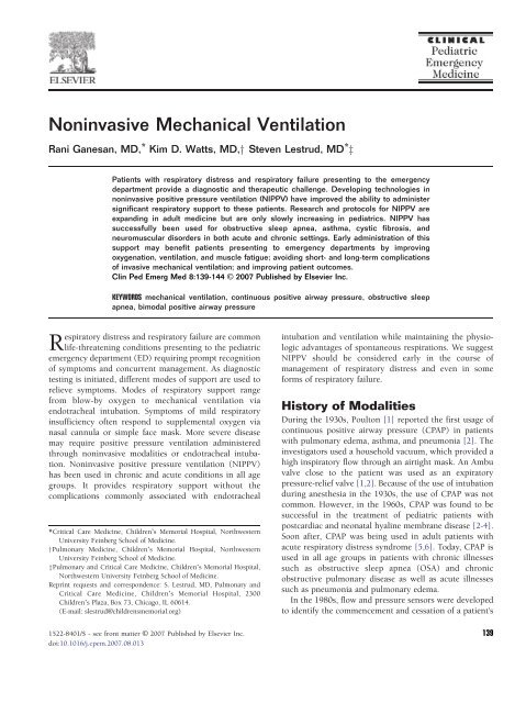
<strong>Noninvasive</strong> <strong>Mechanical</strong> <strong>Ventilation</strong><br />
Rani Ganesan, MD, ⁎ Kim D. Watts, MD,y Steven Lestrud, MD ⁎ z<br />
Patients with respiratory distress and respiratory failure presenting to the emergency<br />
department provide a diagnostic and therapeutic challenge. Developing technologies in<br />
noninvasive positive pressure ventilation (NIPPV) have improved the ability to administer<br />
significant respiratory support to these patients. Research and protocols for NIPPV are<br />
expanding in adult medicine but are only slowly increasing in pediatrics. NIPPV has<br />
successfully been used for obstructive sleep apnea, asthma, cystic fibrosis, and<br />
neuromuscular disorders in both acute and chronic settings. Early administration of this<br />
support may benefit patients presenting to emergency departments by improving<br />
oxygenation, ventilation, and muscle fatigue; avoiding short- and long-term complications<br />
of invasive mechanical ventilation; and improving patient outcomes.<br />
Clin Ped Emerg Med 8:139-144 C 2007 Published by Elsevier Inc.<br />
KEYWORDS mechanical ventilation, continuous positive airway pressure, obstructive sleep<br />
apnea, bimodal positive airway pressure<br />
Respiratory distress and respiratory failure are common<br />
life-threatening conditions presenting to the pediatric<br />
emergency department (ED) requiring prompt recognition<br />
of symptoms and concurrent management. As diagnostic<br />
testing is initiated, different modes of support are used to<br />
relieve symptoms. Modes of respiratory support range<br />
from blow-by oxygen to mechanical ventilation via<br />
endotracheal intubation. Symptoms of mild respiratory<br />
insufficiency often respond to supplemental oxygen via<br />
nasal cannula or simple face mask. More severe disease<br />
may require positive pressure ventilation administered<br />
through noninvasive modalities or endotracheal intubation.<br />
<strong>Noninvasive</strong> positive pressure ventilation (NIPPV)<br />
has been used in chronic and acute conditions in all age<br />
groups. It provides respiratory support without the<br />
complications commonly associated with endotracheal<br />
⁎Critical Care Medicine, Children's Memorial Hospital, Northwestern<br />
University Feinberg School of Medicine.<br />
†Pulmonary Medicine, Children's Memorial Hospital, Northwestern<br />
University Feinberg School of Medicine.<br />
‡Pulmonary and Critical Care Medicine, Children's Memorial Hospital,<br />
Northwestern University Feinberg School of Medicine.<br />
Reprint requests and correspondence: S. Lestrud, MD, Pulmonary and<br />
Critical Care Medicine, Children's Memorial Hospital, 2300<br />
Children's Plaza, Box 73, Chicago, IL 60614.<br />
(E-mail: slestrud@childrensmemorial.org)<br />
intubation and ventilation while maintaining the physiologic<br />
advantages of spontaneous respirations. We suggest<br />
NIPPV should be considered early in the course of<br />
management of respiratory distress and even in some<br />
forms of respiratory failure.<br />
History of Modalities<br />
During the 1930s, Poulton [1] reported the first usage of<br />
continuous positive airway pressure (CPAP) in patients<br />
with pulmonary edema, asthma, and pneumonia [2]. The<br />
investigators used a household vacuum, which provided a<br />
high inspiratory flow through an airtight mask. An Ambu<br />
valve close to the patient was used as an expiratory<br />
pressure-relief valve [1,2]. Because of the use of intubation<br />
during anesthesia in the 1930s, the use of CPAP was not<br />
common. However, in the 1960s, CPAP was found to be<br />
successful in the treatment of pediatric patients with<br />
postcardiac and neonatal hyaline membrane disease [2-4].<br />
Soon after, CPAP was being used in adult patients with<br />
acute respiratory distress syndrome [5,6]. Today, CPAP is<br />
used in all age groups in patients with chronic illnesses<br />
such as obstructive sleep apnea (OSA) and chronic<br />
obstructive pulmonary disease as well as acute illnesses<br />
such as pneumonia and pulmonary edema.<br />
In the 1980s, flow and pressure sensors were developed<br />
to identify the commencement and cessation of a patient's<br />
1522-8401/$ - see front matter C 2007 Published by Elsevier Inc. 139<br />
doi:10.1016/j.cpem.2007.08.013
140 R. Ganesan et al.<br />
respiratory efforts. Pressure support ventilation proved to<br />
be beneficial in invasive mechanical ventilation by Kacmarek<br />
[7]. These technological advances were then applied<br />
to noninvasive mechanical ventilation modalities. The use<br />
of flow-triggered inspiratory support with continuous<br />
positive airway pressure has proven beneficial.<br />
More recently, bimodal positive airway pressure (BiPAP)<br />
provides inspiratory positive airway pressure (IPAP) and<br />
expiratory positive airway pressure (EPAP). This mode of<br />
ventilation is similar to the combination of CPAP and<br />
pressure support ventilation. IPAP delivers positive pressure<br />
during the inspiratory phase of the respiratory cycle.<br />
EPAP delivers end-expiratory positive pressure during the<br />
expiratory phase. This differs from CPAP, as CPAP provides<br />
end expiratory positive pressure during the entire respiratory<br />
cycle.<br />
Modern-day CPAP/BiPAP machines (Respironics Inc,<br />
Murrysville, PA) are electrically controlled, pneumatically<br />
powered, low-pressure ventilators [8]. These machines are<br />
designed to augment the patient's respiratory effort. Flow<br />
transducers in the patient's circuit are used to sense the<br />
patient's effort. The data collected by the flow transducer is<br />
analyzed to determine the amount of pressure needed to<br />
generate the tidal volume or pressure preset by the clinician.<br />
The machine continuously recalibrates to adjust for leaks<br />
and changes in lung compliance and airway resistance.<br />
In recent years, systems developed to provide high-flow,<br />
high-humidity nasal cannula supplemental gas has supplanted<br />
the use of CPAP. These systems deliver 1 to 40<br />
L/min of flow rate, producing positive pressure support<br />
as well as supplemental oxygen. This mode of<br />
noninvasive positive pressure supplementation has<br />
been used more typically in newborns and infants but<br />
likely will find increased usage in all patients with<br />
respiratory distress.<br />
Physiology of Modalities<br />
Respiratory disease may involve the upper airway, lower<br />
airway, or both. Regardless of location of disease in the<br />
respiratory tract, airway resistance and lung compliance<br />
can be affected. Increased inflammation, debris, and<br />
bronchospasm associated with respiratory disease result<br />
in a higher transpulmonary pressure (difference between<br />
proximal airway pressure and alveolar pressure) required<br />
to produce a unit flow of gas through the airways of the<br />
lung. This is demonstrated by an increased airflow<br />
resistance [8]. As the properties of the alveoli may change<br />
during disease, the amount of unit lung volume change<br />
decreases for unit change in transalveolar pressure (the<br />
difference between alveolar pressure and pleural pressure).<br />
This is defined as a decrease in lung compliance [8]. The<br />
resultant decrease in lung compliance and increase in<br />
airway resistance in respiratory disease also require the<br />
patient to generate higher intrathoracic pressures to<br />
maintain alveolar ventilation and oxygenation. Muscle<br />
fatigue from increased work of breathing leads to inability<br />
to augment minute ventilation and adjust for increasing<br />
carbon dioxide.<br />
The combination of increased airway resistance and<br />
decreased lung compliance lead to a decrease in functional<br />
residual capacity (FRC). When the FRC drops below the<br />
critical closing volume, alveolar collapse occurs [9]. Critical<br />
closing volume is altered by age, disease, and position [9].<br />
With exhalation, intrathoracic pressure changes will result<br />
in airway closure. This occurs first in dependent areas of the<br />
lungs around the level of residual volume. During disease<br />
states, increased transpleural pressures result in increased<br />
closing volumes. These closing volumes may occur at the<br />
level of FRC, which result in atelectasis, subsequent<br />
ventilation/perfusion (V/Q) mismatch, and eventual<br />
hypoxemia and hypercarbia [9].<br />
<strong>Noninvasive</strong> positive-pressure ventilation has been<br />
shown to improve oxygenation, alveolar ventilation, and<br />
work of breathing [10,11]. The flow provided by NIPPV<br />
bypasses upper airway obstruction. The increase in upper<br />
airway pressure leads to a drop in airway resistance.<br />
NIPPV also promotes recruitment of collapsed alveoli.<br />
When effective alveolar recruitment is achieved, FRC<br />
increases and surpasses the critical closing volume [9].<br />
This leads to alveolar patency, alveolar stability, increase<br />
in lung compliance, and improvement in V/Q mismatch.<br />
To recruit alveoli effectively, the pressure delivered must<br />
be higher than the critical opening pressure of the alveoli<br />
[9]. The delivery of fixed FIO 2 through a closed circuit<br />
improves patient oxygenation. The increase in open<br />
alveoli leads to redistribution of pulmonary blood flow<br />
to these areas of improved ventilation. This also improves<br />
patient ventilation and oxygenation. The addition of<br />
CPAP/BiPAP may decrease work of breathing by several<br />
mechanisms including relief of upper airway obstruction,<br />
alteration of thoracic closing volumes, and decrease<br />
in diaphragmatic work. All of these factors together<br />
improve oxygenation, ventilation, and overall systemic<br />
and cellular acidosis.<br />
Administration and Monitoring of<br />
Modalities<br />
Modern NIPPV ventilators provide different modes to<br />
accommodate different patient physiology. The spontaneous<br />
mode can provide IPAP, EPAP or both, but only in<br />
response to the patient's respiratory effort. In the<br />
spontaneous/timed mode, pressure delivery is also controlled<br />
by patient effort. However, if no effort is detected<br />
within a set time, a breath is delivered to the patient. In<br />
timed mode, a breath is delivered at a set interval regardless<br />
of patient effort.<br />
The decision to use BiPAP, CPAP, or high-flow nasal<br />
cannula is dependent on the patient, specific disease, and
<strong>Noninvasive</strong> mechanical ventilation<br />
141<br />
clinical expertise. NIPPV may be delivered via nasal or fullface<br />
mask. Both modes of administration can deliver<br />
adequate support. However, full-face masks provide more<br />
constant and reliable pressure delivery. Nasal masks are best<br />
suited for patients who are able to comfortably keep their<br />
mouths closed. These masks are considered more comfortable<br />
by patients. Infants, who are obligate nose breathers,<br />
generally benefit more from nasal prong CPAP or high-flow<br />
nasal cannula.<br />
Patients requiring NIPPV are generally critically ill.<br />
These patients need to be closely monitored for worsening<br />
respiratory failure. Patients should be monitored with<br />
serial lung exams, continuous pulse oximetry, and<br />
transcutaneous carbon dioxide monitoring. If a patient's<br />
status worsens or does not improve, endotracheal intubation<br />
with mechanical ventilation is warranted.<br />
Clinical Uses of NIPPV<br />
<strong>Noninvasive</strong> positive pressure ventilation is used in the<br />
pediatric population to address both acute and chronic<br />
respiratory compromise. It is important for the ED physician<br />
to be knowledgeable about the uses of NIPPV. Understanding<br />
the indications for NIPPV will facilitate its use as a<br />
treatment modality in the acute setting. By understanding<br />
the limitations of NIPPV, better care can also be given to<br />
those children with chronic respiratory conditions requiring<br />
mask ventilation who present to the ED with a change in<br />
clinical status. The use of home ventilation is therefore an<br />
important part of patient history taking in the ED setting.<br />
In the acute setting, the benefits of NIPPV over<br />
endotracheal intubation include reduced risk of nosocomial<br />
infection, shorter intensive care unit (ICU) and<br />
hospital stay, and decreased mortality [12]. It is important<br />
to note that NIPPV is not a replacement for endotracheal<br />
intubation in patients who require airway protection and<br />
have hypercarbia or life-threatening hypoxemia (PAO 2<br />
b60 mm Hg on rebreathing facemask). NIPPV should<br />
not be attempted in patients who are rapidly deteriorating<br />
or who are somnolent or confused. It should also be<br />
avoided in patients who are hypotensive or have cardiac<br />
dysrhythmias [13]. Previous studies have demonstrated<br />
that BiPAP will lead to improvement of symptoms within<br />
hours, so trials of NIPPV should remain short in nature and<br />
intubation with subsequent mechanical ventilation should<br />
be instituted if indicated for the clinical situation.<br />
Literature supporting the use of NIPPV in the pediatric<br />
population primarily emanates from either the pediatric<br />
intensive care unit (PICU) as a treatment of respiratory<br />
distress or from the home setting as a management for<br />
chronic respiratory conditions such as OSA and neuromuscular<br />
disorders. Studies of NIPPV use in the ED are<br />
mostly confined to the adult literature.<br />
A 2006 article by Maheshwari et al [14] sought to<br />
investigate the use of noninvasive mechanical ventilation<br />
in acute care hospitals in the United States [14]. This<br />
survey showed great variation in use among hospitals<br />
within the same region. In this study, use of NIPPV for<br />
acute respiratory failure varied from less than 5% to greater<br />
than 50% of patients. Of those who used this modality in<br />
less than 15% of the patients, lack of physician knowledge<br />
and equipment availability were cited as the top reasons for<br />
this practice pattern. The site of initiation of NIPPV was<br />
most often the ICU (55%) followed by the ED (26%).<br />
Other studies have shown ED use to be higher [15]. In<br />
these facilities, protocol-driven application and monitoring<br />
were associated with better success.<br />
Factors that influenced success of noninvasive ventilation<br />
were also examined in a 2005 article by Merlani et al<br />
[16]. In this retrospective analysis, patients admitted to the<br />
ED for acute respiratory failure and treated with NIPPV<br />
were studied. Failure of NIPPV was defined as the need to<br />
undergo endotracheal intubation. In this study, 31% failed<br />
NIPPV. Factors associated with failure included a pH less<br />
than 7.35 and a respiratory rate greater than 20 after 1 hour<br />
of NIPPV. In adult ED patients, NIPPV is most often<br />
instituted for cardiogenic pulmonary edema, acute asthma,<br />
chronic obstructive pulmonary disease, immunocompromised<br />
states, and pneumonia [14,17,18].<br />
Studies evaluating treatment failure of NIPPV in the<br />
pediatric population have been conducted primarily in the<br />
pediatric intensive care setting. A small prospective study<br />
of infants and children in the ICU undergoing NIPPV for<br />
respiratory failure demonstrated that inspired oxygen<br />
(FIO 2 ) after 1 hour of NIPPV may be a predictor for<br />
outcome, with a requirement for an FIO 2 greater than 80%<br />
being associated with NIPPV failure [19]. A larger study of<br />
NIPPV in PICUs found that acute respiratory distress<br />
syndrome and a high pediatric logistic organ dysfunction<br />
(PELOD) score were independent predictive variables of<br />
NIPPV failure [11]. This study noted that there was<br />
improvement in breathing pattern and in gas exchange in<br />
some patients on NIPPV in the first few hours. Most of the<br />
patients who failed did so within the first 48 hours. These<br />
findings support the need for close monitoring of patients<br />
on NIPPV and the need to consider discontinuation if<br />
improvement is not noted in the short term.<br />
A similar study of predictive factors for success of<br />
NIPPV in the pediatric ED does not exist to our knowledge<br />
beyond small disease-specific cohorts. Therefore, it is<br />
important to understand the specific indications for NIPPV<br />
in pediatrics and the literature that supports its use in<br />
certain disease states.<br />
Asthma<br />
<strong>Noninvasive</strong> mechanical ventilation functions to bridge the<br />
gap between maximal medical management and mechanical<br />
ventilation in asthma. By avoiding intubation in an<br />
asthmatic, complications such as bronchospasm, barotrauma,<br />
ventilator-induced lung injury, cardiovascular<br />
instability, and nosocomial infection can be reduced. The
142 R. Ganesan et al.<br />
use of NIPPV in asthmatic patients works by applying<br />
positive pressure, which decreases the workload of fatigued<br />
muscles used for inspiration including the diaphragm and<br />
accessory muscles. The positive pressure relieves the need<br />
for the patient to “auto-positive end expiratory pressure”<br />
(PEEP) to keep airways open during exhalation. This allows<br />
for improvement in V/Q mismatch by a direct bronchodilator<br />
effect and recruitment of smaller airways and collapsed<br />
alveoli. <strong>Noninvasive</strong> ventilation may also improve delivery of<br />
bronchodilators to smaller lung airways. This modality can<br />
be a reasonable alternative for the 5% to 10% of patients with<br />
acute asthma who fail conventional therapy.<br />
There are some studies to support the use of BiPAP<br />
for acute asthma exacerbations in the pediatric population<br />
[20,21]. In a descriptive, 1-year, retrospective chart<br />
review of ED patients with acute asthma refractory to<br />
conventional treatment subsequently treated with BiPAP,<br />
safety, tolerance, and benefit were studied. Nasal BiPAP<br />
and continuous albuterol were applied to these otherwise<br />
healthy pediatric asthma patients. Most of the<br />
patients tolerated the device; more than 70% had<br />
improvement in respiratory rate and almost 90%<br />
improved their oxygen saturation. Most patients were<br />
subsequently transferred to the PICU; however, almost<br />
25% were weaned off BiPAP in the ED and transferred to<br />
the pediatric general ward without any complications,<br />
adverse events, or deaths.<br />
These patients were started at an inspired positive<br />
airway pressure of 10 cm H 2 O and an expired positive<br />
airway pressure at 5 cm H 2 O with inspiratory pressure<br />
adjusted to achieve an exhaled tidal volume of 6 to<br />
9 mL/kg. A nasal mask was used to reduce the risk of<br />
gastric insufflation, vomiting, and aspiration. Albuterol<br />
was placed in the circuit between the whisper valve and<br />
nasal mask. This study was limited, however, because of its<br />
retrospective nature, differences in administration of<br />
magnesium and terbutaline, and the lack of an objective<br />
measure on when to start BiPAP therapy [22].<br />
In 2004, a prospective crossover study was performed<br />
involving 20 children in the PICU randomized to either<br />
2 hours of nasal BiPAP (10cm/5cm) followed by 2 hours of<br />
standard therapy or the reverse. A clinical asthma score<br />
was used to evaluate patients. All patients were on<br />
continuous albuterol and intravenous steroids but other<br />
therapies were at the treating physician's discretion. This<br />
study found that there were decreased signs of work of<br />
breathing and dyspnea in the BiPAP group as compared<br />
with the standard therapy group. Discontinuation of BiPAP<br />
after 2 hours in the group who initially received it was<br />
associated with an increase in respiratory rate and the<br />
clinical asthma score. In the group receiving conventional<br />
therapy first, only 2 children showed improvements in<br />
respiratory rate and clinical asthma score, and only with a<br />
BiPAP trial. BiPAP was not associated with significant<br />
differences in oxygen saturation and transcutaneous<br />
carbon dioxide monitoring [23].<br />
A prospective, randomized, placebo-controlled trial of<br />
BiPAP in an adult ED patient population with acute asthma<br />
has been reported. It was found that in these adult patients,<br />
the use of BiPAP for 3 hours improved lung function,<br />
alleviated symptoms of the asthma attack more quickly than<br />
in the control group, and reduced the rate of admission [24].<br />
Neuromuscular Disorders<br />
A variety of studies and case reports have supported the use of<br />
NIPPV in neuromuscular diseases such as spinal muscular<br />
atrophy, myasthenia gravis, congenital myopathies, and<br />
muscular dystrophies [25-28]. In neuromuscular disorders,<br />
the discussion regarding ventilation, invasive or noninvasive,<br />
should ideally be made at a nonemergent time. This scenario,<br />
however, is not always possible. It is important to recognize<br />
the role that NIPPV can serve in patients with respiratory<br />
failure and neuromuscular disorders. NIPPV works in<br />
neuromuscular patients by improving ventilatory mechanics,<br />
resting fatigued respiratory muscles, enhancing ventilatory<br />
sensitivity to carbon dioxide, and improving sleep stage<br />
disturbances [28]. The initiation of NIPPV in these patients<br />
has been found to reduce symptoms, such as sleepiness and<br />
headaches, hospitalizations, and health care costs in those<br />
who use the device [26]. In patients with neuromuscular<br />
disorders, care should be given when initiating NIPPV. BiPAP,<br />
rather than CPAP, is the mode of choice because high<br />
continuous pressure may be difficult for weak muscles to<br />
overcome to generate a breath.<br />
Obstructive Sleep Apnea/Airway Obstruction<br />
Obstructive sleep apnea is the major indication for NIPPV<br />
in airway obstruction. It is estimated that OSA syndrome<br />
affects about 2% of children. This can be secondary to<br />
neuromuscular airway control or from anatomic narrowing<br />
of the airway. In most children, obstruction is caused<br />
by enlarged tonsils and adenoids, and surgical intervention<br />
generally improves the condition. However, with increasing<br />
rates among children, obesity is playing a greater role in<br />
pediatric OSA.<br />
If pediatric OSA is not alleviated by tonsillectomy and<br />
adenoidectomy, CPAP or BiPAP can be used. A review<br />
article from Guilleminault et al [29] in 2005 gives an<br />
overview of the symptoms of OSA associated with different<br />
age groups. These clinical symptoms along with findings on<br />
polysomnography confirm the diagnosis of sleep-disordered<br />
breathing. The use of nasal CPAP for OSA in children<br />
has been studied, and the training of parents and patients,<br />
frequent reevaluation and fitting, and attention to midface<br />
growth are important. A recent study investigating<br />
adherence and effectiveness of these modalities in children<br />
with OSA found poor compliance even with intensive home<br />
support by health care professionals. They also found that<br />
of those who do use the equipment, the duration was less<br />
than prescribed. This study did show that NIPPV was<br />
effective in improving clinical symptoms of OSA and was<br />
associated with improved polysomnographic findings [30].
<strong>Noninvasive</strong> mechanical ventilation<br />
143<br />
There are case reports and studies that investigate the<br />
use of NIPPV in children with laryngomalacia, tracheomalacia,<br />
and bronchomalacia [31-33]. NIPPV was found to<br />
relieve the load on respiratory muscles caused by severe<br />
laryngomalacia and was associated with clinical improvement<br />
in sleep and growth when upper airway obstruction<br />
is associated with alveolar hypoventilation [31].<br />
In the ED, it is important to recognize severe upper<br />
airway obstruction, especially in infants, as an etiology for<br />
respiratory distress and failure to thrive. Prompt intervention<br />
with NIPPV can provide acute relief until further<br />
studies and possible surgical interventions can be applied.<br />
Bronchiolitis<br />
Bronchiolitis is a common diagnosis in the ED with only a<br />
small percentage of children worsening into respiratory<br />
failure. Little research has been done to investigate the use<br />
of NIPPV in bronchiolitis in the ED. A randomized<br />
crossover trial comparing CPAP to standard therapy<br />
found that CPAP decreased hypercarbia in infants with<br />
bronchiolitis compared to standard therapy [34]. A study<br />
by Martinon-Torres et al [35] found that there was an<br />
improvement in clinical score, tachypnea and hypercarbia<br />
in patients with refractory bronchiolitis treated with heliox<br />
and CPAP.<br />
Postoperative<br />
A retrospective study of patients undergoing tonsillectomy<br />
and adenoidectomy found that BiPAP was safe and effective<br />
in patients that were predisposed to postoperative postobstructive<br />
airway complications. These factors include<br />
obesity, young age, asthma, and neurologic compromise<br />
[36]. It was also found that the use of NIPPV in pediatric<br />
patients after liver transplantation helped resolve atelectasis<br />
and improved outcomes [37]. NIPPV has also been<br />
found to be useful postoperatively in patients with spinal<br />
cord surgery and laryngotracheal reconstruction procedures<br />
[38,39].<br />
Cystic Fibrosis<br />
The use of NIPPV in cystic fibrosis has been found to be<br />
beneficial not only in those patients with end-stage<br />
disease awaiting lung transplant but also earlier in the<br />
course of the disease to improve nocturnal oxygenation<br />
and sleep quality and rest respiratory muscles [40,41].<br />
These findings suggest that there may be a short-term<br />
benefit of NIPPV in cystic fibrosis. In the ED setting, in<br />
the case of severe respiratory distress not necessitating<br />
intubation, it may be reasonable to try NIPPV as a means<br />
of improving oxygenation as well as improving work of<br />
breathing [42].<br />
Mediastinal Mass<br />
There is no current evidence to support the use of<br />
noninvasive mask ventilation in the case of respiratory<br />
distress caused by obstruction secondary to a mediastinal<br />
mass. This type of obstruction, resulting in compression of<br />
the bronchi, may cause wheezing that could be misinterpreted<br />
as asthma. It is important to note that<br />
wheezing caused by a fixed obstruction is monophonic,<br />
rather than polyphonic. A chest x-ray could help to<br />
differentiate if needed. Currently, there is no indication for<br />
NIPPV in this setting.<br />
Ingestion/Toxin<br />
Ingestion and poisoning causing respiratory failure are not<br />
indications for NIPPV. A full face mask device puts patients<br />
at a high risk for aspiration if vomiting occurs. If<br />
consciousness is impaired, NIPPV is of little use unless<br />
BiPAP with a physiological back-up rate is applied. However,<br />
if this is needed, conventional ventilation with protection of<br />
the airway with an endotracheal tube is indicated.<br />
Foreign Body Aspiration<br />
The size and location of an aspirated foreign body dictate<br />
treatment. However, if a foreign body is producing<br />
significant respiratory distress with concern of impending<br />
respiratory failure, NIPPV is not indicated and will not<br />
provide significant relief past a fixed tracheal obstruction.<br />
Summary<br />
Patients presenting to pediatric EDs with respiratory<br />
distress represent an array of diagnostic and therapeutic<br />
challenges. With the development of NIPPV, there is an<br />
effective tool in our armamentarium to provide significant<br />
respiratory support. NIPPV has demonstrated benefits in<br />
decreasing work of breathing, relieving fatigued muscles of<br />
respiration, improving oxygenation, and possibly avoiding<br />
common complications of endotracheal intubation. There<br />
are clear clinical scenarios in which NIPPV is contraindicated,<br />
such as an obtunded patient, certain postoperative<br />
patients, patients with vomiting, and patients in<br />
which mask ventilation is not tolerated. More frequently, it<br />
is the patient with pulmonary disease resulting in<br />
respiratory distress that is amenable to this therapy. It is<br />
important to obtain any history of NIPPV usage in the ED.<br />
Increasing numbers of children are on nocturnal settings<br />
for OSA and support settings for chronic respiratory<br />
insufficiency with diseases such as myopathies, cystic<br />
fibrosis, and bronchiectasis. Advancements in the knowledge<br />
of NIPPV management strategies for respiratory<br />
distress and early initiation of mask ventilation in the ED<br />
will enable the emergency physician to approach respiratory<br />
distress in a similar manner as sepsis; using goaldirected<br />
therapies to improve oxygenation, ventilation, and<br />
comfort of breathing.<br />
References<br />
1. Poulton EP. Left-sided heart failure with pulmonary edema: its<br />
treatment with the “pulmonary plus pressure” machine. Lancet<br />
1936;231:981-3.
144 R. Ganesan et al.<br />
2. Duke GJ, Berstein AD. Non-invasive ventilation for adult acute<br />
respiratory failure. Part I. Crit Care Resus 1999;1:187-98.<br />
3. Allen LP, Blake AM, Durbin GM, et al. Continuous positive airway<br />
pressure and mechanical ventilation by facemask in newborn infants.<br />
Br Med J 1975;3:137-9.<br />
4. Crew AD, Wall E, Varkonyi PI. Continuous positive airway pressure<br />
breathing (CPAP). Apparatus for use in neonates and adults.<br />
Anaesthesia 1975;30:67-72.<br />
5. Schmidt GB, Bombeck CT, Bennett EJ, et al. Continuous positive<br />
airway pressure in the prophylaxis of the adult respiratory distress<br />
syndrome. Langenbecks Arch Chir 1975:S439-42.<br />
6. Venus B, Jacobs K, Lim L. Treatment of the adult respiratory distress<br />
syndrome with continuous positive airway pressure. Chest 1979;76:<br />
257-61.<br />
7. Kacmarek RM. Inspiratory pressure support: does it make a clinical<br />
difference. Intensive Care Med 1989;15:337-9.<br />
8. Fuhrman BP, Zimmerman J. Pediatric critical care. 3rd ed.<br />
Philadelphia (Pa): Mosby Elsevier; 2006.<br />
9. West JB. Respiratory physiology—the essentials. Baltimore (MD):<br />
Williams and Wilkins; 1995.<br />
10. Fortenberry JD, Del Toro T, Jefferson LS, et al. Management of<br />
pediatric acute hypoxemic respiratory insufficiency with bi-level<br />
positive pressure (BiPAP) nasal mask ventilation. Chest 1995;108:<br />
1059-64.<br />
11. Essouri S, Chevret L, Durand P, et al. <strong>Noninvasive</strong> positive pressure<br />
ventilation: five years of experience in a pediatric intensive care unit.<br />
Pediatr Crit Care Med 2006;7:329-34.<br />
12. Antonelli M, Conti G, Rocco M, et al. A comparison of noninvasive<br />
positive-pressure ventilation and conventional mechanical ventilation<br />
in patients with acute respiratory failure. N Engl J Med 1998;339:<br />
429-35.<br />
13. Marik PE, Varon J, Fromm Jr R. The management of acute severe<br />
asthma. J Emerg Med 2002;23:257-68.<br />
14. Maheshwari V, Paioli D, Rothaar R, et al. Utilization of noninvasive<br />
ventilation in acute care hospitals: a regional survey. Chest 2006;129:<br />
1226-33.<br />
15. Paus-Jenssen ES, Reid JK, Cockcroft DW, et al. The use of noninvasive<br />
ventilation in acute respiratory failure at a tertiary care center. Chest<br />
2004;126:165-72.<br />
16. Merlani PG, Pasquina P, Granier JM, et al. Factors associated with<br />
failure of noninvasive positive pressure ventilation in the emergency<br />
department. Acad Emerg Med 2005;12:1206-15.<br />
17. Yosefy C, Hay E, Ben-Barak A, et al. BiPAP ventilation as assistance for<br />
patients presenting with respiratory distress in the department of<br />
emergency medicine. Am J Respir Med 2003;2:343-7.<br />
18. Collins SP, Mielniczuk LM, Whittingham HA, et al. The use of<br />
noninvasive ventilation in emergency department patients with acute<br />
cardiogenic pulmonary edema: a systematic review. Ann Emerg Med<br />
2006;48:260-9.<br />
19. Bernet V, Hug MI, Frey B. Predictive factors for the success of<br />
noninvasive mask ventilation in infants and children with acute<br />
respiratory failure. Pediatr Crit Care Med 2005;6:660-4.<br />
20. Akingbola OA, Simakajornboon N, Hadley EF, et al. <strong>Noninvasive</strong><br />
positive-pressure ventilation in pediatric status asthmaticus. Pediatr<br />
Crit Care Med 2002;3:181-4.<br />
21. Carroll CL, Schramm CM. <strong>Noninvasive</strong> positive pressure ventilation<br />
for the treatment of status asthmaticus in children. Ann Allergy<br />
Asthma Immunol 2006;96:454-9.<br />
22. Beers SL, Abramo TJ, Bracken A, et al. Bi-level positive airway<br />
pressure in the treatment of status asthmaticus in pediatrics. Am J<br />
Emerg Med 2007;25:6-9.<br />
23. Thill PJ, McGuire JK, Baden HP, et al. <strong>Noninvasive</strong> positive-pressure<br />
ventilation in children with lower airway obstruction. Pediatr Crit<br />
Care Med 2004;5:337-42.<br />
24. Soroksky A, Stav D, Shpirer I. A pilot prospective, randomized,<br />
placebo-controlled trial of bi-level positive airway pressure in acute<br />
asthmatic attack. Chest 2003;123:1018-25.<br />
25. Young HK, Lowe A, Fitzgerald DA, et al. Outcome of noninvasive<br />
ventilation in children with neuromuscular disease. Neurology<br />
2007;68:198-201.<br />
26. Piastra M, Conti G, Caresta E, et al. <strong>Noninvasive</strong> ventilation<br />
options in pediatric myasthenia gravis. Paediatr Anaesth 2005;15:<br />
699-702.<br />
27. Hardart MK, Burns JP, Truog RD. Respiratory support in spinal<br />
muscular atrophy type I: a survey of physician practices and attitudes.<br />
Pediatrics 2002;110(2 Pt 1):e24.<br />
28. Simonds AK. Recent advances in respiratory care for neuromuscular<br />
disease. Chest 2006;130:1879-86.<br />
29. Guilleminault C, Lee JH, Chan A. Pediatric obstructive sleep apnea<br />
syndrome. Arch Pediatr Adolesc Med 2005;159:775-85.<br />
30. Marcus CL, Rosen G, Ward SL, et al. Adherence to and effectiveness of<br />
positive airway pressure therapy in children with obstructive sleep<br />
apnea. Pediatrics 2006;117:e442-51.<br />
31. Fauroux B, Pigeot J, Polkey MI, et al. Chronic stridor caused by<br />
laryngomalacia in children: work of breathing and effects of<br />
noninvasive ventilatory assistance. Am J Respir Crit Care Med<br />
2001;164(10 Pt 1):1874-8.<br />
32. Aaseboe K, Berstad AK, Skadberg BT. <strong>Noninvasive</strong> treatment of<br />
bronchomalacia, successful ventilation of a severely ill infant. Acta<br />
Paediatr 2007;96:310-2.<br />
33. Masters IB, Chang AB. Interventions for primary (intrinsic)<br />
tracheomalacia in children. Cochrane Database Syst Rev 2005:<br />
CD005304.<br />
34. Thia LP, McKenzie SA, Blyth TP, et al. Randomised controlled trial of<br />
nasal continuous positive airways pressure (CPAP) in bronchiolitis.<br />
Arch Dis Child (in press).<br />
35. Martinon-Torres F, Rodriguez-Nunez A, Martinon-Sanchez JM. Nasal<br />
continuous positive airway pressure with heliox in infants with acute<br />
bronchiolitis. Respir Med 2006;100:1458-62.<br />
36. Friedman O, Chidekel A, Lawless ST, et al. Postoperative bi-level<br />
positive airway pressure ventilation after tonsillectomy and adenoidectomy<br />
in children—a preliminary report. Int J Pediatr Otorhinolaryngol<br />
1999;51:177-80.<br />
37. Chin K, Uemoto S, Takahashi K, et al. <strong>Noninvasive</strong> ventilation for<br />
pediatric patients including those under 1-year-old undergoing liver<br />
transplantation. Liver Transpl 2005;11:188-95.<br />
38. Bach JR, Sabharwal S. High pulmonary risk scoliosis surgery: role of<br />
noninvasive ventilation and related techniques. J Spinal Disord Tech<br />
2005;18:527-30.<br />
39. Hertzog JH, Siegel LB, Hauser GJ, et al. <strong>Noninvasive</strong> positive-pressure<br />
ventilation facilitates tracheal extubation after laryngotracheal<br />
reconstruction in children. Chest 1999;116:260-3.<br />
40. Fauroux B, Pigeot J, Polkey MI, et al. In vivo physiologic comparison<br />
of two ventilators used for domiciliary ventilation in children with<br />
cystic fibrosis. Crit Care Med 2001;29:2097-105.<br />
41. Gozal D. Nocturnal ventilatory support in patients with cystic<br />
fibrosis: comparison with supplemental oxygen. Eur Respir J 1997;10:<br />
1999-2003.<br />
42. Caronia CG, Silver P, Nimkoff L, et al. Use of bilevel positive airway<br />
pressure (BIPAP) in end-stage patients with cystic fibrosis<br />
awaiting lung transplantation. Clin Pediatr (Phila) 1998;37:<br />
555-9.