Traumatic Intercostal Pulmonary Hernia - Semes
Traumatic Intercostal Pulmonary Hernia - Semes
Traumatic Intercostal Pulmonary Hernia - Semes

You also want an ePaper? Increase the reach of your titles
YUMPU automatically turns print PDFs into web optimized ePapers that Google loves.
CARTAS AL EDITOR<br />
LETTERS TO THE EDITOR<br />
<strong>Traumatic</strong> intercostal pulmonary<br />
hernia<br />
Sir,<br />
Pneumocele is defined as the protrusion of a<br />
portion of lung parenchyma through an abnormal<br />
opening of the chest wall. The herniated segment<br />
is almost always covered by parietal pleura<br />
to form the bag and the skin cover. Pneumocele<br />
can be classified according to etiology as congenital<br />
and acquired; the latter in turn may be traumatic,<br />
postsurgical and spontaneous or pathological.<br />
Depending on location, it can be cervical,<br />
costal or diaphragmatic 3 . We present a case of<br />
costal lung hernia secondary to chest trauma.<br />
Symptoms are usually few and infrequent. The<br />
diagnosis is usually made by physical examination<br />
and can be confirmed using chest radiography or<br />
computed tomography (CT) scan.<br />
The case involved a 73-year-old man with a history<br />
of hypertension, hypercholesterolemia, hiatus hernia, intermittent<br />
claudication, polyarthrosis, multiple rib fractures<br />
(2nd to 6th right ribs), adaptive anxiety-depression<br />
disorder, Parkinson’s of unknown etiology and<br />
appendectomy. He visited the emergency department<br />
for left chest trauma following an accidental fall. He<br />
was tachypneic, with intense rib pain that increased<br />
with deep inspiration, Valsalva manoeuvres and palpation<br />
of the affected area; he was conscious and oriented,<br />
normally hydrated, afebrile, with blood pressure of<br />
155/90 mmHg, heart rate of 84 beats per minute, without<br />
jugular vein engorgement, audible heart sounds<br />
with frequent extrasystole, breath sounds preserved bilaterally<br />
with right basal crackles, and unremarkable abdomen<br />
except for an appendectomy scar. Physical examination<br />
also showed moderate but noticeable left<br />
gynecomastia (not previously reported by the patient or<br />
relatives) with clear crepitus on palpation of the area,<br />
swelling and sub-mammary bruising extending to the<br />
anterolateral area.<br />
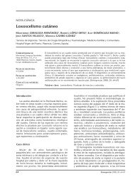
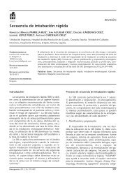
Chest X-ray (Figure 1) showed left pulmonary contusion<br />
related to a fracture of the left 7th rib and image<br />
compatible with intercostal hernia of the 7th intercostal<br />
space and old rib fractures on right rib cage. Chest CT<br />
scan (Figure 1) confirmed the existence of a traumatic<br />
intercostal uncomplicated hernia without pneumothorax,<br />
hemothorax or pleural effusion. The patient was<br />
hospitalized for six days, with complete bed rest, oxygen<br />
therapy and analgesic treatment, without complications<br />
and showed good improvement. Follow up X-<br />
ray showed no evidence of chest complications; given<br />
hit<br />
Figure 1. Plain chest X-ray (top) showing left lung contusion<br />
secondary to a fracture of the 7th left rib, and image compatible<br />
with intercostal hernia in the 7th left intercostal space<br />
(arrow). Computed tomography scan of the lower chest (below).<br />
<strong>Traumatic</strong> intercostal hernia (hit), uncomplicated and<br />
without pneumothorax, hemothorax or associated pleural effusion.<br />
the good clinical outcome and agreement of the patient<br />
and family, surgery was ruled out in favour of a<br />
conservative approach.<br />
154 Emergencias 2010; 22: 154-160
LETTERS TO THE EDITOR<br />
Lung hernia is a rare entity, with approximately<br />
300 cases reported in the literature 4 . According to<br />
Morel-Lavelle, it may be divided into congenital<br />
and acquired; the latter in turn may be pathological,<br />
traumatic or spontaneous. Spontaneous hernias<br />
are associated with manoeuvres involving extreme<br />
pressure on the chest wall (such as<br />
coughing or sneezing) causing the fracture of one<br />
or more ribs or intercostal muscle tear. Generally,<br />
this presents in patients with senile osteoporosis<br />
and / or chronic coughs. Protrusion of the lung<br />
wall may occur through the intercostal spaces<br />
(65% of cases), supraclavicular region (35% of cases)<br />
6 or through diaphragmatic wall defects (rare).<br />
<strong>Traumatic</strong> lung hernia, such as in our case, is<br />
rare and mostly due to high-energy, blunt chest<br />
trauma 5 . It does not necessarily present immediately<br />
after the impact, and may appear months or<br />
even years later 1,7 . The etiology may be explained<br />
by the anatomy of the intercostal spaces. The external<br />
and internal intercostal muscles that line<br />
the internal intercostal space are somewhat shorter<br />
than the ribs, so that one end of the space is<br />
covered by only one of the muscles and the other<br />
by aponeurosis (sinew). <strong>Intercostal</strong> spaces also have<br />
perforations through which chest wall vessels<br />
and nerves pass. Precisely these places are the<br />
most fragile parts of intercostal spaces, and the<br />
most susceptible to high pressure manoeuvres.<br />
They do not represent a serious problem unless<br />
incarceration and / or strangulation occurs,<br />
which cause pain and sometimes haemoptysis 8 .<br />
Besides good clinical examination, diagnosis<br />
usually requires chest X-ray, with oblique projections<br />
being most useful 9 . However, the diagnostic<br />
tool of choice is chest CT scan since it allows visualization<br />
of the pneumocele, its exact location<br />
and the size of the chest wall defect 10,11 .<br />
With regard to therapeutic approach, there is<br />
sharp controversy 12 . Some authors prefer a conservative<br />
approach with immobilizing bandage, and<br />
if this fails and / or complications arise, then opt<br />
for surgical repair. However, others claim that surgery<br />
is indicated in all anterior intercostal hernias,<br />
even if they are asymptomatic, by means of surgical<br />
repair or prosthetic (PTFE) patch repair with<br />
open surgery or thoracoscopy.<br />
References<br />
1 Taylor DA, Jacobson HG. Post-traumatic herniation of the lng. Radiology.<br />
1962;5:896-9.<br />
2 Morel-Lavelle A. <strong>Hernia</strong>s du Pomon. Bull Soc Chir Paris. 1847;1:75.<br />
3 Moncada R, Vade A, Gimenez C, Rosado W, Demos TC, Turbin R.<br />
Congenital and acquired lung hernias. J Torac Imaging. 1996;11:75-<br />
82.<br />
4 Reardon MJ, Fabre J, Reardon PR, Baldwin JC. Video-assisted repair of<br />
a traumatic intercostals pulmonary hernia. Ann Thorac Surg.<br />
1998;65:1155-7.<br />
5 Jiménez Aguero R, Hernández Ortiz C, Izquierdo Elea JM, Cabeza<br />
Sánchez R. <strong>Hernia</strong> pulmonar intercostal espontánea: aportación de<br />
un caso. Arch Bronconeumol. 2000;36:354-6.<br />
6 Munnell ER. <strong>Hernia</strong>tion of the lung. Ann Thorac Surg.<br />
1968;5(Supl):204.<br />
7 Reynolds, J, Davis JT. Injuries of the chest wall, pleura, pericardium,<br />
lung bronchi and esophagus. Radiol Clin North Am. 1966;4:383-<br />
401.<br />
8 Francois B, Desachy A, Cornu E, Ostyn E, Niquet L, Vignon P. <strong>Traumatic</strong><br />
pulmonary hernia: surgical versus conservative management. J<br />
Trauma. 1998;44:217-9.<br />
9 Allen GS, Fischer RP. <strong>Traumatic</strong> lung herniation. Ann Thorac Surg.<br />
1997;63:1455-6.<br />
10 Hauser M, Weder W, Largiader F, Glazer GM. Lung herniation<br />
through a postthoracoscopy chest wall defect: demostratio with spiral<br />
CT. Chest. 1997;112:558-60.<br />
11 Tamburro F, Grassi R, Romano R, Del Vecchio W. Adquired spontaneus<br />
intercostal hernia of the lung diagnosed on helical CT. Am J Roentgenol.<br />
2000;174:876-7.<br />
12 Goverde P, Van ZIL P, Van der Brande F, Vanmaele R. Chronic of the<br />
lung in patint with chronic obstructive pulmonary disease. Case report<br />
and review of the literature. Thorac Cardiovasc Surg.<br />
1988;46:164-6.<br />
Javier GIL DE BERNABÉ LÓPEZ 1 ,<br />
Luis Manuel CLARACO VEGA 1 ,<br />
Amos URTUBIA PALACIOS 2 ,<br />
María Isabel FERNÁNDEZ ESTEBAN 3 ,<br />
Pedro PARRILLA HERRANZ 1 ,<br />
Javier GARCÍA TIRADO 4<br />
1<br />
Servicio de Urgencias. Hospital Universitario Miguel Servet.<br />
Zaragoza, Spain. 2 Servicio Urgencias. Hospital General<br />
Elda. Alicante, Spain. 3 Médico de Familia. Área 16.<br />
Alicante, Spain. 4 Servicio de Cirugía Torácica. Hospital<br />
Universitario Miguel Servet. Zaragoza, Spain.<br />
Post-choking atelectasis<br />
Sir,<br />
Foreign body aspiration is a frequent problem<br />
and its consequences may range from mild to<br />
very severe, including cardiopulmonary arrest<br />
(CPA) from asphyxiation, depending on the location<br />
and degree of obstruction caused by the<br />
body sucked into the airway. It has a bimodal pattern,<br />
with a peak in infants aged less than one year<br />
and another in patients aged about 75 years.<br />
Any smallish object can be aspirated and such aspiration<br />
is especially associated with food intake.<br />
Most cases are resolved by coughing or basic resuscitation<br />
1 , but occasionally this is not enough<br />
and cardiopulmonary resuscitation (CPR) and<br />
post-resuscitation measures are needed.<br />
PA 62-year-old patient choked while eating. On arrival<br />
of the out-of-hospital emergency services, the patient had<br />
CPA with asystole; after approximately 10 minutes of CPR,<br />
heart rate recovered. Prior to orotracheal intubation, a<br />
lump of bread was extracted from the airway. The patient<br />
was admitted to intensive care where X-ray showed com-<br />
Emergencias 2010; 22: 154-160 155
LETTERS TO THE EDITOR<br />
Protruding atrial thrombus in the left<br />
ventricle<br />
Sir,<br />
As recently published in EMERGENCIES 1 , ultrasound<br />
(US) as a diagnostic procedure can be<br />
most useful and we here suggest a number of potential<br />
indications for use in the emergency department<br />
conducted ED physicians 2 . We present a<br />
case with rare US findings which significantly conditioned<br />
our therapeutic approach and probably<br />
averted dire consequences.<br />
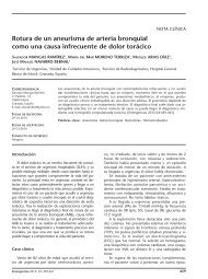
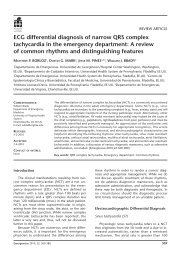
Figure 1. Admission chest X-ray (above) showing right lung<br />
atelectasia, and (below) X-ray after bronchoscopy showing<br />
subsequent evolution.<br />
plete atelectasis of the right lung, partially resolved with<br />
bronchoscopy (Figure 1), during which more bread and<br />
copious secretions were removed.<br />
The migration of aspirated foreign bodies into<br />
the tracheobronchial tree, most often to the right<br />
side, may cause obstructive atelectasis that requires<br />
bronchoscopy for resolution 2 . Occasionally, longstanding<br />
aspiration of a foreign body with irreversible<br />
changes in the lung wall may require surgery 3 .<br />
References<br />
1 Handley AJ, Koster R, Monsieurs L, Perkins GD, Davies S, Bossaert L,<br />
et al. European Resuscitation Council Guidelines for Resuscitation<br />
2005. Section 2. Adult basic life support and use of automated external<br />
defibrillators. Resuscitation. 2005;67(Supl. 1):S7-S23.<br />
2 Sauret Valet J. Cuerpos extraños. Arch Bronconeumol. 2002;38:285-7.<br />
3 Montero Cantú CA, Garduño Chávez B, Elizondo Ríos A. Broncoscopia<br />
rígida y cuerpo extraño. ¿Procedimiento obsoleto? Cir Ciruj.<br />
2006;74:51-3.<br />
Rosa María GARCÍA FANJUL,<br />
Eva GARCÍA PINEY<br />
Servicio de Medicina Intensiva. Hospital de Cabueñes,<br />
Gijón. Asturias, Spain.<br />
The patient was a 66 year-old woman without<br />
known cardiovascular risk factors, a history of atrial<br />
fibrillation (AF), treated with aspirin 100 mg/24 h<br />
and atenolol 25 mg/24 h. She was referred to our<br />
ED for fatigue and dyspnea with moderate exertion,<br />
with no other clinical symptoms. Physical examination<br />
and baseline laboratory results were normal, and<br />
with AF under control. Echocardiogram showed apparently<br />
normal contractility without valve lesions,<br />
but also dilated left atrium containing a mobile mass<br />
of 2 cm near the mitral annulus. The study was therefore<br />
completed with transesophageal echocardiography<br />
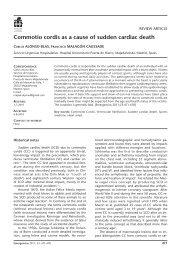
(Figure 1A) by the cardiology department, reporting<br />
a mobile thrombus occupying the left atrial<br />
appendage which protruded into the left atrium,<br />
crossing the mitral ring and visible in the left ventricle<br />
during diastole (Figure 1B). The patient received<br />
immediate treatment with enoxaparin (1 mg/kg/12<br />
hours). During her hospital stay there was no embolic<br />
event with clinical impact. Transthoracic US follow<br />
up one week later showed no evidence of thrombus,<br />
and the patient initiated long-term anticoagulation<br />
therapy with warfarin. The patient was discharged<br />
with sinus rhythm.<br />
In this case, there were no indication for anticoagulation<br />
factors before our echocardiographic<br />
findings, since the patient did not present<br />
risk factors classified as moderate (age 75 years,<br />
hypertension, heart failure, low ejection<br />
fraction or diabetes mellitus) or high (previous<br />
stroke, arterial embolism, mitral stenosis or<br />
prosthetic heart valve), according to the ACC /<br />
AHA / ESC guideline recommendations of 2006<br />
on the prevention of thromboembolic phenomena<br />
in AF3. It was the use of echocardiography<br />
in the emergency department that allowed<br />
us to suspect and then confirm the<br />
presence of atrial thrombus with very high risk<br />
of embolism in a patient with AF 4-6 and initiate<br />
appropriate treatment immediately, thus avoiding<br />
potentially fatal consequences, especially<br />
thromboembolism.<br />
156 Emergencias 2010; 22: 154-160
LETTERS TO THE EDITOR<br />
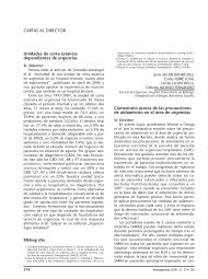
Figure 1. Transesophageal echocardiography. Panel A: Systole. Thrombus in the appendage which protrudes into the left atrium.<br />
Panel B: Diastole. The thrombus passed through the mitral ring and appears in the left ventricle. AI: left atrium; OAI: left atrium appendage;<br />
VI: left ventricle; Tr: thrombus. VM: mitral valve.<br />
References<br />
1 Nogué Bou R. La ecografía en medicina de urgencias: una herramienta<br />
al alcance de los urgenciólogos. Emergencias. 2008;20:75-7.<br />
2 García Martín LA, Campo Linares R, Rayo Gutiérrez M. Pericarditis<br />
purulenta: diagnóstico ecográfico precoz en el servicio de urgencias.<br />
Emergencias. 2008;20:135-8.<br />
3 New ACC/AHA/ESC Guidelines for the managemnet of atrial Fibrillation:<br />
Highlghting Stroke Prevention. Nueva York: Medscape Cardiology;<br />
2006.<br />
4 García Quintana JA, González Morales L, Castro Bueno N, Medina<br />
Fernández-Acytuno. Tromboembolismo pulmonar con trombo móvil<br />
e la aurícula. Med Intensiva. 2006;30:183-6.<br />
5 Gómez R, Penas, JL, Fleitas C, Cascallana JE. Trombo libre en aurícula<br />
izquierda. Biomédica. 2005;25:293-4.<br />
6 Alexis Llamas J. Papel de la ecocardiografía convencional y transesofágica<br />
en la fibrilación auricular. Rev Col Cardiol. 2007;14(Supl.<br />
3):165-70.<br />
Rubén GÓMEZ IZQUIERDO 1 ,<br />
Lorenzo ALONSO VEGA 2 ,<br />
Íñigo GÓMEZ ZABALA 3 ,<br />
Carlos TEJA SANTAMARÍA 4<br />
1<br />
Servicio de Cardiología. 2 Servicio de Urgencias.<br />
3<br />
Médico de Familia. Hospital de Laredo. Cantabria, Spain.<br />
Subacute headache in a young woman<br />
normal neurologic signs<br />
Sir,<br />
LHeadache is a frequent reason for consultation<br />
in the emergency department 1-3 , and for daily<br />
practice it is important to differentiate between<br />
potentially dangerous causes and those which are<br />
not. In addition to the ever-important medical<br />
history, we must rely on neurologic examination<br />
and imaging techniques 4,5 .<br />
MWe present the case of a 34 year-old woman with<br />
progressive headache of four days duration, holocraneal<br />
location and oppressive, which increased throughout<br />
the day, with periods of remission and unrelated to the<br />
Valsalva manoeuvre. She reported no vomiting or fever.<br />
She had suffered two spontaneous miscarriages in the<br />
previous five years. Physical examination and neurological<br />
examinations were unremarkable. Laboratory tests<br />
and chest X-ray were normal, with negative pregnancy<br />
test. Computed tomography (CT) scan revealed a<br />
hyperdense image in the right transverse and sigmoid<br />
venous sinuses (Figure 1). Nuclear magnetic angio-resonance<br />
imaging (MRI) of the skull showed acute thrombosis<br />
of these sinuses. Jugular venous Doppler ultrasound<br />
showed alterations. She received anticoagulation<br />
with intravenous sodium heparin, and later with Coumadin,<br />
with good outcome. Thrombophilia test was<br />
normal [ESR, antithrombin III, D dimer, Protein C, Protein<br />
S, lupus anticoagulant, antibodies, hepatitis B and<br />
C, HIV, RPR, anti-Treponema pallidum, anticardiolipin<br />
and antinuclear antibodies (Ab), vitamin B12, folic acid,<br />
serum ferritin, immunoglobulin (Ig) A, G and M, plasma<br />
homocysteine, TSH, B2-microglobulin, V-Leiden gene<br />
factor and prothrombin mutation 202 106], so that<br />
the patient was finally diagnosed with idiopathic cerebral<br />
venous thrombosis (CVT).<br />
LCVT is an underdiagnosed entity that may<br />
affect patients at any age, and both the clinical<br />
picture and neuroimaging techniques show great<br />
variability, which makes the diagnosis a real<br />
challenge 2,3 . CT is the first test to be performed<br />
since it allows ruling out other causes as well as<br />
showing the underlying source; 80% will be abnormal,<br />
but CT findings are not always diagnostic.<br />
MRI is the technique of choice: MR venography<br />
increases the sensitivity of the study,<br />
Emergencias 2010; 22: 154-160 157
LETTERS TO THE EDITOR<br />
Subphrenic abscess<br />
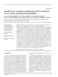
Figure 1. Cranial CT showing a hyperdense image at the sigmoid<br />
venous (arrow) and right transverse sinus (arrow).<br />
and should be done before the suspicion of<br />
CVT, regardless of the CT result. Treatment is<br />
with anticoagulant therapy, sodium heparin in<br />
the acute phase followed by oral anticoagulation<br />
(INR2-3) for at least six months, when it<br />
becomes known as CVT without thrombophilic<br />
disorder 4,5,7 . In conclusion, CVT is a potentially<br />
dangerous cause of headache that requires the<br />
use of complex imaging tests, and may be present<br />
despite normal neurological examination<br />
results.<br />
Sir,<br />
An 80 year-old man was admitted to the emergency<br />
department with abdominal pain and febrile<br />
syndrome. Medical history included an episode of acute<br />
gallstone cholecystitis two months before with favorable<br />
evolution after conservative treatment. On arrival at<br />
the ED, physical examination revealed no abdominal<br />
pain on palpation, no peritonitis, and a body temperature<br />
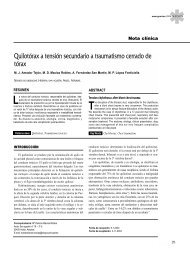
of 37.2 º C. Chest X-ray showed a level of fluid in<br />
the upper quadrant suggestive of right subphrenic abscess<br />
(Figure 1). Abdominal ultrasound showed a right<br />
subdiaphragmatic collection of 14 cm in diameter, near<br />
the midline, with abundant echogenic content containing<br />
air related with a subphrenic abscess. The gallbladder<br />
was not identified. The study was completed<br />
with computed tomography (CT) scan (Figure 1),<br />
which showed anterior subphrenic collection with<br />
hydro-aerial level and a 9 cm lesion in the vesicular<br />
Pablo FRANQUELO MORALES,<br />
Margarita ALCÁNTARA ALEJO,<br />
Carlos HERRÁIZ DE CASTRO,<br />
Félix GONZÁLEZ MARTÍNEZ<br />
Servicio de Urgencias. Hospital Virgen de la Luz.<br />
Cuenca, Spain.<br />
References<br />
1 Dolera C, Peiro LZ, Antón JL, Navarro M. Trombosis de los senos venosos<br />
cerebrales: una emergencia neurológica poco frecuente. Med<br />
Intensiva. 2008;32:198.<br />
2 Sánchez JP, Espina Riera B, Valle San Román N, Gutiérrez Gutiérrez<br />
A. Trombosis de los senos venosos cerebrales. Medicine.<br />
2003;8:4987-94.<br />
3 Laín Terés N, Julián Jiménez A, Núñez Acebes AB, Barrero Raya C,<br />
Aguilar Florit JL. Trombosis venosa cerebral. Un realizad en Urgencias.<br />
Emergencias. 2007;19:99-103.<br />
4 González Hernández A, Fabre Pi O, López Fernández JC, Arana Toledo<br />
V, López Veloso C, Suárez Muñoz J. Prevalencia de los trastornos<br />
de la coagulación en una serie de trombosis de senos venosos cerebrales.<br />
Rev Neurol. 2007;45:661-4.<br />
5 González Martínez F. Cefalea. En: Moya Mir MS, editor. Manejo Integral<br />
de las Urgencias. Madrid: Adalia Ed; 2007. p. 67-71.<br />
6 Pérez D, Cambra L, Noguera Julián A, Palomeque Rico A, Toll Costa<br />
T, Campistol J, et al. Trombosis venosa cerebral en niña portadora de<br />
la mutación 20210GA del gen de la protrombina, tratada mediante<br />
fibrinolisis local del seno sagital superior. Rev Neurol. 2002;35:913-<br />
7.<br />
7 Manzano P, Egido H, Sáiz A, Jorquera M. Ataque isquémico transitorio<br />
como expresión de trombosis de senos venosos durales. Neurología.<br />
2006;21:155-8.<br />
Figure 1. Chest X-ray (upper image) showing a hydro-aerial<br />
level in the right upper quadrant suggestive of subphrenic<br />
abscess, confirmed by CT scan (lower image).<br />
158 Emergencias 2010; 22: 154-160
LETTERS TO THE EDITOR<br />
bed, with air and multiple images suggestive of calcium<br />
stones. We performed puncture and aspiration of the<br />
lesion which drew out purulent fluid, so a percutaneous<br />
drain was placed. The final diagnosis was cholecystitis<br />
complicated by a subphrenic abscess.<br />
For the diagnosis of subphrenic abscess it is<br />
especially important to correlate information obtained<br />
from medical history, examination, laboratory<br />
data and the findings of imaging tests. One<br />
needs to bear in mind that the existence of septic<br />
intra-abdominal or pelvic inflammatory processes,<br />
hollow visceral perforations, abdominal<br />
trauma - primarily of the hypochondria - or abdominal<br />
surgery may favour the development of<br />
this picture. Thus subphrenic abscess should be<br />
suspected in any patient with a history such as<br />
that here described and the signs and symptoms<br />
of sepsis; physical examination shows limited respiratory<br />
movements, pain on compression of the<br />
base of the chest, upper quadrant percussion<br />
pain or elevation of the hemidiaphragm on the<br />
affected side. Treatment is aimed at controlling<br />
sepsis with antibiotics and abscess drainage either<br />
percutaneously under ultrasound or CT control,<br />
or surgery.<br />
References<br />
1 Gervais DA, Ho CH, O’Neill MJ, Arellano RS, Hahn PF, Mueller PR.<br />
Recurrent abdominal and pelvic abscesses: incidence, results of repeated<br />
percutaneous drainage, and underlying causes in 956 drainages.<br />
Am J Roentgenol. 2004;182:463-6.<br />
2 Cinat ME, Wilson SE, Din AM. Determinants for successful percutaneous<br />
image-guided drainage of intra-abdominal abscess. Arch Surg.<br />
2002;137:845-9.<br />
Chiladiti sign<br />
Estela FERNÁNDEZ CUADRIELLO,<br />
Ángela MEILÁN MARTÍNEZ,<br />
Bonel ARGÜELLES GARCÍA,<br />
Susana Ana ÁLVAREZ GONZÁLEZ<br />
Servicio de Urgencias. Hospital Universitario<br />
Arnau de Vilanova. Lleida, Spain.<br />
Sir,<br />
Chilaiditi sign is a rare radiological finding that<br />
may be confused with images leading to misdiagnosis<br />
of chest and abdominal trauma.<br />
A 45 year-old man with unremarkable medical<br />
history was admitted to our centre after a motorcycle<br />
accident in which he crashed into a wall. Initial<br />
examination showed: Glasgow Coma Scale score of<br />
15, blood pressure 140/80 mmHg, heart rate 93<br />
bpm, respiratory rate 30 rpm, arterial SaO 2 96% and<br />
FiO 2 0.5. He presented chest pain, left dorsal erosion<br />
and hypophonesis, subcutaneous emphysema, good<br />
mechanical ventilation without cervical tracheal deviation<br />
or jugular engorgement, correct cardiac auscultation<br />
and pulses, abdomen soft and depressible,<br />
without pain or peritonitis, and stable pelvis, no evidence<br />
of spinal injury, with multiple contusion and<br />
erosion of the limbs. Plain chest X-ray showed a<br />
small pneumothorax and left pleural effusion, fractured<br />
left ribs 3, 4, 5, 6, 7, and an image of air between<br />
the liver and the diaphragm on the right (Figure<br />
1) obtained with computed tomography (CT) scan<br />
which was diagnosed as Chilaiditi sign. Follow up CT<br />
studies showed left lung contusion and a space-occupying<br />
hepatic lesion, compatible with hemangioma.<br />
After chest drainage, epidural analgesia and ventilatory<br />
physiotherapy, he regained physiological<br />
respiration.<br />
Chilaiditi sign is found in 0.28% of standing<br />
chest X-rays 1 , predominantly in men over 65<br />
years of age 2 . First described in 1910 by the radiologist<br />
Demetrius Chilaiditi Demetrius, it is a<br />
positional anomaly of the colon, and rarely the<br />
small intestine, characterized by the interposition<br />
of a loop of the colon between the right<br />
hemidiaphragm and the liver 3 , rarely found on<br />
the left side 4 . It is usually asymptomatic although<br />
some patients report abdominal discomfort,<br />
insidious constipation and nausea that<br />
are usually self-limiting, although the clinical relationship<br />
with the colon interposition is controversial<br />
3 . As predisposing factors, certain authors<br />
have cited liver atrophy, abnormal colon<br />
position and elongation, paralysis of the phrenic<br />
nerve, hypothyroidism, obesity and mental<br />
disorders 4 . The emergence of volvulus 5 is a rare<br />
complication. The absence of abdominal<br />
symptoms, or free intra-abdominal fluid/gas on<br />
Eco FAST and CT scan in the ED in our case, ruled<br />
out pneumoperitoneum. Despite the scarce<br />
clinical symptoms associated with traumatic<br />
diaphragmatic hernia6 and the limited diagnostic<br />
capacity of X-ray and CT scan for this condition<br />
7 , radiological study and the mechanism of<br />
impact with high left chest injuries, less frequent<br />
on the right (5 - 20% of traumatic<br />
diaphragmatic rupture (0.8 to 3.6%) 8 , right<br />
traumatic diaphragmatic rupture with herniation<br />
of bowel contents was considered unlikely.<br />
Other lesions such as subphrenic abscess or<br />
hydatid cyst 9 were ruled out by the absence of<br />
findings in the laboratory tests and medical history.<br />
Some authors recommend supine chest x-<br />
ray for diagnosis 10 . The use of CT scan helps to<br />
confirm the diagnosis. Caution should be taken<br />
when performing invasive techniques in the<br />
Emergencias 2010; 22: 154-160 159
LETTERS TO THE EDITOR<br />
A<br />
area, such as liver biopsy, to avoid iatrogenic<br />
lesions 10 .<br />
Jordi MELÉ OLIVÉ,<br />
Fernando HERRERÍAS GONZÁLEZ,<br />
Cristina GAS RUÍZ,<br />
Enrique SIERRA GRAÑÓN<br />
Servicio de Cirugía General y Aparato Digestivo. Hospital<br />
Universitario Arnau de Vilanova. Lleida, Spain.<br />
References<br />
B<br />
1 Vessal K, Borhanmanesh F. Hepatodiagramatic interposition of the intestine<br />
(Chilaiditi's syndrome). Clin Radiol. 1976;27:113-6.<br />
2 Torgensen J. Suprahepatic interposition of the colon and volvulus of<br />
the coecum. Am J Roentgenol. 1951;66:747-51.<br />
3 Tzimas T, Baxevanos G, Akritidis N. Chilaiditi’s Sign. Lancet.<br />
2009;373:836.<br />
4 Kamiyoshihara M, Ibe T, Takeyoshi I. Chilaiditi’s Sign mimicking a<br />
traumatic diaphragmatic hernia. Ann Thorac Surg. 2009;87:959-61.<br />
5 Haddad CJ, Lacle J. Chilaiditi’s syndrome: a diagnostic challenge.<br />
Postgrad Med. 1991;89:249-50.<br />
6 Mele Olive J, Nogué Bou R. Rotura diafragmática traumática asociada<br />
a gastro-colo-espleno-tórax. Emergencias. 2005;17:96-7.<br />
7 Rodríguez Morales G, Rodríguez A, Shatney CH. Acute rupture of<br />
the diaphragm in blunt trauma: analisis of 60 patients. J Trauma.<br />
1986;26:438-44.<br />
8 Kozak O, Mentes O, Harlak A, Yigit T, Kilbas Z, Aslan I, et al. Late<br />
presentation of blunt right diaphragmatic rupture (hepatic hernia).<br />
Am J Emerg Med. 2008;26:638.<br />
9 Shridar G, Leow CK. Free air or colon? Ann Coll Surg. 2003;7:136-8.<br />
10 Prassopoulos PK, Raissaki MT, Gourtsoyiannis NC. Hepatodiaphragmatic<br />
interposition of the colon in the upright and supine position. J<br />
Comput Assist Tomogr. 1996;20:151-3.<br />
Figure 1. Chest X-ray: A) Note the right subdiaphragmatic<br />
air and right rib fractures and left pleural effusion. B) Lateral<br />
view in which the haustral folds were identified.<br />
160 Emergencias 2010; 22: 154-160