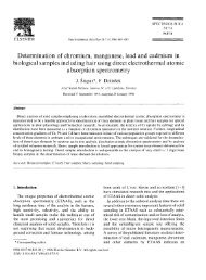
3344 NOTES J. CLIN. MICROBIOL. FIG. 2. PAS-stained section of a portion of the tissue biopsy sample (stratum corneum) showing hyphal elements. Bar, 10 m. Sensitabs; Rosco Diagnostica, Denmark) and Shadomy agar (2). The strain was sensitive to amphotericin B and was resistant to 5-fluorocytosine, fluconazole, itraconazole, and ketoconazole. The lesions regressed after 6 months of topical treatment with both the 10% solution of iodine in alcohol and ketoconazole. Sand samples from the tank used to maintain the turtle were obtained for microbial culture with the aim of selectively isolating <strong>Fusarium</strong> spp. living in the habitat of the turtle. For this purpose, the sand samples were inoculated on malt extract agar supplemented with chloramphenicol and malachite green agar 2.5 supplemented with chloramphenicol, a new selective culture medium recently designed in our laboratory for <strong>Fusarium</strong> spp. (3). All of the inoculated plates for both culture media yielded growth of fungal colonies belonging to <strong>Fusarium</strong> sp., although in malt extract agar, colonies belonging to other genera, such as Aspergillus sp. and Penicillium sp., were isolated. Culture of all of the <strong>Fusarium</strong> isolates on potato dextrose agar and synthetic nutrient-poor agar for identification generated characteristic colonies with conidial structures belonging to F. solani. The origin of this opportunistic infection may be related to the presence of F. solani in the tank and to the immunosuppressive state of the turtle due to the traumatic lesions suffered, the surgical treatment applied, and other stress conditions associated with transportation or rehabilitation of these marine animals, which may alter their immunocompetence, as happens in marine mammals (5). F. solani, like other <strong>Fusarium</strong> spp., is considered to be cosmopolitan in distribution (14). However, in the mycological control sample of the sand of the tank used to maintain the turtle, F. solani was the only <strong>Fusarium</strong> species isolated. This species has been also found in beach sands (17), and it has been isolated from marine life as diverse as lobsters and shrimp (20), sharks (19), and gray seals (13). In human infection, a case of an invasive infection produced <strong>by</strong> F. solani associated with an injury <strong>by</strong> a stingray barb has been described (9). FIG. 3. Characteristic conidiogenous cell and microconidia in false heads of F. solani. Bar, 8 m. Downloaded from jcm.asm.<strong>org</strong> <strong>by</strong> on January 1, 2010
VOL. 35, 1997 NOTES 3345 was related to the tank or pool facilities: the water filter in one case (17) and the sand of the tank in our case. It would be advisable to microbiologically control and periodically clean up the water, filters, and/or sand of the tanks or basins used for maintaining these animals, especially when the animals are at a high risk of infection. FIG. 4. Characteristic macroconidia of F. solani. Bar, 8 m. P. fluorescens and other bacteria have been isolated from skin lesions due to biting (traumatic ulcerative dermatitis) in farmed marine turtles. No fungal species was isolated from these lesions (7). Bacterial shell ulceration due to Pseudomonas sp. has been reported for a tortoise (10). It is not easy to judge the efficiency of the topical ketoconazole treatment in the regression of the lesions of the animal studied in our report. There are several factors that are related to the nature of the animal studied. In effect, the special characteristics of the turtle’s skin together with aquatic environmental conditions made the skin healing in these animals a slow process. On the other hand, hyalohyphomycosis infections due to <strong>Fusarium</strong> spp. are frequently refractory to antifungal therapy, particularly in granulocytopenic patients and in animal models. Experimental antifungal therapy with amphotericin B, fluconazole, and itraconazole reveals <strong>Fusarium</strong> spp. to be refractory to these compounds (12). <strong>Fusarium</strong> infections in other aquatic animals, such as California sea lions and gray seals, appeared to be refractory to topical as well as systemic antifungal treatment. In some cases, the regression of the lesions seemed to be seasonal and probably is not related to the therapy, being a self-remitting process (13). Although in general the in vitro antifungal susceptibilities of the different pathogenic fungi can be a valuable guide for the practitioner, reliable antifungal susceptibility testing is still poorly developed, especially for filamentous fungi (16). Recently, some testing conditions have been proposed as guidelines for a reference broth microdilution method (4). Nevertheless, in vitro resistance to different antifungal agents, such as 5-fluorocytosine, ketoconazole, fluconazole, and itraconazole, <strong>by</strong> <strong>Fusarium</strong> spp. determined <strong>by</strong> different methods has been repeatedly mentioned (4, 16, 18). Finally, in the two cited cases of cutaneous hyalohyphomycosis caused <strong>by</strong> F. solani in sea turtles, the source of infection REFERENCES 1. Borst, G. H. A., C. Vrolge, F. G. Paelma, P. Zwart, V. J. Strik, and L. C. Peters. 1972. Pathological findings on animals in the Royal Zoological Gardens of the Rotterdam Zoo during the years 1963, 1964 and 1965. Acta Zool. Pathol. Antverp. 56:3–19. 2. Casals, J. B. 1979. Tablet sensitivity testing of pathogenic fungi. J. Clin. Pathol. 32:719–722. 3. Castellá, G., M. R. Bragulat, M. V. Rubiales, and F. J. Cabañes. Malachite green agar, a new selective medium for <strong>Fusarium</strong> spp. Mycopathologia, in press. 4. Espinel-Ingroff, A., M. Bartlett, R. Bowden, N. X. Xin, C. Cooper, Jr., A. Fothergill, M. R. McGinnis, P. Menezes, S. A. Messer, P. W. Nelson, F. C. Odds, L. Pasarell, J. Peter, M. A. Pfaller, J. H. Rex, M. G. Rinaldi, G. S. Shankland, T. J. Walsh, and I. Weitzman. 1997. Multicenter evaluation of proposed standardized procedure for antifungal susceptibility testing of filamentous fungi. J. Clin. Microbiol. 35:139–143. 5. Frasca, S., J. L. Dunn, J. C. Cooke, and J. D. Buck. 1996. Mycotic dermatitis in an Atlantic white-sided dolphin, a pygmy sperm whale, and two harbor seals. J. Am. Vet. Med. Assoc. 208:727–729. 6. Frelier, P. F., L. Sigler, and P. E. Nelson. 1985. Mycotic pneumonia caused <strong>by</strong> <strong>Fusarium</strong> moniliforme. J. Med. Vet. Mycol. 23:399–402. 7. Glazebrook, J. S., and R. S. F. Campbell. 1990. A survey of the diseases of marine turtles in northern Australia. I. Farmed turtles. Dis. Aquat. Org. 9:83–95. 8. Glazebrook, J. S., R. S. F. Campbell, and A. T. Thomas. 1993. Studies on an ulcerative stomatitis-obstructive rhinitis-pneumonia disease complex in hatchling and juvenile sea turtles Chelonia mydas and Caretta caretta. Dis. Aquat. Org. 16:133–147. 9. Hiemenz, J. W., B. Kennedy, and K. J. Kwon-Chung. 1990. Invasive fusariosis associated with an injury <strong>by</strong> a stingray barb. J. Med. Vet. Mycol. 28:209–213. 10. Holt, P. E., J. E. Cooper, and J. R. Needham. 1979. Diseases of tortoises: a review of seventy cases. J. Small Anim. Pract. 20:269–286. 11. Jacobson, E. R. 1980. Mycotic diseases of reptiles, p. 283–289. In R. J. Montali and G. Migaki (ed.), The comparative pathology of zoo animals. Smithsonian Institution Press, Washington, D.C. 12. Matsumoto, T., L. Ajello, T. Matsuda, P. J. Szaniszlo, and T. J. Walsh. 1994. Developments in hyalohyphomycosis and phaeohyphomycosis. J. Med. Vet. Mycol. 32(Suppl. 1):329–349. 13. Montali, R. J., M. Bush, J. D. Strandberg, D. L. Janssen, D. J. Boness, and J. C. Whitla. 1981. Cyclic dermatitis associated with <strong>Fusarium</strong> sp. infection in pinnipeds. J. Am. Vet. Med. Assoc. 179:1198–1202. 14. Nelson, P. E., T. A. Toussoun, and W. F. O. Marasas. 1983. <strong>Fusarium</strong> species: an illustrated manual for identification. The Pennsylvania State University Press, University Park. 15. Nirenberg, H. I. 1981. A simplified method for identifying <strong>Fusarium</strong> spp. occurring on wheat. Can. J. Bot. 59:1599–1609. 16. Pujol, I., J. Guarro, C. Llop, L. Soler, and J. Fernández-Ballart. 1996. Comparison study of broth macrodilution and microdilution antifungal susceptibility tests for the filamentous fungi. Antimicrob. Agents Chemother. 40:2106–2110. 17. Rebell, G. 1981. <strong>Fusarium</strong> infections in human and veterinary medicine, p. 210–220. In P. E. Nelson, T. A. Tousson, and R. J. Cook (ed.), <strong>Fusarium</strong>: diseases, biology and taxonomy. The Pennsylvania State University Press, University Park. 18. Sekhon, A. S., A. A. Padhye, A. K. Garg, H. Ahmad, and M. Moledina. 1994. In vitro sensitivity of medically significant <strong>Fusarium</strong> species to various antimycotics. Chemotherapy 40:239–244. 19. Smith, A. G., A. G. Muhvich, K. H. Muhvich, and C. Wood. 1989. Fatal <strong>Fusarium</strong> solani infections in ba<strong>by</strong> sharks. J. Med. Vet. Mycol. 27:83–91. 20. Smith, J. M. B. 1989. Opportunistic mycoses of man and other animals, p. 196–197. CAB International Mycological Institute, Kew, United Kingdom. Downloaded from jcm.asm.<strong>org</strong> <strong>by</strong> on January 1, 2010