Cutaneous Hyalohyphomycosis Caused by Fusarium ... - Seaturtle.org
Cutaneous Hyalohyphomycosis Caused by Fusarium ... - Seaturtle.org
Cutaneous Hyalohyphomycosis Caused by Fusarium ... - Seaturtle.org

Create successful ePaper yourself
Turn your PDF publications into a flip-book with our unique Google optimized e-Paper software.
JOURNAL OF CLINICAL MICROBIOLOGY, Dec. 1997, p. 3343–3345 Vol. 35, No. 12<br />
0095-1137/97/$04.000<br />
Copyright © 1997, American Society for Microbiology<br />
<strong>Cutaneous</strong> <strong>Hyalohyphomycosis</strong> <strong>Caused</strong> <strong>by</strong> <strong>Fusarium</strong> solani in a<br />
Loggerhead Sea Turtle (Caretta caretta L.)<br />
F. J. CABAÑES, 1 * J. M. ALONSO, 2 G. CASTELLÁ, 1 F. ALEGRE, 2 M. DOMINGO, 1<br />
AND S. PONT 2<br />
Departament de Patologia i de Producció Animals, Facultat de Veterinària, Universitat Autònoma<br />
de Barcelona, Bellaterra, 1 and Centre de Recuperació d’Animals Marins (CRAM),<br />
Premiá de Mar, 2 Barcelona, Spain<br />
Received 2 July 1997/Returned for modification 3 September 1997/Accepted 23 September 1997<br />
<strong>Fusarium</strong> solani was reported as the agent of a cutaneous infection in an injured sea turtle collected in the<br />
Mediterranean Sea. The turtle was treated with both a topical 10% solution of iodine in alcohol and ketoconazole.<br />
The source of the causal agent was traced to the sand in the tank in which the turtle was maintained.<br />
The strain was only sensitive in vitro to amphotericin B and was resistant to 5-fluorocytosine, fluconazole,<br />
itraconazole, and ketoconazole.<br />
<strong>Fusarium</strong> species are common soil saprophytes and plant<br />
pathogens. However, in recent years, they have been reported<br />
with increasing frequency as causes of opportunistic infections<br />
in humans and in animals (20), including reptiles (1, 6, 11),<br />
turtles (8, 17), and other marine animals (5, 13, 19). Among all<br />
of the references reviewed, only Rebell (17) mentioned infection<br />
of the shells and skin of ba<strong>by</strong> marine turtles from Bimini<br />
(Bahamas) produced <strong>by</strong> <strong>Fusarium</strong> solani (Mart.) Sacc. No<br />
other fungal species have been mentioned as a cause of a<br />
cutaneous mycosis in turtles. In this paper, a cutaneous hyalohyphomycosis<br />
caused <strong>by</strong> F. solani in a subadult loggerhead sea<br />
turtle (Caretta caretta L.) collected in the Mediterranean Sea is<br />
reported.<br />
In September 1996, a subadult (26.3-kg) loggerhead sea turtle<br />
(Caretta caretta L.) was found floating off the coast of<br />
Barcelona, Spain (Mediterranean Sea). The turtle had a fishing<br />
hook anchored in the proximal esophagus and a traumatic<br />
injury with important loss of shell tissues. The hook was removed<br />
<strong>by</strong> surgical procedures, and debridement of the necrotic<br />
shell tissues was carried out. Treatment consisted of supportive<br />
therapy and control of secondary infections (amoxicillin, 22<br />
mg/kg of body weight, intramuscularly, once a day, for 8 days,<br />
doxycycline, 250 mg, orally once a day for 30 days). During<br />
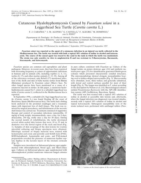
the second month of rehabilitation, the turtle developed<br />
several white-scaled skin lesions that were 10 to 35 mm in<br />
diameter over the dorsal region of the neck and head (Fig.<br />
1). Samples of skin scrapings of the skin lesions for routine<br />
microbial culturing and biopsy specimens were obtained.<br />
Histologic sections of biopsy material were stained with<br />
hematoxylin and eosin and periodic acid-Schiff stain (PAS).<br />
KOH-lactophenol-, hematoxylin and eosin-, and PAS-stained<br />
preparations revealed the presence of numerous hyaline septate<br />
hyphae in the keratin layers of the stratum corneum<br />
(Fig. 2).<br />
Samples were inoculated on Sabouraud glucose agar supplemented<br />
with chloramphenicol, blood agar, and MacConkey<br />
agar. Cultures on Sabouraud glucose agar supplemented with<br />
chloramphenicol yielded numerous vinaceous fungal colonies<br />
in pure culture consistent with <strong>Fusarium</strong> sp. Culture of the<br />
fungal isolate on potato dextrose agar (14) and synthetic nutrient-poor<br />
agar (15) for identification generated bluish-green<br />
colonies which presented characteristic conidial structures.<br />
The micromorphology showed elongate monophialides bearing<br />
oval to kidney-shaped microconidia (Fig. 3). Macroconidia<br />
were abundant, stout, thick walled, and generally cylindrical,<br />
with dorsal and ventral surfaces parallel for most of their<br />
length (Fig. 4). The fungus was identified as F. solani according<br />
to the description <strong>by</strong> Nelson et al. (14). Bacteriological cultures<br />
yielded Pseudomonas fluorescens with the API 20E identification<br />
system (API, bioMérieux, Barcelona, Spain).<br />
The turtle was first treated with a topical 10% solution of<br />
iodine in alcohol at accessible skin lesions and, afterwards,<br />
when the fungal infection was diagnosed, was treated simultaneously<br />
with a topical 10% solution of iodine in alcohol and<br />
topical ketoconazole. Subsequent susceptibility tests of the<br />
strain isolated were performed with antifungal tablets (Neo-<br />
Downloaded from jcm.asm.<strong>org</strong> <strong>by</strong> on January 1, 2010<br />
* Corresponding author. Mailing address: Departament de Patologia<br />
i de Producció Animals, Facultat de Veterinària, Universitat Autònoma<br />
de Barcelona, E-08193 Bellaterra, Barcelona, Spain. Phone:<br />
34 3 5811749. Fax: 34 3 5812006. E-mail: F.J.CABANES@CC.UAB<br />
.ES.<br />
FIG. 1. Lesions on the dorsal regions of the neck and head.<br />
3343
3344 NOTES J. CLIN. MICROBIOL.<br />
FIG. 2. PAS-stained section of a portion of the tissue biopsy sample (stratum corneum) showing hyphal elements. Bar, 10 m.<br />
Sensitabs; Rosco Diagnostica, Denmark) and Shadomy agar<br />
(2). The strain was sensitive to amphotericin B and was resistant<br />
to 5-fluorocytosine, fluconazole, itraconazole, and ketoconazole.<br />
The lesions regressed after 6 months of topical treatment<br />
with both the 10% solution of iodine in alcohol and<br />
ketoconazole.<br />
Sand samples from the tank used to maintain the turtle were<br />
obtained for microbial culture with the aim of selectively isolating<br />
<strong>Fusarium</strong> spp. living in the habitat of the turtle. For this<br />
purpose, the sand samples were inoculated on malt extract<br />
agar supplemented with chloramphenicol and malachite green<br />
agar 2.5 supplemented with chloramphenicol, a new selective<br />
culture medium recently designed in our laboratory for <strong>Fusarium</strong><br />
spp. (3). All of the inoculated plates for both culture<br />
media yielded growth of fungal colonies belonging to <strong>Fusarium</strong><br />
sp., although in malt extract agar, colonies belonging to other<br />
genera, such as Aspergillus sp. and Penicillium sp., were isolated.<br />
Culture of all of the <strong>Fusarium</strong> isolates on potato dextrose<br />
agar and synthetic nutrient-poor agar for identification generated<br />
characteristic colonies with conidial structures belonging<br />
to F. solani.<br />
The origin of this opportunistic infection may be related to<br />
the presence of F. solani in the tank and to the immunosuppressive<br />
state of the turtle due to the traumatic lesions suffered,<br />
the surgical treatment applied, and other stress conditions<br />
associated with transportation or rehabilitation of these<br />
marine animals, which may alter their immunocompetence, as<br />
happens in marine mammals (5).<br />
F. solani, like other <strong>Fusarium</strong> spp., is considered to be<br />
cosmopolitan in distribution (14). However, in the mycological<br />
control sample of the sand of the tank used to maintain<br />
the turtle, F. solani was the only <strong>Fusarium</strong> species isolated.<br />
This species has been also found in beach sands (17), and it<br />
has been isolated from marine life as diverse as lobsters and<br />
shrimp (20), sharks (19), and gray seals (13). In human<br />
infection, a case of an invasive infection produced <strong>by</strong> F.<br />
solani associated with an injury <strong>by</strong> a stingray barb has been<br />
described (9).<br />
FIG. 3. Characteristic conidiogenous cell and microconidia in false heads of<br />
F. solani. Bar, 8 m.<br />
Downloaded from jcm.asm.<strong>org</strong> <strong>by</strong> on January 1, 2010
VOL. 35, 1997 NOTES 3345<br />
was related to the tank or pool facilities: the water filter in one<br />
case (17) and the sand of the tank in our case. It would be<br />
advisable to microbiologically control and periodically clean up<br />
the water, filters, and/or sand of the tanks or basins used for<br />
maintaining these animals, especially when the animals are at<br />
a high risk of infection.<br />
FIG. 4. Characteristic macroconidia of F. solani. Bar, 8 m.<br />
P. fluorescens and other bacteria have been isolated from<br />
skin lesions due to biting (traumatic ulcerative dermatitis) in<br />
farmed marine turtles. No fungal species was isolated from<br />
these lesions (7). Bacterial shell ulceration due to Pseudomonas<br />
sp. has been reported for a tortoise (10).<br />
It is not easy to judge the efficiency of the topical ketoconazole<br />
treatment in the regression of the lesions of the animal<br />
studied in our report. There are several factors that are related<br />
to the nature of the animal studied. In effect, the special characteristics<br />
of the turtle’s skin together with aquatic environmental<br />
conditions made the skin healing in these animals a<br />
slow process. On the other hand, hyalohyphomycosis infections<br />
due to <strong>Fusarium</strong> spp. are frequently refractory to antifungal<br />
therapy, particularly in granulocytopenic patients and in animal<br />
models. Experimental antifungal therapy with amphotericin<br />
B, fluconazole, and itraconazole reveals <strong>Fusarium</strong> spp. to<br />
be refractory to these compounds (12). <strong>Fusarium</strong> infections in<br />
other aquatic animals, such as California sea lions and gray<br />
seals, appeared to be refractory to topical as well as systemic<br />
antifungal treatment. In some cases, the regression of the lesions<br />
seemed to be seasonal and probably is not related to the<br />
therapy, being a self-remitting process (13).<br />
Although in general the in vitro antifungal susceptibilities of<br />
the different pathogenic fungi can be a valuable guide for the<br />
practitioner, reliable antifungal susceptibility testing is still<br />
poorly developed, especially for filamentous fungi (16). Recently,<br />
some testing conditions have been proposed as guidelines<br />
for a reference broth microdilution method (4). Nevertheless,<br />
in vitro resistance to different antifungal agents, such<br />
as 5-fluorocytosine, ketoconazole, fluconazole, and itraconazole,<br />
<strong>by</strong> <strong>Fusarium</strong> spp. determined <strong>by</strong> different methods has<br />
been repeatedly mentioned (4, 16, 18).<br />
Finally, in the two cited cases of cutaneous hyalohyphomycosis<br />
caused <strong>by</strong> F. solani in sea turtles, the source of infection<br />
REFERENCES<br />
1. Borst, G. H. A., C. Vrolge, F. G. Paelma, P. Zwart, V. J. Strik, and L. C.<br />
Peters. 1972. Pathological findings on animals in the Royal Zoological Gardens<br />
of the Rotterdam Zoo during the years 1963, 1964 and 1965. Acta Zool.<br />
Pathol. Antverp. 56:3–19.<br />
2. Casals, J. B. 1979. Tablet sensitivity testing of pathogenic fungi. J. Clin.<br />
Pathol. 32:719–722.<br />
3. Castellá, G., M. R. Bragulat, M. V. Rubiales, and F. J. Cabañes. Malachite<br />
green agar, a new selective medium for <strong>Fusarium</strong> spp. Mycopathologia, in<br />
press.<br />
4. Espinel-Ingroff, A., M. Bartlett, R. Bowden, N. X. Xin, C. Cooper, Jr., A.<br />
Fothergill, M. R. McGinnis, P. Menezes, S. A. Messer, P. W. Nelson, F. C.<br />
Odds, L. Pasarell, J. Peter, M. A. Pfaller, J. H. Rex, M. G. Rinaldi, G. S.<br />
Shankland, T. J. Walsh, and I. Weitzman. 1997. Multicenter evaluation of<br />
proposed standardized procedure for antifungal susceptibility testing of filamentous<br />
fungi. J. Clin. Microbiol. 35:139–143.<br />
5. Frasca, S., J. L. Dunn, J. C. Cooke, and J. D. Buck. 1996. Mycotic dermatitis<br />
in an Atlantic white-sided dolphin, a pygmy sperm whale, and two harbor<br />
seals. J. Am. Vet. Med. Assoc. 208:727–729.<br />
6. Frelier, P. F., L. Sigler, and P. E. Nelson. 1985. Mycotic pneumonia caused<br />
<strong>by</strong> <strong>Fusarium</strong> moniliforme. J. Med. Vet. Mycol. 23:399–402.<br />
7. Glazebrook, J. S., and R. S. F. Campbell. 1990. A survey of the diseases of<br />
marine turtles in northern Australia. I. Farmed turtles. Dis. Aquat. Org.<br />
9:83–95.<br />
8. Glazebrook, J. S., R. S. F. Campbell, and A. T. Thomas. 1993. Studies on an<br />
ulcerative stomatitis-obstructive rhinitis-pneumonia disease complex in<br />
hatchling and juvenile sea turtles Chelonia mydas and Caretta caretta. Dis.<br />
Aquat. Org. 16:133–147.<br />
9. Hiemenz, J. W., B. Kennedy, and K. J. Kwon-Chung. 1990. Invasive fusariosis<br />
associated with an injury <strong>by</strong> a stingray barb. J. Med. Vet. Mycol.<br />
28:209–213.<br />
10. Holt, P. E., J. E. Cooper, and J. R. Needham. 1979. Diseases of tortoises: a<br />
review of seventy cases. J. Small Anim. Pract. 20:269–286.<br />
11. Jacobson, E. R. 1980. Mycotic diseases of reptiles, p. 283–289. In R. J.<br />
Montali and G. Migaki (ed.), The comparative pathology of zoo animals.<br />
Smithsonian Institution Press, Washington, D.C.<br />
12. Matsumoto, T., L. Ajello, T. Matsuda, P. J. Szaniszlo, and T. J. Walsh. 1994.<br />
Developments in hyalohyphomycosis and phaeohyphomycosis. J. Med. Vet.<br />
Mycol. 32(Suppl. 1):329–349.<br />
13. Montali, R. J., M. Bush, J. D. Strandberg, D. L. Janssen, D. J. Boness, and<br />
J. C. Whitla. 1981. Cyclic dermatitis associated with <strong>Fusarium</strong> sp. infection in<br />
pinnipeds. J. Am. Vet. Med. Assoc. 179:1198–1202.<br />
14. Nelson, P. E., T. A. Toussoun, and W. F. O. Marasas. 1983. <strong>Fusarium</strong> species:<br />
an illustrated manual for identification. The Pennsylvania State University<br />
Press, University Park.<br />
15. Nirenberg, H. I. 1981. A simplified method for identifying <strong>Fusarium</strong> spp.<br />
occurring on wheat. Can. J. Bot. 59:1599–1609.<br />
16. Pujol, I., J. Guarro, C. Llop, L. Soler, and J. Fernández-Ballart. 1996.<br />
Comparison study of broth macrodilution and microdilution antifungal susceptibility<br />
tests for the filamentous fungi. Antimicrob. Agents Chemother.<br />
40:2106–2110.<br />
17. Rebell, G. 1981. <strong>Fusarium</strong> infections in human and veterinary medicine, p.<br />
210–220. In P. E. Nelson, T. A. Tousson, and R. J. Cook (ed.), <strong>Fusarium</strong>:<br />
diseases, biology and taxonomy. The Pennsylvania State University Press,<br />
University Park.<br />
18. Sekhon, A. S., A. A. Padhye, A. K. Garg, H. Ahmad, and M. Moledina. 1994.<br />
In vitro sensitivity of medically significant <strong>Fusarium</strong> species to various antimycotics.<br />
Chemotherapy 40:239–244.<br />
19. Smith, A. G., A. G. Muhvich, K. H. Muhvich, and C. Wood. 1989. Fatal<br />
<strong>Fusarium</strong> solani infections in ba<strong>by</strong> sharks. J. Med. Vet. Mycol. 27:83–91.<br />
20. Smith, J. M. B. 1989. Opportunistic mycoses of man and other animals, p.<br />
196–197. CAB International Mycological Institute, Kew, United Kingdom.<br />
Downloaded from jcm.asm.<strong>org</strong> <strong>by</strong> on January 1, 2010