Reproductive Biology and Embryology of the ... - Seaturtle.org
Reproductive Biology and Embryology of the ... - Seaturtle.org
Reproductive Biology and Embryology of the ... - Seaturtle.org

You also want an ePaper? Increase the reach of your titles
YUMPU automatically turns print PDFs into web optimized ePapers that Google loves.
STAGES OF EMBRYONIC OEVELOPMENT [AFTER EGG LAYING)<br />
411<br />
1ni'm"<br />
STAGE 6 7 8/9 10<br />
L:;:::(L~~0-- (\<br />
RF~<br />
bl:J~ ~<br />
1mm<br />
STAGE 12 13 14 15<br />
/<br />
~0_<br />
\ \<br />
ffrt<br />
16 17 18 19<br />
LHf\~>l~~~~~r<br />
R' rrmn<br />
\<br />
23 2mm 24 26-28<br />
STAGE 20 21 22 25<br />
<strong>and</strong> between species in association with definitive scale <strong>and</strong> scute patterns.<br />
Pigment is also present ventrally <strong>and</strong> on all limb elements.<br />
Limbs. Scales are present on <strong>the</strong> proximal <strong>and</strong> distal elements. The nail<br />
tips <strong>of</strong> <strong>the</strong> feet have a slight distal elevation (Fig. 23).<br />
Jaws. Sensory papillae are present along <strong>the</strong> lateral jaw margins <strong>and</strong><br />
scales are evident on <strong>the</strong> gular skin (Fig. 24J).<br />
Caruncle. The caruncle is located on a smooth white base (Fig. 26£).<br />
Brain. The midbrain is visible as a white bulge at <strong>the</strong> back <strong>of</strong> <strong>the</strong><br />
cranium because <strong>the</strong> overlying skin is poorly pigmented <strong>and</strong> <strong>the</strong> osseous<br />
cranial ro<strong>of</strong> is incomplete.<br />
Pericardial Sac. The pericardial sac is three-fourths withdrawn into <strong>the</strong><br />
body cavity.<br />
Palate. An extensive plexus <strong>of</strong> blood vessels <strong>and</strong> sensory papillae has<br />
formed (Fig. 25G).<br />
Species Differences. In C. johnsoni <strong>and</strong> C. porosus <strong>the</strong> scales along <strong>the</strong> jaw<br />
margins are triangular with <strong>the</strong> apex <strong>of</strong> <strong>the</strong> triangle toward <strong>the</strong> jaw margins,<br />
giving <strong>the</strong> latter a serrated appearance. The external genitalia are<br />
larger <strong>and</strong> more differentiated than in A. mississippiensis.<br />
St;ags 24 (Figs. 21, 23, <strong>and</strong> 24KJ<br />
Coloration. Pigmentation is now denser so that embryos appear blacker<br />
in color. Various patterns occur both within <strong>and</strong> between species.<br />
Limbs. The nails on <strong>the</strong> h<strong>and</strong> also have elevations at <strong>the</strong>ir tips (Fig. 23)<br />
<strong>and</strong> <strong>the</strong>se elevations are starting to form <strong>the</strong> curves at <strong>the</strong> tip <strong>of</strong> <strong>the</strong> nails.<br />
Brain. The midbrain, enclosed by bone, is overlain by pigmented skin.<br />
Pericardial Sac. The pericardial sac is fully withdrawn into <strong>the</strong> body<br />
cavity <strong>and</strong> <strong>the</strong> ventral thoracic wall is closing in <strong>the</strong> midline.<br />
Yolk. A large volume <strong>of</strong> yolk lies outside <strong>the</strong> body <strong>and</strong> <strong>the</strong> ventral<br />
umbilical area is large.<br />
Scales <strong>and</strong> Scutes. The scales <strong>and</strong> scutes are now very evident as elevations<br />
all over <strong>the</strong> embryo (Fig. 24K).<br />
l~<br />
~.<br />
~<br />
Ai<br />
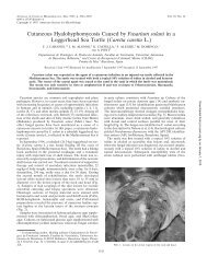
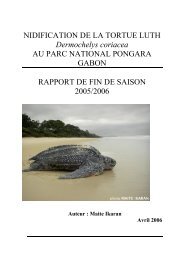
Fig. 23. The typical (diagnostic) appearances <strong>of</strong> crocodilian right fore <strong>and</strong> left hind limbs<br />
(including h<strong>and</strong>s, feet <strong>and</strong> nails) at various stages <strong>of</strong> development. Stages 6 to 11 are views<br />
from <strong>the</strong> dorsal aspect <strong>of</strong> <strong>the</strong> embryo <strong>and</strong> depict <strong>the</strong> projection <strong>of</strong> <strong>the</strong> limb anlage from <strong>the</strong><br />
flank. Stages 12 to 17 are lateral views <strong>of</strong> <strong>the</strong> sides <strong>of</strong> <strong>the</strong> embryo <strong>and</strong> depict <strong>the</strong> proximal <strong>and</strong><br />
distal elements <strong>of</strong> <strong>the</strong> limb anlage. Stages 18 to 28 are views <strong>of</strong> <strong>the</strong> h<strong>and</strong>s, feet, <strong>and</strong> nails. Up to<br />
Stage 17, no structures except <strong>the</strong> digital mesodermal condensations (at Stages 16 <strong>and</strong> 17) are<br />
visible macroscopically in <strong>the</strong> limbs, <strong>the</strong>reafter anlage for <strong>the</strong> limb <strong>and</strong> digit cartilages, for <strong>the</strong><br />
nails <strong>and</strong> for <strong>the</strong> interdigital webbing are evident, hence <strong>the</strong> change in diagram style at Stage<br />
18. Based on observation <strong>of</strong> Alligator mississippiensis, Crocodylus porosus, <strong>and</strong> C. johnsoni, although<br />
some (Stages 18 to 28) drawings are adapted from Voeltzkow's (1899) work on C.<br />
niloticus. AER, Apical ectodermal ridge; 0, mesodermal condensations for <strong>the</strong> digits; DC, digit<br />
cartilages; N, nail anlage.