Reproductive Biology and Embryology of the ... - Seaturtle.org
Reproductive Biology and Embryology of the ... - Seaturtle.org
Reproductive Biology and Embryology of the ... - Seaturtle.org

Create successful ePaper yourself
Turn your PDF publications into a flip-book with our unique Google optimized e-Paper software.
378<br />
IlA<br />
OlA<br />
If. I<br />
J I",<br />
ALB \ 7./////,<br />
REPROOUCTIVE BIOLOGY ANO EMBRYOLOGY OF CROCOOILIANS<br />
BV FZ YS vav Y EEC BV<br />
' ..<br />
'\.<br />
'111.'.'....'<br />
A<br />
ALB<br />
( / I - J I SAC<br />
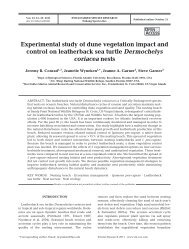
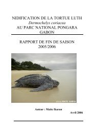
Fig. 10. Alligator mississippiensis. Diagrammatic longitudinal section through a 40-day egg.<br />
At this age <strong>the</strong> chorion <strong>and</strong> related blood vessels extend over <strong>the</strong> entire inner aspect <strong>of</strong> <strong>the</strong><br />
egg, <strong>and</strong> <strong>the</strong> eggshell is entirely opaque. The disposition <strong>of</strong> <strong>the</strong> chorionic blood vessels <strong>and</strong><br />
opaque eggshell b<strong>and</strong> illustrated are from an earlier age <strong>and</strong> for diagrammatic purposes only.<br />
(A) Amnion (solid black); AC, amniotic cavity; ALC, allantoic cavity; ALB, albumen; BV,<br />
chorio-allantoic blood vessels; C, chorion (dotted); CE, calcified eggshell (opaque b<strong>and</strong> in<br />
cross hatched area); EEC, extraembryonic coelom (potential space between yolk sac membrane<br />
<strong>and</strong> <strong>the</strong> chorion, largely occupied by <strong>the</strong> allantois); ESM, shell membrane (opaque in<br />
cross hatched area); FZ, fusion zone (fusion <strong>of</strong> chorion, outer <strong>and</strong> inner layers <strong>of</strong> allantois <strong>and</strong><br />
amnion, all <strong>of</strong> which have a firm attachment to <strong>the</strong> shell membrane); GL, gut loop (herniated<br />
out <strong>of</strong> embryo); ILA, inner layer <strong>of</strong> <strong>the</strong> allantois (fused to <strong>the</strong> amnion); OLA, outer layer <strong>of</strong> <strong>the</strong><br />
allantois (fused to <strong>the</strong> chorion); SAC, seroamniotic caVity (potential space between amniun<br />
<strong>and</strong> chorion, largely occupied by <strong>the</strong> allantois); VBV, vitelline blood vessels; Y, yulk; YS, yolk<br />
sac membrane.<br />
At <strong>the</strong> time <strong>of</strong> laying, eggs have approximately equal volumes <strong>of</strong> yolk<br />
<strong>and</strong> albumen, with <strong>the</strong> yolk being significantly denser than <strong>the</strong> albumen.<br />
Within <strong>the</strong> first day, <strong>the</strong>se ratios change as water passes across <strong>the</strong> vitelline<br />
membrane from <strong>the</strong> albumen. The vitelline fluid accounts for approximately<br />
3% <strong>of</strong> <strong>the</strong> total egg content weight at day 1, 40/0 at day 2, 7% at day<br />
3, 15% at day 6, <strong>and</strong> 27% at day 10. During this period, <strong>the</strong> volume <strong>and</strong><br />
weight <strong>of</strong> albumen decreases from 50% <strong>of</strong> <strong>the</strong> total egg content weight at<br />
day 1, to 45% at day 2, 37% at day 3, 31 % at day 6, <strong>and</strong> finally to 18% at day<br />
10. Concurrently, <strong>the</strong> volume <strong>and</strong> weight <strong>of</strong> yolk remains fairly constant at<br />
around 45 to 55%, <strong>and</strong> <strong>the</strong> total egg weight changes by only 0.5% (G.<br />
Webb, personal communication). The most aqueous vitelline fluid, being<br />
appreciably less dense than <strong>the</strong> yolk granules, remains separated from<br />
THE EBB CONTENTS AND EXTRAEMBRYONIC MEMBRANES<br />
379<br />
<strong>the</strong>m. Thus <strong>the</strong> yolk rotates, bringing <strong>the</strong> embryo up toward <strong>the</strong> top <strong>of</strong> <strong>the</strong><br />
egg regardless <strong>of</strong> its original position. The vitelline fluid remains beneath<br />
<strong>the</strong> embryo, <strong>and</strong> its assimilation is responsible for dispersing <strong>the</strong> albumen<br />
to <strong>the</strong> poles <strong>of</strong> <strong>the</strong> egg <strong>and</strong> also for increasing its viscosity. As a result, <strong>the</strong><br />
vitelline membrane adheres to <strong>the</strong> inner surfaces <strong>of</strong> <strong>the</strong> shell membrane<br />
<strong>and</strong> produces <strong>the</strong> opaque spot. The latter keeps exp<strong>and</strong>ing as more <strong>and</strong><br />
more fluid crosses <strong>the</strong> vitelline membrane (Webb, personal communication).<br />
Assimilation <strong>of</strong> vitelline fluid is an active process, which explains<br />
why infertile eggs never become opaquely b<strong>and</strong>ed. The opacity reflects<br />
dehydration <strong>of</strong> shell <strong>and</strong> shell membranes, caused by <strong>the</strong> loss <strong>of</strong> <strong>the</strong> central<br />
layer <strong>of</strong> albumen. Chemical changes occur later <strong>and</strong> include a more permanent<br />
opacity (Ferguson, 1982a). Thus, <strong>the</strong> length <strong>of</strong> <strong>the</strong> opaque b<strong>and</strong> parallels<br />
<strong>the</strong> regression <strong>of</strong> albumen early in incubation; later it also reflects <strong>the</strong><br />
expansion <strong>of</strong> <strong>the</strong> chorioallantois <strong>and</strong> <strong>the</strong> movement <strong>of</strong> minerals from <strong>the</strong><br />
shell to <strong>the</strong> embryo. Dehydrated or infected eggs (whe<strong>the</strong>r fertile or not)<br />
may become erratically b<strong>and</strong>ed or blotchy as albumen regresses <strong>and</strong> <strong>the</strong><br />
yolk sticks to <strong>the</strong> shell membrane.<br />
If an alligator egg is placed with its top surface (as laid in <strong>the</strong> nest)<br />
uppermost <strong>and</strong> <strong>the</strong> widest (blunt) end <strong>of</strong> <strong>the</strong> egg pointing toward <strong>the</strong><br />
investigator, <strong>the</strong>n <strong>the</strong> head <strong>of</strong> <strong>the</strong> embryo lies distally <strong>and</strong> its snout points<br />
to <strong>the</strong> right in 98% <strong>of</strong> cases. This predictable relationship permits eggs to be<br />
windowed with accuracy. Because <strong>of</strong> <strong>the</strong> advanced stage <strong>of</strong> development<br />
at oviposition, it is unknown whe<strong>the</strong>r <strong>the</strong> early embryonic axis <strong>of</strong> crocodilians<br />
is formed in a constant fashion relative to egg position in <strong>the</strong> oviduct<br />
as in birds (R. Bellairs, 1971).<br />
After Stage 11, <strong>the</strong> embryonic gut projects through <strong>the</strong> body wall into<br />
<strong>the</strong> umbilical stalk <strong>and</strong> contacts <strong>the</strong> yolk (Figs. 10, 20, 21, <strong>and</strong> 341) <strong>and</strong> <strong>the</strong><br />
paired vitelline blood vessels (Figs. 20, 21, <strong>and</strong> 341). Presumably, <strong>the</strong>se<br />
structures participate in <strong>the</strong> breakdown <strong>of</strong> <strong>the</strong> yolk <strong>and</strong> its transport to <strong>the</strong><br />
embryo. The yolk sac encloses <strong>the</strong> yolk; <strong>the</strong> paired vitelline vessels are<br />
continuous with <strong>the</strong> paired intestinal omphalomesenteric vessels at <strong>the</strong><br />
yolk stalk (Figs. 10, 21, <strong>and</strong> 341). It is unknown whe<strong>the</strong>r <strong>the</strong>re are yolk sac<br />
septae as in birds (Patten, 1925; Huettner, 1949), or if <strong>the</strong>re are superficial<br />
<strong>and</strong> deep layers <strong>of</strong> yolk as in o<strong>the</strong>r reptiles (R. Bellairs, 1971). Between <strong>the</strong><br />
yolk sac <strong>and</strong> <strong>the</strong> chorion lies <strong>the</strong> extra-embryonic coelom, <strong>and</strong> between <strong>the</strong><br />
amnion <strong>and</strong> chorion lies <strong>the</strong> sero-amniotic cavity (Fig. 10). Both are occupied<br />
eventually by <strong>the</strong> enlarging allantois. The alligator has two b<strong>and</strong>s <strong>of</strong><br />
firm fusion between <strong>the</strong> shell membrane, chorion, outer <strong>and</strong> inner layers <strong>of</strong><br />
allantois, <strong>and</strong> <strong>the</strong> amnion (where present) (Fig. 10). These two b<strong>and</strong>s are<br />
opposite each o<strong>the</strong>r <strong>and</strong> <strong>the</strong> lower fusion zone adheres less strongly to <strong>the</strong><br />
yolk sac (Fig. 10) than does <strong>the</strong> upper fusion zone to <strong>the</strong> amnion. The two<br />
fusion zones hold <strong>the</strong> embryo in a fairly constant position <strong>and</strong> must be cut<br />
to remove an embryo or yolk from a fixed egg (Ferguson, 1982a).<br />
Several days before hatching, <strong>the</strong> embryonic intestines <strong>and</strong> <strong>the</strong> small<br />
yolk sac are withdrawn into <strong>the</strong> body cavity through <strong>the</strong> umbilicus (Fig.