Noosia banksiae - Fungal Planet
Noosia banksiae - Fungal Planet
Noosia banksiae - Fungal Planet

You also want an ePaper? Increase the reach of your titles
YUMPU automatically turns print PDFs into web optimized ePapers that Google loves.
138 Persoonia – Volume 26, 2011<br />
<strong>Noosia</strong> <strong>banksiae</strong>
<strong>Fungal</strong> <strong>Planet</strong> description sheets<br />
139<br />
<strong>Fungal</strong> <strong>Planet</strong> 83 – 31 May 2011<br />
<strong>Noosia</strong> Crous, R.G. Shivas & McTaggart, gen. nov.<br />
Conidiophoris ad cellulas conidiogenas reductis, solitariis, lateralibus vel<br />
integratis, inconspicuis, lateralibus vel terminalibus, poris minutis, verruciformibus,<br />
0.5 µm diam, conidiis solitariis vel in catenis brevibus efferentibus.<br />
Conidiis dimorphis; conidiis primariis aseptatis, primo globosis, subhyalinis,<br />
levibus, deinde fusoidibus-ellipsoideis, brunneis, verruculosis, solitariis vel in<br />
catenis brevibus; conidiis secundariis (phragmoconidiis) ex cellulis hypharum<br />
disarticulantium formantibus, brunnescentibus et verruculosis.<br />
Etymology. Named after the town Noosa, in Queensland (Australia),<br />
where this fungus was collected.<br />
Associated with leaf spots. Mycelium consisting of hyaline,<br />
smooth, branched, 2–5 µm diam hyphae, becoming brown and<br />
verruculose with age, frequently aggregating in hyphal strands<br />
of up to 20. Conidiophores reduced to conidiogenous cells that<br />
are solitary, lateral, or integrated, inconspicuous, lateral and<br />
terminal, with small, pimple-like pores of up to 0.5 µm diam,<br />
giving rise to conidia that can be solitary or in short chains of up<br />
to 5. Conidia dimorphic; primary conidia aseptate, initially globose,<br />
subhyaline, smooth, becoming fusoid-ellipsoidal, brown,<br />
verruculose, solitary or in short, branched chains; apex obtuse,<br />
base truncate with minute, unthickened pore; secondary conidia<br />
arising as phragmoconidia from disarticulating hyphal cells that<br />
become brown and verruculose.<br />
Type species. <strong>Noosia</strong> <strong>banksiae</strong>.<br />
MycoBank MB560172.<br />
<strong>Noosia</strong> <strong>banksiae</strong> Crous, R.G. Shivas & McTaggart, sp. nov.<br />
Conidiophoris ad cellulas conidiogenas reductis, solitariis, lateralibus vel<br />
integratis, inconspicuis, lateralibus vel terminalibus, poris minutis, apiculatoidibus,<br />
0.5 µm diam, conidiis solitariis vel in catenis brevibus (ad 5) efferentibus.<br />
Conidiis dimorphis; conidiis primariis aseptatis, primo globosis,<br />
subhyalinis, levibus, deinde fusoidibus-ellipsoideis, brunneis, verruculosis,<br />
solitariis vel in catenis brevibus, ramosis, (4–)7–10(–13) × (3.5–)4(–5) µm;<br />
conidiis secundariis (phragmoconidiis) ex cellulis hypharum disarticulantium<br />
formantibus, brunnescentibus et verruculosis, 5–15 × 4–5 µm.<br />
Etymology. Named after the host genus from which it was collected,<br />
Banksia.<br />
Immersed mycelium on potato-dextrose agar consisting of<br />
hyaline, smooth, up to 5 µm diam hyphae; aerial mycelium consisting<br />
of hyphae that are smooth, branched, septate, hyaline,<br />
2–3 µm diam; hyphae become brown and verruculose with age,<br />
frequently aggregating in hyphal strands of up to 20. Conidiophores<br />
reduced to conidiogenous cells that are solitary, lateral,<br />
or integrated, inconspicuous, lateral and terminal, with small,<br />
pimple-like pores of up to 0.5 µm diam, giving rise to conidia that<br />
can be solitary or in short chains of up to 5. Conidia dimorphic;<br />
primary conidia aseptate, initially globose, subhyaline, smooth,<br />
becoming fusoid-ellipsoidal, brown, verruculose, solitary or in<br />
short, branched chains, (4–)7–10(–13) × (3.5–)4(–5) µm; apex<br />
obtuse, base truncate with minute, unthickened pore; secondary<br />
conidia arising as phragmoconidia from disarticulating hyphal<br />
cells that become brown, verruculose, 5–15 × 4–5 µm;<br />
secondary conidia in short chains when young, but forming<br />
directly on conidiogenous cells that can be reduced to loci on<br />
hyphae when mature.<br />
Culture characteristics — (in the dark, 25 °C, after 2 wk):<br />
Colonies spreading, erumpent, with sparse to moderate aerial<br />
mycelium and lobate margins, reaching 30 mm diam; on malt<br />
extract agar smoke-grey, reverse grey olivaceous with dirty<br />
white outer margin; on oatmeal agar olivaceous grey in centre,<br />
dirty white in outer region; on potato-dextrose agar isabelline<br />
on surface and reverse.<br />
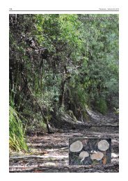
Typus. Australia, Queensland, Noosa, S 26°34'14.0" E 153°4'21.6", on<br />
leaves of Banksia aemula, 13 July 2009, P.W. Crous, R.G. Shivas & A.R.<br />
McTaggart, holotype CBS H-20587, culture ex-type CPC 17282 = CBS<br />
129526, ITS sequence GenBank JF951147 and LSU sequence GenBank<br />
JF951167, MycoBank MB560173.<br />
Notes — Based on a megablast search of NCBI’s GenBank<br />
nucleotide database, the closest hits using the ITS sequence<br />
are species of Periconia, albeit with poor coverage across the<br />
sequence length. A similar search using the LSU sequence<br />
gives as closest hits Sporidesmium tengii (DQ408559; Identities<br />
= 847/856 (99 %), Gaps = 1/856 (0 %)), Massarina igniaria<br />
(DQ810223; Identities = 825/845 (98 %), Gaps = 2/845<br />
(0 %)), Byssothecium circinans (AY016357; Identities = 863/895<br />
(96 %), Gaps = 12/895 (1 %)) and Corynespora smithii (GU323201;<br />
Identities = 856/890 (96 %), Gaps = 5/890 (1 %)). <strong>Noosia</strong> has<br />
some resemblance to the genera Conioscypha (it forms similar<br />
strange phialides in vitro, but never in vivo), Trichobotrys (but<br />
setae lacking, and supporting cells lacking at maturity), and<br />
Periconiella s.l., which is also a generic complex (Seifert et al.<br />
2011). Based on the fact that the present fungus is distinct from<br />
those presently known, and that the DNA sequence could not<br />
be matched with any currently deposited in GenBank, a new<br />
genus is herewith introduced to accommodate it.<br />
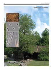
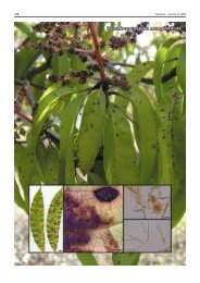
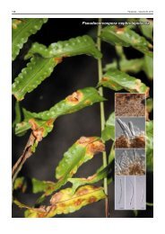
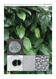
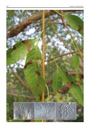
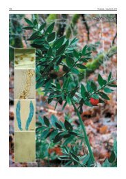
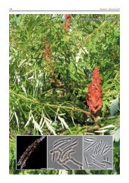
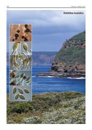
Colour illustrations. Leaf spots on Banksia aemula in Noosa National<br />
Park; hyphae giving rise to short chains of conidia, or breaking up into phragmospores.<br />
Scale bars = 10 µm.<br />
Pedro W. Crous & Johannes Z. Groenewald, CBS-KNAW <strong>Fungal</strong> Biodiversity Centre, P.O. Box 85167, 3508 AD Utrecht, The Netherlands;<br />
e-mail: p.crous@cbs.knaw.nl & e.groenewald@cbs.knaw.nl<br />
Roger G. Shivas & Alistair R. McTaggart, Agri-Science Queensland, Ecosciences Precinct, Dutton Park 4102, Queensland, Australia;<br />
e-mail: roger.shivas@deedi.qld.gov.au; Alistair.McTaggart@gmail.com<br />
© 2011 Nationaal Herbarium Nederland & Centraalbureau voor Schimmelcultures