Microcyclospora rhoicola - Fungal Planet
Microcyclospora rhoicola - Fungal Planet
Microcyclospora rhoicola - Fungal Planet

Create successful ePaper yourself
Turn your PDF publications into a flip-book with our unique Google optimized e-Paper software.
188 Persoonia – Volume 29, 2012<br />
<strong>Microcyclospora</strong> <strong>rhoicola</strong>
<strong>Fungal</strong> <strong>Planet</strong> description sheets<br />
189<br />
<strong>Fungal</strong> <strong>Planet</strong> 148 – 20 December 2012<br />
<strong>Microcyclospora</strong> <strong>rhoicola</strong> Tanney, sp. nov.<br />
Etymology. Named after the host from which it was collected, Rhus<br />
typhina.<br />
Colonies on Rhus typhina forming dark, sometimes slimy, crust<br />
on petiole and twig surfaces, forming a stroma-like sheath on<br />
trichomes, textura prismatica in surface view. Mycelium consisting<br />
of pale brown, branched, thick-walled (c. 1 μm), septate<br />
hyphae, (2–)4–6.5(–7.5) μm diam, smooth. Micromorphology<br />
identical to that in culture, described below.<br />
Colonies on malt extract agar (MEA). Mycelium consisting<br />
of pale brown, branched, septate hyphae, 1.5–3.5 μm<br />
diam, smooth. In older cultures, hyphae becoming darker,<br />
thick-walled (c. 1 μm), ossiform, and fragmenting to form a<br />
yeast-like colony. Conidiophores reduced to conidiogenous<br />
cells. Conidiogenous cells integrated, lateral on hyphae, solitary,<br />
subdenticulate, 3–5 μm tall, 2–3 μm wide, pale brown,<br />
smooth. Conidia (0–)1–3(–6)-septate, 3-septate conidia most<br />
frequent, 5–6-septate conidia rarely observed, aseptate conidia<br />
(8–)9–17.5(–29.5) × (2–)2.5–3 μm, 1-septate conidia<br />
(10.5–)11.5–22.5(–36) × (2–)2.5–3(–3.5) μm, 2-septate conidia<br />
(17–)19–28(–32) × (2.5–)3–3.5(–4), 3-septate conidia<br />
(19–)26–35.5(–40) × (2–)2.5–3(–3.5) μm, 4-septate conidia<br />
(36–)37.5–44(–47.5) × (2.5–)2.5–3(–3.5) μm, 5-septate<br />
conidia (47–)48.5–56(–57) × 2.5–3 μm, 6-septate conidia<br />
48.5 × 3 μm, hyaline, smooth, cylindrical, straight to variously<br />
curved, apex obtuse, base truncate, older conidia somewhat<br />
constricted at septa, guttulate, aggregated in mucoid masses;<br />
hila neither thickened nor darkened; anastomosis among conidia<br />
sometimes observed; microcyclic conidiation commonly<br />
observed.<br />
Culture characteristics — (in the dark, 25 °C after 2 wk on<br />
MEA): Colonies convex, with moderate to woolly aerial mycelium;<br />
surface irregular, slimy, dark grey to olive (1F1–1F3)<br />
(Kornerup & Wanscher 1978), aerial mycelium greyish offwhite<br />
to pastel grey (1B2–1B3), margin diffuse; reverse dark<br />
grey (1F1); diam up to 4 mm. In older colonies (> 6 wk), aerial<br />
mycelium becoming yellowish brown to tobacco brown (5E8–<br />
5F6), collapsing, centre carbonaceous, slimy and yeast-like,<br />
margin lobate.<br />
Typus. Canada, Ontario, Ottawa, Dominion Arboretum, on twigs of Rhus<br />
typhina var. laciniata (Anacardiaceae), 20 Oct. 2011, J.B. Tanney, holotype<br />
DAOM 242272, dried culture ex-type DAOM 242276, ITS sequence Gen-<br />
Bank KC012605, LSU sequence GenBank KC012606, TEF1 sequence<br />
GenBank KC012604, MycoBank MB801439.<br />
Notes — <strong>Microcyclospora</strong> was first described in 2010, with<br />
three species causing sooty blotch on Malus domestica fruit<br />
(Frank et al. 2010). The genus is characterised by 1–multiseptate,<br />
smooth, pale brown, scolecosporous to cylindrical<br />
conidia borne from reduced and integrated mono- to polyblastic<br />
conidiogenous cells. Conidia occur in mucoid masses and<br />
microcyclic conidiation is common (Frank et al. 2010). Morphologically,<br />
M. <strong>rhoicola</strong> conforms with the generic concept of<br />
<strong>Microcyclospora</strong> and can be differentiated from other species<br />
by its shorter conidia with fewer septa (Table 1). The discovery<br />
of M. <strong>rhoicola</strong> represents the first record of <strong>Microcyclospora</strong> in<br />
North America and on its host, Rhus typhina.<br />
The phylogenetic analysis below is based on internal transcribed<br />
spacer (ITS) sequences derived from two M. <strong>rhoicola</strong><br />
isolates (specimens collected c. 250 km apart) and previously<br />
published data (Frank et al. 2010, Crous et al. In press). <strong>Microcyclospora</strong><br />
<strong>rhoicola</strong> has distinct ITS sequences from those<br />
sequenced to date and appears to be rather distant from the<br />
currently described species.<br />
Several cercosporoid fungi are described from Rhus spp.<br />
in North America (Farr et al. 1989), including Cercosporella<br />
toxicodendri and Pseudocercospora rhoina. Both species<br />
occur as leaf spots and have more complex conidiophores<br />
compared to the reduced and integrated conidiogenous cells<br />
characterising M. <strong>rhoicola</strong>.<br />
Fig. 1 Consensus phylogram (50 % majority rule) of 15 002 trees resulting<br />
from a Bayesian inference analysis of an ITS sequence alignment using<br />
MrBayes v. 3.1.2. Posterior probabilities indicated with colour-coded<br />
branches (see legend).<br />
Table 1 Comparison of hosts, distribution and micromorphology of currently described <strong>Microcyclospora</strong> species.<br />
Species Host Origin Morphology Reference<br />
Conidial dimensions (µm)<br />
Conidial septation<br />
M. malicola Malus Germany, Slovenia 45–75 × 2.5 (1–)5–7(–13) Frank et al. (2010)<br />
M. pomicola Malus Germany 50–75 × 2.5–3 1–13 Frank et al. (2010)<br />
M. quercina Quercus Netherlands 30–45 × 2.5–3 (1–)3–4(–11) Crous et al. (In press)<br />
M. <strong>rhoicola</strong> Rhus Canada 26–36 × 2.5–3 (0–)1–3(–6) Present study<br />
M. rumicis Rumex Iran 37–54 × 2.5 1–10 Arzanlou & Bakhshi (2011)<br />
M. tardicrescens Malus Slovenia 35–55 × 2 1–9 Frank et al. (2010)<br />
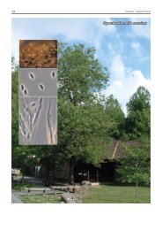
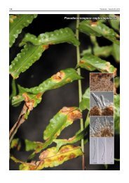
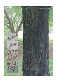
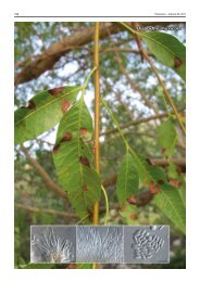
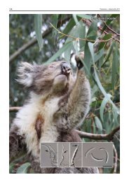
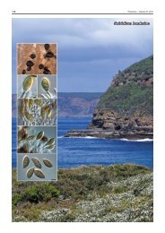
Colour illustrations. Rhus typhina var. laciniata at the Dominion Arboretum,<br />
Ottawa, Ontario, Canada (type host, photo K. Seifert); mycelium on<br />
individual trichomes (scale bar = 100 μm); conidiogenous cells and conidia<br />
exhibiting microcyclic conidiation. Scale bars = 10 μm.<br />
Joey B. Tanney, Biodiversity (Mycology & Botany), Agriculture & Agri-Food Canada, 960 Carling Ave., Ottawa, Ontario K1A 0C6, Canada;<br />
e-mail: Joey.Tanney@agr.gc.ca<br />
© 2012 Nationaal Herbarium Nederland & Centraalbureau voor Schimmelcultures