Sclerostagonospora cycadis - Fungal Planet
Sclerostagonospora cycadis - Fungal Planet
Sclerostagonospora cycadis - Fungal Planet

You also want an ePaper? Increase the reach of your titles
YUMPU automatically turns print PDFs into web optimized ePapers that Google loves.
136 Persoonia – Volume 26, 2011<br />
<strong>Sclerostagonospora</strong> <strong>cycadis</strong>
<strong>Fungal</strong> <strong>Planet</strong> description sheets<br />
137<br />
<strong>Fungal</strong> <strong>Planet</strong> 82 – 31 May 2011<br />
<strong>Sclerostagonospora</strong> <strong>cycadis</strong> Crous & G. Okada, sp. nov.<br />
<strong>Sclerostagonospora</strong>e leucadendri similis, sed conidiis minoribus, (6–)7–<br />
10(–13) × 3–4(–4.5) µm.<br />
Etymology. Named after the host from which it was collected, Cycas.<br />
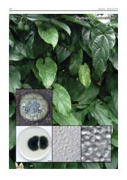
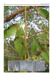
On oatmeal agar. Conidiomata pycnidial, globose, solitary,<br />
brown, 60–300 µm diam, opening mostly by means of a single,<br />
central ostiole, up to 30 µm diam, lined with hyaline, 0–1-septate<br />
periphyses, 2–2.5 µm wide; wall consisting of 2–3 layers<br />
of brown textura angularis. Conidiophores reduced to annellides.<br />
Conidiogenous cells ampulliform to subcylindrical, 3–6<br />
× 3–5 µm, hyaline, smooth, becoming brown, with 1–3 apical,<br />
percurrent proliferations. Paraphyses interspersed among<br />
conidiogenous cells, 0–3-septate, simple or branched, hyaline,<br />
10–30 × 2–2.5 µm. Conidia ellipsoid to subcylindrical (apex<br />
obtuse, base truncate), smooth, medium brown, (0–)1–3-septate,<br />
becoming constricted at septa with age, (6–)7–10(–13)<br />
× 3–4(–4.5) µm.<br />
Culture characteristics — (in the dark, 25 °C, after 1 mo):<br />
Colonies on potato-dextrose agar and oatmeal agar spreading,<br />
reaching 40–50 mm diam, with sparse aerial mycelium, smooth,<br />
with catenulate margins; surface buff to honey with patches of<br />
mouse-grey; reverse honey with patches of mouse-grey.<br />
Typus. Japan, Umihotaru Parking Area, Tokyo Bay Aqualine highway,<br />
on living leaves of Cycas revoluta, 22 Oct. 2005, P.W. Crous & G. Okada,<br />
holotype CBS H-20161, culture ex-type CPC 12388 = CBS 123538, ITS<br />
sequence GenBank FJ372393 and LSU sequence GenBank FJ372410,<br />
MycoBank MB560171.<br />
Notes — The present fungus is placed in <strong>Sclerostagonospora</strong><br />
due to the presence of pycnidia, conidiogenous cells with<br />
percurrent proliferations, and pigmented conidia. The anamorph<br />
genus <strong>Sclerostagonospora</strong> has been linked to Leptosphaeria<br />
(Crous & Palm 1999, Crous et al. 2004) and Montagnula (Huhndorf<br />
1992), and is paraphyletic.<br />
Presently nine species of <strong>Sclerostagonospora</strong> are listed in Index<br />
Fungorum, none of which occur on Zamiaceae, or resemble<br />
S. <strong>cycadis</strong> in morphology. BLASTn results of the ITS sequence<br />
revealed an identity of 99 % with <strong>Sclerostagonospora</strong> sp. (Gen-<br />
Bank accession DQ286767; Identities = 532/538 (99 %), Gaps<br />
= 3/538 (1 %)) and <strong>Sclerostagonospora</strong> opuntiae (GenBank accession<br />
DQ286768; Identities = 531/538 (99 %), Gaps = 3/538<br />
(1 %)). The LSU sequence has 99 % identity to the latter two<br />
GenBank sequences as well as sequences of Phaeosphaeria<br />
species. <strong>Sclerostagonospora</strong> <strong>cycadis</strong> is morphologically similar<br />
to Hendersonia togniniana, which was described from Cycas<br />
revoluta plants cultivated in a botanical garden in Italy. Conidia<br />
of the latter, however, are brown, oblong-ellipsoidal, 3-septate,<br />
10–12 × 6–7 µm, thus being wider than that of the present<br />
species (Saccardo 1899).<br />
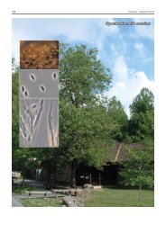
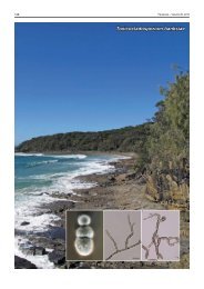
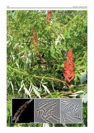
Colour illustrations. Cycas revoluta growing at Sakae-cho, Asaka, Saitama;<br />
colony on oatmeal agar; conidiogenous cells and conidia. Scale bar = 10 µm.<br />
Pedro W. Crous & Johannes Z. Groenewald, CBS-KNAW <strong>Fungal</strong> Biodiversity Centre, P.O. Box 85167, 3508 AD Utrecht, The Netherlands;<br />
e-mail: p.crous@cbs.knaw.nl & e.groenewald@cbs.knaw.nl<br />
Gen Okada, Microbe Division / Japan Collection of Microorganisms, RIKEN BioResource Center, Wako, Saitama 351-0198, Japan;<br />
e-mail: okada@jcm.riken.jp<br />
© 2011 Nationaal Herbarium Nederland & Centraalbureau voor Schimmelcultures