- Page 1 and 2:
Haematology and Blood Transfusion 2
- Page 3 and 4:
Dr. Rolf Neth, Universitäts-Kinder
- Page 5 and 6:
Participants of the Meeting Aaronso
- Page 7 and 8:
Kurth, R, Friedrich-Miescher-Labora
- Page 9 and 10:
Wilsede Scholarship Holders (Grante
- Page 11 and 12:
Preface Gut ist eine Lehrart, wo ma
- Page 13 and 14:
Personal and scientific discussion
- Page 15 and 16:
Acknowledgement We should like to t
- Page 17 and 18:
Wilsede, June 21 1978 Memorial Trib
- Page 19 and 20:
limiting benign lymphoproliferative
- Page 21 and 22:
this most unlikely, however (for re
- Page 23 and 24:
longer subject to the same control
- Page 25 and 26:
Bregula U, Wiener F, Harris H (1971
- Page 27 and 28:
fresh lymph node biopsies from ten
- Page 29 and 30:
Fig. 3. Binucleate mitosis in an ob
- Page 31 and 32:
y Fig. 4. Schematic diagram of post
- Page 33 and 34:
than that among patients with Stage
- Page 35 and 36:
Tests of binding affinity, agglutin
- Page 37 and 38:
N Engl J Med 297: 963-968 - Green I
- Page 39 and 40:
Clinical Aspects
- Page 41 and 42:
"ring chromosome". "Mar" is marker,
- Page 43 and 44:
191 chromosomally abnormal patients
- Page 45 and 46:
tumors of both African and non-Afri
- Page 47 and 48:
Haematology and Blood Transfusion V
- Page 49 and 50:
Retrospective group 26/93 evolved l
- Page 51 and 52:
Haematology and Blood Transfusion V
- Page 53 and 54:
Treatment
- Page 55 and 56:
advantage in terms of duration of f
- Page 57 and 58:
leukaemia. An analysis of 168 patie
- Page 59 and 60:
11. Treatment Program Chemotherapy
- Page 61 and 62:
efractory congestive heart failure
- Page 63 and 64:
period (Sequence II) was gene rally
- Page 65 and 66:
children with a multiple drug proto
- Page 67 and 68:
DAYS 1 5 10 15 20 25 30 35 , I I I
- Page 69 and 70:
Biologically Drug Outcome fit" sens
- Page 71 and 72:
may counterbalance the potential be
- Page 73 and 74:
Ht (0--0) 50 40 30 20 10 0 Platelet
- Page 75 and 76:
10 7 ,-----------------------------
- Page 77 and 78:
11. Haematological and Biochemical
- Page 79 and 80:
0\ 0\ INTERFERON ... 10 3 rl E "'
- Page 81 and 82:
Haematology and Blood Transfusion V
- Page 83 and 84:
("pre-B" phenotype) has been descri
- Page 85 and 86:
human bone marrow cells exhibit the
- Page 87 and 88:
10 9.,0 MBKCC .. .,4 B prot:oool 1'
- Page 89 and 90:
demann W, Büehner T, Arlin Z, Gee
- Page 91 and 92:
-...l 00 a-tLDRENS CAt«:ER S TU>Y
- Page 93 and 94:
prognostic characteristics under in
- Page 95 and 96:
significantly higher in HR} (14.5%,
- Page 97 and 98:
normal or elevated immunoglobulin l
- Page 99 and 100:
References Andreeff M, Darzynkiewic
- Page 101 and 102:
R GROUP 1 ( 1970 - 1971 ) 17 PTS. R
- Page 103 and 104:
Table 3. Influence of initial featu
- Page 105 and 106:
LIJ (f) a.. < -1 LIJ Q: u. 0 >- I-
- Page 107 and 108:
Haematology and Blood Transfusion V
- Page 109 and 110:
me nt indicated that additional the
- Page 111 and 112:
42:2123-2134 - Chessells JM, Cornbl
- Page 113 and 114:
Baum et al. 1979). This study was b
- Page 115 and 116:
1. 0 '--'1--" 0.9 e: o .- VI VI E C
- Page 117 and 118:
e: o e: 1. 0 ,.--...-- 1'"'\ ......
- Page 119 and 120:
In conclusion, we can state that th
- Page 121 and 122:
Haematology and Blood Transfnsion V
- Page 123 and 124:
INDUCTION VCR + PRED + ASNASE (wk 3
- Page 125 and 126:
SCHED.INT 1 MTX • ! Adria MTX •
- Page 127 and 128:
~ looh---------ooooc>----o---c>----
- Page 129 and 130:
Consoll- Irra .tlon t) .tlon Indu
- Page 131 and 132:
Highrisk Standard risk Table 1. Cli
- Page 133 and 134:
Cumulo/ive comp/el9 remission 1.0 .
- Page 135 and 136:
10° 8 = 6 ö 4 Non T Cell .~ :ö
- Page 137 and 138:
Haematology and Blood Transfusion V
- Page 139 and 140:
% SURVIVAL 100 ~ ~II 0 80 60 40 - -
- Page 141 and 142:
a) Percentage of donor cell mitoses
- Page 143 and 144:
Table 5. Results of bone marrow tra
- Page 145 and 146:
...... w N Table 7. Clinical result
- Page 147 and 148:
agent had to be added and that in t
- Page 149 and 150:
Haematology and Blood Transfusion V
- Page 151 and 152:
C. Chronic Myelogenous Leukemia The
- Page 153 and 154:
Patients with a suitable donor rece
- Page 155 and 156:
No. Recurrent Leukaemia Other death
- Page 157 and 158:
followed by dialysis against isoton
- Page 159 and 160:
Haematology and Blood Transfusion V
- Page 161 and 162:
HS, HL- Heme >_ 40 0 HS I ;:. =====
- Page 163 and 164:
Haematology and Blood Transfusion V
- Page 165 and 166:
knowing at present. These genes may
- Page 167 and 168:
esistance to antifolates. In Vitro
- Page 169 and 170:
a DNA probe specific for the gene t
- Page 171 and 172:
translocation between chromosomes 9
- Page 173 and 174:
Table 1. Subdivision of 1827 Myelo-
- Page 175 and 176:
NORMAL CELL ®
- Page 177 and 178:
some change of the primary type, in
- Page 179 and 180:
Haematology and Blood Transfusion V
- Page 181 and 182:
F. Transformation of Mouse Bone Mar
- Page 183 and 184:
Haematology and Blood Transfusion V
- Page 185 and 186:
were carried out with 3H-MTX in the
- Page 187 and 188:
Haematology and Blood Transfusion V
- Page 189 and 190:
the lysozyme cDNA prepared from ovi
- Page 191 and 192:
Haematology and Blood Transfusion V
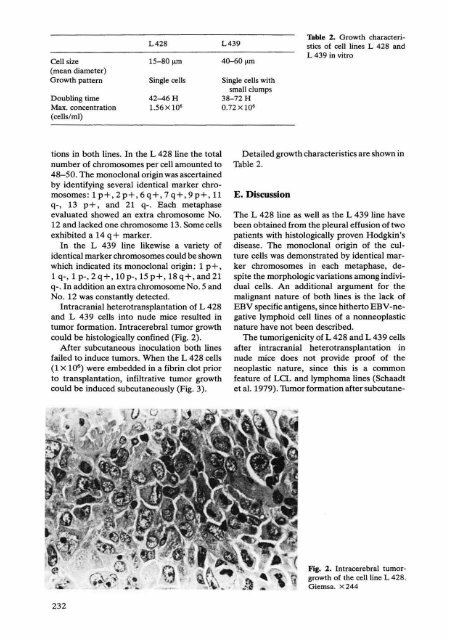
- Page 193 and 194: et al. 1976). The BJAB cellline in
- Page 195 and 196: dalton bands corresponding to light
- Page 197 and 198: 1979). The EBV-carrying cell lines
- Page 199 and 200: producer lines (DAUDI and P3HR-1),
- Page 201 and 202: G, Giovanella B, Westman A, Stehlin
- Page 203 and 204: Haematology and Blood Transfusion V
- Page 205 and 206: Dc;bt CT + i J.I9 Raji; 1. i .. 100
- Page 207 and 208: c. Discussion During infectious mon
- Page 209 and 210: Haematology and Blood Transfusion V
- Page 211 and 212: C. Nonepisomal Viral DNA We have us
- Page 213 and 214: A 260 3.0 A B 1670 70 N2 2.0 c: E .
- Page 215 and 216: vivo. Nature 260:302-306 - Kirk JTO
- Page 217 and 218: Fig. 1. a Polyacrylamide gelelectro
- Page 219 and 220: Haematology and Blood Transfusion V
- Page 221 and 222: 1979). The lymphocyte subpopulation
- Page 223 and 224: lysin 0 antigens are found, and mix
- Page 225 and 226: Pope JH (1978) Long-term T cell-med
- Page 227 and 228: Haematology and Blood Transfnsion V
- Page 229 and 230: Charaeteristie Morphologie maturati
- Page 231 and 232: after TPA-induction (Table 3). The
- Page 233 and 234: PA (1978) Reactivity of a rabbit an
- Page 235 and 236: Blood Monocyte. B lood Monocytes 1-
- Page 237 and 238: Haematology and Blood Transfusion V
- Page 239 and 240: and nonhistone proteins can serve a
- Page 241 and 242: Haematology and Blood Transfusion V
- Page 243: The findings of the evaluation of s
- Page 247 and 248: Cell biological and Immunological A
- Page 249 and 250: consideration. Sanel (1973) obtaine
- Page 251 and 252: endotoxin serum, were used. A parti
- Page 253 and 254: monocytic leukemia cell line in cul
- Page 255 and 256: B. Isolation, Characterization and
- Page 257 and 258: Haematology and Blood Transfusion V
- Page 259 and 260: don al hemopathies generally displa
- Page 261 and 262: nitors. Blood Cells 6:595-607 - Min
- Page 263 and 264: \I) ....I ....I IU Jl 200 r 100 90
- Page 265 and 266: their haemopoietic cells' ability t
- Page 267 and 268: normal normal ALL ALL ALL persons p
- Page 269 and 270: 64.5 { V)
- Page 271 and 272: References Biermann E, Neth R, Gros
- Page 273 and 274: ehambers, the growth pattern observ
- Page 275 and 276: References Anderssen LC, 10kinen M,
- Page 277 and 278: REH CELL UNE TO CALLb~Ö ~q~ V.M. c
- Page 279 and 280: Haematology and Blood Transfusion V
- Page 281 and 282: the macrophage series in which the
- Page 283 and 284: Haematology and Blood Transfusion V
- Page 285 and 286: Table 1. Production of factor depen
- Page 287 and 288: Haematology and Blood Transfusion V
- Page 289 and 290: Table 1. Characteristics of the adh
- Page 291 and 292: Fig. 3. A human marrow culture at 6
- Page 293 and 294: 300 250 • HYPERPROLIFERATIVE PATT
- Page 295 and 296:
Fig. 9. Nonadherent population from
- Page 297 and 298:
The cobblestone areas were often no
- Page 299 and 300:
L (1976) Induction of colony format
- Page 301 and 302:
B. Materials and Methods J. Tissue
- Page 303 and 304:
Fig. 2. Ultrastructural appearance
- Page 305 and 306:
Haematology and Blood Transfusion V
- Page 307 and 308:
Haematology and Blood Transfusion V
- Page 309 and 310:
Table 1. Monoclonal antibody reacti
- Page 311 and 312:
al. 1979; Janossy et al. 1979) is e
- Page 313 and 314:
to which this is an exact replica o
- Page 315 and 316:
inger JL (1976) Distribution of la-
- Page 317 and 318:
I region antigens were found to be
- Page 319 and 320:
have been reported to be present on
- Page 321 and 322:
V. FunctionalAssays Assays for PHA
- Page 323 and 324:
Functional studies of UCHT1 separat
- Page 325 and 326:
Haematology and Blood Transfusion V
- Page 327 and 328:
40 ,...... (5 L.. C o u 30 .9 ~ .~
- Page 329 and 330:
with immunohistochemieal methods (L
- Page 331 and 332:
Haematology and Blood Transfusion V
- Page 333 and 334:
can be dissected by cloning. Utiliz
- Page 335 and 336:
Table 1. Origin and marker profile
- Page 337 and 338:
Haematology and Blood Transfusion V
- Page 339 and 340:
complete identity of gp 100 from no
- Page 341 and 342:
Table 1. Reaction of single anti se
- Page 343 and 344:
Haematology and Blood Transfusion V
- Page 345 and 346:
media. Following 24-h incubation, c
- Page 347 and 348:
nonleukemic eells (Greaves 1979). L
- Page 349 and 350:
Haematology and Blood Transfusion V
- Page 351 and 352:
Fig. 2. Cytocentrifuged smears of b
- Page 353 and 354:
Modification of amino-groups of hum
- Page 355 and 356:
disturb this balance in the directi
- Page 357 and 358:
The majority of human blood lymphoc
- Page 359 and 360:
of the EBV infected B cells. Howeve
- Page 361 and 362:
and immunoglobulins. J Immunol 120:
- Page 363 and 364:
" K 10 • j ~ ALL RNlL AL wtfh les
- Page 365 and 366:
tion akuter Leukämiezellen in "spo
- Page 367 and 368:
D. Results and Discussion J. Acute
- Page 369 and 370:
selectively responsive to different
- Page 371 and 372:
Haematology and Blood Transfusion V
- Page 373 and 374:
l1J ~ 100 ~ Q. ::) l1J Z 0 ~ z >- .
- Page 375 and 376:
Table 2. Growth inhibition of S49 e
- Page 377 and 378:
Reverse Infectious ASC/2X1Q6 transc
- Page 379 and 380:
Haematology and Blood Transfusion V
- Page 381 and 382:
munized animals to 1316 tumor cells
- Page 383 and 384:
Haematology and Blood Transfnsion V
- Page 385 and 386:
Fig. 3. Expression of retroviral gp
- Page 387 and 388:
pg70. J Exp Med 143: 151-166 - McGr
- Page 389 and 390:
Table 1. Expression of tumor antige
- Page 391 and 392:
Virological and Molecularbiological
- Page 393 and 394:
Table 1. Oncogenic properties of re
- Page 395 and 396:
also Fig. 1). It followed that the
- Page 397 and 398:
whether this sequence is related to
- Page 399 and 400:
zed in infected cells argue that th
- Page 401 and 402:
a specific viral transforming prote
- Page 403 and 404:
detect viral RNA as the only charac
- Page 405 and 406:
Eisen H (1980) Myeloproliferate vir
- Page 407 and 408:
p,60- -34K - 1 2 3 4 Fig. 1. In vit
- Page 409 and 410:
.. p - • a'T ... I .t'l Fig. 4. D
- Page 411 and 412:
Haematology and Blood Transfusion V
- Page 413 and 414:
Fig. 2. Photomicrographs of NY68 in
- Page 415 and 416:
DEAE (o(lJmn 'liI GI -2 2 o e s 1
- Page 417 and 418:
vity was deterrnined. For endogenou
- Page 419 and 420:
pr 180 - - - P 12/15 - A B c D E F
- Page 421 and 422:
85,- 66- -27 - -66 40- 27- -1'9 19-
- Page 423 and 424:
Haematology and Blood Transfusion V
- Page 425 and 426:
= 0.5 o E Q. o ILI 0.4 (J) « ILI .
- Page 427 and 428:
Table 1. Oncogenic potential of avi
- Page 429 and 430:
100 III GI c: o "8 80 > w
- Page 431 and 432:
Expt. No. Infecting virus Proportio
- Page 433 and 434:
Haematology and Blood 'fransfusion
- Page 435 and 436:
sequences, provided its gag portion
- Page 437 and 438:
77:3009-3013 - Hu SSF, Moscoviei C,
- Page 439 and 440:
RI 2.2 md and Hind III 2.6 md leuke
- Page 441 and 442:
Haematology and Blood Transfusion V
- Page 443 and 444:
RELATIONSHIP OF ENDOGENOUS AND EXOG
- Page 445 and 446:
E ........ E Cl. U E :::l .... Q) (
- Page 447 and 448:
References Astrin S (1978) Endogeno
- Page 449 and 450:
the same animal were examined (e.g.
- Page 451 and 452:
I) 'IQ a \0 - CL -- Fig. 2. Restrie
- Page 453 and 454:
~ ..................... ~ Cell 3' 5
- Page 455 and 456:
a :Sa,C! + . ~ 111 i' ( " '~ .J. b
- Page 457 and 458:
Table 1 Sampie Tissue TS fragment"
- Page 459 and 460:
a b Sac I / rep ._-_.-_ K J nl rep
- Page 461 and 462:
Haematology and Blood 'fiansfusion
- Page 463 and 464:
Osteopetrosis and osteosarcomas occ
- Page 465 and 466:
Lymphoma cellline Table 1. T and B
- Page 467 and 468:
Table 2. In vivo growth and oneogen
- Page 469 and 470:
Haematology and Blood Transfusion V
- Page 471 and 472:
Table 1. Transforming aetivity of c
- Page 473 and 474:
Table 3. Transformation aetivity of
- Page 475 and 476:
- Shoemaker C, Goff S, Gilboa E, Pa
- Page 477 and 478:
indieate a shifting of target eell
- Page 479 and 480:
Table 2. Expression of Jl and Sourc
- Page 481 and 482:
Haematology and Blood Transfusion V
- Page 483 and 484:
~ i ,..1---------,,':::::::::::::::
- Page 485 and 486:
fragment of pBR322 DNA, a 3.0-kbp S
- Page 487 and 488:
Mak TW (1979) Proc Natl Acad Sci US
- Page 489 and 490:
Table 1. Transformation of 3T3 cell
- Page 491 and 492:
inserts in the genome of rapidly tr
- Page 493 and 494:
pulations, it seemed likely that th
- Page 495 and 496:
contains antigens which react with
- Page 497 and 498:
Haematology and Blood Transfusion V
- Page 499 and 500:
le of reacting in sensitive radioim
- Page 501 and 502:
Haematology and Blood Transfusion V
- Page 503 and 504:
gic approach. In: Hiatt HH, Watson
- Page 505 and 506:
12 Eco R [ Fig. 1. Hybridization pa
- Page 507 and 508:
Haematology and Blood Transfusion V
- Page 509 and 510:
10 - '1~ )( -~ S:! E A U I o 1 2
- Page 511 and 512:
Haematology and Blood 'fransfusion
- Page 513 and 514:
cultures. These cells did not conta
- Page 515 and 516:
indication of malignant T-cells in
- Page 517 and 518:
Fig. 3. Thin-section electron micro
- Page 519 and 520:
Table 3. Lack of detectable related
- Page 521 and 522:
J. HTLV Was Present in the Primary
- Page 523 and 524:
specific signals into nonspecific s
- Page 525 and 526:
Table 2. Distribution of HTLV-relat
- Page 527 and 528:
u 10 ~ 20 ~ :c 30 a 40 o Fig. 1. Ki
- Page 529 and 530:
Haematology and Blood Transfusion V
- Page 531 and 532:
SSV TrS-gp labeled effectively with
- Page 533 and 534:
Haematology and Blood Transfusion V
- Page 535 and 536:
60 HFF/SSV cDNA CELL RNAs: 0 HF/SSV
- Page 537 and 538:
Antisera a Table 1. Immunofluoresce
- Page 539 and 540:
Haematology and Blood 'ftansfusion
- Page 541 and 542:
y competition RIA yielded virtually
- Page 543 and 544:
SiS'V' gp70: ANTIGEN, ANTI BODY, IM
- Page 545 and 546:
type-C virus p30 antigen in cells f
- Page 547 and 548:
c. Results The fractionated whole-b
- Page 549 and 550:
induced antibodies as well as exper
- Page 551 and 552:
- Fig. la-f. Retravirus particles p
- Page 553 and 554:
eplication of infecting murine leuk
- Page 555 and 556:
Haematology and Blood Transfusion V
- Page 557 and 558:
immunodeficiency, multic10nal lymph
- Page 559 and 560:
Bovine leukemia --, proteins of 499
- Page 561 and 562:
Human T- cell - -, characterization
- Page 563 and 564:
RNA -, of tumor virus 439 Rous sarc
- Page 565 and 566:
Haematology andBlood Transfusion Su