Bacterial infection of mudfish Clarias gariepinus (Siluriformes ...
Bacterial infection of mudfish Clarias gariepinus (Siluriformes ...
Bacterial infection of mudfish Clarias gariepinus (Siluriformes ...

You also want an ePaper? Increase the reach of your titles
YUMPU automatically turns print PDFs into web optimized ePapers that Google loves.
<strong>Bacterial</strong> <strong>infection</strong> <strong>of</strong> <strong>mudfish</strong> <strong>Clarias</strong> <strong>gariepinus</strong><br />
(<strong>Siluriformes</strong>: Clariidae) fingerlings in tropical nursery ponds<br />
Gabriel Ikpi & Benedict Offem<br />
Department <strong>of</strong> Fisheries, Faculty <strong>of</strong> Agriculture, Cross River University <strong>of</strong> Technology, Obubra Campus, Cross River<br />
State, Nigeria; benbeff06@yahoo.com, gikpi@yahoo.com<br />
Received 29-i-2010. Corrected 01-xi-2010. Accepted 14-xii-2010.<br />
Abstract: <strong>Bacterial</strong> <strong>infection</strong> among the most common cultured <strong>mudfish</strong> <strong>Clarias</strong> <strong>gariepinus</strong> in Africa, has<br />
become a cause <strong>of</strong> concern, because it constitutes the largest economic loss in fish farms. In order to provide<br />
useful biological data <strong>of</strong> the pathogens for good management practices, samples were collected monthly between<br />
January 2008 and December 2009 in three monoculture nursery ponds, located in three different positions:<br />
upriver (A, grassland), mid-river (B, mixed forest and grassland) and downriver (C, rainforest) along 200km<br />
length <strong>of</strong> Cross River floodplains, Nigeria. A total <strong>of</strong> 720 fingerlings between 15.1 and 20.7g were analyzed<br />
to determine the degree <strong>of</strong> <strong>infection</strong>. The bacterial pathogens were taken from their external surfaces, and<br />
were isolated and identified by standard methods. The caudal fins <strong>of</strong> fingerlings from pond A had the highest<br />
bacterial load (5.8x10 3 cfu/g), while the least counts (1.2x10³cfu/g) were identified on the head <strong>of</strong> fish from<br />
pond C, with Flexibacter columnaris as the major etiological agent. Pseudomonas fluorescens, Aeromonas<br />
hydrophila, Escherichia coli, Staphylococcus aureus and Micrococcus luteus were identified as co-isolates with<br />
P. fluorescens as dominant (0.7x10 2 cfu/mL) co-isolates in pond water. Clinical signs <strong>of</strong> five white spots with red<br />
periphery appeared on the external surface <strong>of</strong> infected fish. All the fish sampled, died after 4 to 9 days. There<br />
was no significant difference in the bacterial counts between different ponds, but the difference between fish<br />
organs/parts examined was significant. Fish from these ponds are therefore potentially dangerous to consumers<br />
and highly devalued, with the economic impact to producers. Preventive methods to avoid these <strong>infection</strong>s are<br />
recommended. Rev. Biol. Trop. 59 (2): 751-759. Epub 2011 June 01.<br />
Key words: <strong>Clarias</strong> <strong>gariepinus</strong> fingerlings, Flexibacter colummnaris, bacteriogical examination, fish farm.<br />
<strong>Clarias</strong> <strong>gariepinus</strong> (Burchell, 1822) is<br />
a species <strong>of</strong> great economic importance in<br />
Africa and South-East Asia, especially as a<br />
food fish and vital in the local sustainability<br />
<strong>of</strong> the aquaculture activity (Teugels 1986,<br />
Venden Bossiche & Bernacsek 1990). Their<br />
aquaculture attributes include ability to withstand<br />
handling stress, disease resistance, high<br />
growth rate, fecundity and palatability. There<br />
is acute reduction <strong>of</strong> these species in inland<br />
natural water bodies in Nigeria because <strong>of</strong><br />
the over-exploitation methods <strong>of</strong> indigenous<br />
fishers that destroy the habitat and fisheries<br />
resources (Viser 1970). Nowadays, the effort<br />
made by the Nigerian government to conserve<br />
and propagate the aquaculture <strong>of</strong> this species<br />
is being hindered, since there is little information<br />
available to producers, on the species<br />
ecology and disease issues in Nigerian waters<br />
(Fagbenro et al. 1993, Okaeme & Obiekezie<br />
1990). Studies conducted by Ugwuzor et al.<br />
(1990), Ogbondeminu et al. (1991), Ogbondeminu<br />
(1993), Ikpi & Offem (2008), revealed<br />
incidence <strong>of</strong> Flexibacter columnaris, Pseudomonas<br />
sp., Aeromonas sp., Vibrio sp., enterobacteriaceae<br />
and Gram positive bacteria as<br />
common fish pathogens responsible for different<br />
bacterial diseases in fish farms in Nigeria.<br />
Fedoruk (1981), Plumb & Olah (1984),<br />
Hanson & Grizzle (1985), MacMillan & Tucker<br />
Rev. Biol. Trop. (Int. J. Trop. Biol. ISSN-0034-7744) Vol. 59 (2): 751-759, June 2011<br />
751
(1985), Noga (2000) revealed that Flexibacter<br />
collumnaris and many other bacterial diseases<br />
<strong>of</strong> fish, have a patho-physiological background<br />
related to environmental stressors. Examples <strong>of</strong><br />
such stressors might be: husbandry factors like<br />
crowding (Ventura & Grizzle 1987), capture<br />
and hauling, water related problems like toxicants<br />
(Tucker et al. 1984, Faisal et al. 1988),<br />
temperature and oxygen extremes (Walters &<br />
Plumb 1980, Plumb & Olah 1984), transport<br />
(Blazer 1992) and rapid environmental changes<br />
(Ciembor et al. 1995).<br />
The prevalence <strong>of</strong> infectious diseases<br />
depends on the interaction between fish<br />
pathogen and the environment (Klesius 1992,<br />
Mqolomba & Plumb 1992, Boon & Huisman<br />
1996, Noga 2000). Besides, the pathogen<br />
spreading, mostly under unpredictable circumstances,<br />
can result in a sudden onset <strong>of</strong> a disease<br />
in an obviously healthy population.<br />
This investigation was developed after<br />
the outbreak <strong>of</strong> an uncommon disease <strong>of</strong> fish<br />
fingerlings purchased from the fish farms under<br />
investigation, also had clinical signs on their<br />
external surface. The study therefore includes<br />
isolation and identification <strong>of</strong> the bacterial<br />
pathogens and determination <strong>of</strong> the degree <strong>of</strong><br />
<strong>infection</strong> <strong>of</strong> the fingerlings and to advice on<br />
management.<br />
MATERIALS AND METHODS<br />
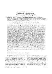
Study area: The study site is the Cross<br />
River floodplain located at the Southeastern<br />
part <strong>of</strong> Nigeria (4 º .25’ - 7 º .00’ N; 7 º .15’ - 9 º .30’<br />
E) (Fig. 1). It is bounded in the South by the<br />
Atlantic Ocean, East by the Republic <strong>of</strong> Cameroun,<br />
the Nigerian states <strong>of</strong> Benue in the North,<br />
Ebonyi and Abia in the West and Akwa Ibom<br />
in the Southwest. Climate <strong>of</strong> the study area is<br />
characterized by a dry season from November-<br />
March and a wet season from April-October.<br />
Highest precipitation (3 050±230mm) occurs<br />
in August, and lowest (300±23mm) in March.<br />
7º30’<br />
8º00’ 8º30’ 9º00’ 9º15’ E<br />
Benue State<br />
6º30’<br />
Ebonyi<br />
State<br />
River<br />
5º00’<br />
AKWA<br />
State<br />
Cross<br />
180 m<br />
B<br />
C<br />
A<br />
Fish Pond - A<br />
Fish Pond - B<br />
Fish Pond - C<br />
United Republic <strong>of</strong> Cameroon<br />
6º30’<br />
5º00’<br />
Atlantic<br />
Ocean<br />
Bakassi<br />
Scale: 1:2 500<br />
4º30’<br />
Fig. 1. Cross River State showing study sites.<br />
752 Rev. Biol. Trop. (Int. J. Trop. Biol. ISSN-0034-7744) Vol. 59 (2): 751-759, June 2011
The mean annual temperature ranged from<br />
15.5±7.6 о C in wet period to 32.6±5.4 о C in dry<br />
period. As obtained in river-floodplain system<br />
Cross River comprised a wide range <strong>of</strong> habitat<br />
types, from small upland streams, smooth<br />
glides <strong>of</strong> the middle sections, and broad, meandering<br />
river stretch, <strong>of</strong> the lowland. The principle<br />
driving force for the productivity <strong>of</strong> major<br />
biota in Cross River floodplain systems is the<br />
seasonal variation <strong>of</strong> the river flows or “flood<br />
pulse,” which produces periodic inundations <strong>of</strong><br />
the floodplain. The bulk <strong>of</strong> the productivity is<br />
derived directly or indirectly within the floodplain<br />
itself. In this study, the three ponds (A, B,<br />
C) investigated were randomly sampled, each<br />
from the floodplains upriver, mid-river and<br />
downriver. The pond sites were located 18km<br />
apart with dimensions <strong>of</strong> 30x10 x1.7m (A),<br />
15x10x1.5m (B) and 20x10x1.5m (C).<br />
Sample Collection: Samples were collected<br />
monthly (8am-10am) between January<br />
2008 and December 2009 from three monoculture<br />
nursery ponds located in three reaches; A:<br />
upriver (grassland), B: mid-river (mixed forest<br />
and grassland) and C: downriver (rainforest)<br />
along 200km length <strong>of</strong> Cross River floodplains,<br />
Nigeria. A total <strong>of</strong> 720 <strong>Clarias</strong> <strong>gariepinus</strong><br />
fingerlings <strong>of</strong> weights between 15.1 and 20.7g<br />
were sampled. Samples were held in six hapas<br />
(a net-like container used to confine fish within<br />
its natural environment for experimental purposes)<br />
placed in each <strong>of</strong> the three different<br />
fish ponds. The six fish were placed in the<br />
hapas, one in each hapa. The size <strong>of</strong> hapa was<br />
30x15x10cm 3 . Specimens were transported to<br />
the fisheries laboratory and held one in each<br />
rectangular glass aquaria with volume capacity<br />
<strong>of</strong> 35x26cm 3 . Sampling <strong>of</strong> fingerlings in the<br />
three fish ponds was achieved using a table<br />
<strong>of</strong> random numbers, as described by Akindele<br />
(1989). All samples were then fixed in 10%<br />
formalin, and later preserved in 70% ethanol,<br />
and deposited in the Fisheries Museum (Catalogue<br />
No.: CRUTECH 1090) at the Fisheries<br />
Department <strong>of</strong> Cross River University, Obubra<br />
Campus, Nigeria.<br />
Sampling methodology: As stated, fingerlings<br />
where held in hapas and observed until<br />
appearance <strong>of</strong> clinical signs. The specimens<br />
that showed early lesions were sampled as<br />
described in Noga (2000). The same process<br />
was repeated in fingerlings held in aquaria in<br />
the laboratory after a four hours acclimatization<br />
period. Areas examined for bacterial <strong>infection</strong><br />
in fish to determine pathogenicity were the<br />
gills, head, body and caudal fin. They were all<br />
observed during the period <strong>of</strong> mortality.<br />
Bacteriological examination: The examination<br />
was conducted to isolate, identify and<br />
confirm bacterial isolates. Early lesions were<br />
aseptically inoculated for culture on Cytophaga<br />
Agar (CA) medium. The inoculated media was<br />
cultured at 25 o C for 18 to 48 hr. A random<br />
selection <strong>of</strong> colonies from various samples,<br />
were re-streak into fresh agar relates to ensure<br />
purity. Pure cultures <strong>of</strong> the bacterial organisms<br />
were identified using the standard procedures<br />
(Barrow & Feltham 1993). The test employed<br />
for the identification <strong>of</strong> isolates, was the Gram<br />
stain, mobility test, biochemical test, sensitivity<br />
analysis, pigments and colony morphology.<br />
The process was repeated for co-isolation <strong>of</strong><br />
other bacterial species with samples <strong>of</strong> other<br />
external surfaces without lesions and the pond<br />
water. Co-isolations were aseptically inoculated<br />
for culture on Trypticase Song Agar (TSA)<br />
and McConkey Agar (McC). The inoculated<br />
media was cultured at different temperatures <strong>of</strong><br />
25 and 35 o C for 18 to 48 hr.<br />
Biochemical and sensitivity test: Biochemical<br />
tests using Cytophaga agar (CA)<br />
and Tripticase Song agar (TSA) were made<br />
following the procedures described by Roberts<br />
(1989) and Inglis et al. (1994). The culture<br />
medium was autoclaved for 15 min at 121 o C<br />
and 115Hg. Cooled slants were inoculated<br />
with bacterial culture suspension by streaking<br />
slant and stabling bull. They were incubated at<br />
between 25 o C and 35 o C for 18 to 48 hr. Sensitivity<br />
test was carried out to confirm the isolation<br />
<strong>of</strong> Flexibacter columnaris. The antibiotic<br />
used was Penicillin.<br />
Rev. Biol. Trop. (Int. J. Trop. Biol. ISSN-0034-7744) Vol. 59 (2): 751-759, June 2011<br />
753
<strong>Bacterial</strong> enumeration: Total viable<br />
count (TVC) <strong>of</strong> bacteria from the external<br />
surface <strong>of</strong> <strong>Clarias</strong> gariepinu fingerlings and<br />
pond water were determined on Cytophoga<br />
Agar (CA). Trypticase Song Agar (TSA) and<br />
McConkey Agar (McC) by the plate count<br />
technique (Roberts 1989, Inglis et al. 1994).<br />
Colony forming units (cfu) were counted with a<br />
Gallenkamp colony counter. Estimation <strong>of</strong> the<br />
bacterial populations was reported as Cfu/mL<br />
or Cfu/g <strong>of</strong> sample.<br />
Water quality tests: The water quality<br />
parameters <strong>of</strong> the ponds and the aquaria<br />
tanks included: temperature, pH, dissolved<br />
oxygen, total suspended solids, total hardness<br />
and conductivity were determined following<br />
the method described by APHA (1980). Water<br />
used in the aquaria, was tap water acclimated<br />
for three days before use.<br />
Data obtained from bacterial counts made<br />
from the external surface and parts <strong>of</strong> fish,<br />
pond water and aquaria were analyzed using<br />
descriptive statistics. In addition, analysis <strong>of</strong><br />
variance was used to compare bacterial counts<br />
in the fish ponds and aquaria, as well as among<br />
the various parts <strong>of</strong> the fish. The statistical<br />
method used in the analysis was the Complete<br />
Randomize Block Design (CRBD)<br />
RESULTS<br />
Pathogenicity and clinical symtoms: The<br />
results showed that Flexibacter columnaris<br />
species was pathogenic as clinical signs were<br />
observed in the external surface <strong>of</strong> fingerlings<br />
in the different fish ponds and suspected fish<br />
held in the aquarium. The clinical signs were<br />
observed in the gills, head, body and caudal<br />
fin (Table 1). The clinical signs on the external<br />
surface <strong>of</strong> <strong>Clarias</strong> <strong>gariepinus</strong> fingerlings were<br />
white spots, white spots with red periphery,<br />
lesions and death <strong>of</strong> fish specimens (Table 2<br />
and 3). The number <strong>of</strong> white spots and white<br />
spots with red periphery were between one to<br />
five for fish specimens from the different fish<br />
ponds, and between one to three for fish held<br />
in aquarium. Lesions ranged between one to<br />
three, and one to two for fish in fish ponds<br />
and aquaria, respectively. There was a 100%<br />
mortality for fish samples in the different fish<br />
ponds and aquaria, but there was a significant<br />
(p
TABLE 2<br />
Clinical signs <strong>of</strong> <strong>Clarias</strong> <strong>gariepinus</strong> fingerlings in the different fish farms<br />
Fish Farm Pond A Pond B Pond C<br />
Fish organ/Part: g h b cf g h b cf g h b cf<br />
Clinical Signs<br />
White Spots 1 2 3 5 1 1 2 3 _ _ 2 2<br />
(1) (1) (1) (1) (2) (2) (2) (2) (-) (-) (3) (3)<br />
White spot with 1 2 3 5 _ 1 2 2 _ _ 2 2<br />
red periphery (2) (2) (2) (2) (-) (3) (3) (3) (-) (-) (4) (4)<br />
Lesion 1 1 2 3 1 1 1 2 1 1 1 1<br />
(3) (3) (3) (3) (4) (4) (4) (4) (5) (5) (5) (5)<br />
Necrotic lesion _ _ _ _ _ _ _ _ _ _ _ _<br />
(-) (-) (-) (-) (-) (-) (-) (-) (-) (-) (-) (-)<br />
Death <strong>of</strong> fish All All All<br />
(4) (5) (6)<br />
g: gills, h: head, b: body and cf: caudal fin. Values in bracket indicate number <strong>of</strong> days <strong>of</strong> appearance <strong>of</strong> clinical signs.<br />
TABLE 3<br />
Clinical signs <strong>of</strong> <strong>Clarias</strong> <strong>gariepinus</strong> fingerlings in aquaria<br />
Fish tanks Aquarium A Aquarium B Aquarium C<br />
Fish Part g h b cf g h b c g h b cf<br />
Clinical Signs<br />
White spot _ _ 2 3 _ _ 1 1 _ _ _ 1<br />
(-) (-) (2) (2) _ _ (3) (3) _ _ _ (4)<br />
White spot with _ _ 2 3 _ _ 1 1 _ _ _ 1<br />
Red periphery (-) (-) (3) (3) (-) (-) (4) (4) _ _ _(5)<br />
Lesion 1 1 1 2 2 1 1 1 1 1 _ 1<br />
(4) (4) (4) (4) (5) (5) (5) (5) (6) (-) (6) (6)<br />
Necrotic lesion _ _ _ _ _ _ _ _ _ _ _ _<br />
(-) (-) (-) (-) (-) (-) (-) (-) (-) (-) (-) (-)<br />
Death <strong>of</strong> fish All All All<br />
(6) (8) (9)<br />
g: gills, h: head, b: body and cf: caudal fin. Values in bracket indicate number <strong>of</strong> days <strong>of</strong> appearance <strong>of</strong> clinical signs.<br />
aquaria C was observed. There was no significant<br />
difference (p>0.05) between log bacterial<br />
counts in fish from different fish ponds but the<br />
difference between log counts for different fish<br />
organs/parts was significant (p
Staphylococcus aureus, Micrococcus luteus<br />
and Escherichia coli (Table 4). The dominant<br />
co-isolated bacterial species in the pond water<br />
and the fish body <strong>of</strong> the three ponds was P.<br />
flurescens in pond A, with a bacterial load <strong>of</strong><br />
0.7x10 2 cfu/mL and 0.3x10 2 , respectively.<br />
Biochemical properties <strong>of</strong> F. columnaris:<br />
F. columnaris is a Gram-ve bacteria and reacted<br />
positively with Cytochrome Oxidase, Catalase,<br />
Voges-Proskauer reaction, H 2<br />
S production,<br />
Nitrite reduction and negatively to Indole and<br />
oxidative/fermentative reaction. Its sensitivity<br />
analysis showed that, it was sensitive to Penicillin.<br />
Examination <strong>of</strong> wet mounts from lesions<br />
showed gliding motion.<br />
The biochemical properties for the co-isolates<br />
showed that P. fluorescens, A. hydrophila<br />
and E. coli are all Gram-ve bacteria while S.<br />
aureus and M. luteus are Gram+ve bacteria.<br />
P. fluorescens, A. hydrophila and M. luteus,<br />
reacted positively with Cytochrome oxidase,<br />
Catalase and Nitrite reduction oxidative/<br />
fermentative reaction, Vogues Proskauer and<br />
H 2<br />
S production. Although P. fluorescens reacted<br />
negatively with H 2<br />
S production and oxidative/fermentative<br />
reaction, E. coli and S. aureus<br />
reacted negatively with Cytochrome oxidase,<br />
Vogues-Proskauer, and H 2<br />
S production. However,<br />
S. aureus showed positive reaction with<br />
Vogues-Proskauer and Nitrite reduction.<br />
Motility test showed that M. luteus and S.<br />
aureus are non-motile while P. fluorescens, A.<br />
hydrophila and E. coli are motile.<br />
Water quality parameters recorded during<br />
the study were temperature, pH, Dissolved<br />
Oxygen, total suspended solids, total hardness<br />
and conductivity (Table 5).<br />
DISCUSSION<br />
The study showed that Flexibacter columnaris<br />
is a Gram-ve bacteria, and was pathogenic<br />
with the appearance <strong>of</strong> clinical signs on<br />
the external surface and gills, followed by the<br />
TABLE 4<br />
Co-isolation <strong>of</strong> Flexibacter columnaris with other bacteria in pond water<br />
<strong>Bacterial</strong> species (×102)<br />
Pond A Pond B Pond C<br />
pw fb g pw fb g pw fb g<br />
Pseudomonas flurescens 0.7 0.3 0.2 0.4 0.1 – 0.2 _ _<br />
Aeromonas hydrophila 0.5 0.2 – 0.2 0.2 _ 0.2 0.1 _<br />
Staphylococcus aureus 0.3 0.2 0.1 0.2 0.2 0.1 0.1 _ 0.1<br />
Micrococcus leteus 0.2 _ _ 0.1 0.1 _ 0.1 _ _<br />
Escherichia coli 0.2 0.1 0.2 0.3 _ 0.1 0.1 _ 0.1<br />
pw: pond water (cfu/mL), fb: fish body (cfu/g) and g: gills (cfu/g).<br />
TABLE 5<br />
Water quality parameters <strong>of</strong> the fish farms<br />
PA AA PB AB PC AC<br />
Temperature ( о C) 28+1.2 26+0.1 27.8+0.2 26+0.1 27.1+0.2 26.1+0.1<br />
pH 7.6+0.0 6.6+0.1 7.2+0.1 6.6+0.1 6.7+0.1 26.0+0.1<br />
Dissolve Oxygen (mg/L) 5.7+0.1 4.2+0.1 4.9+0.1 4.2+0.1 4.7+0.1 4.2+0.1<br />
TSS (mg/L) 19.7+0.1 23.2+0.1 15.3+0.2 23.1+0.1 14.2+0.2 23.1+0.1<br />
Total Hardness (mg/L) 70.2+0.2 31+1 71.3+0.1 31+1 70.2+01 31+1<br />
Conductivity (µS/cm) 102+2 65+4.2 94+1 56+2.2 73+1.0 65+4.2<br />
TSS: Total dissolved solids, PA: Pond A, AA: Aquarium A, PB: Pond B, AB: Aquarium B, PC: Pond C, AC: Aquarium C.<br />
756 Rev. Biol. Trop. (Int. J. Trop. Biol. ISSN-0034-7744) Vol. 59 (2): 751-759, June 2011
eventual death <strong>of</strong> the fish specimens in four to<br />
six days, and six to nine days, at temperatures<br />
between 21.1 and 28.7 o C for the fish farms and<br />
aquaria respectively. These observations were<br />
in accordance with those reported by Wakabayashi<br />
(1993) on the death <strong>of</strong> Weather fish<br />
within seven days at 15 o C and one day at 35 o C.<br />
Chowdhury & Wakabayashi (1988) reported<br />
that <strong>infection</strong> <strong>of</strong> Weather Fish with F. columnaris<br />
was also found to vary with different water<br />
quality conditions.<br />
The outbreak <strong>of</strong> F. columnaris disease in<br />
the fish farms may have been a result <strong>of</strong> purchase<br />
<strong>of</strong> fingerlings from infected fish farms,<br />
poor husbandry practices and environmental<br />
factors (Ventura & Grizzle 1987, Ciembor et<br />
al. 1995 and Noga 2000). The appearance <strong>of</strong><br />
white spots on the gills, head, body and caudal<br />
fin, with the later having higher numbers at the<br />
first indication <strong>of</strong> <strong>infection</strong> and the white spots<br />
surrounded by a zone <strong>of</strong> red tinge on the second<br />
day <strong>of</strong> <strong>infection</strong> were similar as those reported<br />
by Inglis et al. (1994). <strong>Bacterial</strong> counts<br />
<strong>of</strong> 5.8x10 3 cfu/g and 4.1x10 3 cfu/g for <strong>Clarias</strong><br />
<strong>gariepinus</strong> fingerlings in fish farms and aquaria<br />
respectively, was higher than 3.5x10 3 cfu/g<br />
and 2.9x10 3 cfu/g for fish cultured in a similar<br />
experiment by Ikpi & Offem (2008). Also, gill<br />
<strong>infection</strong> was common but less severe in this<br />
study than that reported by Noga (2000), who<br />
observed that although gill <strong>infection</strong> occurred,<br />
was not common in most fishes. Lesions<br />
appeared within three and four days and were<br />
not necrotic for fish in fish farms and aquaria.<br />
Similar observations has been made by Roberts<br />
(1989), Ugwuzor et al. (1990), Olufemi et al.<br />
(1991), Inglis et al. (1994) and Noga (2000)<br />
but within 24 hours for Inglis et al.(1994)<br />
and Noga (2000).<br />
Co-isolation <strong>of</strong> F. columnaris with other<br />
bacteria revealed five different bacterial species,<br />
which were Pseudomonas florescens,<br />
Aeromonas hydrophila, Escherichia coli,<br />
Staphylococcus aureus and Micrococcus luteus,<br />
the first three are Gram-ve while the<br />
last two bacterial species are Gram+ve with<br />
P. fluorescens being dominant. This was in<br />
accordance with Wiklund & Lönnström (1994)<br />
who co-isolated Pseudomonas anguilliseptica<br />
with four bacterial species which were Vibrio<br />
anguillarum, Aeromonas salmonicida, Pseudomonas<br />
sp. and Aeromonas sp. from different<br />
species <strong>of</strong> fish in finnish farms. Chowdhury<br />
& Wakabayashi (1990), also observed that F.<br />
columonaris successfully invaded fish in the<br />
presence <strong>of</strong> other bacterial species.<br />
Although the <strong>mudfish</strong> <strong>Clarias</strong> <strong>gariepinus</strong><br />
is known to be infectious-disease resistant,<br />
with the increasing trend <strong>of</strong> catfish culture in<br />
this region without embarking on a training<br />
programme for local fish farmers, that seem to<br />
have difficulties in the dynamics <strong>of</strong> good aquaculture<br />
management practices.<br />
ACKNOWLEDGMENT<br />
We sincerely thank the Dean <strong>of</strong> Faculty <strong>of</strong><br />
Agriculture and Forestry, Cross River University<br />
<strong>of</strong> Technology, Obubra Campus, for allowing<br />
us the use <strong>of</strong> part <strong>of</strong> the facility and research<br />
grants to carry out this work.<br />
Resumen<br />
Las infeccines bacterianas son comunes en el pez de<br />
cultivo <strong>Clarias</strong> <strong>gariepinus</strong>, el cual es el más cultivado en<br />
Africa y se han convertido en una causa de preocupación,<br />
ya que constituye la mayor pérdida económica en las<br />
granjas piscícolas. Se proporcionan datos biológicos de los<br />
agentes patógenos con el fin de proporcionar información<br />
útil para buenas prácticas de gestión en las granjas. Las<br />
muestras fueron recolectadas mensualmente entre Enero<br />
2008 y Diciembre 2009 en tres viveros de estanques de<br />
monocultivo, situados en tres posiciones diferentes: río<br />
arriba (A, pastizales), mitad del río (B, bosque mixto y<br />
pastos) y aguas abajo (C, bosque) a lo largo de 200km de<br />
longitud en las llanuras de inundación del río Cross, Nigeria.<br />
Un total de 720 alevines de entre 15.1 y 20.7g fueron<br />
analizados para determinar el grado de infección. Los patógenos<br />
bacteriales fueron tomados de las superficies externas,<br />
y fueron aislados e identificados por métodos estándar.<br />
Las aletas caudales de los alevines del estanque A tuvieron<br />
la mayor carga bacteriana (5.8x10 3 cfu/g), mientras el<br />
menor conteo de bacterias (1.2x10³cfu/g) fue identificado<br />
en la cabeza de los peces del estanque C, con Flexibacter<br />
columnaris como el agente etiológico más importante.<br />
Pseudomonas fluorescens, Aeromonas hydrophila, Escherichia<br />
coli, Staphylococcus aureus y Micrococcus luteus<br />
se identificaron como co-aislamientos con P. fluorescens,<br />
como dominantes (0.7x10²cfu/mL) co-aislados en el agua<br />
Rev. Biol. Trop. (Int. J. Trop. Biol. ISSN-0034-7744) Vol. 59 (2): 751-759, June 2011<br />
757
del estanque. Los signos clínicos fueron cinco puntos<br />
blancos con la periferia roja y aparecieron en la superficie<br />
externa de los peces infectados. Todos los peces de la<br />
muestra, murieron después de 4 a 9 días. No hubo diferencia<br />
significativa en los recuentos bacterianos entre los diferentes<br />
estanques, pero la diferencia entre los órganos y las<br />
partes de los peces examinados fue significativa. Los peces<br />
de estos estanques son potencialmente peligrosos para los<br />
consumidores y con alta devaluación, con un impacto económico<br />
para los productores. Se recomiendan métodos de<br />
prevención para evitar estas infecciones.<br />
Palabras clave: alevines de <strong>Clarias</strong> <strong>gariepinus</strong>, Flexibacter<br />
colummnaris, análisis bacteriológico, pez de cultivo.<br />
REFERENCES<br />
Akindele, S.O. 1989. Basic experimental designs in agricultural<br />
research. Montem, Akure, Nigeria.<br />
APHA. American Public Health Association. 1980. Standard<br />
Methods for the Examination <strong>of</strong> Water and<br />
Wastewaters. American Public Health Association,<br />
Washington D.C., USA.<br />
Barrow, G.I. & R.K.A. Feltham. 1993. Cowan and Steel’s<br />
Manual for the Identification <strong>of</strong> Medical Bacteria.<br />
Cambridge, Cambridge, England.<br />
Blazer, V.S. 1992. Nutrition and disease resistance. Ann.<br />
Rev. Fish Dis. 1: 309-323.<br />
Boon, J.H. & E.A. Huisman. 1996. Viral, bacterial and<br />
fungal diseases <strong>of</strong> Siluroidei, cultured for human consumption.<br />
Aquat. Living Resour. 9: 153-164.<br />
Chowdhury, M.B.R. & H. Wakabayashi. 1988. Effects <strong>of</strong><br />
competitive bacteria on the survival and infectivity <strong>of</strong><br />
Flexibacter columnaris. Fish Pathol. 24: 9-15.<br />
Chowdhury, M.B.R. & H. Wakabayashi. 1990. Effects <strong>of</strong><br />
co-existing bacteria on Flexibacter columnaris <strong>infection</strong><br />
in loach Misgurnus anguillicaudatus, p. 651-654.<br />
In T. Hirano & I. Hanyu (eds.). The Second Asian<br />
Fisheries Forum. Asian Fisheries Society, Manila,<br />
Philippines.<br />
Ciembor, P.G., V.S. Blazer, D. Dawe & E.B. Shotts. 1995.<br />
Susceptibility <strong>of</strong> channel catfish to <strong>infection</strong> with<br />
Edwardsiella ictaluri: Effect <strong>of</strong> exposure methods. J.<br />
Aquat. Anim. Health 7: 132-140.<br />
Fagbenro, O.A., C.O. Adedire, E.O. Owoseni, E.O. Ayotunde.<br />
1993. Studies on the Biology and Aquacultural<br />
Potential <strong>of</strong> Feral Catfish, Hetrobranchus bidosalis<br />
(Ge<strong>of</strong>froy St. Hilare 1809). (Clariidae). Trop. Zool.<br />
6: 67-79.<br />
Faisal, M., E.L. Cooper, M. El-M<strong>of</strong>ty & M.A. Sayed. 1988.<br />
Immunosupressor <strong>of</strong> <strong>Clarias</strong> lazera (Pisces) by a<br />
mulluscicite. Develop. Comp. Immunol. 12: 85-97.<br />
Fedoruk, A.N. 1981. A management perspective on<br />
the stress and infectious diseases in <strong>Clarias</strong> farming.<br />
National Inland Fisheries Institute, Bangkok,<br />
Thailand.<br />
Hanson, L.A. & J.M. Grizzle. 1985. Nitrite-induced predisposition<br />
<strong>of</strong> channel catfish to bacterial diseases.<br />
Progr. Fish. Cult. 47: 98-121.<br />
Ikpi, G.U. & B.O. Offem. 2008. <strong>Bacterial</strong> <strong>infection</strong> <strong>of</strong><br />
cultural fishes is the fish farm <strong>of</strong> the Cross River University<br />
<strong>of</strong> Technology. Egyp. J. Microbiol. 21: 57-63.<br />
Inglis, V., R.J. Roberts & N.R. Bromage. 1994. Bacteria<br />
diseases <strong>of</strong> fish. Blackwell, London, England.<br />
Klesius, P. 1992. Carrier state <strong>of</strong> channel catfish infected<br />
with Edwardsiella ictaluri. J. Aquat. Anim. Health<br />
4: 227-230.<br />
MacMillan, J.R. & C.S. Tucker. 1985. Infectious diseases<br />
in channel catfish culture. Dev. Aquacult. Fish. Sci.<br />
15: 405-496.<br />
Mqolomba, T.N. & J.A. Plumb. 1992. Longevity <strong>of</strong><br />
Edwardsiella ictaluri in the organs <strong>of</strong> experimentally<br />
infected channel catfish, Ictalurus punctatus. Aquaculture<br />
101: 1-6.<br />
Noga, E.J. 2000. Fish disease diagnosis and treatment.<br />
Iowa State University, Iowa, USA.<br />
Ogbondeminu, F.S. 1993. <strong>Bacterial</strong> flora associated with<br />
the production <strong>of</strong> <strong>mudfish</strong> (<strong>Clarias</strong> anguillaris L.) in<br />
a hatchery in Bio. Res. Comm. 5: 33-38.<br />
Ogbondeminu, F.S., C.T. Madu & A.N. Okaeme. 1991.<br />
Bacteriological aspects <strong>of</strong> cultured fingerlings <strong>of</strong> <strong>Clarias</strong><br />
anguillaris L. in a hatchery complex in Nigeria.<br />
J. Aqua. Trop. 6: 45-54.<br />
Okaeme, A.N. & A.O. Obiekezie. 1990. Some emergent<br />
Diseases and Management Problems <strong>of</strong> Oreochromis<br />
niloticus, Sarotherodon galileus and <strong>Clarias</strong> species<br />
in Nigeria J. Aquat. Sci. 2: 153-161.<br />
Olufemi, B.E., D.A. Akinlabi & S.A. Agbede. 1991. Aerobic<br />
bacterial Pathogens isolated from the African<br />
Catfish <strong>Clarias</strong> <strong>gariepinus</strong> (Burch). Trop. Vet. 9:<br />
177-180.<br />
Plumb, J.A. & J. Olah. 1984. Pathogens and environment<br />
in European Polyculture. Relationship <strong>of</strong> Water quality<br />
and infectious diseases in cultured channel catfish.<br />
Symp. Biol. Hung. 23: 182-199.<br />
758 Rev. Biol. Trop. (Int. J. Trop. Biol. ISSN-0034-7744) Vol. 59 (2): 751-759, June 2011
Roberts, R.J. 1989. The Pathophysiological and Systemic<br />
Pathology <strong>of</strong> teleosts, p. 56-134. In R.J. Roberts (ed.).<br />
Fish Pathology. Bailliere Tindall, London, England.<br />
Tucker, C., S. MacMillan & T.E. Schwedler. 1984. Influence<br />
<strong>of</strong> Edwardsiella ictaluri septicemia on nitriteinduced<br />
methaemoglobinaemia in channel catfish<br />
(Ictalurus punctatus). B. Environ. Contam. Tox. 32:<br />
669-673.<br />
Teugels, G.G. 1986. A systematic revision <strong>of</strong> the African<br />
species <strong>of</strong> the genus <strong>Clarias</strong> (Pisces: Alariidae).<br />
Koninklijk Museum Voor Midden-Africa Tervuren,<br />
Belgie. Zool. Weten. Ann. 247: 35-41.<br />
Ugwuzor, N.G., D.L. Anadu & C. Ejike. 1990. Pseudomonad<br />
<strong>infection</strong> <strong>of</strong> catfish <strong>of</strong> the genus <strong>Clarias</strong> <strong>gariepinus</strong><br />
(Teugels 1984). J. Aquat. Sci. 5: 11-13.<br />
Ven den Bossche, J.P. & G.M. Bernacsek. 1990. Source<br />
book for the inland fisheries resources <strong>of</strong> Africa 2.<br />
CIFA Technical paper 18.2. FAO Fisheries Department,<br />
Rome, Italy.<br />
Ventura, M.T. & J.M. Grizzle. 1987. Evaluation <strong>of</strong> Portals<br />
<strong>of</strong> entry <strong>of</strong> Aeromonas hydrophila in channel catfish.<br />
Aquaculture 65: 205-214.<br />
Viser, S.A. 1970. Kainji, a Nigerian man-made lake. Kainji<br />
Lake studies. Nigerian Institute <strong>of</strong> Social and Economic<br />
Research, Ibadan.<br />
Wakabayashi, H. 1993. Columnaris disease, p. 23-39. In V.<br />
Inglis, R.J. Roberts & N. Bromage (eds.). <strong>Bacterial</strong><br />
Diseases <strong>of</strong> Fish. Wiley-Blackwell, New York, USA.<br />
Walters, G.R. & J.A. Plumb. 1980. Environmental stress<br />
and bacterial <strong>infection</strong> in Channel Catfish, Ictalurus<br />
punctatus Rafinesque. J. Fish Biol. 17: 177-185.<br />
Wiklund, T. & L. Lönnström. 1994. Occurrence <strong>of</strong> Pseudomonas<br />
anguilliseptica in Finnish fish farms. Aquaculture<br />
126: 211-217.<br />
Rev. Biol. Trop. (Int. J. Trop. Biol. ISSN-0034-7744) Vol. 59 (2): 751-759, June 2011<br />
759