View - Biogeosciences
View - Biogeosciences
View - Biogeosciences

Create successful ePaper yourself
Turn your PDF publications into a flip-book with our unique Google optimized e-Paper software.
g-2008-0022<br />
1300 L. Corbari et al.: Bacteriogenic iron oxides<br />
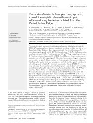
Corbari et al., Figure 3<br />
a<br />
b<br />
c<br />
d<br />
e<br />
500 nm<br />
f<br />
500 nm<br />
Fig. 3. TEM views illustrating the different ways the minerals may be deposited on the rod-shaped bacteria in Rimicaris exoculata. (a)<br />
Mineral deposition in direct contact with the bacteria cell walls. (b) Mineral deposition on secreted bacterial substance. (c) and (d) Methanotrophic<br />
bacteria surrounded by mineral deposits. (e) and (f) Bacterial ghosts coated with minerals and bacterial recolonization of the mineral<br />
sheaths.<br />
The upper level of the mineral crust contains very large<br />
concretions with diameters of up to 2 µm (Fig. 2e and f). The<br />
grape-like concretion shapes with deep indentations suggest<br />
that they result from the aggregation of several smaller ones.<br />
TEM images reveal that the bacteria become very rare in this<br />
upper level. As may be observed in the horizontal sections,<br />
almost the only bacteria present are a few large, thin, bacterial<br />
filaments that perforate throughout the mineral crust.<br />
Moreover, the minerals are not in direct contact with the filament<br />
cell walls but form large sheaths at some distance from<br />
the cell walls (Fig. 2e). Even though the three step-levels<br />
of the mineral crust have been arbitrarily defined, they are<br />
<strong>Biogeosciences</strong>, 5, 1295–1310, 2008<br />
www.biogeosciences.net/5/1295/2008/