View - Biogeosciences
View - Biogeosciences
View - Biogeosciences

You also want an ePaper? Increase the reach of your titles
YUMPU automatically turns print PDFs into web optimized ePapers that Google loves.
g-2008-0022<br />
L. Corbari et al.: Bacteriogenic iron oxides 1299<br />
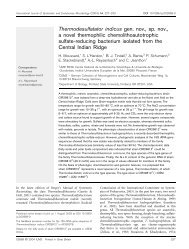
Corbari et al., Figure 2<br />
a<br />
b<br />
2 µm<br />
c<br />
d<br />
5 µm<br />
e<br />
f<br />
5 µm<br />
Fig. 2. The three levels of the mineral crust. Comparison between the electron back scattering images of the polished-thin sections (vertical<br />
sections) revealing the mineral densities and structures (left column) and the TEM micrographs of horizontal cross-sections exhibiting<br />
mineral associated with bacterial community (right column) at the lower level (a and b), the median level (c and d) and the upper level (e<br />
and f).<br />
The median level of the crust exhibits larger mineral concretions,<br />
globular in shape, (Fig. 2c and d). These globular<br />
concretions often meet to form larger ones that exhibit<br />
a botryoidal structure. In the horizontal cross-sections, the<br />
aggregate density appears rather heterogeneous and consists<br />
of highly mineralised patches interspersed with bacteria rich<br />
areas. The bacterial density in the median layer is always<br />
smaller than in the lower layer; there are fewer rod-shaped<br />
bacteria. Moreover, ghosts of bacteria are also observed<br />
(Fig. 3e and f) in TEM images. These ghosts have bacterial<br />
shapes that are completely enclosed in a heavy mineral<br />
sheath. Sometimes the bacteria are still present but appear<br />
either to be damaged or as membrane remain (Fig. 3e and f).<br />
In other cases, the mineral sheath appears to be empty or to<br />
have been recolonised by other rod-shaped bacteria. These<br />
observations suggest that mineral formation may influence<br />
the survival rate of the bacteria.<br />
www.biogeosciences.net/5/1295/2008/ <strong>Biogeosciences</strong>, 5, 1295–1310, 2008