extended abstract - Station Biologique de Roscoff
extended abstract - Station Biologique de Roscoff
extended abstract - Station Biologique de Roscoff

You also want an ePaper? Increase the reach of your titles
YUMPU automatically turns print PDFs into web optimized ePapers that Google loves.
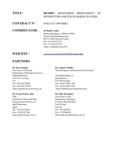
TITLE :<br />
CONTRACT N° :<br />
COORDINATOR :<br />
WEB SITE :<br />
PICODIV: MONITORING BIODIVERSITY OF<br />
PICOPHYTOPLANKTON IN MARINE WATERS<br />
EVK3-CT-1999-00021<br />
Dr Daniel Vaulot<br />
<strong>Station</strong> <strong>Biologique</strong>, CNRS et UPMC<br />
Oceanic Phytoplankton team,<br />
BP 74, 29682 <strong>Roscoff</strong>, France<br />
Tel: +33 2 98 29 23 23<br />
Fax: +33 2 98 29 23 24<br />
email: vaulot@sb-roscoff.fr<br />
www.sb-roscoff.fr/Phyto/PICODIV/in<strong>de</strong>x.html<br />
PARTNERS :<br />
Dr. Dave Scanlan<br />
University of Warwick<br />
Department of Biological Sciences,<br />
Gibbet Hill Road,<br />
CV4 7AL Coventry<br />
UK<br />
Tel: +44-1203 528363<br />
Fax: +44-1203-523701<br />
email: dp@dna.bio.warwick.ac.uk<br />
Dr. Carles Pedrós-Alió<br />
CSIC ,<br />
Institut <strong>de</strong> Ciències <strong>de</strong>l Mar,<br />
Passeig Joan <strong>de</strong> Borbó s/n,<br />
08039 Barcelona,<br />
Spain<br />
Tel: +34-93-2216416<br />
Fax: +34-93-2217340<br />
email: cpedros@icm.csic.es<br />
Dr. Linda K. Medlin<br />
Alfred Wegener Institute for Polar Research<br />
Am Han<strong>de</strong>lshafen 12<br />
Bremerhaven,<br />
D-27570 Germany<br />
Tel: +49-471-4831-1443<br />
Fax: +49-471-4831-1425<br />
email: lmedlin@awi-bremerhaven.<strong>de</strong><br />
Dr. Jahn Throndsen<br />
University of Oslo<br />
Department of Biology,<br />
P.O. Box 1069, Blin<strong>de</strong>rn,<br />
N-0316 Oslo,<br />
Norway<br />
Tel: +47 22854741<br />
Fax: +47 22854438<br />
email: jahn.throndsen@bio.uio.no
PICODIV:<br />
EXPLORING THE DIVERSITY OF PICOPLANKTON<br />
D. Vaulot 1 , L. Guillou 1, 6 , N. Simon 1 , F. Not 1 , H. Felman 1 , F. Le Gall 1 , D.Scanlan 2 , R. Howarth 2 , L.K.<br />
Medlin 3 , R. Scharek 3 , K. Valentin 3 , C. Pedrós-Alió 4 , R. Massana 4 , M. Latasa 4 , J Throndsen 5 , B.<br />
Edvardsen 5 , W. Eikrem 5<br />
1 <strong>Station</strong> <strong>Biologique</strong>, CNRS et UPMC, <strong>Roscoff</strong>, France, 2 University of Warwick, Department of<br />
Biological Sciences, Coventry, UK, 3 Alfred-Wegener-Institut, Bremerhaven, Germany, 4 Institut <strong>de</strong><br />
Ciències <strong>de</strong>l Mar, CSIC , Barcelona, Spain, 5 University of Oslo, Section of Marine Botany,<br />
Department of Biology, Norway, 6 Present address: IFREMER, DRV, Plouzané, France<br />
INTRODUCTION<br />
Picoplankton (<strong>de</strong>fined operationally hereafter as cells that pass through a 3 µm filter) dominate the<br />
photosynthetic biomass in many marine ecosystems, not only in the very oligotrophic regions of the<br />
world oceans, such as the central Pacific gyre (Campbell et al. 1994) or the Eastern Mediterranean<br />
Sea, but also in mesotrophic areas, such as the high chlorophyll - low nutrient equatorial regions.<br />
However, picophytoplankton are clearly not exclusively restricted to oceanic environments. In many<br />
coastal regions, they are present throughout the year and constitute a 'background' population<br />
(Agawin et al. 1998), onto which episodic phenomena, such as the spring bloom <strong>de</strong>velops. In some<br />
environments, such as coastal lagoons, picoplankton are a major component of biomass and<br />
productivity for most of the year. Picophytoplankton are also very relevant from the human point of<br />
view, because some bloom-forming picoplankters, such as Aureococcus spp. are toxic (Bricelj and<br />
Lonsdale 1997).<br />
Photosynthetic picoplankton encompass both prokaryotic and eukaryotic species:<br />
• Prokaryotes. Only two major genera are important for the picoplanktonic community in<br />
marine waters: Synechococcus and Prochlorococcus. Whereas Prochlorococcus dominates<br />
over Synechococcus in most oligotrophic regions, except at high latitu<strong>de</strong>s, the reverse is true<br />
un<strong>de</strong>r meso- and eutrophic conditions (Partensky et al. 1999). With such wi<strong>de</strong> ecological<br />
distributions, these two genera display a large genetic and phenotypic variability, that is just<br />
beginning to be assessed.<br />
• Eukaryotes. In contrast to prokaryotes, the taxonomic diversity of picophytoplanktonic<br />
eukaryotes is much broa<strong>de</strong>r. In fact, nearly every algal division has picoplanktonic<br />
representatives (Figure 1). Still, a vast number of taxa undoubtedly remain unknown and<br />
un<strong>de</strong>scribed.<br />
To date fewer than 30 species of picophytoplankton have been <strong>de</strong>scribed (see Table 1). This<br />
number pales in comparison to the more than 4,000 marine phytoplankton species that have been<br />
<strong>de</strong>scribed to date and to the over 100,000 that are believed to exist. A clear proof of our poor<br />
knowledge of picophytoplankton diversity is revealed by the discovery of three novel algal classes<br />
in the last ten years <strong>de</strong>scribed from picophytoplanktonic taxa (to put this into perspective, to ignore<br />
an algal class corresponds to ignoring the existence of mammals or birds among the vertebrates):<br />
1991 class Pedinophyceae based on Resultor mikron 2 µm (Moestrup 1991)<br />
1993 class Pelagophyceae based on Pelagomonas calceolata 2 µm (An<strong>de</strong>rsen et al. 1993)<br />
1999 class Bolidophyceae based on Bolidomonas pacifica 1.5 µm (Guillou et al. 1999)
Table 1: Chronology of taxonomic picoplankton knowledge.<br />
Year Name Class Size<br />
µm<br />
1952 Chromulina pusilla Butcher<br />
Prasinophyceae 1-1.5<br />
(renamed Micromonas Manton & Parke)<br />
Nannochloris atomus Butcher,<br />
Eustigmatophyceae 1.5-4<br />
N. maculata Butcher<br />
(renamed Nannochloropsis Hibberd)<br />
1955 Nannochloris oculata Droop<br />
Eustigmatophyceae 1.5-4<br />
(renamed Nannochloropsis Hibberd)<br />
1957 Monallantus salina Bourelly<br />
Eustigmatophyceae 1.5-4<br />
(renamed Nannochloropsis Hibberd)<br />
1967 Hillea marina Butcher Cryptophyceae 2-2.5<br />
1969 Pedinomonas mikron Throndsen<br />
Prasinophyceae 1.5-2.5<br />
(renamed Resultor Moestrup)<br />
Scourfieldia marina Throndsen<br />
Prasinophyceae 2-3<br />
(renamed Pseudoscourfieldia Manton)<br />
1974 Imantonia rotunda Reynolds Prymnesiophyceae 2-4<br />
1977 Pelagococcus subviridis Norris Pelagophyceae 2.5-5.5<br />
1978 Chlorella nana Butcher Chlorophyceae 1.8-2.6<br />
1979 Discovery of oceanic picoplankton<br />
marine Synechococcus Naegeli Cyanophyta 0.8-1.2<br />
1982 Nannochloropsis gaditana Lubian Eustigmatophyceae 2.5-5<br />
Nanochlorum eucaryotum Willhelm et al.<br />
Chlorophyceae<br />
(renamed Nannochloris Naumann)<br />
1987 Triparma Booth & Marchant spp,<br />
Chrysophyceae 2.2-4.7<br />
Tetraparma pelagica Booth<br />
1988 Prochlorococcus marinus Chisholm Cyanophyta 0.5-0.7<br />
Aureococcus anophagefferens Hargraves & Sieburth Pelagophyceae 2-4<br />
1990 Bathycoccus prasinos Eikrem & Throndsen Prasinophyceae 1.5-2.5<br />
Pycnococcus provasolii Guillard Prasinophyceae 1- 4<br />
1993 Pelagomonas calceolata An<strong>de</strong>rsen & Saun<strong>de</strong>rs Pelagophyceae 1.3-3<br />
1995 Ostreococcus tauri Courties & Chrétiennot-Dinet Prasinophyceae 0.8-1.1<br />
1996 Prasino<strong>de</strong>rma coloniale Hasegawa & Chihara Prasinophyceae 2.5-5.5<br />
Nannochloropsis granulata Karlson & Potter Eustigmatophyceae 2- 4<br />
1997 Aureoumbra lagunensis Stockwell et al. Pelagophyceae 2.5-5<br />
1999 Bolidomonas pacifica Guillou & Chrétiennot-Dinet Bolidophyceae 1.5<br />
B. mediterranea Guillou & Chrétiennot-Dinet<br />
2000 Picophagus flagellatus Guillou & Chrétiennot-Dinet Chrysophyceae 2<br />
Symbiomonas scintillans Guillou & Chrétiennot-Dinet Bicosoecid 2<br />
Because so little is known about the taxonomy and systematics of picophytoplankton we have very<br />
little data to estimate the levels of its biodiversity un<strong>de</strong>r natural conditions and to un<strong>de</strong>rstand how<br />
the picophytoplankton might be affected by environmental variability linked to either anthropogenic<br />
influence or to larger scale phenomena, such as those linked to climate change or global warming.<br />
However we have some indications that picophytoplankton species (and therefore<br />
picophytoplankton biodiversity) may react sharply to changes in marine systems:<br />
• The prokaryote Prochlorococcus consists of at least two different genotypes/phenotypes,<br />
each one dominates un<strong>de</strong>r different environmental conditions: i.e., one is present un<strong>de</strong>r the<br />
high light/low nutrient conditions of the marine surface layer, and the other un<strong>de</strong>r the low<br />
light/higher nutrient conditions of the bottom of the euphotic zone. Thus Prochlorococcus is<br />
able to partition its niche genetically so that it is phenotypically adapted to its environment.
• The abundance of<br />
Synechococcus in the<br />
equatorial Pacific<br />
<strong>de</strong>creases during El<br />
Niño Southern<br />
Oscillation episo<strong>de</strong>s.<br />
• The potentially toxic<br />
brown picoplanktonic<br />
alga Aureococcus was<br />
unknown before 1985,<br />
but since then it has<br />
bloomed repeatedly in<br />
US coastal waters<br />
(Bricelj and Lonsdale<br />
1997).<br />
Our ignorance<br />
concerning<br />
picophytoplankton<br />
diversity is<br />
mostly explained by the fact<br />
that, because of their very<br />
small size, picophytoplankton<br />
cells most<br />
often lack any distinguishing<br />
features and are very<br />
difficult to i<strong>de</strong>ntify by<br />
classical methods. In fact<br />
many have evolved to small<br />
"green or brown ball"<br />
morphotypes that mask a<br />
broad taxonomic diversity<br />
(Potter et al. 1997). Our<br />
present state of knowledge<br />
regarding<br />
picophytoplanktonic<br />
biodiversity is in fact<br />
analogous to that prevailing<br />
ten years ago for eubacteria<br />
and archea. Until the early<br />
1990's, the taxonomy and<br />
un<strong>de</strong>rstanding of bacterial<br />
diversity was based primarily<br />
on species isolated into<br />
Figure 1: A tree showing the phylogenetic affinities of eukaryotic<br />
picophytoplankton species (in bold). Note that only one picoplanktonic<br />
Prymnesiophyte has been isolated yet (I. rotunda), but a number of<br />
picoplanktonic Prymnesiophyte sequences have been recovered from<br />
oceanic samples (Moon-van <strong>de</strong>r Staay et al. 2000). Source: Guillou<br />
unpublished<br />
culture. No-one could have predicted the vast diversity of these organisms in nature (Giovannoni et<br />
al. 1990).<br />
In or<strong>de</strong>r to remedy to this very poor state of knowledge concerning a group that, in many<br />
ecosystems, accounts for up to 60 to 80% of photosynthetic biomass and production, there is a very<br />
urgent need to <strong>de</strong>velop efficient monitoring tools of picophytoplankton diversity. This problem is in<br />
fact very analogous to that encountered by microbiologists who cannot tell apart bacteria based on<br />
their shape or even on their metabolic requirements. The latter have relied more and more in recent<br />
years on molecular biology techniques to i<strong>de</strong>ntify and <strong>de</strong>tect bacteria in the environment
(Giovannoni et al. 1990; Amann and Kuhl 1998). We plan during the course of this project to expand<br />
this very successful approach to picophytoplankton.<br />
STRATEGY<br />
Our strategy is encapsulated in the following four steps:<br />
(1) Obtain SSU rDNA sequences for as many as possible picophytoplankton taxa from both<br />
cultures and natural samples. Novel taxa will be assessed using a combination of methods<br />
including in particular pigment analysis and electron microscopy.<br />
(2) Using this sequence database, <strong>de</strong>velop hierarchical probes recognizing successive taxonomic<br />
groupings having picophytoplanktonic representatives<br />
(3) Develop fast and efficient techniques to quantify the fraction of the pico-phytoplankton<br />
recognized by the probes in natural samples.<br />
(4) Test and validate these probes on time series of picophytoplankton biodiversity in three coastal<br />
ecosystems.<br />
We will focus on the picophytoplankton from coastal European waters that has been much less<br />
studied in comparison to that of offshore oligotrophic waters. For this purpose we have selected<br />
three sites located in the following regions:<br />
• English Channel (<strong>Roscoff</strong>)<br />
• North Sea (Helgoland)<br />
• Western Mediterranean Sea (Blanes Bay)<br />
These sites have been carefully selected as offering a wi<strong>de</strong> range of environmental conditions<br />
representative of EU coastal waters. Moreover, all have been extensively monitored in the past and<br />
abundant background information is available on environmental conditions as well as phytoplankton<br />
populations. One of them (Helgoland) has been <strong>de</strong>signated as a flagship site for long-term and largescale<br />
marine biodiversity research at a recent European meeting on biodiversity because its longterm<br />
sampling program stretches back at least 26 years.<br />
Although these three sites will serve as focal points for our project, we are also taking advantage<br />
of oceanographic cruises planned outsi<strong>de</strong> this project to examine the diversity of picophytoplankton<br />
in other environments. In particular we have begun sample the following ecosystems:<br />
• Mediterranean Sea (PROSOPE 99, MATER99, HIVERN00)<br />
• Red Sea<br />
• North Atlantic Ocean (PROSOPE 99)<br />
• Celtic Sea (PROPHEZE D246)<br />
First, for probe <strong>de</strong>sign we need to obtain SSU rDNA sequences covering the full taxonomic<br />
spectrum of picophytoplankton. For this purpose, we have adopted a two pronged approach:<br />
a - We are obtaining sequences from fully characterized laboratory strains. We are in particular<br />
securing all picoplanktonic strains available from international culture collections, such as the CCMP<br />
(Center for Cultures of Marine Phytoplankton, Bigelow USA). However, we know that such<br />
collections only feature a limited number of picoplanktonic strains, because very little effort has been<br />
<strong>de</strong>voted to this size class to date. Therefore we need to embark on a very strong effort of strain<br />
isolation. For this purpose, we are establishing cultures of both prokaryotic and eukaryotic<br />
picophytoplankton from the environments listed above using methods that have already proved<br />
very successful for this purpose (prefiltration of natural samples, monitoring of cultures by flow<br />
cytometry). Once established, the cultures are screened by a variety of techniques (flow cytometry,<br />
electron microscopy, pigment analysis, molecular methods) to assess their taxonomic position.<br />
Those that obviously contain novel taxa are further purified by dilution or plating and more fully<br />
studied (electron microscopy sections), sequenced and <strong>de</strong>scribed formally.<br />
b - As we know that a large number of planktonic organisms still escape culture due to the lack of<br />
optimum culture conditions, we are also using the molecular approach that has been so successful
for bacteria i.e., environmental ribosomal RNA gene cloning and sequencing. These sequences are<br />
being obtained from the same environments from which we obtain cultures. It is highly likely that<br />
this will reveal novel groups that we can then target for culturing.<br />
Second, using the sequence database obtained both from cultures and natural samples, we will<br />
use or <strong>de</strong>sign hierarchical probes for each taxonomic level containing picophytoplanktonic<br />
representatives (e.g., classes, such as the Pelagophyceae or species, such as Micromonas pusilla).<br />
These probes will be validated against cultured strains.<br />
Third, we are <strong>de</strong>veloping methods to assess the fraction of the marine pico-phytoplankton<br />
recognized by a given probe. We focus on very recent techniques allowing quantitative and<br />
extensive probe measurements (fluorescent in situ hybridization, probe array, quantitative PCR).<br />
Fourth, we will apply these methods in the second phase of the project to assess<br />
picophytoplankton diversity during a full year at our three coastal sites (English Channel, North Sea,<br />
Mediterranean Sea). At the same time, the composition and abundance of the picophytoplankton<br />
will be studied with more conventional techniques, such as electron microscopy or pigment analysis,<br />
and alternate molecular methods (DGGE). These data will permit a validation of the data obtained by<br />
the molecular probe approach. We will interpret then the biodiversity patterns as a function of the<br />
other environmental parameters of the site sampled. We will <strong>de</strong>termine in particular whether there is<br />
a succession of groups and species (as is the case for the larger nano and micro-phytoplankton) or<br />
whether a small group of ubiquitous species are always present and merely change their abundance<br />
(but not their diversity) in response to environmental changes.<br />
REFERENCES<br />
Agawin, N. S. R., C. M. Duarte, and S. Agusti. 1998. Growth and abundance of Synechococcus sp. in<br />
a Mediterranean Bay: seasonality and relationship with temperature. Mar. Ecol. Prog. Ser. 170:<br />
45-53.<br />
Amann, R., and M. Kuhl. 1998. In situ methods for assessment of microorganisms and their activities.<br />
Curr. Opin. Microbiol. 1: 352-358.<br />
An<strong>de</strong>rsen, R. A., G. W. Saun<strong>de</strong>rs, M. P. Paskind, and J. Sexton. 1993. Ultrastructure and 18S rRNA<br />
gene sequence for Pelagomonas calceolata gen. and sp. nov. and the <strong>de</strong>scription of a new algal<br />
class, the Pelagophyceae classis nov. J. Phycol. 29: 701-715.<br />
Bricelj, V. M., and D. J. Lonsdale. 1997. Aureococcus anophagefferens: Causes and ecological<br />
consequences of brown ti<strong>de</strong>s in US mid-Atlantic coastal waters. Limnol. Oceanogr. 42: 1023-<br />
1038.<br />
Campbell, L., H. A. Nolla, and D. Vaulot. 1994. The importance of Prochlorococcus to community<br />
structure in the central North Pacific Ocean. Limnol. Oceanogr. 39: 954-961.<br />
Giovannoni, S. J., T. B. Britschgi, C. L. Moyer, and K. G. Field. 1990. Genetic diversity in Sargasso Sea<br />
bacterioplankton. Nature, Lond. 345: 60-63.<br />
Guillou, L., M.-J. Chrétiennot-Dinet, L. K. Medlin, H. Claustre, S. Loiseaux-<strong>de</strong> Goër, and D. Vaulot.<br />
1999. Bolidomonas: a new genus with two species belonging to a new algal class, the<br />
Bolidophyceae (Heterokonta). J. Phycol. 35: 368–381.<br />
Moestrup, Ø. 1991. Further studies of presumedly primitive green algae, including the <strong>de</strong>scription of<br />
Pedinophyceae class. nov. and Resultor gen. nov. J. Phycol. 27: 119-133.<br />
Moon-van <strong>de</strong>r Staay, S. Y., G. W. M. van <strong>de</strong>r Staay, L. Guillou, D. Vaulot, H. Claustre, and L. K.<br />
Medlin. 2000. Abundance and diversity of prymnesiophytes in the picoplankton community from<br />
the equatorial Pacific Ocean inferred from 18S rDNA sequences. Limnol. Oceanogr. 45: 98-109.<br />
Partensky, F., W. R. Hess, and D. Vaulot. 1999. Prochlorococcus, a marine photosynthetic<br />
prokaryote of global significance. Microb. Mol. Biol. Rev. 63: 106-127.<br />
Potter, D., T. C. Lajeunesse, G. W. Saun<strong>de</strong>rs, and R. A. An<strong>de</strong>rsen. 1997. Convergent evolution masks<br />
extensive biodiversity among marine coccoid picoplankton. Biodiv. Conserv. 6: 99-107.