Pancreatitis review NEJM 2006.pdf - SASSiT
Pancreatitis review NEJM 2006.pdf - SASSiT
Pancreatitis review NEJM 2006.pdf - SASSiT

You also want an ePaper? Increase the reach of your titles
YUMPU automatically turns print PDFs into web optimized ePapers that Google loves.
The new england journal of medicine<br />
clinical practice<br />
Acute <strong>Pancreatitis</strong><br />
David C. Whitcomb, M.D., Ph.D.<br />
This Journal feature begins with a case vignette highlighting a common clinical problem.<br />
Evidence supporting various strategies is then presented, followed by a <strong>review</strong> of formal guidelines,<br />
when they exist. The article ends with the author’s clinical recommendations.<br />
A 56-year-old woman presents with severe epigastric pain and vomiting of 14 hours’<br />
duration, symptoms that had developed shortly after dinner the previous night. She<br />
has no history of alcohol use, takes no medications, and has no family history of pancreatitis.<br />
On physical examination, she has a heart rate of 110 beats per minute and<br />
moderate epigastric abdominal tenderness without peritoneal signs. The white-cell<br />
count is 16,500 per cubic millimeter, and the hematocrit is 49 percent. The serum<br />
amylase level is 1450 IU per liter, the serum lipase level is 3200 IU per liter, the serum<br />
alanine aminotransferase level is 280 IU per liter, and the serum lactate dehydrogenase<br />
level is 860 IU per liter. Calcium, albumin, triglyceride, and electrolyte values are<br />
normal. How should the patient be further evaluated and treated?<br />
The Clinical Problem<br />
From the Division of Gastroenterology,<br />
Hepatology, and Nutrition, University<br />
of Pittsburgh, Pittsburgh. Address reprint<br />
requests to Dr. Whitcomb at the<br />
University of Pittsburgh Medical Center<br />
Presbyterian, Mezzanine Level 2, C Wing,<br />
200 Lothrop St., Pittsburgh, PA 15213, or<br />
at whitcomb@pitt.edu.<br />
N Engl J Med 2006;354:2142-50.<br />
Copyright © 2006 Massachusetts Medical Society.<br />
Acute pancreatitis accounts for more than 220,000 hospital admissions in the United<br />
States each year. 1 The disease occurs at a similar frequency among various age<br />
groups, but the cause of the condition and the likelihood of death vary according<br />
to age, sex, race, body-mass index (the weight in kilograms divided by the square<br />
of the height in meters), and other factors.<br />
The most important risk factors for pancreatitis in adults are gallstones and excessive<br />
alcohol use, although clinically detected pancreatitis never develops in most<br />
persons with these risk factors. 2,3 The incidence of gallstone pancreatitis is increased<br />
among white women over the age of 60 years 4,5 and is highest among patients with<br />
small gallstones (less than 5 mm in diameter) or microlithiasis. 3,5 Excessive alcohol<br />
use as a cause of pancreatitis is more common among men than women 6 ; the association<br />
between alcohol consumption and acute pancreatitis is complex but appears<br />
to be dose-dependent. Other causes include metabolic aberrations (e.g., hypertriglyceridemia),<br />
duct obstruction (e.g., related to a tumor or pancreas divisum), medications<br />
(e.g., azathioprine, thiazides, and estrogens), and trauma. In children, the<br />
distribution of causes differs from that in adults, with systemic diseases and trauma<br />
particularly common. 7 About 20 percent of cases in adults remain idiopathic, although<br />
this classification is expected to become less common as factors of genetic<br />
predisposition and environmental susceptibility are elucidated. 8<br />
Overall, about 20 percent of patients with acute pancreatitis have a severe course,<br />
and 10 to 30 percent of those with severe acute pancreatitis die. Despite improvements<br />
in intensive care treatment during the past few decades, the rate of death has not<br />
significantly declined. 9<br />
The pathogenesis of acute pancreatitis relates to inappropriate activation of trypsinogen<br />
to trypsin (the key enzyme in the activation of pancreatic zymogens) and a<br />
lack of prompt elimination of active trypsin inside the pancreas. 8 Activation of diges-<br />
2142<br />
n engl j med 354;20 www.nejm.org may 18, 2006<br />
Downloaded from www.nejm.org by MARTIN BRAND MD on December 15, 2007 .<br />
Copyright © 2006 Massachusetts Medical Society. All rights reserved.
clinical practice<br />
tive enzymes causes pancreatic injury and results<br />
in an inflammatory response that is out of proportion<br />
to the response of other organs to a similar<br />
insult. The acute inflammatory response itself<br />
causes substantial tissue damage and may progress<br />
beyond the pancreas to a systemic inflammatory<br />
response syndrome, multiorgan failure,<br />
or death.<br />
Strategies and Evidence<br />
Diagnosis<br />
The clinical diagnosis of acute pancreatitis is based<br />
on characteristic abdominal pain and nausea, combined<br />
with elevated serum levels of pancreatic enzymes.<br />
In gallstone pancreatitis, the pain is typically<br />
sudden, epigastric, and knife-like and may<br />
radiate to the back. In hereditary or metabolic cases<br />
or in those associated with alcohol abuse, the<br />
onset may be less abrupt and the pain poorly localized.<br />
Serum amylase levels that are more than<br />
three times the upper limit of normal, in the setting<br />
of typical abdominal pain, are almost always<br />
caused by acute pancreatitis. Lipase levels are also<br />
elevated and parallel the elevations in amylase levels.<br />
The levels of both enzymes remain elevated<br />
with ongoing pancreatic inflammation, with amylase<br />
levels typically returning to normal shortly<br />
before lipase levels in the resolution phase.<br />
Tests that are more specific for acute pancreatitis<br />
but less widely available evaluate levels of trypsinogen<br />
activation peptide 10 and trypsinogen-2. 11<br />
Abdominal imaging by computed tomography<br />
(CT), magnetic resonance imaging (MRI), or transabdominal<br />
ultrasonography is useful in confirming<br />
the diagnosis of pancreatitis or ruling out<br />
other intraabdominal conditions as the cause of<br />
pain or laboratory abnormalities. Such imaging<br />
may also identify the cause of pancreatitis or its<br />
associated complications.<br />
Management<br />
Determination of the cause is important for guiding<br />
immediate management and preventing recurrence.<br />
An elevated alanine aminotransferase level<br />
in a patient without alcoholism who has pancreatitis<br />
is the single best laboratory predictor of biliary<br />
pancreatitis; a level of more than three times<br />
the upper limit of normal has a positive predictive<br />
value of 95 percent for gallstone pancreatitis. 12<br />
However, the presence of normal alanine aminotransferase<br />
levels does not reliably rule out the diagnosis.<br />
4 Laboratory testing may reveal hypertriglyceridemia<br />
or hypercalcemia as possible causes<br />
of pancreatitis, although pancreatitis may also<br />
cause mildly elevated triglyceride levels.<br />
Imaging Studies<br />
CT or MRI can identify gallstones or a tumor (an<br />
infrequent cause of pancreatitis), as well as local<br />
complications. MRI may also identify early duct<br />
disruption that is not seen on CT. 13 Transabdominal<br />
ultrasonography is more sensitive than either<br />
CT or MRI for identifying gallstones and sludge<br />
and for detecting bile-duct dilatation, but it is insensitive<br />
for detecting stones in the distal bile<br />
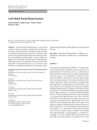
duct. 4,5 Endoscopic ultrasonography may be the<br />
most accurate test for diagnosing or ruling out<br />
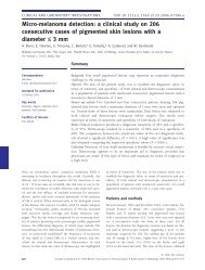
biliary causes of acute pancreatitis (Fig. 1) and may<br />
guide the emergency use of endoscopic retrograde<br />
cholangiopancreatography (ERCP). 14<br />
A<br />
B<br />
Figure 1. Endoscopic Ultrasonography of the Gallbladder<br />
and Common Bile Duct from the Duodenum.<br />
Microlithiasis (sludge) is shown within the gallbladder<br />
(Panel A, arrow) and within the common bile duct<br />
(Panel B, arrow). Also visible in Panel B are the head of<br />
the pancreas (curved arrow) and the pancreatic duct<br />
(arrowhead). (Images courtesy of Neeraj Kaushik, M.D.)<br />
n engl j med 354;20 www.nejm.org may 18, 2006 2143<br />
Downloaded from www.nejm.org by MARTIN BRAND MD on December 15, 2007 .<br />
Copyright © 2006 Massachusetts Medical Society. All rights reserved.
The new england journal of medicine<br />
Table 1. Scoring Methods for the Prediction of Severe Acute <strong>Pancreatitis</strong>.<br />
Criterion and Marker Threshold Value Severe <strong>Pancreatitis</strong><br />
Atlanta criteria*<br />
Indicated by any positive factor listed<br />
Ranson’s score†<br />
≥3<br />
APACHE II score‡<br />
≥8<br />
Organ failure<br />
Shock<br />
Blood pressure of 177 μmol/liter (2 mg/dl)<br />
after hydration<br />
Systemic complications<br />
Disseminated intravascular coagulation Platelet count of ≤100,000/mm 3<br />
Fibrinogen level of 80 μg/ml<br />
Metabolic disturbance<br />
Calcium level of ≤7.5 mg/dl<br />
Local complications<br />
Pancreatic necrosis<br />
Present<br />
Pancreatic abscess<br />
Present<br />
Pancreatic pseudocyst<br />
Present<br />
Ranson’s score†<br />
Indicated by a total score ≥3, with<br />
1 point for each positive factor<br />
At presentation<br />
Age<br />
>55 yr<br />
Blood glucose level<br />
>200 mg/dl (10 mmol/liter)<br />
White-cell count >16,000/mm 3<br />
Lactate dehydrogenase level<br />
>350 IU/liter<br />
Alanine aminotransferase level<br />
>250 IU/liter<br />
Within 48 hr after presentation<br />
Hematocrit<br />
>10% decrease<br />
Serum calcium<br />
4 mEq/liter<br />
Blood urea nitrogen<br />
>5 mg/dl (1.8 mmol/liter) increase<br />
Fluid sequestration<br />
>6 liters<br />
Partial pressure of arterial oxygen§<br />
clinical practice<br />
Table 1. (Continued.)<br />
Criterion and Marker Threshold Value Severe <strong>Pancreatitis</strong><br />
CT severity index Indicated by a total score of >6<br />
(CT grade plus necrosis score)<br />
CT grade<br />
Normal pancreas (grade A)<br />
0 points<br />
Focal or diffuse enlargement (grade B)<br />
1 point<br />
Intrinsic change; fat stranding (grade C)<br />
2 points<br />
Single, ill-defined collection of fluid (grade D)<br />
3 points<br />
Multiple collections of fluid or gas in or adjacent<br />
4 points<br />
to pancreas (grade E)<br />
Necrosis score<br />
No pancreatic necrosis<br />
0 points<br />
Necrosis of one third of pancreas<br />
2 points<br />
Necrosis of one half of pancreas<br />
4 points<br />
Necrosis of >one half of pancreas<br />
6 points<br />
APACHE II score‡<br />
Indicated by a score of ≥8<br />
Initial values of 12 routine physiological measurements,<br />
age, and previous health status<br />
* Data are from Bradley. 21 The Atlanta criteria were adopted in 1992 by the International Symposium on Acute <strong>Pancreatitis</strong>. The presence of<br />
any condition in the five main categories indicates severe acute pancreatitis.<br />
† Data are from Ranson et al. 22 The original Ranson’s score is based on 11 clinical signs (5 measured on admission and 6 in the 48 hours after<br />
admission), with a higher score indicating greater correlation with the incidence of systemic complications and the presence of pancreatic<br />
necrosis. The relationship between Ranson’s score and the CT severity index 23 is given in Table 3.<br />
‡ Data are from Knaus et al. 24 and Larvin and McMahon. 25 The Acute Physiology and Chronic Health Evaluation (APACHE II) score is based<br />
on initial values of 12 routine physiological measurements, age, and previous health status, with a score of 8 or more commonly used as<br />
the threshold for classification as severe pancreatitis.<br />
§ The test was performed without the use of supplemental oxygen.<br />
The CT severity index 23 is a combination of the sum of the necrosis score and points assigned to five grades of findings on CT. The index<br />
ranges from 0 to 10, with higher scores indicating a greater severity of illness.<br />
course of acute pancreatitis, including an inhibitor<br />
of platelet-activating factor (lexipafant 19 ), somatostatin<br />
and its analogues, and protease inhibitors<br />
20 ; treatment is primarily supportive. Patients<br />
should receive nothing by mouth and receive intravenous<br />
pain medication and aggressive hydration<br />
to treat or prevent hemoconcentration (e.g.,<br />
a bolus of fluids to achieve hemodynamic stability,<br />
followed by 250 to 500 ml of crystalloid solutions<br />
per hour in an average-sized patient without<br />
substantial kidney or heart disease). Fluid balance<br />
should be maintained and pulse oximetry should<br />
be considered, especially when narcotic analgesics<br />
are used.<br />
Predicting Severe Acute <strong>Pancreatitis</strong><br />
The severity of acute pancreatitis is defined by the<br />
presence or absence of organ failure, local complications,<br />
or both 21-25 (Table 1). It is critical to<br />
identify patients who are at high risk for severe<br />
disease, since they require close monitoring and<br />
possible intervention. Recognized markers of the<br />
risk of severe acute pancreatitis include specific<br />
laboratory values that measure the systemic inflammatory<br />
response (such as C-reactive protein),<br />
scoring systems that assess inflammation or organ<br />
failure (such as Ranson’s score), and findings<br />
on imaging studies 13,23 (Table 2). The Acute Physiology<br />
and Chronic Health Evaluation score (based<br />
on initial values of 12 routine physiological measurements,<br />
age, and previous health status) is<br />
among the best predictors of severity on admission,<br />
whereas elevated C-reactive protein levels are equally<br />
useful when measured 24 to 48 hours after the<br />
onset of symptoms. 27 Severity scores are useful in<br />
predicting both complications and death (Table 3).<br />
Other markers that are not included in standard<br />
scoring systems should also be considered. Obesity<br />
(a body-mass index of more than 30) is associated<br />
with an increase in the risk of a severe clinical<br />
n engl j med 354;20 www.nejm.org may 18, 2006 2145<br />
Downloaded from www.nejm.org by MARTIN BRAND MD on December 15, 2007 .<br />
Copyright © 2006 Massachusetts Medical Society. All rights reserved.
The new england journal of medicine<br />
Table 2. Value of Various Scoring Systems and Inflammatory Markers in the Prediction of Severe Acute <strong>Pancreatitis</strong>.*<br />
Scoring System Sensitivity Specificity<br />
Positive<br />
Predictive<br />
Value<br />
percent<br />
Negative<br />
Predictive<br />
Value<br />
Accuracy<br />
On admission<br />
APACHE II score†<br />
≥6 83–99 33–54 28–40 80–97 45–65<br />
≥8 68–71 48–67 30–40 84–87 53–68<br />
≥10 52–63 66–81 32–64 81–89 63–77<br />
Interleukin-6 level >400 pg/ml 89 87 80 93 88<br />
At 24 hours<br />
APACHE II score ≥8 63 73 38 88 71<br />
C-reactive protein level >150 mg/dl 65 73 37 90 72<br />
PMN elastase >300 μg/liter 93 99 97 98 98<br />
Urinary TAP >35 nmol/liter 68 74 44 89 73<br />
At 48 hours<br />
APACHE II score ≥8 56–78 52–64 30–33 85–88 58–63<br />
Ranson’s score ≥3 75–89 54–71 37–49 91–96 62–75<br />
Modified Glasgow score ≥3‡ 45–75 63–89 28–66 79–93 59–84<br />
C-reactive protein level >150 mg/dl 65 73 37 90 72<br />
* PMN denotes polymorphonuclear leukocyte, and TAP trypsinogen activation peptide.<br />
† Data are from Knaus et al. 24 and Larvin and McMahon. 25 The Acute Physiology and Chronic Health Evaluation<br />
(APACHE II) score is based on initial values of 12 routine physiological measurements, age, and previous health status,<br />
with a score of 8 or more commonly used as the threshold for the classification of severe pancreatitis.<br />
‡ The Modified Glasgow score is similar to Ranson’s score but can be completed on admission. The score ranges from<br />
0 to 8, with scores of 3 or more indicating a greater severity of illness. 26 Data are from Papachristou and Whitcomb. 27<br />
course by a factor of 2 to 3. 29 A hematocrit above<br />
44 percent is a clear risk factor for pancreatic necrosis,<br />
30 although it is a poor predictor of the severity<br />
of disease. Preliminary evidence suggests that<br />
genetic factors, such as polymorphisms in the chemokine<br />
monocyte chemotactic protein 1 (MCP-1)<br />
gene, 31 may also predict severity, although such<br />
genetic testing is not currently used in practice.<br />
Several clinical findings — including thirst,<br />
poor urine output, progressive tachycardia, tachypnea,<br />
hypoxemia, agitation, confusion, a rising hematocrit<br />
level, and a lack of improvement in symptoms<br />
within the first 48 hours — are warning<br />
signs of impending severe disease. If such symptoms<br />
develop, admission to an intensive care unit<br />
should be considered. Intensive care may also be<br />
warranted in patients at risk for rapid deterioration<br />
in their condition, including those over the<br />
age of 55 years, 22 those who need ongoing volume<br />
resuscitation or invasive monitoring of fluid status<br />
(e.g., central venous pressure monitoring), or<br />
those with renal failure or respiratory compromise.<br />
15<br />
Pancreatic-Fluid Collections, Pseudocysts,<br />
and Necrosis<br />
Up to 57 percent of patients who are hospitalized<br />
with acute pancreatitis will have fluid collections,<br />
with 39 percent having two areas involved and 33<br />
percent having three or more. 32 Fluid collections<br />
are initially ill defined, 21 evolve over time, and are<br />
usually managed conservatively. If the fluid collections<br />
continue to enlarge, cause pain, become<br />
infected (as suggested by the presence of unexplained<br />
fever, leukocytosis, or gas in the fluid collection),<br />
or compress adjacent organs, then medical,<br />
endoscopic, or surgical intervention may be<br />
needed. 33,34 Fluid collections with very high levels<br />
of pancreatic enzymes are usually associated<br />
with pancreatic-duct disruptions and may eventually<br />
form pseudocysts (usually over a period of<br />
several weeks), ascites, or pleural effusions. 34<br />
2146<br />
n engl j med 354;20 www.nejm.org may 18, 2006<br />
Downloaded from www.nejm.org by MARTIN BRAND MD on December 15, 2007 .<br />
Copyright © 2006 Massachusetts Medical Society. All rights reserved.
clinical practice<br />
Asymptomatic pseudocysts can be managed conservatively,<br />
whereas symptomatic pseudocysts can<br />
often be drained endoscopically. 35 ERCP may help<br />
to define the anatomy of the pancreatic duct and<br />
identify any duct disruptions to guide further intervention.<br />
33,34<br />
Pancreatic necrosis, occurring as diffuse or focal<br />
areas of nonviable pancreatic parenchyma, 21<br />
is an important complication that can develop during<br />
the first few days of pancreatitis; the condition<br />
is associated with late complications and death if<br />
the necrotic tissue becomes infected. The development<br />
of necrosis is associated with pancreatic<br />
inflammation, hypovolemia, and hypotension from<br />
the shunting of blood from other organs, vascular<br />
spasm, and hemoconcentration. 30 Pancreatic<br />
necrosis can be demonstrated by a loss of tissue<br />
perfusion on contrast-enhanced CT. 23<br />
Infection of necrotic tissue is suspected when<br />
there is fever, leukocytosis, and a failure to improve<br />
or unexpected deterioration — usually after the<br />
first week of illness. Visualization of gas bubbles<br />
within the necrotic tissue on CT is evidence of infection.<br />
The diagnosis of infected necrosis is usually<br />
made by fine-needle aspiration of the necrotic<br />
area guided by either CT or ultrasonography, with<br />
Gram’s staining and culture of the aspirate. 36<br />
Lack of Improvement<br />
If the condition of a patient whose pancreatitis is<br />
predicted to be mild fails to improve within two or<br />
three days, then contrast-enhanced CT (“pancreas<br />
protocol”) should be considered to identify fluid<br />
collections, pancreatic necrosis, or other complications<br />
that may require intervention. Antibiotic<br />
therapy and nutritional support also warrant consideration<br />
in patients whose condition fails to improve<br />
promptly or in whom complications develop.<br />
Table 3. Relationship between Severity Scores and Outcomes in Acute <strong>Pancreatitis</strong>.*<br />
Index<br />
Score<br />
Ranson’s score 0–2 3–4 5–6 7–8<br />
percent of patients<br />
Intensive care for >7 days and 1 24 53 NA<br />
survival<br />
Death 3 16 40 100<br />
Either intensive care for >7 days 4 40 93 100<br />
or death<br />
CT severity index 0–3 4–6 7–10 NA<br />
Complications 8 35 92 NA<br />
Death 3 6 17 NA<br />
* Data are from Balthazar et al. 23 and Ranson. 28 NA denotes not applicable.<br />
Use of Antibiotics<br />
The proper role of antibiotics in acute pancreatitis<br />
remains controversial. No antibiotics are indicated<br />
in mild cases. However, infectious complications<br />
are an important concern in severe cases,<br />
especially cases of pancreatic necrosis. A potential<br />
role for prophylactic antibiotics in severe pancreatitis<br />
was initially given support by a randomized<br />
trial demonstrating that the administration of<br />
imipenem reduced infectious complications, including<br />
central-line sepsis, pulmonary infection,<br />
urinary tract infection, and infected pancreatic necrosis.<br />
37 Subsequent trials yielded mixed, but generally<br />
confirmatory, results. 38 However, a recent<br />
randomized trial failed to demonstrate differences<br />
in outcome among patients treated with ci p-<br />
rofloxacin plus metronidazole, as compared with<br />
placebo, leading some experts to recommend<br />
against the routine use of prophylactic antibiotics.<br />
39 Some centers use antifungal therapy as well<br />
as antibacterial therapy, but this practice has not<br />
been validated by randomized trials.<br />
Nutritional Support<br />
Ensuring adequate nutrition is important in patients<br />
with severe or complicated pancreatitis, but<br />
the optimal means of doing so remains controversial.<br />
40 Two small trials involving a total of 70<br />
patients showed a nonsignificant reduction in adverse<br />
outcomes with enteral feeding through nasoenteric<br />
feeding tubes, as compared with total<br />
parenteral nutrition. 41 More recent meta-analyses<br />
of six randomized trials involving a total of<br />
263 patients demonstrated improved outcomes<br />
with enteral nutrition, 42,43 including decreased<br />
rates of infection 42,44 and surgical intervention, 42<br />
a reduced length of hospital stay, 42 and reduced<br />
costs (20 percent of the costs associated with total<br />
parenteral nutrition). 43 Enteral feeding is usually<br />
well tolerated in patients with ileus. 40 However,<br />
total parenteral nutrition may be necessary<br />
for patients who cannot obtain sufficient calories<br />
through enteral nutrition or in whom enteral access<br />
cannot be maintained. 45<br />
Surgery<br />
Surgical intervention is indicated in patients with<br />
infected pancreatic necrosis. In most cases, the di-<br />
n engl j med 354;20 www.nejm.org may 18, 2006 2147<br />
Downloaded from www.nejm.org by MARTIN BRAND MD on December 15, 2007 .<br />
Copyright © 2006 Massachusetts Medical Society. All rights reserved.
The new england journal of medicine<br />
agnosis is confirmed by fine-needle aspiration before<br />
surgical intervention, but because false negative<br />
results can occur (reported sensitivity, 88<br />
percent), 46 surgery also warrants consideration<br />
when there is a high index of suspicion of infected<br />
necrosis even if infection is not documented.<br />
Surgery within the first few days after the onset<br />
of severe acute pancreatitis is associated with rates<br />
of death up to 65 percent. 47 Furthermore, there is<br />
no clear demarcation between viable and nonviable<br />
tissue early in the course of acute pancreatitis. 47<br />
Observational data support delaying surgical débridement<br />
of necrotic tissue for at least two weeks<br />
if possible while the patient’s medical condition is<br />
optimized and viable pancreatic tissue becomes<br />
evident. 47 This approach appears to improve survival<br />
and maximize organ preservation. 47<br />
Discharge Planning<br />
Whenever possible, the cause of pancreatitis should<br />
be determined and plans to prevent recurrence<br />
should be devised before the patient is discharged<br />
from the hospital. In patients with acute pancreatitis<br />
caused by gallstones, cholecystectomy should<br />
be considered before discharge in those with mild<br />
cases or within a few months in those with more<br />
severe or complicated cases to allow inflammatory<br />
processes or fluid collections to organize or<br />
resolve. 47 ERCP with sphincterotomy is an alternative<br />
in patients who are not surgical candidates<br />
or in whom surgery must be delayed. 47 If the cause<br />
is hypertriglyceridemia, then dietary measures,<br />
cessation of alcohol intake, weight reduction, and<br />
possibly, treatment with the administration of gemfibrozil<br />
or fenofibrate should be initiated. 48 The<br />
identification of hypercalcemia requires attention<br />
to the underlying cause, such as hyperparathyroidism<br />
or cancer. Medications associated with acute<br />
pancreatitis should be discontinued. 49 Recurrent<br />
pancreatitis — in the absence of biliary disease,<br />
alcoholism, and toxic or metabolic causes — suggests<br />
other causes, such as strictures, pancreas divisum,<br />
duct-obstructing masses, autoimmune pancreatitis,<br />
and genetic susceptibility. 50 Systematic<br />
approaches to idiopathic and recurrent acute pancreatitis<br />
have been <strong>review</strong>ed elsewhere. 50-52<br />
Patients can be discharged when their pain is<br />
controlled with oral analgesics and they are able<br />
to eat and drink. Oral feeding can be started when<br />
abdominal tenderness diminishes and the patient<br />
becomes hungry. Clinical experience provides support<br />
for a recommendation that patients eat small,<br />
low-fat meals of carbohydrates and proteins, with<br />
a gradual increase in quantity over a period of<br />
three to six days as tolerated. 40 Patients who are<br />
unable to eat because of persistent pain or gastric<br />
compression from a pseudocyst have been successfully<br />
treated as outpatients with nasoenteric<br />
feeding tubes, surgical jejunal tubes, or total parenteral<br />
nutrition.<br />
Areas of Uncertainty<br />
Data from randomized trials are needed to identify<br />
ways to improve the management of acute pancreatitis,<br />
including the optimization of nutritional<br />
support and the prevention and treatment of infections<br />
and other complications.<br />
Guidelines<br />
The prophylactic use of antibiotics in patients with<br />
pancreatic necrosis is supported by the guidelines<br />
of the International Association of Pancreatology<br />
for the surgical management of acute pancreatitis<br />
47 and the Japanese Society of Abdominal Emergency<br />
Medicine 53 but is discouraged by an expert<br />
panel of the American Thoracic Society and other<br />
organizations. 15 No consensus was reached by the<br />
United Kingdom Working Party on Acute <strong>Pancreatitis</strong>.<br />
17 The last three organizations 15,17,53 favor<br />
the use of enteral nutrition over total parenteral<br />
nutrition in patients with severe acute pancreatitis<br />
whenever possible. Early intervention for gallstone<br />
pancreatitis with bile-duct obstruction with<br />
the use of ERCP with endoscopic sphincterotomy<br />
is consistently recommended.<br />
Summary and Recommendations<br />
In a patient presenting with acute pancreatitis,<br />
such as the woman in the vignette, immediate considerations<br />
include assessment of the severity and<br />
cause of the condition. The patient in the vignette<br />
has a Ranson’s score that indicates a high risk of<br />
severe disease on the basis of her age, white-cell<br />
count, and levels of lactate dehydrogenase and alanine<br />
aminotransferase. She should be admitted to<br />
the hospital, receive aggressive hydration, and be<br />
closely monitored. Given her sex, age, absence of<br />
alcohol intake, and alanine aminotransferase levels,<br />
gallstones are the likely cause, and transabdominal<br />
or endoscopic ultrasonography should be<br />
performed to look for stones or sludge in the gall-<br />
2148<br />
n engl j med 354;20 www.nejm.org may 18, 2006<br />
Downloaded from www.nejm.org by MARTIN BRAND MD on December 15, 2007 .<br />
Copyright © 2006 Massachusetts Medical Society. All rights reserved.
clinical practice<br />
bladder. If the findings on imaging or the clinical<br />
presentation provide support for a biliary cause,<br />
consultation or transfer to a facility with an experienced<br />
therapeutic endoscopist is warranted, since<br />
emergency treatment with ERCP is useful in such<br />
patients. Nasoenteric feedings are recommended<br />
for most patients with severe pancreatitis; among<br />
patients whose condition is stable, such feedings<br />
should be started within two to three days after<br />
presentation. Data and clinical guidelines conflict<br />
with respect to whether antibiotics are indicated<br />
in severe acute pancreatitis. Pending more<br />
data to inform this decision, the use of antibiotics<br />
should be reserved for patients with necrosis of<br />
more than 30 percent of the pancreas, since small<br />
areas of necrosis seldom become infected; the<br />
use of imipenem was associated with the prevention<br />
of infectious complications in two randomized<br />
trials. 37,54<br />
Dr. Whitcomb reports having received consulting and lecture<br />
fees from Solvay Pharmaceuticals. He reports being listed on a<br />
patent for genetic testing for hereditary pancreatitis, which is<br />
licensed to Ambry Genetics. No other potential conflict of interest<br />
relevant to this article was reported.<br />
I am indebted to Drs. Veronique Morinville, Kevin McGrath,<br />
Georgios Papachristou, Scott Cooper, and Adam Slivka for their<br />
critical <strong>review</strong> of the manuscript.<br />
References<br />
1. DeFrances CJ, Hall MJ, Podgornik MN.<br />
2003 National Hospital Discharge Survey.<br />
Advance data from vital and health statistics.<br />
No. 359. Hyattsville, Md.: National<br />
Center for Health Statistics, 2005.<br />
2. Lankisch PG, Lowenfels AB, Maisonneuve<br />
P. What is the risk of alcoholic pancreatitis<br />
in heavy drinkers? Pancreas 2002;<br />
25:411-2.<br />
3. Venneman NG, Buskens E, Besselink<br />
MG, et al. Small gallstones are associated<br />
with increased risk of acute pancreatitis:<br />
potential benefits of prophylactic cholecystectomy?<br />
Am J Gastroenterol 2005;100:<br />
2540-50.<br />
4. Chwistek M, Roberts I, Amoateng-<br />
Adjepong Y. Gallstone pancreatitis: a community<br />
teaching hospital experience. J Clin<br />
Gastroenterol 2001;33:41-4.<br />
5. Levy P, Boruchowicz A, Hastier P, et<br />
al. Diagnostic criteria in predicting a biliary<br />
origin of acute pancreatitis in the era<br />
of endoscopic ultrasound: multicentre prospective<br />
evaluation of 213 patients. Pancreatology<br />
2005;5:450-6.<br />
6. Drinking in the United States: main<br />
findings from the 1992 National Longitudinal<br />
Alcohol Epidemiologic Survey<br />
(NLAES). Vol. 6. Bethesda, Md.: National<br />
Institutes of Health, 1998. (NIH publication<br />
no. 99-3519.)<br />
7. Whitcomb DC, Lowe ME. <strong>Pancreatitis</strong>:<br />
acute and chronic. In: Walker WA, Goulet<br />
O, Kleinman RE, Sherman PM, Shneider<br />
BL, Sanderson IR, eds. Pediatric gastrointestinal<br />
disease: pathophysiology, diagnosis,<br />
management. 4th ed. Hamilton,<br />
Ont., Canada: B.C. Decker, 2004:1584-97.<br />
8. Whitcomb DC. Value of genetic testing<br />
in management of pancreatitis. Gut<br />
2004;53:1710-7.<br />
9. McKay CJ, Imrie CW. The continuing<br />
challenge of early mortality in acute pancreatitis.<br />
Br J Surg 2004;91:1243-4.<br />
10. Neoptolemos JP, Kemppainen EA,<br />
Mayer JM, et al. Early prediction of severity<br />
in acute pancreatitis by urinary trypsinogen<br />
activation peptide: a multicentre<br />
study. Lancet 2000;355:1955-60.<br />
11. Kemppainen E, Hedstrom J, Puolak-<br />
kainen P, et al. Increased serum trypsinogen<br />
2 and trypsin 2-alpha 1 antitrypsin<br />
complex values identify endoscopic retrograde<br />
cholangiopancreatography induced<br />
pancreatitis with high accuracy. Gut 1997;<br />
41:690-5.<br />
12. Tenner S, Dubner H, Steinberg W. Predicting<br />
gallstone pancreatitis with laboratory<br />
parameters: a meta-analysis. Am J<br />
Gastroenterol 1994;89:1863-6.<br />
13. Arvanitakis M, Delhaye M, De Maertelaere<br />
V, et al. Computed tomography and<br />
magnetic resonance imaging in the assessment<br />
of acute pancreatitis. Gastroenterology<br />
2004;126:715-23.<br />
14. Romagnuolo J, Currie G. Noninvasive<br />
vs. selective invasive biliary imaging for<br />
acute biliary pancreatitis: an economic<br />
evaluation by using decision tree analysis.<br />
Gastrointest Endosc 2005;61:86-97.<br />
15. Nathens AB, Curtis JR, Beale RJ, et al.<br />
Management of the critically ill patient with<br />
severe acute pancreatitis. Crit Care Med<br />
2004;32:2524-36.<br />
16. Ayub K, Imada R, Slavin J. Endoscopic<br />
retrograde cholangiopancreatography<br />
in gallstone-associated acute pancreatitis.<br />
Cochrane Database Syst Rev 2004;4:<br />
CD003630.<br />
17. Working Party of the British Society<br />
of Gastroenterology, Association of Surgeons<br />
of Great Britain and Ireland, Pancreatic<br />
Society of Great Britain and Ireland,<br />
Association of Upper GI Surgeons of<br />
Great Britain and Ireland. UK guidelines<br />
for the management of acute pancreatitis.<br />
Gut 2005;54:Suppl 3:iii1-iii9.<br />
18. NIH state-of-the-science statement on<br />
endoscopic retrograde cholangiopancreatography<br />
(ERCP) for diagnosis and therapy.<br />
NIH Consens State Sci Statements 2002;<br />
19(1):1-26.<br />
19. Johnson CD, Kingsnorth AN, Imrie<br />
CW, et al. Double blind, randomised, placebo<br />
controlled study of a platelet activating<br />
factor antagonist, lexipafant, in the<br />
treatment and prevention of organ failure<br />
in predicted severe acute pancreatitis. Gut<br />
2001;48:62-9.<br />
20. Steinberg W, Schlesselman S. Treat-<br />
ment of acute pancreatitis: comparison of<br />
animal and human studies. Gastroenterology<br />
1987;93:1420-7.<br />
21. Bradley EL III. A clinically based classification<br />
system for acute pancreatitis:<br />
summary of the International Symposium<br />
on Acute <strong>Pancreatitis</strong>, Atlanta, Ga., September<br />
11 through 13, 1992. Arch Surg<br />
1993;128:586-90.<br />
22. Ranson JH, Rifkind KM, Turner JW.<br />
Prognostic signs and nonoperative peritoneal<br />
lavage in acute pancreatitis. Surg<br />
Gynecol Obstet 1976;143:209-19.<br />
23. Balthazar EJ, Robinson DL, Megibow<br />
AJ, Ranson JH. Acute pancreatitis: value<br />
of CT in establishing prognosis. Radiology<br />
1990;174:331-6.<br />
24. Knaus WA, Draper EA, Wagner DP,<br />
Zimmerman JE. APACHE II: a severity of<br />
disease classification system. Crit Care<br />
Med 1985;13:818-29.<br />
25. Larvin M, McMahon MJ. APACHE-II<br />
score for assessment and monitoring of<br />
acute pancreatitis. Lancet 1989;2:201-5.<br />
26. Blamey SL, Imrie CW, O’Neill J, Gilmore<br />
WH, Carter DC. Prognostic factors in<br />
acute pancreatitis. Gut 1984;25:1340-6.<br />
27. Papachristou GI, Whitcomb DC. Predictors<br />
of severity and necrosis in acute<br />
pancreatitis. Gastroenterol Clin North<br />
Am 2004;33:871-90.<br />
28. Ranson JH. Etiological and prognostic<br />
factors in human acute pancreatitis:<br />
a <strong>review</strong>. Am J Gastroenterol 1982;77:633-<br />
8.<br />
29. Martinez J, Sanchez-Paya J, Palazon<br />
JM, Suazo-Barahona J, Robles-Diaz G,<br />
Perez-Mateo M. Is obesity a risk factor in<br />
acute pancreatitis? A meta-analysis. Pancreatology<br />
2004;4:42-8.<br />
30. Baillargeon JD, Orav J, Ramagopal V,<br />
Tenner SM, Banks PA. Hemoconcentration<br />
as an early risk factor for necrotizing<br />
pancreatitis. Am J Gastroenterol 1998;93:<br />
2130-4.<br />
31. Papachristou GI, Sass DA, Avula H, et<br />
al. Is the monocyte chemotactic protein-1<br />
–2518 G allele a risk factor for severe<br />
acute pancreatitis? Clin Gastroenterol<br />
Hepatol 2005;3:475-81.<br />
n engl j med 354;20 www.nejm.org may 18, 2006 2149<br />
Downloaded from www.nejm.org by MARTIN BRAND MD on December 15, 2007 .<br />
Copyright © 2006 Massachusetts Medical Society. All rights reserved.
clinical practice<br />
32. Robert JH, Frossard JL, Mermillod B,<br />
et al. Early prediction of acute pancreatitis:<br />
prospective study comparing computed<br />
tomography scans, Ranson, Glascow,<br />
Acute Physiology and Chronic Health<br />
Evaluation II scores, and various serum<br />
markers. World J Surg 2002;26:612-9.<br />
33. Jacobson BC, Baron TH, Adler DG, et<br />
al. ASGE guideline: the role of endoscopy<br />
in the diagnosis and the management of<br />
cystic lesions and inflammatory fluid collections<br />
of the pancreas. Gastrointest Endosc<br />
2005;61:363-70.<br />
34. Kozarek RA. Endoscopic therapy of<br />
complete and partial pancreatic duct disruptions.<br />
Gastrointest Endosc Clin N Am<br />
1998;8:39-53.<br />
35. Baron TH, Harewood GC, Morgan DE,<br />
Yates MR. Outcome differences after endoscopic<br />
drainage of pancreatic necrosis,<br />
acute pancreatic pseudocysts, and chronic<br />
pancreatic pseudocysts. Gastrointest<br />
Endosc 2002;56:7-17.<br />
36. Banks PA, Gerzov SG, Langevin RE,<br />
Silverman SG, Sica GT, Hughes MD. CTguided<br />
aspiration of suspected pancreatic<br />
infection: bacteriology and clinical outcome.<br />
Int J Pancreatol 1995;18:265-70.<br />
37. Pederzoli P, Bassi C, Vesentini S,<br />
Campedelli A. A randomized multicenter<br />
clinical trial of antibiotic prophylaxis of<br />
septic complications in acute necrotizing<br />
pancreatitis with imipenem. Surg Gynecol<br />
Obstet 1993;176:480-3.<br />
38. Bassi C, Larvin M, Villatoro E. Antibiotic<br />
therapy for prophylaxis against infec-<br />
tion of pancreatic necrosis in acute pancreatitis.<br />
Cochrane Database Syst Rev 2003;<br />
4:CD002941.<br />
39. Nathens AB, Curtis JR, Beale RJ, et al.<br />
Executive summary: management of the<br />
critically ill patient with severe acute pancreatitis.<br />
Proc Am Thorac Soc 2004;1:289-<br />
90.<br />
40. Meier R, Beglinger C, Layer P, et al.<br />
ESPEN guidelines on nutrition in acute<br />
pancreatitis: European Society of Parenteral<br />
and Enteral Nutrition. Clin Nutr 2002;<br />
21:173-83.<br />
41. Al-Omran M, Groof A, Wilke D. Enteral<br />
versus parenteral nutrition for acute<br />
pancreatitis. Cochrane Database Syst Rev<br />
2003;1:CD002837.<br />
42. Marik PE, Zaloga GP. Meta-analysis of<br />
parenteral nutrition versus enteral nutrition<br />
in patients with acute pancreatitis.<br />
BMJ 2004;328:1407.<br />
43. Mayerle J, Hlouschek V, Lerch MM.<br />
Current management of acute pancreatitis.<br />
Nat Clin Pract Gastroenterol Hepatol<br />
2005;2:473-83.<br />
44. Windsor AC, Kanwar S, Li AG, et al.<br />
Compared with parenteral nutrition, enteral<br />
feeding attenuates the acute phase<br />
response and improves disease severity in<br />
acute pancreatitis. Gut 1998;42:431-5.<br />
45. McClave SA, Dryden GW. Issues of nutritional<br />
support for the patient with acute<br />
pancreatitis. Semin Gastrointest Dis 2002;<br />
13:154-60.<br />
46. Rau B, Pralle U, Mayer JM, Beger HG.<br />
Role of ultrasonographically guided fine-<br />
needle aspiration cytology in the diagnosis<br />
of infected pancreatic necrosis. Br J<br />
Surg 1998;85:179-84.<br />
47. Uhl W, Warshaw A, Imrie C, et al. IAP<br />
guidelines for the surgical management<br />
of acute pancreatitis. Pancreatology 2002;2:<br />
565-73.<br />
48. Batiste MC, Schaefer EJ. Diagnosis<br />
and management of lipoprotein abnormalities.<br />
Nutr Clin Care 2002;5:115-23.<br />
49. McArthur KE. Drug-induced pancreatitis.<br />
Aliment Pharmacol Ther 1996;10:<br />
23-38.<br />
50. Morinville V, Whitcomb DC. Recurrent<br />
acute and chronic pancreatitis: complex<br />
disorders with a genetic basis. Gastroenterol<br />
Hepatol 2005;1:195-205.<br />
51. Somogyi L, Martin SP, Venkatesan T,<br />
Ulrich CD II. Recurrent acute pancreatitis:<br />
an algorithmic approach to identification<br />
and elimination of inciting factors.<br />
Gastroenterology 2001;120:708-17.<br />
52. Draganov P, Forsmark CE. “Idiopathic”<br />
pancreatitis. Gastroenterology 2005;128:<br />
756-63.<br />
53. Mayumi T, Ura H, Arata S, et al. Evidence-based<br />
clinical practice guidelines<br />
for acute pancreatitis: proposals. J Hepatobiliary<br />
Pancreat Surg 2002;9:413-22.<br />
54. Nordback I, Sand J, Saaristo R, Paajanen<br />
H. Early treatment with antibiotics<br />
reduces the need for surgery in acute necrotizing<br />
pancreatitis — a single-center<br />
randomized study. J Gastrointest Surg<br />
2001;5:113-20.<br />
Copyright © 2006 Massachusetts Medical Society.<br />
COLLECTIONS OF ARTICLES ON THE JOURNAL’S WEB SITE<br />
The Journal’s Web site (www.nejm.org) sorts published articles into<br />
more than 50 distinct clinical collections, which can be used as convenient<br />
entry points to clinical content. In each collection, articles are cited in reverse<br />
chronologic order, with the most recent first.<br />
2150<br />
n engl j med 354;20 www.nejm.org may 18, 2006<br />
Downloaded from www.nejm.org by MARTIN BRAND MD on December 15, 2007 .<br />
Copyright © 2006 Massachusetts Medical Society. All rights reserved.