sepsis and the kidney.pdf - SASSiT
sepsis and the kidney.pdf - SASSiT
sepsis and the kidney.pdf - SASSiT

You also want an ePaper? Increase the reach of your titles
YUMPU automatically turns print PDFs into web optimized ePapers that Google loves.
212<br />
klenzak & himmelfarb<br />
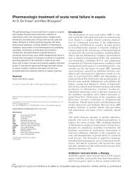
Pathophysiology<br />
Tubular epi<strong>the</strong>lium <strong>and</strong> acute tubular necrosis<br />
The clinical syndrome of ARF in <strong>the</strong> setting of critical illness, manifested by<br />
rising serum creatinine <strong>and</strong> decreasing urine output, results from injury to <strong>the</strong><br />
tubular epi<strong>the</strong>lial cells, or acute tubular necrosis (Fig. 1). Ischemic or toxic injury<br />
primarily affects this renal compartment, both because this area is most dependent<br />
on downstream blood flow, <strong>and</strong> <strong>the</strong>se cells are highly metabolically active,<br />
engaged in solute <strong>and</strong> water transport. The tubular epi<strong>the</strong>lial cells most vulnerable<br />
to ischemia line <strong>the</strong> S3 segment of <strong>the</strong> proximal tubule.<br />
Lethal injury to <strong>the</strong>se cells (necrosis or apoptosis) leads to loss of cell adhesion<br />
to <strong>the</strong> tubular basement membrane <strong>and</strong> subsequent shedding into <strong>the</strong> lumen. The<br />
denuded cells appear in <strong>the</strong> urine intact as tubular epi<strong>the</strong>lial cell casts, or <strong>the</strong>y<br />
may degrade leading to excretion of granular casts, both of which are typically<br />
found in <strong>the</strong> urine of patients with acute tubular necrosis. Such casts may cause<br />
micro-obstruction to urine flow. The damaged tubular basement membrane may<br />
fill with cast material, cellular debris, <strong>and</strong> Tamm-Horsfall protein. Sublethal<br />
injury results in loss of <strong>the</strong> brush border, which is <strong>the</strong> site of much energyconsuming<br />
metabolic activity.<br />
The mechanisms of injury to tubular epi<strong>the</strong>lial cells in <strong>sepsis</strong> are difficult to<br />
reproduce in <strong>the</strong> laboratory. Laboratory models of acute tubular necrosis have<br />
Pathophysiology of Ischemic Acute Renal Failure<br />
MICROVASCULAR<br />
Glomerular<br />
Medullary<br />
Vasoconstriction in response to:<br />
endo<strong>the</strong>lin, adenosine,<br />
angiotensin II, thromboxane A2,<br />
leukotrienes, sympa<strong>the</strong>tic nerve<br />
activity<br />
Vasodilation in response to:<br />
nitric oxide, PGE2, acetylcholine<br />
bradykinin<br />
Endo<strong>the</strong>lial <strong>and</strong> vascular smooth<br />
muscle cell structural damage<br />
Leukocyte-Endo<strong>the</strong>lial adhesion<br />
vascular obstruction, leukocyte<br />
activation, <strong>and</strong> inflammation<br />
O 2<br />
Inflammatory<br />
<strong>and</strong><br />
vasoactive<br />
mediators<br />
TUBULAR<br />
Cytoskeletal breakdown<br />
Loss of polarity<br />
Apoptosis <strong>and</strong> Necrosis<br />
Desquamation of viable<br />
<strong>and</strong> necrotic cells<br />
Tubular obstruction<br />
Backleak<br />
Fig. 1. Pathophysiology of ischemic acute renal failure. PGE2, prostagl<strong>and</strong>in E 2 .