Left-Sided Portal Hypertension - SASSiT
Left-Sided Portal Hypertension - SASSiT
Left-Sided Portal Hypertension - SASSiT

You also want an ePaper? Increase the reach of your titles
YUMPU automatically turns print PDFs into web optimized ePapers that Google loves.
1142 Dig Dis Sci (2007) 52:1141–1149<br />
major splanchnic veins was 13%, with the splenic vein being<br />
occluded in 8% of patients, the portal vein in 4%, and the<br />
superior mesenteric vein in 1% [10].<br />
Synonyms<br />
LSPH has also been referred to as segmental [11], sinistral<br />
[7], regional [4], localized [12], compartmental [13], lineal<br />
[14], or splenoportal hypertension [15].<br />
Anatomy<br />
The splenic vein is a large and nontortuous vessel formed<br />
by five or six tributaries from the spleen. It lies inferior to<br />
the splenic artery and, after leaving the splenic hilus, runs<br />
behind the tail and the body of the pancreas. It is approximately<br />
0.5 cm in diameter and 12 cm long. The splenic vein<br />
crosses anterior to the left kidney, being separated from the<br />
left sympathetic trunk and crus by the left renal vessels and<br />
from the abdominal aorta by the superior mesenteric artery<br />
and left renal vein [16]. The tributaries of the splenic vein<br />
include the short gastric, left gastroepiploic, pancreatic, and<br />
inferior mesenteric veins. Behind the neck of the pancreas<br />
the splenic vein joins the superior mesenteric vein to form the<br />
portal vein. Since the splenic vein is contiguous with the<br />
pancreas throughout that organ’s entire length, pancreatic<br />
disorders contribute the main etiology, and any significant<br />
pancreatic pathology may be complicated with venous obstruction<br />
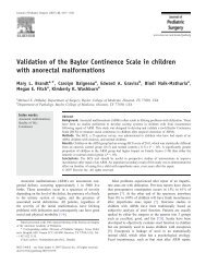
(Fig. 1). There is also close approximation between<br />
the splenic vein and the neighboring pancreatolienal lymph<br />
nodes. Therefore, even retroperitoneal diseases may contribute<br />
to splenic vein occlusion [17–21].<br />
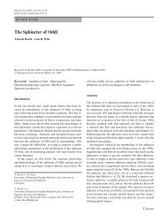
Fig. 1 Illustration of splenic venous thrombosis and fundal varices.<br />
CV, coronary veins; GEV, gastroepiploic vein; PV, portal vein; SV,<br />
splenic vein; SMV, superior mesenteric vein<br />
Pathophysiology<br />
Blood flow through the splenic vein may be blocked secondary<br />
to either thrombosis formation or neighboring mass<br />
effect. Splenic vein occlusion results in venous hypertension<br />
in collateral pathways that carry splenic arterial blood to<br />
the superior mesenteric and portal veins including the short<br />
gastric, coronary, and gastroepiploic veins and the veins located<br />
in the upper half of the stomach. Following obstruction,<br />
splenic blood typically drains through the short gastric veins<br />
to the stomach. In the gastric wall veins of the fundus, blood<br />
flow and pressure increase and submucosal structures consequently<br />
dilate, producing gastric varices. Eventual decompression<br />
into the portal system occurs through the coronary<br />
and epiploic veins. The coronary vein drains to different parts<br />
of the portal system (directly to the portal vein, to the junction<br />
of the splenic and portal veins, and to the splenic vein).<br />
When the coronary vein drains distal to the obstruction in<br />
the splenic vein, esophageal varices may occur alone or in<br />
combination with gastric varices [2, 7, 22]. However, due to<br />
several anatomic variations, obstruction of the splenic vein<br />
may not always result in portal hypertension or formation of<br />
varices.<br />
Etiology<br />
The main cause of LSPH is splenic vein thrombosis (SVT).<br />
Rare causes of LSPH include compression of the splenic<br />
vein by other organs, edema, enlarged lymphadenopathies,<br />
and splenic artery aneurysm. There is a strong association<br />
between pancreatic disorders and SVT because of the splenic<br />
vein’s location. Because the splenic vein is posterior to the<br />
pancreas and in direct contact with it, any type of pancreatic<br />
disease is likely to involve the splenic vein [3, 8, 16].<br />
Acute and chronic pancreatitis and pancreas neoplasms are<br />
the most common causes of SVT [3, 8, 10, 16, 23–26]. In<br />
an early report in 1970, Sutton et al. found that 35% of their<br />
cases of isolated SVT were caused by tumors and only 17%<br />
by pancreatitis [4]. More recent reviews have found acute<br />
or chronic pancreatitis to be the probable cause of isolated<br />
SVT in the majority of cases [2]. In a study by Moosa et al.,<br />
pancreatitis—diagnosed with biopsy or operation—was the<br />
etiology in 87 (60%) of 144 cases, while pancreas malignancy<br />
was detected in only 13 (9%) of the patients [16].<br />
The reason for this difference may be due to increases in the<br />
incidence of pancreatitis and in diagnostic activities, as well<br />
as to improvements in diagnostic procedures [27].<br />
Single episodes of acute pancreatitis may lead to SVT,<br />
and the risk of SVT does not correlate with the severity of<br />
pancreatitis. Also, SVT may occur silently, as a complication<br />
of mild pancreatitis [7, 8].<br />
Springer