Gray-scale and color Doppler US findings of amniotic sheets
Gray-scale and color Doppler US findings of amniotic sheets
Gray-scale and color Doppler US findings of amniotic sheets

You also want an ePaper? Increase the reach of your titles
YUMPU automatically turns print PDFs into web optimized ePapers that Google loves.
Diagn Interv Radiol DOI 10.4261/1305-3825.DIR.4696-11.1<br />
© Turkish Society <strong>of</strong> Radiology 2011<br />
FETAL IMAGING<br />
ORIGINAL ARTICLE<br />
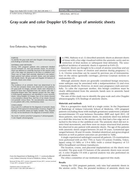
<strong>Gray</strong>-<strong>scale</strong> <strong>and</strong> <strong>color</strong> <strong>Doppler</strong> <strong>US</strong> <strong>findings</strong> <strong>of</strong> <strong>amniotic</strong> <strong>sheets</strong><br />
Esra Özkavukcu, Nuray Haliloğlu<br />
PURPOSE<br />
To identify the gray-<strong>scale</strong> <strong>and</strong> <strong>color</strong> <strong>Doppler</strong> ultrasonography<br />
(<strong>US</strong>) <strong>findings</strong> <strong>of</strong> <strong>amniotic</strong> <strong>sheets</strong>.<br />
MATERIALS AND METHODS<br />
Among 1201 pregnant patients who underwent detailed<br />
second trimester <strong>US</strong>, nine had <strong>amniotic</strong> <strong>sheets</strong>. An <strong>amniotic</strong><br />
sheet was defined as a shelf-like structure in the uterine cavity<br />
with a free edge not attached to the fetus or umbilical cord.<br />
There was no major fetal anomaly observed in any patient.<br />
Eight patients had solitary <strong>amniotic</strong> <strong>sheets</strong>, <strong>and</strong> one patient<br />
had double <strong>sheets</strong>. All gray-<strong>scale</strong> <strong>and</strong> <strong>Doppler</strong> <strong>US</strong> features <strong>of</strong><br />
<strong>amniotic</strong> <strong>sheets</strong> were noted.<br />
RESULTS<br />
The incidence <strong>of</strong> an <strong>amniotic</strong> sheet was determined to be<br />
0.75% (ten <strong>amniotic</strong> <strong>sheets</strong> were observed in nine patients).<br />
On gray-<strong>scale</strong> <strong>US</strong> images, <strong>amniotic</strong> <strong>sheets</strong> were observed as<br />
b<strong>and</strong>s <strong>of</strong> tissue that originated from the uterine wall with a<br />
triangular-shaped base that tapered toward the free edge.<br />
A three-layered appearance was identified in seven <strong>amniotic</strong><br />
<strong>sheets</strong>. Using <strong>Doppler</strong> <strong>US</strong> images, four <strong>of</strong> ten <strong>sheets</strong> showed<br />
a low-resistance arterial flow, <strong>and</strong> five <strong>of</strong> ten <strong>sheets</strong> showed<br />
non-pulsatile venous flows. No vascularization was observed<br />
in one patient with a thin, membranous sheet.<br />
CONCL<strong>US</strong>ION<br />
<strong>Gray</strong>-<strong>scale</strong> <strong>US</strong> is sufficient for the diagnosis <strong>of</strong> <strong>amniotic</strong> <strong>sheets</strong><br />
because <strong>of</strong> the typical <strong>US</strong> characteristics; however, <strong>Doppler</strong> <strong>US</strong><br />
<strong>findings</strong> <strong>of</strong> <strong>amniotic</strong> <strong>sheets</strong> are highly variable. Thus, <strong>Doppler</strong><br />
<strong>US</strong> may not be beneficial in the diagnosis <strong>of</strong> <strong>amniotic</strong> <strong>sheets</strong>.<br />
Key words: • uterine synechiae • pregnancy • ultrasonography<br />
• <strong>color</strong> <strong>Doppler</strong> ultrasonography<br />
From the Department <strong>of</strong> Radiology (E.Ö. eozkavukcu@gmail.<br />
com), Ankara University School <strong>of</strong> Medicine, Ankara, Turkey.<br />
Received 29 May 2011; revision requested 24 June 2011; revision received<br />
15 July 2011; accepted 19 July 2011.<br />
Published online 6 October 2011<br />
DOI 10.4261/1305-3825.DIR.4696-11.1<br />
In 1985, Mahony et al. (1) described <strong>amniotic</strong> <strong>sheets</strong> as aberrant <strong>sheets</strong><br />
<strong>of</strong> tissue with a free edge visualized within the <strong>amniotic</strong> cavity <strong>and</strong> no<br />
restriction <strong>of</strong> fetal motion or subsequent fetal deformity. The sonographical<br />
incidence <strong>of</strong> <strong>amniotic</strong> <strong>sheets</strong> is reported as 0.6% (2).<br />
Amniotic <strong>sheets</strong> are thought to be a result <strong>of</strong> uterine synechiae <strong>and</strong> are<br />
covered with two layers <strong>of</strong> chorion <strong>and</strong> amnion during pregnancy (1,<br />
3, 4). Uterine synechiae can be caused by previous use <strong>of</strong> instrumentation<br />
on the uterus (generally curettage), previous Cesarean sections or<br />
endometritis (2).<br />
Although <strong>amniotic</strong> <strong>sheets</strong> are generally considered benign structures,<br />
some subtypes may be associated with a malpresentation (5) <strong>and</strong> even<br />
intrauterine death (6). However, in most cases pregnancy ends uneventfully.<br />
To calm the expectant mother, this benign condition must be<br />
clearly differentiated from the <strong>amniotic</strong> b<strong>and</strong>s seen in <strong>amniotic</strong> b<strong>and</strong><br />
syndrome (ABS).<br />
The aim <strong>of</strong> this study was to identify the gray-<strong>scale</strong> <strong>and</strong> <strong>color</strong> <strong>Doppler</strong><br />
ultrasonography (<strong>US</strong>) <strong>findings</strong> <strong>of</strong> <strong>amniotic</strong> <strong>sheets</strong>.<br />
Materials <strong>and</strong> methods<br />
This is a prospective study held at a single center. In the Department<br />
<strong>of</strong> Radiology <strong>of</strong> Ankara University School <strong>of</strong> Medicine, 1201 pregnant<br />
patients (excluding those with multiple gestations) underwent a detailed<br />
second-trimester <strong>US</strong> scan between December 2009 <strong>and</strong> April 2011. Of<br />
these patients, nine had <strong>amniotic</strong> <strong>sheets</strong>. An <strong>amniotic</strong> sheet was defined<br />
as a shelf-like structure in the uterine cavity that had a free edge not attached<br />
to the fetus or the umbilical cord. The <strong>amniotic</strong> sheet did not restrict<br />
fetal movements, <strong>and</strong> there were no major structural fetal anomalies<br />
observed in any patient with <strong>amniotic</strong> <strong>sheets</strong>. The age <strong>of</strong> the patients<br />
with <strong>amniotic</strong> <strong>sheets</strong> ranged between 24 <strong>and</strong> 39 years. Gestational ages<br />
ranged between 18 <strong>and</strong> 24 weeks. Detailed obstetrical <strong>and</strong> gynecological<br />
histories as well as patient outcomes are provided in Table.<br />
A single experienced radiologist scanned all patients. <strong>US</strong> examinations<br />
were performed with an SSA 770A or Xario <strong>US</strong> system (Toshiba, Tokyo,<br />
Japan) using 3.5 MHz or 1.9–6 MHz (with a central frequency <strong>of</strong> 3.5<br />
MHz) broadb<strong>and</strong> curvilinear transducers.<br />
The location, course, <strong>and</strong> placental implantations on the <strong>sheets</strong> were<br />
recorded. The gray-<strong>scale</strong> <strong>US</strong> features <strong>of</strong> the <strong>amniotic</strong> <strong>sheets</strong> were recorded.<br />
A <strong>color</strong> <strong>Doppler</strong> <strong>US</strong> with a spectral analysis was also performed on<br />
all <strong>amniotic</strong> <strong>sheets</strong>.<br />
The ethics committee <strong>of</strong> our institution approved this study.<br />
Results<br />
Among the 1201 pregnant patients, only nine had <strong>amniotic</strong> <strong>sheets</strong> in<br />
the uterine cavity. Thus, the prenatal incidence <strong>of</strong> <strong>amniotic</strong> <strong>sheets</strong> was
a<br />
b<br />
Figure 1. a, b. Sagittal <strong>US</strong> views <strong>of</strong> the patient no. 2 showing an <strong>amniotic</strong> sheet originating from the uterine wall with a triangular-shaped base<br />
(arrows, a) <strong>and</strong> a tapered, membrane-like <strong>amniotic</strong> sheet (curved arrow, b).<br />
determined to be 0.75%. All <strong>amniotic</strong><br />
<strong>sheets</strong> were classified as incomplete<br />
<strong>sheets</strong>, according to the classification<br />
outlined by Tan et al. (6). Eight patients<br />
had solitary <strong>amniotic</strong> <strong>sheets</strong>, whereas<br />
one patient had two <strong>sheets</strong>. Five <strong>amniotic</strong><br />
<strong>sheets</strong> were located in the transverse<br />
plane, whereas five <strong>sheets</strong> were longitudinally<br />
positioned in the uterine cavity.<br />
On the gray-<strong>scale</strong> <strong>US</strong>, <strong>amniotic</strong> <strong>sheets</strong><br />
were observed as b<strong>and</strong>s <strong>of</strong> tissue originating<br />
from the uterine wall with a triangular-shaped<br />
base (Fig. 1a). Amniotic<br />
<strong>sheets</strong> tapered toward the free edge,<br />
with some resembling the thinness <strong>of</strong><br />
a membrane (Fig. 1b). Sonographically,<br />
three layers were identified in seven<br />
<strong>of</strong> the ten <strong>sheets</strong> with two echogenic<br />
lines <strong>and</strong> a hypoechoic line located in<br />
between (Fig. 2). Placental extension <strong>of</strong><br />
Figure 2. A sagittal <strong>US</strong> view <strong>of</strong> the patient no. 3 showing the three-layered <strong>amniotic</strong> sheet<br />
(arrow) with two echogenic lines <strong>and</strong> a hypoechoic line located in between.<br />
Table. The obstetrical <strong>and</strong> gynecological history <strong>of</strong> study patients<br />
Previous Previous Previous<br />
Spontaneous Fetal Mode <strong>of</strong> delivery<br />
No. Gravidity Parity D/C C/S uterine surgery<br />
abortion outcome (indication)<br />
1 4 1 1 0 0 1 N C/S (cephalopelvic disproportion)<br />
2 3 2 0 0 0 0 N C/S (breech presentation)<br />
3 2 0 1 0 0 0 N Vaginal delivery<br />
4 1 0 0 0 0 0 N Vaginal delivery<br />
5 3 1 1 0 0 0 N/A N/A<br />
6 1 0 0 0 0 0 N/A N/A<br />
7 3 1 1 0 0 0 N Vaginal delivery<br />
8 3 1 0 0 0 1 N C/S (previous C/S)<br />
9 2 0 1 0 0 0 N/A N/A<br />
D/C, dilatation <strong>and</strong> curettage; C/S, Cesarean section; N, normal; N/A, not available (two patients were lost during follow-up, <strong>and</strong> one has not yet delivered)<br />
ii • Diagnostic <strong>and</strong> Interventional Radiology<br />
Özkavukcu <strong>and</strong> Haliloğlu
Figure 3. A sagittal <strong>US</strong> view <strong>of</strong> the patient no. 1 showing the placenta<br />
(arrow) extending into the <strong>amniotic</strong> sheet.<br />
Figure 4. A sagittal <strong>US</strong> view <strong>of</strong> the patient no. 5 showing the “thick”<br />
free edge <strong>of</strong> the incomplete <strong>amniotic</strong> sheet (arrows).<br />
a<br />
b<br />
Figure 5. a, b. A sagittal <strong>US</strong> view (a) <strong>of</strong> the patient no. 2 showing a relatively thick <strong>amniotic</strong> sheet (arrows) with placental extension. A <strong>color</strong><br />
<strong>Doppler</strong> <strong>US</strong> view (b) showing substantial arterial vascularization. Spectral analysis is not shown.<br />
the <strong>amniotic</strong> <strong>sheets</strong> was noted in three<br />
<strong>of</strong> the nine patients with <strong>amniotic</strong><br />
<strong>sheets</strong> (Fig. 3), <strong>and</strong> we did not observe<br />
any “bulbous tips” (Fig. 4). Fetuses always<br />
moved freely, independent <strong>of</strong> the<br />
<strong>amniotic</strong> <strong>sheets</strong>, <strong>and</strong> no major malformations<br />
were noted in any fetus.<br />
On the <strong>color</strong> <strong>Doppler</strong> <strong>US</strong>, the patients<br />
with relatively thick <strong>sheets</strong> also<br />
had placental implantations on the<br />
<strong>amniotic</strong> <strong>sheets</strong>, <strong>and</strong> substantial arterial<br />
vascularization (similar to that observed<br />
at the uteroplacental junction)<br />
was also noted (Fig. 5). However, thin<br />
parts <strong>of</strong> the <strong>sheets</strong> showed poor vascularization<br />
on the <strong>color</strong> <strong>and</strong> power<br />
<strong>Doppler</strong> <strong>US</strong> images. A spectral analysis<br />
<strong>of</strong> these thin <strong>sheets</strong> revealed a lowresistance<br />
arterial flow (in four <strong>of</strong> ten<br />
<strong>sheets</strong>) (Fig. 6) or non-pulsatile venous<br />
flow (in five <strong>of</strong> ten <strong>sheets</strong>) (Fig. 7). The<br />
heart rates obtained from the arterial<br />
flows were always <strong>of</strong> maternal origin,<br />
suggesting that the <strong>amniotic</strong> <strong>sheets</strong> are<br />
<strong>of</strong> a maternal rather than fetal origin<br />
(Fig. 6b). No vascularization was noted<br />
on the <strong>Doppler</strong> <strong>US</strong> in one patient with<br />
a thin, membranous sheet.<br />
Discussion<br />
In our study, the incidence <strong>of</strong> <strong>amniotic</strong><br />
<strong>sheets</strong> was 0.75%. This percentage<br />
is comparable to those reported in<br />
previous retrospective studies (2, 5–7).<br />
There are a few reasons for the negligible<br />
difference. First, a single radiologist<br />
with 10 years <strong>of</strong> experience in obstetrical<br />
<strong>US</strong> imaging performed all <strong>US</strong><br />
scans in our study; thus, there were no<br />
interobserver variations. Furthermore,<br />
<strong>amniotic</strong> <strong>sheets</strong> were specifically examined<br />
for during all <strong>US</strong> scans.<br />
Tan et al. (6) classified <strong>amniotic</strong><br />
<strong>sheets</strong> into complete <strong>and</strong> incomplete<br />
<strong>sheets</strong>. A complete sheet only has a<br />
small perforation that is not visible<br />
on <strong>US</strong> scans, whereas an incomplete<br />
sheet has a free-floating edge. They<br />
suggested that although the incomplete<br />
<strong>sheets</strong> are benign, complete <strong>amniotic</strong><br />
<strong>sheets</strong> may be associated with<br />
intrauterine death. The authors hypothesized<br />
that the small defects on<br />
the complete <strong>sheets</strong> may predispose<br />
the mother <strong>and</strong> fetus to cord prolapse<br />
<strong>and</strong> intrauterine death (6). None <strong>of</strong><br />
our patients had a complete sheet<br />
or poor pregnancy outcome, which<br />
suggests that complete <strong>sheets</strong> are extremely<br />
rare.<br />
<strong>Gray</strong>-<strong>scale</strong> <strong>and</strong> <strong>color</strong> <strong>Doppler</strong> <strong>US</strong> <strong>findings</strong> <strong>of</strong> <strong>amniotic</strong> <strong>sheets</strong> • iii
a<br />
b<br />
The most important differential diagnosis<br />
<strong>of</strong> <strong>amniotic</strong> <strong>sheets</strong> is ABS. ABS<br />
is thought to result from a defect in<br />
the <strong>amniotic</strong> membrane that exposes<br />
the fetus to the chorionic cavity.<br />
iv • Diagnostic <strong>and</strong> Interventional Radiology<br />
Figure 6. a, b. A sagittal<br />
gray-<strong>scale</strong> <strong>US</strong> view (a)<br />
<strong>of</strong> the patient no. 5<br />
showing the double<br />
<strong>amniotic</strong> <strong>sheets</strong> (arrows),<br />
located close to one<br />
another (C, cervix). A<br />
<strong>color</strong> <strong>Doppler</strong> <strong>US</strong> <strong>and</strong><br />
spectral analysis (b) <strong>of</strong><br />
the thicker <strong>amniotic</strong><br />
sheet revealed a lowresistance<br />
arterial flow<br />
<strong>and</strong> a heart rate <strong>of</strong><br />
maternal origin.<br />
Figure 7. A <strong>color</strong> <strong>Doppler</strong> <strong>US</strong> <strong>of</strong> the patient no. 6 <strong>and</strong> spectral analysis <strong>of</strong> the <strong>amniotic</strong> sheet<br />
showing a non-pulsatile venous flow.<br />
Serious fetal malformations can occur<br />
in ABS, such as limb, trunk, vertebral,<br />
cranial, facial, <strong>and</strong> abdominal abnormalities<br />
(1). The <strong>amniotic</strong> b<strong>and</strong>s in<br />
ABS are observed as membranes that<br />
flap with fetal movements or attach to<br />
the fetus <strong>and</strong> restrict fetal movement<br />
(1). Conversely, <strong>amniotic</strong> <strong>sheets</strong> do not<br />
attach to the fetus or umbilical cord,<br />
<strong>and</strong> the fetus can move independently.<br />
The number <strong>of</strong> <strong>amniotic</strong> b<strong>and</strong>s in ABS<br />
can range from one to many. Amniotic<br />
<strong>sheets</strong> are usually solitary, but multiple<br />
<strong>sheets</strong> have also been reported<br />
(8). There are no major structural fetal<br />
anomalies that accompany <strong>amniotic</strong><br />
<strong>sheets</strong>. Because <strong>amniotic</strong> <strong>sheets</strong> consist<br />
<strong>of</strong> two layers <strong>of</strong> chorion <strong>and</strong> amnion,<br />
whereas <strong>amniotic</strong> b<strong>and</strong>s consist<br />
<strong>of</strong> only a single layer <strong>of</strong> amnion (9),<br />
<strong>amniotic</strong> <strong>sheets</strong> appear thicker than<br />
<strong>amniotic</strong> b<strong>and</strong>s (3). Amniotic <strong>sheets</strong><br />
are similar to the inter-twin membrane<br />
seen in dichorionic-di<strong>amniotic</strong> twins<br />
(3). This layered <strong>and</strong> relatively thick<br />
shelf-like appearance was noted in all<br />
<strong>of</strong> our patients, with the exception<br />
<strong>of</strong> one patient who had a thin, taut<br />
sheet. Additionally, the sheet base was<br />
similar to the “twin peak sign” seen in<br />
dichorionic-di<strong>amniotic</strong> twins. Other<br />
useful sonographic features <strong>of</strong> <strong>amniotic</strong><br />
<strong>sheets</strong> are the broad base at the<br />
origin on the uterine wall <strong>and</strong> the bulbous<br />
free edge or tip (1, 3, 9, 10). In our<br />
study, the triangular broad base was always<br />
present, except in the one patient<br />
with a thin, membranous <strong>amniotic</strong><br />
sheet; no patients exhibited a bulbous<br />
free edge. The free edge was difficult to<br />
see <strong>and</strong> required scanning in different<br />
planes. The free edges always appeared<br />
thick <strong>and</strong> taut rather than flapping.<br />
Occasionally, the placenta appeared<br />
to be implanted on the <strong>amniotic</strong> sheet<br />
(9, 10). Korbin et al. (10) found that<br />
the placenta appeared to be implanted<br />
on the <strong>amniotic</strong> sheet in 26.1% <strong>of</strong> the<br />
cases. They also concluded that pregnancy<br />
outcomes are similar in patients<br />
with <strong>and</strong> without placental implantation<br />
on the <strong>amniotic</strong> sheet (10). In our<br />
study, three <strong>of</strong> the nine patients had<br />
placental implantation on the <strong>amniotic</strong><br />
sheet.<br />
Previous publications regarding<br />
<strong>Doppler</strong> analysis <strong>of</strong> the <strong>amniotic</strong><br />
<strong>sheets</strong> were case reports (11, 12), thus<br />
making this the first original article<br />
on <strong>Doppler</strong> analysis <strong>of</strong> the <strong>amniotic</strong><br />
<strong>sheets</strong> in a series <strong>of</strong> patients. Some<br />
authors claimed that <strong>color</strong> <strong>Doppler</strong><br />
<strong>US</strong> imaging with spectral analysis has<br />
an important role in the differential<br />
diagnosis <strong>of</strong> intrauterine membranes<br />
<strong>of</strong> undetermined origin during pregnancy<br />
(11, 12). We also showed that<br />
Özkavukcu <strong>and</strong> Haliloğlu
all <strong>amniotic</strong> <strong>sheets</strong> that had an arterial<br />
flow revealed a maternal heart rate, indicating<br />
the maternal origin <strong>of</strong> these<br />
benign structures; however, five <strong>of</strong><br />
ten <strong>sheets</strong> showed only venous flows.<br />
Because the <strong>Doppler</strong> <strong>findings</strong> varied,<br />
the use <strong>of</strong> <strong>Doppler</strong> <strong>US</strong> to diagnose<br />
<strong>amniotic</strong> <strong>sheets</strong> is debatable. Other<br />
conditions that can mimic <strong>amniotic</strong><br />
<strong>sheets</strong> include chorio<strong>amniotic</strong> separation,<br />
vanished twin, uterine septum,<br />
<strong>and</strong> circumvallate placenta (2, 3). The<br />
course <strong>of</strong> the sheet in the uterus may<br />
help to differentiate an <strong>amniotic</strong> sheet<br />
from a septate uterus. The septum <strong>of</strong><br />
a septate uterus is <strong>of</strong>ten in the fundus<br />
<strong>and</strong> is oriented in the sagittal plane (8,<br />
10). Amniotic <strong>sheets</strong> in our study were<br />
oriented either in the transverse or sagittal<br />
planes. Korbin et al. (10) suggested<br />
that the myometrial tissues can <strong>of</strong>ten<br />
be seen extending into the base <strong>of</strong> the<br />
septum in a septate uterus <strong>and</strong> that<br />
previous radiological examinations <strong>of</strong><br />
the uterus may also be helpful in differential<br />
diagnosis. Nonetheless, it may<br />
not always be possible to differentiate a<br />
septate uterus from an <strong>amniotic</strong> sheet<br />
using only <strong>US</strong> scans.<br />
There were two major limitations<br />
in our study. First, the structure <strong>of</strong> an<br />
<strong>amniotic</strong> sheet has not been proven<br />
histopathologically. Second, there was<br />
a small number <strong>of</strong> patients enrolled in<br />
our study.<br />
In conclusion, gray-<strong>scale</strong> <strong>US</strong> <strong>findings</strong><br />
alone are sufficient for the diagnosis <strong>of</strong><br />
<strong>amniotic</strong> <strong>sheets</strong> because they have typical<br />
<strong>US</strong> appearances. <strong>Doppler</strong> <strong>US</strong> images<br />
<strong>of</strong> <strong>amniotic</strong> <strong>sheets</strong> show the variable<br />
flow patterns <strong>of</strong> arterial or venous<br />
origins; thus, <strong>Doppler</strong> <strong>US</strong> may not be<br />
beneficial in the diagnosis <strong>of</strong> <strong>amniotic</strong><br />
<strong>sheets</strong>.<br />
Conflict <strong>of</strong> interest disclosure<br />
The authors declared no conflicts <strong>of</strong> interest.<br />
References<br />
1. Mahony BS, Filly RA, Callen PW, et al. The<br />
<strong>amniotic</strong> b<strong>and</strong> syndrome: antenatal sonographic<br />
diagnosis <strong>and</strong> potential pitfalls.<br />
Am J Obstet Gynecol 1985; 152:63–68.<br />
2. Sistrom CL, Ferguson JE. Abnormal membranes<br />
in obstetrical ultrasound: incidence<br />
<strong>and</strong> significance <strong>of</strong> <strong>amniotic</strong> <strong>sheets</strong> <strong>and</strong><br />
circumvallate placenta. Ultrasound Obstet<br />
Gynecol 1993; 3:249–255.<br />
3. R<strong>and</strong>el SB, Filly RA, Callen PW, et al.<br />
Amniotic <strong>sheets</strong>. Radiology 1988; 166:633–<br />
636.<br />
4. Stamm E, Waldstein G, Thickman D, et<br />
al. Amniotic <strong>sheets</strong>: natural history <strong>and</strong><br />
histology. J Ultrasound Med 1991; 10:501–<br />
504.<br />
5. Lazebnik N, Hill LM, Many A, et al. The<br />
effect <strong>of</strong> <strong>amniotic</strong> sheet orientation on<br />
subsequent maternal <strong>and</strong> fetal complications.<br />
Ultrasound Obstet Gynecol 1996;<br />
8:267–271.<br />
6. Tan KB, Tan TY, Tan JV, et al. The <strong>amniotic</strong><br />
sheet: a truly benign condition?<br />
Ultrasound Obstet Gynecol 2005; 26:639–<br />
643.<br />
7. Ball RH, Buchmeier SE, Longnecker M.<br />
Clinical significance <strong>of</strong> sonographically<br />
detected uterine synechiae in pregnant<br />
patients. J Ultrasound Med 1997; 16:465–<br />
469.<br />
8. Brown DL, Felker RE, Emerson DS.<br />
Intrauterine shelves in pregnancy: sonographic<br />
observations. AJR Am J Roentgenol<br />
1989; 153:821–824.<br />
9. Finberg HJ. Uterine synechiae in pregnancy:<br />
exp<strong>and</strong>ed criteria for recognition<br />
<strong>and</strong> clinical significance in 28 cases. J<br />
Ultrasound Med 1991; 10:547–555.<br />
10. Korbin CD, Benson CB, Doubilet PM.<br />
Placental implantation on the <strong>amniotic</strong><br />
sheet: effect on pregnancy outcome.<br />
Radiology 1998; 206:773–775.<br />
11. Abuhamad AZ, Romero R, Shaffer WK, et<br />
al. The value <strong>of</strong> <strong>Doppler</strong> flow analysis in<br />
the prenatal diagnosis <strong>of</strong> <strong>amniotic</strong> <strong>sheets</strong>. J<br />
Ultrasound Med 1992; 11:623–624.<br />
12. Sherer DM, Lysikiewicz AJ. <strong>Doppler</strong> flow<br />
velocimetry assisted diagnosis <strong>of</strong> an intrauterine<br />
synechia during pregnancy. Am J<br />
Perinatol 2002; 19:421–426.<br />
<strong>Gray</strong>-<strong>scale</strong> <strong>and</strong> <strong>color</strong> <strong>Doppler</strong> <strong>US</strong> <strong>findings</strong> <strong>of</strong> <strong>amniotic</strong> <strong>sheets</strong> • v