Radiological report: expectations of clinicians - Diagnostic and ...
Radiological report: expectations of clinicians - Diagnostic and ...
Radiological report: expectations of clinicians - Diagnostic and ...

Create successful ePaper yourself
Turn your PDF publications into a flip-book with our unique Google optimized e-Paper software.
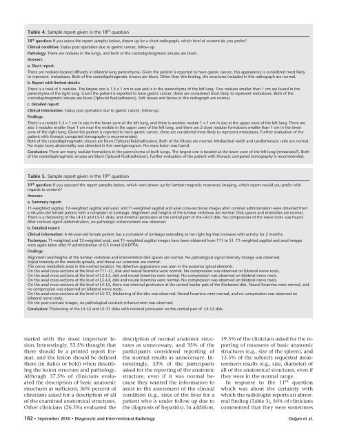
Table 4. Sample <strong>report</strong> given in the 18 th question<br />
18 th question: If you assess the <strong>report</strong> samples below, drawn up for a chest radiograph, which level <strong>of</strong> content do you prefer?<br />
Clinical condition: Status post operation due to gastric cancer, follow-up.<br />
Pathology: There are nodules in the lungs, <strong>and</strong> both <strong>of</strong> the costodiaphragmatic sinuses are blunt.<br />
Answers:<br />
a. Short <strong>report</strong>:<br />
There are nodules located diffusely in bilateral lung parenchyma. Given the patient is <strong>report</strong>ed to have gastric cancer, this appearance is considered most likely<br />
to represent metastases. Both <strong>of</strong> the costodiaphragmatic sinuses are blunt. Other than this finding, the structures included in this radiograph are normal.<br />
b. Report with limited details:<br />
There is a total <strong>of</strong> 5 nodules. The largest one is 1.5 x 1 cm in size <strong>and</strong> is in the parenchyma <strong>of</strong> the left lung. Two nodules smaller than 1 cm are found in the<br />
parenchyma <strong>of</strong> the right lung. Given the patient is <strong>report</strong>ed to have gastric cancer, these are considered most likely to represent metastases. Both <strong>of</strong> the<br />
costodiaphragmatic sinuses are blunt (?pleural fluid/adhesion). S<strong>of</strong>t tissues <strong>and</strong> bones in this radiograph are normal.<br />
c. Detailed <strong>report</strong>:<br />
Clinical information: Status post operation due to gastric cancer, follow-up.<br />
Findings:<br />
There is a nodule 1.5 x 1 cm in size in the lower zone <strong>of</strong> the left lung, <strong>and</strong> there is another nodule 1 x 1 cm in size at the upper zone <strong>of</strong> the left lung. There are<br />
also 3 nodules smaller than 1 cm near the nodule in the upper zone <strong>of</strong> the left lung, <strong>and</strong> there are 2 close nodular formations smaller than 1 cm in the lower<br />
zone <strong>of</strong> the right lung. Given the patient is <strong>report</strong>ed to have gastric cancer, these are considered most likely to represent metastases. Further evaluation <strong>of</strong> the<br />
patient with thoracic computed tomography is recommended.<br />
Both <strong>of</strong> the costodiaphragmatic sinuses are blunt (?pleural fluid/adhesion). Both <strong>of</strong> the hiluses are normal. Mediastinal width <strong>and</strong> cardiothoracic ratio are normal.<br />
No major bony abnormality was detected in this roentgenogram. No mass lesion was found.<br />
Conclusion: There are many nodular formations in the parenchyma <strong>of</strong> both lungs. The largest one is located at the lower zone <strong>of</strong> the left lung (metastasis?). Both<br />
<strong>of</strong> the costodiaphragmatic sinuses are blunt (?pleural fluid/adhesion). Further evaluation <strong>of</strong> the patient with thoracic computed tomography is recommended.<br />
Table 5. Sample <strong>report</strong> given in the 19 th question<br />
19 th question: If you assessed the <strong>report</strong> samples below, which were drawn up for lumbar magnetic resonance imaging, which <strong>report</strong> would you prefer with<br />
regards to content?<br />
Answers:<br />
a. Summary <strong>report</strong>:<br />
T1-weighted sagittal, T2-weighted sagittal <strong>and</strong> axial, <strong>and</strong> T1-weighted sagittal <strong>and</strong> axial cross-sectional images after contrast administration were obtained from<br />
a 46-year-old female patient with a complaint <strong>of</strong> lumbago. Alignment <strong>and</strong> heights <strong>of</strong> the lumbar vertebrae are normal. Disk spaces <strong>and</strong> intensities are normal.<br />
There is a thickening <strong>of</strong> the L4-L5 <strong>and</strong> L5-S1 disks, <strong>and</strong> minimal protrusion at the central part <strong>of</strong> the L4-L5 disk. No compression <strong>of</strong> the nerve roots was found.<br />
After contrast agent administration, no pathologic enhancement was observed.<br />
b. Detailed <strong>report</strong>:<br />
Clinical information: A 46-year-old female patient has a complaint <strong>of</strong> lumbago extending to her right leg that increases with activity for 2 months.<br />
Technique: T1-weighted <strong>and</strong> T2-weighted axial, <strong>and</strong> T1-weighted sagittal images have been obtained from T11 to S1. T1-weighted sagittal <strong>and</strong> axial images<br />
were again taken after IV administration <strong>of</strong> 0.5 mmol Gd-DTPA.<br />
Findings:<br />
Alignment <strong>and</strong> heights <strong>of</strong> the lumbar vertebrae <strong>and</strong> intervertebral disk spaces are normal. No pathological signal intensity change was observed.<br />
Signal intensity <strong>of</strong> the medulla spinalis, <strong>and</strong> thecal sac extension are normal.<br />
The conus medullaris ends in the normal location. No defective appearance was seen in the posterior spinal elements.<br />
On the axial cross-sections at the level <strong>of</strong> T11–L1, disk <strong>and</strong> neural foramina were normal. No compression was observed on bilateral nerve roots.<br />
On the axial cross-sections at the level <strong>of</strong> L2–L3, disk <strong>and</strong> neural foramina were normal. No compression was observed on bilateral nerve roots.<br />
On the axial cross-sections at the level <strong>of</strong> L3–L4, disk <strong>and</strong> neural foramina were normal. No compression was observed on bilateral nerve roots.<br />
On the axial cross-sections at the level <strong>of</strong> L4–L5, there was minimal protrusion at the central basilar part <strong>of</strong> the thickened disk. Neural foramina were normal, <strong>and</strong><br />
no compression was observed on bilateral nerve roots.<br />
On the axial cross-sections at the level <strong>of</strong> L5–S1, thickening <strong>of</strong> the disc was observed. Neural foramina were normal, <strong>and</strong> no compression was observed on<br />
bilateral nerve roots.<br />
On the post-contrast images, no pathological contrast enhancement was observed.<br />
Conclusion: Thickening <strong>of</strong> the L4–L5 <strong>and</strong> L5–S1 disks with minimal protrusion on the central part <strong>of</strong> L4–L5 disk.<br />
started with the most important lesion.<br />
Interestingly, 53.5% thought that<br />
there should be a printed <strong>report</strong> format,<br />
<strong>and</strong> the lesion should be defined<br />
there (in italics or bold) when describing<br />
the lesion structure <strong>and</strong> pathology.<br />
Although 37.5% <strong>of</strong> <strong>clinicians</strong> evaluated<br />
the description <strong>of</strong> basic anatomic<br />
structures as sufficient, 36% percent <strong>of</strong><br />
<strong>clinicians</strong> asked for a description <strong>of</strong> all<br />
<strong>of</strong> the examined anatomical structures.<br />
Other <strong>clinicians</strong> (26.5%) evaluated the<br />
description <strong>of</strong> normal anatomic structures<br />
as unnecessary, <strong>and</strong> 35% <strong>of</strong> the<br />
participants considered <strong>report</strong>ing <strong>of</strong><br />
the normal results as unnecessary. Interestingly,<br />
32% <strong>of</strong> the participants<br />
asked for the <strong>report</strong>ing <strong>of</strong> the anatomic<br />
structure, even if it was normal because<br />
they wanted the information to<br />
assist in the assessment <strong>of</strong> the clinical<br />
condition (e.g., sizes <strong>of</strong> the liver for a<br />
patient who is under follow up due to<br />
the diagnosis <strong>of</strong> hepatitis). In addition,<br />
19.5% <strong>of</strong> the <strong>clinicians</strong> asked for the <strong>report</strong>ing<br />
<strong>of</strong> measures <strong>of</strong> basic anatomic<br />
structures (e.g., size <strong>of</strong> the spleen), <strong>and</strong><br />
13.5% <strong>of</strong> the subjects requested measurement<br />
results (e.g., size, diameter) <strong>of</strong><br />
all <strong>of</strong> the anatomical structures, even if<br />
they were in the normal range.<br />
In response to the 11 th question<br />
which was about the certainty with<br />
which the radiologist <strong>report</strong>s an abnormal<br />
finding (Table 3), 56% <strong>of</strong> <strong>clinicians</strong><br />
commented that they were sometimes<br />
182 • September 2010 • <strong>Diagnostic</strong> <strong>and</strong> Interventional Radiology<br />
Doğan et al.