Chemoembolic lobectomy - Diagnostic and Interventional Radiology
Chemoembolic lobectomy - Diagnostic and Interventional Radiology
Chemoembolic lobectomy - Diagnostic and Interventional Radiology

You also want an ePaper? Increase the reach of your titles
YUMPU automatically turns print PDFs into web optimized ePapers that Google loves.
Diagn Interv Radiol DOI 10.4261/1305-3825.DIR.3166-09.1<br />
© Turkish Society of <strong>Radiology</strong> 2010<br />
INTERVENTIONAL RADIOLOGY<br />
CASE REPORT<br />
<strong>Chemoembolic</strong> <strong>lobectomy</strong>: imaging findings of hepatic lobar<br />
volume reduction after transcatheter arterial chemoembolization<br />
Ron C. Gaba, Jason J. Carroll, James T. Bui, Tami C. Carrillo, M. Grace Knuttinen, Charles A. Owens<br />
ABSTRACT<br />
Hepatic lobar atrophy-hypertrophy complex formation is an<br />
uncommonly reported sequella of hepatic arterial embolotherapy<br />
procedures. Whereas radiation-induced hepatic lobar<br />
ablation has been described after intra-arterial therapy with<br />
yttrium-90 microspheres, this phenomenon has not been reported<br />
after transcatheter arterial chemoembolization. Here,<br />
we report a case of prominent hepatic lobar atrophy with<br />
contralateral lobar hypertrophy after chemoembolization <strong>and</strong><br />
suggest a mechanism by which arterial embolization contributes<br />
to the volumetric response.<br />
Key words: • chemoembolization • liver • atrophy • hypertrophy<br />
• <strong>lobectomy</strong><br />
From the Department of <strong>Radiology</strong> (R.C.G. rongaba@yahoo.<br />
com), University of Illinois at Chicago, Chicago, IL, USA.<br />
Received 12 November 2009; revision requested 22 November 2009;<br />
revision received 22 November 2009; accepted 23 November 2009.<br />
Published online 3 August 2010<br />
DOI 10.4261/1305-3825.DIR.3166-09.1<br />
Recently, there has been much interest in the concept of radiation-induced<br />
hepatic lobar ablation with marked volumetric loss<br />
<strong>and</strong> contralateral compensatory lobar hypertrophy (1–3). This<br />
phenomenon, termed radiation <strong>lobectomy</strong>, has been described to occur<br />
after yttrium-90 ( 90 Y) microsphere radioembolization <strong>and</strong> has been associated<br />
with high rates of tumor response <strong>and</strong> improved long-term patient<br />
survival in preliminary investigations (1). At present, the causative<br />
relationship between the administered radiation dose <strong>and</strong> the volumetric<br />
change is unknown, <strong>and</strong> the degree to which vascular embolization<br />
contributes to the observed changes remains uncertain. We recently encountered<br />
a striking case of hepatic lobar atrophy-hypertrophy complex<br />
formation after transcatheter arterial chemoembolization that suggests<br />
a possible contribution of arterial embolization to the volumetric response,<br />
which we present herein.<br />
Case report<br />
A 50-year-old man with a history of type 2 diabetes mellitus, hypertension,<br />
<strong>and</strong> hepatitis B virus liver disease was referred to interventional<br />
radiology for liver-directed therapy for the treatment of biopsy-proven<br />
hepatocellular carcinoma (HCC). The patient initially presented for assessment<br />
after screening computed tomography (CT) scan demonstrated<br />
a 4-cm right hepatic lobe mass, which was confirmed as HCC by image-guided<br />
core needle biopsy. Metastatic disease was ruled out with a<br />
chest CT <strong>and</strong> bone scan. The patient evaluation demonstrated an Eastern<br />
Cooperative Oncology Group performance status of zero (4), <strong>and</strong> the<br />
lab examination showed normal synthetic liver function (total bilirubin<br />
0.9 mg/dL, albumin 4.2 g/dL, <strong>and</strong> prothrombin time 10.0 s). The initial<br />
-fetoprotein level was only slightly elevated, measuring 15.5 ng/mL.<br />
Treatment options were discussed, <strong>and</strong> transcatheter arterial chemoembolization<br />
was elected for local tumor control.<br />
Subsequently, the patient underwent drug eluting bead chemoembolization<br />
using 300–500-micron LC beads (Angiodynamics; Queensbury,<br />
New York, USA) loaded with 50 mg doxorubicin <strong>and</strong> mixed with 50<br />
mg cisplatin <strong>and</strong> 20 mg mitomycin C in suspension. For chemoembolization,<br />
a st<strong>and</strong>ard right common femoral artery approach was used to<br />
position a 5 French reverse curve catheter in the celiac artery. The chemoembolic<br />
material was administered through a 2.8 French microcatheter<br />
placed coaxially in a segmental distribution via a right hepatic artery<br />
ascending branch (Fig. a) to a static angiographic endpoint. The patient<br />
had an uneventful post-procedure hospital course <strong>and</strong> was discharged<br />
24 hours after treatment.<br />
The patient follow-up included serial CT scans <strong>and</strong> lab assessment,<br />
which were performed initially at one month post-procedure <strong>and</strong> then<br />
at approximately three-month intervals. Although the one-month post-
a<br />
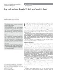
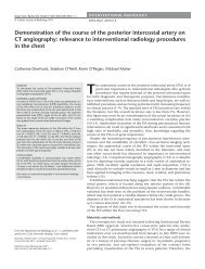
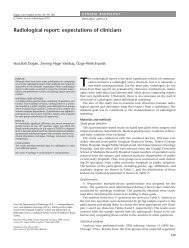
Figure. a–c. Fifty-year-old man with right lobe hepatocellular<br />
carcinoma. Right hepatic arteriogram (a) performed<br />
during the initial chemoembolization procedure showing<br />
the hypervascular tumor (arrowheads). One month posttreatment<br />
contrast-enhanced axial <strong>and</strong> maximum intensity<br />
projection CT scans (b) demonstrating low attenuation in the<br />
right lobe tumor, which indicates treatment response. The<br />
concurrent right <strong>and</strong> left hepatic lobe volumes were 796 mL<br />
<strong>and</strong> 437 mL, respectively. CT scan (c) obtained 35 months<br />
following initial therapy revealing complete tumor response<br />
with concomitant marked progressive right lobe atrophy to<br />
230 mL (71% reduction) <strong>and</strong> left lobe hypertrophy to 1129<br />
mL (158% enlargement).<br />
b<br />
c<br />
treatment CT scan showed good tumor<br />
response (Fig. b), the patient underwent<br />
two additional right hepatic lobe<br />
chemoembolization treatment sessions<br />
at three months (again using drug eluting<br />
beads) <strong>and</strong> seven months (a 20-mL<br />
volume of cisplatin, doxorubicin, <strong>and</strong><br />
mitomycin C solution mixed 1:1 with<br />
emulsifying iodized oil) after the initial<br />
therapy due to local residual <strong>and</strong>/or recurrent<br />
tumors. Additionally, because<br />
vague areas of atypical right hepatic<br />
lobe contrast enhancement were evident<br />
in the 12-month follow-up imaging,<br />
the patient underwent a right hepatic<br />
lobe biopsy, which demonstrated<br />
chemotherapy-related parenchymal fibrosis<br />
<strong>and</strong> a giant cell response without<br />
neoplasm. Surveillance imaging was<br />
continued, <strong>and</strong> a CT scan performed<br />
35 months (Fig. c) following the initial<br />
chemoembolization demonstrated<br />
marked temporal changes in hepatic<br />
lobar volumes, including right lobe<br />
atrophy with concomitant left lobe<br />
hypertrophy. Concomitantly, a complete<br />
tumor response was evident based<br />
on necrosis criteria, <strong>and</strong> the laboratory<br />
analysis indicated that the enlarged<br />
left lobe functionally compensated for<br />
the shrunken right lobe, as evidenced<br />
by the maintenance of liver synthetic<br />
function (total bilirubin 1.6 mg/dL,<br />
albumin 3.5 g/dL, prothrombin time<br />
13.7 s). The patient is currently alive<br />
with excellent performance status 38<br />
months following the initial chemoembolization.<br />
Discussion<br />
The liver atrophy-hypertrophy complex<br />
represents the hepatic regenerative<br />
response following parenchymal<br />
injury (5). This process may occur due<br />
to any liver disease that induces atrophy,<br />
including biliary, portal vein, <strong>and</strong><br />
hepatic vein obstruction. The subsequent<br />
restorative process involves cel-<br />
ii • <strong>Diagnostic</strong> <strong>and</strong> <strong>Interventional</strong> <strong>Radiology</strong><br />
Gaba et al.
lular hyperplasia, which occurs as a<br />
result of growth factor <strong>and</strong> cytokine<br />
production (including hepatocyte<br />
growth factor, transforming growth<br />
factor , <strong>and</strong> interleukin 6), signaling<br />
pathway initiation (including nitric<br />
oxide <strong>and</strong> mitogen activated protein<br />
kinase pathways), <strong>and</strong> transcription<br />
factor activation (including NF-b, AP-<br />
1, <strong>and</strong> STAT3), leading to hepatocyte<br />
proliferation (5). Hepatic lobar atrophy-hypertrophy<br />
complex formation<br />
is a well-described sequela of portal<br />
vein embolization (PVE) but has been<br />
less commonly reported <strong>and</strong> analyzed<br />
after hepatic arterial embolotherapy<br />
procedures (1, 6–8).<br />
Recent studies that investigated the<br />
effects of 90 Y radioembolization on the<br />
liver have shown that this therapy may<br />
result in changes in the hepatic parenchymal<br />
volume, including ipsilateral<br />
lobar atrophy <strong>and</strong> contralateral lobar<br />
hypertrophy, as well as the induction<br />
of liver fibrosis <strong>and</strong> portal hypertension<br />
(7). The term “radiation <strong>lobectomy</strong>”<br />
has been proposed to describe<br />
the use of internal brachytherapy radiation<br />
to obliterate tumor-containing<br />
liver tissue on a lobar level with<br />
concomitant hypertrophy of the nonradiated<br />
hepatic lobe. Investigators<br />
have recently described the findings<br />
of radiation <strong>lobectomy</strong> in a cohort of<br />
20 patients with primary liver malignancies<br />
(1). In the reported series, radiation<br />
<strong>lobectomy</strong> occurred with an<br />
incidence of at least 6%, <strong>and</strong> a median<br />
cumulative dose of 132 Gy resulted in<br />
a 52% median volumetric reduction<br />
in the treated right lobes, with a median<br />
40% compensatory volumetric<br />
increase in the untreated left lobes. Radiation<br />
<strong>lobectomy</strong> was associated with<br />
good clinical outcomes, with tumor<br />
objective response rates ranging from<br />
55–70% <strong>and</strong> 90%, for size <strong>and</strong> necrosis<br />
criteria, respectively.<br />
Transcatheter arterial chemoembolization<br />
represents a recognized locoregional<br />
therapy for hepatocellular carcinoma<br />
with proven survival benefits<br />
(9, 10). Similar to radioembolization,<br />
this therapy utilizes the discrepancy<br />
in blood supply between hypervascular<br />
neoplastic tissue, which derives the<br />
majority of its blood supply from the<br />
hepatic artery, <strong>and</strong> non-cancerous liver<br />
parenchyma, which is predominantly<br />
perfused by the portal vein, to administer<br />
a targeted tumor therapy consisting<br />
of a combination of chemotherapeutic<br />
<strong>and</strong> embolic agents (11–13). Embolic<br />
materials slow the blood flow through<br />
the tumor <strong>and</strong> sequester chemotherapy<br />
agents to achieve high localized<br />
drug concentrations within the tumor,<br />
which then act through specific cellular<br />
mechanisms to induce cell death<br />
(11). Tumor necrosis rates following<br />
chemoembolization have been reported<br />
to range from 60–100% (12).<br />
Unlike radioembolization, marked parenchymal<br />
atrophy in the treated lobes<br />
has not been reported following chemoembolization.<br />
The mechanism by which ispilateral<br />
hepatic lobar atrophy <strong>and</strong> contralateral<br />
hypertrophy are induced after<br />
intra-arterial chemoembolization remains<br />
unknown. In PVE, these volumetric<br />
alterations are explained based<br />
on the diversion of a significant portion<br />
of the hepatic blood supply from<br />
the portal vein away from the diseased<br />
liver toward the non-diseased future<br />
liver remnant, culminating in contralateral<br />
regeneration <strong>and</strong> hypertrophy of<br />
non-embolized hepatic parenchyma.<br />
We speculate that the similar changes<br />
observed after chemoembolization in<br />
our case could be related to the greater<br />
dependence of the cirrhotic liver on<br />
the hepatic artery. It is known that a<br />
decrease in portal venous blood flow<br />
in the setting of cirrhosis results in an<br />
increase in hepatic arterial flow in a<br />
phenomenon termed the hepatic arterial<br />
buffer response (14). Given these<br />
changes in hepatic hemodynamics, it<br />
is possible that with the appropriate<br />
degree of hepatic arterial dependence<br />
<strong>and</strong> lack of portal venous flow, arterial<br />
embolization may induce volumetric<br />
changes in the liver similar to<br />
PVE when arterial flow is shunted from<br />
a diseased hepatic lobe to a non-tumorous<br />
lobe. Despite the single case<br />
experience presented herein, this hypothesis<br />
is corroborated by the more<br />
significant volumetric changes observed<br />
herein as compared to a study<br />
that reported volumetric changes following<br />
hepatic arterial coil embolization<br />
in non-diseased livers, in which<br />
the mean reported degrees of atrophy<br />
<strong>and</strong> hypertrophy were 10% <strong>and</strong> 37%,<br />
respectively. It is notable that a biopsy<br />
of the treated right hepatic lobe after<br />
three chemoembolization sessions revealed<br />
evidence of hepatic fibrosis,<br />
suggesting at least a partial role for<br />
chemotherapy-induced liver scarring<br />
in the atrophy process. However, given<br />
the use of embolic microspheres <strong>and</strong><br />
the static angiographic chemoembolization<br />
endpoint, we believe that the<br />
role of embolization is a more important<br />
factor in the development of the<br />
observed volumetric changes. Finally,<br />
it should be mentioned that the similar<br />
hemodynamic alterations in the cirrhotic<br />
liver might explain the observed<br />
volumetric changes in radiation <strong>lobectomy</strong><br />
but with radiation-induced liver<br />
fibrosis causing arterial diversion as opposed<br />
to arterial embolization, given<br />
the generally less embolic nature of 90 Y<br />
microspheres.<br />
In both radiation <strong>lobectomy</strong> <strong>and</strong> our<br />
case of chemoembolic <strong>lobectomy</strong>, the<br />
observed imaging changes were associated<br />
with improved patient survival. A<br />
40.6% five-year survival rate has been<br />
reported for cases of hepatocellular carcinoma<br />
with radiation <strong>lobectomy</strong> (1),<br />
<strong>and</strong> our patient remains alive <strong>and</strong> well<br />
38 months following chemoembolization.<br />
At present, uncertainty remains<br />
concerning the true pathophysiology<br />
<strong>and</strong> natural history of hepatic <strong>lobectomy</strong><br />
after intra-arterial therapy. Further<br />
investigations of this phenomenon, including<br />
its incidence, contributing factors,<br />
relationship with hepatic arterial<br />
<strong>and</strong> portal venous hemodynamics, <strong>and</strong><br />
association with clinical outcome, are<br />
vital to achieving a better underst<strong>and</strong>ing<br />
of this condition.<br />
References<br />
1. Gaba RC, Lew<strong>and</strong>owski RJ, Kulik LM, et al.<br />
Radiation <strong>lobectomy</strong>: preliminary findings<br />
of hepatic volumetric response to lobar<br />
yttrium-90 radioembolization. Ann Surg<br />
Oncol 2009; 16:1587–1596.<br />
2. Siddiqi NH, Devlin PM. Radiation <strong>lobectomy</strong><br />
– a minimally invasive treatment<br />
model for liver cancer: case report. J Vasc<br />
Intervent Radiol 2009; 20:664–669.<br />
3. Gaba RC, Bui JT, Knuttinen MG, Owens<br />
CA. Re: Radiation <strong>lobectomy</strong> – a minimally<br />
invasive treatment model for liver cancer. J<br />
Vasc Intervent Radiol 2009; 20:1394–1396;<br />
author reply 1396.<br />
4. Oken MM, Creech RH, Tormey DC,<br />
et al. Toxicity <strong>and</strong> response criteria of the<br />
Eastern Cooperative Oncology Group. Am<br />
J Clin Oncol 1982; 5:649–655.<br />
5. Kim RD, Kim JS, Watanabe G, Mohuczy<br />
D, Behrns KE. Liver regeneration <strong>and</strong> the<br />
atrophy-hypertrophy complex. Semin<br />
Intervent Radiol 2008; 25:92–103.<br />
6. Anaya DA, Blazer DG, Abdalla EK. Strategies<br />
for resection using portal vein embolization:<br />
hepatocellular carcinoma <strong>and</strong> hilar<br />
cholangiocarcinoma. Semin Intervent<br />
Radiol 2008; 25:110–122.<br />
<strong>Chemoembolic</strong> hepatic <strong>lobectomy</strong> • iii
7. Jakobs TF, Saleem R, Atassi B, et al. Fibrosis,<br />
portal hypertension, <strong>and</strong> hepatic volume<br />
changes induced by intra-arterial radiotherapy<br />
with 90 yttrium microspheres. Dig<br />
Dis Sci 2008; 53:2556–2563.<br />
8. Vogl TJ, Balzer JO, Dette K, et al. Initially<br />
unresectable hilar cholangiocarcinoma:<br />
hepatic regeneration after transarterial embolization.<br />
<strong>Radiology</strong> 1998; 208:217–222.<br />
9. Llovet JM, Real MI, Montana X, et al.<br />
Arterial embolisation or chemoembolisation<br />
versus symptomatic treatment in<br />
patients with unresectable hepatocellular<br />
carcinoma: a r<strong>and</strong>omised controlled trial.<br />
Lancet 2002; 359:1734–1739.<br />
10. Lo CM, Ngan H, Tso WK, et al. R<strong>and</strong>omized<br />
controlled trial of transarterial lipiodol<br />
chemoembolization for unresectable hepatocellular<br />
carcinoma. Hepatology 2002;<br />
35:1164–1171.<br />
11. Bruix J, Sherman M; Practice Guidelines<br />
Committee, American Association for the<br />
Study of Liver Diseases. Management of<br />
hepatocellular carcinoma. Hepatology<br />
2005; 42:1208–1236.<br />
12. Ramsey DE, Kernagis LY, Soulen MC,<br />
Geschwind JH. Chemoembolization of<br />
hepatocellular carcinoma. J Vasc Interv<br />
Radiol 2002; 13:S211–S221.<br />
13. Breedis C, Young G. The blood supply of<br />
neoplasm of the liver. Am J Pathol 1954;<br />
30:969–985.<br />
14. Zipprich A. Hemodynamics in the isolated<br />
cirrhotic liver. J Clin Gastroenterol 2007;<br />
41:S254–S258.<br />
iv • <strong>Diagnostic</strong> <strong>and</strong> <strong>Interventional</strong> <strong>Radiology</strong><br />
Gaba et al.