ivisw nepuri r
ivisw nepuri r
ivisw nepuri r

Create successful ePaper yourself
Turn your PDF publications into a flip-book with our unique Google optimized e-Paper software.
<strong>ivisw</strong> <strong>nepuri</strong> r
UD. C<br />
535.37:3)9.12«
December 1971<br />
Rise Report No. 2IS<br />
Part II (pp. 444-879;<br />
Proceedings of the<br />
Third International Conference on Luminescence Dosimetry<br />
The Danish Atomic Energy Commission<br />
Research Establishment Ris5<br />
October 11-14 1971<br />
Sponsored by<br />
The Danish Atomic Energy Commission<br />
and<br />
International Atomic Energy Agency<br />
Editor<br />
V. Mejdahl
ISBN 87 550 01 20 3<br />
ISBN 87 550 01 23 8
CONTENTS<br />
PARTI<br />
Page<br />
MECHANISM OF THERMOLUMINESCENCE I<br />
Chairman: S. Watanabe, University of Sao Paulo, Brazil<br />
Interpretation of Resolved Glow Curve Shapes in LiF (TLD-100)<br />
from 1 00° to 500°K. E. B. Podgorsak, P. R. Mo ran and J. R.<br />
Cameron 1<br />
Analysis of Thermoluminescence Kinetics of CaF«: Mn Dosimeters.<br />
G. Adam and J. Katriel 9<br />
Investigation of Thermoluminescent Lithium Borate Glasses using<br />
Electron Spin Resonance. Douglas R. Shearer 16<br />
A Simple Thermoluminescence Model and its Application in<br />
Thermoluminescent Dosimetry. R. Abedin-Zadeh 41<br />
Efficiency Variations of Thermoluminescent LiF Caused by<br />
Radiation and Thermal Treatments.<br />
Per Sparine and C. _*<br />
Carlsson 48<br />
MECHANISMS OF TL II<br />
Chairman: A. Moreno y Moreno, Inst, of Physics, Univ. of<br />
Mexico, M xico<br />
Continuous Model for TL Traps. Shigueo Watanabe and Spero<br />
Penha Morato 58<br />
The Influence of Hydroxide Impurities on Thermoluminescence in<br />
Lithium Fluroide. L. A. DeWerd and T. G. Stoebe 78<br />
Influence of OH Anion on the Thermolumiscence Yields of Some<br />
Phosphors. Toshiyuki Nakajima 90<br />
Abnormal Thermoluminescence Fading Characteristics. A. G.<br />
Wintle, M.J. Aitken and J. Huxtable 105<br />
Fading in Thermoluminescent Dosimetry.<br />
Zdenek Spumy and<br />
Josef Novotny 132
Effects of Deep Traps on Supralinearity, Sensitisation and<br />
Page<br />
Optical Thermoluminesconce in LiF TLD. C. M. Sunta, V. N.<br />
Bapat and S. P. Kathuria 146<br />
Supralinearity and Sensitization. V. K. Jain and J. B. Sasane ... 156<br />
Re-estimation of Dose in LiF. G.S. Linsley and E.W. Mason .. 157<br />
Properties of Some Deep Traps in Lithium Fluoride. E. W.<br />
Mason and G. S. Linsley 164<br />
TL INSTRUMENTATION<br />
Chairman: T. Higashimura, Research Reactor Institute,<br />
Kyoto University, Osaka, Japan<br />
Possible Elimination of the Annealing Cycle for Thermoluminescent<br />
LiF. G. A. M. Webb and H. P. Phykitt 1 85<br />
Significant Changes in TLD Readings Produced by AC Heater<br />
Currents. J. E. Saunders 209<br />
Photon Counting as Applied to Thermoluminescence Dosimetry.<br />
T. Schlesinger, A. Avni, Y. Feige and S. S. Friedland 226<br />
Dosimeter and Reader by Hot Air Jet. H. Oonishi, O. Yamamoto,<br />
T. Yamashita and S. Hasegawa 237<br />
The Emission Spectra of Various Thermoluminescence Phosphors.<br />
K. Konschak, R. Pulzer and K. Hflbner 249<br />
IMPROVED TL MATERIALS I<br />
Chairman: Z. Spumy, Nuclear Research Institute, Prague,<br />
Czechoslovakia<br />
Some Thermoluminescent Properties of Quartz and its Potential<br />
as an "Accident" Radiation Dosimeter. D.J. McDougall 255<br />
Thermoluminescent Enamels. M. Mihailovic and V. Kosi 277<br />
Thermoluminescent Phosphors based on Beryllium Oxide.<br />
Y. Yasuno and T. Yamashita 290<br />
A Study of Silver, Iron, Cobalt and Molybdenum as Lithium<br />
Borate Activators for its use in Thermoluminescent Dosimetry.<br />
A. Moreno y Moreno, C. Archundia and L. Salsberg 305
Page<br />
IMPROVED TL MATERIALS II<br />
Chairman: T. Schlesinger, Soreq Nuclear Research Centre,<br />
Yavno, Israel<br />
Sintered TL Dosimeters.<br />
T. Niewiadomski, M. Jasinska<br />
and E. Ryba 332<br />
Studies of the Thermoluminescence of Lithium Fluoride Doped<br />
With Various Activators. M. E. A. Robertson and W. B. Gilboy .. 350<br />
A New TL LiF (NTL-50) Which is Unnecessary of Annealing,<br />
its Properties Especially for Application and the Results of<br />
Several Practical Cases. Katsumi Naba 357<br />
Thermoluminescent Response of Natural Brazilian Fluorite to<br />
i 37 Cs Gamma-Rays. S. Watanabe and E. Okuno 380<br />
Thermoluminescence of Natural CaF, and its Applications.<br />
C. M. Sunta 392<br />
Improvement of Sensitivity and Linearity of Radiothermoluminescent<br />
Lithium Fluoride. G. Portal, F. Berman, Ph.<br />
Blanchard and R. Prigent 410<br />
Further Studies on the Dosimetric Use of BeO as a Thermoluminescent<br />
Material. G. Scarpa, G. Benincasa and<br />
L. Ceravolo 427<br />
PART II<br />
PROPERTIES OF TL MATERIALS<br />
Chairman: C. Carlsson, Univ. of Link&ping,<br />
LinkBping, Sweden<br />
Dose Relationship, Energy Response and Rate Dependence of<br />
LdF-100, LiF-7 and CaS0 4 -Mn from 8 KeV to 30 MeV.<br />
G. Eggermont, R. Jacobs, A. Janssens, O. Segaert and<br />
G. Thielens 444<br />
On the Non-Linearity and LET Effects of the Thermoluminescence<br />
Response. Toshiyuki Nakajima 461<br />
On the Sensitivity Factor Mechanism of Some Thermoluminescence<br />
Phosphors. Toshiyuki Nakajima 466
Page<br />
The TSEE Respon e of Ceramic BeO covered with Different<br />
Absorbers During Gamma and X-Ray Irradiation. E. Rotondi<br />
and T. Suppa 480<br />
Low Temperature Monitoring Using Thermoluminescent<br />
Materials. Robert D. Jarrett, J. Halliday and J. Tocci 490<br />
Dependence of the Response of LiF TLD 100 Powder,<br />
Incorporated in Silicone Rubber, on Grain Size. P. Bassi,<br />
G. Busuoli, A. Cavallini, L. Lembo and O. Rimondi 504<br />
Manufacture of Uniform, Extremely Thin, Thermomminescence<br />
Dosimeters by a Liquid Moulding Technique. Geoffrey A. M.<br />
Webb and George Bodin 518<br />
1<br />
The Consistency of the Dosimetric Properties of LiF in Teflon<br />
Discs over Repeated Cycles of Use. T. O. Marshall, K.B. Shaw<br />
and E. W. Mason 530<br />
Influence of Size of CaF 0 :Mn Thermoluminescence Dosimeters<br />
60<br />
on Co Gamma-Ray Dosimetry in Extended Media.<br />
Margarete Ehrlich 550<br />
THERMALLY STIMULATED EXOELECTRON EMISSION<br />
Chairman: R. Maushart, Berthold-Frieseke<br />
Vertriebsgesellschaft GmbH, Karlsruhe, Germany<br />
Exoelectronic Properties of AljOj-Solids. G. Holzapfel<br />
and E. CrySBou 561<br />
Chemically, Thermally and Radiation-Induced Changes in the<br />
TSEE Characteristics of Ceramic BeO . R. B. Gammage,<br />
K. Becker, K, W. Crase and A. Moreno y Moreno 573<br />
Exoelectron Dosimetry with Oxide Mixtures. M. Euler,<br />
W. Kriegseis and A. Scharmann 589<br />
Low-Z Activated Beryllium Oxide as a High Sensitive Radiation<br />
Detector in TSEE Dosimetry. D. F. Regulla, G. Drexler and<br />
L. Boros 601<br />
TSEE Dosimetry Studies. T. Niewiadomski 612<br />
The Optical Stimulation of Exoelectron Emission. J. Kramer ,., 622
Characteristics of Selected Phosphors for Stimulated Exoelectron<br />
Emission Dosimetry. P. L. Ziemer, W.C. McArthur, V. L.<br />
McManaman and G. D. Smith 632<br />
Problems in the Use of Proportional Counters for TSEE Measurements.<br />
L. D. Brown Gf>4<br />
Trapping Centers in CaF,:Mn from Thermoluminescence and<br />
Thermally Stimulated Exoelectron Emission Measurements on<br />
Undoped and Mn Doped CaF„ Samples. K. J. Puite and<br />
J. Arends 680<br />
RADIOPHOTOLUMINESCENCE<br />
Chairman: K. Becker, Oak Ridge National Lab.,<br />
Oak Ridge. U. S. A.<br />
Formation Kinetics of Color Centers in RPL Glass Dosimeters.<br />
A. M. Chapuis, M. Chartier and H. Francois 692<br />
A RPL Dosimetry System with Fully Automated Data Evaluation.<br />
M. Dade, A. Hoegl and R. Maushart 693<br />
New Type of High-Sensitive and Soil-Insensitive RPL Glass<br />
Dosimetry. R. Yokota, Y. Muto, Y. Koshiro and H. Sugawara .. 709<br />
Laser Pulse Excitation of Radiation Induced Photoluminescence<br />
in Silver-Activated Phosphate Glasses. F. Hillenkamp and<br />
D. F. Regulla 718<br />
The Response of Radiophotoluminescent Glass to Co V-<br />
and 1 0-30 MeV Electron Radiation. L. Westerholm and<br />
G. Hettinger 727<br />
Some Ways of Applying the Capabilities of Various Luminescence<br />
Methods in Personnel Monitoring. M. Toivonen 741<br />
Radiation-Induced Optical Absorption and Photoluminescence of<br />
LiF Powder for High-Level Dosimetry. E. W. Claffy, S. G.<br />
Gorbics and F. H. Attix 756
Page<br />
TL IN CLINICAL AND PERSONNEL DOSIMETRY<br />
Chairman: F. H. Attix, U. S. Naval Res. Lab.,<br />
Washington, D. C, U.S.A.<br />
Two Years Experience of Clinical Tiermoluminescence<br />
Dosimetry at the Radiumhemmet, Stockholm.<br />
Bengt-Inge Ruden 781<br />
Thermoluminescence Dosimetry for Clinical Use in Radiation<br />
Therapy. D. S. Gooden and T. J. Brickner 793<br />
TLD - Calcium-Fluoride in Neutron Dosimetry; TLD -<br />
Calcium-Sulphate in Health Protection Service.<br />
D.K. Tewley and E. Blum 815<br />
Lithium Fluoride Dosimeters in Clinical Radiation Dose Measurements.<br />
N. Suntharalingam and Carl M. Mansfield 81 6<br />
A Personal Dosimeter System Based on Lithium Fluoride<br />
Thermoluminescent Dosimeters (TLD). A. R. Jones 831<br />
Progress Towards Automatic TLD Processing for Large-Scale<br />
Routine Monitoring at Riso. Lars Bøtter-Jensen and<br />
Poul Christensen 851<br />
UV Induced Thermoluminescence in Natural Calcium Fluoride.<br />
Emico Okuno and Shigueo Watanabe 864<br />
A Current Look at TLD in Pe.soimal Monitoring. F. H. Attix ... 879<br />
PART III<br />
DATING AND BACKGROUND RADIATION MONITORING<br />
Chairman: M. Aitken, University of Oxford, Oxford, England<br />
New Techniques of Thermoluminescent Dating of Ancient Pottery:<br />
I. The Substraction Method. S. J. Fleming and D. Stoneham ... 880<br />
New Techniques of Thermoluminescent Dating of Ancient Pottery:<br />
II. The Predoae Method. S.J.Fleming . 895<br />
Progress in TL Dating at Ris5. Vagn Mejdahl 930<br />
Some Uncertainties in Thermoluminescence Dating.<br />
Mark C. Han and Elizabeth K. Ralph 948
Page<br />
Environmental and Personnel Dosimetry in Tropical Countries.<br />
Klaus Becker, Rosa Hong-Wei Lu and Pao-Shang Weng 900<br />
Natural Radiation Background Dose Measurements With<br />
CaF 2 :Dy TLD. D.E. Jones, C. L. Lindeken and R. E. McMillen . 985<br />
Impurities and Thermoluminescence ii. Lithium Fluoride.<br />
M.J. Rossiter, D.B. Rees-Evans, and S.C Ellis 1002<br />
CHARGED PARTICLE, NEUTRON AND UV RESPONSE<br />
Chairman: N. Suntharalingam, Thomas Jefferson University<br />
Hospital, Philadelphia, Pennsylvania, U.S.A.<br />
The Measurement of Dose from a Plane Alpha Source.<br />
J. R. Harvey and S. Townsend 1 01 5<br />
Thermoluminescent Research of Protons and Alpha-Particles<br />
with LiF (TLD - 700). B. Jahnert 1031<br />
Thermal Neutron Dosimetry by Phosphor Activation.<br />
M. R. Mayhugh, S. Watanabe and R. Muccillo 1040<br />
Determination of the Sensitivity of the CaF 2 :Mn Thermoluminescent<br />
Dosimeter to Neutrons. M. Prokic 1051<br />
Triplet Exciton Annihilation Fluorescence Changes Induced by<br />
Fast Neutron Radiation Damage in Anthracene. D. Pearson,<br />
P. R. Moran and J. R. Cameron 1 063<br />
Mixed Neutron-Gamma Dosimetry. S. K. Dua, R. Boulenger,<br />
L. Ghoos and E. Mertens 1074<br />
Energy Response of Certain Thermoluminescent Dosimeters<br />
and Their Application to the Dose Measurements. H. K. Pendurkar,<br />
R. Boulenger, L. Ghoos, W. Nicasi and E. Mertens ... 1089<br />
Tm-and Dy-Activated CaS0 4 Phosphors for UV Dosimetry.<br />
K. S. V. Nambi and T. Higashimura 1107<br />
Transferred Thermoluminescence in CaF,:nat as a Dosimeter<br />
of Biomedically Interesting Ultraviolet Radiation. Edwin C.<br />
McCullough, Gary D. Fullerton and John R. Cameron 1118
MISCELLANEOUS PROPERTIES. EFFECTS AND APPLICATIONS<br />
Chairman: H. Francois, C. E. A., Paris, France<br />
Storage Stability of TL and TSEE from Six Dosimetry Phosphors.<br />
A. E. Nash, V. H. Ritz and F. H. Attix 11 22<br />
Optical Absorption and ESR Properties of Thermoluminescent<br />
Natural CaF2 after Heavy Gamma Irradiation. Ks. S. V. Nambi<br />
and T. Higashimura 1155<br />
Methodological Aspects on Measurements of Steep Dose Gradients<br />
at Interfaces Between two Different Media by Means of Thermoluminescent<br />
LiF. Gudrun Aim Carlsson and Carl A. Carlsson .. 11 63<br />
Kapis as a Thermoluminescent Dosimeter. N. T. Bustamante,<br />
R. Petel and Z. M. Bartolome 1177<br />
Experimental Modification of Thermoluminescence by Static<br />
and Explosive Deformation. D. J. McDougall 11 93<br />
Some Dosimetric Properties of Sintered Activated CaF»<br />
Dosimeters. D. Uran, M. Knezevic, D. Susnik, and D. Kolar .. 1195<br />
Panel Discussion , 1209<br />
Author List 1217<br />
List of Participants 1220<br />
List of Exhibitors 1
- 444 -<br />
Dpw Relationship, Energy Response and Rate<br />
Dependence of LiF-100 f LiF-7 and<br />
CaSO.-Hn fro« 8 KeV to 50 MeV.<br />
by<br />
G. Eggeraont f R. Jacobs, A. Janssens , 0. Segaert, G. Thi el ens.<br />
Dept. of Radiologic si Protection and Control and Natuurkundig Laboratoriua(+)<br />
Ghent State University, Proeftuinstraat, 86, 9000 Gent, Belgium<br />
Abstract<br />
The energy response, the dose relationship and the dose rnte dependence of<br />
commercially available Harshaw LiF-lOO ribbons, Con-Fad LiF-7 teflon rods<br />
and Con-Red CaS0.-Mn teflon rods are investigated within the energy region<br />
fro« 8 keV to 30rMeV. The energy response calculations for different grain<br />
sizes are based on the general cavity theory for gamma ray energies fron<br />
10 keV to 3 HeV and for electron energies between 10MeV and 50 HeV. The<br />
experimental results obtained with CaSO.-lta confira the Burlin theory and<br />
the grain size dependence of the T.L. doseaeter response. The decreased<br />
sensitivity of LiF-100 at high electron energies can only partially be<br />
explained by the cavity theory.<br />
Dose response .curves are given for doses ranging from 10 rads to 10 radr<br />
forLiF-7 for 8.3 keV off X rays and cobalt-60 and for UF-100 for cobalt-60<br />
gsaaa rays and 15*5MeV electrons . Dose estimation is aade by evaluation<br />
of the integrated area and the peak value of the glow curve. Variation of<br />
Bupralinearlty and saturation is established for the different radiation<br />
qualities.<br />
The modification of the Con-Rad 5100 reader extending its sensitivity and<br />
ita possibilities is discussed briefly.<br />
Introduction<br />
In order to obtain a consistent view on the dose relationship, energy<br />
response and rate dependence of thermoluminescent materials a systematic<br />
theoretical and experimental study is made on Con-Rad CaS0.-Mn teflon rods,<br />
LiF-7 teflon rods and Harshaw LiF-100 ribbons.<br />
The experimental work by Zanelli indicates a grain size dependence of the<br />
theraoluninescence doseaeter response. A theoretical approach based on the<br />
cavity theory was published by Chan and Burl in , The present investigation<br />
was undertaken in order to predict the theoretical dose response for a<br />
nuaber of thermoluminescent materials of different grain sizes, to make<br />
a ooaparison with the experimental results and to test the validity of the<br />
cavity theory* Attention is also given to the energy dependence of<br />
supralinearity of LiF-100 and LiF-7 phosphors.
"M5 -<br />
Thsoretioel troatnont of the cr-ergy dependence of theraolasinaBcent<br />
reapoaao.<br />
If tba energy dependence of tharaolisuaescent dosentera varies with the<br />
grain sine of the themolusineseent nsterial together with the nature of<br />
the surrounding substance and if the grain foras a cavity in the<br />
irradiated nediua (e.g. a teflon aatrii or a perspei phantoa etc.) then<br />
the cavity theory relates the dose in the TL grain D to the dose in the<br />
•ediua D through the relationship D . f°D , where f* is the energy<br />
and grid! Bias dependent stopping power ratio, cavity to aediua. The<br />
f-r&lue can be calculated exactly for every aaterial and size by Mans of<br />
the general cavity theory. Since the TL response is proportional to D c (<br />
the stopping power ratio f£ is proportional to ( TL response / D )•<br />
In order to evaluate the energy dependence of a given TL aaterial to its<br />
cobelt-£0 response, stopping power ratios aunt be calculated for all<br />
energies including the eobalt-60 photon energy and for the specific size<br />
and nature of phosphor and sedim.<br />
f° (T .A)<br />
Fron the equation derived by Barlin *<br />
• »..llfT.AKstopping<br />
power ratios are calculated for Conrad CaSO,-Kn in teflon rods,<br />
Oon-flad LiF>7 teflon rods and Harshaw LiF-100 ribbons in perspex.<br />
In the cass of electron beans tbs ratio of the nass energy absorption<br />
ooefficienta is replaced by the ratio of the electron densities, hence<br />
the last tern in the equation vanishes . For initial electron energies<br />
T «j the energy A at which electrons will on the average just cross the<br />
osvity, the integral fros A to T is replaced by zero. The ratio of the<br />
total to tin prinary electron fib S (T , T) is calculated for teflon and<br />
perspex using the expression deriveO>y° Spencer end Attix', An/Tis the<br />
nass energy absorption coefficient derived fron the tables of Store and<br />
Esrsel 6<br />
i<br />
K W„, T-)<br />
B a (T o , T) . 1 • t"' /ix'[l-t(t'-t)']<br />
B, t f )<br />
, T'<br />
With X • TA 0 and X . , auusing charged particle aquilihriua,<br />
T<br />
o<br />
secundary electron energtee less than 1/2 and neglecting breasstrshlungoorreotions.<br />
This Volterra integral equation is solved as indicated by<br />
Spenser and Faro'.<br />
'
- 446 -<br />
The nunerical calculations are executed on a PDF computer with an<br />
accuracy better than 1 $.<br />
Stopping power ratios are evaluated for 1°) Conrad CaSC^-Hn (4 * by weight)<br />
in a teflon Matrix with an unknown grain size distribution (maximum<br />
disaster of 75 micron with a mean value of 5 micron) for grain diameters<br />
of 1.2$ micron, 5 micron and 49 micron. The teflon medium has a density<br />
of 2-09 g/cm?.<br />
2°) Conrad LiF-7 (4 * by weight)<br />
teflon rods with an unknown grain size distribution (maximum diameter of<br />
75 micron with a mean value of 12 micron) for grain diameters of 12 micron,<br />
56 micron and 1,4 micron.<br />
3») LiP-100 ribbons of (3.175 nm<br />
x 3.175 mm x • 89 mm).<br />
The stopping numbers B(T) are directly deduced from the continuous<br />
slowing down approximation collision stopping power data tabulated by<br />
Pages**. The Values for the cavity sizes are derived from the values<br />
for air tabulated by Spencer and Attlx"' 10 , simply by multiplication<br />
with the ratio of the continuous slowing down approximation ranges in the<br />
medium to the corresponding values in air. The range-energy relations<br />
used in.the derivation of the weighting factor d are taken from Katz and<br />
Penfold 11 for electron energies from 10 keV to 2*5MeV and from Marcutil?<br />
in the region from 2.5 MeV to 30 MeV.<br />
The stopping power ratio must be averaged over the energy spectrum of the<br />
electrons generated by the incident monoenergetic gamma rays. The contributions<br />
to the mass stopping power ratio of the photoelectric, Compton and<br />
pair production processes are weighted by their respective mass energy<br />
absorption coefficients.<br />
The photoelectric electron energy is the gamma ray energy minus the<br />
binding energy , for the Compton process the average Compton recoil energy<br />
and for pair production the middle of the symmetric energy distribution<br />
is chosen. From the calculations of stopping power ratios for Compton<br />
spectra and for monoenergetic electrons made by Spencer and AttixlO can<br />
be concluded, that for the low Z materials under consideration, this<br />
approximation rather than taking into account the whole spectrum does<br />
not introduo« errors greater than 1 %<br />
Instrumentation and experimental techniques<br />
a) Irradiation facilities<br />
In order to cover the wide range of X, gmrna and electron energies<br />
different sources and machines were used. 1) X-ray I (contact therapy<br />
machine) t KaXm - 50 kVp, E^f m 8.3 keV, I - 2 mA, no added filter.<br />
2) X-ray II (dental X-ray machine) %„, - 60 kVp, Eeff. . 15 -45 keVeff.<br />
by adding filters. 3) X-ray II (diagnostic X-ray machine) %ax. *<br />
93 kVp, E«ff. • 22 and 38 keV eff. by adding filters. 4) X-ray IV<br />
(therapy machine) E,^ * 200 kVop, added filter 2mm Al + 0.5 mm Cu 1<br />
84 keV eff., Eg« » 100 kVcp, added filter 2mm Al 1 40 koV eff.<br />
5) Cobalt source (gamma cell 220) cylindrical shape 3400 Ci dose rate<br />
50 rads/s • 6) Radium source, 300 mCi point source. 7) Linear accelerator<br />
E - 10 - 30 MeV energy resolution 1 £ , I (max.) a 1 A A, pulsewidth<br />
0,25 /t sec, 50 Ha, collimated beam) used with perspex phantom following<br />
A.A.P.M. recommendation«!? •
) The TLD read-out systea<br />
For the read out of the doseaeters a Cou-Rad 5100 reader is used. The<br />
integrator and the cathode follower circuit are aodified into an active<br />
integrator using an operational amplifier. This allowed to obtain an<br />
increase of its sensitivity by a factor of 35, the extension of its range<br />
up to 9 decades and the possibility of autoaatic range switching within<br />
5 decades 1 '<br />
c) Calibration<br />
Vietoreen Radocon ionisation chambers for low, aedisa and high energy were<br />
used together with a Tictoreen Condenser Hatemter and a Farmer secondary<br />
standard* In addition to this, ferrous sulfate Fricke solution in perspez<br />
cells were used at the eobalt-60 irradiation facility and at the linear<br />
accelerator* In order to express the dose in rad in the aedion e&vity<br />
theory was taken into account*<br />
Correction factors are calculated accounting for the attenuation (both<br />
in the wall and in the cavity) and the divergence of the bean* The choice<br />
of the effective attenuation coefficients is justified on account of the<br />
following statements.<br />
Fricke solutions in cylindrical perspez cells of different sizes and wall<br />
thicknesses received a constant dose within the cylindrical oobalt-60<br />
unit. For the celle used the stopping power ratios (Fricke solution to<br />
perspex ) are nearly independent of the cavity size, consequently the<br />
systes&tic difference between the dossseter responses are due to<br />
variations of attenuation and absorption for different saeple geoaetries.<br />
Correction factors were evaluated using respectively attenuation<br />
coefficients or absorption coefficients for vail and cavity. The deviation<br />
is minimised and brought back within the experimental error, by using<br />
the attenuation coefficient excluding coherent seatting for the perspex<br />
wall and using the energy absorption coefficient for the cavity.<br />
For the cylindrical shape of the cobalt^O source a eoaputer progrsaa was<br />
worked out to evaluate these correction factors.<br />
1) Energy dependence<br />
Theoretical and experimental results<br />
Froa equation (1) it can be shown that theory and experiment are related<br />
by the equation<br />
K<br />
rad«<br />
I<br />
_ — . — (2)<br />
rad,<br />
Co<br />
Ina ratio tM/tCo i» the »topping pover ratio noraaliead to tho »topping<br />
povejr ratio of coBalt-60 deduced fro« the calculations baaad upon tho<br />
general oarity theory. In order to interocnpare the experimental and<br />
theoretical result« , the fjv-value ia derived froji the total spectral<br />
"adi il, ionV rr ** i, * 10n >ilmiB8 for<br />
*** *'<br />
lov * r mut O scattered
- 448 -<br />
At 1.2$ MeV this results in a fc0 value difference of 0,01 * down to<br />
1*3 i, according to size and wall thickness*<br />
The theoretical surres of fg/fco for different grain sizes for each of<br />
the three theraoluainescent asterials are shown in figures 1, 2 and 3,<br />
together with the •/** n /f - ratio,<br />
\(^)c^^)i«]K : &"S~*A"y ? jyj^ which is usualy taken as the<br />
theoretical expression for the energy dependence of the doseaeter response.<br />
The theoretical evaluation of the energy dependence of T^-100 ribbons was<br />
Bade using tbe average pathlength across the cavity and U3ing a pathlength<br />
equal to the thickness of the cavity In the direction of the incident beaa.<br />
For eleetroaagnetic radiation this nakes no difference (cfr. figure 1).<br />
In figure 2 the representation of (/*ei»/f )- ratio curve for CaSO^-Hn,<br />
coinoides practically with curve 3- In figure 1 the (/*«* /f )- ratio<br />
curve for LiF-100 coincides with the grafical representation of the<br />
stopping power ratio*<br />
For reason of coaforaity with current litterature the energy dependence<br />
of IiF-7.<br />
with reference to rads in teflon (figure 5) is converted to its<br />
energy dependence with reference to rads in air (figure 4)*<br />
The autual distance between the grains within a teflon aatrix is<br />
appxtudaately equal to the average range of secondary electrons with an<br />
energy of 400 keV. At energies greater than 400 keV, the possible<br />
influence of surrounding grains on the charged particle equilibria« flux<br />
generated in teflon can be neglected; the stopping power ratio of both<br />
materials does not differ greatly at energies above 400 keV. If there is<br />
any influence it should tend the resulting stopping power ratio nearer<br />
to one.<br />
The reproducibility of the response and the degree of absolute accuracy<br />
of the calibration are illustrated by the experimental data (cfr. figure<br />
1 and 2).<br />
For CaSQi-Mn** special attention was given to the diagnostic X-ray energy<br />
region where for different KVp settings and added filters the response<br />
is given at the corresponding effective energies.<br />
The administered doses and irradiation tiaes are kept constant, fading<br />
corrections are accounted for .<br />
The experiaental results obtained with LiF-100 ribbons in the diagnostic<br />
X*>ray region and at 84 keV effective are shown in figure 1, together with<br />
the dose response value obtained with 20 MeV electron bean incident on a<br />
perspax phantoa. At the position of the thermoluainescent aatarial the<br />
electron energy is reduced about 15.3 MeV. At this energy the experiaental<br />
value shown on the graph is taken at a dose of 10* rad. An experiaental<br />
value at a lower dose, within the linear sensitivity region, could not<br />
be established, due to the l~'-i ations of the transfer instrument at the<br />
linear accelerator.<br />
As the experiaental error on the dose response data for LiF-7 is greater<br />
than the difference between tbe theoretical stopping power ratio curve<br />
and the /*••••/* - ratio curve, the experinental results are irrelevant.<br />
these results are not inserted in the grafical representation.
- 445 -<br />
2) Dos« response<br />
For U.F-7 the experimental dos« responsa data, expressed in rad in teflon,<br />
for an 8.3 keT effective X-ray spectrin and for cobalt-60 gamma radiation<br />
in the region froa 10 rad to 10* rad are shown in figure 5- For oobalt-<br />
60 the experiaental curve can be extended down to 10*2 rall> froB 102 rad<br />
down to 10*2 rad ti,e sensitivity remains constant. The constant sensitivity<br />
ratio in the linear region is taken equal to the ratio predicted<br />
by the cavity theory, for, a small error in the energy measurement at<br />
8,3 keT off. gives rise to a great uncertainty concerning the<br />
attenuation correction factor applied to the calibration. If the<br />
attenuation correction is evaluated at 6 keV and at 10 keT, it appears<br />
that the theoretical value at 6.3 keT lies between the, for 8 keT<br />
attenuation , and the for 10 keV attenuation corrected experinental value,<br />
The doae responae measurements based upon the evaluation of the maximum<br />
of the glow curve did not differ froa those baaed upon the evaluation<br />
of the integrated area. Exception is made in the saturation region where<br />
they oross each other. This effect is explained by the reduction of the<br />
integrated area due to the fixed read out tlae.<br />
In the superlinear region a high tenperature part is observed which<br />
increases at higher doses. If not stated otherwise doses are evaluated<br />
using the integrated area.<br />
In figure 6 dose response curves for LiF-100 for cobalt-60 and 15.3 HeV<br />
electrons froa 10* to 10° rad are given.For Co-60 radiation a constant<br />
sensitivity was observed fro« 10- 2 to 3 . 1# rad. The sensitivity curve<br />
is derived froa the dose response curve, and is given in figure 7.<br />
3) Rate dependence<br />
At the present state of the investigation the results on the rate<br />
dependence of the thernoluninesoent material do not allow any conclusive<br />
statement.
- 4bC -<br />
Discussion<br />
From the experimental and theoretical results the grain size dependence<br />
of the thermoluminescent response is evident, particularly for CaSO^-Kn<br />
were the effect is striking. The numerical values of the calculated<br />
stopping power ratios are of course dependent on the input data used<br />
in the Burl in equation. This mav explain the difference in shape of our<br />
curves, for gamma ray energies higher than 50 keV, compared to those<br />
published by Chan and Burl in 2 .<br />
This fact does not alter our conclusions since most of our measurements<br />
were performed at energies below 50 keV effective«<br />
In the comparison of theory and experiment two approximations are made :<br />
• the experimental results obtained with continuous X-ray spectra<br />
characterised by a given effective energy are compared with the<br />
corresponding theoreticaly predicted values for monoener getic gamma<br />
rays .<br />
- the results for a teflon matrix with a distribution of grain sizes<br />
is compared with the theoretical values for the mean grain size,<br />
however upper and lower limits are given.<br />
The grain size dependence may explain the discrepancies in the<br />
experimental results on energy dependence shown by Attix*'.<br />
Insufficient information on grain sizes and experimental conditions does<br />
not allow to compare these data with the theoretical predictions of the<br />
cavity theory. The selection of grain sizes during the preparation<br />
process of the thermoluminescent dosemeter^ could lead to the production<br />
of dosemeters with a definite response within an appropriate energy<br />
region*<br />
The 10 f> smaller response of LiF for high electron energies deduced from<br />
our experiments are in agreement with the measurements of Crosby 18 . This<br />
reduction can only partially be explained by the 4 r smaller response<br />
predicted by our cavity calibrations.<br />
Aoknowledgements<br />
The authors are indebted to Prof. Dr. J.U Varhaeghe, director of the<br />
n Centrale Dienst voor Fysische Controle" and of the " Natuurkundig<br />
Laboratorium N of the Ghent State University, and to Prof. Dr. C.<br />
Felerents of the "Fakulteit Landbouwwetenschappen" for the use of the<br />
°PCo irradiation facility.<br />
Two of the authors. G. Eggermont and A. Janssens, are grateful1 to their<br />
sponsor, the Inter university Institute for Nuclear Sciences, Belgium.<br />
We acknowledge A. Hasood (Bhabha Atomic Research Centre, Bombay) for his<br />
helpfull advice and collaboration.<br />
We also want to thank the linear accelerator group together with the<br />
technical staff for their assistance.
- 451 -<br />
1. G.D. Zanelli, Phys. Med. Mol., 1J_ , 393-399 096a)<br />
2. F.K. Chan, T.E. Burliii, Health Physics, 18, 323-332 (1970)<br />
3. T.E. Burlln, Brit. J. Badiol., 22, 727-734 (1966)<br />
4. T.E. Burlin, F.X. Chan, Int. J. Appl. Rad. Isot., 20, 767-775 (1969)<br />
3. L.V. Spencer,F.K. Attiz, Rad. Res. , 3. , 239-234 (1953)<br />
6. E. Stora, 1.1. Israel, LA - 3753 (1967)<br />
7. L.V. Spencer, V. Fano, Phys. Review, 21 , 1172-1181 (1954)<br />
8. L. Pages, B. Bertel, H. Joffre, L. Sklavsntis, CEA-R-5942 (1970)<br />
9. NBS Handbook 7£ , 46 (1961)<br />
10. NBS Handbook 85. ; 8-10 (1962)<br />
11. Z. lata, A.S. Penfold, Rev. Modern Phys., 24. , 28-44 (1952)<br />
12. J.S. Laughlln, in Rad. Dosiaetry, F.H. Attlz, S.Toohilln ed.,i, 99(1969)<br />
13. A.A.P.M. (SCHAD), Phys. Hed. Bio]., 11. , 305-520 (I966)<br />
14. T.E. Burlln, F.X. Chan, Int. J. Appl.Badiat. Isot., 22,, 73-83 (1971)<br />
15. R. Jaoobs et el, to be published<br />
16. 0. Kggexaont et al, to be published<br />
17. F.H. Attix, Health Physics, 1£, 49-56 (1968)<br />
18. E.H. Crosby, F.R, Alaoad, R.J. Shalek, 11, 131-132 (1966)
. 452 -<br />
Webb<br />
Your curve for l.k ym grain size of CaSO. :Mn in teflon did not show a flat<br />
energy response. What grain size would you need to have to get the uniform<br />
response in the diagnostic region you just indicated was possible«<br />
Fowler<br />
How far can you go towards achieving a flat response (i.e. independent of<br />
photon energy) in the low keV range by choosing the grain size distribution?<br />
Can you reduce the response of LiF at 30 keV by 3o06,.'for example, so a? to<br />
eliminate its energy dependence altogether?<br />
Eggermont<br />
Although we concluded that a flat TL response in a limited energy region could<br />
be achieved by approiate choice of grain size distribution, up till now we<br />
have not made any calculations on this matter. With much smaller grain sizes<br />
(« 1 tat) the response will approach unity, but this might be limited by<br />
practical difficulties.<br />
Suntharalingam<br />
In one of your slides you indicated that the response per rad for 15 NeV<br />
electrons was about 10£ lower than for Co-60 gamma radiation. Ch another<br />
slide showing the TL response as a function of dose, where you compared 15 MeV<br />
electrons and Co-60 gamma radiation, it appeared that there was closer agreement<br />
at certain doses. Does this imply that the energy response is also a<br />
function of dose?<br />
Eggeraont<br />
Over the doee region considered the experimental decrease in energy response<br />
varies between 0.38 and 0.92* This range in efficiency is well within the<br />
experimental error* The dose response at the doses you are referring to includes<br />
a greater experimental inaccuracy due to limitations of the transfer<br />
instrument of the linac at low dose rates« We have reached no definite conclusion<br />
on the agreement between theory and experiments for electrons, and the
- «J -<br />
problem will be studied again with a more sensitive transfer instrument in the<br />
linear TL region. However, I see no reason why a dose dependence of energy<br />
response should exist.<br />
Carlsson, C.<br />
With C0-6O gamma radiation the dose distribution within your grains depends<br />
on the direction of the incident photons as shown by Dutreix and Bernard.<br />
The Burlin formula neglects this effect of electron scattering« For this<br />
reasr • your normalization of experiments and calculations at 1.25 MeV may be<br />
questioned*<br />
Eggermont<br />
Ve agree that the Burlin theory in its actual form is not yet sufficiently<br />
refined. In our opinion the corrections for lov-Z- materials are negligible<br />
and within the large experimental error.
30<br />
001<br />
i i i 11<br />
Q1<br />
• i i 11—<br />
1.0<br />
K> 20 30<br />
20<br />
*<br />
10 20 30<br />
10<br />
electromagnetic radiation<br />
*J_<br />
01<br />
10<br />
ENERGY (MeV)<br />
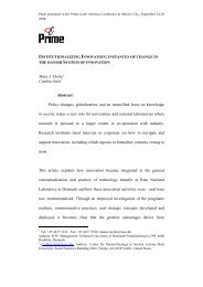
Calculated Relativ« Energy Bespoase of UF-100 in Ferspex<br />
(noraalised to é0 Co). Saapla thickness : 0.8? at<br />
Electrons t 1 average pathlength<br />
2 thiekneas pathlength
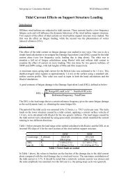
Ft«, 2<br />
001 01<br />
PHOTON ENERGY (MeV)<br />
Calculator Belatlre Energy Eeaponae of OaSO.-Mn in teflon<br />
(noraaliaeft to ""Co) *<br />
Orain aixee i (l)i 1.2$ />• (2)i 5.0 *» Burlin oavitjr theory<br />
n»<br />
.(5, 3 l( *J!>) I !( /}&)<br />
P t.3ø/ ^ yT«fl.o nonaalisad to ^Co<br />
Ixperlaintal pointa for Way epeetra. with an #ff eotire energy<br />
Olttal to the aonoanergetie energy for uhieh the; are plotted.
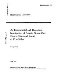
Fig. I<br />
PHOTON ENERGY (MeV)<br />
Calculated Relative Energy Response of LiP-7 in Teflon (normalised<br />
to °
* «r-<br />
HK. 4<br />
Q1 1.0<br />
PHOTON ENERGY (MEV!<br />
Calculated Relativ« Energy Response of IdF-7 in Teflon (nomaliaed<br />
to »
- «o -<br />
ABSORBED<br />
DOSE IRAD IN TEFLON)<br />
Fig- 5 Dos« Raspoas« Curve of IdT-7 for °°Co and 8.3 IteV «ff. X-ray* .
- «9 -<br />
ABSORBED DOSE |RAO<br />
M FERSPEX)<br />
ngs_6<br />
DOM Reepom« Carre of Ltf-100 for 60 !* and 15.5MeV el.ctrona.
20<br />
a<br />
S t5<br />
i<br />
3<br />
I 10<br />
Si<br />
os<br />
10'<br />
I I I I I I I 11 I I I I I I I<br />
Tl=i=!=pXT<br />
T ' '—' ' i ' i i| 1 p—i—i ini| , 1 i i i i M<br />
10 s<br />
o ntgatiw dtetran* »5 M«V<br />
• ^o gamma rays<br />
I0 6<br />
20<br />
15<br />
05<br />
ooL<br />
' * 1 • f ' • ' ' \ 1 > • i i i i I —1_<br />
K?<br />
Flfc_I<br />
OOSE (rad in ptrspex)<br />
-j i—i i i 111<br />
6o„<br />
S.nsitlvity Curv« of UF-100 for 60 Co gani rays and 15 K«V electrons,
On the Non-Linearity; and LET Effects of the Themioluminescence<br />
Response<br />
Toshiyuki Nukajima<br />
Division of Physics, National Insitute of Radiological Sciences<br />
9-1, 4-chome, Anagawa, Chiba-shi, Japan<br />
Abstract<br />
A releasing probability change model has bee proposed through investigation<br />
of the properties of LiF glow curve. In this work, it is mathematically<br />
discussed on the releasing probability change model for the<br />
problems of the thermoluminescence response. It has been revealed that<br />
this model can qualitatively explain these problems better than other<br />
models.<br />
Introduction<br />
Recently, thennoluminescence dosimeter has been widely utilized in<br />
the radiation dosimetry. However, it has been found the following phenomena<br />
by many of investigator;<br />
CI) transformation of the glow curve due to the irradiating dose of<br />
low LET radiation, 1<br />
(2) absorbed energy independence of the glow curve of the phosphor<br />
irradiated with high LET radiation,2<br />
(3) dependence of thermoluminescence response on the absorbed energy<br />
or LET, 3<br />
(4) decreasing non-linearity of the response with increasing the LET<br />
of irradiating radiation,^<br />
C5) influence of exposure on the energy dependence of the phosphor,^<br />
(6) dependence of sensitization factor on either the absorbed energy<br />
or LET of the post- and test-radiations,S<br />
To explain the dose dependence and IJBT effects of the sensitivity of<br />
the phosphor, Cameron and his co-workers, and Attix et al. proposed the<br />
competing trap model and track model, respectively.5»6 But these<br />
models have not been satisfactory to explain these phenomena. Recently,<br />
it has been proposed a model through investigation of the experiment<br />
which has been designed to measure the transformation of the glow curve<br />
due to the energy absorbed in the phosphor.*<br />
This paper reports the result of mathematical analysis of the this<br />
model on the above phenomena.<br />
Notations<br />
A: number of electron produced by irradiation per unit energy which<br />
the phosphor absorbed,<br />
D; the energy absorbed in the phosphor,<br />
1^: the thermoluminescence yields due to electron released from trapping<br />
level, Ei,<br />
k: Boltanann constant,<br />
n^: number of captured electron in the trapping level, hi,<br />
N^: number of electron trapping level, Ei,<br />
R: dose rate of irradiating radiation or absorbed energy rate,
SJ: frequency factor of the trapping level, Ej.,<br />
S: thermoluminescence response of post irradiated phosphor,<br />
Sø: thermoluminescence response of the virgin phosphor,<br />
t, t': irradiating period,<br />
Tn: irradiating temperature,<br />
T Y : maxiimjn heating temperature,<br />
B heating rate of phosphor,<br />
*t: releasing probability of trapped electron in trapping level, Ei,<br />
at irradiating temperature,<br />
ey: intensity fraction of photon energy, j, in thennoluminescence<br />
emitted by releasing electron from trapping level, Ei,<br />
YJ_: escaping probability from trapping level, Ej, at a given temperature,<br />
oi: capturing probability of trapping level, Ei,<br />
5i'. quantum efficiency of photo-detector for the emitted photon of<br />
energy, e^,<br />
øijitransition probability of electron remained in the level, Ei, to Ej.<br />
Theory<br />
The radiation produces clusters of electron and hole along the track<br />
of energetic recoiled electrons, A concentration of these carrier in the<br />
cluster increases with increase of either LET of irradiating radiation or<br />
energy absorbed in the phosphor. Each traj Ing level near or in the cluster<br />
has a proper capturing probability of electron which contributes to thermoluninescence,<br />
and this probability of each trap varies with change in<br />
either the concentration of electron near or in the cluster, or the<br />
absorbed energy as a result of interaction between each other of the clusters.<br />
Let us formulate a mathematical description of the model. According<br />
to Randall and WilJcins' model of thennoluminescence mechanism ,6the thermoluminescence<br />
yield, I, is represented by fbllowings;<br />
- j T n dt (1)<br />
where dt and Y are defined as follows, dt = dT/p, Y - s exp(-E/kT).<br />
Now, when their model applies to all the trapping levels in the phosphor,<br />
and when I is corrected with quantum efficiency of a photcoultiplier<br />
tube and with a fraction of each emission in thermDiuminescence, total<br />
light yields detected by the photomultiplier tube are represented as<br />
follows;<br />
all<br />
f 1 '<br />
2 2 Ei Uj c V e 'J'.-'i dT<br />
C2)<br />
Now, if it is assumed that the c.. is unchanged against the absorbed in<br />
the phosphor and against LET of radiation, eq.(2) is proportional to the<br />
concentration or number of trapped electrori in the trapping levels. Therefore,<br />
it has need to know the concentration of the electrons in each trapping<br />
level in order to study the dose dependence on the response and so on.<br />
Changes in the concentration of the trapped electron in each trancing<br />
level are given by eq. (3),<br />
•.»•H^UI«
dnj/dt «= - «i i^ • AQL - n^ Ji R . (3)<br />
The boundary conditions can be given by the xollowings, t = 0, ni = 0.<br />
The nunber of the trapped electron in each trapping level is presented by<br />
the following equation,<br />
nA * (oi A Ni R/(«i + °i A R))(l - exp(-c. AR - ^H)<br />
(4)<br />
If it is assumed that the nunber of the trapped electron, which is releasing<br />
from the trappling level, Ei, for irradiating period is very small,<br />
the concentration of the trapped electron is approximate I v given by eq.<br />
CS).<br />
^ = NjCl - expC-oi ARt)), as ct^ A R t4t 1<br />
ni = Oi A ^ R t = ej A Nj D (5)<br />
When the phosphor is heated from T to T* with the dosimetric reader,<br />
the dosimetric thermoluminescence light yields, Ij, are presented by the<br />
following,<br />
Id = Z S K^ D oi (6)<br />
where Kij is A^ij ^ijr fri/B) dt.<br />
It may be revealed that when the experimental results of the phenomena<br />
mentioned in the Introduction are mathematically expressed, OJ in LiF<br />
phosphoT is characterized by the followings;<br />
O »i/»D)hlgh ^ = Oa/ HJ),^ ^ SS 0, (?)<br />
C » °i /3D >low LET > (3 V 3D) high LET - °> C8)<br />
in case of low irradiation dose range,<br />
& "i/^'low LET - &>å/ 3D)l0W ^ - 0, (9)<br />
in case of high dose region,<br />
O °i/3D)l0K un- > 0°d/ 3D)low ^ - 0. (10)<br />
Let us consider various phenomena of the response in LiF thermoluminescence<br />
dosimeter.<br />
(1) Non-Linearity of Response<br />
In case of irradiation of low dose with lower LET radiation, the cluster<br />
has lower concentration of electron created in the phosphor and<br />
scarcely interact with each other, because each track is so far separated<br />
from each. Accordingly, a value of i is independent of the absorbed<br />
energy within some ranges. Now, if the response is defined as 7-j/S, the<br />
dose dependence of the response is represented as follow,<br />
(aS/SD)^ -"2S Kij Ooi/SD)^ (11)<br />
In this case, eq.(11) from eq.(9) is nearly equal to zero. The response<br />
is independent of the absorbed energy. On the other hand, in case of<br />
the high doeeregion of low LET radiation, the eq.(ll) from eq.(10) is<br />
larger than zero. Accordingly, the sensitivity of the phosphor irradiated<br />
with low LET radiation becomes to reveal the non-linearity.
Let us suppleoent on ai from Carlsson's results in addition in eqs.<br />
(7). (8), (9) and (10). Namely, it is follow;<br />
(sl. (12)<br />
in these equations, ai is of the dosimetric trap and ai is of the nondosimetric<br />
deep trap.<br />
In case of irradiation with higher LET radiation, eq.(ll) from eqs.<br />
(7) and (12 )is approching to zero with increasing LET of radiation.<br />
Therefore, the dependence of the response on the absorbed energy decreases<br />
with increasing LET.<br />
(2) Dependence of Energy Dependence an Dose<br />
In general, the energy dependence of the response is given by a ratio<br />
between theraioluminescence responses, S^ and Sn, of the phosphors irradiated<br />
with X- and 60 Co gamna-rays, respectively,<br />
VSQTsi Kij oi/2s K'ij a 'i • ay<br />
j i j i<br />
The dose dependence of the energy dependence is considered by followings;<br />
(3i*/aDV( asco/æ) - 2 2 Kij (aoi/ao)/ Ky^'j/sD) (14)<br />
i i<br />
In the energy range of 150 keV or below, LET of X-rays is larger than<br />
60&J gamma-rays. Accordingly, a function F(D) is an increasing function<br />
due tc eqs. (7),(8) and (9),<br />
dF(D) = (3=1/30)0, - (s* j/ S D) x (15)<br />
Therefore, it may be reaveled that eq.(13) is a decreasing function of the<br />
exposure.<br />
(3) Sensitization Factor of Lif Phosphor<br />
Author reveales that the cause of the sensitization factor and the of<br />
changes in the sensitivity of the phosphor due to repeated uses is a<br />
radio-stimulated thenuoluminescence due to the trapped electrons in the<br />
non-dosimetric deep trapping levels. 1 Therefore the sensitization<br />
factor, S/Sn, is given by the followings;<br />
Y = s/so -1 •(£ 2, E<br />
Kj k 8ij Ni(i-Pi>i a, /» /so<br />
(16)<br />
Where Pi ?s releasing probability of trapped electron due to annealing.<br />
In eq. (16), the second ttrm is presented the radio-stimulated thermoluiiiinescence<br />
yieldj of the non-dosimetric deep trapping levels.<br />
Dose dependence of the sensitization factor, Y, is given by the followings;<br />
( 3 Y/sDp)- 2 K;j k ey N i (l-
function of the dose, but in case of irradiating of high LET radiation<br />
eq. (17) from eqs. (7) and (12) decreases with increasing LET of post<br />
radiation. These result may reveal that mechanism for the sensitization<br />
factor can be explained by the releasing probability change model.<br />
References<br />
1. T. Nakajima, Radiation Physics Research, 3, No. 2, 15-22 (1970).<br />
2. C.A. Carlsson and G.A. Carlsson, Proc. 2nd~~Intem. Conf. on Lirain.<br />
Dosim., 302-309 (1968).<br />
3. J.R. Cameron and D.W. Zimmerman, USAEC Report COO-110S-113(part-l)<br />
(1966;.<br />
4. W.R. Hendee, G.S. Ibbott and D.B. Gilbert, Intem. J. Appl.<br />
Radiat. S Isot., 19, 431- 436 (19<br />
(1968). ~<br />
5. N. Suntharalingam and J.R. Cameron, Phys. Med. Biol., 14, 397-410<br />
(1969). ~<br />
6. J.T. RandaU and M.H.F. Wilkins, Proc. Roy. Sac, (London) A184<br />
366-389 (1945).
On the Sensitivity Factor Mechanism of Some Themolvminescence<br />
Phosphors<br />
Toshiyuki Nakajima<br />
Division of Physics, National Institute of Radiological Sciences,<br />
9-1, 4-chcme, Anagawa, Chiba-shi, Japan<br />
Abstract<br />
The sensization factor of MgjS^CTb) and LiF is studied. It is obtained<br />
that the factor diminishes with increasing annealing temperature, and is<br />
dependent on linear energy transfer of both the previous and test radiations<br />
and that the sensitization factor of the sensitized phosphor with radiation<br />
of higher LET is greater than un-sensitized one even in the low dose region<br />
of the previous dose. The sensitization factor phenomena of LiF and Mg2Si04<br />
phosphors are caused by the radio-stimulated thermolumijiescence of electron<br />
and hole captured in the deeper trapping levels.<br />
Introduction<br />
It has been reported that sensitivity of LiF TLD phosphor, annealed at low<br />
temperature after irradiation, increases with decrease of the annealing temperature,<br />
and that one of causes of changes in the sensitivity is radio-stimulated<br />
théimoluriinescence of trapped electron in deep non-dosimetric traps. 1 On<br />
the other hand, Cameron and his co-workers have found the sensitivity factor<br />
phenomena in LiF phosphor which are similar to the sensitization of the annealed<br />
phosphor after irradiation, and have tried to explain them with the<br />
competing trap model. Sut mechanism for the sensitivity factor and sensitization<br />
of the annealed phosphor after irradiation is not yet clarified.<br />
The present experiment was made to obtain information on the mechanism<br />
for the sensitization (sensitivity) factor of LiF and Mg 2 s i04(Tb) phosphors.<br />
Experiments<br />
Powdered MgjSiCjCTb) phosphors, enclosed in a glass capsule of about 15<br />
m length and 2 urn m dia, and powdered TLD-100 LiF phosphor were used in this<br />
experiment. The sensitization of ItøSiO.tTb) phosphor has been especially investigated,<br />
The sources of y-ray radiation were both 2000 curie 137 Cs and 60 Co units<br />
designed for the radiation theraphy. The X-ray radiation of 38 keV was obtained<br />
from a machine designed for the theraphy. The exposure to the phosphor was<br />
measured with Victoreen condenser chamber. Sad Con chamber or Fricke dosimeter<br />
according to the order of irradiating dose.<br />
After irradiation at room temperature and subsequently thermal treatment
at the temperature from 300 °C to 500 °C, the phosphors were irradiated with<br />
a test radiation and subsequently their thermoluminescence yields were measured<br />
to obtain the sensitization factor with a Dai Nippon toryo TLD Reader<br />
of model 1200 as a TLD reader.<br />
Experiment on radio-stimulated thermoluminescence from the irradiated<br />
phosphor is undertaken to obtain some information on the mechanism for the<br />
sensitization factor. The radio-stimulated thermoluminescence is obtained by<br />
following processes:<br />
1) The phosphor is irradiated at room temperature with 10"* or 10 R of<br />
Y-rays from 60co<br />
source and sensitized.<br />
2) The irradiated and sensitized phosphor is thermally treated at 300 °C"<br />
or 350 °C for one hour.<br />
3) Both the treated and virgin phosphors are irradiated again with either<br />
X-ray or Y-rays to compare die glow curves between them.<br />
4) The irradiated phosphors are heated with a constant heating rate of<br />
, . 10 .°C/min and its glow curves are recorded.<br />
Results<br />
1) Dependence of S/Sø on Anneal ing' 3tanperature<br />
Fig. 1 presents the changes in the sensitization factor of N^SiO^CTb)<br />
phosphor, irradiated with one R of the test radiation, as a function of annealing<br />
temperature and as a parameter of the previous and sensitizing irradiation<br />
dose.<br />
As can be seen in Fig, 1, it is observed that the sensitization factors<br />
diminish with increasing the annealing temperature regardless of the sensitizing<br />
irradiation dose. However, a tendency of the deterioration of the sensitization<br />
factors markedly varies with the sensitizing irradiation dose. In<br />
case of the phosphors which are thermally treated at a relatively lower temperature,<br />
the sensitization factor of the phosphor, irradiated with high dose<br />
of the sensitizing radiation, is very large and is greater than that irradiated<br />
with lower dose.<br />
In fig. 2, presents also the dependence of the sensitization factor of<br />
Mg?Si04(Tb) phosphor as a function of the given dose of sensitizing radiation<br />
and as a parameter of annealing temperature. From Fig. 2, also, the sensitization<br />
factor of the annealed phosphor at higher temperature is smaller than<br />
that at lower temperature.<br />
2) Dependence of S/SQ on Irradiation Dose of Sensitizing radiation<br />
Effect of sensitizing irradiation dose on the sensitization factors of<br />
Mg2SiO.(Tb) phosphor is observed.<br />
Fig. 3 shows the changes in the sensitization factor of Mg2SiC>4(Tb)<br />
phosphor, as a function of the sensitizing irradiation dose.<br />
Qirve A in Fig. 3 is the changes in the sensitization factor of the<br />
phosphor irradiated with the sensitizing radiation of 38 keV X-rays and curve<br />
B is of Y-rays from *>0co source.<br />
In case of using the Y-rays for the sensitizing irradiation, the sensitization<br />
factor is nearly equal to unit value of one in the sensitizing dose<br />
range from 3 R to about 100 R, but it gradually increases with increasing the<br />
sensitizing dose.<br />
On the other hand, the sensitization factor of the sensitized phosphor<br />
with the radiation of 38 keV X-rays is greater than the unit value in the low<br />
dose region of the sensitizing irradiation. This fact reveals that the sensitivity<br />
of these treated phosphors with higher LET radiation is greater than<br />
the un-treated one even in the low dose region of the sensitizing irradiation.<br />
However, a gradient of the 38 keV X-ray irradiated sensitization factor on<br />
the sensitizing dose was smaller than that of the 60 Co y-ray factor. In the<br />
sensitizing dose region of 600 R or over, the X-ray sensitization factor was
smaller than that Y-ray factor.<br />
It is obtained from this result that, in the region of low sensitizing<br />
irradiation dose, difference in linear energy transfer(LET) of the sensitizing<br />
radiation brings about the different sensitization factor of the phosphor.<br />
But, Cameron and his co-workers have reported that difference in LET of the<br />
sensitizing radiation dose not bring about the different sensitization factor.<br />
3) Dependence of S/Sn on Test-Irradiation Dose<br />
Fig. 4 presents the changes in the sensitization factor of NføSiCtø (Tb)<br />
phosphor as a function of the test radiation dose. The crystal was thermally<br />
treated at 350 °C for one hour after irradiated with 10* R of either Y-rays<br />
or X-rays. In fig. 4 the vertical axis reveals a thermoluminescence yield<br />
divided by the test radiation dose. Curve A is the dependence of the sensitization<br />
factor of the ir-ray irradiated phosphor on the test irradiation dose<br />
of Y-rays from 137Cs source, after irradiation of 38 keV X-ray. Curves B and<br />
C show the changes in the sensitization factor of the sensitized phosphors<br />
with Co Y-rays due to different LET of the test radiation.<br />
As shown in curves B and C of Fig, 4, in case of the different LET<br />
of the test'radiation, the sensitization factor is different from each other<br />
even though the phosphor is irradiated with the sensitizing radiation of the<br />
sane LET, Furthermore, it is obtained from curves A and B of Fig. 4 that, in<br />
case of the different LET of the sensitizing radiation, although the LET of<br />
the test radiation is same, the factor differs from each other.<br />
In case of the former results, when the phosphor is irradiated with the<br />
test radiation of higher LET, the sensitization factor is neaTly equal to one<br />
in the dose region from one roentgen to about 50 R. But after reaching the<br />
maximum value at 300 R, the factor decreases with increasing the test radian<br />
tion dose. On the other hand, in case of the latter, it is observed that when<br />
the phosphor is irradiated with the test radiation of same low LET, the sensitization<br />
factor of the sensitized phosphor with the radiation of higher LET<br />
is greater than that of lower LET.<br />
4) Radio-Stimulated Thermoluminescence as Mechanism for the Factor<br />
It has been found difference of the sensitization factor of LiF phosphor<br />
due to irradiation of different LET radiation. But the cause' on the difference<br />
is not yet clarified.<br />
Experiment has been undertaken to obtain some information on the causes<br />
of this difference.<br />
Fig, 5 shows a glow curve of the Harshaw TLD-100 LiF irradiated with 10<br />
R after annealing at 300 °C for 30 min in the atmosphere of 10-2 jm Hg vacuum.<br />
In general, the peaks which appear in the temperature region from room<br />
temperature to about 300 °C, are used for the dosimetry. Accordingly, the<br />
glow peaks in the region of 300 °C or over is not used for the dosimetry and<br />
the trapped electron which contributes to these glow peaks remains in the<br />
phosphor. Actually, the glow peak is observed from the annealed LiF and Mgi-<br />
Si04(Tb) at about 300 °C and 400 °C, respectively, after irradiation.<br />
To obtain the sensitization factor, the phosphor has been thermally<br />
treated at 350 °C or below for one houT. But it is clear from Fig$. 5 and 6<br />
that the annealing temperature is not suitable for releasing the trapped electrons<br />
in all trapping levels of the phosphor.<br />
Figs, 7 and 8 show the effects of the re-irradiation on the remained<br />
electron captued in the deeper traps.<br />
It is known that photo-stimulated thermoluminescence is emitted from the<br />
irradiated crystal, when it is heated after illumination at lower temperature<br />
than the irradiated temperaure. The photo-stimulated thermoluminescence<br />
has been reported to be caused by the photo-stimulation of the deeper trapped<br />
electron due to illumination. 1 As can be seen in curve of Figs. 7 and 8, the<br />
dosimetric glow peaks from the sensitized phosphor are greater than that from
the un-sensitized one. Especially, all glow peaks of the sensitized Mg^SiC^<br />
(Tb) phosphor were higher than the un-sensitized one.<br />
These results may reveal that when the phosphor which involves the electron<br />
captuTed in the trapping levels, is irradiated again, the re-excited<br />
electrons with radiation is re-trapped in the shallow trapping levels. Accordingly,<br />
the glow peak height from the sensitized phosphor is composed of the<br />
intrinsic and the radio-stimulated heights. Namely, it is concluded that the<br />
phenomena of the sensitization is caused by the radio-stimulated thermoluminescence<br />
due to re-trapping the remained electrons in the deeper traps into<br />
the shallow traps.<br />
Discussion<br />
The experimental results described in the present work has been provided<br />
information that should help in explaining the causes of the sensitization.<br />
The causes of the sensitization phenomena can be explained as follows;<br />
When the thermolnnrjiescence phosphors are excited with ionizing radiation.<br />
many of the electron are trapped at tlie trapping levels. Now, if the trapped<br />
electrons in the trapping levels between Eo and E^C^Eo) are released by<br />
thermal treatment, the electrons in the levels of Ej(>Ei) will remain in the<br />
phosphor.<br />
When the remained electrons in the deeper trapping levels Ej are excited<br />
again with irradiation the radiation, the electron of Ej transfer into the<br />
trapping levels below Ej. The transfered electrons contribute to the thermoluminescence<br />
for the dosimetry. Therefore, the gross dosimetric thermoluminescence<br />
yield for the dosimetry is the sum of the intrinsic thermoluminescence<br />
yield due to irradiation and the radio-stimulated the..i»luminescence yields.<br />
Next, causes on the different sensitization factor due to different LET<br />
of both test and sensitizing radiations will be discussed.<br />
To obtain the relative glow peak heights from the glow curve, the glow<br />
curve is recorded through • heating the irradiated phosphor with heating<br />
rate of 10 °C/min. When the crystal of LiF is irradiated with y-rays, its<br />
relative glow peak heights are changed with increasing the absorbed dose, as<br />
can be seen in Fig. 9. The relative heights in the higher temperature region<br />
— the non-dosimetric peak temperature region increase with the absorbed<br />
dose.<br />
The behaviour of the glow peak heights has been found in the Mg2SiC>4 (Tb)<br />
phosphor, also. As abovementioned, if the cause on the sensitization factor<br />
is due to the radio-stimulated thermoluminescence, it will be easily understood<br />
that the sensitization factor of the phosphor with higher dose of the<br />
sensitizing radiation is greater at lower annealing temperature than that with<br />
lower dose.<br />
On difference in the sensitization factor due to different LET of the<br />
sensitizing radiation, it will be explained by following;<br />
The radio-stimulated thermoluminescence yields are increased with the concentration<br />
of the trapped electron which stores in the non-dosimetric deep<br />
traps of the sensitized phosphor. According to the Carlsson's results ,3 in<br />
case of irradiation of high LET radiation, the relative concentration of the<br />
trapped electron between the deeper and shallow traps is scarcely dependent<br />
on the dose.<br />
On the other hand, in case of low LET radiation, as shown in Fig. 9, the<br />
relative concentration of the trapped electron in each trap increases with the<br />
absorbed energy. Therefore, after annealing, the relative concentration of the<br />
trapped electron which remained in the traps is influenced by both LET and<br />
absorbed energy of the sensitizing radiation. If the absorbed energy of the<br />
phosphor due to irradiation of the sensitizing radiation is a constant, the<br />
concentration will be increased with increasing the LET. Therefore, the sensitization<br />
factor of the phosphor with the higher LET of the test radiation
is greater than that with the lower LET. If the LET of the sensitizing radiation<br />
is a constant, the concentration per dose increases with increasing the<br />
absorbed energy, as can be seen in Fig. 9. Therefore, the sensitization factor<br />
increases with the dose of the sensitizing radiation<br />
Conclusion<br />
The sensitization factor has been found to be diminished with annealing<br />
temperature, and to be differed with changing the LET of both sensitizing and<br />
test radiations. Furthermore, it is obtained that in the low dose of sensitizing<br />
irradiation the factor of the sensitized phosphor with 38 keV X-rays is<br />
greater than that of the un-sensitized one.<br />
It nay be concluded that the sensitization factor phenomena are caused by<br />
the radio-stimulated thermoluminescence due to re-exciting the trapped electrons<br />
remained in the deeper trapping levels. Various phenomena in the sensitization<br />
of the phosphor response have been found to be easily understood<br />
by the radio-stimulated thermoluminescence and our model for the non-linear<br />
response of the phosphor.<br />
Acknowledgment<br />
The author is indebted to Dr. Y. Yamamoto, of the Tokyo Metropolitan Isotope<br />
Research Centers, for helpful discussions on the interpretation of the<br />
results. Grateful acknowledgement is also due to Miss I. Taneichi for her<br />
assistance of experiment<br />
References<br />
1. D.W, Zimmerman, C.R. Rhyner and J.R. Cameron, Health Physics, 12^<br />
525 - 531 (1966)<br />
T. Nakajima, Health Physics, 16, 509 - 51 (1969)<br />
2. N. Suntharalingam and J.R. Cameron, Phys. Med. Biol., 14, 397-410(1969)<br />
3. C.A, Carlsson and G.A. Carlsson, Proc. 2nd Intern. CoKf. on Lumin.<br />
Dosim., 302-309 (1968)
10<br />
\<br />
10<br />
300 400 500<br />
ANNEALING TEMR (°C)<br />
Fig, 1, Effect of annealing temperature on the sensitization factor<br />
of Mg 2 SiO. (Tb) phosphor as a parameter of the sensitizing<br />
irradiation-doseCThese phosphors vere thermally treated one<br />
hour after irradiation of gamma-rays from Co)
o —<br />
Mg 2 Si0 4 (Tb)<br />
350"Ctreatment<br />
10° 10 10 2 10 3<br />
IRRADIATION DOSE (R)<br />
Fig. 2.<br />
(Slanges in the sensitization factor of Mg-SiO.(Tb) as a<br />
function of the sensitizing irradiation dose and as a parameter<br />
of annealing temperature<br />
K
Fig. 3.<br />
100 1000<br />
PREVIOUS EXPOSURE (R)<br />
Changes in the sensitization factor of Mg-SiO.CTb) as a<br />
function of the sensitizing irradiation dose and as a parameter<br />
of the LET of the sensitizing radiation<br />
10000
38keV-<br />
X-ray<br />
10 100 1000<br />
RRADIATION DOSE (R)<br />
Fig. 4. Influence of the teat irradiation dose on the sensitization factor of MgoSi0.(Tb)(A: the teat radiation is<br />
137 4.<br />
.-rays of Cs after irradiation of 10 R X-ray and subsequently annealed for ooe hour at 550 C, B: the<br />
test radiation of 38 keV is used after<br />
4 1^7<br />
ray« is used after gamma-ray irradiation of 10 •ray R). irradiation of 10 R, C; the test radiation of Cs gamma-
H-vacuunCIO ' nm Hg)-300 'C - 30 min<br />
100 200 300<br />
TEMPERATURE CC)<br />
Fig. 5.<br />
Thermoluminescence glow curve of Harshaw TLD-10Q LiF irradiated<br />
with 10 R after thermal treatment at 300 °C for 30 min in<br />
vacuum of 10" nm Hg<br />
400
Mg Siq(Tb)<br />
100<br />
200 300<br />
TEMPERATURE CC)<br />
Fig. 6. Thermoluninescence glow curve of the Mg^SiO.CTb)<br />
irradiated with 10 R<br />
400<br />
phosphor
00 -st CN<br />
A1ISN31NI TI<br />
Fig, 7, Radio-stimulated theimoluminescence of T1D-100 LiFfcurve A:<br />
the virgin of the crystal irradiated with 10 R after thennal<br />
treatment at 300 °C for one hour in argon gas, B: irradiated<br />
with 10 R after irradiation and sensitization with 10 R<br />
and subsequently annealing at 300 °C for one hour in argon gas)
RADIO-STIMULATED T.L.<br />
Mg 2 SiQ!i(Tb)<br />
200 300<br />
TEMPERATURE (°C)<br />
400<br />
Fig. 8. Radio-stimilated thermoluminescence of Mg.SiO. (Tb) (curve A:<br />
3 3<br />
the virgin crystal irradiated with 10 R, B: with 10 R after<br />
irradiation and sensitization with 10 R, and subsequently<br />
annealing at 400 °C for 30 min)
10" 10° 10<br />
EXPOSURE (R)<br />
Fig. 9. The ratio between each glow peak height and 250 °C one in TLL-<br />
100 LiF phosphor as a function of absorbed energyf A: 3I0H<br />
peak at 250 °C, B: at 310 "C, C: at 340 »C, D: 370 °C, axA E:
The T3EE Response of Ceramic *e0 cover«" with Different Absorbers During<br />
Gamma and X-Ray Irradiation<br />
by<br />
E. Rotondi - T. Suppa<br />
Lab. Dosimetria e Standardizzazione<br />
C.N.G.N. - C.S.N. Casfeccia<br />
Roma (Italy)<br />
Abstract<br />
Owing to the very thin layer involved in exoemission process, the<br />
response of a TSEE detector is largely influenced by the material used<br />
as cover during y and X rays irradiation.<br />
In this paper a study was conducted on the TS5E response of ceramic<br />
BeO covered with materials of different atomic number such as aluminium<br />
and gold.<br />
For X rays of 66 keV effective energy, the response with gold cover<br />
is 18 tines higher than with aluminium. With a gold cover the response to<br />
X rays is much higher than to cobalt. It is also shown that the simultaneous<br />
use of two BeO detectors covered respectively with aluminium and gold can<br />
be utilized for evaluating the low energy X ray component in an unknown<br />
field of photon radiation. It has been observed the diffusion of the<br />
gold in the BeO produces the same effect of covering the BeO with gold<br />
layer during irradiation.<br />
INTRODUCTION<br />
The interactions of photons with the materials covering TSEE detectors<br />
give rise to cany electrons, which reach the sensitive layer and<br />
contribute to *,h« exoemicsion phenomenon.<br />
At photon energies wher* the photoelectric process predominates,
this contribution varies strongly with the atomic number.<br />
This paper is mainly concerned with the response variations produced<br />
by covering ceramic BeO with gold or aluminium during irradiation.<br />
Moreover, measurements nave been carried out with gold impregnated<br />
samples, to investigate whether the gold diffused in the BeO increases<br />
the response enhancing the exoemission probability or giving rise to more<br />
electrons available to be trapped.<br />
1. EXPERIMENTAL<br />
The measurements have been carried out on both bare and gold plated<br />
samples of sintered ceramic beryllium oxide , Thermalox 995 (discs 0.9 cm<br />
in diameter and 0.5 cm thick).<br />
The apparatus, reported elsewhere consisted of a *as flow Geiger<br />
counter provided with a linear heating rate system.<br />
To obtain a good thermal annealing the BeO was maintained, before<br />
being used, at 600 C for 20 minutes. Each reading was terminated at 550 C<br />
and the sample was reused without annealing.<br />
The irradiations have been performed with Co and X rays of SS keV<br />
effective energy.<br />
2. RESULTS AND DISCUSSION<br />
2.1. BeO bare sample - Co° irradiation<br />
During the cobalt irradiation the BeO discs have been covered with<br />
either an aluminium or gold layer of thickness 600 mg/cm .<br />
The exoemission curves reported in Fig. 1 are relative to 1 H exposure,<br />
the curve 1 refers to the sample covered with gold, the curve<br />
2 to that covered with aluminium. The corresponding integral counts are<br />
19000 and I6OOO, with standard deviation a = 6#.<br />
The results show that at the cobalt energy the TSEE response is<br />
rather insensitive to the different Z of the covers. Indeed in this case<br />
Compton interactions and pair production are predominant and the mass<br />
energy absorption coefficient varies slowly with the atomic number.<br />
P.2. BeO bare cample - X rays irradiation<br />
For low energy X rays the situation is quite different since the
photoelectric process, which is Btrongly energy ana atomic number dependent,<br />
predominates.<br />
In Fig. ? the results obtained with BeO samples covered respectively<br />
with 10 mg/cm^ of aluminium and gold are reported. The exposure was 50 mH,<br />
the curve 1 (integral counts ?6000» a = 6%) corresponds to the sample<br />
covered with gold, the curve 2 (integral counts 2000, a = €%) to that<br />
covered with aluminium. The response with gold ie 18 times higher than that<br />
with aluminium, in good agreement with mass energy absorption coefficient<br />
ratio of the two metals at the considered energy.<br />
Comparing X rays and cobalt results and taking into account the<br />
different exposures involved, it follows :hat the response to X rays is<br />
higher than to cobalt. This effect is greatly increased when using gold<br />
as a cover, pointing out the strong energy dependence introduced by the<br />
presence of a high Z material during the irradiation.<br />
From the above considerations the possibility arises of using the<br />
energy and Z dependence to evaluate the low energy X rays component in an<br />
unknown field of photon radiation.<br />
The measurements carried out in this connection have been reported<br />
in Fig. 3. The BeO sample, covered in turn with aluminium and gold, was<br />
irradiated with 1 R of cobalt plus 50 mR of X rays. The curve 1 refers<br />
to the sample covered with 10 mg/cm' of gold, the curve 2 to that covered<br />
with 10 mg/cm of aluminium. The difference between the two curves is due<br />
essentially to the 50 mR of X rays component since 1 ft of cobalt gives in<br />
both cases approximately similar contributions, as seen before.<br />
2.3. Gold Plated Sample<br />
p<br />
The BeO discs were plated with 50 vg/cm of gold and heated for two<br />
hours at 600 C to achieve the diffusion of the gold in the BeO.<br />
Concerning the cobalt irradiation the gold plated samples give the<br />
same response as bare ones. The gold diffusion in the BeO does not produce<br />
any change in the sensitivity.<br />
For X rays, 1 R exposure, the response of the goJ.d plated sample<br />
was three times higher than the bare one, as shown in Fig. k.<br />
To investigate whether the increase in the response can be attributed<br />
entirely to the photon interaction with gold, the following experiment was<br />
performed.<br />
The bare and plated samples were covered w-"th a gold layer of 10 rag/em'<br />
thick a.»i exposed to 50 mR of 7 rays. The results obtained are shown in<br />
Fig 5 and it can be seen, that the response of the two samples is very
similar.<br />
Therefore ve nay conclude that the difference in the response observed<br />
in Fig. *t is only due to the photoelectric process which takes place in<br />
the 30 lig/cm of gold diffused in the sample.<br />
In speculating whether or not the gold diffusion process modifies<br />
the physical properties of the sensitive layer, we can say, as far as our<br />
experiment is concerned, that if changes occur they do not influence the<br />
ezoemission process.<br />
References<br />
1) K. Becker, J.S. Cheka, K.U. Crase, R.B. Gammage - Advances in Physical<br />
and Biological Radiation Detectors.<br />
IAEA Vienna 25 - 35 (197C).<br />
2) E. Rotondi, F. Bordoni.<br />
CNEH-RT/F1 7-15 (1770).
Fowler<br />
Do you know whether the diffusion of the gold occurs efficiently at 600 C?<br />
Vhj did you choose this temperature?<br />
Rotoodi<br />
I cannot give an answer about the efficiency of the diffusion, but, as reported<br />
by other investigators, it is likely that diffusion takes place at<br />
least into the sensitive layer. The temperature of 600 C has been chosen to<br />
conform with the thermal treatment used for annealing of the bare samples.<br />
Different temperature treatments would introduce sensitivity changes not<br />
related to gold diffusion in the BeO.
Be O Bare sample<br />
CO 1200|o.<br />
6<br />
111<br />
HI<br />
(0<br />
600|-<br />
100 200 300 400<br />
T(°C)<br />
Fig« 1 Response to cobalt, exposure 1 R»<br />
1 - gold cover 600 mg/em<br />
2 - aluminium cover 600 rag/cm
Be O Bara sample<br />
100 200 300 400<br />
T(°C)<br />
Pig. ?- Response to X rays, exposure 1 Hi<br />
1 - gold cover 10 mg/cm<br />
p<br />
P - aluminium cover 10 mg/cir.
- 487 -<br />
CO<br />
ri.<br />
1200<br />
Be O<br />
Bare sample<br />
ID<br />
CC<br />
UJ<br />
600<br />
100 200 300 400<br />
T(°C)<br />
Fig. 5 Response to 1 R of cobalt plus 50 mR of X raysi<br />
1 - gold cover 10 mg/cm<br />
2 - aluminium cover 10 rag/cm
- 436 -<br />
'a) Be O gold plated sample<br />
b) Be O Bare sample<br />
Pig. *<br />
200 300<br />
T(°C)<br />
Response to X rays, exposure 1 Bt<br />
a - gold plated sample<br />
o - bare sample<br />
400
CO 1200-<br />
d<br />
a) Be O gold plated sample<br />
b) Be O Bare sample<br />
UJ<br />
%<br />
UJ 600<br />
UJ<br />
CO<br />
100 200 300<br />
T(°C)<br />
400<br />
Fig* 5<br />
Response to X rays, exposure 50 mR, gold cover 10 mg/cm t<br />
a - gold plated sample<br />
b - bare sample
- 490 -<br />
Um Temperature Monitoring Using<br />
Thermoluminescent Materials<br />
by<br />
Roben D. Jarrett, J. Halliday and J, Tocci<br />
U. s. Army Natick Laboratories<br />
Natick, Massachusetts<br />
Abstract<br />
Organoleptic testing of radappertized foods has shown that acceptability<br />
is highly correlatible with irradiation temperature. In general,<br />
irradiation temperatures in the region of -30*C yield the most acceptable<br />
product. Thus, a requirement has been generated, particularly in cummin ial<br />
applications, for a dosimeter capable of being used in radiation field<br />
napping and production dcse Bonitering at these teaperatures.<br />
Of the various dosimetry systeas considered for this application, a<br />
thermoluminescent systev shows the aiost pronise. Samples of the coanercially<br />
available materials were subjected to a screening and evaluation procedure<br />
to determine their applicability. The results of this study are discussed<br />
including calibration data for these themoluninescent naterials for gamma<br />
irradiations at various tenperetures. for example, a sanple of TLD-10O in<br />
its loose powder fora yields values twenty to forty percent lower when<br />
irradiated at -196*C than when irradiated at 25"C. Such results show the<br />
potential usefulness of thermoluminescent materials but point to the critical<br />
dependence of the calibration function on temperature.<br />
A standard procedure for "reading out" thermoluminescent materials is<br />
discussed. This procedure involves, among its other features, a ten minute<br />
annealing period at 100'C prior to readout. This was found necessary in<br />
order to eliminate a number of low energy traps that contribute to spurious<br />
results.<br />
Again, for commercial radio-sterilization applications, a suitable<br />
total dose range is necessary. The potential applicability of certain high<br />
dose (in the megarads region) naterials is discussed. For example, our evaluation<br />
of Isomet liF in both its single crystal and loose powder forms will be<br />
contrasted with results on this material as reported by Tochlin.<br />
This paper demonstrates the requirement for a low-temperature dosimetry<br />
system and evaluates the presently available thermoluminescent materials
(both inajMiiiially available or those in a developmental stage) with respect<br />
to their potential for aeeting this requirement.<br />
General Introduction<br />
In nuatrous applications of ionizing radiation there exists the need<br />
for a dosimetry system with a rather specialized set of properties - the<br />
capability of measuring doses in the aegarad region and at sub-zero centigrade<br />
irradiation teaperatures. It is the intent of the present work to explore<br />
the applicability of theraoluminescent materials in fulfilling this dual requireaent.<br />
Ihe developaent of a satisfactory readout technique and some of<br />
the aore significant pitfalls encountered are discussed. Several selected<br />
thermoluminescent materials are evaluated and discussed relative to their<br />
"ambient** and low temperature dose response.<br />
This work was performed using the irradiation facilities of the U. S.<br />
Army Nmtick Laboratories, Natick, Massachusetts, U. S. A. The Irradiation<br />
laboratory bas a nine kilowatt electron linear accelerator capable of producing<br />
12 MeV electrons, three Cobalt-60 sources (1.5 megacuries, 35 kilocuries,<br />
and 7 kilocuries) and a 200 kilocurie Cesium-137 source. All gamma<br />
irradiations were made using the two smaller Cobalt-60 research irradiators<br />
which have dose rates of 4.4 kilorads and 0.56 kilorads per second.<br />
Introduction<br />
The basic phenomenon underlying thermoluminescent dosimetry (TLD) is<br />
the freeing of trapped electrons by thermal stimulation. A plot of the<br />
intensity of die light emitted by the luminescent material as a function of<br />
its hmat treataent is the familiar "glow* 1 curve. Since the heat treatment<br />
involves a teaperature-tiae relationship, the glow curve and hence the results<br />
of a theraoluminescent dosiaeter reading must be considered relative to the<br />
aode of heat treatment. A large number of temperature profiles are possible;<br />
three basic types are illustrated in figure 1, The first type (Figure la)<br />
features a preheat rate from A to B followed by the primary heating rate<br />
from B to C. A second type (Figure lb) involves only the primary heating rate<br />
froa A to C. A aore versatile type (Figure Ic) inserts an annealing plateau<br />
between the preheat part of the cycle and the commencement of the primary<br />
heating. In all cases C represents the maximum temperature achieved and is<br />
us> ally the point where the integration of the light output is terminated.<br />
The basic commercial instrument used was the Harshaw Model 2000<br />
Analyzer (I). It was operated in the temperature range 40"C to 410*C using<br />
various primary heating rates. Samples of the thermoluminescent powder were<br />
dispensed onto the heating pan and then subjected to the desired temperature<br />
profile.<br />
Post Irradiation Annealing<br />
It has been suggested (2) that annealing the material for 10 minutes<br />
at 100*C after irradiation and before reading the samples would improve the<br />
results. This improvement is based upon the fact that heating at 100'C<br />
would greatly reduce or remove the traps below that temperature and give a<br />
reliable base line. We evaluated the effectiveness of this annealing procedure<br />
by dispensing a sample of irradiated TLD-100 powder into the heating<br />
pan of the reader and observing the output signal of the photoauLtiplier tube<br />
as a function of time when the sample was heated to and held at 100 C. It<br />
was observed that the signal approached a constant value after 8.5 minutes.
These results demonstrate that if the staple is held at 100'C for 10 minutes<br />
the base Una is stable and any inaccuracy in tiling would have a negligible<br />
effect.<br />
Based upon the fa« that post annealing stabilited the base line wo<br />
modified our reader to allow post annealing to be perform«! in the reader as<br />
typified by Figure Ic. It is possible to vary the post annealing temperature<br />
(•) to any value and the annealing time (B to B 1 ) to any period. These modifications<br />
also allowed us to reduce the post anneal time to less than 10<br />
minutes as the samples are now held for an exact time which eliminates the<br />
inaccuracies which would be involved in heating the samples in an oven for<br />
less than 10 minutes and transferring them to the reader. For most of the<br />
results reported we used this procedure with an annealing time of 0.S minutes.<br />
It should be noted that the coulomb meter operates only during the period B 1<br />
thru C. An additional advantage in using this readout procedure is that the<br />
temperature of the pan and sample, prior to the start of readout, has less<br />
effect an the peak height and integrated charge values. Therefore, we<br />
improved the readout reproducibility with this heating procedure.<br />
As is obvious from the preceding description of an optimum readout<br />
technique, aa integrated peak area as well as peak height(s) is a useful<br />
parameter in correlating luminescence with dose level. Based upon the standard<br />
error of ten readings at a given dose it was, in fact, determined that the<br />
integrated charge (output from the photomultiplier tube) gave the more reproducible<br />
results. The standard error using peak height measurements was<br />
approximately 5% as contrasted to a value of 2% using integrated charge<br />
measurements. The doses used for the evaluation were in excess of ten kilorads.<br />
There are several effects observed incidental to the heat treatment<br />
which might lead to inatrumental artifacts. These involve mainly the<br />
composition and pre-hi story of the heating pans and the contribution of infrared<br />
radiation at the higher temperatures.<br />
During the course of these studies we were supplied with experimental<br />
type heating pans from Harshaw. While evaluating the use^ilness of these<br />
pans at high readout temperatures we observed a rather impressive effect on<br />
our readings as a result of pan condition. This effect was observed by<br />
making a number of readings using irradiated TU) powder, that had been heated<br />
in an oven at 100*C for 10 minutes, in both new and used pans. The readings<br />
showed that the condition of the heating pan can affect the peak height<br />
values by as much as 35 percent and the integrated charge values by at much<br />
as 25 percent. Calibration curves made with the different pans had the same<br />
general shape but were displaced by various anounts.<br />
According to the procedure described by Webb (3), the best readout procedure<br />
is that which integrates the total area under the glow curve peaks but<br />
excludes the rising contribution occuring at high teaperatures. This artifact<br />
at the end of the glow curve is attributable to the omission of infrared<br />
radiation from the phosphor and pan. Hence, the optimal readout conditions<br />
for the higher dose levels must be compromised in order to minimize the contribution<br />
of this effect.<br />
When quantative comparisons are made between glow curves, particularly<br />
when certain peaks are to be identified and compared, the calibration of pan<br />
temperature indicating device is crucial. A convenient alternative to the<br />
use of calibrated thermocouples if a paper thermometer (*). These devices<br />
are available in ten degree Centigrade) increments and may be conveniently<br />
placed directly on the pan. Gross errors in the pan temperature may be
- 493 -<br />
quickly discovered using these thermometers.<br />
TLD Materials Evaluated<br />
Various thermoluminescent materials were evaluated with respect to<br />
their fulfillment of part of the dual dose range-irradiation temperature<br />
requirement. In addition to regular lithium flouride (LiF Harshaw TLD-100),<br />
the dose response of lithium boTate (Li 2 Bj|0 7 :Mn), isomet lithium flouride,<br />
strontium fluoride and lead fluoride .was studied.<br />
Hie thermoluminescence powders weTe dispensed into gelatin capsules<br />
5 x IS mm) for irradiation. Each capsule contained sufficient powder for<br />
seven readings. Exposed powder was dispensed in 28 mg. lots into the builtin<br />
heating pan for analyzing the radiation induced thermoluminescence.<br />
The capsules of powder were irradiated to the desired doses in electron<br />
equilibrium shields using one of the Cobalt-60 irradiators previously<br />
mentioned. Doses were based on chemical dosimetry using the Fricke dosimeter<br />
(G value • 15.6}<br />
Most of the low temperature irradiations were conducted at -40*C using<br />
cold nitrogen gas to regulate the temperature of the irradiation chamber.<br />
The several comparisons made at 196*C were achieved by irradiating the<br />
samples immersed in liquid nitrogen.<br />
Lithium Florida - TLD-100<br />
This material is one of the most researched and used thermoluminescent<br />
materials available. He therefore initiated our studies using this material<br />
and observed many interesting phenomena. Our first temperature response<br />
studies showed that the loose powder samples irradiated at -196*C gave results<br />
that were twenty to forty percent lower than samples irradiated at 25*C. These<br />
studies were over the limited dose range of 10 3 to 5 x 10 1 * rads. To evaluate<br />
the temperature response in a temperature range more nearly approaching that<br />
to be used in radappertized foods we selected -40°C. It is observed in<br />
Figure 2 that in the dose range from 10 3 to 3 x 10 s rads the integrated<br />
charge values of samples irradiated at -40°C are sixteen to thirty percent<br />
lower than similar samples irradiated at 25'C. Above 3 x 10 s rads it appears<br />
that radiation damage to the crystal is more pronounced in the 25*C samples<br />
as evidenced by the reduced response. The peak height values differ quite<br />
considerably between the two temperatures as shown in Figure 3.<br />
Lithium Borate<br />
Two commercial forms of lithium borate were evaluated for their usefulness<br />
at doses greater than 10 3 Tads and at two temperatures 25* and -40'C.<br />
They are loose powder and Conrad's teflon disks.<br />
The powdered lithium borate was treated prior to irradiation by baking<br />
for 15 minutes at 300'C. The irradiated samples were annealed in an oven at<br />
100'C for 10 minutes and read out with nitrogen gas purging the sample. The<br />
response curves of this material (Figure 4) show that it may be used into the<br />
megarad range depending upon the accuracy required in the dose measurement.<br />
The difference in response between samples irradiated at the two temperatures<br />
is approximately ^6% of an average value. This is approximately equal to<br />
the standard deviation of our peak height readings and three times the<br />
standard deviation of our integrated charge readings.
- 434 -<br />
The lithiua borate teflon disks show the lwest sensitivity to irradiation<br />
temperature of any of the systens we tested (figure 5). All the integrated<br />
charge values for these disks between 10 3 and 3 x lO* rads were within<br />
the normal fluctuation of readings. Above 3 x 10* rads the 2S C samples give<br />
•ore light output per red than the samples irradiated at -40 C.<br />
Isomet Lithiun Fluoride<br />
In our search for a high dose phosphor we obtained a sample of the<br />
Isoaet LiF suggested by Goldstein (S). This material was »ported to be<br />
usable in the megarad range and to have a high temperature peak between 400 and<br />
450"C. Tochlin reported (6) reading out 16 mg samples by preheating the<br />
sample to 350*C to erase the earlier dominant peak, and then reheating the<br />
samples to read out the 450*C peak, he irradiated samples in the megarad<br />
range and using Tochlin*s procedures tried without success to locate the 450°C<br />
peak. He tried using a fast heating rate of approximately 40 C* peT second<br />
and a slow rate of approximately 7 C* per second. The maximum temperature<br />
peak we were able to locate was at approximately 340*C (figure 6).<br />
It was observed from the glow curves that the height of the 340*C peak<br />
was a function of the irradiation dose, while the lower temperature peaks<br />
remained relatively constant. Following the reasoning previously stated with<br />
regard to annealing the low energy traps, the samples were dispersed onto the<br />
heater pan and rapidly heated to 240*C. They were held at this temperature<br />
for 0.5 minutes and then heated to a temperature maximum of 390*C at a rate<br />
of 5*C per second. Figure 7 shows response curves we obtained for samples<br />
irradiated at 25* and -40*C, using the peak height values at the 540*C peak<br />
and the integrated charge values above 240°C. These curves show that there<br />
is considerable effect on the response of this material as a function of the<br />
irradiation dose. In the dose range of 10 s to 3 x 10 e rads, the samples<br />
irradiated at 25*C response is approximately four times that of similar<br />
samples irradiated at -40°C. It is noteworthy that using the above procedure<br />
this material is a potential high dose phosphor. Our main concern is the<br />
availability of similar material in the future.<br />
In an effort to put the phosphor in a form which would be more usable<br />
than the loose powder we had Tyco Laboratories (7) grow single crystals .125<br />
x .25 x 1.5 inches. These crystals were made by the continuous growth method<br />
(8,9) which allows the dimension of the crystal to be controlled independent<br />
of the growth rate. The glow curves for these single crystals might indicate<br />
that the concentration of the impurities in the original Isomet crystal are<br />
modified by the crystal growth method. It was observed that the main response<br />
peak to irradiation dose in these crystals is at ISO'C in contrast to peak<br />
position at 340*C in the loose powder.<br />
While we were not successful in our initial attempt to incorporate the<br />
Isomet material into single crystals that had a megarad dose response, Tyco's<br />
growth process shows two potential areas for development. First they are<br />
able to control the impurities in the growth crystals under some conditions<br />
so that they can conceivably dope the crystals. Secondly, these crystals axe<br />
very transparent and could be grown in shapes which would allow the crystals<br />
to be read spectrophotoasterically as well as thermally. These crystals'<br />
dose response resembles more the TLD-100 results than the original Isoaet<br />
powder results. Figure S.<br />
Strontium and Lead Fluoride<br />
Samples of strontium fluoride and lead fluoride were obtained from
-495 -<br />
Harshaw as materiels that Bight be useful in aeasuring doses in the aegarad<br />
nag*. These staples »ere irradiated with dose betveen 0.S and 7 aegarads<br />
and evaluated using Harshaw's standard readout procedure of heating to 100*c<br />
rapidly and than to teaperature aaiiawa using a loner heating rate. The<br />
results obtained were negative in that there uas no appreciable change in the<br />
tlienoluwinescence as a function of dose.<br />
Conclusion<br />
These studies ra-oaphasize the iaportance of careful control of the<br />
readout equipment, especirlly the condition of the heater pan. It nay be<br />
advisable to maintain a calibrated phosphor that is read periodically to<br />
assure that the pan has not degraded to a point that it is giving erroneous<br />
readings. In order to reduce the background, calibrations should be made for<br />
each phosphor using the optiaua set of reader conditions. It has been deaonstrated<br />
that an iaproved readout procedure is obtained by holding a saaple at<br />
an annealing teaperature in the reader (without integrating the signal) for<br />
a predetermined tine and then continuing the heating at a fixed rate while<br />
integrating the signal. The reproducibility of the readings is iaproved in<br />
this nanner and the contribution fron the low energy traps is reduced or<br />
eliminated.<br />
Cuamiii ially available TIP aaterial nay be used at low irradiation<br />
teaperatures provided they are calibrated at the teaperature. More studies<br />
need to be conducted to determine the decrease in response as a function of<br />
temperature.<br />
References<br />
1. Harshaw Chemical Company, Cleveland, Ohio<br />
2. R. A. Arnold, private conn.<br />
3. G. A. Webb, J. Sci. Instr. 44 (1967)<br />
4. Paper Thermometer Co., 10 Stagg Dr., Natick, Mass. 01760<br />
5. H. Goldstein, E. Tochlin, and *. G. Miller, Health Physics 14, 159-162<br />
(1968)<br />
6. E. Tochlin, private conn.<br />
7. Tyco Corporate Technology Center, Naltham, Mass.<br />
*. H. E. LeBelle, A. I. Mlavski, Material Research Bull. 6_, 571 (1971).<br />
9. H. E. LeBelle, ibid, 5(1 (1971).
-49« •<br />
111<br />
- 497 -<br />
10 3 E<br />
-40°<br />
3 ,0 r r<br />
+ 25«<br />
o<br />
o<br />
K io-<br />
O<br />
i<br />
I<br />
.//<br />
S io-'<br />
o<br />
tal<br />
I- -<br />
Z IO -2<br />
10*31 i i nniil i iit nm ii ml i i i IIIII<br />
10° IO 4 I0 B IO 6<br />
DOSE RADS<br />
Figure 2.<br />
Response curve of LiF-TLD-lOO as a function of irradiation<br />
tevperature.
498<br />
10"<br />
-40*(200*J<br />
CO<br />
Ul<br />
K<br />
UJ<br />
0.<br />
S<br />
<<br />
r-<br />
X<br />
O<br />
UJ<br />
X<br />
<<br />
UJ<br />
0.<br />
10"<br />
I0-«<br />
io-'<br />
J<br />
- jf<br />
ti<br />
tf<br />
io-» -1/<br />
'•tf 1/<br />
/ / 2S«(I50»)<br />
V \-40»(l50»)<br />
A<br />
25* (200*)<br />
10"<br />
• lOl . i in.i.l I I i i 11 nul i i 11 nu<br />
10'<br />
IO 8 10* » 9 10" 10 10«<br />
DOSE RADS<br />
Figure 3. LiF-TLD-100 peak height neasurements read at two temperatures<br />
(150* and 200'C) as a function of irradiation temperature.
PEAK HEIGHT- AMPERES<br />
I l I 11111J—l l i lllllj—i i i Mini<br />
O E i i 11 mil i i I mill i i i mill—i i mini—i nl<br />
O O<br />
» i<br />
N —<br />
_ o o<br />
O ra w<br />
INTEGRATED CHARGE-MICRO COULOMB
- 500 -<br />
I0 3 i<br />
|Q-*I * i i i iniil * ' • ' I—i i iiiwl<br />
10* I0 4 10* 10* I0 7 I0 8<br />
DOSE RADS<br />
' Figure 5. Response curve of teflos disks impregnated with lithiua borate<br />
when irradiated at 25* and -40*C.
- 501-<br />
I a*<br />
'-I<br />
„-B<br />
100*<br />
figura 6.<br />
I I<br />
I I • I<br />
M 75<br />
TIK-StCMDS<br />
A aaapla of laonat LiF looaa povdar prahaatad to<br />
170°C and taald for 30 saeondsi tban haatad to a<br />
taaparatura Hxinua of 530 C at a rata of 6 dagraaa<br />
par aaeond.<br />
100
PEAK HEIGHT -AMPERES<br />
i nil)<br />
i i mm|—i i uiiii|<br />
O<br />
i<br />
øl<br />
O<br />
I<br />
£ 5<br />
i ' • i mil—> i IIIIIII—i i 11 mil—• ' ••••••' —' • • ••••••<br />
INTEGRATED CHARGE-MICRO-COULOMB<br />
O<br />
w
- 503 -<br />
O<br />
o<br />
K.<br />
O<br />
111<br />
©<br />
at<br />
<<br />
o<br />
x<br />
o<br />
bl<br />
r-<br />
<<br />
C<br />
- 504 -<br />
Dependence of the Response of LiF TLD 100 Powder, Incorporated<br />
in Silicone Rubber, on Grain Size<br />
by<br />
P.Bassi, G.Busuoli, A.Cavallini, L.Lento<br />
Laboratorio Fisica Sanitaria, C.N.E.N., Bologna<br />
and<br />
O.Rimondi<br />
Istltuto di Fisica dell'Universita.Bologna<br />
Abstract<br />
The response as a function of energy of a detector for electromagnetic<br />
radiations made up of L1F TLD 100 powder incorporated in silicone rubber has<br />
been calculated and measured. It has been shown that the response Is due<br />
not only to the atomic composition of L1F, silicone rubber and the respective<br />
percentages by weight, but also to the mean weight diameter of the incorporated<br />
powder. Within the errors, the measurements fit the calculations<br />
which show that the crucial parameter is the mean weight diameter of the<br />
powder. In practice, if the mean weight diameter of the powder is larger<br />
than 70-80 urn, the detector has a response almost similar to that of the<br />
L1F TLD 100 used alone.<br />
I. Introduction<br />
A thermoluntnescence powder, incorporated in silicone rubber or other<br />
materials, makes it possible to prepare even very thin radiation detectors<br />
fyr various uses in dosimetry >. These detectors have a response as a func-<br />
/J?2 "L^f. e l e S trom,snet,c rsd1at1l ». determined by the kind of powder<br />
(L1F: Mg, L12»407: Nn, etc.), the kind of binder (silicone rubber, teflon.
- 505 -<br />
etc.) and the respective percentages by weight. Moreover, the response<br />
depends also on the grain size of the powder 2. in this research the dependence<br />
of the response on the various parameters has been calculated and<br />
measured with particular reference to the geometric parameter (the grain<br />
size of the powder).<br />
II. Method of Calculation<br />
The calculated energy dependence (S = -j-SL—• medlu "' ) 3 of LiF TLD 100<br />
lu en' "air<br />
(curve a), of silicone rubber (curve c) and of a homogeneous mixture: LiF<br />
plus silicone rubber, 301 and 70% by weight respectively (curve b) are shown<br />
in F1g.l. The silicone rubber RTV-615 used in this work 1s produced by the<br />
General Electric Company and it has the following composition by weight:<br />
C-31.3I, N-7.3«, 0=21.9«, Si=38.7%.<br />
The experimental response of the detector, made up of L1F and silicone<br />
rubber (30* and 70% by weight respectively), does not fit the curve b)<br />
calculated for the homogeneous mixture, because, as has been said, it depends<br />
also on the grain size of the powder. A homogeneous mixture means that the<br />
LiF molecules are homogeneously distributed among those of silicone rubber.<br />
The detector response may be correlated to the curves of Fig.2 or Fig.3.<br />
These curves have been calculated with two different approximations, but,<br />
In both cases, bearing in mind the powder grain size. The ordinate gives<br />
the ratio I between the energy actually absorbed by the powder grains, and<br />
the energy they would absorbe if they stayed in a homogeneous medium, i.e.<br />
In LiF. The H values have been normalized to that of Co-60. The parameter<br />
Is the diameter D of the powder grains. The larger the diameter, in comparison<br />
with the range of the electrons arising in the medium, the more the ratio<br />
approaches unit value, I.e. the response of the loose powder. In other<br />
words the energy absorbed in the grains may be subdivided into two components:<br />
one Imparted by the electrons generated in the silicone rubber and entering<br />
the grain, the second imparted by the electrons arising in the same grain.<br />
When the grain size increases, in comparison with the electron range, the<br />
first component becomes much lower than the second. Obviously, by keeping<br />
constant the grain diameter and varying the radiation energy, we have the<br />
former or the latter situation; this accounts for the bell shape of the curves<br />
of Fig.2 and Fig.3. The calculations have been made with J.L.Howarth's *<br />
method, because of its simplicity 5"6. The Howarth method gives the dose<br />
distribution inside a spherical grain. The distribution function is:<br />
H (x) = 1 + I, K, G, (1)<br />
where, H (x) is the ratio between the dose actually absorbed at the point P<br />
that has a distance x from the spherical grain surface and the dose there<br />
would be at P, if the grain were surrounded by LiF only (homogeneous medium).<br />
The sua Is extended to any group 1 of electrons having Initial energy T,;<br />
G, 1s a geometric factor that depends on T. and x; K, 1s a function of<br />
miss energy absorption coefficients of the two mediums.<br />
the<br />
The mean value of H (x), within a grain, corresponds to the detector<br />
experimental response. The mean value 1s expressed by<br />
D<br />
H . i / H (x) d x (2)
- 50* -<br />
Dependence of the Response of LiF TLD 100 Powder. Incorporated<br />
in Silicone Rubber, on Grain Size<br />
by<br />
P.Bassi, G.Busuoli, A.Cavallini, L.Lembo<br />
laboratory o Fisica Sanitaria, C.N.E.N., Bologna<br />
and<br />
O.Rimondi<br />
Istituto d1 Flslca dell'University,Bologna<br />
Abstract<br />
The response as a function of energy of a detector for electromagnetic<br />
radiations made up of L1F TLD 100 powder Incorporated in silicone rubber has<br />
been calculated and measured. It has been shown that the response is due<br />
not only to the atomic composition of LiF, silicone rubber and the respective<br />
percentages by weight, but also to the mean weight diameter of the Incorporated<br />
powder. Within the errors, the measurements fit the calculations<br />
which show that the crucial parameter Is the mean weight diameter of the<br />
powder. In practice, if the mean weight diameter of the powder is larger<br />
than 70-80 \m, the detector has a response almost similar to that of tie<br />
L1F TLD 100 used alone.<br />
1. Introduction<br />
A thernoluminescence powder. Incorporated in silicone rubber or other<br />
materials, makes it possible to prepare even very thin radiation detectors<br />
for various uses in dosimetry '. These detectors have a response as a function<br />
of the electromagnetic radiation, determined by the kind of powder<br />
(LIF: Hg, L1 Z» 40 7: Hn, etc.), the kind of binder (silicone rubber, teflon.
- 3U3 -<br />
etc.) and the respective percentages by weight. Moreover, the response<br />
depends also on the grain size of the powder 2 . In this research the dependence<br />
of the response on the various paraaeters has been calculated and<br />
measured with particular reference to the geometric parameter (the grain<br />
size of the powder).<br />
II. Method of Calculation<br />
(u / p) j - ^<br />
The calculated energy dependence (S = -r-2>—eaiun ( -J of LiF TL0 100<br />
lu en' "air<br />
(curve a), of silicone rubber (curve c) and of a homogeneous mixture: LiF<br />
plus silicone rubber, 30* and 70S by weight respectively (curve b) are shown<br />
in Fig.l. The silicone rubber RTV-615 used in this work is produced by the<br />
General Electric Company and it has the following composition by weight:<br />
C=31.3X, 11=7.3%, 0=21.W, Si=38.7».<br />
The experimental response of the detector, made up of LiF and silicone<br />
rubber (301 and 701 by weight respectively), does not fit the curve b)<br />
calculated for the homogeneous mixture, because, as has been said, it depends<br />
also on the grain size of the powder. A homogeneous mixture means that the<br />
LiF molecules are homogeneously distributed among those of silicone rubber.<br />
The detector response may be correlated to the curves of Fig.Z or Fig.3.<br />
These curves have been calculated with two different approximations, but,<br />
in both cases, bearing in mind the powder grain size. The ordinate gives<br />
the ratio H between the energy actually absorbed by the powder grains, and<br />
the energy they would absorbe if they stayed in a homogeneous medium, i.e.<br />
in LiF. The T values have been normalized to that of Co-60. The parameter<br />
is the diameter D of the powder grains. The Urger the diameter, in comparison<br />
with the range of the electrons arising in the medium, the more the ratio<br />
approaches unit value, i.e. the response of the loose powder. In other<br />
words the energy absorbed in the grains may be subdivided into two components:<br />
one imparted by the electrons generated in the silicone rubber and entering<br />
the grain, the second imparted by the electrons arising in the same grain.<br />
When the grain size increases, in comparison with the electron range, the<br />
first component becomes much lower than the second. Obviously, by keeping<br />
constant the grain diameter and varying the radiation energy, we have the<br />
former or the latter situation; this accounts for the bell shape of the curves<br />
of F1g.2 and Fig.3. The calculations have been made with J.L.Hawarth's *<br />
method, because of its simplicity 5"6. The Nowarth method g-'ves the dose<br />
distribution inside a spherical grain. The distribution function is:<br />
H (x) = 1 • I. K, G, (1)<br />
where, H (x) 1s the ratio between the dose actually absorbed at the point P<br />
that has a distance x from the spherical grain surface and the dose then<br />
would be at P, 1f the grain were surrounded by LiF only (Homogeneous medium).<br />
The sum is extended to any group j. of electrons having initial energy T,;<br />
G* is a geometric factor that depends on T. and x; K, Is i function of the<br />
mass energy absorption coefficients of the two mediums.<br />
The mean value of H (x), within a grain, corresponds to the detector<br />
experimental response. The mean value Is expressed by<br />
D<br />
H • i / H (x| d x (Z)
All the calculations have been made with the computer. First of all, in<br />
order to test the computer program, the marrow dose in spherical bone<br />
cavity 50 urn in diameter has been calculated. The results obtained for a 5<br />
50 KeV electromagnetic radiation is 2.4 rad/R. F.W.Spiers gives 2.28 rad/R J<br />
for the same situation. The slight difference is probably due to the different<br />
choice of the parameters employed (*).<br />
Thefl values of Tab.I have been calculated by supposing that the LiF<br />
grains are spherical and the binder boundless. In other words the distance<br />
betwe-n one grain and the nearest one is larger than the maximum electron<br />
range, whatever the radiation energy. However this is not the actual situation.<br />
The order of magnitude of the mean distance between the grains may<br />
be evaluated by supposing,to make calculations easier,that the grains are<br />
cube shaped (side 0) and arranged at the lattice points of a simple cubic<br />
space lattice of side L.<br />
It results:<br />
L = D +n (3)<br />
/i^r<br />
where PS=1, Ps= 70%, p.-2.6, pf=30% are the densities and the percentages<br />
by weight of silicone rubber and LiF respectively. In this case L -<br />
2.6 D. When I is shorter than the electron range, the electrons emerging<br />
from a grain may reach the neighbours. This situation may be approximated<br />
by supposing that the LIF grains are embedded in a homogeneous mixture<br />
(L1F plus silicone rubber, 301 and 70% by weight respectively) instead of<br />
the silicone rubber only. These calculations are shown in Fig.3 or Table II.<br />
Both figures 2 and 3 show that the influence of the grain size begin to be<br />
important when the diameters are lower than 40 no.<br />
HI. Measurements and Discussion of the Results<br />
The detectors have the shape of a disc 7.5 mm in diameter and 0.9mm<br />
thick. All the discs have the same percentage of LiF (30* by weight). The<br />
preparation method is described in a previous paper 1. Two sets of detectors<br />
have been used, the first with grains having a mean weight diameter Tj, •<br />
- 6 urn and the second T), = 70 m». The mean weight diameter ^ is defined<br />
by<br />
7 Z< n( D4 3<br />
* '/-Mr - c«<br />
where n, is the number of particles having diameter 0..<br />
The Incorporated powder was extracted from the commercial powder by<br />
means of calibrated sieves. The powder 1s not monodlspersed and the size<br />
distribution was measured in the following way. A layer of L1F particles,<br />
laid on a slide, was photographed throught a microscope (Fig.4). The<br />
employed and the computer program will<br />
•eport.
particle diameters were measured on the photographs by means of a Zeiss<br />
particles analyzer TGZ 3 °. The size distribution is sown in Table III.<br />
From this table and the curves of F1g.2 or Fig.i 1t Is possible to calculate<br />
the response H of the detector (for a certain energy of the radiation) by<br />
means of: 9<br />
II =i I,, m. Hj (5)<br />
v<br />
g m i i i<br />
'<br />
where: m- is the total mass of the particles having a diameter that ranges<br />
in the i-th interval, m = IJ m., TTj is the ordinate of the curve having the<br />
parameter corresponding to the 1-tn interval.<br />
The detectors were exposed to Co-60, to filtered X-rays and to fluorescence<br />
X-rays. The caracterlstics of the beams are shown 1n Tab.IV and Tab.V.<br />
The exposure measurements were made by means of a cavity chamber, calibrated<br />
in comparison with the standards of other European Laboratories 9. The<br />
expo-ure measurements with the cavity chamber are accurate to within a few<br />
percent. The exposure at the various energies was about 10 R.<br />
Before Irradiation the detectors were annealed at 300°C for 10 min,then<br />
at 100°C for two hours. After irradiation, before the reading, the detectors<br />
were kept at 100°C for 10 min. The reading was taken with a Harshaw 2OO0.<br />
The maximum heating temperature was 260°C and the heating time 60 sec.<br />
The results of the first set of detectors are shown in Table VI. The<br />
experimental values (column B) are normalized to that of Co-60. The error<br />
is the standard deviation of the ratio. Column C shows the ratio between the<br />
values of column B and column F (LiF calculated energy dependence). The L1F<br />
TLD 100 energy dependence has been calculated with the atomic composition<br />
given by C.E.KUck et al.l". The TL values calculated with reference to the<br />
curves of Fig.2 and Fig.3 are shown in column D and E respectively. As nay<br />
be seen the detector response (column C) stays between the values of column<br />
D and E, as foreseen by the previous considerations.<br />
As further proof of the results of Table VI the detectors were exposed<br />
to fluorescence X-rays. These measurements are shown in Table VII and<br />
confirm the previous ones.<br />
The results of the detectors, with large grains (E, " 70 are snown<br />
>")•<br />
In Tab.VIII. As can be seen the response fits the calculated values within<br />
the experimental errors. Also for this set, the experimental results are<br />
normalized to the respective value of Co-60, so as to account for the large<br />
rise 1n light yield of LiF with the increase of the particle sizes •'. The<br />
light yield of TJ, and U, detectors to Co-60 is respectively 10 + I and 23 +<br />
+ 1.6 arbitrary unit per R. The light yield would also depeniTon the radiation<br />
energy but, at least in our case, this effect is contained within the<br />
experimental errors.<br />
In conclusion the previous calculations and measurements show that the<br />
determining parameter of the detector response 1s the mean weight diameter<br />
of the powder, whereas the LiF percentage affects the detector sensitivity<br />
only.<br />
From the practical point of view the results suggest the criterion for<br />
choosing the most convenient size distribution of the powder to be incorporated<br />
1n silicone rubber. In other words, 1f the mean weight diameter of the<br />
powder is larger than 70-80 um , the detector has a response as a function<br />
of energy almost similar to that of L1F used alone.<br />
ACKNOW' DGEHEHT<br />
The authors gratefully acknowledge the assistance of Mr.I.Kaganzanl who<br />
wrote the computer program used for the calculations.
REFERENCES<br />
1 - P.Bassi, G.Busuoil, A.Cavalllnl, O.RImondl, Glornaie d1 Fisica Sanitaria<br />
e Protezlone contro le Rad1az1on1, J3 n.4, 276-281, 1969.<br />
2 - -ICRU, International Commission on Radiological Units and Measurements,<br />
Handbook 78 NBS, pg.6, 1959.<br />
3 - F.H.Attlx, Health Physics, J5, 49-56, 1968.<br />
4 - J.L.Howarth, Radiation Research 24, 158-183, 1965.<br />
5 - F.H.Spiers, in F.H.Attix and E.Tochlin "Radiation Dosimetry" Vol.Ill<br />
pg.840. Academic Press 1969.<br />
6 - F.H.Spiers, Br.J.Radiol. 39, 216-211, 1966.<br />
7 - R.*.C««e "Particle Stze" pg.30, Chapnann i Hall. 1965.<br />
8 - F.Endter and H.Gebauer, Optik, 97-101, 1965.<br />
9 - G.Busuoli, A.Cavallim", L.Lembo, Glornaie di Fisica Sanitaria e Protezio<br />
ne contro le Radiaz1on1, K, n.l, 28-32, 1971.<br />
10 - C.C.KUck et all., Journal Applied Physics, 38, 3867-3874, 1967.<br />
11 - G.D.ZanelH, Phys. Med. Biol.J3, 393-399, 1968.
509-<br />
3.5<br />
a) LiF<br />
b) SILICONE BOBBER +<br />
tojBY WEIGHT LiF<br />
•) SILICONE BUBBER<br />
• • ' ' ' '<br />
1000<br />
Fig.l - Energy dependence of LIF (curve a), of a homogeneous<br />
mixture: silicone rubber plus L1F (curve b) and<br />
silicone rubber alone (curve c).
10 100<br />
ENERGY (KeV)<br />
1000<br />
Fif *<br />
F1g. 2 - L1F powder Incorporated In silicone rubber.<br />
Calculations performed for spherical Interface.
B.fi<br />
-<br />
2.0<br />
-<br />
18<br />
1.6<br />
1.4<br />
1.2<br />
1<br />
**" / / /<br />
/ / /<br />
t,m' J J ,<br />
Fig. 4<br />
Photograph of the LiF powder.<br />
Fig. 4 - Photograph of the LiF powder.
TMIE<br />
IT<br />
|I]WIHIMIII ftctar |mtewfcn« i» HCW* wmxnt nmn.i.i.),<br />
!»•!• Mi«** I.) •** 1.1 f*r alt thi kta*i
Iw**<br />
lb<br />
5. S.K<br />
EKt. talt.<br />
I«)<br />
«<br />
*<br />
Mai,<br />
fm Filfcr<br />
(-)<br />
0.1 11<br />
0-lSt»<br />
•M*> nitM'<br />
1")<br />
B.II ai<br />
0.11 RI<br />
IHU (I<br />
to rillen« i-ti»» •* »t PWIÉIT af *«• ifefdht Oiwtar 1^.1<br />
M
^. 0<br />
„<br />
» JO<br />
«0<br />
M<br />
•o<br />
to<br />
100<br />
at<br />
300<br />
«n<br />
MO<br />
HO<br />
•M<br />
..<br />
2i<br />
1.37<br />
1.10<br />
t.n<br />
I.M<br />
t.u<br />
t.t§<br />
i.«<br />
i.is<br />
1.0*<br />
1.01<br />
I.OD<br />
I.OD<br />
1.00<br />
1.00<br />
1.00<br />
3 u<br />
l.O<br />
l.ss<br />
1.10<br />
2.15<br />
l- 01<br />
1ST<br />
1.47<br />
IIS<br />
1.02<br />
1.01<br />
1.00<br />
I.OD<br />
1.00<br />
1.00<br />
1.00<br />
s.<br />
I.M<br />
I.M<br />
1.01<br />
I.M<br />
Z.O*<br />
t.H<br />
1.4*<br />
1.«<br />
1.00<br />
1.01<br />
I.OS<br />
1.00<br />
I.0B<br />
1.00<br />
1.00<br />
i.n<br />
t.x<br />
t.x<br />
I.H<br />
Z.l*<br />
I.M<br />
1-41<br />
I.K<br />
I.OI<br />
1.0)<br />
1.M-<br />
1.00<br />
1.«<br />
I.N<br />
1.00<br />
'°-<br />
1.11<br />
I.K<br />
I.M<br />
1.11<br />
1-70<br />
I.M<br />
1.«<br />
1.02<br />
I.OI<br />
1.00<br />
1.00<br />
1.00<br />
1.00<br />
1.00<br />
a u<br />
,M<br />
1.07<br />
1.14<br />
1.»<br />
I.M<br />
1.(0<br />
I.M<br />
I.K<br />
t.07<br />
1.01<br />
1.00<br />
1.00<br />
I.M<br />
1.00<br />
1.00<br />
3D..<br />
I.«<br />
1.0*<br />
1.1S<br />
1.11<br />
I.M<br />
1.3«<br />
1.11<br />
I.OI<br />
1.01<br />
1.00<br />
1.00<br />
I.OD<br />
1.00<br />
40 u. K<br />
1.0) r<br />
I.Ot 1<br />
1.11 1<br />
l.lt !<br />
1.1? 1<br />
I.M<br />
1 19 t<br />
t<br />
1.0) 1<br />
1.01 1<br />
1.0« t<br />
I.OD 1<br />
1.00 1<br />
1.00 t<br />
i.oo r<br />
01<br />
01<br />
« 0*<br />
01<br />
07<br />
07<br />
Oi<br />
01<br />
00<br />
00<br />
00<br />
00<br />
t^iS**^<br />
Rui<br />
« for UF<br />
».<br />
n IUWjMInit MtnUrt (UF * Silicon* Mfter)<br />
..<br />
10 u<br />
ZOu 31<br />
U<br />
K<br />
u<br />
80 u<br />
10<br />
10<br />
1.13<br />
I.K<br />
1.27<br />
i.oo<br />
1.17<br />
l.U<br />
1.10<br />
1-2«<br />
I.M<br />
1.11<br />
I.K ' 1 01 I 01<br />
; os i 03 1 01<br />
1.01<br />
1.01<br />
10<br />
I.M<br />
i.n<br />
1.07<br />
I.M<br />
I.M<br />
1.10 t X<br />
I 0«<br />
1.01<br />
40<br />
T.M<br />
1.95<br />
1.11<br />
1.19<br />
I.IS<br />
1.H 1 11 1 OB<br />
1.03<br />
50<br />
1.B3<br />
i.«<br />
l.7f<br />
1.7*<br />
l.tt<br />
1.47 I IS 1 10<br />
1.0«<br />
•0<br />
00<br />
100<br />
200<br />
MO<br />
MD<br />
1*0<br />
MS<br />
I.K<br />
i.n !-»<br />
1.02<br />
1.00<br />
i.oo<br />
1.00<br />
1.00<br />
1.65<br />
l.»<br />
l.lt<br />
I.Ot<br />
1.00<br />
i.oo<br />
i.oo<br />
1.00<br />
1.«]<br />
1.34<br />
1.18<br />
i.n<br />
I.OO<br />
1.00<br />
1.00<br />
1.00<br />
1.81<br />
1.34<br />
l.tt<br />
1.01<br />
1.00<br />
1.00<br />
1.00<br />
1.00<br />
I.S7<br />
1.31<br />
i.n<br />
1.02<br />
1.00<br />
I.M<br />
1.00<br />
I.OO<br />
1.44 1 35 1 13<br />
1.7J- 1 2S 1 21<br />
1.16 1 IS 1 14<br />
1,(7 1 02 1 01<br />
1.« 1 00 1 00<br />
1,
11"<br />
•<br />
17.1<br />
H.I<br />
n.o<br />
Https«! to Flwrnctnc* 1-Uf of Om Porttr «f mm Htlakt IHwtttr 1,- i i*<br />
••/ knlw<br />
(<br />
I.M* 1«<br />
1.79* Ilt<br />
l.W*<br />
lit<br />
1.00* 101<br />
11 r<br />
c<br />
i.n* i«<br />
i.»i<br />
i.w*<br />
nt<br />
in<br />
1.00 T 1M<br />
0<br />
i.n<br />
1.42<br />
I.M<br />
1.00<br />
\<br />
1.19<br />
i.a<br />
I.M<br />
tmmmc*<br />
T<br />
1.11<br />
l-»<br />
l.»<br />
1.00<br />
TMLE »III<br />
IMWMM to FllWrt M-hyi «f tM ftMtr af NtM *lf>t (»MUT u> 70 L»
- MY -<br />
Scarpa<br />
Does the grain size used in the Mixture affect also the mechanical properties<br />
of the mixture?<br />
Burandi<br />
Tes* It would be »ore convenient to use small grains because the grains<br />
would then be more homogeneously distributed, and the mixture would be more<br />
compact.
Manufacture of Uniform, Extremely Thin, Thermoluminescence<br />
Dosimeters by a Liquid Moulding Technique<br />
by<br />
Geoffrey A. M- Webb and George Bodin<br />
Teledyne Isotopes, Westwood, N.J-<br />
Abstract<br />
A technique is described for preparing uniform thin thermoluminescence<br />
dosimeters It is shown possible to fabricate dosimeters containing 30% by<br />
weight phosphor as thin as 15 microns with a standard deviation in weights of<br />
only 3*6&. In larger sizes the measured standard deviation of a batch of<br />
dosimeters was generally 3 to 4%.<br />
The silicone polymer before curing has a viscosity high enough to maintain<br />
the phosphor in suspension but low enough to enable it to be poured and moulded<br />
easily. After curing it is a tough elastic solid able to ~thstand readout<br />
and 300"C annealing. The optical transmission is nearly 100% and it has<br />
a similar refractive index to most common phosphors The efficiency and minimum<br />
detectable doses for different compositions with different phosphors are<br />
given. The low specific gravity of the silicone binder enables dosimeters to<br />
be nade with a lower effective thickness for a given physical thickness. A 20<br />
micron CaSO.iDy - Silicone dosimeter is only 2.1 mg/cm 2 effective thickness.
Introduction<br />
Thermoluminescence dosimeters provide potential sources ot : solid state<br />
3ragg-Gray Cavity Chambers for radiation research. The major difficulty<br />
in their use has been the manufacture of uniform, high sensitivity dosimeters<br />
thin enough to not perturb the surrounding electron equilibrium but robust<br />
enough to be handled-<br />
The thinnest dosimeter available at present is the Phosphor—Teflon "I'ltrathin"<br />
dosimeter^. These have been shown to agree well 2 with the Solid State<br />
Cavity Chamber theory developed by Burlin'*- They hcive also been used in interface<br />
dosimetry by Schulz*.<br />
The phosphor-Teflon dosimeters are manufactured by mieroromiiiy from a<br />
solid bar of phosphor-Teflon of the appropriate diameter {normally t>mm).<br />
This technique can produce dosimeters of 20 micron nominal thickness but<br />
the variations in thicknesses between dosimeters is very great and necessitates<br />
weighing and correcting each reading to a normalized weight- The production<br />
is also very laborious and results in a very high unit cost for the<br />
dosimeters. A disadvantage of Teflon as a carrier in this instance is the<br />
relatively poor light transmission. Since each dosimeter contains only about<br />
0.3 mg of phosphor it is obviously desirable to get as much light out as<br />
possible•<br />
Most of the problems of variation among a batch of dosimeters seemed to us<br />
to be attributable to the manufacturing technique of machining from a solid<br />
so we have investigated a new technique which enables sheets of material to<br />
be manufactured directly to the required thict ss. The dosimeters then<br />
merely have to be punched to the appropriate diameter which is a trival<br />
task.<br />
NakajimaS described thin dosimeters of phosphor in a silicone matrix but<br />
his dosimeters could not be made extremely thin because of the relatively<br />
high viscosity of his material. In addition the finished dosimeters were<br />
brittle and hard to handle. We have manufactured dosimeters using a silicone<br />
elastomer which has a low viscosity before curing and which produces<br />
a flexible dosimeter which is very tough. The object of this paper is to<br />
report on the manufacture and properties of the dosimeter. No measurements<br />
using the dosimeters as cavity chambers or in interface dosimetry have been<br />
carried out.<br />
2. Manufacture of Dosimeters by Liquid Moulding<br />
In preparing the phosphor powder for manufacture of dosimeters, some<br />
preliminary screening must be carried out. Since the suspension of powder<br />
in the silicone is directly formed to the required thickness, phosphor<br />
grains of a larger diameter than this thickness should be removed by sieving.<br />
It night be expected from the results of Zanelli^ with LiF that use of phosphor<br />
with maximum grain size of 15 micron would cause a significant reduction<br />
in light output compared with the normal "fine" powder ranging up to<br />
75 micron grain size ueed in manufacture of phosphor-Teflon dosimeters. The<br />
results for CaS0.:Dy did not show this reduction (see section 7).
The preparation of the compound follows the following steps<br />
1. Mix silicone poemer with correct proportion of curing agent in<br />
a clean container.<br />
2. Add phosphor powder in the correct proportion by weight and mix<br />
thoroughly.<br />
3. Deaerate the mixture by reducing pressure to less than 20 mm for<br />
15 to 20 minutes<br />
4. Pour mixture into mould taking care not to introduce air bubbles.<br />
The moulds used have generally been optically flat glass plates with<br />
a spacer of the correct thickness on the lower plate. Excess material<br />
is placed inside the spacer which has a gap to allow this excess<br />
to escape during pressing*<br />
5. Press on the top mould. This is another optically flat glass plate<br />
which is pressed down to touch the spacer all round.<br />
6. Cure at room temperature for 7 days or at elevated temperature<br />
e.g. 100°C for 1 hour.<br />
7. Punch out dosimeters of the appropriate diameter.<br />
For making relatively thick dosimeters no support material has been used.<br />
For very thin dosimeters, however, the handling is much improved by placing<br />
on the lower mould under the spacer a metal foil (0.001" Aluminium). The<br />
elastomer bonds with the foil during curing and the foil imparts more rigidity<br />
enabling cutting and handling to be carried out more readily. The foil<br />
can be removed before use or left in place. In most of this work it was<br />
left in place as it appears to assist the heat distribution during heating<br />
for readout and also makes the very thin dosimeters more convenient to handle.<br />
Dosimeters have been manufactured by this technique in thickness from<br />
0.02 mm to 0.5 mm, containing percentages from 0.60% by weight of CaSO.iDy<br />
phosphors.<br />
3. Physical Characteristics of Phosphqr-Silicone Dosimeters<br />
The main physical property made use of during the manufacturing process<br />
is tiie viscosity of approximately 3500 centipoise. This is sufficiently high<br />
to hold the powder in suspension with no observable settling out,, but low<br />
enough to enable the mixture to be poured and moulded with relative ease.<br />
After curing the silicone elastomer becomes a tough transparent solid,<br />
with a slightly "tacky" surface. The transmission of visible light is almost<br />
100% in the thicknesses we are considering and the refractive index of 1.4<br />
is very close to the indices of most thermoluminescence phosphor materials.<br />
The material is non-conducting electrically and a relatively poor thermal<br />
conductor.
- 521 -<br />
The resistance to temperature is not very great, however it can withstand<br />
temperatures of 300°C in an oven for periods of 2 hours with no apparent<br />
deterioration. Higher temperatures can cause the material to turn brittle<br />
and crack. In addition dosimeters have been put through many readout<br />
cycles without affect. The material is therefore capable of withstanding<br />
the temperatures necessary for readout and annealing of all common phosphors-<br />
The readout cycle with anneal described in another paper at this meeting 7<br />
is ideally suited for these dosimeters as the post-readout anneal of 20 seconds<br />
at 305*C proves to be optimum in zeroing the dosimeter without causing<br />
'thermal damage.<br />
The dosimeters are insoluble in water and resist attack by most common<br />
solvents.<br />
4. _ Readout Technique<br />
Most of the results in this paper were obtained on Teledyne Isotopes<br />
Model 7100TS reader. This uses a constant heater current through a resistive<br />
heating element for a fixed time and integrates and displays the light<br />
emission. The temperature ar" ^low curve were displayed on a dual channel<br />
recorder. The reader was se_ up for minimum contribution from residual thermoluminescence<br />
and incandescence emission.<br />
Since the phosphor^-ailicone dosimeters are slightly tacky they tend to<br />
stick, to the heater tray and provide good thermal contact. In the case of<br />
the very thin dosimeters these were generally left on their Aluminium backing<br />
for irradiation and readout. It was found that as long as the metal<br />
touched the heater in two or three places the thermal conductivity was good<br />
enough to provide uniform heating.<br />
In the newer model 7300 reader 7 which has a ramp and hold heating pattern<br />
the heating characteristics should be even better.<br />
5. Thermoluminescence from the Silicone<br />
In some earlier dosimeters using Silicone materials the limiting property<br />
at low levels was the inherent luminescence of the Silicone itself. In the<br />
material we are using there seems to be no inherent luminescence property.<br />
Irradiation of discs made from pure silicone elastomer with no phosphor to<br />
100 R did not produce any increase in signal above background- Irradiation<br />
of the same discs to laboratory fluorescent lighting also produced no detectable<br />
signal increase over discs stored in dari-ness. The sensitivity was such<br />
that a signal from a 5 mR exposure of a similar dosimeter containing 3U% by<br />
weight CaS0.:Dy would have been detectable.<br />
6. Light Output Efficiency<br />
The increased light output efficiency was measured by comparison with<br />
standard CaS0.;Dy Teflon dosimeters containing the same quantities of the<br />
same phosphor. The results show that the light output per mg of phosphor<br />
from the Phosphor-Silicone dosimeter is approximately 3 times that from an<br />
equivalent Phosphor-Teflon dosimeter. This is directly attributable to the<br />
improved optical characteristics of the silicone matrix.
This result can be used in two ways for standard dosimeters. If performance<br />
is the objective then the increased signal/noise ratio permits lower<br />
minimum detectable levels. If cost reduction is the objective then the same<br />
performance can be obtained with one third of the phosphor content, resulting<br />
in lower costs.<br />
For thin dosimeters the phosphor cost is insignificant and performance<br />
improvement is the only criterion-<br />
7. Effect of Phosphor Loading<br />
To test the effect of different phosphor contents on detection efficiency<br />
and mechanical strength of the dosimeter, the phosphor content was varied<br />
from 0 to 60%. Figure 1 shows the relative sensitivity corrected for dosimeters<br />
of different weights after exposure to the same dose as a function of<br />
phosphor content.<br />
Although results have some variability the light output is roughly<br />
proportional to the loading.<br />
Even for 60% loadings the phosphor does not interfere with the mechanical<br />
properties of the silicone.<br />
A further test was the effect of different phosphor grain sizes on the<br />
efficiency. Figure 2 shows the results normalized to light output per unit<br />
dose per unit mass of phosphor for dosimeters with different maximum grain<br />
sizes. It can be seen that the marked fell in efficiency observed by Zanelli 6<br />
for grain sizes below 20 microns is not observed in these circumstances.<br />
8.M Uniformity of Production<br />
One advantage of the production process is the much closer tolerance on<br />
thickness which can be obtained* Since the dosimeters are punched to exactly<br />
the same diameters a good check on the thickness is the weight of the<br />
dosimeter. Actual measurements of thickness are difficult as the silicone<br />
is elastic and compresses rather easily.<br />
Table 1 shows the standard deviation of weights obtained in several<br />
batches of dosimeters of different types
- 523-<br />
TABLE 1.<br />
Standard Deviations of weights and measured thermoluminescence<br />
of different batches of Phosphor-Silicone Dosimeters-<br />
Phosphor Loading Thickness Standard Deviation Standard Deviation of<br />
% (microns) Weight (%) Signal Corrected fur<br />
Weights (*>)<br />
20%<br />
20%<br />
30%<br />
30%<br />
30%<br />
40%<br />
7S<br />
15<br />
75<br />
75<br />
50<br />
75<br />
1.8%<br />
3.68!<br />
2.5%<br />
1.7%<br />
1.5?.<br />
0.8S<br />
1.7"..<br />
2.8:;.<br />
2.1*;.<br />
1.8:.,<br />
l.of,<br />
0.5?,<br />
Table 1 also shows the results of the measured thermoluminescence signal<br />
from different batches of dosimeters. The results show the excellent reproducibility<br />
of dosimeters produced by this technique- The production batches<br />
were generally small (50-100 dosimeters) and the batch to batch variation can<br />
be anticipated to be worse than the results shown in Table 1.<br />
9. Minimum Detectable Dose<br />
There are several definitions of this but one which is acceptable is<br />
three times the Standard deviation of the background from unirradiated dosimeters-^<br />
Table 2 shows the results according to this definition for minimum<br />
detectable dose for phosphor-Silicone dosimeters. Similar results for some<br />
phosphor-Teflon dosimeters are included for comparison.<br />
TABLE 2.<br />
Comparison of minimum detectable doses for phosphor-Teflon and<br />
phosphor-Silicone dosimeters.<br />
Dosimeter Matrix Phosphor Content Minimum De-<br />
% mg tectable dose<br />
, „ mB<br />
D-CaS04:Dy-0.4 Teflon 30 30 0.5<br />
UT-CaS04:Dy Teflon 30 0.3 45<br />
75 micron CaS0.:Dy Silicone 30 3.0 1.5<br />
15 micron CaSO ;Dy Silicone 30 0.27 20
• J*-* —<br />
10. Application to extremity Dosimetry<br />
The current interest in accurate skin dosimetry 9 and the excellent results<br />
obtained by Marshall and Docherty 10 '* 1 using 40 micron thick LiF-Teflon<br />
dosimeters suggest the possibility of using LiF-Silicone dosimeters for this<br />
purpose- The increased light output should enable doses as low as 10 to 20<br />
mRad to be detected- The effect of the Silicone binder on the energy dependence<br />
of the phosphor would have to be investigated before this application<br />
could be pursued In this connection it is interesting to conjecture<br />
whether a material with an inherent under-response such as Lithium Borate (Mn)<br />
in a Silicone matrix would result in a dosimeter with relatively good energy<br />
response characteristics. A further advantage of Silicone as a binder is<br />
a considerable reduction in the effective thickness in mg/cnr for a phosphor-<br />
Silicone dosimeter of the same measured thickness as a phosphor-Teflon dosimeter.<br />
Table 3 shows the comparison of measured thickness, weight and effective<br />
thickness for the two dosimeter types- The difference is due to the low<br />
specific gravity of the Silicone (1-02) compared with Teflon (2.7)<br />
TABLE 3.<br />
Comparison of measured and effective thickness for phosphor-<br />
Silicone and phosphor-Teflon dosimeters<br />
Dosimeter Diameter Thickness Weight Effective Thickness<br />
CaS04:Dy Teflon 12.0 m 0.4 mm 100 mg 90 mg/cm 2<br />
CaSCvDy Teflon 6.0 mm 20 micron 1.25 mg 4.5 mg/cm<br />
CaS04:Dy Silicone 11.0 mm 0.4 mm 53 mg 43 ag/cn 2 2<br />
CaS04:Dy Silicone 11.0 mm 20 micron 2.6 mg 2.1 mg/cm<br />
This decrease in effective thickness for a given physical thickness<br />
is an advantage when attempting skin dosimetry. For example a 40 micron<br />
thick Phosphor-Silicone dosimeter under a 2 rag/cm 2 window is an almost perfect<br />
replica of the basal layer skin depth found 9 in recent measurements for<br />
the trunk.<br />
11. Conclusions<br />
The preparation and properties of a new Phosphor-Silicone dosimeter<br />
type have been described. It has been shown that it is possible to fabricate<br />
dosimeters as thin as 15 micron with a standard deviation in weight of only<br />
3.6%. In larger sizes the measured standard deviation of a batch of phosphor<br />
Silicone dosimeters was generally 3 to 4%.<br />
The principal advantage of the silicone binder in manufacture is its<br />
viscosity which is high enough to maintain the phosphor in suspension but low<br />
enough to enable it to be poured and moulded easily.
525<br />
After curing the dosifteter is a tough elastic solid- The optical transmission<br />
is almost 100% and the refractive index is suitable for optimum light<br />
collection from the phosphor-<br />
The lower specific gravity of the Silicone matrix enables dosimeters to<br />
be constructed with a lower effective thickness for a given physical thickness,.<br />
For example a 20 micron thickness dosimeter of CaSO.iDy-Silicone is 2.1 mg/cm"<br />
compared with 4.5 mg/cm 2 for a 20 micron CaS0.:Dy-Teflon dosimeter- This<br />
capability has potential for more precise skifl dose measurements than hitherto<br />
possible.
^ s. m -<br />
References<br />
1. B. £• Bjarngard and D. Jonea. Synp on Solid State and Cheaical Radiation<br />
Dosimetry in Hed. and Biol. IAEA Vienna (1966)<br />
2. T. E. Burlin and F. K. Chan British J. Radiol 40 SS6 (1967)<br />
3. T. E. Burlin British J. Radiol 39 727 (1966)<br />
4. R. J. Schulz Proceedings of the Conference "MicrodosiBetry" Ispra (1967)<br />
5. T. Nakajiaa Int. J. Appl. Rådn. and Isotopes 19 789 (1968)<br />
6. G. D. Zanelll Ihys. Med. Biol 13 Mo. 3 393 (1968)<br />
7. G- A. M. Webb and H. P. Phykitt 3rd Int. Conf- on Luminescence<br />
Dosimetry Riso (1971)<br />
8. B. E. Bjarngard and D. Jones Jhys. Med. Biol 13 No. 3 461 (1968)<br />
9. Mrs. J. T. Nhitton, J.R. Harvey, J.D. Everall, E. Galabova and H. Meek<br />
Preceed'ngs I.R.P.A. Congress Brighton, England (1970)<br />
10. J. A. B- Gibson, M. Marshall and J. Docherty. Synposium on Mew Dev. in<br />
Physical and Biol. Radn. Detectors Vienna (1970)<br />
11. H. Marshall and J. Docherty Ihys. Med. Biol. 16 No. 3 503 (1971)
28<br />
P<br />
e<br />
o 15<br />
><br />
S 101- - -""<br />
\«. s y<br />
s'<br />
s'<br />
A<br />
/<br />
yi i i i i i L_<br />
0 20 40 60<br />
CoS0 4:Dy PHOSPHOR CONTENT (WEIGHT %)<br />
Figure 1.<br />
The relative sensitivity corrected for dosimeters of<br />
different weights as a function of phosphor content<br />
for CaS04:Dy - Silicone dosimeters.
• WS-<br />
40<br />
**<br />
o<br />
E<br />
ri i<br />
% 30<br />
o<br />
i •<br />
S<br />
i<br />
SENSITIVITY<br />
N<br />
O<br />
RELATIVE<br />
o<br />
i<br />
i<br />
so »o<br />
MAXIMUM GRAIN SIZE (MICRONS)<br />
Kigure 2. Relative sensitivity of Casualty - Silicone dosimeter*<br />
for different maximum phosphur grain sizes*
-529-<br />
Suntharalingam<br />
Is the uniformity of response of these nevly fabricated dosimeters the same<br />
as the - 3*5% yon stated for the uniformity of weight in the fabrication?<br />
Webb<br />
Full infonnation is given in the paper on the uniformity of radiation response.<br />
A typical figure for 15 pm dosimeters is - j% after correction for weight<br />
variation or - 6% with no corrections applied.
The Consistency of the Dosimetric Properties of<br />
7LJF in Teflon Discs over Repeated Cycles of Use<br />
T.O. Marshall and K.B. Shaw<br />
National Radiological Protection Board<br />
South Eastern Centre<br />
Clifton Avenue, Belmont,<br />
Surrey, U.K.<br />
and<br />
E.W. Mason<br />
National Radiological Protection Board<br />
Scottish Centre<br />
9j West Graham Street,<br />
Glasgow, C.4,<br />
U.K.<br />
Abstract<br />
A series of anneal and read-out conditions has been applied to<br />
7 LiP in teflon discs in order to determine the feasibility of using<br />
this thermoluminescent device as the basis of a personal dosemeter<br />
for routine whole-body monitoring. It has been demonstrated with<br />
six different read/anneal combinations that, with the discs protected<br />
from light, little fading occurs and a sufficiently constant sensitivity<br />
and an acceptable background can be maintained over numerous<br />
cycles of use for average doses up to 1 rad per cycle. Of the six<br />
read/anneal combinations some involved the conventional high plus low<br />
temperature anneal conditions but in other combinations these anneals<br />
were replaced by high and low temperature holds introduced into the<br />
read cycle* It still remains to be demonstrated, however, that the<br />
performance observed does not deteriorate under operational conditions,<br />
1. Introduction<br />
This paper is an interim report on an investigation being<br />
carried out in order to determine the feasibility of using 7 LiF in<br />
teflon discs as the basis of a personal dasemeter for routine whole-<br />
' jdy monitoring. In considering the suitability of such a dosemeter<br />
the cost must be taken into account, not only that of the dos ene ter<br />
itself, but also *ie cost of processing it. Thus the dosimetric<br />
material must either he inexpensive or be capable of repeated use.<br />
^.ie price of LiF discs is, at present, such that they must be capable<br />
of being u*ed at least 35 times if they are to form an economically
531 •<br />
viable basis of a 2 element dosemeter. The aim of the study was,<br />
therefore, to see if a combination of anneal and read cycles could be<br />
specified which would give rise to little fading and to a constant<br />
sensitivity and an acceptable background over at least 25 cycles of<br />
use.<br />
2. Experimental programme<br />
The programme included four anneal conditions based on the<br />
recommendations of the manufacturers of the discs and on procedures<br />
in current use or being tried in various laboratories. The anneal<br />
conditions chosen weie:-<br />
Ax - 400°C for 30 mins, followed by B0°C for 16h<br />
k^ - 300°C " " " " " "<br />
A<br />
- 80°C for 16h<br />
A. - No pre-irradiation anneal.<br />
Similarly a range of read cycles were chosen. Each read cycle<br />
can be split into four parts and the following conditions were chosen<br />
for each part.<br />
(i) Pre-read treatment Rx low temperature trip* (130 C)<br />
or R_<br />
low temperature hold** (130 C for 15s<br />
before trip)<br />
(ii)<br />
Heating rate<br />
R _i„<br />
10 °^s<br />
or R_2M<br />
20°G/S<br />
(iii) High temperature R x 260°C<br />
trip<br />
(iv) Post read-out R x Nil<br />
treatment<br />
or R - High temperature hold (at trip<br />
temperatare for 15s)<br />
The anneal conditions Aj. and A2 are typical of those in current<br />
use in a number of laboratories« conditions A3 and A4 have been<br />
suggested by other authors 1 with the idea of keeping the anneal<br />
process as simple as possible, thereby reducing processing costs.<br />
In the case of anneal A3 the high temperature anneal is replaced by<br />
a high temperature hold in the read cycle while in the case of A,,<br />
the high and low temperature anneals axe replaced by the high and low<br />
temperature holds xespectively in the read cycle. The combination of<br />
anneal and read cycles which were investigated are shown in Table 1.<br />
•Low and high temperature trips are the temperatures between which<br />
the sicnal is integrated,-<br />
**A temperature hold refers to a period in the read cycle during<br />
which the temperature is held constant.
- 532 -<br />
The signal was integrated between the low and high temperature<br />
tripe or holds as appropriate.<br />
The study was split into four parts to observe:-<br />
(i) The variation of background<br />
(ii) The consistency of sensitivity<br />
(iii) The degree of fading<br />
(iv) The useful disc life.<br />
It was intended that each read/anneal (RA) combination would be<br />
subjected to the individual parts of the study in the order indicated.<br />
Failure to provide satisfactory results for any part would eliminate<br />
the RA combination concerned from the remainder of the study.<br />
However, more of the RA combinations than expected proved to be<br />
satisfactory so that the programme had to be reduced where possible.<br />
Details of the modifications made are given in Section 5 Results.<br />
3.1. The variation of background<br />
3. Experimental method<br />
Ten new discs were allotted to each RA combination investigated<br />
and the experiment was carried out as follows in each case:-<br />
Anneal<br />
Read 1<br />
Anneal<br />
Dose<br />
Read 2<br />
Anneal<br />
Read 3<br />
Anneal<br />
Read 4<br />
Anneal<br />
Read 5<br />
(prior to dose)<br />
(post dose)<br />
(1st Background)<br />
(2nd Background)<br />
(3rd Background)<br />
B o<br />
D (1 rad)<br />
S<br />
B l<br />
B 2<br />
B 3<br />
The experiment was thus designed to give information on the<br />
sensitivity of the system from the values of S and the magnitude and<br />
constancy of the background from the values of Blt Bj and B- for<br />
each combination. It was recognised that, for a combination to be<br />
acceptable, it must enable a threshold dose if a few tens of mrad to<br />
be maintained. This necessitates a very low background or a very<br />
constant one which is independent of dose history.
- 553 -<br />
3.2. The consistency of sensitivity<br />
Ten new discs were allotted to each RA combination being investigated;<br />
each batch was subjected to the following:-<br />
Anneal<br />
Dose (1 rad)<br />
Read (S )<br />
Read background (B)<br />
Anneal<br />
Dose (1 rad}<br />
Repeat three times to obtain<br />
After the second anneal the background was read for comparison with<br />
the values obtained in the previous experiment. The consistency of<br />
the sensitivity of the discs was obtained from the values of Sx to Ss.<br />
A particular RA combination was considered satisfactory at this stage<br />
provided the values of S shoved no systematic change in sensitivity<br />
exceeding + 15« about the overall mean sensitivity.<br />
3.3. The degree of fading<br />
New discs were taken for each RA combination undergoing this<br />
experiment and were annealed and exposed to a dose of 1 rad. These<br />
were read over a period of 26 days, a standard light source being used<br />
to check the stability of the reader. A particular RA combination<br />
was considered satisfactory provided the fading did not exceed 10S<br />
over the 28 days.<br />
3.4. The useful disc life<br />
This experiment was similar to the sensitivity experiment<br />
described in 3.2 but involved a larger number of cycles (at least 25)<br />
The effects of different dose levels and of cleaning the discs by means<br />
of washing we i also to be observed. The discs were also to be<br />
examined at various stages during the experiment to observe discolouration<br />
or any other form of deterioration.<br />
Thirty-six new discs were allotted to each of the RA combinations<br />
under investigation. These were split into two batches of 18, one to<br />
be washed and the other unwashed. These were further divided into<br />
batches of 9, one to be expos«! to low doses and the other to high.<br />
At each irradiatic stage, 3 discs in the low dose batch were exposed<br />
to each of the following doses: 0, 100 and 500 mrad. Doses chosen<br />
for the high dose batch were 0, 500 and 2,500 mrad. The disc* in Che<br />
high and low dose batches were randomly chosen for exposure to thr<br />
Prescribed doses; the average dose per cycle was 200 mrad in the low<br />
.S.<br />
simply<br />
m*VS?<br />
agitation lf in<br />
?"<br />
methanol<br />
"^ ln thS for hl9h approximately<br />
'<br />
The «"**»-<br />
a minute<br />
P*°cSur"<br />
after<br />
,«,<br />
.,hieh
- 534 -<br />
the discs were allowed to dry.<br />
prior to each anneal.<br />
The washing procedure was carried out<br />
The experimental procedure was as follows:-<br />
Anneal<br />
Dose<br />
Read<br />
It was intended that this should be repeated at least 25 times<br />
or until, for some reason, a particular RA combination was found to<br />
be unsatisfactory and therefore eliminated from the experiment.<br />
4. The sensitivity to light of LiF in teflon discs<br />
Initially an attempt was made to carry out the background<br />
experiment taking no precautions to prevent exposure of the discs<br />
to light. The RA combinations being investigated at that stage were<br />
those involving both high and low temperature anneals, i.e. A^ and A2-<br />
The backgrounds observed were high and variable and such that a dose<br />
threshold of a few tens of mil lir ads could not be maintained. Light<br />
effects were suspected and simple experiments, in which the behaviour<br />
of discs handled in normal light conditions was compared with that of<br />
discs handled in subdued light conditions (i.e. under photographic<br />
safe lights), showed that the backgrounds were almost entirely due to<br />
the sensitivity of the discs to light* Some of these experiments were<br />
repeated with discs of pure teflon and similar counts were observed,<br />
indicating that the light sensitivity is mainly due to the teflon.<br />
Several methods of eliminating the effect were tried. The only<br />
satisfactory method was that of handling the discs in total darkness<br />
or in subdued light. (Aider these conditions the signal increased<br />
linearly with radiation dose and the backgrounds were at or near zero<br />
so that a dose threshold of a few tens of mrads could easily be<br />
maintained, A possible compromise solution was obtained with a post<br />
irradiation anneal of 15 mins at 130°C. This was found to reduce the<br />
sensitivity by a factor of about 2 but the response remained linear<br />
and the backgrounds were sufficiently constant to be subtracted.<br />
Discs handled *n normal and subdued light conditions are compared<br />
in Table 2 and the behaviour of discs exposed to light and then<br />
subjected to a post irradiation anneal of 15 mins at 130°C are shown<br />
in Table 3. It can be seen from Table 2 that it would be difficult<br />
to maintain a dose threshold below 100 mrad if the discs are not<br />
protected from light.<br />
At this stage the possibility of investigating the light effect<br />
in detail was considered as well as the possibility of designing a<br />
system for personal dosimetry in which the discs could be handled in<br />
subdued light conditions. It was decided, as a monitoring system<br />
designed to keep the discs in subdued light conditions was feasible,<br />
to re-start the study taking precautions to prevent exposure of the<br />
discs to light.<br />
5. Results<br />
The main feature of the study was the experiment to determine
- 555 -<br />
the useful disc life; the other three experiments were included in<br />
order to eliminate quickly unsatisfactory RA combinations. It is<br />
sufficient, therefore, to comment merely on the findings of the<br />
"background", "consistency of sensitivity" and "fading" experiments<br />
and to give detailed results only on the useful disc life experinent.<br />
5.1. RA combinations involving anneal conditions Ai and A2<br />
RA combinations Rii2l/ A l» R i21l/ A l» R ll21 yl ' A 2 and R 121t^A2 were<br />
investigated using a disc reader designed at the Atomic Energy<br />
Establishment, Winfrith^. in this reader a disc, when being read,<br />
is clamped between upper and lower elements which are heated<br />
electrically. The lower element is a nichrome block and the upper a<br />
nickel mesh. The temperature of the lower element is monitored by<br />
means of a thermocouple and controlled electronically. The reader<br />
is housed in an air-conditioned room and the temperature of the photomultiplier<br />
tube is controlled by means of a water-cooled fridgistor<br />
in order to stabilize its gain and dark current.<br />
All four RA combinations were subjected to the background experiment<br />
(Section 3.1). In all cases the values of BQ to B3 were at or<br />
near zero indicating that a threshold dose of a few tens of mrads<br />
could easily be maintained.<br />
All four combinations were next subjected to the consistency of<br />
sensitivity experiment (Section 3,2) which showed that the sensitivity<br />
in all cases was sufficiently constant to continue with the remainder<br />
of the study. However, it was decided that, with the effort available,<br />
the number of combinations subjected to the "fading" and "useful disc<br />
life" experiments should be limited to two. Those chosen were<br />
R 112l/ A i atid R 1211-^*2• Both were subjected to the fading experiment<br />
and, in each case, the fading observed over 28 days was less than 10%.<br />
During the "useful disc life" experiment there was an interval<br />
of 1 to 3 days between each cycle and 31 cycles were carried out in<br />
all. As indicated in Section 3.4, the discs were divided into<br />
washed and unwashed groups, each group being subdivided further into<br />
high and low dose batches. However, no significant differences in the<br />
behaviour of washed, unwashed, high and low dose batches were observed.<br />
Therefore, the readings of all the discs for a given cycle and RA<br />
combination have been combined and expressed as counts per mrad.<br />
These are shown plotted against cycle number in Figure 1 for RA<br />
combinations Rn2l/*1 an< * ^lSll/^* Etror bars corresponding to one<br />
standard deviation are also shown. The counts for zero dose were<br />
practically zero in all cases as observed in the background experiment.<br />
After completing the 31 cycles both sets of discs appeared to be<br />
in good condition. Discolouration was only slight and the indications<br />
were that many more cycles could have been successfully carried out.<br />
The optical density was measured at intervals throughout the experiment.<br />
The values stayed reasonably constant with cycle number at<br />
densities of about o»5 and 0-7 for R1121/A1 and Ri2ii/ A 2 respectively.<br />
The sensitivity of the discs was found to be a factor 2 greater<br />
for R1121/A1 than for R i2ii/ A 2» *^e<br />
overall mean sensitivities<br />
indicated by the dotted lines in Figure 1 being 1*44 and 0*71 cts/mrad<br />
respectively. This enhanced sensitivity is due mainly to the different
anneal conditions as the consistency of sensitivity experiment<br />
showed only a marginal increase in the sensitivity for combination<br />
RH21/A2 over R 1211 /A 2» i - e * °* 91 cts /» rad as compared * ith °' 71<br />
cts/mrad. The higher value associated with the Aj anneal could, to<br />
sone extent, be due to the lower optical density of the discs<br />
annealed at 400°C.<br />
The dotted lines in Figure 1 show the overall mean sensitivities<br />
for the two RA combinations concerned. The scatter of the points (the<br />
mean value for each cycle) about the overall mean shows no systematic<br />
change in sensitivity with cycle number. The random spread of the<br />
individual means about the overall mean is less with the 400°C anneal;<br />
the standard deviation expressed as a percentage of the overall mean<br />
sensitivity is 3% whereas it is 7% in the case of the 300°C anneal.<br />
The spread in the values for each cycle is also less for the 400°C<br />
anneal; if is typically 6JB whereas it is 956 in the case of the 300°C<br />
anneal.<br />
5.2. RA combinations involving anneal conditions A4<br />
RA combinations R2112/ A 4» H 2212 i^A4» R 2122^A4 and<br />
meIe<br />
^2222/^A<br />
investigated using a powder reader designed by the Atomic Energy<br />
Establishment, Winfrith3. which had been modified to take discs. The<br />
planchet was shaped for discs and a nickel mesh was welded to it to<br />
form an upper heating element. When being read discs are positioned<br />
in the planchet and beneath the nickel mesh, both of which are heated<br />
electrically. The temperature of the planchet is monitored by means<br />
of a thermocouple and controlled electronically. The photowultiplier<br />
tube is cooled by means of an air-cooled fridgistor in order to<br />
stabilize its gain and dark current.<br />
All four combinations were subjected to the background experiment.<br />
The values of BQ to Bj (Section 3.1) obtained were higher than observed<br />
with the RA combinations involving anneal conditions A± and A,* This<br />
was thought to be due to the behaviour of the reader and not the discs<br />
^ince, to obtain a sensitivity in the region of 1 ct/mrad, it was found<br />
necessary to select a high PM tube BUT setting with a resulting increase<br />
in the dark current. However, the values of BQ to B, were approximately<br />
constant such that a threshold of a few tens of mrads could be maintained.<br />
At this stage it was decided that a maximum of two RA combinations<br />
could be subjected to the disc life experiment. Combinations R2112/A4<br />
and 82222/^4 ei * chosen as these represent the two txtrernes of the<br />
read cycles under consideration. Both these combinations were taken<br />
successfully through the "consistency of sensitivity" experiment. It<br />
was considered sufficient to subject combination R2113/A- to the<br />
"fading" experiment which it completed satisfactorily, the experiment<br />
was of particular interest because it enabled a comparison to be made<br />
of combinations in which the 80*>C anneal is replaced by the 130OC<br />
15 s hold.<br />
The »useful disc life" experiment was carried out without including<br />
the washing procedure since the results presented in Section 5.1<br />
showed that this was unimportant. However, the high and low dose<br />
batches were again employed but again no differences in the behaviour<br />
of the two groups were observed, Therefore, the readings of all the<br />
discs for given cycles and RA combinations have been combined and
- 537 -<br />
expressed as cts/nrad. These are shown plotted against cycle number<br />
in Figures 2 and 3 for combinations R211^A4 and R 2222^A4<br />
respectively<br />
for the 25 cycles completed. Error bars corresponding to one<br />
standard deviation are also shown. The counts for zero dose wore<br />
high and varied from day-to-day. This was thought to be due to the<br />
high PM tube EHT setting and not to the discs. The background<br />
remained sufficiently constant throughout a given day for a dose<br />
threshold of a few tens of a rads to be maintained. There was also<br />
very little difference between the zero dose reading for the high ami<br />
low dose groups. At the end of the experiment the discs ivoro in aood<br />
condition and the indications were that more cycles couM have been<br />
carried out.<br />
The dotted lines in Figures 2 and 3 show the overall mean<br />
sensitivities of the two combinations concerned. The scatter of the<br />
points (the mean value for each cycle) about the overall mean shows<br />
no systematic change in sensitivity with cycle number in Figure 2,<br />
i.e. for ^2112^*4" Superficially it appears that the sensitivity<br />
indicated in Figure 3 is less constant than that of Figure 2 but the<br />
change in sensitivity is not sufficient to be considered serious as<br />
far as a personal doseraeter is concerned.<br />
The mean
- 536 -<br />
A. and A are identical in that this is limited to a high temperature<br />
hold in the read cycle.<br />
The dotted lines in Figure 4 show the overall mean sensitivities.<br />
The scatter of the points about the overall mean shows no systematic<br />
change in sensitivity with cycle nunber so far. The mean fl~ values<br />
for the spread in the readings for individual cycles were 9« and 12-5«<br />
for RU12/A, and R1122/A3 respectively. The
- 539 -<br />
low temperature holds in the read cycles while maintaining a<br />
sufficiently constant sensitivity, acceptable background and little<br />
fading over at least 25 cycles of use for average doses up to 1 rad<br />
per cycle.<br />
7. Conclusions<br />
The experiments described have shown that six of the twelve RA<br />
combinations given in Table 1 could be used with lithium fluoride in<br />
teflon discs to form the basis of a personal dosemeter for routine<br />
whole body monitoring. The other six combinations have not been<br />
investigated so far but the work reported here suggests that these<br />
will also prove satisfactory. The choice of read and anneal cycles<br />
should depend on the type and size of personal monitoring service<br />
envisaged. It still remains to be demonstrated, however, that the<br />
performance observed does not deteriorate under operational<br />
conditions.<br />
Acknowledgement s<br />
The work reported here was part of the technical development<br />
programme of the National Radiological Protection Board, The<br />
authors are grateful to Mr. E.E. Smith for his support and help in<br />
preparing the manuscript and to Dr. G.S. Linsley, Mrs. G. Flinn and<br />
Mr. S.N. Sketchley for their assistance in carrying out the experimental<br />
work.<br />
References<br />
1. G. Portal, H. Francois, and P. Blanchard, Reemploi Sans<br />
Regeneration du LiF Radiothermoluminescent, CEA-R-3746 (1969).<br />
2. K.E.G. Perry, Reading Equipment for Thermoluminescent Dosimetry<br />
AEEW-R607 (1968).<br />
3. K.E.G.Perry and E. George, An Experimental System for Thermoluminescent<br />
Dosimetry, AEEW-R411 (1965).
- 540<br />
Table 1<br />
Paginations of anneal and read cycles<br />
Anneal conditions<br />
Read cycles<br />
A i<br />
R 112l<br />
R 1211<br />
*2<br />
H 112l<br />
R 1211<br />
*3<br />
R 1112<br />
R 1212 R 1122 B 1222<br />
A 4<br />
"2112<br />
R 2212 R 2122 R 2222
-541 -<br />
Table 2<br />
Comparison of discs handled in dark and sunlight<br />
Dose<br />
Discs kept in dark<br />
Count<br />
Discs exposed to several<br />
hours sunlight<br />
Background<br />
20 mrad<br />
1O0 Mrad<br />
0, 1, 1, 1, 0<br />
12, 15, 16, 12, 15<br />
93, 86, 89, 97, 107<br />
116, 146, 176, 214, 100<br />
204, HO, 147, 117, 102<br />
170, 265, 152, 160, 193<br />
1000 »tad<br />
1087, 1013, 989, 991, 1017 989. 1036, 1039, 983, 912
-542<br />
Table 3<br />
Effect of post irradiation anneal at 130°C for 15 Bin<br />
Dose<br />
Count<br />
Hean count<br />
Hean - background<br />
Background<br />
18, 15, 13<br />
10, 4, 6<br />
11<br />
-<br />
20 arad<br />
26, 25, 26<br />
23, 21, 25<br />
24<br />
13<br />
50 Brad<br />
39, 30, 43<br />
35, 40, 46<br />
39<br />
28<br />
100 arad<br />
64, 54, 66<br />
64, 67, 74<br />
65<br />
54<br />
1000 Brad<br />
585, 608, 513<br />
508, 590, 511<br />
553<br />
542
543 -<br />
Table 4<br />
Backgrounds (zero dose readings) for Rmy^<br />
Cycle Nunber<br />
1<br />
2<br />
3<br />
4<br />
5<br />
6<br />
7<br />
8<br />
9<br />
io<br />
11<br />
12<br />
13<br />
14<br />
15<br />
16<br />
Mean background<br />
Low dose batch<br />
29<br />
33<br />
35<br />
35<br />
32<br />
35<br />
31<br />
1<br />
31<br />
31<br />
32<br />
35<br />
36<br />
31<br />
30<br />
30<br />
readings (etc.)<br />
High dose batch<br />
28<br />
33<br />
39<br />
38<br />
36<br />
33<br />
34<br />
5<br />
34<br />
31<br />
32<br />
38<br />
36<br />
29<br />
30<br />
31
- 5 4 4 -<br />
Table 5<br />
Relevant rvalues for the varions RA conbinations<br />
RA<br />
combinations<br />
0" values as % of appropriate Mean sensitivity<br />
Mean tf" for individual<br />
cycles<br />
-5«5 -<br />
Counts per mrad<br />
Flour« 1 Useful disc Ufa results for K 112 i/>' 1 »nd R 1 211 //A Z*
-546-<br />
Pigure 2 Useful disc life results for «2ng/\.
- 547 -<br />
Counts per mrad<br />
Figure 3<br />
Useful disc life results for R^^^^*
10<br />
0-5<br />
-U-f-J-M+jj-f-H-rrW-<br />
R 1112 A 3<br />
•o<br />
a<br />
10<br />
c<br />
3<br />
o<br />
O<br />
0-5<br />
J JJJ -Ht M +rrrr<br />
R 1122 A 3<br />
5 10<br />
15<br />
Cycle Number<br />
Figure 4 Useful disc life results for S 1122 /*3 «xi Rm2 /A 3-
-549-<br />
Suntharal i ngam<br />
It is well known that teflon has a softening point close to 530°C. Could you<br />
please tell us how you have been able to carry out the **00 C annealing for the<br />
teflon disc dosimeters you described? Are these dosimeters different from<br />
those commercially available?<br />
Marshall<br />
We used the commercially available discs. They do soften during the koo°C<br />
annealing, which means that they nave to be supported and kept separate on a<br />
flat plate during the process. Our discs were supported in one of the annealing<br />
stacks manufactured by D*A* PLtmaim Zitd«<br />
Webb<br />
First a comment on the previous question. As the manufacturer of the LiFteflon<br />
discs we are aware of the *KX> C annealing possibility, hut it must be<br />
used with care as the dosimeters may turn brown if they are heated under pressure,<br />
and also the large change in optical density must he kept the same for<br />
all dosimeters of a batch. With regard to the lifetime of the dosimeters<br />
we have taken them through one hundred readout and annealing cycles in a<br />
reader with preheating and annealing with a standard deviation of sensitivities<br />
of - 3%.
-550<br />
Influence of Sige of CaF.:Mn Thermolumlnescence Dosimeters on<br />
Co Ganen-Ray Dosimetry In Extended Media<br />
by<br />
Margarete<br />
Ehrllch<br />
National Bureau of Standards, Washington, D.C. 20234, U.S.A.<br />
Abstract<br />
Absorbed-dose distributions vere measured in CaF-rMh TU) of thicknesses<br />
jjetveen 0. S and 3.5mm, Irradiated with an essentially plane-parallel beam of<br />
6Qco gamma rays In media of aluminum (homogeneous case), polystyrene, copper,<br />
and lead. Change of average absorbed dose with dosimeter thickness was<br />
deduced from the dose distribution. Since dosimeter sices were of the order<br />
of secondary-electron ranges, total energy absorption was found to be<br />
critically affected by Interface effects, which caused a loss of linearity<br />
ri energy absorbed with dosimeter thlcfcness for polystyrene and lead. A<br />
relatively strong asymmetry in the absorbed-dose distributions »as found<br />
near front and rear Interfaces, absorbed dose being ~ 10 percent higher in<br />
front for polystyrene, and ~ 15 and ~ 30 percent lower for copper and lead,<br />
respectively. Comparisons of relative experimental values of average<br />
absorbed dose for different dosimeter thicknesses with values computed<br />
according to Burlin's scheme (which does not consider front-rear asymmetries)<br />
were not conclusive for polystyrene and lead, since the assumptions regarding<br />
the electron spectrum at the dosimeter site proved to be critical for<br />
polystyrene, and the assumptions regarding the photon spectrum critical for<br />
lead. For copper, there was agreement to within the limits of experimental<br />
reproducibility.<br />
Introduction<br />
Detailed information on the distribution of absorbed dose In the<br />
vicinity of Interfaces between media of different atomic composition« has<br />
bean obtained la the past with thin ion chambers with opposite walls<br />
consisting of different m»terlal«1.2, with suitably embedded sheets of<br />
Perspax.J of anthracene In gelatine.* or of radiochromlc-dy« dosimeters'<br />
Ih« Ionisation method requires precision equipment and measurements The
- 551-<br />
thln-fllm method is simple and capable of furnishlog dose distributions of<br />
high spatial resolution, but it is insensitive, requiring megarads of<br />
absorbed dose. Also, when solid systems are used for the dosimetry of x or<br />
gamma rays, dosimeter dimensions often are comparable to the range of the<br />
secondary electrons produced by the photon interaction In the dosimeter<br />
proper and in its immediate surroundings. As a consequence, it is no longer<br />
possible to deduce absorbed dose from Spencer-Attix theory, and the<br />
observable effects are small. Nevertheless, by computing absorbed dose by<br />
means of Burlin's extension of cavity theory,6 Chan and Burlin, and<br />
Ilii-Popovic*,* obtained good agreement with experiment for dosimeter thicknesses<br />
comparable to secondary-electron ranges, even when the difference in<br />
atomic number between dosimeter material and medium was large. They employed<br />
gamma-ray irradiators delivering high dose rates in cylindrical<br />
geometries. This limits the generality of their results, since the influence<br />
of directional effects known to exist at interfaces ' is removed. Burlin's<br />
theory, too, is formulated for diffuse radiation incidence, but this fact is<br />
not stated expllcitely.<br />
This paper deals with studies of absorbed-doBe distributions and<br />
average absorbed dose as a function of dosimeter dimensions, employing hotpressed<br />
CaF2:Mn thermoluminescence dosimeters (TLDs) in media Irradiated In<br />
a plane-parallel geometry. Burlin and Chan's earlier work with TU)s was<br />
inconclusive because of lack of reproducibility. 7 However, when properly<br />
calibrated, some TU)s are comparable in reproducibility to Perspex, but are<br />
more sensitive by orders of magnitude; therefore, they should lend themselves<br />
well to interface studies in a plane-parallel geometry, which is easier to<br />
obtain with a weaker radiation source. Also, since TIJ) materials of<br />
different atomic number (Z) are available, they could be used in the future<br />
to simulate both the hlgher-and the lower-Z sections of the interface.<br />
Experimental Setup<br />
The experiment was performed with individually calibrated hot-pressed<br />
CaFqiMn samples of dimensions 6.25mm x 6.25mm x 0.5am. Readout was<br />
accomplished with a linear heating ramp extending from about 100 to 400<br />
degrees C, and the heating time was chosen so as to produce essentially<br />
complete annealing during a single readout. Both glow curves and integral<br />
readings were obtained. Most of the data presented here are based on<br />
integral readings over a preselected temperature interval. The relative<br />
standard deviation for a series of Individual readings was about 0.8<br />
percent.*<br />
Figure 1 shows the Irradiation geometry. In order to achieve a<br />
configuration suited for comparison of the experimental data with future<br />
theoretical results for a semi-infinite geometry, the individual TID samples<br />
were pressed into cutouts in the center of 5cmx5cm aluminum sheets of the<br />
same thickness as the dosimeter samples. Stacks of these TID-loaded sheets<br />
* The TID reader used for most of this work was designed for variabletemperature<br />
"on" and "off" gating of an analogue-to-digital converter, which<br />
furnished the number of pulses (counts) proportional to the ares under the<br />
portion of the glow curve between pre-selected minimum and maximum temperatures.<br />
With this arrangement, number of counts was found to be more<br />
reproducible than peak height.
- 552 -<br />
could be sandwiched be :ween layers of polystyrene, aluminum, copper, or<br />
lead, and irradiated with a nearly plane-parallel beam of 60 Co gamma radiation<br />
of a cross section large in comparison to the lateral extension of the<br />
medium. On the side facing the source, the layers were approximately equal<br />
in thickness to the range of the most energetic secondary electrons produced<br />
in the medium; the layers in back of the aluminum sheets were at least ten<br />
times as thick. Since the average Z of Cat2 *» approximately equal to that<br />
of aluminum, the TLD-loaded aluminum sheets sandwiched between layers of<br />
aluminum may be considered to constitute a homogeneous block.* A single<br />
TU>-loaded aluminum sheet, or stacks of two, four, or seven sheets were<br />
used, providing a range of dosimeter thicknesses from 0.5 to 3.5 mm<br />
(corresponding to mass per unit area from about 0.16 to 1.12 g/cm?). In<br />
order to simulate a semi-infinite geometry in the case of TIJ)-loaded aluminum<br />
sheets in a polystyrene medium (constituting an assembly essentially<br />
equivalent to bone in tissue), a block of larger lateral dimensions (30cm x<br />
30cm) was used as well, again in conjunction with a beam of a cross section<br />
larger than the irradiated block.<br />
a- Absorbed-Dose Distributions<br />
Results<br />
The measurements furnish information on the response of individual<br />
(0.5 mm thick) dosimeter samples placed inside different blocks of extended<br />
media, in stacks of varying thickness; or, if one thinks of each stack as<br />
one dosimeter, they furnish Information on the response distribution over<br />
dosimeter depth. Figure 2 Bhows CaF2:Ma TL response for the stacks of<br />
different thicknesses contained in polystyrene, copper, or lead blocks,<br />
relative to the response of the singly exposed samples In the same media.<br />
For the homogeneous case of TID in aluminum, the response across the<br />
dosimeter stack was constant to within a relative standard deviation of 0.8<br />
percent.** Therefore, since the TL response is known to be proportional to<br />
absorbed dose over wide ranges of electron and photon energies,*»' these<br />
response distributions reflect the absorbed-dose distributions within the<br />
dosimeter stacks. The values shown in figure 2 for each step represent an<br />
average of several readings (five in most cases; in some only three or four).<br />
Most of these measurements did not reproduce to better than + 3 to ± 5<br />
percent of the values shown. In view of the 0.8 percent relative standard<br />
* It may be noted here that, because of differences In mass fractions, equal<br />
average atomic numbers do not necessarily result in equal stopping powers<br />
and attenuation coefficients. However, since measurements in aluminum<br />
revealed no interface effects, one must conclude that, for the present<br />
experiment, an aluminum medium containing CaF2:Hn dosimeters may be<br />
considered homogeneous.<br />
** For the relatively large source-to-dosimeter distance (~ 150cm) and the<br />
small stack depth employed (3.5mm or less), inverse-square reduction In dose<br />
and beam attenuation over the dosimeter thickness were smaller than 1<br />
percent.
- 555 -<br />
deviation of individual dosimeter readings and of stack-dosimeter readings<br />
in the homogeneous case, this is a relatively large error. Data from the<br />
two series of experiments performed with the large polystyrene blocks, In<br />
which components could be more precisely assembled, were within ± 2 percent<br />
of the indicated values. This suggests that contact and alignment of the<br />
individual components of each block assembly are rather critical in this<br />
type of experiment, and that one could improve reproducibility by a more<br />
careful experimental design.<br />
In spite of the larger-than expected uncertainties, the experiment<br />
yielded information on dose distribution in the dosimeters which is of some<br />
Interest. In figure 2, in which relative TL response (absorbed dose) Is<br />
shown aB a function of order number of the samples In stacks of different<br />
thicknesses embedded in the media of different average z, a definite frontrear<br />
asymmetry of the distributions is apparent near the Interfaces. The<br />
dosimeter layers in the rear are seen to read higher than in front by as<br />
much as IS percent for copper and 30 percent for lead, while, in the case<br />
of polystyrene, the front layer shows a reading about 10 percent higher than<br />
the rear layer. One may note that absorbed dose at any given depth In the<br />
30cm x 30cm polystyrene block is about 10 percent higher than in the 5cm x<br />
5cm block, but the trend of absorbed doBe with depth Is not very different.<br />
A qualitative analysis of this effect is possible if one assumes that,<br />
relative to CaF2, electron back scattering prevails over forward scattering<br />
in copper and lead, while, in polystyrene, only forward scattering is of<br />
importance.<br />
These results confirm earlier findings obtained with ionization<br />
chambers^, and suggest that, in a general cavity theory, the angular<br />
distribution of the secondary electrons may have to be taken into account.<br />
They also suggest that, If TU> powder Is used In a cavity of a medium of<br />
different atomic number, considerable non-uniformity in the readings of<br />
equal aliquots of powder may occur, unless the powder is thoroughly mixed<br />
prior to readout, or is contained in a TLD-equlvalent capsule of suitable<br />
thickness. For a bone-equivalent dosimeter material in a soft-tissue medium,<br />
these non-uniformities could amount to more than 10 percent.<br />
b- Average Absorbed Dose in Dosimeter Stacks of Different Thicknesses<br />
By adding the readings of all dosimeters in a given stack, the results<br />
of the measurements also may be used to obtain information on total energy<br />
absorption, proportional to total response of dosimeters of varying thickness<br />
Irradiated in extended media of different Z,* and of average absorbed dose,<br />
proportional to response per unit mass ("'Pacific response"). This<br />
procedure was used to arrive at the data showt. in figure 3 from those of<br />
figure 2. In figure 3, specific TL response (proportional to average<br />
* The quantities obtained by this summation procedure would be proportional,<br />
for instance, to the total response of TLD powder, filling cavities of<br />
varying slse inside the different media, or to the response one would obtain<br />
with solid TLD blocks of the different thicknesses, if self-absorption of<br />
the luminescence were negligible.
-554-<br />
absorbed dose In CaF2:*ta embedded In the different media) Is plotted as a<br />
function of dosimeter thickness, relative to the specific response of the<br />
thinnest dosimeter need (the single 0.5m sample). The solid straight line<br />
through unity represents the ideal response for the homogeneous case, for<br />
which average absorbed dose is not a function of dosimeter size. In the<br />
copper medium, average absorbed dose is seen to be essentially equal to that<br />
in the homogeneous case (the points straying about this line by no more than<br />
the experimental error). In polystyrene and lead, average absorbed dose<br />
decreases as the else of the dosimeters Is increased. While, for<br />
polystyrene, the downward trend with increasing thickness is within the<br />
limits of experimental reproducibility, its consistency suggests that It may<br />
be real. For lead, the decrease in average absorbed dose amounts to almost<br />
20 percent.<br />
Comparison with Theory<br />
In order to examine whether the asymmetry effects demonstrated In<br />
figure 2 materially influence the trends of average absorbed dose with<br />
dosimeter else shown in figure 3, absorbed dose was computed following<br />
surlin's approach,* which does not take asymmetry effects Into account,<br />
absorbed dose in the TIB stacks (representing cavities comparable in size<br />
to the range of secondary electrons) was computed as<br />
B-dCi) +«-"»> G«,) • < l ><br />
where Otfd^l, and the symbols s and u__ are ratios of average stopping<br />
powers and energy-absorption coefficients. Since It was Intended to compare<br />
the results of the present computations with those of other authora^^,<br />
it was important to make comparable choices for the values of d, i, and ji^.<br />
Using the (not-too-realistic) expression suggested by Burlin, d values<br />
from 0.2S to 0.03 were obtained for dosimeters ranging in thickness from<br />
0.5 to 3.5mm.* In the limit of d - 1 (small cavity), eq I 1B to furnish<br />
absorbed dose according to Spencer-Attlx theory; therefore, the value for B<br />
has to be computed for a cavity small compared to secondary-electron ranges.<br />
Conversely, In the limit of d » 0, eq 1 is to furnish the absorbed dose in<br />
a cavity large compared to secondary-electron ranges; therefore, the ft<br />
values are to be taken for the photon spectrum at the cavity site in * n<br />
the absence of a cavity.<br />
Neither Chan and Burl in nor Hic'-popovic' state their assumptions<br />
regarding cavity sise (and associated electron-energy cutoff) for their<br />
Spencer-Attix-type computations of the relative stopping-power values.<br />
Therefore, It was decided to compute the two limiting cases of (1) electrons<br />
having the average energy of the initial Compton-and photoelectrons, and<br />
(2) electrons having an equilibrium slowing-down spectrum (cavity size<br />
infinitesimal). Similarly, the values for the ratio of energy-absorption<br />
* Since, in the present experiment, the dosimeter material essentially was<br />
•end-infinite, the average electron-track length appearing in the expression<br />
for d was computed as 1.2 times the dosimeter thickness; the factor 1,2-<br />
1/0.8 is the one suggested by Spencer and Attlx.
555-<br />
coefficients were computed for the cases of (a) a pure Co gamma-ray<br />
spectrum, as assumed by Tlic-Popovic', and (b) the spectrum measured by<br />
Costrell* 0 , which was the one assumed by Chan and Burlln^.* The electronstopping<br />
powers used were those of Berger and Seltzer,** the electron<br />
slowing-down spectra those of McGinnies'2, and the energy-absorption<br />
coefficients thoae of Hubbell, published by Evans. •*<br />
The computations resulted in four sets of absorbed-dose values for each<br />
dosimeter thickness. Depending on the medium used, these values were rather<br />
insensitive either to the choice of the electron spectrum or the photon<br />
spectrum, or both. Therefore, in figure 3, only the sets resulting in the<br />
largest trends are shown (broken lines). The values not plotted would<br />
agree with these lines to within 1 or 2 percent.<br />
For copper, for which neither the choice of stopping powers nor energyabsorption<br />
coefficients was critical, the plotted case(la) is seen to agree<br />
with the experimental data to within the relatively large limits of<br />
experimental error. Since, for copper, the choice of d la not too critical<br />
either (the two extreme dose values for d - 1 and d = 0 being within ± 6<br />
percent), one must conclude that, within the limits of experimental error,<br />
asymmetry effects are unimportant.<br />
For polystyrene, the choice of stopping powers proved to he critical.<br />
The plotted trends for the cases (la) and (2a) are seen to bracket the<br />
experimental trend. Therefore, for drawing any conclusions from the trend<br />
of experimental and computed data, a decision would be required on how best<br />
to compute the stopping-power term in eq 1.<br />
For lead, on the other hand, the choice of energy-absorption<br />
coefficients was critical. The plotted trend for case (la) is seen to<br />
coincide with the experimental data, although, from preliminary spectral<br />
measurements, it is known that the "°Co gamaa-ray beam employed is not free<br />
of scattered radiation. The Cos tre 11 spectrum (lb) is seen to produce a<br />
considerably larger trend. Therefore, for drawing any conclusions from<br />
either agreement or disagreement between computed and experimental trends,<br />
a more precise knowledge of the photon spectrum at the site of the cavity<br />
would be required.<br />
* Because of the plane-parallel beam geometry chosen for the present<br />
experiment and the shallow depth in the medium chosen as the site of the<br />
dosimeters, there is reason to assume that our photon spectrum at the<br />
dosimeter site was purer than that of Chan and Burlin, and not more<br />
degraded than that of lll£-Popovic. For this reason, the energy-absorption<br />
coefficients computed for the selected gamma-ray spectra are considered to<br />
represent upper and lower limits.
556<br />
Reference«<br />
1- J. Dutrelx, A. Dutrelx, H. Bernard, Phys. Wed. Biol. 1, 69 (1962).<br />
2- C.L. wingate, U. Gross, G. Faille, Radiology 79, 946 (1962).<br />
3- F.K. Chan, T.E. Burlin, Brit. J. Radiol. 43, 34 (1970).<br />
4- J.Ilic'-Popovlc', Int. J. appl. Radlat. Isotopes 22, 457 (1971).<br />
5- W.L. McLaughlin, in Manual on Radiation Dosimetry, N.W. Holm,<br />
R.J. Berry, editors, Marcel Dekker Inc., New York (1970).<br />
6- T.E. Burlin, in Radiation Dosimetry I, F.H. Attix, W.C. Roesch,<br />
editors, Academic Press, New York and London (1968).<br />
7- T.E. Burlin, F.K. Chan, Br.J. Radiol. 40, 556 (1967).<br />
8- M. Ehrlich, R.C. Placlous, Health Physics 15, 341 (1968).<br />
9- P.R. Almond, K. MeCray, Phys. Med. Biol. IS, 335 (1970).<br />
10- L. Costrell, Health Phys. 8, 261 (1962).<br />
11- H.J. Berger, S.H. Seltser, NASA SP-3036, National Aeronautic« and<br />
Space Administration, Washington, D. C. (1966).<br />
12- R.T. McGinnies, National Bureau of Standards Circular 597, 0. S.<br />
Government Printing Office, Washington, D.C, (1959).<br />
13- R.D. Evan«, in Radiation Dosimetry I, F.H. Attix, W.C. Roesch,<br />
editors, Academic Press, New York and London (1968).
- 557 -<br />
Æ <br />
3 >t<br />
o-S<br />
•* —«—<br />
- 556 -<br />
CC 1.2<br />
UJ<br />
I-<br />
UJ i i<br />
1<br />
o i.o<br />
UJ<br />
-10.9<br />
o<br />
z<br />
WO*<br />
POLYSTRENE<br />
-i<br />
r<br />
^30 cm x 30 cm<br />
1 f5cm x 5cm<br />
••'r— COPPER<br />
X<br />
1.0-1<br />
o<br />
H 0.9<br />
UJ<br />
><br />
H |_ Ml Ur—<br />
S LEAD<br />
lil i n<br />
W 1.0—1<br />
DC<br />
UJ<br />
(O 0.9<br />
O<br />
8s 0 - 8<br />
UJ<br />
DU<br />
°=o.7l 0 2 0 2<br />
0 1 0 ORDER NUMBER<br />
0 2 4<br />
SAMPLES<br />
Fig.2 IL response distribution (aboorbed-dose distribution)<br />
relative to that of singly exposed dosimeter in medium of<br />
5cm x 5cm cross section. Abscissa: Order number of<br />
0.5mm samples In stacks. Radiation Incident from left.<br />
Each step represents an average of several meae- —ementB,<br />
straying by up to *i percent (solid lines) and ±> percent<br />
(dashed lines ,
559<br />
i<br />
—!—<br />
i<br />
1<br />
DOSE<br />
—<br />
POLYSTYRENE<br />
—<br />
o<br />
)RBE<br />
£0.9<br />
<<br />
Ul<br />
"in<br />
z<br />
o<br />
Q.<br />
UjO.9<br />
o<br />
i<br />
{<br />
>«—<br />
;<br />
•••v.<br />
-°""=<br />
O<br />
__Q_<br />
COPPER<br />
LEAD<br />
0<br />
o<br />
—<br />
(J<br />
UJ<br />
WO.9<br />
UJ<br />
><br />
f0.8<br />
<<br />
WO.7<br />
—<br />
Fig. 3<br />
\ V<br />
•>*<br />
1<br />
|<br />
~"~~1~<br />
*0~<br />
I 2 3<br />
DOSIMETER THICKNESS, mm<br />
Average TL rasponaa par unit naaa (average absorbed doaa)<br />
1<br />
relative to that of alngly exposed dosimeter; »olid lines --<br />
honogeneoua eaaa. Raaponse estlaated froa Burlln'a exteasion<br />
of cavity theory: dashed Hue* — pure 60 Co gaana-ray •pactrua,<br />
average Initial electron energy (case la); dotted line-- pure<br />
6Qco geaaa-ray ipectrua. equlllbrlua elovlng-dovnelectron<br />
•pectrua (cese 2a); dash-dot ted line— Costrell<br />
60 Co gaaaa-ray<br />
spectrum, average Initial electron energy (case lb).
- 560 -<br />
Pttite<br />
Will you also extend these experiments into the X-ray region? In my opinion,<br />
this would be very valuable.<br />
Erhlich<br />
I plan an extension to high-energy electrons, but not to low-energy X-rays.<br />
An extension into the X-ray region would require very thin dosimeters.
-561-<br />
Exoelectronic Properties of Al.C,->ol ids<br />
by<br />
o.Holzapfel and E.Chryssou<br />
Physikalisch-Technische Bundesanstalt, Berlin, Germany<br />
Technical University of Athens, Fhy5„LaIi.I t Athens, Grerce<br />
Abstract<br />
Besides BeO-materials, Al ? C--solids seem to br appropriate<br />
to ToEE dosimetry- The exopassive ** -modification changes continuously<br />
to the stable and exoactive oC-modification between<br />
800°C and 1200°C. For powder materials as we I as for ceramics<br />
and single crystals an intense double glow p^ak arr and 450 C<br />
is characteristic and may be correlated to electron traps within<br />
a thin oxygen-deficient cubic surface layer which is mismatched<br />
to the underlying hexagonal bulk material. This peak<br />
may be convenient especially for high temperature dosimetry and<br />
another double peak around 250°C for ordinary 7".EC dosimetry.<br />
Introduction<br />
In integrating solid state dosimetry Berylliumoxide<br />
(BeC)<br />
has been most frequently used as TSEE material<br />
. Mixtures of<br />
Berylliumoxide and graphite powders (BeO/C) seem to be more<br />
effective than nonconductive BeO-ceramics with respect to radiation<br />
response linearity and reproducibility. On the other<br />
hand, dispersed materials like powders are sometimes dangerous<br />
to handle because of the toxidity of 3e0. Therefore, non-toxic
.»w -<br />
TSEE materials with low effective atomic number are investigated<br />
now. Despite of apparent lower chemical stability, Alumlnumoxide<br />
(Al,0j) is believed to be a promising TSEE material.<br />
Among the different AljOj-modifications<br />
»• A1,0,.1H,0 -f e-Kl.O. ••-
Nature of Traps<br />
In order to get some information on the nature of electron<br />
traps in ot -Al-O, we analyzed the glow spectrum according to<br />
3 4<br />
the method of Balarin and Zetzsche * . In table 1 the normalized<br />
glow peak locations, T (1°C/S), and the term data<br />
(activation energy E and frequency factor K ) of the peaks at<br />
moderate temperatures are listed. The strongly overlapped high<br />
temperature peak could not be evaluated exactly up to date.<br />
The frequency factors reach values close to the Debye-frequency<br />
of the bulk material. From that we exclude traps in sorption<br />
layers which generally exhibit much lower frequency factors<br />
3 ' 5 .<br />
Since TSEE is an effect in the surface region, the atomic<br />
structure of the disordered surface is of major importance.<br />
Several authors ' have suggested that the traps in A1~0 3 consist<br />
in oxygen vacancies (F-centers). Recently, from low energy<br />
electron diffraction (LEED-) studies on ot -Al.Og single crys-<br />
Q<br />
tals it was concluded that by annealing at high temperatures,<br />
especially with Al- or 5i-contamination 9 the surface structure<br />
is changed chemically within a few atomic layers from hexagonal<br />
Al 2°3 to cubic A1 2° or A10 » Fi 9- 2, This stable and very<br />
thin overlayer is strongly mismatched to the underlying hexagonal<br />
bulk material. Therefore, a multitude of lattice defects<br />
within the range of exoemlssion depth of some 100 X is expected<br />
which may act as electron traps.<br />
Al-0,-Single Crystals<br />
Evidence for the existence of this kind of electron traps
-5Mis<br />
given from our experiments on Al 20 3-single crystals, Pig.3.<br />
Single crystals, grown at our institute and annealed after preparation<br />
at 1200°C exhibit only a high temperature double peak.<br />
The structure at moderate temperatures in the glow curve of polycrystalline<br />
material is missing. After producing fresh surfaces<br />
which never have seen high temperatures, by smashing the<br />
same single crystal in a ball mill the high temperature double<br />
peak disappears and a pronounced emission is raised at moderate<br />
temperatures. But, additional annealing of the fresh surfaces<br />
at 1200°C regenerates the high temperature emission obviously<br />
by restoring the discussed thin surface layer.<br />
Al^O.,-Ceramics<br />
The glow curve of Al-O,-ceramics (MRC Superstrate 7800)<br />
Fig.4, does not undergo a change by smashing and successive<br />
annealing. Especially, the high temperature glow peak is always<br />
present. This can be explained because ceramics consist of sintered<br />
grains which have been mechanically treated and heated<br />
already during fabrication, i.e. smashing does not really produce<br />
new surfaces ard the discussed exoactive surface overlayer<br />
may be inherent to the ceramic material.<br />
Potential Applications in TSEE Dosimetry<br />
Stalling from powder materials we notice first that by proper<br />
annealing at temperatures between 800°C and 1200°C, see Fig.l,<br />
the sensitivity can be adjusted to values within three orders<br />
of magnitude. This procedure is more effective than diluting<br />
the exoactive material by foreign exopassive admixtures. Those<br />
admixtures may be preferably applied for varying the effective
atomic number.<br />
Dose response linearity and reproducibility of dose measurements<br />
depend on the conductivity of the material. We observed<br />
that the emission on the high temperature peak around 450°C in<br />
the glow curve of Al-O, powders, ceramics and single crystals,<br />
Fig.5, is much less impaired by this effect than the emission<br />
at moderate temperatures. Evidently, increasing conductivity<br />
above 300 - 400°C eliminates disturbing surface charging.<br />
When powder materials with conductive exopassive admixtures<br />
(graphite) are used the double peak around 250 a C may be convenient<br />
for the main dose read out and the double peak around<br />
450 C for dose information storage only. However, rugged solids<br />
like ceramics and single crystals without conductive additives<br />
may also be applicable, especially for high temperature<br />
dosimetry. In this case, the intense high temperature double<br />
peak around 450 C has to be used for the main read out.<br />
References<br />
1. K.Becker, J.S.Cheka, and R.B.Gammage, Proc.Third Internat.<br />
Symp. Exoelectrons, PTB-Mitteilungen 80, 334 (1970)<br />
2. E.Chryssou, and G.Holzapfel, to be published in phys.stat.<br />
sol. (a)<br />
3. G.Holzapfel, Z.angew.Phys. £9, 107 (1970)<br />
4. H.Balarin, and A.Zetzsche, phys.stat.sol. 2 f 1670 (1962)<br />
5. G.Holzapfel, and R.Nink, phys.stat.sol.(a) K 181 (1970)<br />
6. L.Grunberg, and K.H.R.Wright, acta physica austriaca 10,<br />
375 (1957)
7. T.Lewowskl, acta phyalca polonlca 20, 161 (1961)<br />
8. T.M.French, and G.A.Somorjai, J.Fhys.Chem. 74, 2489 (1970)
-5CT-<br />
Fig. 1<br />
100 200 300 400 500 'C<br />
Temperature<br />
TSEE glow spectra of Al2°3 recorded parallel to the<br />
jf- -Ow phase transformation between 800°C and<br />
12O0°C. Parameter: Annealing temperature. Annealing<br />
time: 4 hours. Normalized to equal X-ray doses,<br />
approx. 10 *.<br />
Heating rate of glow curve recording: 0.34<br />
C/s
-5*8-<br />
Table 1<br />
W n°c/s)<br />
105<br />
161<br />
267<br />
313<br />
E<br />
eV<br />
» • *<br />
1.2 0<br />
15,<br />
«S<br />
s-1<br />
310 11<br />
610 12<br />
6 10«<br />
HO 13<br />
Term data of electron traps in<br />
Jf/OL -Al^O,<br />
1<br />
Al 2 0 v (AlO) cubic<br />
surface<br />
V////////////// 7<br />
AI2O3 hexagonal<br />
zone of<br />
mismatch<br />
8}<br />
T.M.French and G.ASomorjoi<br />
Pig. 2 A1 2 0- or AlO-overlayer on o£-Al 2 0 3 -single crystal,<br />
see Ref. 8.
»9 -<br />
A^O^ Single Crystal<br />
ol<br />
b| smashed without successive anneoling<br />
c) smashed with successive annealing<br />
n.2.<br />
Fig. 3<br />
~i5T ZOO 300 ~m 500 »C<br />
Temperature<br />
TSEE glow spectra of solid and dispersed Al-Osingle<br />
crystals. Heating rate: 0.34 °C/s.<br />
Annealing temperature: 120O°C. Annealing time:<br />
4 hours.
-570-<br />
Al2 O3 -Ceramics<br />
ol annealed<br />
j!yj££i<br />
s<br />
10 3 4<br />
bl smashed without successive annealing<br />
c I smashed with successive annealing<br />
n.2.<br />
10'<br />
"lira"<br />
15 300~ 400 500 «T<br />
Temperature<br />
Pig. 4<br />
TSEE glow spectra of solid and dispersed Al20,-ceramics.<br />
Heating rate: 0.34 °C/s.<br />
Annealing temperature: 1200°C. Annealing time:<br />
4 hours
571 -<br />
anises<br />
S<br />
a -M2O3- Powders<br />
I Merck 109SI<br />
jr/a - Al 2 O3-Powders<br />
(Merck 10951<br />
A^Oj-Ceramics<br />
IMRC Superstate I<br />
W z<br />
AljOj -Single-Cristal<br />
[PTB-Petzl<br />
TOO ' 200 300 400 S00 °C<br />
Temperoture<br />
Pig. 5<br />
TSEE glow spectra of different AljO,-materials.<br />
Heating rate: 0.24 °C/s.
-572-<br />
fiotondi<br />
Tou mentioned that a TSEE material Cor dosimetry purposes Bust be characterized<br />
by a low atonic number. X should like to remark that the electron<br />
build-up available for trapping in the sensitive layer is largely dependent<br />
on the encapsulating or covering material during irradiation*.<br />
Bolsapfel<br />
Tes, you are right. TSBB seems to be less dependent on the effective atomic<br />
number, at least in the thin sensitive surface region. But it is probably<br />
preferable to use low-Z material also for TSEE in order to get correlation<br />
with the related and vell-establisned phenomenon of XL in dosimetry.
-573-<br />
Third International Conference on Luminescence Dosinetry<br />
Rise, Denmark<br />
October 11-14, 1971<br />
Chemically, Thermally and Radiation-Induced Changes<br />
in the TSEE Characteristics of Ceramic BeO 1<br />
R. B. Gaaaage, K. Becker, K. w. Crase, and A. Moreno y Moreno<br />
Health Physics Division<br />
Oak Ridge National Laboratory<br />
Oak Ridge, Tennessee 37830<br />
Abstract<br />
Of the Materials studied so far at ORNL, sone commercial ceramic berylliua<br />
oxides (Brush Theraalox 995) exhibited the aost premising properties for<br />
practical TSEE dosiaetry. It has been found that a pronounced TSEE peak at<br />
%325*C in this material is closely associated with the presence of Si02 in<br />
the emitting surface. The peak can be destroyed and an unusual "selfexcitation"<br />
effect induced by removal of the SiOj. Both effects are reversible<br />
by restoring the initial Si02 concentration in the surface. Heat sensitization<br />
(optiaua sensitivity is obtained by several hours of preheating at<br />
1400*C), can be related to the phase diagram of the BeO-"i02 system, in particular<br />
the formation of phenacite (Be2SiO*). Of several other potential<br />
activators, only Li* was shown to have a pronounced effect in increasing the<br />
high temperature peaks at *450 and *5S0*C. The sensitivity can be optimized<br />
at a diffusion temperature of 10OO*C.<br />
Unlike X, beta and gamma radiation, high doses of heavy particles create<br />
semipermanent changes in the TSEE peak ratios. In BeO:Si, bombardment with lv M<br />
rad or more of 200 keV deuterons, for example, destroys aost of the silicon<br />
traps. Annealing at 950*C increases the TSEE peak at M50*C, which disappears<br />
after further annealing at 1400*C. A brief interpretation of these effects<br />
is given.<br />
1 Research sponsored by the U.S. Atonic Energy Commission under contract with<br />
the Union Carbide Corporation.<br />
2 Visiting scientist. University of Mexico, Mexico D.F.
-574-<br />
Introduction<br />
It has been pointed out in previous publications 1 " J that a commercial<br />
ceramic Beryllium oxide, which is made in different sizes and shapes under<br />
the trade name of Theraalox 995 by the Brush Beryllium Company, is superior<br />
in its dosimetric properties to all other materials which we investigated so<br />
far.<br />
It is the purpose of this paper to summarize the results of some recent<br />
studies concerning the effect of various parameters on the characteristics of<br />
this material.<br />
In particular, we will describe attempts to optimize the sensitivity<br />
and stability of the radiation response by heating, activation, and/<br />
or exposure to a high flux of heavy ions.<br />
Heat Treatment<br />
In the following experiments, usually ceramic disks 12.4 mm in diameter<br />
have been used and evaluated, after exposure to ^100 mR of gamma radiation,<br />
in a gas-flow GM counter at a heating Tate of l*C/sec.<br />
Heating of these<br />
disks to temperatures exceeding ^800*C for several hours in air prior to exposure<br />
resulted in an increase of sensitivity.<br />
If the sensitivity after four<br />
hours of heating is plotted as a function of temperature (Fig. 1), a sharp<br />
peak at 14Q0°C results.<br />
The same maximum sensitivity can be obtained if a<br />
sample is first heated to 1S00*C, followed by heating to 1400°".<br />
Preheating<br />
not only increases the sensitivity, but also increases the reproducibility of<br />
the reading from about ± 20% standard deviation to about 5% for the same group<br />
of disks.<br />
This heat sensitization, resulting in a hydrophobic surface, is partially<br />
reversible when the surface equilibrates with water.<br />
Still, a substantial increase<br />
of sensitivity by about a factor of four remains, indicating that only<br />
traps in the immediate surface region are affected by the interaction frith<br />
water.<br />
No chemically induced fading can be observed in sensitized and equilibrated<br />
samples at storage temperatures up to 1O0°C.<br />
and hydration are completely reversible processes.<br />
Of course, dehydration<br />
It is interesting to note
that heat sensitization and hydration only affects the main TSEE peak at<br />
t£25°C* (Fig. 2), leaving the smaller high-temperature peaks at i45D and<br />
*»635 0 C (not resolved in Fig. 2) unaffected.<br />
Silicon Activation<br />
The TSEE curve of Thermalox 995 which contains as a main impurity 0.21<br />
silicon, has been compared (Fig. 3) with that of another, purer preparation of<br />
the same ceramic which contains thirty times less Si (Thermalox 998).<br />
Not<br />
only is the characteristic peak at *325°C lacking in the purer material, but<br />
the reproducibility of the readings is much less and a strange, non-radiation<br />
induced TSEE signal is observed during multiple reading cycles {"self-excitation").<br />
This effect has some similarity to the "cold cathode discharge" as observed in<br />
ceramic A1203, MgO and BeO.^<br />
An electron microprobe analysis shows that the actual surface concentration<br />
of Si, in particular at growth steps and grain boundaries, is even higher<br />
than in the bulk of Thermalox 995, averaging about 1% of silicon.<br />
In order to<br />
remove the SiO, without affecting the BeO, samples have been etched with hydrofluoric<br />
acid.t ' jjy this treatment, the dominant TSEE peak at ~325*C (Fig. 4)<br />
could be eliminated without affecting the smaller high temperature peaks.<br />
The<br />
material becomes very similar in behavior to the Thermalox 998 with its much<br />
lower Si content.<br />
The peak at ^325°C can be fully restored by impregnating the surface with<br />
SiO- again (a 350 A layer of silicon was vacuum deposited onto the etched surface,<br />
oxidized and diffused into the BeO at 1400*C).<br />
Etching and reinpregnation<br />
are reversible.<br />
In the insensitive 99S, silicon coating, oxidation and<br />
heat treatment at 1400°C also induced as expected a strong TSEE peak at *325*C,<br />
thus making it almost identical to the 995.<br />
*A11 temperatures given are those measured by a thermocouple at the bottom of<br />
the sample; due to the thermal gradient in the sample, the actual teaperature<br />
of the emitting surface layer is lower.
A thin layer of silica itself does not give appreciable TSEE signals. Selfexcitation<br />
also occurs and disappears as the Si-concentration in the surface<br />
is changed.<br />
One concludes that it is the presence of silicon in BeO which<br />
giver rise to strong exoelectron emission at ^325°C.<br />
For an explanation of the role of silicon and the effects of heat treatment,<br />
reference should be made to the phase diagram 1<br />
' for bulk BeQ-Si02 (Fig.<br />
5): There is a eutectoid at 1560°C, below which 995 is composed of BeO and<br />
2 BeO-SiO_ (phenacite). Above 1560°C and below the eutectic temperature<br />
(1670X), the phenacite decomposes to solid BeO * solid SiD2- Solid BeO +<br />
liquid form above the eutectic point.<br />
It appears necessary for Si02 to be in<br />
the form of 2 BeO-SiO- to act as a TSEE "activator", because the production<br />
and decomposition of phenacite appears to coincide with the increase and decrease<br />
of the TSEE peak at V525°C.<br />
One can speculate why silicon is such a good activator.<br />
BeO is one of<br />
the few oxides crystallizing in the wurtzite structure.<br />
The small size of the<br />
beryllium ion (0.31 A radius) and the nearly perfect packing of the anions<br />
suggests that BeO might be relatively free from substitutional impurities.<br />
Si 4 * CO.41 X) is a particularly suitable candidate for substitution because<br />
of its closeness in size to the Be * ions and its preference for tetrahedTal coordination.<br />
It is suggested that within the exoelectron emitting region, sub-<br />
4+ 2+<br />
stitutional Si in place of Be acts as an electron trap giving rise to the<br />
*325°C TSEE peak because of its excess positive charge with respect to the normal<br />
lattice sites. We do not know, however, whether the Si trap is located in<br />
the BeO, the phenacite, or in the interface region between both.<br />
Other Activators<br />
Experiments are being carried out with modified BeO/SiO, systems, in particular<br />
with ceramics containing larger amounts of SiO., and with ceramics to<br />
which other low-Z metal oxides have been added with or without the presence of<br />
Si02><br />
So far, it appears that an increase of SiO- by a factor of two further
increases the sensitivity, hut that additions of MgO, A1_0<br />
and CaO have only<br />
relatively little influence on the sensitivity.<br />
Kork with some high-Z activators<br />
such as Ti and Mn is also in progress, hut has not yet lead to subs tan-<br />
(7)<br />
tial results. '<br />
So far, distinct success in influencing the TSEE spectra with other elements<br />
than Si has only been achieved by diffusing Li + into the BeO. ' Lithium<br />
activation has the effect of increasing the intensity of the TSEE peaks at<br />
M50 and ^35*C up to 50 and 20 fold, respectively, without affecting the<br />
peak at t>325°C, if the Li activation is carried out at 1QC0°C (Fig. 6). The<br />
exoelectron trap is probably an oxygen vacancy formed to preserve charge neutrality<br />
during substitution of Be<br />
by Li . Unfortunately, the Li-induced<br />
TSEE response is somewhat erratic, which makes it unlikely that lithium activation<br />
has much practical value for dosimetry.<br />
Perhaps lithium imparts hydrophi<br />
lie character to the surface because of its highly polar character, thus<br />
making the surface-doped BeO more prone to chemical attack by atmospheric<br />
constituents.<br />
Radiation Effects<br />
It can be speculated that heavy particle exposure should be effective in<br />
creating oxygen vacancies which act as in lithium doping.<br />
4<br />
more than 10<br />
Indeed, exposure to<br />
rad of alpha radiation also increased the high temperature TSEE<br />
peak at *v535*C.<br />
BeO 995 detectors, after sensitization at 1400°C, were also<br />
exposed to high fluences of deuterons (about 7 x 10 to 2 x 10 d/cm ,<br />
corresponding approximately to doses of 10 to 10 rads in BeO). Subsequent<br />
exposure to gamma radiation resulted in TSEE curves differing significantly<br />
from those obtained prior to deuteron bombardment (Fig. 7). A deuteron fluence<br />
of 10 d/cm caused the peak at ^325*C to be reduced in intensity by a<br />
factor of approximately 2.3 x 10 , a peak intensity at M50°C to be slightly<br />
enhanced and the ^535'C peak to be of greatly reduced intensity.
- 578 -<br />
To study the effect of annealing on sensitivity and peak ratios, the<br />
radiation-damaged samples were heated at temperatures between 650° and 1400°C<br />
for one hour. The results are summarized in Fig. 8. The M50*C peak increases<br />
to maximum intensity at 950°C. At this point, sensitivities of 'HO counts/mR/<br />
cm 2 are obtained. Obviously, a peak at M50°C is not subject to thermal fading<br />
at storage temperatures up to 200 to 250"C, making this material suitable<br />
for dose measurements at high ambient temperatures.<br />
The peak at 325°C peak which was severely<br />
reduced in intensity as a result of deuteron bombardment begins to grow back<br />
at 1000'C until at 1400**C the original intensity is fully restored.<br />
The intensity changes in the t>325*C peak can be explained as follows:<br />
During deuteron irradiation a«, currents of ^10 yA, severe nearinstantaneous<br />
heating and cooling occurs on a localized scale within the range<br />
of the deuteron (
- 579 -<br />
References<br />
1. K. Becker, J. S. Cheka and R. B. Ganuage, Proc. Int. Symp. Exoelectrons,<br />
PTB-Mitt. 80, 335 (1970).<br />
2. K. Becker, J. S. Cheka, K. W. Crase and R. B. Gammage, Paper SM 143/37,<br />
IAEA Synp. on New Radiation Detectors, Vienna 1970.<br />
3. K. W. Crase, K. Becker and R. B. Gamage, ORNL-TM, in press (1971).<br />
4. D. V. Mayer, Proceed. 5th. Nat. Conf. Electron Tube Techniques, Pergamon<br />
Press 1961, p. 70.<br />
5. R. B. Gaanage. K. N. Crase and K. Becker, Health Phys., in press.<br />
6. E. M. Levin, C. R. Rolskins, and H. F. McMurdie, Phase Diagrans for<br />
Ceranists, An. Ceramic. Soc, Colinbus 1964.<br />
7. J. S. Nagpal and J. S. Cheka, unpublished.<br />
B. A. Noreno y Noreno, J. S. Cheka, R. B. Gaanage, J. S. Nagpal and K. Becker,<br />
0RNL-1M, in press.
- 5B0-<br />
'1 -10090<br />
NL-DWG 7<br />
IE<br />
O<br />
i V<br />
__,><br />
u<br />
c<br />
a<br />
<<br />
><br />
i<br />
Ul<br />
c \<br />
o i<br />
I<br />
<<br />
O UJ<br />
a i N<br />
«- o:<br />
o.<br />
U<br />
i<br />
\<br />
h<br />
C »<br />
i<br />
(<br />
a<br />
\<br />
o « o* 0<br />
-'<br />
o<br />
XV3d 0o9E£~ JO A1ISN31NI 33S1 3AI1V13M<br />
O<br />
O<br />
n S;<br />
K<br />
U III<br />
* i'<br />
UJ<br />
I-<br />
6*<br />
O O<br />
1<br />
' • S<br />
11!
300<br />
ORNL-DWG 70-H483<br />
200<br />
fe<br />
LU 100<br />
fe<br />
CC<br />
- 582 -<br />
2000<br />
OML-DWG 71-228<br />
lOOmrad<br />
(500<br />
\<br />
m iRMAU » 995<br />
500<br />
1<br />
0 J.<br />
»THER IALOX« 98<br />
400<br />
T("C1<br />
~s<br />
Fig. 5. TSEE curves of untreated BeO Thermalox 995 and 998.
ORNL-DWG 71-227<br />
1000<br />
100 mrad<br />
2°C/sec<br />
995, UNTREATED<br />
til<br />
CO<br />
500<br />
995, ETCHED WITH HF-<br />
200 300 400<br />
T(°C)<br />
500 600<br />
Fig. 4. TSEE curves of BeO Thermalox 995 before and after etching for<br />
several seconds in 40% HF.
ORNL-DWG 71 -6876<br />
R.A.MORGAN AND F.A.HUMMEL, J.AM. CERAM. SOC., 32 [s] 255(1949)<br />
2300<br />
2100<br />
O<br />
• k- 1900<br />
\ U i i l i i<br />
_ ^<br />
N<br />
s.<br />
UOUID<br />
X<br />
\<br />
— \ \ \<br />
_<br />
\<br />
BeO + LIQUID \<br />
1 1<br />
—<br />
\ —<br />
\ \<br />
\<br />
V<br />
1700<br />
— 1670*<br />
1900<br />
- 1560' SOLID (BeO+ Si02)<br />
SOLID (2 BeO-SK^t BeO)<br />
~ 1 1 1 1 1 1 1<br />
( > 20 40 ^60<br />
SOLID (2BeO • SiOg + Si02)<br />
1 l -<br />
80 10<br />
BeO 2BeO-Si02 Si02<br />
PERCENT Si02 —•<br />
Fig. S. Phase diagram of BeO-Si02 (after ref. 6).
- 585 -<br />
~ 450* PEAK—-»V\<br />
ORNL-OWG 71-5905*<br />
BeO THER» DALOX 995<br />
1<br />
~5K ) - PEAK^<br />
A<br />
1<br />
!<br />
rBEFORE ADDITION OF LITHIUM<br />
r i i<br />
S<br />
/<br />
/<br />
200 400 600 800 (000<br />
ACTIVATION TEMPERATURE PC)<br />
/<br />
\<br />
V<br />
1200<br />
Fig. 6.<br />
Increase of the high-teaperature TSEE peaks in BeO Theraalox<br />
995 as a function of the teaperature at which Li is diffused<br />
into the atter!*!.
-586-<br />
0RML-0W6 71-6124<br />
~ 3000<br />
id<br />
g<br />
S<br />
in<br />
IC<br />
§j 2000<br />
BEFORE DEUTERON<br />
BOMBARDMENT<br />
AFTER DEUTERON<br />
BOMBARDMENT TO K)<br />
dsuterons/cm<br />
I<br />
1<br />
995 BeO<br />
1 ) 13'Cs y<br />
J<br />
!<br />
Fig. 7.<br />
/<br />
A<br />
1<br />
1<br />
i<br />
J > s\j II »\ r\<br />
300<br />
7-CO<br />
14 2<br />
Effect of 10 deuterons/cm irradiation on the TSEE curve of<br />
Thenalox 99S BeO (heating rate l'C sec.).
- 587 -<br />
ORNL-DWG 71-40089<br />
600 1000 1400<br />
ANNEALING TEMPERATURE CC)<br />
Fig. S. Relative peak intensities in deuteron irradiated Thermalox 995<br />
BeO, normalized for the ^325*C peak after 1400°C treatment and<br />
the i450*C peak after heating at 950°C, as a function of postirradiation<br />
annealing temperature.
588 -<br />
Attix<br />
What heat treatment was use« 9 , for the Thermalox 998 discs?<br />
Becker<br />
Neither of the two samples compared in figure 3 got any heat treatment.
- 589 -<br />
Exoelectron Dosimetry with Oxide Mixtures<br />
by<br />
M. Euler, W. Kriegseis, and A. Scharmann<br />
I. Physikalisches Institut der Universitat Ciessen, Germany<br />
Results describing the dependence of the thermally stimulated<br />
exoelectron emission (TSEE) of BeO on various oxide and silicate<br />
admixtures after high temperature pretreatment at 1100 °C are<br />
reported.<br />
The intensity of emission is not affected by low concentrations<br />
of MgO, Al_03,or Si02f but is reduced by high concentrations.<br />
A mixture of Be0+Zn0+2 SiO, was considerably more sensitive than<br />
all its components. Although during the heat pretreatment<br />
Be2SiO. and Zn-SiO. are formed, the increase of sensitivity is<br />
only correlated to the combination of ZnO and BeO. This could<br />
be concluded from a comparison of the individual TSEE-curves of<br />
the mixture - components and was verified by an investigation of<br />
the system BeO+ZnO.
-590-<br />
BeO is one of the most suitable materials for TSEE - dosimetry<br />
due to its high sensitivity, chemical stability, and favourable<br />
fading characteristics. However, investigations of BECKER<br />
indicated, that as well the temperature of the emission maximum<br />
as the sensitivity of the available commercial BeG-powders and<br />
BeO-ceramics vary from each other<br />
The relative impure Thermaload - powder (Brush Beryllium Co.)<br />
yielded a considerably higher emission than the purer BeO-powder,<br />
produced by Merck. It can be assumed that the TSEE of BeO strongly<br />
depends on chemical impurities. After the diffusion of evaporated<br />
Au, Pd or Pt into ceramic samples, BECKER also observed increased<br />
sensitivity, which was explained by a reduction of the work<br />
function . Apart from these experiments very little is known<br />
about the correlation of the TSEE of 3eO with defined impurities.<br />
Therefore we investigated the TSEE of the rather pure MercJc-BeO<br />
ana its dependence on admixtures of various oxides. These<br />
experiments should enable the development of new substances with<br />
increased sensitivity compared to the materials so far available.<br />
For our experiments the powder-mixtures of BeO with other oxides<br />
were heated for a feu days at 11O0 °C in Al-Q3-vessels in a<br />
closed furnace. To avoid contaminations, we placed the Al-O, -<br />
vessels in quartz tubes, which were open at one end. The<br />
substances were pulverised after the high temperature treatment<br />
and sedimented on graphite discs of 19 mm diameter and 0.8 nun<br />
thickness. These samples were pre-annealed for 30 minutes at<br />
60O C, and. afterwards slowly cooled down to room temperature<br />
during 30 minutes, that no backround emission could be induced<br />
by quenching. They were irradiated by a 50 kV - X - ray tube<br />
(Dermopan, Siemens) or a b0 Co - source. The TSEE was measured in<br />
a high vacuum apparatus (heating rate 0.5 °K • sec" 1 ) by an open<br />
multiplier and a combination of amplifier, counter, and printer.<br />
First we investigated mixtures, which contained one of the main<br />
impurities Mg, Al or Si of the Thermaload-powder. After doping<br />
with small amounts of MgO, Al^Oj or Si02we could not observe<br />
any important changes of the TSEE compared to the undoped heated
- 591 -<br />
BeO-powder of Merck. Higher concentrations of oxide admixtures<br />
reduced the sensitivity as is pointed cut for Al-O, by fiq. 1.<br />
This results was astonishing at least in the case of Al-O-. We<br />
had expected a remarkable increase of sensitivity, because<br />
is substituted by Al . The Al -ions act as electron traps and<br />
thus should contribute to an increase of the concentration of<br />
exoelectron emission-centres. However, our negative results may<br />
be caused by the fact, that possibly the temperature of llOO °C<br />
is not high enough for the substitution of a sufficient amount<br />
of Be<br />
2+<br />
-ion<br />
3+<br />
of Be -ions by Al - This may also be valid for the incorporation<br />
of Mg or Si.<br />
We also investigated a mixture of ZnO+BeO (Merck) + 2 Si02,<br />
which was heated for 3 days at 1100 °C. X-ray diffraction<br />
analysis proved that it was mainly composed of Zn^SiO. and<br />
0-, and in a<br />
2SiO from<br />
BeO and SiO, is formed at temperatures not lower than 1500 °C.<br />
If ZnO is present, the conversion takes place already at 900 °C .<br />
Our mixture is as sensitive as Thermaload, A comparison with the<br />
TSEE of Merck-BeO is presented in fig. 2. The broad main peak at<br />
about 280 °C indicates a substructure, and therefore can be<br />
assumed to be due to various electron emission centres. Probably<br />
the most active of these traps is not present in the original<br />
materials. At 280 °C neither ZnO nor BeO have maximum emission,<br />
only SiO_ which is, however, far too insensitive to cause the<br />
observed increase of the emission current (fig. 3).<br />
A correlation of the new emission centres to the silicates,<br />
which are formed during the heat pretreatment at 110 °C, can<br />
also be excluded. We compared the TSEE of the oxide-silicatemixture<br />
to that of pre-annealed 'In^SiO^ (produced by van Baerle<br />
k Co) and Be^SiO* (Prof. Bauer, Institut flir Anorganische Chemie,<br />
TH Karlsruhe) (fig. 4) . Doses of 300 R can hardly be detected<br />
with Zn2Si04 (curve a). The TSEE-curve of the Be2Si04 powder also<br />
exhibits an emission peak a*. 280 °C,but with lower intensity
592 -<br />
than the oxide-silicate-mixture (curve L) . After admixture of<br />
Zn2SiO. this peak vanishes completly (curve c).<br />
Prom all these results we concluded that the increased<br />
sensitivity of our silicate-oxide-mixture is due to the system<br />
of BeO+ZnO. This statement is verified by fig. 5. The TSEE-curve<br />
of a mixture of BeO (Merck) and ZnO, which was fired for 12 hours,<br />
is very similar to the Ther ma load-curve in shape and intensity.<br />
After a longer heating period the maximum of emission shifts<br />
towards 28o °c. The peak of Be-SiO. at this temperature is<br />
caused by rests of Zno and BeO.<br />
In fig. 6 the response of our silicate-oxide-mixture is plotted<br />
for exposures tc various doses D of the radiation of a<br />
Co - source. The values on the ordinate are proportional to<br />
the sum of counted TSEE-pulses in the temperature range of 250°<br />
- 350 C. We did not receive linearity of the response, but a<br />
D * - relation. The upper and lower detection limit depend<br />
mainly on the electronic device and can be improved.<br />
Our work on this subject is not yet finished. We recently<br />
discovered that a mixture of Thermaload + ZnO is even more<br />
sensitive than Thermaload itself. Further improvement also seems<br />
to be possible by optimizing the lenght of the heat treatment.
- 593 -<br />
References<br />
1) K. Becker, J. S. Cheka, and M. Oberhofer, Health Phys. ^^<br />
391 - 403 (1970)<br />
2) R. C. Du Varney, A.K. Garrison, and S.B. Harem, phys. stat.<br />
sol. (b) 45j. 259 - 264 t 1971)<br />
3) G. B. Fonda, J. electrochem. soc. 95, 3o4 - 315 (1949)<br />
A. P. Rice, J. electrochem. soc. 96, 114 - 122 (1949)
- 594 •<br />
200 300 "C<br />
Temperature<br />
Fig. 1 : Dependence o£ the TSEE of Beo (Kerck) on various<br />
admixtures of A1203
s<br />
arb.<br />
units<br />
"I—<br />
alBeO<br />
b) Zn-Be-silicate<br />
.200R-X (50kV)<br />
b/<br />
a<br />
50 100 150 200 250 300 °C 3S0<br />
Temperature<br />
F:g. 2: TSEE of the mixture BeO+ZnO+2 Si0 2 conpared to BeO
596<br />
300 M0»C 400<br />
Twnptratur«<br />
Fig. 3:<br />
TSEE of BeO, ZnO, and SiOj
•<br />
ard<br />
units<br />
a) Zn StO<br />
2 4<br />
b)Be 2 SK) (<br />
c)Zn 2 S» t*B» 2SiO t<br />
/<br />
f<br />
d<br />
"^v<br />
d)BeO*ZnO«SiO,<br />
/<br />
30OR-X(5OI< V)<br />
/ -<br />
' / * V<br />
* i X s 1<br />
/<br />
/ /<br />
//<br />
/<br />
A<br />
#<br />
:<br />
\<br />
^***JL<br />
/^^^<br />
s..' ^ ^ _<br />
l , '<br />
*<br />
* *<br />
* m*<br />
•<br />
**<br />
»"^^•ifc^<br />
/ %"•<br />
* *-<br />
* Si<br />
J*^ri<br />
0<br />
100 200 300<br />
Temperatur*<br />
'Z «»<br />
Fig. 4:<br />
TSEE of the mixture BeO+ZnO+2 S10 2 compared to<br />
Zn 2Si0 4,Be 2Si0 4, and Zn 2Si0 4/Be 2Sio 4
- 598 -<br />
2<br />
arb.<br />
units<br />
!<br />
a)BeO(Merck)+ZnO 0:1)<br />
12 h fired<br />
b)BeO(Thermaload)<br />
1<br />
65 h fired<br />
c)BeO (Merck)<br />
65 h fired<br />
300R-X(50» cV)<br />
a<br />
/f\<br />
f \<br />
1<br />
II<br />
II<br />
"*S<br />
/<br />
1<br />
1 /<br />
M/<br />
100 200 300<br />
Temperature<br />
Pig. 5s<br />
TSEE of BeO+ZnO+2 SiOj and of Thermaload-powder<br />
after heat treatment of 12 hours<br />
c<br />
400
-599 -<br />
8<br />
1 1<br />
Response of silicate-mix ture<br />
*tf "fe *9<br />
uojssMig<br />
t V H<br />
"e<br />
7 s<br />
•fe
- 600 -<br />
Becker<br />
It is obviously not only the chesical composition, but equally or even »ore<br />
important, other factors such as grain size, fine structure of the emission<br />
surface, etc. which affect the sensitivity of TSEE detectors.<br />
chance study these parameters?<br />
Did you by any<br />
Kriegseia<br />
Indeed TSEK is governed by even more parameters« ttie of the most important<br />
for a lot of materials is sorption, since it causes decisive changes of surface<br />
potentials. So far we did not examine any of these parameters* but we<br />
intend to investigate the influence of sorption. It is well known that at the<br />
surface of 2n0 chemisorption of oxygen takes place. Therefore it Bay be possible<br />
that the high sensitivity of our BeO-7-nO-SiCL powder depends on the distribution<br />
of oxygen along the grain boundaries of the powder.<br />
Holzapfel<br />
Do you think that a better mixture of nondestructive BeO and conductive ZnO<br />
could improve the dose-rssponse linearity?<br />
Kriegseis<br />
We did not observe any relation between dose response linearity and ZnO<br />
content* The ZnO-to-BeO mixing ratio mainly affects the sensitivity.
- 601 -<br />
Low-2 Activated Beryllium Oxide as a High Sensitive Radiation Detector<br />
in TSEE'Dosimetry<br />
D. F.Regulla, G. Orexler and L. Bo ros<br />
Institut fur Strahlenschutz<br />
Gesellschaft fur Strahlen und Umweltforschung mbH Munchen,<br />
D-8042 Neuherberg, Ingolstadter Landstr. 1, West-Germany<br />
Abstract<br />
To improve reproducibility and sensitivity of ceramic BeO used in TSEE<br />
dosimetry several coating and activation procedures have been described<br />
in literature based upon doping the matrix material with high-Z metals.<br />
In order to avoid unwanted effects of the high-Z material sue!, u; reduction<br />
of tissue equivalency, a procedure of activating BeO with low-Z material<br />
has been developed. By this new activation technique the flat energy response<br />
of the matrix material could be maintained; moreover, a marked<br />
gain in sensitivity was attained. For multiple re-use of the detectors high<br />
reproducibility could be achieved by a preirradiation treatment based on<br />
"oxidation" and "hydration" of the deiector surface.<br />
The paper reports glow curves and sensitization factors achieved with BeO<br />
by the new activation technique as well as the technology of preparation of<br />
the detectors concerning the activation material, the activation procedure<br />
and optimum conditions due to thermal treatment and activation atmosphere.<br />
Furthermore, some results on the dosimetric properties of the low-Z<br />
activated BeO are presented, i. e. the relation between exposure and total<br />
counts, the conditions for dose-independent total output /expo s ure unit, the<br />
detection limits and the directional and energy dependences.<br />
Present address: Radiological Clinic of Medical University, Budapest,<br />
OllA ut 78, Hungary
- 602 -<br />
Introduction<br />
Much work has been done up to now necessary for an application of the TSEE<br />
phenomenon of BeO to dosimetry. Especially, properties like sensitivity and<br />
reproducibility had to be improved.<br />
KRAMER<br />
first succeeded in reaching a linear dose-effect relation by adding<br />
non-emitting, electrically and thermally conductive material, for instance<br />
2 3<br />
graphite to BeO. BECKER et al. ' described promising results after metal<br />
impregnation with platinum, palladium and gold: These processes of coating<br />
and activating yielded remarkable gains in sensitivity, but there was found<br />
an jver-sensitivity for low energy photons and, partly, a non-linear doseeffect<br />
relation. Own experiences confirmed data given by BECKER .that the<br />
sensitivity of ceramic BeO increases up to a factor of 7 by solely a thermal<br />
treatment of the detectors; however, we could not find the high reproducibility<br />
reported by him. We explained this poor reproducibility by dehydration<br />
and creation of hydrophobic detector surfaces. In this case, there should<br />
be an influence of ambient humidity,which has positively been proven for<br />
4 s<br />
aluminium surfaces by RAMSEY , LINKE and MEYER .<br />
Philosophy of Present Investigations<br />
The possibility of an amelioration of detector properties by doping seems<br />
to be evident. In contrary to other authors, however, we assumed that this<br />
activation should be done with low-Z material because of better diffusion<br />
caused by compatible ionic radii of the activation and matrix material. Moreover,<br />
to avoid an over-sensitivity for low energy photons the atomic number<br />
of the activation material should be similar to Be. Due to these reasons we<br />
decided to investigate lithium as activation material.<br />
Moreover, the oxidation and hydration of the detector surface has obviously<br />
major influence on the reproducibility of measurement. From literature study<br />
and experiment we learned that it is H20 which plays an important part in the<br />
TSEE emission process, the adsorption of which obviously presumes an oxide<br />
layer. As for adsorbed oxygen our assumption agrees with explanations of<br />
HOL2APFEL 8 and BECKER et al. 7 .
- 603 -<br />
In order to be independent of casual influences of ambient water vapour and<br />
oxygen, as well as of the counting gas atmosphere, we introduced a preirradiation<br />
treatment to the detectors consisting out of the following processes:<br />
1) "Oxidation" in air at 400°C for 20 sec<br />
2) Definite cooling down to room temperature<br />
3) "Hydration" in water at room temperature for 2 min.<br />
Technology of Li-Activation<br />
BeO ceramic discs of the wellknown Brush Thermalox 995 type (dimensions:<br />
10 mm dia. x 0. 5 mm) and of Berylco type {purity: 96 %; dimensions:<br />
6 mm dia. x 1, 5 mm) has been used as basic material. For sensitization the<br />
lithium was applied as Li-dispersion in vaseline.<br />
Already the virgin BeO eraitts TSEE after exposure. However, by the activation<br />
process a gain in sensitivity by a factor 50 for Brush and a factor 200<br />
for Berylco BeO could be achieved. Pig. 1 shows the glow curves of both<br />
detector materials used: The one of Brush BeO: Li hae a single, gaussian<br />
shaped TSEE emission peak at about 300°C, the one of BeO: Li from Berylco<br />
shows two peaks at 360 C and 410 C, by contrast. These peak temperatures<br />
are high enough to exclude fading even at elevated irradiation and storage<br />
temperatures; moreover, for Brush BeO: Li there is no pronounced second<br />
glow peak at Higher temperature found in Berylco BeO: Li and in some homemadeBeOC<br />
samples being strongly influenced by mechanical treatment and<br />
ambient conditions.<br />
Fig. 2 shows the integral TSEE output against exposure of BeO: Li in comparison<br />
with data published by other authors. The TSEE intensity of BeO: Li<br />
turned out to be proportional to exposure in case of Brush BeO: Li from<br />
about 100 iiR to 10 R, respectively to 100 R with a lower heating rate (1 C/s ,<br />
not indicated in Pig. 2). It became evident that these detectors are even more<br />
sensitive than those doped with noble metal ions.<br />
+ Manufacturer: Brush Beryllium Comp., Elmore, Ohio 43416, USA<br />
"""""Dealer: Deutsche Beryllium GmbH, 6370 Oberursel, W. -Germany
The lowest limit of detection has been achieved with Berylco BeO:Ll which<br />
permitted the detection of exposures as low as 10 «R.<br />
By reducing the relatively high background of our reading apparatus an extension<br />
of the lower limit of detection seems to be possible.<br />
Preparation of Detectors<br />
It has soon been found that activation has to be performed at elevated temperature.<br />
Fig. 3a demonstrates, that maximum sensitivity could resonancelike<br />
be attained by tempering the samples at 1000 C.<br />
Activation at lower temperatures even reduces sensitivity if compared with<br />
the undoped material. The same is true for temperatures between 1000 C<br />
and 1200 C, above which sensitivity increases again. Looking to the optimum<br />
duration of the high temperature treatment it became evident that it must be<br />
applied by about 60 min (Fig. 3b) at which time maximum sensitivity has been<br />
3<br />
found, agreeing somehow with BeO-Pd activation curves of BECKER .Shorter<br />
or extented temper procedures at 1000 C reduce the TSEE sensitivity significantly.<br />
Major influence to the dose dependence of the ratio total counts/exposure unit<br />
was found to arrive from the atmosphere during the high temperature treatment<br />
(Fig. 4). While this ratio decreased with increasing exposure in case of<br />
air, it kept constant for argon atmosphere.<br />
Dosimetric Properties<br />
It has been shown in Fig. 2 that the specific sensitivity of BeO:Li is about<br />
10 counts/cm . R for Brush and 8x10 counts/cm ,R for Berylco detectors.<br />
These sensitivities could not be reproduced, if the detectors were cooled<br />
down in the counting gas atmosphere after reading. Nevertheless, by this<br />
way of re-use the decreasing sensitivity seemed to approach asymtotically<br />
a new sensitivity level, which is about 20 % of the one after activation (Fig, 5),<br />
provided the ambient atmosphere remains constant.
605<br />
After applying the above mentioned pre-irradiation-treatment of "oxy-hydration"<br />
to the detectors, however, a good reproducibility of the previous- radiation<br />
sensitivity was achieved with a standard deviation of 6"— 4.5 "v> from the<br />
mean value over IS readings.<br />
The energy dependence was determined using heavily filtered X-rays from<br />
a 300 kV X-ray facility, and Co-60 gamma rays. All measurements were<br />
performed under conditions of electron equilibrium. From Fig. 6 it can be<br />
seen that only for low photon energies 00 keV the experimentally determined<br />
energy dependence of BeO:Li deviates slightly from the calculated one<br />
of pure BeO, because of the lower Z of Li (Z = 3) compared with Be (Z = 4).<br />
By adding a defined amount of high-Z material to BeO:Li the production of<br />
high sensitive, practically energy independent detectors seems to be possible.<br />
For comparison. Fig. 6 exhibits the energy dependence of BeO doped with<br />
noble metal ions and the one of the wellknown LiF TLD-100.<br />
As for the free air directional dependence to Co-60 radiation the TSEE output/R<br />
of the Brush BeO:Li discs seems to increase by about 15 % for radiation incidence<br />
from backward, i.e. when the radiation passes the detector material before<br />
penetrating into the exoelectron emitting layer (Fig. 7).<br />
All detector evaluations were performed with a two minutes reading cycle up<br />
to 400 C (heating rate: 10 C/s) with the detector not grounded. After reading<br />
the detectors were re-used without any long-term annealing procedure.<br />
Summary<br />
Ceramic BeO activated with Li turned out to be a high-sensitive radiation<br />
detector on TSEE basis characterized as follows:<br />
1. Linearity: TSEE output proportional to exposure<br />
in the range investigated, i. e. between<br />
10 yuR and 100 R,<br />
7 2 8<br />
2. Sensitivity: 10 counts/cm . R (Brush) and 8x10<br />
counts/cm . R (Berylco)<br />
3. Reproducibility: Standard deviation €> ** 4. 5 % with<br />
pre-irradiation treatment
- 606 -<br />
4. Energy dependence: No influence of the doping<br />
material on the energy dependence of<br />
the matrix for photon energies :=*• 30 keV.<br />
As our detectors were evaluated electrically isolated on a silica plate the<br />
often described TSEE concept providing grounding of the detector for reproducible<br />
and dose-proportional measurements seems not to be suitable in<br />
case of BeO:Li, at least.<br />
The stabilizing effect of the oxidation and hydration on the radiation sensitivity<br />
demonstrates the importance of the detector surface and of adsorbed<br />
surface layers on the emission mechanism of thermally stimulated exoelectrons.<br />
References<br />
(1) J.KRAMER: Z. angew. Phys. 20, 411 {1966)<br />
(2) K.BECKER, J. S.CHEKA, K. W.CRASE and R.B.GAMMAGE;<br />
IAEA-Symposium on New Developments in Phys. and Biol. Rad. Detectors<br />
SM-143/37, Vienna, 23-27 Nov. 1970<br />
(3) K.BECKER, J.S.CHEKA, K.W.CRASE and R.B.GAMMAGE:<br />
rd<br />
Proc. 3 Int.Symp. on Exoelectrons at Physikalisch-Technische Bundesarstalt,<br />
Braunschweig, July 6-8.1970<br />
(4) J.A.RAMSEY: Surface Sci. 8, 313 (1967)<br />
(5) E. LINKE and K.MEYER: Surface Sci. 20, 304 (1970)<br />
(6) G.HOLZAPFEL: Diss.D 83, Techn.Univ.Berlin (1968)<br />
(7) K. BECKER, J. S. CHEKA and M. OBERHOFER: Hlth. Phys. 19, 391 (1970)
- 607<br />
£ Fig. 1: TSEE glown><br />
curves of BeO:Li<br />
/<br />
r<br />
å<br />
U-.PI<br />
TIM «98S<br />
B« tarl<br />
/<br />
2 > • \<br />
H<br />
rb<br />
./<br />
f<br />
s<br />
) , /<br />
/<br />
BliO<br />
IhMIIMMI WJ^IMIII<br />
iraiUadHBcdw)<br />
—|<br />
0* 2 510*2 5 ff* 2 5«T»2 SIT 1 2 S «P<br />
Exposure,!?<br />
f<br />
Fig. 2: TSEE sensitivity of BeO (Co-60)
ooo noo<br />
Activation ttmpsratura, *C<br />
MOO 30 00 00 QO 190 WO<br />
Activation timt, min<br />
Fig. 3: Dependence of TSEE sensitivity of BeO:Li<br />
on magnitude and duration of the activation temperature
: io'<br />
Aetivotion tamperatm/timt: WWC/lh<br />
I I I I l_<br />
«T 3 2 5 XT 2 2 5 V 2<br />
Expo««*, R<br />
5 If<br />
Fig. 4: Dependence of TSEE sensitivity of BeO:Li<br />
on activation atmosphere<br />
1"<br />
I«<br />
»< »<br />
V^<br />
V<br />
\<br />
\<br />
1 ___,<br />
T-7<br />
'_•_! /'<br />
V:<br />
i<br />
X^^BtlC it pn-inrt jotion-trao nwnl<br />
•<br />
'<br />
an-OKidati DO and<br />
•on,<br />
Stan ldtv.o.4.5<br />
i<br />
6 •<br />
Numb« off ra-ui<br />
Fig. 5: Reproducibility of BeO:Li with and without prvirradiation<br />
treatment between re-uses
i<br />
o 20<br />
É<br />
i<br />
IS<br />
i-<br />
i<br />
,*\ 1 1 1<br />
/ P B*OOI»rmota99N:M<br />
l in hokMf (Btdiir 1<br />
/<br />
•<br />
\<br />
UF IUHUU \<br />
(•nwrimtnW)<br />
6*0<br />
(colciri<br />
•"•^»0:li<br />
(nptrjimnlal<br />
Si<br />
B 20 SO 00 IH<br />
Efteth* photon intnjy, MV<br />
I<br />
ar<br />
Fig. 6: Calculated and experimental energy<br />
dependence of different EeO compared with<br />
LiF TLD-100<br />
MHi (Bnah ThtnMk* 9951<br />
tufflfn on. x OoiMn<br />
Co-«0<br />
OB 06 04 02 TJ 02 04 08 08 10 U U<br />
Fig. 7: Directional dependence of TSEE sensitivity of<br />
BeO:Li to Co-60 (free air exposure)
- fall -<br />
Becker<br />
The effect of your water treatment appears to be simply a surface hydration,<br />
resulting probably in a tri-molecular layer of water molecules on a previously<br />
partially hydrophobic surface (see Becker et al., Syrap. New Bad. Detectors,<br />
IAEA, Vienna 1970)- Did you observe any changes in the TSEE curve as a result<br />
of the Li treatment as we did, and what are your diffusion procedures (we<br />
evaporated a LiNQ, solution on the surface prior to heating)?<br />
Begulla<br />
I agree with your ideas about surface hydration.<br />
In order to justify these<br />
models experimentally we are going on with specific experiments.<br />
TSKE glow curve it has not been affected by the Li activation.<br />
As for the<br />
The Li treatment<br />
consisted of a covering procedure of the BeO disc surface with metallic<br />
Li dispersed in vaseline with a special iterative temperature programme up to<br />
iooo°c<br />
Scharmaim<br />
Tou suggested that the mechenism of Linke and Meyer (preparation of pure metal<br />
surfaces in UHV by water for oxidation) is also responsible for the results<br />
of your experiments.<br />
atmosphere + water?<br />
What is the explanation in your case: BeO in normal<br />
Begulla<br />
We did not assume identical mechanisms for our results and those of the<br />
mentioned authors at all, but we learnt by studying their papers that oxygen<br />
and water influence the TSKE output. This idea applied to our detectors<br />
enabled us to improve the reproducibility, rather poor so far. Furthermore,<br />
our experiments were primarily performed with an eye to dosimetry application,<br />
up to now without any special ambition to clarify the mechanism. However,<br />
since we have r-sproduceable results, the experiments are going to be extended<br />
in this direction, about which we shall publish later.
- 612 -<br />
ESKB DosiaBtry Studies<br />
by<br />
T. tfiewiadomski<br />
Institute of :.uclear Pnysics, Cracow. Poland<br />
Abstract<br />
an open window Call« counter and a proportional counter for XSB Measurements<br />
were constructed. She G.iu £as flov counter with 0,1 taipt wire operates<br />
with 3&V argon and 2> etaanol mixture. It worses at 1720 V and Gives<br />
0*2 V pulses with 0.3 msec dead tine and 40 cpn cold background, fulses are<br />
amplified /50 d£/ and iuteerally counted or tl« count rate is recorded versus<br />
tenperature. The counter is operated over a nickel sheet working as a neater«<br />
supplied by a controlled power supply i^ving precise linear beating rates<br />
between 0.1 and 5°C/sec and teaperatures up to 750 C.<br />
She proportional counter is a butane-propane flow counter operating at<br />
4 kV« Its pulses are amplified /JO dB/ and recorded similar t J tliose obtained<br />
froa tLe G.ii. counter.<br />
Some dosimetric Grade LIP powders were investigated for their TS3* and<br />
9*SB£ phenomena. it» influence of thermal and cbenioal treatment was investigated<br />
and soue sinilarity with the properties of i£ doped phosphors was found*<br />
Introduction<br />
Jtoeent developments in ISIS and the possibility of very low dose neasuremente<br />
aroused our interest in fSBS dosioetry* Two types of counters were<br />
constructed and some materials investigated, She first experiments perfoxiMd<br />
snowed that the application of the Z3B Phenomenon in radiation dosimetry
- 615 -<br />
oust be preceded by fundamental investigations cf tm properties e-f BXDeuissioru<br />
In our opinion, the asin difficulty in t-iis application is the<br />
iå£h instability of TSES output, fitperieuce shows that the hi^iier the sensitivity<br />
of the materials measured the greater appears to be the instability.<br />
This paper describes our first 1'ttBS investigation concerning LiF phosphors<br />
used for 2L dosimetry. These pliusphors exid.bit relatively reproducible<br />
SSBB output, compared with Bed powders and BeO sintered oiuKs.<br />
G.k. Counter<br />
Ti» G.U. counter shown scheaaticaly in Fi£. 1 is an ar^on /d^W etluuiol<br />
/2iV flew counter with 0,1 mm ft anode wire /2a/. The counter is operated<br />
over a nickel slieei working as a heater /3/ supplied by an electronic controlled<br />
power supply giving precise linear heating rates between 0.1 and 5°C/sec<br />
and temperatures up to 750 C. The heater is mounted in a drawer /5/ which<br />
allows the introduction of samples into the counter voluue. The counter<br />
cathode is at +60 V accelerating potential with respect to tiie heater sheet.<br />
Without this potential the efficiency of eaooelectron counting was 4 tiues<br />
lower. She count rate versus applied voltage as well as tlie dead time influence<br />
are £iven also in 2i& 1. A wording voltage of 1720 V was chosen<br />
at which the pulse amplitude is about 0.2 V, the dead tiaa about 0.3 msec,<br />
and the cold background 40 epeu The pulses were enplified /50 db/ and tlieir<br />
count rate was integrated and registered on an JOE recorder. The counter<br />
plateau measured with a C-14 source and with exoelectrons were practically at<br />
the sawa voltage. The counter was investigated with Lij?:tt£,Ti sintered<br />
disks which appeared to be suitable for this purpose, because of handling<br />
convenience and their sufficient TSEE output reproducibility. Tiiis counter<br />
operates very stably and all the subsequent meaaureueuts were performed with<br />
it.<br />
Proportional Counter<br />
This counter, shown saheoatlcaly :-a -he upper right part of fig. 2, is<br />
2<br />
sisdlar to the proportional counter constructed by Attix . Instead of<br />
»thane, a butane-propane liquid mixture, produced for Kitchen and tourist<br />
purposes was used as the counting gas*<br />
The optimal distance between sample
- 614 -<br />
and anode loop centre is 25 ran* The application of the accelerating potential<br />
also gives some gain in excelectron counting but not so significant as<br />
with G.L. counter. This is due to the much higher operating voltage, amounting<br />
to 4 kV, in this counter. The counter pulses are amplified by a bootstrap<br />
type head amplifier /30 dB/ and a main amplifier /40 OB/. The pulses<br />
are discriminated at the level of 2 V, and the count rate is registered on<br />
the XC recorder or counted. The counting rate characteristics given in the<br />
lower right part of fig. 1 snow the train advantage of this counter over the<br />
G.U. counter, i.e. a broad range of counting due to the slwrt dead tine of<br />
the counters. A. low pulse amplitude /below 0.5 uV/ obtained from this counter<br />
is the main difficulty of its use and sous care should be taken to reduce<br />
the noise, hence the electronic heater supply containing two thyrystors was<br />
replaced by a •eohanioal one. She very narrow pulcas produced by the thyrystors<br />
could not be filtered until the counter voltage hod increased above<br />
4*3 kV« The oounter was investigated using the sacs LiP:l^,Ti sintered<br />
disks* This counter is intended for further dose oeasureuents and only a few<br />
T3EB count rate curves have been taken with it. The sensitivity of the oounter<br />
is about twice that of the G.ii. counter, but the curves obtained are<br />
quite siirtl 3 ar. This means that neither of the counting gases introduce any<br />
changes in the crystal surface ^hich would influence the TSEE curve of the<br />
phosphors investigated.<br />
TSL and TSBB Investigations of lag Phosphors<br />
Two dosioatric grade LiP powders were investigated for connections<br />
between their TSL and TSEE phenomena. UJ:Cu,Ag , having a high single TL<br />
peak at about 120 C which may be used for TL dosimetry without any interoperati&aal<br />
annealing /its glow curve does not change either by read out<br />
nesting or by high dose/, end LiFiIfe.Ti , exhibiting properties similar to<br />
those of the well-known TUMOO, were measured. Two other LiP phosphors,<br />
i.e. TLD-100 and Li?: 200 Ti /(SB production/, have also partly been investigated«<br />
The differences occurring in the TSEE output of all the phosphors<br />
exhibiting the main glow peak at 200°C are of a little significance, and the<br />
results for Li»:Hg,Ti given below are also representative for XUMOO and<br />
U?i200 Ti.<br />
The first ezperlaent was carried out to investigate the thermal stability<br />
of the TSEB output« tin the basis of liohun* s work , we expeoted the thermal
615<br />
treatment to have sreat influence, because the phospliors used are of doped<br />
type« 10 og of long stored /^1 yr/ phosphor was put on a graphite disk /£ -.am<br />
in diameter/, carefully cleaned and warmed out. This sauple was irradiated up<br />
to a dose of 10 rads of Co-bO gamma radiation and promptly read-out, avoiding<br />
any strong li&iit illumination of the sample. Tlie irradiations and read-outs<br />
were repeated several tiaes and several samples were used in order to avoid<br />
any incorrect interpretation due to the influence of external variables. Tiie<br />
results shown in fie. 2 allow the following conclusions to be drawn:<br />
1. Both TSEB curves exhibit some similarity to the corresponding Tl curves<br />
Khea the sauples are heated for the first tiue, but the aain TSEB peaks<br />
appear at slightly higher temperatures than the main Ttfb peaks*<br />
2. Although the LiFtCu.Ag TL output is about 10 tiues hijjier than that of<br />
LiF:i4g,Ti, the 1'SEE main peak amplitudes oi" both phosphors are nearly<br />
of the saue sise.<br />
3. Besides the main.peaks, other peaks appear also on both TSEB curves.<br />
They have no corresponding peaks on the TSL curves.<br />
4. The amplitudes of the main TSEE peaics are reduced significantly as &<br />
consequence cf first heating, but no new peaks are then formed.<br />
5. Thermal annealine of LiF:Ug,Ti phosphor, restoring the "virgin" EL ^luw<br />
curve, does not influence the shape of Wo TSEE curve.<br />
6. The "virgin" TSEB curve shape is not restored until the sauple lias been<br />
stored for at least two weeks.<br />
7. Only one i'SEE peai at about 300 C appears to be cowoon in tiie two phosphors.<br />
There is a question as to what part is played by the centres created by surface<br />
sorption or hydrolysis in TSEE output of JAP phosphors. Tiie influence of the<br />
surface centres is shown on the "first heatinc" curve obtained wiien the phosphors<br />
are stored for few weeks. It seeus that these centres ore thermally<br />
unstable and are disintegrated during beating. Lut it io al&u possible tljit<br />
some Internal chances occur during the heating as is tJie cose in the T3L of<br />
most lAJf phosphors.<br />
3h the next experiment we tried to influence the crystal .»jrfftce by LIQUID<br />
of weak acids and alkalis. For this purpose the cample was i.oistoued with a<br />
drop of huG-, solution /pj - 3/ or LiUil solution /pii - V* dried, and ueoaured
- 616 -<br />
on the next day. With reference to thermolumineseenca it was confirmed that<br />
this influence is vary mak, only e decrease in the 300 C peak on the<br />
LifiligiTi curve being observed after using the acid solution. Conversely,<br />
this influence on TSBB is more evident. The XS8B ouzve changes shorn in<br />
Kgs 3 and 4, are typical for all LU phosphors exhibiting the Bain TL peak<br />
at 200°C* They may be sumnariaed as follows:<br />
1. Acid solution "stabilizes" the TSEB curve to some extent. The acidified<br />
sample may be read out several times without any remarkable TSEB<br />
curve change. The integrated exoelectron yield is slightly lower than<br />
that of the "rixgia" sample.<br />
2. After treatment of the sample with alkaline solution the TSEB output<br />
during the first read out increases but this increase disappears after<br />
the first heating.<br />
These results suggest that the TSBB output enhancement of the "first" read<br />
out may be due to some LiOH groups adsorbed on the crystal surface during<br />
storage in air. These groups are thermally unstable.<br />
There is an interesting fact, why during all the« experiments no new<br />
peaks are formed? An explanation of this fact may be the supposition that<br />
the exoelectxons registered are emitted not by the surface defect, but that<br />
the heteromorphio structure defects adsorbed on the crystal surface diminish<br />
the work function in the places where they are created, and the emission of<br />
exoelectrons increases depending on the number of defects. The defects produced<br />
by acid are thermally more stable than those produced by alkali.<br />
Unfortunately, the experiments performed in this work do not allow an<br />
exact explanation of what kind of surface phenomenon is responsible for the<br />
TSB curve changes described« We are also unable to stabilize the TSB output<br />
so well that the LIP phosphors could be used for dosimetry purposes.<br />
Conclusion<br />
Sne XSSB sensitivity of I4J powders allows doses higher than 0.1 rad to be<br />
read but the reproducibility of the read out of the same sample as well as<br />
that of different samples are unsatisfactory. Work is in iirogress to obtain<br />
a better knowledge of the XSBB propertlee of Up and of some other materials.
-617-<br />
4okre»aadgaHtnt<br />
Ttaanka ara du* to Sr. O.K. Dasxkiawicz for constructing tb« alaetronlc<br />
baailag dnrioa.<br />
Baftrancae<br />
1. I. BiawiadoaBld., H. Jaalaata, K. Byba, tUa Confaranca papar Ko 22.<br />
2. V.H. Attix, Int. J. Appl. Bad. Jaot. ^2, 185. 1971.<br />
3. f. KiawiadooBfcl, Mdaonlka 1£, 281, 1967.<br />
4. M. Jaainaka, I. Kiawiadoaaki, M. Syba, Mnlflannllrw 14, 495, 1969.<br />
5. A. Bohun, P.I.B. Mttallrnigwi 80, 318, 1970.
SM<br />
COUNTER<br />
PROPORTIONAL COUNTER<br />
A.<br />
A<br />
B.<br />
HV ,1<br />
hH.v.<br />
/ TP<br />
IHBHHIÉ<br />
" ^ s ^ ^<br />
^4<br />
-5<br />
^•^f!^_Cj8^L J<br />
,4<br />
-5<br />
^ 6 ^<br />
VOLTAGE CHARACTERISTICS<br />
COUNTNS RATE CHARACTERISTICS<br />
g-« 6<br />
ft. GM COUNTER<br />
Ut-172kV<br />
GAM XdB<br />
Ud-»tf<br />
ARGON •ETHANOL<br />
FLOW RATE Sffr<br />
I,« 5<br />
5KC<br />
A. PROP COUNTER<br />
U.-4W<br />
GAM 70dB<br />
BUTANE PROMNE<br />
FLOW R«E 3Ut»<br />
K)2<br />
?i£. 1. Cuunjers fer laEE und their clisraoteristics.<br />
1 - t&B Inlet, 2a - anode, 2b - loou anode, 3 - iiejiter siieet wit'i<br />
theruoccuple, 4 - HK insulator, 5 - ,U draw, 6 - current conductors.
If<br />
AFTER THE I-ST<br />
MEASUREMENT<br />
STORED 2 WEEKS<br />
JN_AB<br />
cpm °<br />
^^trv_,<br />
amps T.<br />
cpm 4<br />
^t^><br />
F'<br />
J ><br />
ANNEALEO<br />
4HRSA.T 80«C<br />
8<br />
*<br />
y<br />
h<br />
cpm<br />
C--^]<br />
o<br />
* N ^<br />
K S.<br />
amps *<br />
$85|<br />
ANNEAL«<br />
NOT NEED<br />
PHOSPHC<br />
ft *<br />
l-ND MEASUREMENT<br />
cpm 8<br />
amps<br />
S<br />
C ><br />
-*-<br />
ANNEALED ANO<br />
STORED >1 YEAR<br />
I-ST MEASUREMENT<br />
cpm 8<br />
å<br />
o<br />
c<br />
amps T>
WO 200 300<br />
TEMPERATURE (°C)<br />
yig. 3. Kffoct of M> 3 solution /pH * 3/ on SSSE *£ LU?:lfc,Ti<br />
1 - "virgin" curve, 2 - "stabilized" sample by several runs.<br />
3 - acidified sample.
vi<br />
li<br />
I!<br />
p •<br />
I<br />
TSEE INTENSITY (arb. units)<br />
P .—<br />
01 — oi<br />
I?
The Optical Stimulation of Exoeleotron<br />
Emission<br />
by<br />
J. KRAMER<br />
Fhysikalisch-Technische Bundesanstalt<br />
Braunschweig, Germany<br />
Abstract<br />
Until now only the thermal stimulation has been applied to<br />
dosimetry with exoelectrons. Although more expensive, the optical<br />
stimulation has important advantages. It is also of great advantage<br />
for analysing the traps and determining the stimulation<br />
energies.
1. Thermal and optical stimulation<br />
Up to now, when using exoelectrons for dosimetry, the thermal<br />
stimulation (TSEE) has almost always been applied. In view of<br />
our own experience, we too were not in favour of the optical<br />
stimulation (OSES), as the dispersion of the measured values<br />
for TSEE was smaller and it was less expensive. The main reason<br />
for better results with TSEE is due to the fact that all electrons<br />
are emitted when an emission maximum is exceeded. With<br />
OSEE it is not possible with normal light sources to empty the<br />
traps completely in a reasonable time. Fig. 1 shows the difference<br />
between the two kinds of stimulation. To obtain better<br />
results with OSEE also, quantity and spectral distribution of<br />
the stimulating light must be kept constant in order to bring<br />
an equal percentage to emission. If the intensity of light is<br />
reduced, the counting time must be proportionally prolonged.<br />
The reduction of the light intensity is desired in order to<br />
adapt the emission rate to the counting capacity of the counter.<br />
For the reduction of the light intensity without changing the<br />
spectral distribution, neutral density filters are suitable<br />
which are available in the range of 1 to 10 -\ This method has<br />
the disadvantage that at large doses unbearably long counting<br />
times are required. To avoid this difficulty, the part of<br />
electrons used for measurement was kept so small that the optical<br />
bleaching is negligible. It is then possible, by equal counting<br />
time, to multiply the measured emission with the absorption<br />
factor of the neutral density filter without causing a great<br />
error. This procedure was chosen though it reduces the sensitivity.<br />
But it allows a quick evaluation for a wide dose range<br />
using only one O-H-counter.<br />
2. Advantages and disadvantages of optical stimulation<br />
OSEE:<br />
The following 8 points may be regarded as advantages of<br />
1. The range of unknown irradiation dose can be determined by<br />
a first rough measurement without losing the information<br />
of the whole dosemeter.
2. The specific range of the used measuring method, e.g.<br />
G-M-counter, can be adjusted to the dose range by changing<br />
the light intensity with neutral density filters in order<br />
to have the best counting conditions and to enlarge the<br />
range of measurement.<br />
3. A single doseaeter gives a series of values and a mean<br />
value of dose is easily determined.<br />
4. Partial regions of the dosemeter can be covered by an<br />
absorber in order to get infomation about the type of<br />
irradiation.<br />
5. Partial covering with polyethylene or similar materials<br />
enables a discrimination of v-radiation and fast neutrons.<br />
6. The OSEE-doseneter is not heated; any thermally-induced<br />
changes of exo-sensitivity are excluded.<br />
7. Manufacturing OSEE-dosemeters is very cheap, e.g. impregnated<br />
paper may be used. Therefore OSEE-dosemeters should be oneway<br />
dosemeters.<br />
8. Because only a little part of excitation is used for the<br />
measurement, OSEE-dosemeters can be stored for a later<br />
checking.<br />
A disadvantage must be mentioned, namely the lower sensitivity<br />
of OSEE-dosemeters. But very sensitive exo-dosemeters can<br />
be produced so that this disadvantage of OSEE should not be<br />
decisive for normal application. Another disadvantage is that<br />
it needs a device for controlling the light intensity. This,<br />
however, can easily be installed by common electronic means.<br />
3. Conditions for optical stimulation<br />
In contrast to TSEE, the following conditions have to be<br />
observed for OSEE:<br />
1. The dosemeters must in every case show a linear dependence<br />
of the emission on the dose. As non-linearity arises by charges<br />
on the surface, the OSEE-dosemeter must have sufficient electrical<br />
conductivity. Non-linearity which, to our opinion, handicaps
- 625 -<br />
the measurements also for TSEE, renders the application of neutral<br />
density filters by OSEE absolutely impossible, because a<br />
non-linearity depending on the dose also causes a non-linearity<br />
depending on the light intensity.<br />
2. Certain substances, f.i. BeO, produce a spontaneous emission<br />
after intensive irradiation at room temperature without additional<br />
stimulation. This does not matter in the case of ISEE; but<br />
at the far lower emission by OSEE, mainly when neutral density<br />
filters are used, it must be taken into consideration either<br />
by leaving sufficient time after the irradiation, or by measuring<br />
the spontaneous emission and subtracting it from the OSEE.<br />
4. Results using SrSO,, and BeO<br />
In fig. 2, the results using SrSOh and BeO are demonstrated.<br />
The influence of the spontaneous emission is indicated in the<br />
BeO-curve. All results of TSEE are found here: Large dose volume<br />
which is measured here solely with a G-M-counter; beginning of<br />
saturation of SrSO], and beginning of supra-linearity of BeO. The<br />
increase of the sensitivity by an intensive preradiation of BeO<br />
could also be observed. There is, therefore, no difference in<br />
the results using OSEE or TSEE.<br />
5. Paper dosemeters<br />
To profit by the advantages of OSEE over TSEE, special<br />
shapes must be chosen for these dosemeters. Tests have been<br />
carried out to use strips of paper as dosemeters. The strips<br />
were 20 x 60 mm 2 . Arter irradiation they were covered by a<br />
lattice leaving 12 measuring places open which were evaluated<br />
step by step.<br />
Even common commercial paper can be used as a dosemeter,<br />
because most paper, except newsprint and special paper, contain<br />
exo-sensitive substances as filling material. For fig. 3, paper<br />
with 3 % T10 and 2 * BaS0„ has been used; the strips were
. 626 -<br />
irradiated in the middle only. One obtains a great emission and<br />
sees the bleaching. Although these paper strips show local<br />
irregularities of sensitivity, they can even be used for measurements,<br />
because the 12 measured values give an only little dispersing<br />
mean value. For fig. 4, a block of 22 paper strips has<br />
been irradiated together. The absorption of the X-rays and the<br />
intensification of the excitation by a platin foil for the last<br />
paper strip can be seen.<br />
This paper is, of course, not practicable. More suitable<br />
fillings will have to be used and sufficient electrical conductivity<br />
should exist. For these investigations, the paper was<br />
self-produced, or filter paper was spread with a layer consisting<br />
of SrSOjj, graphite and methanol. This paper is sufficiently sensitive,<br />
and linearity can be obtained as some examples on fig. 5<br />
show. Although these tests have been carried out with extremely<br />
simple means, the results are so promising that further tests<br />
are planned together with a paper factory. For practical use,<br />
two strips are welded into plastic foils and only taken out<br />
immediately before evaluation.<br />
6. The optical stimulation for the analysis of traps<br />
OSEE is not only advantageous in dosemetry but also to analyse<br />
traps for the determination of activation energies. Using<br />
wedge-shaped interference filters, the measurement is as simple<br />
as TSEE and does not take more time. The activation energies<br />
can be taken directly, while in the case of TSEE, this determination<br />
is more problematic because of experimental and theoretical<br />
reasons. The resolving power is better for OSEE, as can<br />
be seen in fig. 6. For the same evaluation time, the small<br />
maximum appears only at OSEE, while this can be obtained at<br />
TSEE only by very slow heating.<br />
The optical stimulation offers great advantages for physical<br />
research and for the application of exoelectrons.
Temperature *- Bleaching lime<br />
Pig. 1 Thermal and optical stimulation on haSO . Upticai stimulation<br />
without and with neutral density filter.<br />
'iff* 2<br />
TO" 4 io" 3 n 2 m 1 xf irf tf tf 10*<br />
X-Irraddton In mAs<br />
Emission on SrSO, + graphite and BeO + graphite, optically<br />
stimulated with neutral density filters. The deched part of<br />
the BøC-curv* indicates the spontaneous emission X-rays 10 kV.
. 626 -<br />
105 r i i i • ! "i<br />
Fig. J<br />
Scanning of an irradiated paper strip. Paper with<br />
J % Ti0 2 and 2 % BaSOj,. Ten times scanned.
Number of Poper Layer ^-<br />
Fig. 4 22 paper strips. Irradiated as paper pad.<br />
10 6 -<br />
Tig. 5<br />
io° mAs IO 1<br />
X-ray Intensify »»—<br />
Paper with SrSO,, * graphite. 1: Selfnade paper;<br />
2, 3, and 4: Filter paper covered with SrSO,, * graphit<br />
and Methanol.
. 630 -<br />
feipiratir —<br />
100 200 300 X 400<br />
150 200 250 300 X 350<br />
temperatur "•<br />
Fig. fi C&SOh baked at 940 °C thermally and optically atinulated.
Spumy<br />
It seems to us that commercial paper produced for reprographic purposes which<br />
has a ZnO surface layer should be a sensitive "paper dosimeter".
-6:52 -<br />
Characteristics of Selected Phosphors for Stinulated<br />
Exoelectron Emi salon Dosimetry<br />
P. L. Zlemer, V. C. McArthur, V. L. McManaman, and G. D. Smith<br />
Bionucleonlcs Department, Purdue University, Lafayette, Indiana, USA<br />
Abstract<br />
The sensitivity, linearity, reproducibility, fading and emission curve<br />
characteristics for thermally stimulated exoelectron emission were studied for<br />
reagent grade LIT, and CaP2 powders. Readout was by means of a windowleas<br />
proportional counter using an argon-methane gaa mixture. Sample heating during<br />
readout was accomplished using a linear temperature programmer, A multichannel<br />
analyser operated in tha multiscaler mode was used to obtain the emission<br />
curves.<br />
A mixture of LIP with graphite gave improved precision but sacrificed<br />
sensitivity as compared to the pure powder. Pure CaF2 powder was slightly more<br />
sensitive than L1F while a mixture of CaF2 with graphite again resulted in a<br />
decrease in sensitivity. In all cases the sensitivity was effected by heat<br />
treatments prior to use. In the case of CaFjr the emission curve characteristics<br />
were altered by such preheating.<br />
The responses of the powders as a function of exposure were linear over<br />
only limited exposure ranges of up to 700 R for LiF/graphite and up to 300 R<br />
for CaF/graphite.<br />
Both powders exhibited "fading" or loss of emission as a function of time<br />
after exposure. At room temperature, the fading for LIF was 30X in 6 weeks while<br />
for CaF2 the decrease was about 25% in 122 hours.<br />
Since the time of Kramer's report<br />
Introduction<br />
in 1957 indicating a linear relation<br />
between radiation dose and exoelectron emiasion in CaSO^, a number of materials<br />
have been considered<br />
emission of exoeleetrons (TSEE).<br />
for radiation dosimeters using thermally stimulated<br />
attention include LIF, BeO, BaSO^ as well as CaSO^.<br />
Those materiale which have received the moat<br />
For practical dosimetry,<br />
LIF and BeO appear to hold much promise, due largely to their relative Insensitirity<br />
to environmental influences«<br />
Other materials which have received limited<br />
attention include SrS04, Cap«, BaF2. and SrF2>
In comparing studlea reported in the literature, one notes conflicting<br />
data in sone cases and lack of detailed data in other cases. For example,<br />
lithium flor ide was reported to have an emission peak at 135C in one study<br />
and at 210°C In another study . These same publications reported fading rates<br />
of zero loss of emission for up to 10 days in dark storage at room temperature<br />
in the first case and ZS7L loss of emission in 5 hours at room temperature, in<br />
the second case.<br />
In the present study, we initiated a systematic examination of CaSO,,<br />
BaSO. • LiF, and CaF. powders in an attempt to determine TSEE sensitivity,<br />
linearity, reproducibility, and emission curve characteristics. The CaSO, and<br />
BaSO. proved to be very difficult to work with aa far as obtaining reliable and<br />
reproducible results. Hence the major portion of the study was confined to the<br />
two fluoride compounds.<br />
Instrumentation<br />
The detector was composed basically of a counting chamber and a stainless<br />
steel drawer which contained the dosimeter sample, heater and thermocouples.<br />
A stainless steel adapter plate was machined for a precision match between the<br />
counting chamber and drawer. The counting chamber and anode wire assembly were<br />
taken directly from a standard, commercially available, windowless gas flow<br />
detector.<br />
The loop shaped anode wire was attached to a high voltage connector which<br />
was mounted with screws to the top of the cylindrical counting chamber. The<br />
anode wire and connector assembly could thereby be quickly and easily removed<br />
for cleaning or replacement without disturbing other parts of the detector.<br />
The heating element consisted of a disc-shaped array of nichrome ribbon<br />
strips which were electrically connected in series by spot welding. The<br />
nichrome heater was mechanically connected across two large copper leads which<br />
entered the drawer recess through a hole drilled from the back of the drawer<br />
up to the recess. Two iron-constantan thermocouples, one a control sensor for<br />
the linear temperature programmer and the other for temperature monitoring,<br />
were pressed firmly against the bottom of the nichrome heater by a small piece<br />
of mica which was rain forced with asbestos packing. A thin layer of mica was<br />
placed over the nichrome for electrical insulation from the dosimeter sample<br />
during readout. An aluminum disc 1.25 in. in diameter waa placed over the mica<br />
and was electrically grounded. The dosimeter sample waa then placed upon the<br />
aluminum disc for readout. The distance from the top of the anode loop to the<br />
powder surface measured approximately .69 in.<br />
*Model 200A, Packard Instrument Company, LaGrange, 111.
The block diagram in Figure 1 includes all the components of the complete<br />
counting system. The electronic component« of the system consisted of a 0 -<br />
3000 V high voltage supply * which was connected to the anode through a FET<br />
preamplifier . Detector pulses of negative polarity were fed through a short<br />
length of cable into the preamplifier. Pulses of positive polarity from the<br />
preamplifier were then fed into an amplifier . The amplified pulses were then<br />
sent into a single channel analyzer 0 operated in the integral mode and with<br />
zero lower level discrimination. The standard 6 V output pulses were then fed<br />
e<br />
f<br />
into a scaler which was operated in conjunction with a timer . The same<br />
standard pulses were also sent Into the input of a 400 channel multichannel<br />
analyzer 8 which was operated in the multi-sealer mode. An external variable<br />
frequency pulser provided the necessary negative pulses to advance the<br />
channels at a selected rate, normally 45 channels per min. TSEE curves could<br />
then be directly recorded photographically from the multichannel analyzer<br />
oscilloscope and through an K-Y plotter and indirectly through a high speed<br />
parallel printer . Also the integral count was displayed on the module scaler.<br />
Heating of the dosimeter sample was controlled by a linear temperature<br />
It<br />
programmer . The nichrooe heater w» connected to the power output of the<br />
linear i-^perature programmer through a step down isolation transformer .<br />
Control was obtained through the iron-constantan thermocouple held against the<br />
bottom of the nlchrome heater. Heating rates from 2°C/min to 40°C/min could<br />
be selected by the dial setting, and faster rates could be obtained by connecting<br />
appropriate resistances across an external Input jack. The linear temperature<br />
programmer could also be operated in an isothermal mode at any desired<br />
*Model 446, Ortec, Oak Ridge, Term.<br />
Sfodel 109PC, Ortec, Oak Ridge, Term.<br />
C Model 451, Ortec, Oak Ridge, Tenn.<br />
T4od«l 406A, Ortec, Oak Ridge, Tenn.<br />
e Model 430, Ortec, Oak Ridge, Tenn.<br />
£ Hod«l 482, Ortec, Oak Ridge, Tenn.<br />
8 Model 34-12B, R1DL, Nuclear Chicago Corp., Des Plalnes, 111.<br />
^todel VP2E Var i pulse, W. B. Johnson and Associates, Mountain Lakes, M.J.<br />
Sfodel HR-97, Houston Instrument Corp., Houston, Texas<br />
J Model H43562A, Hewlett-Packard, Palo Alto, Calif.<br />
Stodel 326, WUkens Instrument and Research, Inc., Walnut Creek, Calif.<br />
Slodel P6309, Chicago Standard Transformer Corp., Chicago, 111.
- W) -<br />
temperature.<br />
A linear heating rate of 93°C/min was used in the I.iF studies<br />
and 81°C/min in the CaF2 studies.<br />
A second iron-constantan thermocouple also<br />
positioned beneath the nlchrome heater provided temperature monitoring through<br />
a variable speed strip chart recorder .<br />
Three different counting gases were checked for an optimium response.<br />
These gases Included a mixture of 90% argon and 10% methane, pure methane, and<br />
a mixture of 79.051 helium and 0.951 isobutsne.<br />
The helium/isobutane mixture<br />
gave an erratic response which appeared to be due to insufficient quench.<br />
stable and reproducible response was obtained for pure methane, but no counting<br />
plateau occurred within the range of the high voltage supply. When used with<br />
204<br />
a standard Tl beta source, the argon-methane mixture gave an excellent<br />
characteristic curve with a plateau length of 500 V centering at 1775 V with a<br />
slope of 0.67% per 100 V,<br />
LiF and CaF. exoelectrons.<br />
Characteristic curves were then obtained for both<br />
For L1F exaelectrons, a very narrow "plateau" was<br />
observed having a width of about 100 V and located at about 2100 V.<br />
A<br />
For CaF.<br />
exoelectrons, a narrow "plateau" only 50 V wide was observed at an operating<br />
voltage of 2250 V.<br />
Dosimeter Preparation<br />
The phosphors used in all cases were reagent grade powders obtained<br />
commercially.<br />
Initially, all dosimeters were prepared by evaporating a thin<br />
layer of the powder from an ace tone-powder slurry into a 1/2 inch diameter<br />
stainless steel planchet.<br />
for pure LiF and for LiF mixed with graphite,<br />
It was found that this technique worked very well<br />
in the case of CaF-, the acetone<br />
plating method did not yield dosimeters having a reproducible response.<br />
found that for CaF. the best results were obtained by simply placing known<br />
weights of the loose powder directly into the planchets.<br />
In the experiments described in the remainder of this paper, all LIF<br />
dosimeters were prepared by acetone plating prior to irradiation.<br />
It was<br />
For CaF.,<br />
on the other hand, irradiations were carried out on the loose powder contained<br />
either on planchets or in vials.<br />
to planchets just prior to readout.<br />
In the latter case the powder was transferred<br />
The thickness of the pure LiF powder in the planchets was 10.5 mg/cm .<br />
2<br />
For mixtures of LiF and graphite in a 3:1 ratio, the thickness was 7.2 mg/cm .<br />
In the case of the CaF,, the planchets were filled to a thickness of 14.8<br />
2 2<br />
mg/cm for pure CaF,, and 11.0 mg/cm for mixtures of CaF. and graphite in a<br />
3:1 ratio.<br />
2<br />
"Honeywell Electrotiik 194, Honeywell, Fort Washington, Pa.<br />
b J. T. Baker Chemical Co.
- ej)c -<br />
Irradiation Condition«<br />
The exposures were carried out using a 1663 Ci Co gamma irradiation<br />
facility. The irradiator was the panoramic bean type located in the center<br />
of a shielded room with inside dimensions of about 4 meters X 4 meters. The<br />
pianenets containing the powders were sandwiched between sheets of plexiglass<br />
in groups of 12 dosimeters. The plexiglass holders were positioned at a<br />
distance which provided an exposure rate of 100 aft/sec (a distance of approximately<br />
2 meters from the source). The source vas calibrated with a Victoreen<br />
R-meter.<br />
Results<br />
Lithium Fluoride<br />
A group of typical emission curves for Lip. as recorded on the X-T plotter,<br />
is shown in Figure 2. The main peak position corresponded to a temperature of<br />
140° as determined by a thermocouple placed on the surface of the stainless<br />
steel plancher. A second higher temperature peak was observed at about 260° C<br />
but was not normally used since it vas more convenient to limit the operating<br />
temperatures.<br />
It VMS found that the sensitivity (coutttM per R) and the reproducibility<br />
of the LiF dosimeters could be altered in a number of ways. The addition of<br />
graphite to the LiF powder has been used by others to improve the reproducibility<br />
of exoelectron emission. In so doing, one sacrifices the sensitivity somewhat.<br />
For the detection system described, a mixture of LiF to graphite in a<br />
3:1 ratio by weight and prepared by the acetone plating method resulted in<br />
dosimeters which gave a reproducible response with a standard error of the<br />
mean aa low as 2%. In contrast, the precision of pure LiF fluoride dosimeter<br />
was 17X (standard error of the mean). Sensitivity was better far the pure<br />
powder, however, being 1200 counts/R for LiF for the system described as compared<br />
to 650 counts/R for the graphite mixture.<br />
Preparation conditions play an important role in establishing the sensitivity<br />
of LiF dosimeters. To study this aspect, groups of dosimeters were preheated<br />
for a constant time at selected temperatures from 100° C to 800° C. The<br />
powders were heated for IS minutes at a given temperature, allowed to cool to<br />
room temperature, plated in the planchers using acetone and then given a test<br />
exposure. For the Llp-graphltc mixture, the greatest sensitivity resulted for<br />
powders hasted at 450° C. For the pure LIF powder, the 380° C treatment gave<br />
maximum sensitivity. Using these two optimum temperatures, the heating times<br />
wen then varied for groups of dosimeters. The sensitivity plotted as a function<br />
of heating tin« in both esse indicated a broad peak which remained fairly flat<br />
Model 150 "Gassttbcaa", Atomic Energy of Canada, Ltd., Ottawa, Canada.
- 6?7 -<br />
between 20 minutes and one hour.<br />
For convenience, a 30 minute treatment time<br />
was selected in the preparation of all subsequent LiF dosimeter powders in<br />
order to maximize the sensitivity.<br />
Figure 3 shows the heat treatment effect.<br />
Predosing has been used in themoluminescence dosimetry as a means of<br />
enhancing response.<br />
This technique has been suggested for increasing sensitivity<br />
of TSEE dosimeters also. To investigate this possibility, LiF was<br />
exposed to Co gamma ra>s for total exposures in the range of 5 X 10 R to<br />
3.5 X 10 R. The exposed powder was then annealed, prepared on planchets, and<br />
exposed to a small test exposure (13.2 R).<br />
No significant difference in exoe<br />
lee tron emission was observed for any of the powders treated with hig'u exposure<br />
as compared to non-treated LiF.<br />
Response as a function of total exposure was found to be dependent of the<br />
addition of the conducting material (graphite).<br />
of both pure LiF and of the LiF-graphite mixture.<br />
Figure U indicates the response<br />
For the pure powder, the<br />
response was linear over a range of about 100 mR to 100 R, while the mixture<br />
gave a linear response up to about 700 R.<br />
Stability of the 140°<br />
C emission peak for LiF/C was reasonably good despite<br />
the low temperature of the peak.<br />
The fading of the peak over a period<br />
of about two months under typical room conditions (25° C temperature and 45Z<br />
relative humidity) is shown in Figure 5.<br />
The fading reaches about 30% after<br />
6 weeks. Storage at an elevated temperature (50° C) resulted in rapid fading<br />
with 50% loss of the peak within 10 hours.<br />
Calcium Fluoride<br />
The unsmoothed emission curve of a pure CaF dosimeter is shown in Figure<br />
6> The two main peaks occurred at temperatures o* 79° C and 115° C respectively.<br />
These temperatures were measured with a copper-constantan thermocouple<br />
which was placed directly into the powder of a dosimeter during temperature<br />
calibration runs.<br />
As suggested previously, plating of the CaF- powder with acetone onto<br />
planchets did not produce dosimeters having reproducible response.<br />
addition of graphite in this case did not solve the problem.<br />
Even the<br />
The sensitivity<br />
of the CaF. dosimeters prepared in this way generally decreased with time<br />
following preparation.<br />
for longer times after preparation.<br />
constituents could account for these effects.<br />
They also exhibited an Increasingly erratic behavior<br />
The absorption of water or atmospheric<br />
The best reproducibility was<br />
obtained by preparing dosimeters with loose powder taken directly from covered<br />
glass storage jars.<br />
Even in this case, care had to be taken to kef? the<br />
dosimeters covered after preparation since sensitivity changes were observed<br />
for these dosimeters when they remained for prolonged periods in open air.
- wo -<br />
This effect m clearly demonstrated in an experiment in which groups of<br />
dosimeters (loose powder in planchets) were prepared and stored in open air.<br />
Control groups, prepared in a similar manner,war«stored beneath a flat piece<br />
of lucite, essentially "sealing" the dosimeters from contact vith the room air.<br />
At selected times after preparation, groups of 6 dosimeters were removed from<br />
storage, given a tast exposure (13.1 R of Co gamma irradiation) and immediately<br />
read out. The sensitivity of the covered dosimeters remained essentially<br />
unchanged in time (Fig. 7),while those stored in open room air shoved a continual<br />
drop in sensitivity as a function of atorage time.<br />
The TSEE response of CaF. powder showed a dependence on pre irradiation<br />
heat treatment as was the case for LiF. A study was made of the sensitivity<br />
of responae aa a function of heat treatment over a temperature range of 100° C<br />
to 1000° C. The procedure involved the heating of a 1 g sample of pure CaFj<br />
powder in a small crucible at the selected temperatures for a period of onehalf<br />
hour. The maximum sensitivity, about 2000 counts/R, was obtained for<br />
powders heated at 200° C. (Fig. 8) An examination of emission curves Indicated<br />
that this Increase In sensitivity was not accompanied by any apparent changes<br />
in the emission curve structure with respect to unheated powder. However, the<br />
changes in sensitivity at the higher temperatures (300° C to 1000° C) very<br />
definitely involved changes in the emission curves. At 300° C, the 115° C peak<br />
was reduced while the 79° C peak was Increased. Treatment at 350° C resulted<br />
in these two peaks being approximately the same height. Powder treated at<br />
400° C showed a downward shift of the low temperature peak to a position corresponding<br />
to 69° C with both peaks remaining equal in height. Treatment at 460°<br />
C resulted in a decrease in height of the 115° C peak plus the appearance of a<br />
slight peak at the 178° C positions. Powder treated at 600° C showed a complete<br />
absence of the 115° C peak, but a new peak at 162° c was observed. The 700° C<br />
heating resulted in a substantial reduction in counts in all peaks with almoat<br />
complete loss of the 69° C peak. Finally, the emission curves for the 900° C<br />
treatment showed the appearance of the 69° C peak, the 115° C peak, and a predominant<br />
162° C peak* Typical emission curves for the 600° C and 900° C treatments<br />
are shown In Figures 9 and 10, It should be noted that for powders heated<br />
at 700° C and above, a solid mass formed which required grinding with a morter<br />
and pestle before the material could be used* Since pure CaF3 has a melting<br />
point of 1360° C, the formation of the solid mass was probably due to one of<br />
the impurities present. The presence of CaCl2, which has a melting point of 772°<br />
C, could perhaps be involved.<br />
The response of CaF2 « • function of exposure to *°Co gamma radiation Is<br />
shown in Figure 11. The pure Ca?2 exhibited a linear response for exposures between<br />
25 R and 125 K. The CaF2-graphite mixture gave a linear response between
- 639-<br />
40 R and 310 R.<br />
An experiment was designed to observe the response of irradiated CaF?<br />
dosimeters a« a function of time after irradiation (time fading). Due to the<br />
environmental sensitivity, a snail quantity of CaF2 powder was irradiated inside<br />
a small glass bottle. Dosimeters were prepared from this bottle and read<br />
out at selected times after irradiation. The response as a function of the<br />
time after irradiation is shown in Figure 12. A response decrease of approximately<br />
19X within 32 tar and of approximately 25Z within 122 hr after irradiation<br />
was due primarily to the 79° C temperature peak.<br />
It has been known that TSEE dosimeters are light sensitive after irradiation.<br />
To have some quantitative information on this effect. Cap* dosimeters<br />
were exposed to normal room fluorescent lighting (approximately 100 footcandles)<br />
for different lengths of :ime ranging from 1 min to 60 min. As shown<br />
in Figure 13, a 17X response loss occurred for a 5 min exposure and a 23% response<br />
loss occurred for a 60 min exposure. Essentially all the fading occurred<br />
within the rlrst 5 min of light exposure and appeared to be characteristic of<br />
the 79° C temperature peak, since the peak heights of the 115° C peak remained<br />
constant. The general shape of the glovcurve remained essentially unchanged for<br />
exposures from 5 min to 60 ain.<br />
Discussion<br />
The results obtained with LiF are generally in agreement with other<br />
published reports. The dosimetric properties of LiF were satisfactory with<br />
respect to reproducibility and sensitivity. Furthermore, such dosimeters were<br />
rather easy to handle and use, in that they were seemingly not affected by<br />
normal room environment such as exposure to air or humidity.<br />
The CaF, presents some interesting questions and should be discussed in<br />
more detail. Due to the sparce amount of information available in the literature,<br />
any comparisons of the dosimetric properties of CaF. are extremely limited.<br />
The majority of the previous work on the TSEE of CaF. has been primarily concerned<br />
with glowcurve characteristics for various colorations, while the limited work<br />
of Sujak end Gtcrossynska appears to provide the only available information on<br />
the TREK dosimetric properties.<br />
The decreasing sensitivity with increasing heating temperature is in disagreement<br />
with the findings of Sujsk and Gierossynska who observed a sensitivity<br />
enhancement with Increasing temperature £ttm 27° C to 900° C. This apparent<br />
discrepancy might be explained by the fact that the powder used in the present<br />
work was not of the highest purity; whereas, the powder used by Sujak and<br />
Gterosxynsks was described as "pure." If indeed they did have hiph purity<br />
powder, they would not have experienced any diffusion of impurities with low<br />
melting points to ths surfaces of the crystals. Consequently, on this basis a
- D4U -<br />
sensitivity drop would not be expected. Their observed increased sensitivity<br />
could then possibly be related to the deiorptlon of surface coating« at higher<br />
temperatures resulting in a reduced work function. Also their dosimeters were<br />
read out within 60 sec after Irradiation; whereas, a time of 25 mln elapsed<br />
between irradiation and readout in the present work.. The significance of this<br />
is that, if the work function was Indeed lowered by heating in the present work,<br />
there Is s possibility that a rapid decay of the emission would appear as a<br />
lower sensitivity because of the longer time between irradiation and readout.<br />
A particularly interesting observation associated with the experiment on<br />
radiation fading in time should be noted. As previously shown the sensitivity<br />
of CaF. dosimeters which stand uncovered in normal room sir prior to Irradiation<br />
decreases with time, and a cover over the dosimeters inhibits this loss of<br />
sensitivity. To circumvent the necessity for carefully controlling the exposure<br />
of the dosimeter surfaces to air during a long range fading study, dosimeters<br />
were prepared Juat prior to their readout froa powder which had been previously<br />
irradiated inside s small glass bottle. The excellent reproducibility of<br />
dosimeters so prepared and read out points to the possibility of irradiating<br />
TSEE dosimeters in a manner similar to that of TL dosiwtere. Small encapsulation<br />
tubes containing known weights of the sensitive material can be irradiated<br />
and emptied Into a readout planchet. At least for CaF. it appears that such<br />
handling does not effect the response and that, contrary to other findings« a<br />
smooth undisturbed surface may not be a necessary requirement for reproducible<br />
TSEE dosimeters using CaF-,<br />
References Cited<br />
1. J. Kramer, Acta Phys. Austriaca, \0, 392, (1957).<br />
2. K. Becker, J. Cheka, and M. Oberhofer, Health Phys. 19, 391 (1970).<br />
3. J. Kramer, Froc. Second Int. Conf. Luminescence Dosimetry (COKF-680920),<br />
180, (1968).<br />
4. W. lanle, A. Scharmann, G. Seibert, and J. Slebert, Mukleonlk 8, 129, (1966).<br />
5. K. Becker, Froc. Second Int. Conf, Luminescence Dosimetry (COKF-680920)<br />
200 , (1968).<br />
6. B. Suysk and K. Clerossynska, Acta Phys. Austriaca 10, 427, (1957).<br />
7. B* Sujak and S. Galsor, Acta Phys. Pol, 32, 541, (1967).<br />
8. K. Becker, Health Phys. 16, 527, (1969).<br />
9. M. Frank, P. Knoll, and P. Muller, Isotopenpraxis 2, 369, (1966).
-641 -<br />
High<br />
Voltage<br />
S ngle<br />
Prea-.pl i 1 rier<br />
Timer<br />
Detecto<br />
»<br />
Pulser<br />
Multichannel<br />
Analyzer<br />
to<br />
Transformer<br />
c<br />
É<br />
X<br />
a.<br />
o<br />
u<br />
o<br />
E<br />
*<br />
x-y<br />
Plotter<br />
Linear<br />
Temperature<br />
Prog ran ne r<br />
Strip<br />
Chart<br />
Printer<br />
Figure 1.<br />
Block diagram of TSEE dosimeter readout system.
- 642 -<br />
4000h<br />
50 75 100<br />
CIANNEL NUMBER<br />
Figur« 2.<br />
Exotlectron eaiiølon eurv** for HF.
35<br />
30<br />
9 25 —<br />
8 20 —<br />
£ 15<br />
i 10-<br />
5<br />
0<br />
s<br />
i 1 1<br />
/<br />
/""% •—pure LiF<br />
/<br />
\<br />
/"<br />
/<br />
/ \ \<br />
/ I<br />
/<br />
i<br />
/<br />
f V "<br />
\ \<br />
\ i<br />
A"—'<br />
LiF7C (25%)<br />
1 1<br />
100 £00 300 400 500 600 700 800 900<br />
TEMPERATURE (°C)<br />
Figure 3.<br />
^^<br />
»T«<br />
melting point<br />
840° C<br />
Senaiclvlty of Lithium Fluoride aa a function of preheating<br />
treataent (15 minute heating times).<br />
1 1
- 644 -<br />
0.1 10° I0 1 10* I0 3<br />
EXPOSURE (R)<br />
Figure 4.<br />
Ltthlua fluoride TSEE response as a function of exposure.<br />
(LiF/C pretreated at 450°C compared to untreated LiP)
4-+rilifcc<br />
10 100<br />
STORAGE TIME (hr.)<br />
Figure 5.<br />
Fading of TSEE ae a funcclon of storage time and temperature<br />
fOT It?.
- 646 -<br />
QN003S H3d SlNflOO
- M7 -<br />
i<br />
n 1 ' • • • • • '<br />
O 20 40 60 80 100 120 140<br />
TIME AFTER PREPARATION (hours)<br />
Figure 7, Sensitivity of CaF2 dosimeters stored in air prior to<br />
irradiation.
100 200 300 400 500 600 700 800 900 1000<br />
HEATING TEMPERATURE (°C)<br />
Figure 8.<br />
Semltlvity of CaP2 a« a function of pra-lrradlatlon heat<br />
treatraeat.
I52 r<br />
25 50 75 100<br />
CHANNEL NUMBER<br />
175<br />
Figure 9.<br />
Emission curve of CaF2 treated at 600°C prior to exposure.
75 100 125<br />
CHANNEL NUMBER<br />
200<br />
Figure 10.<br />
luUelon curve of CaF2 created at 900°C prior to exposure.
-651<br />
'6 s r<br />
i 1 i i_<br />
I 10 100 1000<br />
EXPOSURE (R)<br />
Figure 11.<br />
Calcium fluoride tSEE response as • function of exposure.
- 652-<br />
r<br />
tn<br />
D<br />
O<br />
15<br />
O<br />
1-<br />
10^<br />
Figure 12.<br />
J_<br />
20 40 60 80 100 120<br />
TIME AFTER IRRADIATION (hours)<br />
140<br />
Calcium fluoride TSEE reaponae aa a function of tine after<br />
gama Irradiation (atorage In dark).
- 6 5 3 -<br />
20r<br />
I I I I 1 1<br />
10 20 30 40 50 60 70<br />
TIME OF LIGHT EXPOSURE (minutes)<br />
Figure 13.<br />
Calciun fluoride TSEE response as a function of light exposure<br />
time after ganma Irradiation.
-654-<br />
Problems in the Ose of Proportlaaal Counters for TSEI Measurements<br />
L.D.Brown. Edwards Radiation laboratories, Korth Bast London<br />
Polytechnic, Romford Road, London, E.15.*<br />
Abstract.<br />
It has been found that proportional counters designed for the<br />
detection of thermally stimulated exo-electrone mist be operated<br />
»t voltepee above the end of the conventional plateau. Under<br />
these conditions difficulties have been experienced in eliminating<br />
occasional spurious readings. Methods have therefore been<br />
developed for producing geometrically stable camples suitable for<br />
repeated readout which have enabled some of the factors leading<br />
to variations in counter performance to be studied. TSiese inolude<br />
the choice of counting gas, the gas flow dynamics, the counter<br />
head design and the effect of the heating cycle during readout.<br />
Bie reader developed following consideration of these<br />
studies incorporates facilities for the recording and comparison<br />
of both thermoluminescent and exo-electron emission curves using<br />
a snail nultlohennjl analyser. HJ) and TSSS reader heads can be<br />
quickly interoharged and Bamplee can be re-irradlated in the<br />
reader without being removed from the heater cup.<br />
Results reported show that It 1B desirable to avoid the use<br />
of extended anodes^n the counter, that photo-electrons released<br />
as a result of sample thermolumineseenee do not contribute<br />
significantly to exo-eleetron counts and that the simultaneous,<br />
emission of more than one exo-electron appears to be usual.<br />
Introduction<br />
When it ver decided to construct en exo—electron dosimeter<br />
reader at the fTorth East London Folytechnic discussionawere<br />
srranged with members of the Health Physics and L!edieal Division<br />
A.E.B.E. Ilemell. As a result I was invited to make use of the<br />
TSSE reader constructed there by F.iI.Attljc and B.Ulnchley in<br />
1968 and subsequently desoribed by ttr Attix. 1 Results reported<br />
in this paper are based partly on readings made at Harwell and<br />
partly on additional work at the Polytechnic where the reader<br />
described later in tale paper WAS developed. Qie counting<br />
chamber In this second reader was designed with rather different<br />
geometry ana for a considerable time work was proceeding concurrently<br />
in both centres In order to facilitate assessment of<br />
the Importance of reader characteristics on the results obtained.<br />
Although the relatively long dead time associated with<br />
gelger counters renders these unsuitable for the development of<br />
a wide range linear TSEE dosimetry system, proportional counters<br />
have so far been less widely used. Dr Attlx's work at Harwell<br />
and the USNRI hss shown not only that proportional counters oan<br />
be used successfully but that they are more informative than<br />
gelger counters«''hey have therefore been useA throughout the<br />
following v.o»k P been carried on* principally with<br />
«X Bow at Dept. of Kedical rnysiee, TTniversity of Aberdeen.
- 655 -<br />
Conrad thermoluminescent irrade lithium fl'ioricle LiF-7 (referred<br />
to subsequently in this paper as TLB 7 to distinguish it from<br />
the referent grade lithium fluoride which nas also used for eoine<br />
observations) and beryllium oxide in powder form. Some observations<br />
have also 'been made with ceramic BeO discs obtained from<br />
Consolidated Beryllium Ltd. of nilford Haven. Problems arising<br />
from electrostatic effects at the surface of tiie discs during<br />
readout, which Dr tesker's group et Oak 3idge overcame by<br />
applying a thin evaporated metal film appear to nave been<br />
•voided in our work by pressing a fine earthed metal gauze<br />
firmly into contact with it.<br />
Problems of Scurlons Readings.<br />
The early nork showed discrepancies between repeated readings<br />
which exoeeded normal statistical variations in the counts. Riis<br />
could be readily explained if for example tne number of actual<br />
emission sites on the surface of the sample was very much smaller<br />
than the total number- of electrons actually emitted, and was<br />
not regarded as of serious importance. A greater problem arose<br />
with between 555 and 10$ of the samples which gave totally<br />
unexpected and apparantly spurious readings. These wore usually<br />
excessively large although occasional very low counts were also<br />
encountered, If a group of about 10 samples were dosed and read<br />
out several times in succession it would typically be found that<br />
on each run one of the samples pave a result incompatible with<br />
the others but that the sample concerned was not the same on<br />
different runs. Unexpectedly high connte were typically<br />
associated with very short burets of counting during the readout<br />
cycle which appeared to be caused by counter instability.<br />
Low readings were thought to result from electrostatic effeeta<br />
associated with inadequate earthing of the samples. :iowever<br />
alternative explanations were possible-low counts also followed<br />
the "poisoning 1 of prepared samples by exposure to certain<br />
organic vapours such as those from plasticising agentB 3 whilst<br />
high counts could be due to sample inhomogenity permitting a<br />
very few exceptionally efficient emission centres to occur. To<br />
decide conclusively whether these spurious results were rlue to<br />
sample or counter effects a twin programme of investigations t»a<br />
planned the alms of which were:-<br />
(a) To develop samples which were geometrically stable and<br />
would give more closely repeatable readings.<br />
(b) ""o study possible causes of spurious counting in the<br />
proportional counter head.<br />
Sample Preparation.<br />
The most widely reported methods of producing dosimete"<br />
discs from luminescent crystals are deposition on a plannnet<br />
from a suspension in acetone 4 and binding with sodium silicate<br />
solutionis -me former of these was not regarded ae giving<br />
sufficient stability for reliably repeatable readout and tae<br />
latter was found to produce a drastic loss in sensitivity.<br />
Alternative binding agentE were therefore tested, the most<br />
satisfactory being a thin layer of polyimlde .tnermocuring<br />
plastic In the fo"m of Du Font BK 692 Pyre ML varnish a<br />
sample of which was kindly supilied by the manufacturers.<br />
This material.unlike most alternative« tested, is suitable for<br />
repeated heat cycling at up to 600°C.
-«5« -<br />
Sampler evaporate« under vacuum and deposited on etalnleaa<br />
steel planchete were also used successfully. TJIIB process changed<br />
the emission curve for luminescent grade lithium fluoride to a<br />
form very close to that of the laboratory grade showing that the<br />
activators In luminescent grade mate-lal are not carried over In<br />
the evaporation process and hence indicating why evaporation has<br />
not so far proved successful for the preparation of thermoluminescent<br />
dosimeters. (Figure 1. J Hie majority of toe remits<br />
reported here wre however obtained with samples thermally fused<br />
to the planchet by heating to about 800"* for a few minutes. Tfela<br />
technique can be used successfully with both graphite and stainless<br />
steel planchets although leas accurate eont-ol of the heating<br />
process is required with the latter.<br />
Figure E shows the T*KB response of some of these various<br />
samples.<br />
Proportional Counter Design.<br />
The reader constructed at ;iSrwell used pure methane gas<br />
flowing through a hemispherical proportional counter fitted with<br />
a platinum wire loop anode. It had to be operated ulth an applied<br />
voltage greater than that corresponding to the. end of the<br />
counting plateau plotted for a radioactive source- an effect<br />
previously observed by Campion when detecting single electrons<br />
In a proportional counter." Apart from the problem of occasional<br />
spurious results this reader also showed a steady loss of<br />
sensitivity with use necessitating a regular Increase in toe<br />
operating voltage which was attributed to spark polishing of the<br />
wire. Consequently when the Polyteehnie reader was developed it<br />
was decided to investigate the various factors which could affect<br />
counter sensitivity or stability. Uieee Included:-<br />
(a) Counting cnamber shnpe and nature of anode.<br />
Vbj Gas flow dynamics.<br />
(e) Hie effect of the heating cycle on the gas gain.<br />
(d) The choice of counting gae.<br />
(e 1 The relationship between the pulse height spectrum from<br />
the counter and the enerpgr spectrum of the emitted ezoelectrons.<br />
Initially a second counting chamber WSB developed »lth the<br />
same height as the original but only half the diameter. This was<br />
fitted with a needle point anode. (A Millward Hunter S Sharp<br />
hand sewing needle.) It was found to have closely comparable<br />
efficiency to the original chamber but to be less subject to<br />
Instability. Unlike this original it could be used successfully<br />
with an argon-methane gas mixture as well aa with pure methane,<br />
lie plateau for e radioactive source was however shorter tnan for<br />
the original counter and the operating voltage required to detect<br />
single electrons came still farther beyond the end of this plateau.<br />
Replacing thia needle with a etill more sharply pointed surgical<br />
needle led to the plateau being reduced to little more than • kink<br />
In the curve and was not found successful. Figure 3 shows a photomicrograph<br />
of the tips of these two needles.<br />
Although Soost et el 8 have reported that a counting chamber<br />
with a large height to diameter ratio is desirable to minimise<br />
photon induced spurious pulses, the Killward needle anode wnen<br />
fitted into the original counting chamber showed the came<br />
redaction in spinous pulses as observed with the second chamber.
-657-<br />
Thie reduction -es therefore clearly associated \.itn t.ie ure of<br />
the netdle anode end not »1th the Ufference in cnamber crom-.try.<br />
Other anodes were subsequently tested incluilno' a straight •.-.ire<br />
anode, » special wire "node and mounting developed by Dr. Calms<br />
at A.E.''.^. for T-tay spectrometry 3 , end a jiece of raior blade.<br />
'iraree 4-10 show the character-is tic curve* for bsckf-ro•>» 1<br />
counts, radioeetive source eoints, and exo-eleetrm counts<br />
obtained rlth these various anodes. The rnsor bl«fle with a<br />
nominal rsdius of curvature at its edge of CCS inn. rurprielngly<br />
required a his'her operating voltage than lA^Cl. in. dlimeter<br />
wire anodes, tlris tending to confirm the importance of Furface<br />
irregule-ities in controlling the operating point
- 658 -<br />
than thermal electron? cannot tnerefore be explained by the<br />
former prortucin* multiple ionization and is raort readily<br />
accounted for by Attix's ruberhofer M. Health Physics 18 434 (1970)<br />
A sensitive ceramic BeO TSBE dosimeter.<br />
(S) Brown L.D. IRPA/S/TO and Health Physics 19 93 (1970)<br />
The design of a T2EE dosimeter and a comparison of exoeleetron<br />
emission senritlvity with thermoluminescent<br />
sensitivity for various phosphors.<br />
(4) Becker 1C Proe. 2nd. Int. Conf. Lum. Dosimetry, Oatllnburg<br />
19*8. Conf. 1580920. p. 200.<br />
8ome studies on radiation dosimetry by thermally stimulated<br />
exo-electron emission.<br />
(5) Svarcer 7.,fowler .T.?. Proc 1st. Int. Conf. Lum. Dosimetry,<br />
Stanford 196S. Conf. *R0«37. p. 827<br />
Spurious thermoluminescence and tribolumlnescence in LIP<br />
dosimetry powder.
- 6 5 9 -<br />
(6) Kramer .T. Proc. End. Int. Conf. Lum. UoEiæetry, Gatllnburg<br />
1968. Conf. «80980. p.180.<br />
Dosimetry aith exo-electrons.<br />
(7) Campion P..". .Murray D.K. Int. J. App. Radn. * Isotoiee.<br />
18 SC4 (l9fi7)<br />
The detection of single electrons in pulse radiation<br />
detectors.<br />
(8} Rooet K. de, Binok B. > Spernol A. Int. .T. App. Rain. 4<br />
Isotopes BO 38? (1969}<br />
Improvements in 4n t-1 coincidence counting.<br />
(9) Cairns J. A. ,Holloway D.F. ttic. Inets. & Methods. 94 579<br />
(1971)<br />
A new proportional counter anode unit.
Figure 1. TSEE emission curves for fused and evaporated samples<br />
of Conrad TLD 7.
-661-<br />
TSEE RESPONSE CuKVCi<br />
Li F l BeO<br />
Virore S.<br />
TS2B reeponre of various pamjler tettei.
S3<br />
i<br />
Mvu.w*» HAND Sew.«,; Nrø L e Pd.ces.rm SURCCAL HEEDUB.<br />
Figure S.<br />
Biotomlerograph of tips of needle anodes.
663<br />
Læ*<br />
ANCK' McTHlNt G MS. SiStniHiNimt I<br />
Plgure 4,<br />
Characteristic curve? for T3EE reader wltn loop<br />
anode and methane gee.
- 664<br />
STilJII&tfT UlRf AlMI MCTHMC Cu DiKiiHiiMiaK »3 V.<br />
• " —. ^ • • • , , .1 , , _ |<br />
M *» K> « M M M »o<br />
CRT W!<br />
Figure 6.<br />
GhareeteriBtio ourvee for TSEK reader with etraignt<br />
wire anode and methane g«a.
-665<br />
CftlKNS AMOK MfT»«Nt G«> Ecscmn,..«« I*<br />
< .,<br />
GWT KV.<br />
Pigare fi. Characteristic eiirveB for TSBB reader with Cairna<br />
anode and methane
- 666 -<br />
Kazon. £LADE ANMSS. AI&ON-HETMAN£. PiscAiMtfwraA la<br />
tf T ff<br />
Mwwe 7.<br />
Characteristic curves for T8EB reader ritø razor<br />
toleAe anode and emon-nethane gas.
667 -<br />
SURGICAL NcEBlt AnOM-McTMANE<br />
DISUIMINHTM I veer.<br />
&*<br />
»O H H " W »• »> *» "<br />
PiHure s.<br />
Gharaeterlttle curve« for TtSSS reader with aarglcal<br />
needle anode and methane fra s.
NEOXC "Rwr AMOM MW-U*W» rfclSiw-* MFIMAMC<br />
DiKftiwwnnwt IWi<br />
^-<br />
Pigure 9.<br />
Characteristic curve? fer T8EB reader with Hlllwerd<br />
needle anode and methane gee.
-669-<br />
Tlmre 10. Cheracteriptic curvee for TSEB resaer with Klllward<br />
needle »node •«< ar
- rwtSmer<br />
or<br />
tamme £*« FLAW IN COUNT«<br />
»> C*» flow UN*«<br />
4 G» fw DftMkMUM<br />
1-5 «•* »7 I-I H »0 21 *1 M 1«<br />
EH.T. kV.<br />
Plfture 11. Effect of reversing gee flow In counter«
- 671 -<br />
EFFECT or 0."£*jiTiwfi. COUMTO INVMTH*.<br />
. Ntcw-t ?CIHT AHOK. AA&ON MCTHANC DMMIMINATM \mt<br />
Figure IS. Effect of operating; counter Inverted,
- CT2 -<br />
T5EC- Use OF BIASSED GRID IN COUNTER<br />
Ntrai« •R.IT AIM. - AKO" MB»«NC II»V »».IV.<br />
«»| 1<br />
EC<br />
HVMM 'Run ON CouutfR Cni»(vkn;<br />
Plsure IS. Effect of varying electronio (»in.
673 -<br />
ON OF ELCCTPONIC GUIN.<br />
AfiGor: MCTHANC NECBLE TWr AMOK<br />
EH"f 21 kV DlSt«MIN»T«l O S VOLT<br />
•"<br />
Figure 14. nee of biamwd i>ria In counting member.
EH.T<br />
EC.<br />
__— «« ii««<br />
/ / /"' \ / \ / .—.'•''X '•••<br />
Figure 15. Comparative emission curves for different counter voltages.
£IM Cflm-Koa»-TVS?.<br />
ww Rut flwmm CJm<br />
•m HMM«. Cat Mm SMU<br />
^--..<br />
>V^<br />
>•--.-.<br />
4*et<br />
Figure 16. Comparative emission curves for different amplifier gains.
CUM CMUMS R»M» 1L9?<br />
*•""»» OTN Sucwtnumi ScriK<br />
IM> WctiMi« CH.T *l«v. HiimiM<br />
>-::>^c<br />
Figure 17. Comparative emission curves for different discriminator bias settings.
-677-<br />
EXO-ELECTSON Remen HEMS AS;<br />
y<br />
r^ri<br />
rM ttpur<br />
I_J «*•»* «<br />
^0 *.*i» ScnL.<br />
Piffuw 18. Counter hesd developed "or fSrø reader. (Diagram, *
Figure 19.<br />
Photograph of couatar hoad. (Dlsnantled.)
Figure 20. Photograph of complete TSEE reader.
Trapping Centers in CaF-.tMn from Th8ra.o]'— 3 mole jf Hn. The TL is mainly due to holes trapped<br />
at the Hn ions. The high temperature component of the composite TL<br />
glow peak and the TSKE peak are probably due to electrons trapped at<br />
trivalent rare earth ions, such as T***, Ho***, Sm*** and Tm***.<br />
Introduction<br />
The single TL glow peak of the TLD powder CaF2> 3 mole Jt (to does<br />
in fact consist of several unresolved peaks due to different types of<br />
trapping centers. This composite character of the glow peak has been<br />
shown previously by Schulman et al. 1 with a post-exposure annealing of<br />
the HHL Car2iKn ("aval Research Laboratory, BSA), while the possibility
- 681 -<br />
of a second read out after UV exposure of already read out CaF„:Mr. frou<br />
Philips (the Netherlands) has also given evidence for a complex<br />
*><br />
character of the glow peak .<br />
In an attempt to obtain information about the origin of the unresolved<br />
peaks simultaneous measurements of TL and TSEE on CaF0: ~ nole<br />
i» Hn (Philips) have been carried out. Special precautions have been<br />
taken to prevent the light produced by the gas discharge during the<br />
TSEE measurements from interfering with the TL signal.<br />
The TL and TSEE data obtained have been compared with TL and TSEE<br />
data from undoped CaF^ and GaJ"2 doped with 0.1 and 2 mole % Hn. Furthermore<br />
optical density (OD) measurements on single crystals of these<br />
samples have been carried out. The combined measurements have resulted<br />
in some insight in the type of trapping centers, present in the CaF?<br />
samples, especially in the TLB powder CaT„: 3 mole £ Mn.<br />
Methods<br />
The apparatus for measuring the TL and TSEE signals simultaneously<br />
is shown in fig. 1. The gamma irradiated powder was heated in three<br />
small holes which have a diameter of 0*9 mm each. A constant heating<br />
rate of 24 C/min was used with a thermocouple situated at a distance of<br />
3 mm from the holes. The temperature at the position of the powder and<br />
the influence of the gas flow on the temperature at this position was<br />
determined with a second thermocouple in one of the holes. The temperature<br />
difference between the positions of the thermocouples was considerable<br />
e.g. at 250°C a difference of 15°C was measured both with and<br />
without a gas flow. A mixture of helium gas (?8.7 $>) and isobutane<br />
(1.3 %) was flowed through the counter, which was used in the GM region.<br />
A useful plateau could be obtained up to an oven temperature of 500 C.<br />
The TL signal was detected with a photomultiplier (type EMI 9656 R*<br />
super 11 cathode) on top of the counter. An optical filter (LCV, Baird<br />
Atomic) was used which only transmitted the spectral region of the<br />
maximum emission of CaJUs 3 mole i> Hn (485-515 run). The TL signal<br />
proved to be distorted by the light arising from the gas discharges in<br />
the counter. To avoid this a chopper system has been used in order to<br />
decrease the high tension on the vire periodically by 200 V to stop the<br />
discharges« During this period, being 450 msec, only the TL signal was<br />
recorded. When the GH counter had reached its working potential of
1220 T again only the TSEE signal was recorded, also over a period of<br />
450 msec (see also Fig. 1).<br />
Results and Discussion<br />
The peak tenperatures of TL and TSEE for different read out procedures<br />
of the CaP« * 3 nole $ Mn powder are shown in Fig. 2. The results<br />
of a post-irradiation annealing) where the powder was linearly heated<br />
up to different temperatures, are plotted. Due to the composite character<br />
of the TL glow peak J. shift of the TL peak to a higher temperature<br />
is found whan the maxixxm annealing temperature is increased.<br />
Apparently, the position of the TSEE peak is less influenced by this<br />
annealing. The difference between the TL and TSEE peaks seems to be at<br />
minimuu^lO C. It should be noted that the estimated accuracy of the<br />
temperature measurements is + 5 C.<br />
In the same Figure the position of the TL and TSEE peaks, recorded<br />
during a second read out is given, when the gamma irradiated powder was<br />
first completely read out and afterwards exposed to UY light from a<br />
Philips HPLR mercury lamp. The UT transferred TL belongs to an electron<br />
trapping center.<br />
The theory on the shift of the TL peak relative to the TSEE peak<br />
is complicated . Although no direct relationship has been derived the<br />
following two effects play an important rolei<br />
a. a decrease of the luminescence intensity at higher temperatures, due<br />
to the increase in the number of non-radiative transitions compared to<br />
the number of radiative transitions. A measure of this so-called thermal<br />
quenching effect for the DEL CaP^iMn has been obtained by Gorbics et<br />
al. by measuring the X-ray-excited radio luminescence as a function of<br />
the phosphor temperature.<br />
b. the influence of the effective work function
doped and doped with 0.1 dnd 2 mole % Mn, from a chemical analysis of<br />
the CaP2 samples and from TL and TSEE measurements of these samples.<br />
The OD experiments were carried cut at room temperature with a<br />
Unicam SP 700 recording spectrophotometer. Very pure CaP? can hardly be<br />
coloured by irradiation at room temperature. O'Connor and Chen have<br />
demonstrated that an absorption at very high doses can often be ascribed<br />
to the contaminant yttrium X** , which has the same ionic radius as<br />
Ca"*"*". From our samples the undoped CaF„ of Materials Research Corporation<br />
(MRC) and the CaP_: 0.1 mole $ Mn from Semi-Elements both show<br />
the presence of yttrium, while in the crystals from MRC doped with<br />
2 mole # of Mn the yttrium absorption is obviously influenced by the<br />
presence of Mn. A Vinor ^ crystal, which is yttrium free, shows F-center<br />
absorption due to the presence of vacancies introduced by oxygen<br />
(Table 1).<br />
A chemical analysis by neutron activation confirmed the presence<br />
of yttrium in the MRC and Semi-Elements samples. The Philips powder<br />
contained 0.03 Ppm yttrium, while an Harshaw* sample was contaminated<br />
with 0.7 ppm yttrium. The impurities which could be demonstrated eleven<br />
days after the end of the irradiation axe collected in Table 2.<br />
The peak temperatures of the TL and TSEE curves from the CaF,<br />
samples are shown in Table 3- Comparing the two MRC samples and the<br />
sample of Semi-Elements it is clear that the 310°C TSEE peak is due to<br />
the Y** center. The TL peak for this center was found at^500°C. From<br />
the initial rise of the TL glow curve (neglecting thermal quenching) a<br />
trap depth E of 1.0 eY along with frequency factor s of 9 * 10 /sec is<br />
obtained for the t** center in the undoped MRC sample. Substituting<br />
these E and s values in the equation derived by Holzapfel 1<br />
*1. Materials Research Corporation, Orangeburg, New York IO962, U.S.A.<br />
s2. Semi-Elements, Inc. Saxonburg, Pa. 16056, U.S.A.<br />
•5. Vinor Laboratories, Medford, Mass. 02155, U.S.A.<br />
M4. The chemical analysis was kindly carried out by Mr. P. Bruys from<br />
the Philips factories, Eindhoven, the Setherlands.<br />
•5. Philips H.V., Eindhoven, the Netherlands.<br />
•6. The Harshaw Chemical Co., Cleveland 6, Ohio, U.S.A.
with<br />
p (E + »)/H£«* s exp (-EATm)<br />
k = Boltzmann's constant, 8.61 x 10"* 5 eV/°K<br />
p = heating rate<br />
T = peak temperature of TSEE curve,<br />
m<br />
an E + v value of 1.4 eV for the T ++ center of the 310°C TSEE peak is<br />
calculated. This would imply that the value of Mn have been 'corrected 1 for this thermal quenching<br />
while the TSEE curves have also been 'corrected* by dividing this signal<br />
by the factor exp (- v/kT) with
» 005 -<br />
(Table 1). Tterefore, P-center type traps may be emptied in the temperature<br />
region of 150-170°C.<br />
Conclusion<br />
After irradiation of CaFji 3 mole jf Mn (Philips) powder both I H<br />
and Mh''' centers are expected to occur. The TL is mainly due to holes<br />
trapped at the Hn ++ ions. The TSEE, which peaks at 292°C, the IV transferred<br />
TL, which peaks at 281 C and the high temperature components of<br />
the composite TL peak are associated with Y"** and probably with Y** +<br />
Hn** trapped electron centers, while also other divalent rare earth<br />
centers Buch as Ho , Sm and Tin might be present instead ofY" 1 "*"<br />
centers.<br />
Acknowledgements<br />
The authors wish to thank D.L.J.M. Crebolder for his skilful technical<br />
assistance in constructing the apparatus. The discussions with<br />
E.E. Chadwick, E.P. Leenhouts and W.F. Oosterheert (ITAL, Wageningen)<br />
are gratefully acknowledged.<br />
References<br />
1. J.E. Schulman, R.J. Ginther, S.G. Gorbics, A.E. Nash, E.J. West,<br />
F.H. Attix, Int. J. appl. Badiat. Isotopes 20, 525 (1969).<br />
2. E.J. Puite, Int. J. appl. Radiat. Isotopes 19., 597 (1968).<br />
3. B.H. Hosenko and V.Ya. Yaskolko, Opt. i. Spektr. Suppl. 1., lir<br />
(1963).<br />
4. S.G. Gorbics, A.E. Hash and F.H. Attix, Int. J. appl. Radiat.<br />
Isotopes 20, 829 (1969).<br />
5. J.R. O'Connor and J.E. Chen, Fhys. Rev. 150. 1790 (196?).<br />
6. G. Eolzapfel, Fhys. stat. sol. .Jj, 2 35 (1969).<br />
7. J.M. Baker, B. Bleany, F.R.S. and V. Hayes, Proc. Roy. Soc. London,<br />
A 2J1, 141 (1958).
thermocouple<br />
oven<br />
Fig. 1 Apparatus for simultaneous measurement of TL and TSEE.<br />
240 260<br />
(0*24t/mM<br />
Fig, 2<br />
TL and TSEE pyak temperatures of CaFgt 3 mole % Hn (Philips)<br />
using different read out procedures.
0><br />
-tqO<br />
' " • ' • • • • n i i • ' 1_<br />
10<br />
-Jo<br />
o~<br />
6<br />
aov<br />
Fig. 3. M-Band absorption in Leitz LiF. Uonochromator:<br />
450 nm; PM-tube filter: 3 mn Corning #5562.<br />
Curve A, 0 : 6 Co y rays at "> 10 3<br />
X<br />
Curve B, +<br />
rad/sec<br />
43 kV X rays at ** 102 rad/sec<br />
25 kV X rays at " 10 2 rad/sec<br />
2 HeV pulsed electrons at<br />
» 10 14 -10l5 rad/sec<br />
2 HeV-Van de Graaff-electrons<br />
at * 10* rad/sec
Table 1<br />
Color centers in CaJL crystals.<br />
Philips<br />
MRC<br />
San«-El.<br />
HmM»<br />
Y<br />
0.03<br />
r.6<br />
• 3<br />
0.17<br />
LU<br />
'—•—^center<br />
sample -<br />
F<br />
IMn<br />
Y"<br />
^krafl<br />
e~<br />
h*<br />
e"<br />
«•<br />
tfpr]<br />
er<br />
7,6 ppm Y<br />
6<br />
6<br />
TSEE<br />
Tl_<br />
99<br />
106<br />
133<br />
iei ; 174<br />
IS !<br />
213<br />
3PJ<br />
340<br />
363<br />
381<br />
398<br />
3?0<br />
Semi-Element s<br />
OA"fc Mn<br />
8 3 ppm Y<br />
6<br />
6<br />
TSEE<br />
TL<br />
108<br />
90<br />
124<br />
153<br />
III!<br />
206<br />
213<br />
263"<br />
£50<br />
2*4"<br />
222<br />
298<br />
328<br />
355<br />
395<br />
MRC<br />
-2-fcMn<br />
Philips<br />
6<br />
6<br />
6<br />
TSEE<br />
TL<br />
rsEE<br />
»2<br />
_2ij_<br />
._<br />
-3«fcMn<br />
OjQ^ppmX<br />
6<br />
TL<br />
260<br />
idem .with postirr.<br />
annealing<br />
to 270"C<br />
6<br />
6<br />
TSEE<br />
TL<br />
-3C<br />
2;<br />
4,<br />
Vinor<br />
noY<br />
5OO0 TSEE<br />
5000 TL<br />
104<br />
148<br />
175<br />
263<br />
250<br />
309<br />
343<br />
323<br />
379<br />
340<br />
390<br />
Harshaw<br />
0.17 ppmY<br />
5O00 TSEE<br />
9000 TL<br />
152<br />
168<br />
160<br />
304<br />
279<br />
HI<br />
333<br />
• Dg-SOOOkrad heating rate 24*C{mtn<br />
Table 3<br />
TSEE and TL peak temperatures of CaF„ samplea, using a linear<br />
heating rate of 24°C/min. in optical filter with a hand pasB<br />
of 485-515 nm was mounted under the photomultiplier. The main<br />
peake are underlined.
tOrtJ*<br />
asmff 3<br />
aaa»acr 3<br />
Pig. 3<br />
Effects influencing the positions of the TL and TSEE peaks of<br />
CaF„iHh. Both the relative radioluminescence intensity as<br />
measured hy Gorbics et al* in CaF-tHn and the influence of the<br />
work function are shown as a function of the phosphor temperature.<br />
•-<br />
"Bsoo-<br />
$260-<br />
?ig, 4<br />
i out after •<br />
normal jUVaxpomre [<br />
read out i of read |<br />
•out I<br />
post-irradiation<br />
annealing toi<br />
220 240 260<br />
read out procedure (p-24*C/min)<br />
'Corrected' TL and TSEE peak tenperatures of CaJjl 5 mole £ Mn<br />
(Philip«) using different read out procedures.
-691<br />
Moreno<br />
I should like to mention the convenience of using a multichannel analyzer<br />
operated in the pulse height analysis aud multiscaler modes for TSEE<br />
measurements.
692<br />
Formation Kinetics of Color Centers<br />
in RPL Glass Dosimeters<br />
A. M. Chapuis, N. ChartieT, and h. Francois<br />
Department de Protection<br />
Section Dosimetric CEA<br />
France<br />
Theoretical investigations on radiophotoluminescent (RPL)<br />
glass dosimeters betray quite a number of gaps.<br />
They are frequently<br />
carried out in different experimental conditions and<br />
are difficult to be compared.<br />
The authors performed new investigations<br />
with French made metaphosphate (C.E.C.) glass dosimeters.<br />
Fluorescence, optical absorption and paramagnetic electronic<br />
resonance spectra have been studied. The results show several<br />
color centers, and the kinetics of their formation has been<br />
studied.<br />
By modifying the glass compositions and the experimental<br />
irradiation conditions, it is possible to explain the nature<br />
of these centers. The authors have compared their relative<br />
stability with regard to heat annealing processes and to U.V.<br />
irradiation.
- 693 -<br />
A RPL Dosimetry System<br />
with fully automated Data Evaluation<br />
by<br />
M. Dade **<br />
A. Hoegl* J<br />
R. Maushart "'<br />
*J Frieseke A Hoepfner Cab8, Erlaagen-Bruck<br />
*•) BT - Vertriebsgesellscnaft mbH, Karlsruhe<br />
Abstract<br />
A RPL dosimetry systea is described consisting of a dosimeter<br />
and the fully automatic reading device. They have been<br />
designed as corresponding units that together enable the useful<br />
energy radge of the systea to be extended down to 15 keV.<br />
The dose value is displayed by Nixie tubes in steps of 50 aR<br />
up to 100 R, and in steps of 500 aR up to 1000 R. Data printout,<br />
including the coded dosimeter number, is done by scans<br />
of either a typewriter or a tape puncher so that further processing<br />
of the neasured values by .i computer will be possible.<br />
The use of a gas-tight sealing protects the glass fron dirt<br />
and dust so that the cleaning procedure becomes unnecessary.<br />
The light output of the UV lamp is stabilized by a compensation<br />
circuit. After each measuring cycle the photomultiplier<br />
sensitivity it corrected automatically by means of a calibration<br />
glass. Thus any long time drift effects are eliminated.<br />
The system will evaluate 200 dosimeters per hour with an error<br />
of less than 5Z + 50 aR.
- 694 -<br />
1. Present Stand<br />
1.1 Application of RPL-Dosimetry<br />
Phosphate glasses useful in routine personnel dosimetry are<br />
available since nearly 10 years by now. The properties of<br />
the glasses and their behaviour under various conditions<br />
have been thoroughly studied during this period * ' . Nowadays<br />
glass dosimeters rank among the most accurate and reliable<br />
personnel dosimeters.<br />
Nevertheless they have met with relatively little favourable<br />
reception in routine dosimetry. Only in Japan, France and<br />
particularly in Germany where the energy compensation filter,<br />
that is essential tor energy independent and direction independent<br />
dose measurement» had also been developed, the first<br />
endeavours were to be noticed in the early sixties to replace<br />
the film dosimeter method which is likely to give erroneous<br />
results by a glass dosimeter system, especially in<br />
the field of nuclear research and engineering. Here the spherical<br />
casing according to PIESCH has been greatly successful,<br />
which in the meantime has even been improved by the Japanese-*<br />
Nearly 15000 pieces of this kind of dosimeters have hitherto<br />
been manufactured. Even for the international comparison<br />
measurements of the IAEA ,this system is being used.<br />
In other countries, on the other hand, more attention was<br />
paid to the development of thermoluminescense, particularly<br />
as this promised a higher measuring sensitivity and better<br />
properties for the detection of low-energy quantum radiation.<br />
The commercial availability of thermoluminescent dosimeters<br />
and related evaluation equipment is accordingly remarkably<br />
broader and more sophisticated than what is the case in glass<br />
dosimetry. This can certainly be reasoned by the fact that<br />
the principal field of application of thermoluminescent dosimetry<br />
does not lie so much in personnel dosimetry, but in the<br />
region of medical and biological dosimetry for research purposes.<br />
1.2 Evaluation Equipment<br />
While there are equally a good number of diverse dosimeters<br />
and evaluation equipment available with phosphate glasses for<br />
accident dosimetry, namely for the dose range above approx.<br />
0,5 R, which will not be discussed here in detail, in actual<br />
practice there is only a single manufacturer for evaluation<br />
equipment in the lower dose range above approx. 50 mR, as<br />
they are required in routine dosimetry. In any case, however,<br />
the glasses can be measured only one by one and manually.<br />
The resulting cumbersomeness in evaluation with these instruments<br />
might as well be one reason why glass dosimeters are<br />
not particularly favoured in the routine dosimetry, for here<br />
it resolveB itself into a question of time and work saving<br />
processing of a few thousands of dosimeters without causing
- 695 -<br />
any reading and transmitting errors by it. The development of<br />
an automatic evaluation equipment could therefore significantly<br />
contribute in creating the essential technical conditions<br />
for the application of phosphate glasses in routine personnel<br />
dosimetry and ensuring a widespread use that they should receive<br />
by virtue of their other favourable properties.<br />
1.3 Energy Range<br />
But even with this, the facility to detect such low energy radiation<br />
as they are encountered in personnel monitoring in<br />
the field of radiology is yet missing. This is peculiarly important<br />
for the particular reason that a majority of people<br />
who fall under official personnel monitoring are employed in<br />
this field.<br />
Due to the inherent high energy dependence of the useful phosphate<br />
glasses this problem could not so far be overcome through<br />
energy filtering in a single glass. Lithium-boron-glasses offer<br />
rather better properties, but are mechanically too delicate<br />
and moreover hydroscopic to play any role in practical<br />
Application. The assessment of depth dose distribution in<br />
glass' is still too complicated for routine operation.<br />
In the nuclear research centre at Karlsruhe already in 1964 a<br />
system was developed which is based on the difference measurement<br />
with two glasses with different filters. In the meantime<br />
such dosimeters seem to be supplied commercially from Japan 8 .<br />
As in all multifilter systems (also for film dosimeter) the<br />
direction dependence plays a dominant role relating to the<br />
measuring accuracy. However, there is good chance for practical<br />
application if it becomes possible to automate evaluation<br />
process and likewise the computation connected with difference<br />
formation.<br />
A newly developed dosimeter system which takes account of<br />
these considerations will be described in the following.<br />
2. The new Dosimeter System with Automatic Evaluation<br />
2.1 Object<br />
The following were the objectives set in the development of<br />
the evaluation equipment.<br />
1) The evaluation is done automatically. It means that it<br />
should be possible to insert a definite number of dosimeters<br />
into the instrument in a single operation and to<br />
obtain as result, independent of the sequence of insertstion,<br />
a printed or stored pair of figures for each dosimeter<br />
which contains the dose value and the dosimeter<br />
marking.<br />
2) The measured dosimeters must leave the instrument in<br />
working or dispatchable condition.
- 696 -<br />
3) The dosimeter must be sealed dust-tight so that the washing<br />
can be omitted.<br />
4) The measuring instrument must permit a routine long-time<br />
operation reliably, without any need for recalibration.<br />
Facility for calibrations or stabilizations that may eventually<br />
become necessary, must be provided for in the control<br />
system.<br />
5) The measuring range must cover the entire dose range required<br />
by the dosimeter without any change-over operation.<br />
6) Feasibility to extend the useful energy range as far down<br />
as possible that the system can also find application in<br />
personnel dosimetry in the field of radiology.<br />
It was quite evident right at the beginning of the development<br />
that all these requirements could only then be fulfilled<br />
if the evaluation equipment and the dosimeter are considered<br />
as an integral, mutually balanced system. The evaluation<br />
equipment in its present form cannot therefore measure any<br />
desired glasses, but form an integral functional unit with<br />
the related dosimeters.<br />
2.2 Description of the System<br />
2.2.1 Dosimeter (Fig. 1)<br />
The dosimeter has the designation FH 38 B. In its mechanical<br />
construction it resembles somewhat the film badge quite customarily<br />
used in the Federal Republic of Germany. It has, however,<br />
only half the size. A glass base with a phosphate glass<br />
having the dimensions 14 am dia x 1,5 mm, hermetically sealed<br />
in a plastic bag by welding forms the unit which corresponds<br />
to the film. The glass base has bore-holes for coding the<br />
7-digit dosimeter reference number. The plastic bag liea in<br />
a dosimeter casing equipped with energy dependent correction<br />
filters. The outer dimensions of the dosimeter are 46 mm x<br />
32 mm x 6 mm and its weight is 15 gms. The glass base is an<br />
aluminium plate 22 mm x 32 mm large and 2 mm thick with a<br />
14 mm dia cavity for receiving the phosphate glass and a<br />
10 mm wide illumination window. The boreholes on the glass<br />
base permit the marking of the dosimeter with a 7-digit number.<br />
This number is moreover decoded and engraved on the<br />
glass base. The glass base is beveled on one side in order<br />
to avoid wrong arrangement in the magazine.<br />
The metaphosphate glass of Messrs. Schott & Gen. is 14 mm<br />
dia x 15 nm large, has polished edges and a polished phase.<br />
It is flrnly fixed to the glass base so that an unequivocal<br />
allocation of glass and coding warranted.<br />
The glass base together with the phosphate glass is sealed<br />
in s plastic bag by welding. The actual dosimeter casing bas
-697 -<br />
a suitable opening for receiving the plastic bag which ensures<br />
a proper adjustment of the phosphate glass with respect<br />
to the correction filter incorporated in the casing.<br />
The filters are 0,65 nn thick lead discs with 4 mm dia<br />
holes. The dosimeter casing is closed with a self-locking<br />
slide which has likewise a correction filter of the sane<br />
$i*e. Moreover an observation window in the slide permits<br />
to check the dosimeter reference number. The dosimeter<br />
casing is also marked with the name and dosimeter reference<br />
number of the user.<br />
2.2.2 Automatic Measuring Equipment<br />
The measuring equipment has the designation FHT 380 B<br />
(Fig. 2). It comprises an inserting device, a tempering<br />
furnace, an evaluation apparatus with decoder measuring<br />
chamber and data output as veil as the packaging device<br />
for the measured dosimeters.<br />
The block diagram (Fig. 3) shows the functional relationship<br />
between the various units as well as their fundamental<br />
functions.<br />
The insertion of the dosimeter is done from the magazine<br />
which is sent into the instrument by means of the insertion<br />
device. The magazine has a holding capacity of 150<br />
dosimeters. The dosimeter is conveyed from the magazine<br />
to the measuring chamber (Fig. 4) for evaluation. There<br />
it is placed between two phctomultipliers (EMI 9524) and<br />
is stimulated by ultra-violet light from one side. One of<br />
the photomultipliers measures the luminescence radiation<br />
of the entire glass, namely the filtered and unfiltered<br />
components. Solely with this measured value the dose in<br />
the energy range above 40 keV can be assessed. For extending<br />
the measuring range down to 15 keV the second photomultiplier<br />
is additionally used, which measures only the<br />
luminescence component of the unfiltered glass volume and<br />
its output current is combined in a suitable way with<br />
that of the first photomultiplier.<br />
The respective doses will be displayed in luminous digits<br />
and simultaneously printed on the IBM-typewriter together<br />
with the dosimeter reference number. The measuring range<br />
reaches from 0 - 100 R in steps of 50 mR and from 100-1000 R<br />
in steps of 500 mR. Dose values higher than a preselected<br />
value will be printed in red.<br />
2.2.3 Stabilisation<br />
The instrument is automatically stabilised in two ways. In<br />
one, a photoelectric cell which is installed in the incident<br />
radiation path of the ultra-violet light regulates a control<br />
system which fully compensates by itself UV-light fluctuations<br />
of - 30Z. Besides the photomultiplier gain vill be
- 698 -<br />
R8 Combination filtet eff<br />
kV mm Pb mm Cu mm Al keV<br />
29<br />
43<br />
50<br />
0<br />
0<br />
0<br />
0 0,3 13<br />
0 0,6 18 Dermopan<br />
0 1,0 22<br />
50<br />
50<br />
60<br />
70<br />
SO<br />
100<br />
150<br />
0<br />
0<br />
0<br />
0<br />
0<br />
0<br />
0<br />
1,2 5,0<br />
3,5 5,0<br />
0 5,0 33<br />
0,2 2,0 38<br />
0,4 2,0 46<br />
0,7 2,0 55<br />
2,0 2,0 72<br />
7,0 2,0 113<br />
+ 2,0 mm inherent<br />
filteration<br />
of Al<br />
Figure 5, curve b, shows the result of the measurement of<br />
the entire glass volume with the first photomultiplier. An<br />
energy dependence of - 20% between 35 keV and 2 MeV is<br />
achieved.<br />
For the measurenent with the second photomultiplier which<br />
"sees" only the glass volume that has received the unfiltered<br />
radiation (4 mm dia hole), the energy dependence<br />
shown in the curve a is obtained.<br />
By combining both measured values using the equation<br />
p0a - 20
- 699 -<br />
automatically controlled after each measurement with the aid<br />
of a calibration glass and readjusted whenever necessary.<br />
2.3 Evaluation Process<br />
The dosimeters are inserted into the instrument one by one.<br />
The inserting device opens the plastic bag, draws out the<br />
dosimeter and stacks it in a magazine with a holding capacity<br />
of 150 dosimeters.<br />
Now it must be decided whether the magazine should first be<br />
sent to the tempering furnace or directly to the evaluation<br />
chamber.<br />
The tempering including heating and cooling takes an hour<br />
(20 min at 115°C). If the doses are bel ow 100 R tempering<br />
of the dosimeters iff essential only if the period between<br />
exposure and evaluation is less than a few hours. For higher<br />
doses tempering must always be carried out. In actual practice<br />
the dosimeters which show higher doses will therefore<br />
be sorted out after the first evaluation, tempered,and then<br />
evaluated again.<br />
After the magazine is inserted into the evaluation apparatus,<br />
the dosimeters will be drawn out from the magazine automatically<br />
one by one, the code number decoded and printed,<br />
the dose evaluated and printed, and finally the dosimeters<br />
are sealed again in plastic bags by welding. After this the<br />
dosimeters of a magazine will leave the instrument as a<br />
continuous band.<br />
The various devices for the evaluation of t*-e dosimeters are<br />
arranged in the instrument in Fuch a way that they can be<br />
operated by an operator in sitting position. The evaluation<br />
instrument has the height of a typewriter table (68 cm) in<br />
order to permit comfortable working as far as possible. The<br />
operator has enough space for his legs so that he can sit<br />
right in front of the instrument. Altogether the equipment<br />
is 91 cms vide and 70 ens deep.<br />
3. Energy Dependente and Measuring Accuracy<br />
The energy dependence of the dosimeter has been studied with<br />
hard filtered X-raya of various energies in the energy range<br />
of 15 keV to 1,2 MeV. The Stabilipan- and Dermopan-instruments<br />
of Messrs. Siemens have been used for this purpose<br />
with the following combination filters.
- 700 -<br />
6. Working Procedure in Routine Dosimetry<br />
The Central Evaluation Laboratory receives the glass bases<br />
sealed in plastic bags (without dosimeter casing) from the<br />
dosimeter user. These are arranged in containers, separate<br />
for each sender, for ready evaluation. The evaluation can be<br />
done by a skilled worker.<br />
The dosimeters of a particular sender are arranged in the respective<br />
magazine and his address is marked with the corresponding<br />
magazine number. Moreover the address of the sender<br />
is written at the top of the report sheet which receives<br />
all the evaluation results of the dosimeters arranged in a<br />
magazine, with an IBM-typewriter before the evaluation.<br />
When all the dosimeters have been evaluated the sealed dosimeters<br />
will be cut off from the band foil and packed together<br />
with the evaluation report.<br />
The working time including all extra work for the evaluation<br />
of 200 dosimeters is approx. 1 hour. In detail th« working<br />
time will be as shown below. Here an unfavourable case has<br />
been selected where 20 magazines each with 10 dosimeters come<br />
for evaluation.<br />
Extra work<br />
Unpacking<br />
Arranging<br />
Change of<br />
magazine<br />
Writing<br />
address<br />
Cutting off<br />
dosimeters<br />
Packing<br />
Automatic evaluation<br />
10 min Evaluation 60 min<br />
20 min<br />
2 min<br />
10 min<br />
10 min<br />
10 min<br />
62 min 60 min<br />
When tempering is required, an additional time of 1 hour per<br />
magazine will be needed. Complete evaluation time for one<br />
single dosimeter is about one minute.<br />
This work has been supported by the Bayerisches St&atsministeriun<br />
fOr Wirtschaft und Verkehr. The Authors wish to acknowledge this<br />
gratefully.
- 701 -<br />
Bibliography<br />
1 R. Maushart, E. Piesch, RPL Glass Dosimeters, in<br />
IAEA Technical Report Series No. 109, Wien 1970<br />
2 E. Piesch, Developments in RPL Dosimetry, in Progress<br />
in Radiation Dosimetry, Pergamon Press<br />
Nev York, in print<br />
3 J.F. Fowler, F.H. Attix, Solid State Integrating<br />
Dosimeters in Radiation Dosimetry Vol. II, New-<br />
York 1966<br />
4 R. Maushart, E. Piesch, Phosphate Glass Dosimetry<br />
in Nuclear Installations, Prov. Int. Symp. Luminescence<br />
Dosimetry, Stanford 1965<br />
5 R. Yokota, V. Muto, J. Maoi, S. Nishijima (1969):<br />
Some improvements on spherical capsule type glass<br />
dosimeter. J. nucl. Sci. Techno I. 6,46<br />
6 G.A. Dorofeev, S. Somasundaram (1971): IAEA International<br />
Glass Dosimetry Intercomparison Experiment.<br />
1970, Proc. Symp. on nev development in Physics<br />
and Biological Radiation Detectors, IAEA Vienna<br />
7 H. Kiefer, E. Piesch, 2nd Int. Congress of IRPA,<br />
Brighton 1970<br />
8 Toshiba Catalogue
WtftokqW!<br />
PMHtwgtfwwHr'<br />
wind«<br />
The Dosimeter Arrangement
- 70? -<br />
RAD FMTWOB<br />
Fig. 2<br />
tø<br />
The Dosimeter Reading Device
Block diagram of Die UPl-øhs analyzer<br />
JDV-Lamp j<br />
IpUerluK' I<br />
Oiaittl-V<br />
-j Printer<br />
Fig. 3<br />
Slock Diagram of Evaluation Systea<br />
Sdiemotic of Hie weosBring cell<br />
Fig. 4<br />
Block Diagram of Measuring Arrangement
GammQ Energy Response thoraeteristic<br />
Fig. 5 Gamma Energy Response Characteristic
Relative Response<br />
Direction Dependence
-707-<br />
Attix<br />
What type of glass do you use? Is it Toshiba, or if not, is it similar to one<br />
of the Toshiba glasses in characteristics?<br />
Becker<br />
The glass is produced by Jenaer Glaswerke Schott, Mainz Germany. It is cirailar,<br />
tat not identical in composition and properties to the FD-3 glass. For the<br />
exact composition, which I do not recall right now, please contact Dr. Jahn<br />
at Schottrains.<br />
Regulla<br />
Can you tell me in what energy range your impressive lower limit of detection<br />
(50 niR) is valid?<br />
Dade<br />
Above k5 keV.<br />
Naba<br />
For personnel dosimetry beta measurement is necessary. What do you think of<br />
beta measurement by fluoroglass in this automatic system?<br />
Haushart<br />
Beta radiation will not be measured in this system*<br />
Naba<br />
Concerning fluoroglass dosimetry, the most important problem for the accuracy<br />
is washing for the purpose of elimination of extra luminescence caused by<br />
dust, touch of fingers, etc. How can you wash the glasses after irradiation<br />
with the automatic system?
-Toe-<br />
Haushart<br />
By antoaatic opening and sealing again of the plastic foil wrapper the glass<br />
is fully protected against dust and dirt so washing will become unnessary.
- 709 -<br />
New Type of High-Sensitive and Soil-Insensitive<br />
RPL Glass Dosimetry<br />
by<br />
R. Yokota, Y. Muto, Y. Koshiro and H. Sugawara<br />
Toshiba Research and Development Center<br />
Tokyo Shibaura Electric Co. , Ltd.<br />
Komukai, Kawasaki, Japan<br />
Abstract<br />
The 'predose' of RPL* glass dosimetry limits the sensitivity, and the<br />
soil on glass surface must be cleaned thoroughly.<br />
The decay time for the visible luminescence, which is a measure of<br />
the absorbed dose, is tei times longer than the decay times of both the unexposed<br />
low-Z silver-activated phosphate glass and the organic contamination.<br />
By using low cost ultra-violet N2 gas laser, this principle was materialized<br />
experimentally and the exposure dose of 1 mR can be measured by<br />
the new pulse technique, where the fluctuation of the output of the laser is<br />
cancelled.<br />
Introduction<br />
The low-Z silver-activated phosphate glass dosimeter 1 ' has the basic<br />
advantage the permanence of the radiation effect which permits an unlimited<br />
remeasurement and intermittent measurement during long time dose<br />
integration and has the uniform sensitivity and the capability of the incident<br />
energy and direction with the scanning method,2) 3)<br />
The predose of RPL glass dosimeter limits the sensitivity and the soil<br />
on glass surface must be cleaned thoroughly in order to measure the low<br />
dose accurately.
-no-<br />
Kastner et al^fi ise overed the phenomenon that the decay- time for RPL<br />
is 3. 0 psec which is ten times longer than the decay times of the unexposed<br />
dosimeter glass. He used the 3472 A UV pulse derived from a ruby laser<br />
by frequency doubling. This was a proof-of-principle experiment. Later,<br />
Kastner et al 5 * proposed several measuring methods based on this phenomenon<br />
for obtaining the practical measuring systems, but no methods have<br />
been succeeded to measure as low as 1 mR.<br />
UV pulse by N2 gas laser is noticed by us and the measurement of<br />
1 mR of bu Co Y-rays has been succeeded with FD-3 (8x8x4. 7mm 3 ) dosimeter<br />
glass.<br />
Experimental Procedure<br />
After trying several methods including KDP electro-optical shutter,<br />
finally we succeeded with the use of 3371 Å UV pulse generated by N2 gas<br />
laser. The pulse duration is 15 nsec, and peak power is about 5 kW. Ng<br />
gas pressure is 6 Torr. Both ends of laser tube have silica glass window<br />
with Brewster angle.<br />
The power source is relatively simple and cheap. By triggering spark<br />
gap which is Bealed in glass bulb filled with one atmosphere of N2 gas, the<br />
stored energy in coaxial cable excites Nj gas and the UV laser pulse is generated.<br />
One part (about 1%) of the UV pulse is reflected by silica glass plate<br />
and goes to the monitoring photomultiplier, and the remaining part of UV<br />
pulse passes the silica glass and excites the RPL dosimeter glass,<br />
Nd doped glass plate and orange glass filter are located in front of the<br />
monitoring photomultiplier, and UV cut-off fiUer and orange filter are located<br />
in front of the measuring photomultiplier. Both photo multipliers have<br />
multi-alkali photo-cathode. In this case, the luminescence decay time constant<br />
of Nd glass is about 20 fisec. The output by the monitoring photomultiplier<br />
after passing the preamplifier operates the Schmitt circuit and generates<br />
the electric pulse which acts as the time standard.<br />
Then, by the circuit determining the delay time and sampling gate<br />
time, the sampling gate circuit is opened and the sampled part of the luminescence<br />
of Nd glass is stored in the integrator. For the measuring photomultiplier,<br />
after passing the different delay time and th« same sampling gate<br />
time circuit, the one part of RPL luminescence by opening the sample gate<br />
circuit is stored in another integrator.<br />
The voltage ratio of dosimeter glass to monitor is indicated by the<br />
meter, by which the fluctuation of the output of UV pulse is cancelled. The<br />
block diagram of the measuring system is shown in Figure 1,<br />
The luminescence decay curve of FD-3 glass obtained by memory<br />
synchroscope is shown in Figure 2, The reference glass is used as the<br />
calibration of the reader.
-711 -<br />
Thus, we have been able to measure 1 rnR of °°Co 'V-rays. When the<br />
measuring photomultiplier is cooled down to -20"C by the electrical means,<br />
we have been able to detect 0. 2 mR. The input pulse and sampling relation<br />
is shown in Figure 3, where T
- 712 -<br />
The wavelength of Nj gas laser, that is, 3371A is especially suitable<br />
to excite the RPL dosimeter glass, because the absorption of RPL centers<br />
at 3371 Å is very high. 7 )<br />
The electronic disturbance due to the laser excitation does not affect<br />
the measurement, because in our measuring system the sampling measurement<br />
of luminescence for both Nd glass and dosimeter glass is performed<br />
after decay of disturbance with very short duration period.<br />
In conclusion, the M2 gas laser excitation is very suitable for our<br />
measuring system, and 1 mR can be measured, being separated from the<br />
predose and soil contamination effect, experimentally. This measuring<br />
system is considered to be less expensive than those of Kastner et al.<br />
References<br />
1) R. Yokota. S. Nakajima, E. Sakai, Health Phye. 5_, 219(1961).<br />
2) H. Kiefer und E. Piesch, Direct Information 10/67, in: Atompraxis,<br />
Heft 11/12, (1967).<br />
3) H. Kiefer und E. Piesch, Atompraxis, Heft 2, (1969).<br />
4) J. Kastner, D. Eggeberger, A. Longnecker, D. King and D. Scott,<br />
Proc. IAEA Symp. Solid-State and Chemical Dosimetry, Vienna, 115<br />
(1967)<br />
5) J. Kastner, R. K. Langs, B. A. Cameron, M. Paesler and<br />
G. Anderson, Proc. 2nd Int. Conf. Lum. Dosimetry, Gatlinburg, 670<br />
(1966).<br />
6) R. Y ok ota, Y. Muto, J. Naoi and I. Yamaji, Health Phys. 20, 662 (1971).<br />
7) R. Yokota and H. hnagawa, J. Phys. Soc. Japan 23, 1038-1048 (1967).
l t gw later<br />
Nd glow<br />
—Dffl PM. H *«"+<br />
optlcol filter<br />
nr~rTr-jR3fl|R^pr>«np.<br />
I<br />
delay 6t sampling time<br />
Mtting circuit<br />
3"<br />
delay at cwnpllng time<br />
letting circuit<br />
ISchmitt circuit<br />
1<br />
-1 =<br />
linear<br />
amp.<br />
sampling<br />
gate circuit<br />
sampling<br />
gate circuit<br />
Integrator<br />
integrator —• *<br />
ft<br />
•Mica glace<br />
plate<br />
dosimeter glase<br />
Figure 1.<br />
Block diagram of the present measuring system.
714-<br />
Figure 2.<br />
Luminescence decay curve of FD-3 glass.<br />
monitor RM.<br />
proomp. output<br />
Schmitt circuit<br />
output<br />
sampling tim« T I>B<br />
I T d.m<br />
ft<br />
doty timo sampling tim« T«^<br />
dotinrator glow goto pulst<br />
i<br />
T *.g<br />
dolay timt<br />
Figure 3.<br />
Time relation between input pulse and sampling pulse.
-715-<br />
Figure 4.<br />
Photograph of the electric part.<br />
Figure 5.<br />
Photograph of the outer appearance of the optical part.<br />
Figure 6.<br />
Photograph of H z gas laser and it» power »ource.
-TU-<br />
Figure 2.<br />
Luminescence decay curve of FD-3 glass.<br />
monitor RM.<br />
proamp. output<br />
Schmitt circuit<br />
output<br />
•ampling timo T, „<br />
|<br />
T O.*J<br />
doloy fimo<br />
dooimotor glow goto pulw<br />
Figure 3.<br />
! Vo<br />
doloy timo<br />
ft<br />
campling timo Tn, f<br />
Time relation between input pulse and sampling pulse.
-T15-<br />
Figure 4.<br />
Photograph of the electric part.<br />
Figure 5.<br />
Photograph of the outer appearance of the optical part.<br />
Figure 6.<br />
Photograph of N z g»» l»»«r and it« power aource.
716 •<br />
FD-P8-3<br />
FD-P8-7<br />
_L<br />
50 100 150<br />
photon energy (keV)<br />
fr<br />
1250<br />
Figure 7.<br />
Energy response of FD-3 and FD-7 (8x8x4.7nun 3 ) glasses.<br />
Table I Glass compositions of FD-3 and FD-7<br />
used in this experiment.<br />
composition («t%)<br />
Na<br />
Li<br />
P<br />
0<br />
Al<br />
Ag<br />
FD-3<br />
11.00<br />
31.55<br />
51.16<br />
6.12<br />
3.27<br />
FD-7<br />
3.58<br />
34.53<br />
53.51<br />
5.11<br />
0.17
717<br />
Herman<br />
Vhat magnitude of dirt effect is it possible to discriminate against by this<br />
method? Can you detect mB doses with glasses which would give above 1 S dirt<br />
effect on conventional readers?<br />
Becker<br />
According to Dr. Tokota'e paper, surface contamination only interferes if it<br />
results in a significant absorption of either the existing UV or the emitted<br />
RPL light.
- 718 -<br />
Laser Pulse Excitation of Radiation Induced<br />
Photoluminescence in Silver-Activated Phosphate Glasses<br />
F. Hillenkamp and D. F. Regulla<br />
Abteilung fur Koharente Optik and<br />
Institut fiir Strahlenschutz<br />
Gesellschaft fflr Strahlen- und Umweltforschung mbH. Milnchen,<br />
D-8042 Neuherberg, W. German;<br />
Abstract<br />
The detection of small doses of ionizing radiation with phosphate glass<br />
detectors is limited by the "predose fluorescence" exhibited by the nonexposed<br />
glasses under DC UV-excitation, Several authors have recently<br />
reported on the possibility of discriminating the pre-irradiation from the<br />
post-irradiation fluorescence through their different time decay constants<br />
after pulse excitation. This has stimulated interest in the laser excitation<br />
of the glass dosimeters.<br />
Glass cubes of four different manufacturers (Toshiba, Schott, Leitz, CEC)<br />
have been excited with frequency doubled, Q-switched ruby-laser pulses<br />
(X > 347 mm, texc a 2.10'° s). A specially designed sample holder and<br />
detection head and the use of highly discriminating UV filters completely<br />
eliminated laser and other background light. This, along with a very careful<br />
cleaning procedure of the glasses allowed the observation of the complete<br />
fluorescence pulse including the first few hundred nanoseconds as distinguished<br />
from the results reported by others.<br />
The measurement of doses as small as 10 mR is demonstrated and a doseeffect<br />
curve for the range of 50 mR - 2R is shown. As compared to the DC<br />
evaluation of glass dosimeters the laser pulse evaluation leads to a much<br />
less detrimental effect of the regeneration cycles on the non-radiation induced<br />
predose. The time decay of the fluorescence of the various glasses<br />
is discussed and it is concluded, that 'an approximation of the decay curve<br />
by two or more exponential functions does not as yet render any better insight<br />
Into the processes involved.
- 719-<br />
Introduction<br />
The detection of small doses of ionizing radiation with phosphate glass detectors<br />
is limited by the "predose fluorescence" exhibited by the non-exposed<br />
glasses under DC UV-excitation. Several authors (J.KASTNER et al. ,<br />
J. P. BACIOTTI et al. and J. BARTHE et al. 4 ) have recently reported on<br />
the possibility of discriminating the pre-irradiation from the post-irradiation<br />
fluorescence through their different time decay constants after pulse excitation.<br />
This has stimulated interest in the laser excitation of the gl.ss dosimeters.<br />
Experimental Setup<br />
We have conducted experiments of this type with the experimental set -up<br />
shown in Fig. 1. The light of a ruby laser, Q-switched with a Pockels cell<br />
(PC), is frequency doubled by a nonlinear KDP crystal (FD). The resulting<br />
light pulses have a wavelenght of 347 nm and a width of about 20 ns. Because<br />
of the extremely hight intensity available, these pulses produce a strong<br />
fluorescence in the glasses visible even with the naked eye, despite of the<br />
fact, that the wavelenght is rather in the wing of the excitation. The filter<br />
F 1 suppresses the remaining red light by app. 150 db relative to the transmitted<br />
ITV light. Through the stop FS a 2 x 6 mm field of the front surface<br />
of the 8 x 8 x 4.7 mm glasses is illuminated. The laser light transmitted<br />
through the glass P is diffusely reflected by a MgO block onto a fast ITT F-4000<br />
vacuum photodlode (PD). The fluorescing field of the glass P is imaged by the<br />
lens L onto the photocathode of a EMI 9558 type photomultiplier (PM).<br />
With the extremely short, very high intensity excitation light pulses used,<br />
the stray and scattered light is a major problem; moreover, the fluorescence<br />
produced by this scattered light in usual UV absorption filters and the<br />
optics severely interferes with the radiation induced fluorescence from the<br />
phosphate glass. With the specially designed filter F„ we wtre able tr reduce<br />
these signals to a level small compared even to the RPL of only a few<br />
mR' s. Along with this, a very careful cleaning procedure of the glasses<br />
allowed us to register the total fluorescence signal including the first few<br />
hundred nanoseconds following the laser pulse without saturating the multiplier
- 720 -<br />
or the electronics. The Filter F has a sharp edge at 570 nm, transmitting<br />
only the red part of the RPL.<br />
The electrical signal from the laser detector PD is integrated by the elec -<br />
tronic circuit J with a time constant of 75 ns, and then applied to a single<br />
trace fast oscilloscope (O). The fluorescence from the multiplier is electronically<br />
delayed (D) by 150 ns and then added to the laser signal.<br />
Results and Discussion<br />
Fig. 2 shows oscilloscope traces for three glasses: a nonexposed one, one<br />
exposed to 200 mR and one to 2R. The height of the first peak is a measure<br />
of the incident laser pulse energy and is used to normalize the fluorescence<br />
signal. This laser signal has almost decayed to zero, before the fluorescence<br />
signal sets in 150 ns later. The second sharp peak originates from glass fluores -<br />
cence and corresponds to the "predose" measured with the usual DC excitation,<br />
though no quantitative relationship of this value to the predose measured<br />
with a Toshiba Fluoro-Glass Dosimeter FGD-6 could be found. The time dependence<br />
of this fast fluorescence was not resolved by our multiplier with a<br />
bandwidth of about 0-10 MHz, but we intend to use a faster detector in future<br />
experiments. Despite of the fact, that even with a larger number of cleaning<br />
cycles and/or a more elaborated cleaning procedure this peak could not be<br />
made smaller than shown it is quite likely that this fluorescence is due entirely<br />
to surface contaminations. In virgin glasses and ones which have gone through<br />
as many as 10 irradiation-regeneration cycles no "predose fluorescence" with<br />
a decay constant comparable to that of RPL was found. For the exposed glasses,<br />
the signal 1 »s after the onset of the fluorescence was taken as measure of the<br />
applied dose.<br />
Fig. 3 shows a dose-effect curve for Toshiba and Schott glasses. Each value<br />
is the average of nine measurements (3 shots for 3 individual glasses of each<br />
manufacturer). The glasses were regenerated alter each exposure and then<br />
exposed to the next higher dose. These regenerations did not affect the fluorescence<br />
at 1 us. When evaluated with PC excitation on the contrary, the "predose"<br />
to be subtracted from the dose reading increased significantly with increasing
- 721 -<br />
number ol irradiation-regeneration cycles. This observation must be<br />
attributed to the sharp excitation wavelenght of the laser at 347 nm as<br />
compared to a rather broad excitation band around 365 nm in the FGD-6<br />
reader; the wavelength band selected in the emission path was very nearly<br />
identical for both methods.<br />
This seems to make the laser pulse excitation quite superior to the DC<br />
excitation method particularly for the repeated measurements of small<br />
doses. The reproducibility of the values is relatively poor so far. Even<br />
for a single glass and equal laser energy the observed fluctuation of the<br />
fluorescence signal was up to - 15 %. Most likely these fluctuations originate<br />
from the varying energy distribution over the laserbeam cross section<br />
from shot to shot together with the rather severe optical inhomogeneities<br />
of the glasses. This, however, must be verified by future experiments.<br />
Fig. 4 shows time decay curves for glasses of the manufacturers Toshiba,<br />
Schott, Leitz and CEC normalized to the same value at 0.5 .usee. Whereas<br />
the Toshiba, Schott and Leitz glasses show identical behaviour within the<br />
measurement accuracy, the CEC glass differs markedly from these, exhibiting<br />
a much slower decay. For the Toshiba and Schott glasses we did<br />
not observe any significant influence of the applied dose or the number of<br />
preceding irradiation - regeneration cycles on the time decay behaviour.<br />
The Leitz and CEC glasses were not virgin and as yet we have no data on<br />
their behaviour in this respect. In the semi-log diagram the decay curves<br />
show a rather continuous bending toward longer time - constants with increasing<br />
time. We hope, that the analysis of these curves will eventually<br />
help to better understand the processes involved. However, we do not feel<br />
that the approximation of these curves by two or more straight lines, as<br />
attempted by other authors helps very much, because these different components<br />
cannot as yet be correlated to specific fluorescence centers.<br />
Fig. 5 is a demonstration for the capability of the method for the measurement<br />
of doses as small as 10 mR. Because of the increased laser intensity<br />
applied at this experiment, a •jomewhat higher background signal even at
-722-<br />
times beyond 0.5 jusec is apparent for the unexposed glass, but the increase<br />
in fluorescence after irradiation remains striking. It seems men, that the<br />
lower limit of doses detectable with glass dosimeters is not set by the<br />
sensitivity of the measurement procedure, but is rather a function of the<br />
stability of the individual glass cubes and the reproducibility from sample<br />
to sample. Large scale experiments over an extended period of time are<br />
necessary to further clarify this point. The full details of our experiments<br />
will be published elsewhere.<br />
References<br />
1. J.KASTNER, G. EGGENBERGER and A. LONGECKER, Proc.IAEA<br />
Symp. on Solid State and Chemical Radiation Dosimetry in Medicine<br />
and Biology, Vienna, October 3-7, 1966<br />
2. J.KASTNER. M.PAELSLER and R.K.LANGS, Proc. Symp. Microdosimetry,<br />
Euratom, EPRA, November 1967<br />
3. J. P. BACIOTTI, O.BLANC, J. L.TEYSSDSR, H. FRANCOIS and<br />
G.SOUDAIN, Nucl.Instr.Heth. 50,93(1967)<br />
4. J.BARTHE, D.BLANC, L.COMMANAY, J.L.TEYSSTER. Hlth.Phys.<br />
18_, 573 (1970)
- 723<br />
E=>^o|—-hf--^,<br />
Ruby-tastr PC FD F<br />
« i F<br />
! 5<br />
Fig. 1: Schematic diagram of<br />
experimental setup.<br />
D<br />
5<br />
PH PD<br />
05/K/Wn<br />
OmR<br />
(Bntf/OM<br />
200 mR<br />
(tout/On.)<br />
2000 mK<br />
(SOnV/Uv.)<br />
Fig. 2: Time decay of RPL<br />
(Toshiba Glass).
-72* -<br />
V<br />
5<br />
1<br />
•y<br />
so no så mo sooo WOOD<br />
ExpaaanjmR<br />
Lostrpube fire<br />
» fos/woa<br />
.Setoff<br />
Fig. 3: Dose-effect curve for glasses<br />
regenerated after each exposure.<br />
s<br />
s<br />
N<br />
^ N<br />
s v ^<br />
+<br />
Toshil bolt<br />
s Scho<br />
x LtiU<br />
•<br />
^* ^<br />
o t i i t s t f t i i o i i B t i u t s<br />
ikm, iu<br />
Fig. 4: Semi-log representation of time decay<br />
curves of RPL (Exposure: 2R).
- 725 -<br />
O mR<br />
10 mR<br />
Toshiba Glass<br />
Loser: 2V/DIV.<br />
RPL:<br />
SmV/Div.<br />
0.5 M'/Div.<br />
Flg. 5: Luminescence decay curve before and<br />
after a 10 mR exposure.
-726<br />
Becker<br />
Ibere have been claims by Tokota et al. that with a conventional reader doses<br />
as low as 10 mfi can be measured* Do you feel that the relatively snail gain which<br />
appears to be possible by pulsed excitation justifies substantial efforts in<br />
this area from the practical point of view?<br />
Eillenkamp<br />
Tes, we do if one sets out to measure doses below about IB. It has been<br />
shown by other authors, and it is our experience also with the tGD-6, that<br />
the standard error of single measurements below 1 H increases rapidly to at<br />
least a value of about lOjtf at a 100 mfi reading* Because this is essentially<br />
a random error t we do not believe that measured doses of 10 »B obtained as<br />
the difference between two readings around 100 aS have any reliability. We<br />
are sure that the range of the laser evaluation methods can be extended to at<br />
least 1 rafi if the random errors caused by the glasses theaselves can be made<br />
sufficiently small*<br />
Hor an<br />
In studying the decay curve of the fluorescence, have you found any dependence<br />
upon the laser pulse intensity?<br />
Hillenkanp.<br />
Mo* We have varied the incident laser energy within a range of l-loO without<br />
observing changes in the normalised fluorescence and without the decay variation<br />
exceeding the fluctuation within the accuracy of measurement.
-727 -<br />
The Response of Radiophotoluainescent 'ildss to<br />
Co y- and 10-30 MeV Electron Radiation<br />
by<br />
L. Vesterholm and G. Hettinger,<br />
Radiation Physics Department<br />
University of Umeå<br />
S-oOl 85 Umeå. Sweden.<br />
ABSTRACT<br />
Toshiba RPL-dosimeters (FD-R1-1) and a Toshiba reader (PGD-6)<br />
were used in this investigation. A washing procedure was<br />
developed which reduces the fluorescence of deliberately soiled<br />
glass to values characteristic for unsoiled glass. Stabilization<br />
can be performed as long as 2 h after irradiation without<br />
altering the fluorescence yield. The dependence of the<br />
fluorescence yield on temperature during irradiation was found<br />
to be less than - 0.1 £/°C. A special initial pretreatment<br />
(irradiation, repeated heit-treatment with subsequent cooling)<br />
was found necessary in order to assure that the sensitivity
- 72S -<br />
of the dosimeters would be independent of the number of<br />
regenerations. Such pretreated dosimeters have a precision<br />
which is approximately a factor of 2 better than those not<br />
pretreated. No correlation between sensitivity and the mass<br />
of the glass rod was observed. - The relationship between<br />
fluorescence and absorbed dose in the range of 30 to 800 rads<br />
could be approximated by a straight line.<br />
Glass _osimeters were absorbed dose-calibrated at different<br />
depths in a water phantom which was irradiated with electrons,<br />
the incident energy of which was ^h t 2k or 3* MeV v or with<br />
Co y-radiation. The calibration values were found to be in*<br />
dependent of the type of radiation and depth in the phantom.<br />
INTRODUCTION<br />
In the clinical dosimetry of 10-30 HeV electron radiation there<br />
is a need for small, energy-independent dosimeters for the study<br />
of for example the effect of inhomogeneities on the absorbed<br />
dose distribution. The aim of the present study was to investigate<br />
the possibility of using glass dosimeters for clinical<br />
measurement of absorbed dose in the region of approximately<br />
30 to 1000 rads. The response of the dosimeter has therefore<br />
been tested with respect to its regeneration, linearity, dependence<br />
on radiation quality, and precision.<br />
APPARATUS<br />
Toshiba RPL dosimeters (FD-Rl-l) and a Toshiba reader (FDG--6)<br />
were used. The reading instrument was modified by replacing its<br />
high-voltage supply with an external high-voltage aggregate and<br />
replacing Its manual compensation for the current from the photomultiplier<br />
by a eervosystem with a digital display. Measuring<br />
precision was thereby improved by approximately a factor of 2
- 729<br />
over the original instrument.<br />
In such investigations in which the radiation quality was<br />
expected to be of secondary importance (for example, in the<br />
study of stabilization and regeneration), the Co-radiation<br />
source was used.<br />
WASHING PROCEDURE<br />
The precision of the fluorescence measurements depends strongly<br />
upon the cleanliness of the glass surface. With the washing<br />
procedure which was devised (Table 1), the fluorescence of<br />
delibe: ately soiled glass was reduced to values characteristic<br />
of unsoiled glass rods. During the ultrasonic washing, the<br />
dosimeters were placed in plexiglass cassettes. No mechanical<br />
damage to the dosimeters was observed.<br />
STABILIZATION<br />
After irradiation, the fluorescence increases with time at room<br />
temperature, reaches a saturation value after 3 days, and remains<br />
within - 1 $ (S.D.) of that level for at least 80 days<br />
(Fig. 1). It is possible to increase the rapidity of fluore-<br />
1 2<br />
scence build-up by warming the dosimeter * . This stabilization<br />
was carried out by immersing the dosimeters in boiling water<br />
for ten minutes. The stabilization may be carried out any time<br />
during at least the first 2 hours after irradiation and will<br />
result in the same fluorescence yield within - I.7 #<br />
(S.D.).<br />
After stabilization, the fluorescence is approximately 11 #<br />
higher than the saturation value for non-stabilized dosimeters.<br />
The increase in fluorescence after stabilization was found to<br />
be dependent on temperature (+ 0.3 #/°C at<br />
100°~). A change in<br />
the air pressure of - 30 mb gives a change in the boiling point<br />
of the water of less than 1°C and therefore alters the
730<br />
fluorescence yield after stabilization by less than - 0.3 £•<br />
The temperature during storage before and after stabilization<br />
has been maintained at 23 - 2°C.<br />
TEMPERATURE DURING IRRADIATION<br />
The dependence of fluorescence yield on temperature during<br />
irradiation has been studied in the range of + 25 to + ko C.<br />
The dosimeters were placed in a temperature stabilized bath<br />
(- 1°C). The dependence was found to be less than - 0.1 £/ C,<br />
This value is less than those published earlier for dosimeters<br />
3 4 5<br />
with the same chemical composition ' ' . The difference might<br />
possibly be explained by the fact that, in this study, we used<br />
only pretreated dosimeters (see section Regeneration).<br />
REGENERATION<br />
Introduction: Both sensitivity and precision are changed after<br />
regeneration . This is due to several factors, among them the<br />
regeneration temperature and time and the pretreatment. These<br />
factors have received much attention in this investigation.<br />
Time and temperature of regeneration: Dosimeters were irradiated<br />
with an exposure of 300 R after which they were heat-treated at<br />
350, *I00, ^50 or 500°C for 0.2, 0.5, 1, 2 or 3 h. The residual<br />
fluorescence is graphically presented in Fig. 2. At 500 C the<br />
dosimeters were damaged. At shorter times of regeneration than<br />
1 h, the fluorescence values obtained were not reproducable.<br />
Therefore, in all further studies, ^50°C and 1 h were the<br />
temperature and time of regeneration of choice. Using these<br />
parameters, the residual fluorescence is approximately equal to<br />
that from non-ii^adiated dosimeters.
- 731 -<br />
Sensitivity and precision versus number of regenerations: The<br />
effect of up to 10 regenerations has been studied for exposures<br />
of 150 and 600 R (Fig. 3). The sensitivity, which is defined<br />
as the quotient between net fluorescence and exposure decreases<br />
after the first few regenerations and thereafter increases to<br />
a value which exceeds by approximately 7 # that for unregenerated<br />
dosimeters. The difference between extreme values is<br />
approximately 12 #. The precision decreases with the number of<br />
regenerations.<br />
Initial pretreatment: The variations in sensitivity tend to be<br />
stabilized after 7 to 8 regenerations (Fig. 3). The possibility<br />
of reducing the variation during the first 6 regenerations by<br />
an initial pretreatment has been studied. As a first attempt<br />
to such pretreatment, the dosimeters were irradiated with an<br />
exposure of 13 kR ( Co-y), followed by heat-treatment at ^50°C<br />
for 6 h. Subsequently, the sensitivity was again studied in<br />
relation to the number of regenerations. The results showed<br />
that the decrease in sensitivity through the 3rd regeneration<br />
was eliminated but that the increase in sensitivity through<br />
the 6th or 7th regeneration remained. It was therefore determined<br />
that pretreatment should also consist of 5 repeated heattreatments<br />
at 450 C for 1 h. After such pretreatment described<br />
variations in sensitivity are eliminated (Fig. k). The precision<br />
of the dosimeters was improved by about a factor of 2 by this<br />
pretreatment, despite the fact that these dosimeters were<br />
exposed to more extensive handling than those which were not<br />
pretreated.<br />
As is seen in Fig, k, the sensitivity values for 150 R and<br />
600 R differ from each other. The proportionality between the<br />
net fluorescence and exposure has thus been lost through the<br />
pretreatment. If not a proportional but a linear relationship<br />
is applied the results in Fig. 5 are obtained. The distance<br />
between the extreme values during the first 10 regenerations<br />
has now been reduced to 2.5 %•<br />
The effect of the initial pretreatment lasts for at least 6months.<br />
The results for different levels of exposure were the same as
- 732 -<br />
those in Fig. 5 for as many as 20 regenerations.<br />
THE EFFECT OF MASS<br />
The dosimeters used differed in mass by - 3 £ (S.D.). 50<br />
dosimeters which were initially pretreated were divided with<br />
respect to mass into 5 groups with 10 dosimeters in each. The<br />
average sensitivity for each group during the first two regenerations<br />
is presented in Table 2. There is clearly no<br />
correlation between sensitivity and mass.<br />
LINEARITY<br />
A comparison of Fig. 3 and Fig. k reveals a change in the<br />
relationship between net fluorescence and exposure caused by<br />
the initial pretreatment. A separate study of linearity was<br />
therefore indicated. The results, presented in Fig. 6, show<br />
that the relationship between net fluorescence and absorbed<br />
dose in water can be approximated by a straight line within the<br />
range of 30-800 rads with a precision of - 1.5 £ (S.D.). The<br />
established range of linearity is less than that reported by<br />
7<br />
Freytag ' who worked with dosimeters and reader of the same<br />
manufacturer.<br />
Spot checks with glass dosimeters which have been regenerated<br />
more than three times show the same region of linearity. The<br />
linearity of the reader has continuously been checked by making<br />
measurements of quinine-sulphate solutions of different dilution.<br />
DEPENDENCE ON RADIATION QUALITY<br />
Dosimeters were attached to a thin bar of polystyrene to a<br />
maximum depth of 16 ca in a water phantom (30 x 30 x 30 em)
- 735<br />
along the central axis of the radiation beam. The phantom<br />
was irradiated with Co /-radiation or electrons. The energy<br />
of the incident electrons was \k, 2k or 31 MeV. The average<br />
electron energy at different depths in the water phantom was<br />
estimated by the method given by Harder .<br />
The individual glass dosimeters were calibrated against the<br />
absorbed dose distribution in the water phantom which was<br />
determined using an ionization chamber "» * . The results<br />
of the calibration are given in Table 3. (liven limits of error<br />
correspond to the expected standard deviation for a single<br />
glass dosimeter. The same calibration factor within - 1.2 £<br />
(S.D.) was obtained independent of the type of radiation and<br />
depth in the water phantom.<br />
CONCLUSIONS<br />
Initially pretreated dosimeters can for as long as 6 months be<br />
irradiated and regenerated up to about 20 times with the same<br />
sensitivity. The relationship between net fluorescence and<br />
absorbed dose is linear from approximately 30 rads up to 800<br />
rads. Furthermore, the calibration value of the dosimeters is<br />
independent of the type of radiation and depth in the water<br />
phantom in which they are irradiated.<br />
In the estimation of the precision of a single measurement of<br />
absorbed dose, the following sources of uncertainties must be<br />
accounted for, stabilization, fluorometer reading, radiation<br />
quality, regeneration and linearity. The root mean square<br />
uncertainty based on the in Table k presented standard deviations<br />
was found to be about 3.5 $.<br />
Vith pretreated dosimeters and a modified reader, sufficiently<br />
good precision has been achieved for glass dosimeters to be<br />
used in clinical dosimetry.
73*-<br />
ACKNOWLEDCEMENTS<br />
This work was supported by grants from the Swedish Cancer<br />
Society.<br />
REFERENCES<br />
1. K. Becker, Solid State and Chemical Radiation Dosimetry in<br />
Medicine and Biology, IAEA 1967.<br />
2. R. Yokota and H. Imagawa, Journal of the Physical Society of<br />
Japan 5_, 1038-1048 ( 1967)<br />
3. H. Kiefer, R. Haushart und E. Piesch, Atompraxis _n, 88-93<br />
(1965).<br />
4. K. Becker, Nukleonik £, 154-159 (1963).<br />
5. I. Miyanaga and H. Yamamoto, Health Physics £, 965-972 ('963).<br />
6. D. Regulla, H. Pychlau and P. Vachsatann, Solid State and<br />
Chemical Radiation Dosimetry in Medicine and Biology«<br />
IAEA 1967.<br />
7. E. Freytag, Health Physics 20, 93-94 (1970).<br />
8. D. Harder, in Symposium on high energy electrons, p. 26.<br />
Edited by A. Zuppinger and 0. Poretti, Springer Verlag,<br />
Berlin I965.<br />
9. G. Vickman, A digitized unit for precision charge measurements<br />
on ionization chambers. To be published.<br />
10. C, Pettersson and G. Hettinger, Acta Radiol. Ther. Phys.<br />
Biol. 6, I6O-I76 (I966).
-735 -<br />
11. H. Svensson and C. Pettersson, Arkiv for Fysik 3Ji, 377-:)8'»<br />
(19*7).
T ABLE 1 - Washing programme<br />
1. Ultrasonic washing, 5 min<br />
2. Rinsing in tap water<br />
3. Washing in a 50 £ neutral detergent (RBS)( 15 mln<br />
k. Rinsing in tap water<br />
5. -"- in distilled water<br />
6. -"- in special grade alcohol<br />
7. Drying<br />
TABLE 2 - Sensitivity versus mass, Co-60 radiation<br />
Average mass<br />
(relative units)<br />
1 .000<br />
1.020<br />
LOW<br />
1 .060<br />
1 .081<br />
Average sensitivity<br />
(relative units)<br />
1,000<br />
1.000<br />
0.992<br />
1 .006<br />
O.996<br />
TABLE 3 - Absorbed dose calibration factors of Toshiba RPL-glass (FD-R1-1) at different mean<br />
electron energies, £„, and Co-60 7-radiation. E.. is the mean energy of primary<br />
electrons at different phantom depths estimated according to Harder.<br />
Energy interval<br />
(MeV)<br />
Average<br />
value<br />
E.j-5 53.5±Q3
- 737<br />
TABLE k - Estimated uncertainties in the measurement of<br />
absorbed dose with an individual glass rod.<br />
Source of uncertainty<br />
Relative S.D./#<br />
1. Stabilization 1.7<br />
2. Pluorometer reading 1.6<br />
3. Radiation quality 1.2<br />
4. Regeneration 1.9<br />
5. Linearity 1.5<br />
Rms 3.5
-738-<br />
8 '*<br />
• STABILIZED<br />
NON-STABILIZED<br />
2J*C<br />
IV<br />
10 30 40 50 60 70<br />
TIME AFTER IRMOMTION/OAVS<br />
Fig. 1<br />
Relative fluorescence versus time after irradiation<br />
for both stabilized and non-stabilized glass<br />
dosimeters.<br />
• 2k<br />
TEMPtHATURE/C<br />
Fig. 2<br />
Residual fluorescence versus temperature after heattreatment<br />
1 r 2 or 3 hours.
739-<br />
1MB<br />
- ALTERNATING 150 ANO 600 II<br />
- MOR<br />
Fig. 3<br />
K<br />
00% CONFIDENCE^ ,<br />
'* /IMTIS \<br />
--?<br />
0 1 2 3 4 5 6 7 0 9 1 0<br />
NUMBER OF REGENERATIONS<br />
Sensitivity versus the nunber of" regenerations.<br />
Dosimeters were exposed with 150 or 600 R.<br />
I-<br />
o I N *<br />
• ALTERNATING 000 ANO 1MB<br />
° O00R<br />
y*\<br />
a<br />
/*\^-__ji^<br />
****<br />
Fig. k<br />
,.»0%CONF«XNC£X |<br />
r umm ^|<br />
3 4 5 6 7 0 0<br />
NUMBER OF REGENERATIONS<br />
Sensitivity versus the number of regenerations after<br />
an initial pretreatment of the dosimeters. Dosimeters<br />
were exposed with 150 or 600 R.<br />
.
- 740<br />
Pig- 5<br />
Sensitivity versus the number of regenerations.<br />
Linear relationship between fluorescence and exposure<br />
applied.<br />
Fig. 6<br />
Fluorescence versus absorbed dose in water after<br />
0, 1, 2 and 3 regenerations.
- 741 -<br />
Eegulla<br />
Can you tell us something about the level and the fluctuation of the predose<br />
of the glasses in the course of the multiple regeneration processes involved<br />
in your investigation?<br />
Westerholm<br />
The magnitude of tbe predose corresponds to an absorbed dose in water of about<br />
0-5 rads and there was no significant change in this value as a result of<br />
repeated regenerations.
7« -<br />
Sone Ways of Applying the Capabilities of Various<br />
Luminescence Methods in Personnel Monitoring<br />
by<br />
M. Toivonen<br />
Institute of Radiation Physics<br />
Helsinki, Finland<br />
Abstract<br />
The principal advantage of RELD, namely, the possibility<br />
of reading its indication repeatedly, at arbitrary intervals,<br />
throughout a period of long-term doee integration, proved to<br />
be of no value when used as a measuring technique in a personnel<br />
monitoring service. TLD proved nore practical in routine<br />
measurements, even when powdered luminophors and simple readout<br />
arrangements without automation were used. HP1D seems to<br />
be more versatile than TLD if information concerning the type<br />
of radiation and its incidence is needed in interpreting wholebody<br />
or organ doses involving high, abnormal exposures.<br />
A development of dosemeter badges is discussed, details<br />
of the RPL and TL techniques used are described, and the<br />
usefulness of a new general purpose dosemeter approach is<br />
assessed in the light of experience gained in film dosimetry<br />
practice. Studies of the technique of scanning the RFL<br />
distribution in glass blocks are presented and typical results<br />
are stated.
-743<br />
Introduction<br />
The Government institute for radiation protection in<br />
Finland has maintained a personnel monitoring service since 196?.<br />
Phosphate glasses were Introduced as additional badges carried<br />
simultaneously with film dosemeters, with the aim of eliminating<br />
systematic errors due to inadequate filtering in connection with<br />
the film badges with regard to exposures involving mixed 1<br />
radiation and X-rays. In order to avoid the necessity of simultaneously<br />
wearing two separate badge cases we designed multiglass<br />
badges having special characteristics for a range of<br />
different working conditions. This glass method was in routine<br />
use for four years, a maximum 700 persons, out of those 1500 -<br />
2300 who used our facilities during the time in question, being<br />
monitored with glasses. In the cource of last two years the<br />
glasses were partly replaced with TL dosemeters. The original<br />
glass badges and the associated methods of evaluation have been<br />
described elsewhere . However, the results of the study are<br />
included here, in order to provide a basis for discussion of<br />
our recent activities.<br />
The difficulty of separating low-energy X-rays and 0<br />
radiation from the more penetrating photon radiation have been<br />
considered to constitute a principal disadvantage of the HPLD<br />
system. Even so development of the HFL reading technique has<br />
enabled information to be obtained concerning the type of<br />
radiation and its spectrum, as well as the direction of radiation<br />
incidence, from a single glass body if this body has been<br />
irradiated with sufficient exposure in a non-isotropic radiation<br />
field. A method for scanning the fluorescence in the three<br />
dimensions of a glass block, or circumferentially and axially<br />
in the case of a cylindrical glass, has been described by<br />
Kiefer and Piesch * . Yokota et.tsl improved the response of<br />
glasses to 0 radiation by stimulating the RPL with a narrow UV<br />
light beam passing through the effective glass layer with total<br />
reflection from the inner glass surface. We have made experiments<br />
to find the optimum means of applying standard 8 x 8 x<br />
4.7 mm' glass blocks, instead of special glasses with trapezoidal<br />
form, for the same purpose.
744 -<br />
Experiences with the Previous Badges<br />
The badge cases and accompanying dosemeters are illustrated<br />
by Fig.1. Metal filters, 1.6 am of Sn or Cd and 0.1 mm<br />
of Cu + 0.9 mm of Al, could be selected to obtain specialised<br />
dosemeter badges meeting the requirements of measuring accuracy<br />
in different working environments. The most complex badges,<br />
mainly intended for mixed X-ray and v and neutron radiation<br />
fields, proved impractical and they were withdrawn soon after<br />
the requisite competence could be more conveniently attained<br />
with the aid of TL dosemeters. The original bi-glass holder<br />
with an energy compensetion filter, 1.6 mm of Sn or Cd, and<br />
supplemented with a TL dosemeter as shown in Fig.1 was subsequently<br />
the most practical badge. In special X-ray dosemeters<br />
the advantages of RPLD compared with film dosimetry seemed to<br />
be questionable. The sharp fall in sensitivity of the glass at<br />
photon energies below 40 keV, wich is due to the inhomogeneous<br />
absorbtion of energy, was found to introduce e significant<br />
uncertainty in measurements. As regards directional dependency,<br />
however, it was possible to improve the accuracy of measurement<br />
compared to the film dosemeter.<br />
From the point of view of the accuracy of measurement<br />
the experience with RPLD was fairly promising, when the badge<br />
change interval was three months. For instance, comparisons in<br />
practice with LiJJ^O-iHn TI dosemeters enclosed in the badges<br />
between the glasses have shown agreement within + 15 per cent<br />
in medical Ha therapy environments. In the smaller number of<br />
equivalent comparisons which we were able to institute with<br />
dosemeters exposed to X-rays in actual industrial or medical<br />
working conditions, the results were in almost equally good<br />
agreement.<br />
The greatest difficulties were encountered in the<br />
practical arrangements. The main principle of the project, that<br />
is to accomplish the monitoring economically over short intervals,<br />
and with quite high accuracy over long periods, was based<br />
on the special feature inherent in RPL glasses, i.e. the<br />
possibility of repeated readings without changing the dose<br />
memory. It was expedient to provide the badges with individual<br />
identification numbers. Since their dispatching was by mail,
745 -<br />
two dosemeters were needed for each person, of which one was<br />
in use and the other was being measured. These practical<br />
arrangements proved to involve much more work than the film<br />
dosimetry system. There were two main reasons for these<br />
difficulties:<br />
First, for administrative reasons, and sometimes also<br />
as a radiation protection policy, a periodical rotation of<br />
duties or of personnel in certain working environments is often<br />
practiced. This, together with spontaneous personnel replacements,<br />
reduce the need for highly accurate individual dose<br />
measurements over several years. It alfco introduced the risk<br />
of mix-ups of the specialized dosemeters, and the recording of<br />
predose values was difficult.<br />
Secondly the risk of false evaluations increased more<br />
powerfully than was expected with increasing dose memory from<br />
previous periods of use. The potential influence of damaged<br />
glass surfaces could only be controlled with sufficient<br />
dependability for low dose measurements by withdrawing any<br />
glasses displaying increased predose level after extinquishing<br />
of the dose storage luminescence. This reduced the practical<br />
value of the dose storage over consecutive control cycles,<br />
because time cycles shorter than three months for change of<br />
badges could not be applied.<br />
RPL Heading Technique<br />
The experiments aiming at improving performance of the<br />
glass were performed by making the necessary modifications in<br />
a self-built reader' 7 . The principal set-up used is shown in<br />
fig.2. The manner of focusing the light beam was simplified<br />
from the original system by removing the first lens. This<br />
system provided an almoBt parallel beam without harmful reflections<br />
from the holder surfaces if a sufficiently narrow stop<br />
was placed before the lens. The risk of error due to wrong<br />
positioning of the light source or lens is also greatly reduced.<br />
Owing to the favourable focus geometry of the DC-supplied UV<br />
lamp, Osrao 200W/2, the minor movements of the discharge have<br />
so influence on the sensitivity, even though a quite narrov
-7*6-<br />
aperture, 0.5 DB, is used before the glass. The glass holder<br />
and the driving unit with synchronous motor for scanning the<br />
fluorescence are illustrated in Fig.;. The figure also shows<br />
the different ways of placing the glass for photon and B<br />
radiation measurements, respectively.<br />
In order to demonstrate the response of glassess with<br />
different types and energies of radiation, glasses were irradiated<br />
as is shown in Table I. The Co and X-ray irradiations<br />
could be directly measured with secondary standard instruaents<br />
whereby the accuracy was within + 3 per cent. Because of the<br />
low activity of the sources in other instances, these irradiations<br />
were made with a distance of 30 cm in the case of y<br />
irradiation and 10 cm in that of 3 irradiation. The accuracy<br />
is thus not equally good, but is within * 10 per cent for Y<br />
irradiations. The 3 irradiations were measured by means of LiF<br />
TLD-100 powder in plastic sachets, with a ''4 mg/cm FVC window<br />
and alio as a thin uncovered layer on paper. The response<br />
calibrations of Peabody and Preston were utilised. The dose<br />
values stated represent the dose in soft tissue at a depth of<br />
7 mg/cm . The calculated dose rates rroa the '"Sr/Y. and *"T1<br />
sources were in fair agreement with the measurements.<br />
Table I. Types and qualities of radiations used in irradiationa<br />
Source and<br />
equipment<br />
Added<br />
filtration<br />
Radiation type and ouality<br />
keT me,<br />
keT eff W P<br />
^Co<br />
99 Tc<br />
» 1 ta<br />
Diagnostic<br />
X-ray unit<br />
Therapy<br />
X-ray unit<br />
- " -<br />
90 Sr/90v<br />
204 T1<br />
7pm<br />
3 HUD Al<br />
3.8 tarn Al<br />
-<br />
>100mg/cm 2<br />
Y<br />
Y<br />
Y<br />
X<br />
X<br />
X<br />
e<br />
en<br />
3<br />
2250<br />
770<br />
220<br />
1250<br />
140<br />
60<br />
20<br />
100<br />
29<br />
10
7« -<br />
An investigation as to the optimum manner of focusing the<br />
stimulating UV light was made, and the results are given in<br />
Table II. As can be seen, the most favourable sensitivity and<br />
background values were achieved with the old glass holder.<br />
However, the most sensitive glass positioning for differential<br />
evaluation gives equally low predoses, and the higher PHT dark<br />
current is not harmful, especially if the output is read by<br />
means of a recorder. An investigation as to the optimum method<br />
for measuring B radiation was made, using glasses irradiated<br />
204<br />
with a Tl source. The results are given in Fig.4. The<br />
sensitivity depends only slightly on the glass positioning.<br />
The main reason for which the angle of 6° between the UV light<br />
beam and the reflecting glass surface was chosen consisted<br />
thus of the imperfect ability of the recorder to register the<br />
narrow peak at 0° angle.<br />
Fig.5 illustrates the scanning results for photons and<br />
Fig,6 those for 0 radiation. Fig.7 shows some typical background<br />
curves and the lowest predose values that were obtained.<br />
Besides the typical curve measured with the glass in positioning<br />
II, an example has also been shown of the disturbing<br />
effect of surface faults in low dose measurements. The results<br />
were obtained with Toshiba FD-1 glasses', which had been in<br />
practical use for several years and which had thus been heated<br />
several times.<br />
Table II. Results of investigation on optimum method<br />
for stimulating RPL.<br />
Glass positioning<br />
and<br />
opening in the<br />
front of lens (1)<br />
I 0 20 mm<br />
25 mm<br />
30 nm<br />
II 25 x 5 mm 2<br />
III - " -<br />
IV - " -<br />
Relat.<br />
sensit<br />
4,4<br />
7.5<br />
11.1<br />
1.0<br />
1.9<br />
1.2<br />
Background (2) IFMT dark<br />
predose I without (current<br />
| glass (5)<br />
equivalent to Co exposure<br />
(81<br />
0.550 0.008 0.045<br />
0.560 0.012 0.027<br />
0.560 0.065 0.018<br />
0.620
- 746 -<br />
(1) The adjustable light stop is illustrated in Fig.2<br />
(2) PHT dark current subtracted<br />
(3) The glass quenches the influence of light reflections.<br />
Therefore, the values cannot be considered to represent<br />
the "pseudo-predose" from the glass holder.<br />
The energy response measured at various depths below the<br />
glass surface, seen in Fig.8, agrees well with the results of<br />
Kiefer and Piesch . The energy response for 3 radiation seems<br />
204<br />
to agree well, with Tl and at higher p energies, with the<br />
nearly flat response curve obtained by Yokota et.al , but with<br />
147<br />
'Pm P energy our arrangement displayed a significantly lower<br />
sensitivity.<br />
TLD Practice<br />
The TL dosemeters have been read using a self-built<br />
reader , which initially had certain electronic units in common<br />
with the EPL reader. Some significant changes were made after<br />
the RPL measuring head was detached. The heating element, originally<br />
consisting of constantan, was replaced with a Kanthal<br />
strip of 0.1 mm thickness. The greater mass of the new heating<br />
tray made it unnecessary to use a Teflon ring as a heat absorber<br />
around the powder sample, but this ring also confines the volume<br />
of the measuring chamber and improves the purity of the nitrogen<br />
However, the ring was taken off in order to quarantee reproducibility<br />
of the heating procedure over prolonged periods. The<br />
A/D converter, which provides a wide linear range in the measurement<br />
of PMT output charge, was replaced with a new adjustable<br />
unit, Fig.9. Furthermore, the reading programme was curtailed<br />
to have a duration of 30 seconds only. Compared to the<br />
commonest type of TL reader, the sole difference is our beating<br />
programme, with a very rapid rise of the heater temperature up<br />
to desired level.<br />
The FH tube EMI 9558B with S20 photocathode, together<br />
with optical filter Schott BG58, has proved to be suitable for<br />
most TL phosphors. Results of an investigation concerni) i low<br />
dose performance with the light-sum integration method, with<br />
the original reading programme, have been stated earlier'.<br />
Fig.9 shovB the corresponding results when the modified reading<br />
programme and heater were used. Two Li-B^O.iMn materials with
different origins ^,y<br />
- 7W<br />
displayed nearly the same sensitivities,<br />
but a phosphor prepared by BBH Chemicals Ltd had a lower<br />
relative value, 0.8. The corresponding values for LIF:Cu,Ag<br />
10 11<br />
and LiF:Hg,Ti • were 15 and 1.5 compared to 1 for Li^B^Or,:Mn.<br />
The relative sensitivity of LiF TLD-10P was 1.5-P.5, depending<br />
on the pre-annealing procedure.<br />
Powder bags and capsules, and finger dosemeter sachets<br />
with 14 mg/cm windows, were used in practical monitoring<br />
applications. Samples of ?0 æg were dosed nrior to reading, by<br />
using a simple volumetric measuring device, which is rapidly<br />
cleanable in an air flow.<br />
Modified Dosemeter Badges<br />
Fig.1 shows the modified badge cases, the identification<br />
strip and the dosemeters normally used. The glass positioning<br />
is standardized, the rough 8 x 4.7 mm surface facing backwards.<br />
Further significant departures of the new general purpose badge<br />
from the preceding type consist of the two open windows and of<br />
the number aperture. Several types of solid TL doserneters or<br />
powder bags can be inserted under the upper open window.<br />
Furthermore, two additional TL dosemeters can be placed in the<br />
centre of the plastic case if a pair of dosemeters in identical<br />
conditions is required for neutron indication.<br />
Since the individual dOBes are low in most working conditions,<br />
it is mostly of no interest to distinguish the low<br />
energy photon and P radiations from more penetrating radiations,<br />
and mere evaluation of the TL dosemeter under the open window<br />
is sufficient. If necessary, further investigations are possible<br />
by using the KPL glass. At high dose levels a single glass<br />
is equally competent as a relatively complex TL doseneter<br />
system in supplying information wich may be useful in interpreting<br />
abnormal measurements. Phosphate hlsss is also useful<br />
as a neutron dosemeter because the Si (5 activity induced by<br />
fast neutrons can be used for dose evaluation several hours<br />
13<br />
after the exposure, as Piesen has reported.
750<br />
Conclusions<br />
As a result of developments, the competence of the<br />
badges has substantially improved. The risk of false information<br />
on safety due to inadequate understanding of the response<br />
limitations hss thus been reduced. A critical examination of<br />
data obtained with film dosemeters from the field shows,<br />
however, that the information on exposure conditions has quite<br />
often been useful when direct radiation protection measures<br />
have been necessary on the basis of individual dose measurements.<br />
A summary of the reports that have been sent to radiation<br />
users shows that the lucid information furnished by the radiographic<br />
pattern on the film has revealed 10-20 cases of bad<br />
practice every year, while the corresponding number of yearly<br />
or quarterly over-exposures has been less than five. Furthermore,<br />
about 10 per cent of the films used in isotope laboratories<br />
have shown contamination. The best substitute for radiographic<br />
pattern is to organize a short-term control with<br />
sensitive dosemeters, but difficulties will be encountered<br />
when dosemeters have to be issued by mail to a great number of<br />
radiation users.<br />
References<br />
1. M. Toivonen, SFL-A7, Helsinki 196?<br />
2. H. Kiefer, E. Piesch, Direct Information 10/67,<br />
Atompraxis 13 (1967)<br />
J. H. Kiefer, E7 Piesch, IRPA/2/53, Abstracts Health. Phys.<br />
(1970)<br />
4. H. Yokota, Y. Muto, and T. Miyake, Proc. Sec. Int. Conf:<br />
Luminescence Dosimetry, p.775-782, USAEC CONF-680920<br />
5. M. Toivonen, SFL-A13, Helsinki 1969<br />
6. CO. Peabody, H.E. Preston, AEEtf-R 497, Winfrith 1967<br />
7. H. Yokota, and S. Hakajima, Health. Phys. 11 241-25} (1965)<br />
8. H.T. Brunskill, IRPA/2/62, Abstracts, HealtE. Phys. (1970)<br />
9. P. Christensen, Proc. Sec. Int. Conf. Luminescence<br />
Dosimetry, p.90-117, USAEC C0NP-680920<br />
10. T. Niewiadomski, Nukleonika 12 281-301 (1967)<br />
11. M. Jasinska, T. NieviadomskiT"E> Kyba, Nukleonika 1«<br />
995-1009 (1969) ~~<br />
12. E. Piesch, Neutron Monitoring, Proc. Symp., Vienna 1966,<br />
p.471-493, IAEA, Vienna 1967
-T51-<br />
Figure 1. The previous (left) and new (right) dosemeter<br />
badges and associated doseseters.<br />
UV light<br />
source<br />
Osram 200W/2<br />
HV<br />
PH tube<br />
Toshiba PM50<br />
SJ^S<br />
TI- optical filter • I- .1OG5<br />
DI adjustable<br />
**! ;,<br />
light stop<br />
plate<br />
70 cm 8 cm<br />
5mm<br />
[J 5mm<br />
TfT V 6-<br />
Figure 2. Principle of the RPL reading arrangement.
- 752 -<br />
^5= ttf d<br />
I<br />
^<br />
n<br />
7$<br />
^^\<br />
tø £-<br />
Figure 3. The new glass holder of the RPL reader, the driving<br />
unit for differential evaluation and the different<br />
glass positions in relation to the UV beam«<br />
I<br />
SLASS DEPTH<br />
20%,<br />
Figure 4-, Relative response »o £U 7T1 B radiation for<br />
different angles between glass faee and UV beam.
-753 -<br />
n<br />
^7 mm<br />
GLASS OEPTH<br />
Figure 5. Differential RPL evaluation results.
-754 -<br />
rV><br />
Ul<br />
01<br />
z<br />
o<br />
o.<br />
(fl<br />
U)<br />
0£<br />
Ul<br />
><br />
Ul<br />
0 8mm<br />
GLASS DEPTH<br />
Figure 6. Differential RPL evaluation results for P<br />
radiations and comparison of 0 radiation, soft<br />
X-ray and y radiation sensitivities,.<br />
-IR<br />
0 0 4,7 8mm<br />
GLASS DEPTH<br />
Fipire 7. Eauivalence between predose and 60„ ""Co exposure.<br />
Typical curves, the lowest values obtained<br />
and typical effect of surface damage.
Cfc<br />
\<br />
Uj<br />
<br />
O<br />
O.<br />
«0<br />
Uj<br />
0:<br />
.<br />
• 7<br />
,<br />
•4 /<br />
:I''<br />
. 1<br />
X<br />
/<br />
/<br />
- 755 -<br />
T<br />
I<br />
i<br />
.<br />
1<br />
X \<br />
\<br />
\<br />
• *\<br />
% \<br />
1 «<br />
Glass depth<br />
J 0 - 1.2mm<br />
x 4mm<br />
• 7mm<br />
20 50 150 l250keV<br />
PHOTON ENERGY<br />
Figure R. Energy dependence of response of phosphate<br />
glass measured at different glass depths.<br />
nitrogen Mo*<br />
A/D CONVERTER<br />
\r^<br />
S LiF LiP:Be,Ti<br />
ur-.Oi.kt<br />
The luainophors<br />
have beer, shaken<br />
in a plastic<br />
capsule before<br />
readir.<br />
READING PROGR.<br />
lipB^O^,:]<br />
4 m<br />
5.3<br />
0.3 0.7 0.3 0.7 0.? 0.5 0.3 0.7<br />
RELATIVE HEATING POWER<br />
Figure 9. Top: Technical details of the TL reader.<br />
Bottom: Luminescence yield and equivalence<br />
between spurious luminescence and Co exposure<br />
versus heating power.
-756 -<br />
Radiation-Induced Optical Absorption and Photolualnescence<br />
of LiF Powder for High-Level Dosimetry*<br />
by<br />
Esther W. Claffy, Steven G. Gorbics, and Frank H. Attix<br />
V. S. Naval Research Laboratory, Washington, D. C. 20390<br />
Abstract<br />
Undoped LiF powder and the diffuse-reflectance apparatus<br />
described foi-n the basis of a nondestructive technique for dosimetry<br />
in the lO^-lO® rad region. The properties determined are<br />
optical absorption in tbe M and R2 bands and radiophotoluminescence<br />
(RPL) of the M centers. From 10» to 10 7 rad, M-center RPL<br />
is useful; from 10 s to 10? rad, absorption in the M band is applicable;<br />
and absorption in th*» Jtz band from 10' to 10 8 rad. The<br />
stability of both absorption bands and the M-center RPL as a<br />
function of time at room temperature after irradiation is adequate<br />
for most practical applications. The lack of linearity in<br />
absorption or in the RPL vs. dose necessitates the use of a suitable<br />
calibration curve. Absence of measurable dose-rate dependence<br />
is indicated by essentially the same response per rad in<br />
the LiF for 2-HeV electrons, whether in a Van de Graaff beam<br />
depositing 104 rad/sec or in a high-intensity 30-ns pulsed beam<br />
giving 10*4-lo 15 rad/sec. However, "^Co v rays and low-energy<br />
x rays (» 15 keV) yield 2- or 3-fold greater absorption and RPL<br />
than do electrons. This can be characterized either as a difference<br />
between the effects of electrons and photons ,or as an energy<br />
dependence, negligible between IS keV x rays and *>°Co y rays<br />
(which produce an equilibrium secondary-electron spectrum of<br />
" 0.3-UeV average energy) but important between 0.3 HeV and<br />
2 HeV. Use of a powder allows tbe homogenization of large quantities<br />
of material for uniform performance, and at reasonable<br />
cost for only 15-30 sg is needed per dosimeter. Since readout<br />
does not disturb stored signals, thermolumlnescence can be subsequently<br />
measured to verify dose information on a single<br />
dosimeter.<br />
Introduction<br />
It was suggested by Nakajlma that density changes in the<br />
optical absorption bands of LiF crystals might be useful is the<br />
dosimetry of ionizing radiations. Vaughan and Miller' investigated<br />
this possibility with cleaved crystals of Isomet UV-windowgrade<br />
LiF, and found that the R band . (370 nm), H band (450 nm),<br />
and N2 band (550 nm) all Increased in optical density (00) more<br />
or less linearly within the y-ray exposure region 10 B -10 8 R.<br />
Koreover, they reported that little or no change in the measured<br />
optical densities was observed after 24 hours following irradiation.<br />
* Thin work was supported by Defense Atonic Support Agency (now<br />
thy Defense Nuclear Agency) contract NWET/TID Subtask 11XA8X502.
757 -<br />
Goldstein et al. have made use of the same type of LiF, but<br />
ground and sieved to a 100-200 mesh powder, for thermoluminescence<br />
(TL) dosimetry sX high exposures. The height of the glow<br />
peak occurring in the 400-475°C region was the measured parameter,<br />
increasing with exposure over the range 10^-108 Rf with some<br />
variations from sample to sample.<br />
It seemed to us worthwhile to investigate the possibility of<br />
employing optical absorption measurements with powdered samples<br />
of LiF as a dosimetry method, for the following reasons:<br />
(a) Optical absorption measurements can be practically nondestructive<br />
of the stored signal. They could therefore be performed<br />
on a sample before it was heated to obtain the TL reading,<br />
thus achieving desirable replication of dose information especially<br />
in cases where repetition of the exposure would not be<br />
feasible.<br />
(b) The use of single crystals for dosimetry applications<br />
generally has not been found practical because of high cost and<br />
large variability in radiation sensitivity from crystal to crystal.<br />
Thus the future application of the LiF optical absorption<br />
method in dosimetry probably depends on whether it can be shown<br />
to work with powdered samples.<br />
(c) In radiation exposures accompanied by severe mechanical<br />
shock, single crystals are not likely to survive without fracture:<br />
In addition to the foregoing, we wanted to examine the possibility<br />
that the radiophotoluminescence (RPL) of the M center in<br />
LiF, as first reported by Molnar 4 and later by Klick 5 , might also<br />
have dosimetric possibilities. We were unaware until later that<br />
Regulla 6 was also investigating this problem, but little duplication<br />
was involved since he studied single crystals of Harshaw<br />
LiF (TLD-100), while our work was based on powdered samples of<br />
relatively pure LiF. We extended our study to include TLD-100<br />
powder so that a comparison could be made with Regulla's results.<br />
There was the further possibility that the RPL of R2 and N2<br />
centers, which develops under higher radiation exposures, might<br />
serve to extend the dose range to correspondingly higher levels.<br />
Okuda? reported luminescence from R2 centers at low temperature<br />
and Gorlich et al. 8 confirmed this for room temperature measurements.<br />
Luminescence from true N2 centers has been less clearly<br />
established, although excitation in the general region of 550 nm<br />
Tras reported to produce luminescence 8 .<br />
Materials<br />
The LiF employed in the present study w .s obtained through<br />
the courtesy of E. Tochilln*, and was a sample of a large boule<br />
(No. 246B) of Leitz UV-grade LiF which had been ground and<br />
thoroughly mixed for homogeneity, then sieved to 100-200 mesh.<br />
This material was selected partly because of its known ability<br />
to perform as a TL dosiaeter in the 10 6 -10 7 R range of y-ray<br />
* Radiation Detection Company, 385 Logue Avenue, Mountain View,<br />
California 94042.
-758<br />
exposures, since one of our secondary objectives (see paragraph<br />
(a) above) was to verify that the readout of dose Information<br />
through the Measurement of 0.0. was possible without disturbing<br />
the stored TL signal. The availability of a kilogram of homogeneous<br />
LiF powder from a single source for possible later applications<br />
was also viewed as a considerable advantage, should the<br />
method prove to be useful.<br />
Irradiation Techniques<br />
in gelatine capsules, several of which were enclosed in a graphite<br />
pillbox with walls 6.3 mm thick for electronic equilibrium.<br />
This was positioned in the NHL GOCo facility, at a location<br />
having a calibrated exposure rate of 4 X10* R/min, and removed<br />
after various total exposure times to extract one or more of the<br />
samples. The absorbed dose in the LiF was computed from the<br />
relation:<br />
where k<br />
a<br />
(u„ n/P)<br />
'LiF, IH1!i<br />
air<br />
X<br />
I<br />
k(l-a)<br />
"W".!*<br />
0.869 (1- 0.028) §^g£X<br />
- 0.784J£<br />
is the absorbed dose in rad to air<br />
exposed to 1 roentgen of y rays under<br />
electronic equilibrium conditions;<br />
is the fractional reduction in r-ray<br />
exposure due to broad-beam attenuation<br />
in the 6.3 ma graphite wall;<br />
is the mass energy-absorption coefficient<br />
for LiF and air, respectively; and<br />
is the y-ray exposure at the sample<br />
location, in the absence of the graphite<br />
pillbox.<br />
X-Rays. A beryllium-window x-ray tube was operated at constant<br />
potentials of 26 kV and 43 kV, with an added filtration of<br />
0.2b mm of Al and 0.25 mm of Teflon. LiF powder samples were<br />
evenly distributed in a monogranular layer (6 mg/cm z ) in a shallow<br />
Teflon cup, 3 cm from the x-ray window. This close spacing<br />
was required in order to achieve the necessarily large doses<br />
within reasonable exposure periods of a few hours. The exposure<br />
rate (1 X10 6 R/hr.) was calibrated by placing r 25-R VJctoreen<br />
ion chamber with its active volume bisected by the plane of the<br />
phosphor layer. This Ion chamber was in turn calibrated against<br />
the NRL standard free-air ionization chamber at 50 cm from the<br />
x-ray tube, employing the same voltages and filtration. The x-<br />
ray spectra were determined by means of a lithium-drifted silicon<br />
detector, reducing the beam intensity by passing it through a
-759 -<br />
microdiaphragm. The ratio of (u en /P). lr /
- 760 -<br />
electron is then determined from the stopping power of LiF, as<br />
given in published tables 9 .<br />
Apparatus<br />
The diffuse reflectance apparatus used for measuring the<br />
optical absorption and RPL of powdered LiF samples has been<br />
described in detail elsewhere 1 " and will be considered only<br />
briefly here. Figure 1 is a schematic diagram showing its essential<br />
features.<br />
A light beam of appropriate wavelength is passed horizontally<br />
from a monochromator and collimator into a box having a diffusely<br />
reflecting inner surface. The beam is reflected downward by a<br />
front-surface mirror onto the sample, typically 30 mg of LIF<br />
powder evenly distributed over a circular depression in an aluminum<br />
planchet positioned in the plane of the bottom surface of the<br />
box. The depression in the planchet is 0.10 ma deep by 6 mm in<br />
diameter. As little as IS mg of »ample gives satisfactory results.<br />
Light is diffusely reflected from the sample and the<br />
aluminum surface beneath it. At some exciting wavelengths RPL<br />
light is emitted by the sample. The light is diffusely reflected<br />
by the walls of the box and enters the photomultiplier (Pli) tube<br />
window. A two-position optical filter drawer and shutter allows<br />
discrimination against either the incident wavelength or the RPL,<br />
or the complete exclusion of light from the PM cathode-face<br />
(S-20, tri-alkali type; quartz window; spectral sensitivity range<br />
165-850 nm>. Current from the PH tube is measured by means of a<br />
Keithley guarded differential voltmeter, Model 660A.<br />
Optical Absorption Measurements<br />
The concept of optical density as a measure of absorption is<br />
itself less well defined for the case of diffuse reflectance from<br />
a powder than for direct transmission of a flat-surfaced single<br />
crystal. In our apparatus one is measuring the total effect of<br />
many individual rays which have passed through a distribution of<br />
distances in the LiF material. Nevertheless, one can treat the<br />
resulting signal as though it were some measure of "apparent<br />
optical density" (AOD) for convenience in plotting dose-response<br />
curves for dosimetry purposes.<br />
In terms of measured light signals, the AOD due to dose is<br />
given by<br />
• 1O «IO[^T|]<br />
where l„ - Intensity of diffusely reflected light (PH-tube current)<br />
from an undosed virgin sample,<br />
I_ •= Intensity of light from a dosed sample, and<br />
I- - intensity of background scattered light from an<br />
absorbing black velvet disc in place of the sample.<br />
The term AOD is meaningful only as an empirical parameter under<br />
fixed conditions of sample mass, configuration and particle size.<br />
Fig. 2A shows the absorption spectra of two LiF samples which
- 761<br />
received exposures of 6.38 X 10 6 and 6.67 X 10 T rad of 60 Co-y<br />
radiation, respectively. The values plotted are AOD, in terms of<br />
measured PH-tube current, as a function of wavelength for 30-mg<br />
powder samples. The Corning No. 9788 filter excluded the<br />
sample's RPL signal (predominantly a 670 nm band) from the PM<br />
tube.<br />
By comparison with Fig. 2B, which reproduces Vaughan and<br />
Miller's plot of 0D as a function of wavelength for 1.5-mm thick<br />
single crystals of y-irradiated LiF (Isomet, 2 X 10 7 R), it is<br />
evident that the present apparatus does detect absorption bands<br />
in the same regions - the H band, 450 nm; R2 band, 380 nm: N2<br />
band, 5S0 nm. The diffuse-reflectance apparatus gives somewhat<br />
poorer wavelength resolution, however, partly because of the<br />
replacement of the monochromator exit slit by a circular diaphragm<br />
6 mm in diameter used to illuminate the entire sample<br />
surface with a uniform spot of light. Some variation in relative<br />
peak positions and heights also results because the spectra are<br />
uncorrected for the wavelength sensitivity of the PM tube.<br />
In Fig. 3 are shown the changes in optical absorption (AOD)<br />
of the H band in Leitz LiF with increasing dose. The samples<br />
were illuminated with 450-nm light and the reflected light was<br />
viewed by the PM tube through a Corning No. 5562 filter to remove<br />
RPL arising from excited H centers - a predominant emission band<br />
at 670 nm and a lesser band at 520 nm 8 . Curve A delineates the<br />
trend in samples irradiated with 60co y rays (0 points). The<br />
absorption of samples irradiated with 2-HeV pulsed electrons was<br />
sufficiently d iff erect so that a separate curve (B) is drawn<br />
through those points (+). Other samples were irradiated in the<br />
electron beam of the 2-MeV Van de Graaff accelerator, and these<br />
points (O) are indicated for comparison; they tend to follow<br />
Curve B. Samples irradiated with x rays at 25 and 43 kV gave<br />
data points (X,A) lying close to Curve A. The significance of<br />
the difference between Curves .A. and B will be discussed later.<br />
The 60co points represent average values for several measurement*,<br />
which accounts for the evidently smoother fit with Curve ^.<br />
Doses ranged from 2 X 10 4 to 2 X 10 s rad, but below »SJ 10 s rad<br />
the resulting absorption could not 'je clearly determined.<br />
Vaughan and Miller 2 found linear response for the growth of<br />
the H band, based on transmission measureuents of single crystals<br />
in the dose range 10" - 2 X 10 7 R. The M-band curves in Fig. 3,<br />
based on reflectance from powders, only roughly approach linearity<br />
of response in this dose range. A pronounced saturation<br />
effect flattens out the curves at higher doses, unlike the results<br />
of Vaughan and Miller. This apparent saturation probably<br />
results from the multiplicity of path lengths followed by individual<br />
light rays in penetrating the powdered sample, giving rise<br />
to a distribution of absorption thicknesses rather than the<br />
single well-defined thickness traversed in a flat crystal. At<br />
large enough doses the diffusely reflected light signal consists<br />
predominantly of rays which have passed through little or none of<br />
the darkened LiF material, but have been merely reflected from<br />
the crystal surfaces or aluminum tray. This accounts for the<br />
apparent convergence of Curves A and B.<br />
Fig. 4 shows a similar plot of the data for the H2 absorption
-762-<br />
btnd with 377-nm incident light, viewed through a Corning No. 5330<br />
filter to remove the RPL of excited R2 centers (predominant 530<br />
nm band; lesser 670 na band) 8 . Again the data fall along two<br />
curves: A for 60co y irradiation, with a few polrv representing<br />
x-ray dosed samples; Curve B for 2-MeV pulsed-elecuron and Van de<br />
Graaff-electron irradiations. There appear to be several significant<br />
differences from the It-band results shown in Fig. 3:<br />
(a) The R2 band is weaker and the AOD values tend to be generally<br />
lower by about a factor of 8 in the 106-10' rad dose region;<br />
(b) Better linearity, and less saturation at the higher doses, is<br />
evident with the R2 band; (c) The points on Curve B show even<br />
greater scatter in Fig. 4, partly because of (a); and (d) One of<br />
the 25-kV x-ray points, indicated by X, falls more closely on<br />
the B than the A curve, but the AOD value is so small this is<br />
probably not significant. On the other hand, the response to the<br />
pulsed electrons and the Van de Graaff-electrons again tends to<br />
be lower than that for y rays and x rays to a degree comparable<br />
with that for M-center coloration, persisting over the full dose<br />
range studied.<br />
Absorption changes in the N2 band as a function of dose are<br />
similarly displayed in Fig. 5. Here the sample is illuminated<br />
with 550 na light, and viewed through Corning No. 9780 glass to<br />
filter out possible RPL from excited »2 centers (largely a 670 na<br />
band) 8 . The Nj-absorption first becomes detectable at still<br />
higher Irradiation levels than does R2 absorption, and the AOD<br />
values are roughly 30-fold less than for the II band in the lower<br />
part of the dose range, leading to excessive scattering of points.<br />
The x ray data tend to agree more closely with the y-ray curve<br />
than the electron curve, but the agreement is very poor at 10 6<br />
rad. The electron response again appears to be markedly less<br />
than the y-ray response, and again, the points for Van de Graaffelectron<br />
irradiation fall more in line with the pulsed-electron<br />
curve than with that for y rays. The latter is distinctly steeper<br />
than linear for the low-dose end, as was the case for the II band<br />
also.<br />
Stability of Optical Absorption. Kaufman and Clark 11 noted,<br />
among other optical changes, that in Harshaw LiF crystals the<br />
450 nm band increased and then stabilized during the first 30hrs.<br />
post-irradiation, during dark storage at room temperature.<br />
Vaugoan and Miller 2 reported that no measurable change in optical<br />
density was observed in any of the three absorption bands between<br />
24 hrs. and two months after irradiation of the Isomet crystals.<br />
It was deemed desirable therefore to determine what changes in<br />
absorption, if any, took place in the irradiated Leitz LiF powder<br />
used in the current investigation.<br />
Figs. 6 and 7 summarize our findings. Identical samples were<br />
dosed to « 6 X 10⁶ rad with 60 Co y rays and 2-HeV Van de Oraaff<br />
electrons. Initial measurement of AOD at the peak of each band<br />
was made within one hour, after which samples were stored In the<br />
dark at « 25°C (ambient temperature) until subsequent measurements.<br />
Results were the same for both the y and electron-dosed<br />
samples, and these are reproduced In Fig. 6. The curves In Fig. 7<br />
are based on a similar sequence of measurements mads on samples<br />
dosed to « 1.6 X 10? rad with 2-seV electrons from the Van de<br />
Graaff generator. Those samples were stored at a controlled
-763-<br />
temperature of 20°C, but the air-conditioning system failed after<br />
one week and thereafter the ambient temperature was * 25 C. It<br />
will be seen in both figures that the M band increases slowly but<br />
continuously with time to at least one month. In Fig. 6 the R2<br />
absorption fades during the first 5 hrs. and then rises again,<br />
whereas for the larger dose (Fig. 7) a steady rise is observed.<br />
(This difference in behavior may be due to the influencs of the<br />
overlapping short-wavelength tail of the H band.) The N2 absorption<br />
fades during the first week, more or less, and then flattens<br />
out or rises slightly. In no case was the coloration completely<br />
stable with time, as has been reported for single crystals of<br />
(Isomet) LiF after 24-30 hrs delay following irradiation.<br />
We have also observed visual changes in body color of irradiated<br />
LiF powders stored in the dark at ambient temperature,<br />
-764<br />
Regulla 6 reported linear response with dose from 10 2 R up to<br />
10 6 R for the H-center luminescence In single crystals, but Illustrates<br />
the dose-dependence of the weaker 520 nat ealsslon band<br />
only. Our data for TLD-100 powder, as plotted in Fig. 9, show<br />
response approaching linearity In the approximate working range<br />
4 X 10* - 3 X 10 6 rad for the total RPL, but distinctly steeper<br />
variation vs. dose for the individually aeasured 670 and 520 na<br />
bands. The RPL response of Leitz LiF powder to dose, as plotted<br />
in Fig. 8, is approximately linear over part of its range<br />
(2 X 10⁵ - 2 X 10* rad) for the 520 nat band, but the 670 nm band<br />
and the combined output for both bands are linear only in the<br />
immediate vicinity of 10 6 rad, being steeper below that dose and<br />
saturating at higher doses. The steeper-than-linear behavior of<br />
the RPL output at the lower doses appears to be related to an<br />
optical absorption effect, inasmuch as a slightly Irradiated LIF<br />
(
- 765 -<br />
N2 band and only a very weak signal from peak Rg-excitation. In<br />
the dose-range of interest, therefore, RPL from the R2 and N2<br />
bands is not useful for dosimetry application by our technique.<br />
Discussion and Conclusions<br />
Optical Absorption. In some respects, LiF powder as an optical<br />
absorption dosimeter does not fulfill expectations based on<br />
the earlier single-crystal work of Vaughan and Miller 2 . The<br />
linearity of OD vs. dose observed by them is only crudely approximated<br />
In the present results, and the complete time stability of<br />
the coloration (after the first 24 hrs following irradiation)<br />
which those authors reported has not been achieved with the Leitz<br />
LiF powder. Nevertheless, the present results indicate that<br />
(a) The H-band coloration provides a useful measure of the X-<br />
or y-ray dose over the range 4 X 10⁵ - 1 X lo 7 rad, above which<br />
the AOD saturates excessively.<br />
(b) The Rg-band coloration is useful for X- or y-ray dose<br />
measurements over the range 2 X 10 6 - 1 X 10** rad, and perhaps<br />
even somewhat higher.<br />
(c) The N2-band is too weak to be of much practical value as<br />
a dose parameter.<br />
id) There seems to be no significant dependence of the H- or<br />
R2-band absorption response per rad in LiF upon quantum energy in<br />
comparing 6 0Co y rays with 25 and 43-kV x rays. Thus a LiF-dosimeter<br />
calibration with 60 Co y rays is applicable for measuring<br />
soft x rays as well. It is likely that this energy independence<br />
applies as well to the intervening quantum energies, but additional<br />
measurements should be made to verify this.<br />
(e) The optical absorption is significantly less for 2-MeV<br />
electrons at both the exceedingly high rates of 10 14 -10l5 rad/sec,<br />
and the relatively low rates of 104 rad/sec employed in the<br />
present work. The similarity of results at the high and low<br />
electron-dose rates makes it unlikely that the still lower dose<br />
rates of the x and y irradiations can account for the difference<br />
between Curves A and B in Figs. 3-5. Ruling out a dose-rate<br />
effect, one is Teft with either a difference in damage mechanisms<br />
between electrons and photons, or a dependence on the energy of<br />
the radiation. Since x and y rays deposit nearly all of their<br />
dose via secondary electrons, it is difficult to attribute<br />
special damage mechanisms to the photons. Rather, it seems<br />
reasonable to focus attention on the differences in electron<br />
kinetic energy involved. The equilibrium spectrum of the secondary<br />
electrons from 6OC0 y rays has an average energy of about<br />
0,3 MeV and for the x rays it is about 0.01 MeV, as compared with<br />
the 2-MeV electron beans. It thus appears that between electron<br />
energies of 0.3 and 2 MeV a decrease in effectiveness of producing<br />
color centers occurs in the LIF employed in this study,<br />
Ritzl" has reported a decreasing trend of F-center production<br />
efficiency with increasing radiation energy in LiF, which is<br />
qualitatively consistent with the present findings, although<br />
Ritz's irradiations were done at low temperatures.<br />
(f) Combining the conclusions in (d) and (e), it should be<br />
possible to calibrate LiF optical-absorption dosimeters with<br />
w>Co y rays at relatively low dose rates, and then employ that<br />
calibration in interpreting the coloration produced by high-
76« -<br />
Intensity pulses of z rays. This involves the assumption that<br />
the lack of dose-rcte dependence observed with electron beams<br />
will apply as well to x rays t which is not unreasonable.<br />
(g) The increase with tim« in the AOD for the a band, as<br />
shown in Figs. 6 and 7, is practically the sase for the two dose<br />
levels, 6 X 10* and 1.6 X lo 7 rad. Relative to the reading 24<br />
hrs after irradiation, the AOD has increased by 5% at one week<br />
after irradiation, and by about 8% at 1 aonth. This degree of<br />
instability is comparable with that of some other practical<br />
dosimeters (e.g., the Car^iHn TL dosimeter), and can easily be<br />
corrected for. The R2 band shows approximately similar instability<br />
at the higher dose level, but considerably greater time<br />
variations at 6 X 10 s rad, suggesting that where time stability<br />
is important, the R2 band will be most useful for dosimetry<br />
above 10 7 rad, employing the M band for lower doses.<br />
(h) The reflectance apparatus used as a reader for the<br />
present study produces no bleaching or other effect on the<br />
coloration of samples, nor on TL readings which may be made<br />
later on the same samples to further verify dose determination.<br />
Radiophotoluminescence. In the 105-10 7 rad range, the RPL<br />
signal from the • center in LiF powder can provide a useful<br />
measurement of the dose, effectively extending the dose range<br />
of the N-band absorption technique one decade lower. The »2<br />
and »2 Rt* signals are too weak for dosimetry applications.<br />
The M-center RPL response and its build-up with time are similar<br />
to those of the AOD of theII band. Conclusions (d), (e) and (f)<br />
for optical absorption apply likewise to the H-center RPL.<br />
In comparing the RPL data for the Leitz LiF (Fig. 8) with<br />
that for Barshaw TLD-100 LiF (which is doped primarily with<br />
100-200 ppm of Hg) in Fig. 9, we see that the latter is roughly<br />
a factor of two brighter under similar dose and illumination<br />
conditions. Moreover, the combined-band curve for 1LD-100 tends<br />
to be more nearly linear than is the correspondlog curve for<br />
Leitz LIF. Regulla's 6 data for single crystals of LiF (TLD-100)<br />
showed linear RPL response avvr the dose range from 10^ to 10*<br />
rad for the 520-ns band. Be did not publish the correspond"ng<br />
results for the 670-nm or combined bands. Obviously the use of<br />
large (8 a 8 z 4.7 mm) crystals weighing sow 780 mg provides<br />
such greater RPL sensitivity, and allows the measurement of<br />
much smaller doses, than does the small powdered LiF samples<br />
employed in the present experiment. Kegulla's results, furthermore,<br />
give no indication of optical-absorption effects distorting<br />
the linearity of the RPL signal vs. dose, as seems to be the case<br />
in the present work.<br />
Summary<br />
LiF powder and the diffuse-reflectance apparatus offer a<br />
means for high-range dosimetry which allows the dose to be read<br />
out in several nondestructive ways: the optical absorption of<br />
the H band for 10 8 -10 7 rad, the R b 2 » nd 'or 10 e -10 8 rad, and the<br />
H-center RPL for 10 5 -10 7 rad. None of these measuring operations<br />
disturbs the stored signals, nor the 1L signal which may be<br />
measured subsequently by separate means described by Goldstein
-767 -<br />
et al. 3 . Thus, verification of dose information is obtainable,<br />
which is especially valuable for measurement of nonrepeatable<br />
exposures.<br />
The stability of the H-band coloration and Rpi, as a function<br />
of time at room temperature after irradiation is adequate tor<br />
most practical applications. The R 2-band-coloration stability<br />
is also satisfactory in the dose range above 10? rad where it is<br />
most likely to be useful.<br />
The powder form allows the homogenization of large quantities<br />
of material for uniform performance from sample to sample,<br />
at reasonable cost, since only 15-30 mg is needed per dosimeter.<br />
The lack of linearity of the AOD or the RPL vs. dose of<br />
course necessitates the use of a suitable calibration curve<br />
extending over the entire range of doses to be measured. A<br />
60Co y-ray calibration will suffice for use with x rays, at<br />
least in the region of ~ 15 keV.<br />
Indications are that the absorption bands and the RPL as<br />
measured by this technique are independent of dose-rate, but<br />
are somewhat dependent on energy in the range between 60co<br />
V rays and 2-meV electrons.
- 768 -<br />
References<br />
1. T. Nakajiaa, Japan J. Appl. Phys. 7, 1418 (1968).<br />
2. W.J. Vaughan and L.O. Miller, Health Phys. 18, 578 (1970).<br />
3. N. Goldstein, E. Tochilin, and W.G. Miller, Health Phys. 14,<br />
159 (1968).<br />
4. J.P. Molnar, Absorption Spectra of Trapped Electrons in<br />
Alkali Hal ides-, Thesis, MIT, 1940.<br />
5. C.C. Klick, Phys. Rev. 79, 894 (1950).<br />
6. D.F. Regulla, Health Phys. 19, 93 (1970) (Abstract).<br />
7. A. Okuda, J. Phys. Soc. japan 16, 1746 (1961).<br />
8. P. GBrlich, H. Karras and G. KUtitz, Phys. stat. sol. 3,<br />
1803 (1963).<br />
9. M.J. Berger and S.M. Seltzer, NASA SP-3036 (1966).<br />
10. S.G. Gorbics, E.W. Claffy, and F.H. Attix, Diffuse Reflectance<br />
and Fluorescence Measuring Apparatus; in press,<br />
Rev. Sci. Instr.<br />
11. J.V.R. Kaufman and CD. Clark, J. Chen. Phys. 38, 1388<br />
(1963). —<br />
12. V. Ritz, Phys. Rev. 133, A 1452 (1964).
QUARTZ-IODINE<br />
LAMP<br />
L B a L<br />
MONOCHROMATOR<br />
COLLIMATOR<br />
MIRROR<br />
FILTER<br />
PEEP-HOLE PLUG<br />
-DIFFUSE-REFLECTIVE<br />
COATING<br />
MIRROR-ADJUSTMENT<br />
ROD<br />
SAMPLE<br />
Fig. 1. Schematic diagram of apparatus for diffuse reflectance<br />
and fluorescence measurements.
-•no-<br />
0.9<br />
0.8<br />
0.7<br />
"<br />
n<br />
i \<br />
i \<br />
J \<br />
o 0 5 -<br />
O<br />
-771-<br />
300 400 500<br />
X, nm<br />
600<br />
B)Absorption spectrin of Isoaet LIF single crystal,<br />
1.5 aai thick; 2 X 10 7 R (•> 1.6 X 10⁷ rad), w>Co<br />
y rays. (Reproduced froa Vaughan and Miller,<br />
ref. 2, fig. 1, peralssioo Health Phys. Soc.).
-772-<br />
O<br />
OOOI<br />
10« I0 7 10« I0»<br />
ABSORBED DOSE IN LfF , RADS<br />
Fig. 4. R2-B»nd absorption in Leitz LiF. Uonochroaator:<br />
377 na; PH-tube filter: 3 sa Corning #5330.<br />
Curve« and pointa as designated in Fig. 3.
773<br />
J I I I I I III J I I I I I II J 1 ' i i ml<br />
I0 6 I0 7 I0 8<br />
ABSORBED DOSE IN LiF, RADS<br />
I0 9<br />
Fig. 5. H2-Band absorption in Leltz LIF. HonochrouMtor:<br />
550 na; PH-tube filter: 3 m Corning #9780.<br />
Curves and points as In Fig. 3.
1.2<br />
0.12<br />
R 2 (380nm)_<br />
.X-—<br />
0.10<br />
0.06<br />
CO<br />
o<br />
z<br />
<<br />
OD<br />
'4U_<br />
—•<br />
M (450nm)<br />
0.06<br />
N<br />
O<br />
z<br />
<<br />
I DAY<br />
N 2 (550nm)<br />
»-<br />
I WEEK<br />
0.04<br />
—10.02<br />
I MONTH<br />
CM<br />
ae<br />
*<br />
o<br />
<<br />
j • • i i t'<br />
• ' i '<br />
10 I0 2<br />
POST-EXPOSURE STORAGE TIME, HOURS<br />
IQ3<br />
Fig. 6. Time-stability of absorption bands in Leitz LiF.<br />
•II 106 rad, MCo y rays at » 103 rad/sec or<br />
2 MeV Tan de Graaif electrons at « 104 rad/sec.<br />
Ambient temp. (» J5'C). Pit-tube filters:<br />
H - #5562, R 2 - #5330, N 2 - #9780.
1.2 0.30<br />
0.25<br />
S 0.8<br />
<<br />
m<br />
2 0.&h» x"<br />
o<br />
< 0.4<br />
"•*"•—.»+<br />
*•• x -»*<br />
R 2 (380nm) „ __.!! ••—x—"<br />
__X. — .. y<br />
N2(550nm)<br />
— 0.20<br />
0.15<br />
0.10<br />
0.2<br />
i<br />
I DAY<br />
I WEEK<br />
i<br />
I MONTH<br />
0 J 1—I I I I I I I 1 T I—l l i i i i I T_| I ' i i ITI i O<br />
Tj i i i i ITI<br />
I 10 I0 2<br />
I O 3<br />
POST-EXPOSURE STORAGE TIME, HOURS<br />
Fig. 7. Tlne-stabllity of absorption bands In Leitz LtF.<br />
1.6 X 10 7 rad, 2 MeV-Van de Graaff-electrons at<br />
« 10 4 rad/sec. 20 3 C to 1 wk, ambient temp,<br />
(o 25'C) thereafter. Filters as in Fig. 6.<br />
i<br />
0.05
776 -<br />
<br />
< CD<br />
O<br />
O<br />
o _<br />
O S E<br />
CO • •g c<br />
Q h«o<br />
*f a> i i- -<br />
tf t>s
- 777 •<br />
09<br />
>-<br />
ae<br />
<<br />
o:<br />
S<br />
te<br />
<<br />
u<br />
te<br />
c<br />
O<br />
I0 3<br />
10'<br />
10<br />
(670+520 nm) bds.—~^£<br />
J<br />
SS'<br />
"7<br />
B<br />
-670nm-bd.<br />
LINEAR _^,H7 /<br />
RESPONSE^ /*<br />
/? x<br />
520 nm-bd.<br />
a.<br />
J<br />
o.<br />
o:<br />
/<br />
/<br />
./<br />
I0 4 I0 5 I0 6 I O 7<br />
ABSORBED DOSE IN LiF, RADS<br />
i<br />
'<br />
Fig. 9. Radiophotoluninescence of TLO-100 MF alter<br />
60co y-ray exposure. Excitation: 455 nm;<br />
Curves and filters as in Fig. 8.
10 10" 10" 10<br />
ABSORBED DOSE IN LiF, RADS<br />
Fig. 10.<br />
Radlophotoluainaacanca of Laltz LiF attar<br />
differing doa«-rata-and-energy axpoauras.<br />
Coablnad 670 + 520 n> banda (Corning #3384<br />
filter); 4SS na excitation. Curva A (aame<br />
aa A in Fig. 8), 80 Co y raye; Curva B,<br />
2 Mev alectrona. All polnti aa daalgnatad<br />
in Fig. 3.<br />
10" 10"
- 779 -<br />
Hillenkaap<br />
If, as mentioned in your paper, the value measured for the optical density in<br />
reflection depends on grain size, geometrical conditions, etc., would it not<br />
be advisable to use an integration sphere arrangement to eliminate these<br />
effects?<br />
Claffy<br />
A Beckman "IK" reflectance spectrophotometer with integrating sphere was used<br />
in our preliminary survey. Use of an integrating sphere was sacrificed in the<br />
later experimental set-up in order to have a simple, inexpensive apparatus<br />
that would readily accomodate very small amounts of coarse-grained dosimetry<br />
powder as samples so long as it still gave some experimental measure of optical<br />
density.<br />
Begun a<br />
I>id you investigate the emission spectrum of the pure LiF studied by you<br />
which might be different from that of LiF TLD-100 used by us?<br />
Claffy<br />
No, we did not. It is reported in the literature by Garlich and others that<br />
M-centre and R-centre luminescence in pure (undoped) LiF consists of 6?0 nm<br />
and 530 nm emission bands in differing proportions.<br />
Begulln<br />
Just as a comment I want to add that our BEL measurements with single crystals<br />
of LiF TLD-200 showed that there is a linear dose-effect relation between at<br />
least 10 S and more than 10 R« and : t 6e«ne possible to extend the range<br />
further (details will be published in Health Physics).<br />
Carlsson, C.<br />
Bie large difference in apparent optical densities when the sane absorbed<br />
dose in LiF is given by Co-60 gamma rays and 2 NeV electrons is remarkable,
-780-<br />
especially *e there is no difference between the response to 25 kV X-rays and<br />
Co-60 jj.ii—11 rays« If this effect is true, it seems to be an excellent aethod<br />
of showing differences in LSI-distributions in the region of very low LETirradiation«<br />
It seems unlikely that such a saall change in LET-dietribution<br />
can give rise to such a large effect, so I just wonder whether the dosimetry<br />
is correct.<br />
Claffy<br />
Ve believe the dosimetry to be correct« However, the observed differences<br />
between the 2 MeV electron results and those for lower-energy photon<br />
irradiations are probably not related to LET differences, but rather to scae<br />
unidentified energy-threshold effect«
- ?81 -<br />
Two Years Experience of Clinical Thermoluainescence Dosimetry at the<br />
Radiutthemmet. Stockholm<br />
by<br />
Bengt-Inge Ruden<br />
National Institute of Radiation Protection<br />
S-104 01 Stockholm 60, Sweden<br />
Abstract<br />
Thermoluminescent dosimeters (TL]}) have been used extensively for patient<br />
dose measurements at the Radiumhemmet during recent years. About 17 000 clinical<br />
measurements were performed during 1970 and two thirds of these were<br />
made with TLB. The majority of the dose measurements were performed with<br />
dosimeters containing LiF, but Li-B^OyiMn and CaS0.iMn were also employed.<br />
The precision of the dosimeters is nithin - 2$ provided an individual<br />
calibration schedule is used. Calibrations have been performed to ascertain<br />
the differences in sensitivity for various radiation energies. The conversion<br />
of dosimeter reading to absorbed done in tissue is included in these calibrations.<br />
The clinical TLD application that are discussed include routine measurements<br />
for "°Co 7-rays 6 - 42 XV x-rays and 5-39 HeV electrons | verification<br />
through intracavitary and intravenouB measurements of the dose distributions<br />
obtained in patients f determination of absorbed dose from beta<br />
emittersi and investigation cf neutron contamination in the use of high<br />
radiation energies.<br />
Introduction<br />
R. Sievert began experiments with a new method of radiation measurements<br />
in 1926. This lead to the design of an ionisation chamber which was entirely<br />
separated from the measuring instrument. Later on another ionisation chamber<br />
was developed by Sievert , which was usable for larger exposures, 'he latter<br />
chamber become a routine tool in measurements at the Radiumhemmet. The total<br />
number of annual measurements using this so called condenser chamber was<br />
about 10 000 in 1940 and the number has since increased.<br />
Difficulties in the use of these ionization chambers became evident in<br />
conjunction with the commencement of high energy x-ray and electron irradiation<br />
around 1968. Thermoluminescent dosimeters were therefore introduced in<br />
the determinations of doses in routine therapy. During 1970 the number of<br />
clinical measurements amounted to 17 000 of which two thirds were made with<br />
TLB. During 1971 the number of tin measurements has further increased. Both<br />
disc and rod-shaped dosimeters have been used. Tbe majority of the dosimeters<br />
used contains Li? but dosimeters holding I^B^jiMn and CaSO^iMn have also<br />
besn employed.<br />
Read-out apparatus and thermal treatment of the dosimeters.<br />
In clinical routine the tberaoluminescence of the dosimeters was measured<br />
with a Harshaw apparatus (Model 2000). For research purposes a modified Con-<br />
Rad read out unit (Model 5100 A) was used. The modification of the latter implied<br />
that a feed-back operational amplifier (Keithley 301) was snbstituded<br />
for the original electrometer amplifier. A digital voltmeter has been connected<br />
to this amplifier. The advantages are that the drift and noise is considerably<br />
reduced. The mhotomultlpller has also been replaced by the more<br />
sensitive MI 9514SA.<br />
Thermal treatment of the LIF-dosimeters before they have been exposed to<br />
ionizing radiation (pre-annealing) depends on the type of application of the<br />
dosimeters. In some measurements fading influences the accuracy appreciably.<br />
This is, for instance, the case in situations where difficulties are eneoun-
- 782 -<br />
tered in keeping the time between exposure and reed out constant and when<br />
continuous exposure during prolonged periods is used. This occurs for patient<br />
dose measurements as well as for radiation protection measurements. We have<br />
then used the following thermal treatment! for teflon dosimeters 30 min at<br />
3O0°C and far Harshaw ribbons and rods 60 »in at 400°C and 24 hours at 80%<br />
pre^umealihg and 15 win at 80°C before read-out. Separate ovens have been<br />
used for the various thermal treatasnts. In measurements where we were able<br />
to oancel the effect of fading the heating^during the read-out procedure was<br />
used as the only aethod for pre-anneallng . In such amasureaents the time<br />
interval between the irradiation and the read-out of a dosimeter was kept<br />
the same in both calibration and experiment. In order to allow Identical<br />
coding-cyoles for all the dosimeters they have been retained in the read-out<br />
apparatus 1 min after the integration was completed.<br />
The energy response to high energy elmctron and photon radiation of thin<br />
LiF-teflon dosimeters<br />
The dosimeters used in this investigation were 0.1 mm thick Lff-teflon<br />
dosimeters Li7, Lit and LIB. (Isotopes Inc). The thermoluminescenee of the Iff<br />
was measured in the aodified Con-Rad read-out unit. The dosimeters ware Irradiated<br />
with bOco y-rays, high energy electrons and x-rays in a polystyrene<br />
phantoa. When bOco and 6 uV x-rays were employed the dosimeters were placed<br />
at a depth of 5 on. When 42 NT x-rays were used the depth was 10 cat and when<br />
the electron energies at the surface were 5, 7.5 and 10 to 39 meV, the depth<br />
were 1.0, 1.3 and 2.4 on respectively.<br />
The absorbed due in water was obtained from ionisation chamber measurements<br />
(Siemens Sondfingerhntkamuer) in the polystyrene phantom by a time two<br />
step process, first the ionisation measurement was converted to absorbed dose<br />
in polystyrene and secondly the absorbed dose in polystyrene was converted to<br />
absorbed dose in water. In the latter case we use the appropriate dose conversion<br />
factor for polystyrene for photons recommended by the laeriean<br />
Association of Physiolsts in Medioine 5 and the dose conversion factor for<br />
electrons experimentally verified by Almond 4,<br />
The calibration of the electron beam was performad with a Siemens Sond- ,<br />
fingerhutkmamer aooording to the method described by Svenseen and Pettexowon .<br />
The LiT-doslmeters were in all cases given a dose between 180 and 200 rad.<br />
The relative sensitivity was defined as the thermoluminescent response per<br />
rad in water for various radiations of different energies divided by the<br />
thermoluminescent response per rad in mater for °°Co y-rays. The results of<br />
the experimental measurements are listed in Table 1.<br />
fable 1. Measured response of thin Lif-tefIon dosimeters Types 7.6 and » to<br />
various radiations relative to sOCo T-raya<br />
Energy at<br />
surface<br />
meT<br />
4.5<br />
7.4<br />
9.8<br />
11.6<br />
14.3<br />
19.4<br />
28.2<br />
59.2<br />
Kef.<br />
depth<br />
1.0<br />
1.5<br />
2.4<br />
2.4<br />
2.4<br />
2.4<br />
2.4<br />
2.4<br />
Electron radiation<br />
Iv. energy at<br />
ref. depth<br />
laV<br />
2.2<br />
4.1<br />
4.9<br />
6.7<br />
9.6<br />
14.7<br />
23.5<br />
34.5<br />
7<br />
0.94<br />
0.93<br />
0.93<br />
0.93<br />
0.95<br />
0.95<br />
0.97<br />
0.98<br />
6<br />
0.94<br />
0.92<br />
0.94<br />
0.95<br />
0.95<br />
0.95<br />
0.96<br />
0.98<br />
I<br />
0.93<br />
0.92<br />
0.92<br />
0.93<br />
0.94<br />
0.95<br />
0.97<br />
0.98<br />
Photon radiation<br />
Bad.<br />
quality 7 6<br />
»Co<br />
6mT<br />
42MV<br />
1.00<br />
1.00<br />
0.99<br />
1.00<br />
1.00<br />
1.09<br />
•<br />
1.00<br />
1.00<br />
1.01
-783 -<br />
The observation was made that there is no difference (within the experimental<br />
error of * 2 jC) between the response of the different dosimeters fcr various<br />
electron energies. The dosimeters had a 7 - 5 i* decrease in sensitivity between<br />
2 and 15 MeV electron energy and about 3 £ decrease in sensitivity for<br />
higher electron energies. For the studies will be aade to inves titrate the<br />
possibility of explaining these results using cavity theory. The trend in<br />
the experiment is that the change in sensitivity of the dosimeters between<br />
2 and 35 •«¥ is the sane as the change with energy of the aass stopping<br />
power ratio, SJj*<br />
Our results show a decrease in sensitivity of less magnitude than that reported<br />
by Bisks ° and Almond ?, the difference probably being due to differences<br />
in the thickness of the dosimeters« These authors have used loose<br />
powder in a polystyrene tube. The sensitivity of the dosimeters used did not<br />
differ when 6 HT x-rays was employed. Furthermore the sensitivity for 6 MV<br />
x-rays was the same »»for ^^Co y-rays 8 . Li7 has the same sensitivity for<br />
42 HV x-rays as for *°Co y-rays. LiX shows a slight increase and Li6 about<br />
10 % increase in sensitivity. The large increase of the sensitivity for Li6<br />
and the slight increase for LiX for 42 IT x-rays depend on the large cross<br />
section of °Li for thermal neutrons. With high energy roentgen rays, neutron<br />
radiation is produced through nuclear reactions in materials with high Z in<br />
the betatron. This is further discussed in a later section.<br />
Patient dose measurements<br />
The verification of doses aimed at in the routine external beam therapy<br />
has been achieved, on the one hand by entrance and exit dose determinations<br />
and. on the other hand by measurements of doses in accessable body cavities.<br />
LiF-teflon discs ($ 8 am, thickness 0.5 am) have been employed for the entrance<br />
and exit dose measurement* while extruded LiF rods, inserted in presterilized<br />
teflon catheters have been introduced for instance in the esophagus<br />
or in veins. High sensitivity ribbons have been utilised for the determination<br />
of doses, which represent only minor fractions of the therapeutic<br />
dose* Such measurements are of importance for instance for the estimation of<br />
dose contribution to organs, in which relatively small radiation doses night<br />
be particularly undesirable, e.g. the eyes and the gonads.<br />
The dosimeters have been divided into separate groups, each containing 2*.<br />
Calibration constants in rad/digit have been assigned to the individual dosimeters<br />
after calibration procedures. In addition, five dosimeters have been<br />
used for calibration purposes in connection with every measurement occasion.<br />
The change in the calibration constant for these five dosimeters which thereby<br />
was obtained was applied to all the other dosimeters included in the same<br />
group. Such a procedure was made possible by running all dosimeters within _<br />
the group through exactly the same thermal treatment and cooling procedure .<br />
It urns observed that when a new dosimeter was employed its sensitivity increased<br />
markedly during the first applications. Comparing the quotients between<br />
the individual calibration constants and the preceding constants for the<br />
same dosimeters shoved, however, that the variation between the dosimeters of<br />
the changes was always less than - 2 jt. At intervals all the dosimeters have<br />
been recalibrated in order to check their individual calibration constants.<br />
The dosimeters in a few groups have now been utilized more than 150 times but<br />
still the accuracy of dose determination remained at the same level as initially*<br />
When in connection with external 7 and x-ray therapy the reference dose<br />
is to be determined, two LiF-teflon discs are placed in a specially designed<br />
build-up cap (Fig* l). This is attached to the body surface in the.center of<br />
the beam* The thickness of the build-up layer has been 4 ma in a w Co beam<br />
and 15 am in * 6 and 42MV x-ray beam* The thickness of 15 am is too small,<br />
however, when the reference dose from 42MY x-rays is determined* Therefore a<br />
correction factor derived from depth-dose curves must be applied. These<br />
correction factors have been determined through measurements* They are
-784<br />
dependent upon the front-akin distance and upon the size of the field and<br />
amounts to maximum 1.2%<br />
When the reference doae is determined in connection with electron therapy<br />
a build-up cap is not used routinely. The relation between the surface doae<br />
thereby determined and the doae at the maximum build-up level varies, however,<br />
with the energy of the electrons. In order to arrive at Information<br />
about the d«ee at the maximum build-up level, correction factors, maximum<br />
1.30, must be applied. The magnitude of these factors is dependent on both<br />
the energy and on the size of the tube, and they have been determined througb<br />
measurements.<br />
Determination of dose distribution in the pelria in gynaeiologic. intracavi-.<br />
tary radiation therapy<br />
The availability during recent yearn of lithium fluoride thermoluminescent<br />
dosimeters in solid forms in a variety of shapes has led to their use for the<br />
determination of dose in narrow body cavities, such as blood vessels, preferably<br />
on the venous side. LIP rods have been used, for example, for the character<br />
isat ion of pelvic dose distribution at intracavitary treatment of carcinoma<br />
of the uterine cervix 10 * * 1 . The dosimeters in the latter investigatlCBB<br />
at Radiumhemmet were introduced in presterilized teflon catheters, inserted<br />
in the external iliac and common iliac veins from transcutaneous punctures<br />
in the izguinal regions (Fig. 2). The catheters remained in the vessels for<br />
treatment periods of up to 30 hours without any untoward reactions. In addition,<br />
LiF dosimeters were used doss to the irradiators, e.g. in the urinary<br />
bladderand in the rectum. The use of lead spacers in between the LiF rods<br />
made it possible to determine the correlation between measurement locations<br />
and actual pelvic anatomy from ordinary orthogonal radiographs« In an investigation<br />
performed as a cooperative project of the departments of Gynaecology<br />
and Clinical Radiation Physics at the Radiumhemmet and the Unite de<br />
Radiophysique, Institut Gustave Soussy in Paria the correspondanoe was tested<br />
between computer calculated doses in various portions of the pelvis mud the<br />
doses in the same sites measured with the LiF dosimeters. A marked discrepancy<br />
was observed at several cm distance from the irradiators, which necessitated<br />
detailed and precise experimental tests using a polystyrene phantom,<br />
(Fig. 3)* Even though the phantom was constructed with the aim of<br />
arriving at a constant interrelationship between radiation sources and dosimeters<br />
it was observed that in the phantom the average difference between<br />
repeated dose determinations in the same locations was 6 #. The average<br />
variability of the calibration constants for the dosimeters used, however,<br />
was 1.0 %f maximum 2.0
- 785 -<br />
LiF teflon rod dosimeters are convenient for such dose measurements. They<br />
need, however, calibration since the dose absorbed in them and probably also<br />
the relative light output from them will depend on the spectrum of beta particles<br />
that has impinged on them. Furthermore a rod dosimeter which has been<br />
immersed in a beta-radioactive solution will be exposed to a lower dose-rate<br />
than the solution itself due to the replacement of activity with the volume<br />
of the rod« Aqueous solutions of various beta-emitting nuclides were used as<br />
radiation sources. Neglecting second order effects due to the shape of the<br />
beta spectrum, the mean beta-energy should be a good measure of the range of<br />
the electrons*<br />
__ The radionuclides used were (mean energy in MeV given within parentheses)!<br />
"S (0.049), 9*c (O.O94), ^ l (0.24), 5 5 P (0,69) and ** (C90).<br />
The LiF. rods were calibrated individually using "^Co-radiation fro^ a<br />
therapy unit. The beta dose from the different radionuclides was calculated<br />
from the exposure time and dose-rate formula dD/dt » 51.2 x 10^ x 1? x C<br />
rad/day when C is the radionuclide concentration in Ci/kg and B is the mean<br />
energy in MeV.<br />
The results are given in Pig. 4 as the quotient between the dose related<br />
to ""Co-calibration factor (measured dose) and the beta-dose in the surrounding<br />
medium obtained from the dose-rate formula (calculated dose). The<br />
term "dose related to the ^°Co-calibration" refers to the relative light<br />
output normalized in such a way that 1 unit corresponds approximately to 1<br />
rad in the dosimeter from "^Co gamma radiation. The dosimeters act as solid<br />
state cavities and the dose was calculated using the general cavity theory<br />
derived by Burlin ^. The approximation involved in the calculations gives<br />
an error of less than 3 per cent. The precision of the dosimeters is found<br />
to be within - 3 per cent and the maximum error in the quotient is indicated<br />
in the figure. 1<br />
A comparison between the experimental results of Kastner et al. ^ who<br />
used powder and single crystals of LiF (0 0.1 and 0.2 mm), and the results<br />
obtained in this study when the LiP rods were completely surrounded by radioactive<br />
liquid show that the self-shielding effect plays an important role<br />
over the entire electron range studied. The results of theoretical calculations<br />
of Lindgren et al. 14 of the integral dose to infinitely imgteflon<br />
cylinders ($ 1 mm) immersed in homogeneous solutions of 9PSr and ^°Y seem<br />
to indicate that a possible dead surface layer does not yield a substantial<br />
effect since theory and experiment are in good agreement (within the limits<br />
of experimental error). This would also be consistent with a negligible dependence<br />
of light output per rad in the dosimeter material on the incident<br />
electron energy. LiF rods should be useful as dosimeters for pure beta emitters<br />
after calibration on theoretical calculations have been made to account<br />
for self-shielding.<br />
In figure At curve b gives the quotient between the dose related to the<br />
calibration factor and the calculated dose at a point on the interior wall<br />
of the cylinder containing the beta sources (the latter dose is twice that<br />
in the liquid). At mean beta-energies lower than about 0.20 HeV the dose<br />
related to the ^°Co calibration factor is approximately the same for both<br />
the. 8 geometries which means that it does not matter if the dosimeters are<br />
pressed against the interior wall or completely surrounded by radioactive<br />
solution.<br />
Measurements of dose contribution from neutrons with tjiln LiF teflon discs<br />
During radiotherapeutic treatment with high-energy roentgen and electronrays,<br />
neutron and proton radiation with high relative biological efficiency<br />
is produced through nuclear reactions in the patient, the collimators, the<br />
beam flattering filter and in those parts of t! betatron which are made of<br />
materials with high Z. The neutrons are very penetrating and they contribute<br />
irradiation to the whole patient and even cause radiation protection problems<br />
for the staff«
- 786 -<br />
With oar measurements of the neutron radiation produced in the treatment<br />
rooa of the betatron, using 42 ** x-rays we have employed 0.1 mm thick LiF<br />
teflon discs of Li6 and Lt7 (isotopes inc.).<br />
Li6 and Li7 teflon discs have been calibrated for fast and theraal neutrons.<br />
Fast monoenergetic neutrons with energies of 0.13, 1*0 and 2.0 MeV<br />
have been generated using a Van de Graaf apparatus. Thermal neutrons were<br />
obtained froa two Po- Be-sources with activities of 2 Ci each. Folyethene,<br />
water and graphite were used as Moderators. The dosimeters were surrounded<br />
bj a 1 a* layer of tin with the aim, of shielding the« from the influence of<br />
protons. It was shown that Li6 and Li7 had a very low sensitivity for fast „<br />
neutrons. The sensitivity found for fast neutrons was 5 x 10" 10 rad/n x ca<br />
which corresponds to the data published by Sehietaer and Gibson 1 5. The unit<br />
of light output is defined as that obtained after placing the dosimeter in<br />
water and the absorbed dose in water using 1 rad. Using thermal neutrons.<br />
Li6 was observed to be more sensitive than L17 with a factor of 160. In addition,<br />
the sensitivity of Li6 was 530 times higher for thermal neutrons than<br />
for photons (°°Co 7 rays) per rad in water.<br />
Preliminary experiments have been carried out in order to determine the<br />
thermal neutron dose using Li6 and Li7« As a consequence of the low sensitivity<br />
of the dosimeters for fast neutrons the contribution to the total dose<br />
froa the fast neutron fluence generated in the betatron is less than the<br />
least detectable dose for these dosimeters. The photon dose at 42 MV x-rays<br />
was measured with L17 in a water phantom in which dosimeters were placed both<br />
inside and outside the beam. This latter dose value was subtracted from the<br />
total dose measured with Lio". The results are listed in Table 2.<br />
Table 2. Results of measurements in a water phantom. Absorbed dose from thermal<br />
neutrons (araa/rad* in water).<br />
Depth<br />
ca<br />
5<br />
15<br />
40<br />
Inside beea<br />
0>0 °4<br />
0.086 t<br />
0.050 - 0.005<br />
0.027 - 0.008<br />
Oatside beaa<br />
ill<br />
1+1+1+<br />
111<br />
^Photon-dose measured in water at the maximum point of the depth-dose curve<br />
The values measured in the centre of the beam are uncertain because the<br />
difference between total dose and theraal neutron dose ia only 10 ?•>• In comparison,<br />
the quotient Li6/Li7 dose determination outside the beaa amounts to<br />
3 to 5.<br />
Measurements of the neutron dose were also performed in the treatment rooa<br />
of the 42 MeV betatron with the Li6 and Li7 dosimeters surrounded by paraffin<br />
in a 8 ca thick layer. With such an arrangement the dose contribution from<br />
fast neutrons was determined. It was observed that the total neutron dose was<br />
in good agreement with the dose obtained by using a rem counter.<br />
In order to measure the neutron dose affecting various organs in connection<br />
with abdominal irradiation a Temex-phantom was used in a 42 MV x-xay<br />
beam. If a dose of 5000 rad was given the neutron dose the the eyes amounted<br />
to 1.2 rem (QF - 20). The photon dose due to secondary irradiation was observed<br />
to be 3 rem.<br />
From the results of the study it has been verified that Li6 and Li7 dosimeters<br />
can be used lor the determination of the thermal neutron dose when<br />
irradiation is administered with 42 MT x-rays. The contribution froa fast<br />
neutrons can be assessed by using CaSOjsMn teflon dosimeters.<br />
Conclusion<br />
It is apparent from the examples above that TID can be widely applied in
-787-<br />
ttae medical physics and that amny difficult problems in dosimetry can nos be<br />
solved by the use of thermoluminescent dosiseters.<br />
Acknowledgement<br />
This studies supported by grants fron the Cancer Society of Stockholm and<br />
the King Gustaf V Jubilee Fund.<br />
References<br />
1. R. Sievert, Asta Radiol. Suppl. HT (1932).<br />
2. C.A. Carlsson, B. Mårtensson, and G.A. Carlsson, Second Int. Conf. Luminescence<br />
Dosimetry, Gmthlingburg, 936-939 (1968).<br />
3. The American Association of Physicists in medicine, Phys. Med. Biol. 16,<br />
379-396 (1971).<br />
4. P.R. Almond, Int. J. of Appl. Rad. and Isotop. .21., 1-3 (1970).<br />
5. H. Svensson and C. Pettersson, Arki» fBr fysik JJ, 377-384 (1957).<br />
6. C. Binke, Phys. Med. Biol. H, 327-328 (1969).<br />
7. P.B. Almond, and K. Mc Ray, Phys. Med. Biol. TJ, 333-342 (1970).<br />
8. C.J. Karsaark and J. Geisselsoder, Second Int. Conf. Luminescence Dosimetry,<br />
Cathlingburg, 4OO-409 (1968).<br />
9. B. Martensson, Phys. Med. Biol. 14., 119-130 (1969).<br />
10. JJI. Johansson, B.ljl. Lindskoug, and C.E. Bys trom, Acta Radiol. 8, 360-<br />
372 (1969).<br />
11. I. Joelsaon, A. Backstroa, J. Biehl and C. Lagergren, Acta Radiol, j, 33-<br />
54 (WO).<br />
12. T.E. Burlin, Br. J. Radiol. 33, 727-734 (1966).<br />
1% J. Kastner, R. Hukkoo, B.C. Oltaan and Y. Bayal, Radist. Res. ^2, 625-64O<br />
(1967).<br />
14. B. Lindgren, G. Eriksson and L. Ehrenberg, Mutation Res. JO, 335-351 (1970).<br />
15. M. Schietser and H.R. Gibson, HDL^M-52, US1HDL (1968).
- 788-<br />
Figaro 1 • Lvelte doaiawter holders for patient doae<br />
a. The bottoa of a holder eantainlng two dosiaeteri,<br />
b. The holder wed for °**Co f raya.<br />
o. The holder aaed for 6 and 42 HT x-xaya«
789-<br />
Tigaxe 2. Koentgenogru inap projection of one of the patient« during the<br />
course of treatment. The intrauterine radina cylinder and the<br />
intraTaginal box are held in place by aeans of graze taapoon,<br />
packed into the vagina. LiF doslaetera, interspaced with lead<br />
rods, inserted in tephlon catheters, are introduced in the external<br />
and coaaon iliac veins. LiF dosiaeters are also placed<br />
in the urinary bladder and in the rectua.
-790-<br />
Tigvn J. The shamtoa aeed for the lmMtlamtlaa of the eonwpeaanea beteeeB<br />
ealealated aad aeaaaze* dmee aboat the ooaMaatlea of a<br />
erUater aad a box loaded with radiaa ocoaleted of ttn pmrte.<br />
IB the central phentoa loeatioaa eaalralant to bladder aad reetea,<br />
aad in tb* firet »ing pbantoa looation aialeUag tbe.external pel-<br />
Tlc eeiai, »ere axxanged. The eeeoad »lag pbantca arne aoed to r»-<br />
pieaent the eonditlom »matting la the region of the coaaoa<br />
lllae veine.
- 791 -<br />
Q02 0.1 10<br />
Mean beta energy (MeV)<br />
Figure 4- The ratio between the dose related to the Co calibration factor<br />
(measured dose) and calculated dose for LiP powder and LiF teflon<br />
rods (0 1 mm and length 6 mm) as a function of the mean beta energy.<br />
Curve a gives the ratio for dosimeters completely surrounded by<br />
radioactive liquid and curve b the ratio for dosimeters pressed<br />
against the wall of the exposure vessel. Curve o is reproduced<br />
from Kastner et al. who used LiF powder. The ratios for 50Sr and<br />
90v calculated from data given by Lindgren et al. are marked with<br />
4.
- 792<br />
Suntharalingam<br />
Your energy response data for both high-energy photons and high-energy electrons<br />
are different from previously published data* I tend to agree with<br />
your photon data, but could you explain why you see a 6% drop in sensitivity<br />
for 2 and h MeV electrons compared to the 6 MV photon beam?<br />
Ruden<br />
My energy response data for electron energies between 2 end 13 HeV are not<br />
different from previously published data* I cannot at this moment explain<br />
why there ie a &% decrease in sensitivity for 2 and k NeV electrons as compared<br />
to 6 MV X-rays, but I hope the results can be explained by cavity theory.
- 77* -<br />
Tjientioluminescence Dosimetry for Clinical Use<br />
in Radiation Therapy<br />
by<br />
David S. Gooden, Ph.D.* and T. J. Brickner, Jr., M.D.<br />
St. Francis Hospital<br />
and<br />
The William K. Warren Medical Research Center<br />
Tulsa, Oklahoma 74135, U.S.A.<br />
Abstract<br />
A complete thermoluminescence dosimetry system for use in<br />
radiation therapy is presented with a description of new techniques<br />
for dosimeter pairing, grouping, calibration and annealing.<br />
TLD-100 High Sensitivity Ribbons are prop sed as the<br />
dosimeter of choice for routine dosimetry within a radiation<br />
therapy facility. The importance of a TL reader with a "two<br />
cycle" readout in which the luminescence associated with low<br />
temperature peaks is ignored is discussed. This TLD system<br />
lends itself well to routine dosimetry of a multitude of treatment<br />
techniques in a busy clinical practice. The entire system<br />
can be operated by a trained radiation therapy technician. The<br />
system is used within our department for external beam dosimetry,<br />
intracavitary dosimetry, calibration of treatment cones, experimental<br />
set-ups and others. It has shown an accuracy of better<br />
* Consultant to the Eberllne Instrument Corporation.
794-<br />
than ±2X under experimental conditions, and an estimated clinical<br />
accuracy of better than ±4J.<br />
Introduction<br />
It appears that a relation between thernoluminescence and<br />
ionizing radiation may have been observed as early as 1904 ;<br />
however, it was not suggested as a dosimetry technique until<br />
much later. We are all familiar with the work of Farrington<br />
Daniel at the University of Wisconsin in the late 1940's and<br />
early 1950's and his unsuccessful attempts to interest people<br />
in the use of thermoluminescence techniques for radiation dosimetry.<br />
In the 1960's Dr. John Cameron revitalized the work<br />
which had been begun by Daniels and gave thermoluminescence<br />
techniques the impetus they needed to become a recognized dosimetry<br />
method.<br />
Even now we feel that thermoluminescence dosimetry (TLD)<br />
has still not obtained the acceptance it deserves. This is<br />
especially true In the area of medical radiation therapy. The<br />
capabilities of good TLD are needed everywhere radiation therapy<br />
Is practiced. Such capabilities can help assure good patient<br />
care and a more uniform delivery and reporting of radiation<br />
doses between existing radiation therapy facilities. It would<br />
be foolish to say that good radiation therapy cannot be practiced<br />
without the capabilities of good TLD; however, we certainly<br />
feel that these capabilities compliment a total prograa.<br />
Unfortunately, we who would most like to see the techniques<br />
of thermoluminescence dosimetry widely accepted are largely to<br />
blame for the lack of acceptance. Even the most thorough reading<br />
of the literature available on the subject would leave many<br />
confused and reluctant to engage in the setting up of a TLD<br />
program for routine use. We feel that individuals who are<br />
knowledgeable in thermoluminescence techniques have an obligation<br />
to put the "dosimetry" back in TLD.<br />
In light of what luminescence dosimetry has come to mean<br />
In the last half decade, neither of us can hold ourselves to be<br />
experts 1n this extensive field. However, we do feel qualified<br />
to discuss the thermoluminescence dosimetry of X- and gamma<br />
radiation within a radiation therapy facility.
-795 -<br />
Choice of Dosimeters for Routine Dosimetry<br />
Although the TL reader is the heart of any T_D system we<br />
choose to discuss it last since the other considerations often<br />
influence the choice of readers. One of the most important<br />
decisions the TID user must make is the choice of dosimeter<br />
material and its physical form. The user is faced with a wide<br />
selection of theraoluminesrence dosimeters which include not<br />
only different compounds, such as lithium fluoride, calcium<br />
fluoride, and others, but also many different physical forms<br />
and shapes. Physical forms include powder, impregnated teflon<br />
dosimeters, glass encapsulated dosimeters and relatively new<br />
2<br />
extruded solid dosimeters . Harshaw Chemical Company* now<br />
supplies three different solid dosimeters as lithium fluoride<br />
(TLD-100, TLD-600, and TLD-700). Extruded dosimeters in other<br />
compounds are also available.<br />
One of the most recently developed solid lithium fluoride<br />
dosimeters is the "High Sensitivity Ribbon" . The standard<br />
geometry for this material is 1/8" x 1/8" x 0.035" and the mass<br />
of the dosimeters is approximately 15 milligrams (see Figure 1).<br />
The dosimeter consists of lithium fluoride without dilutant<br />
binder and is made of standardized lithium fluoride TLO powder<br />
using controlled temperature and pressure**.<br />
We feel that presently lithium fluoride TLD-100 High Sensitivity<br />
Ribbon Is unequivocally the best dosimeter fnr routine<br />
dosimetry in radiation therapy. Some of the advantages of the<br />
High Sensitivity Ribbon include optical transparency, mechanical<br />
ruggedness, capability of individualized calibration, small<br />
2<br />
size, extended use and handling convenience .<br />
The characteristics of TLD-100 have been well documented<br />
and are essentially "made to order" for radiation therapy appli-<br />
3 4 5<br />
cations. These characteristics include wide exposure ranges,<br />
6 7 8 9<br />
high exposure rate Independence, low energy dependence, '<br />
approximate tissue equivalence and long term response retention.<br />
10,11 The superlinearity with increasing exposure for<br />
1 2 12<br />
TLD-100 has been discussed by several investigators •<br />
* 6801 Cochran Road, Solon, Ohio 44139.<br />
** U.S. Patent No. 3320180.
• 796 -<br />
FIGURE 1: HARSHAW'S HIGH<br />
SENSITIVITY RIBBONS<br />
(1/8* x 1/8" x 0.035")<br />
g lo 2 -<br />
10* -<br />
10" I I I llllll—I ' ' llllll I I 11 Illll<br />
10" 10* 10' 10"<br />
60 Co EXPOSURE IN ROENTGENS<br />
FIGURE 2:<br />
TL RESPONSE vs. 60 Co EXPOSURE
- 797 -<br />
There is general agreement that superlinearity occurs above<br />
1,000 R. However, in the exposure range of interest in radiation<br />
therapy (10-1,000 R), we have found the response of TLD-100<br />
High Sensitivity Ribbons to be strictly linear (see Figure 2).<br />
The TL response versus photon energy for TLD-100 is also a<br />
parameter which has been investigated and reported. 1. Again,<br />
it is generally agreed that lithium fluoride exhibits the best<br />
linearity of response for photons with energies from 10 keV to<br />
many meV available today. However, for high accuracy dosimetry<br />
in radiation therapy sections the TL response per/R must be<br />
checked for each modality used to deliver radiation.<br />
Readout Mechanism for Routine Dosimetry<br />
The type of instrument readout is very important for routine<br />
dosimetry. A recording of the glow curve carries the most<br />
information; however, it is awkward and time consuming for<br />
routine dosimetry uses. The total integrated "counts" under<br />
the glow curve can also be used as the readout mecnanism. This<br />
is convenient for routine applications; however, it has the<br />
disadvantage of including information from low temperature peaks<br />
which are rather unsuitable for dosimetry purposes. The "peak<br />
count rate" readout eliminates the problem for low temperature<br />
peaks but necessitates a highly reproducible temperature cycle<br />
and does not include all usable information associated with the<br />
high temperature peaks.<br />
Me feel that for dosimetry purposes the best readout mechanism<br />
is a "two cycle" one. During the first part of the cycle,<br />
the pan temperature is elevated and the unstable low temperature<br />
peaks are dumped with no counts recorded. During the second<br />
cycle, the pan temperature rises rapidly and the total counts<br />
are Integrated for the luminescence associated with the high<br />
temperature peaks.<br />
Figure 3 shows the two cycle readout sequence used in our<br />
TLD program. During the first part of the cycle (10 seconds),<br />
the pan temperature rises to 150° C. The low temperature peaks<br />
are dumped during this cycle with no counts recorded. During<br />
the second portion of the cycle (10 seconds), the pan temperature<br />
rises to 250° C. All luminescence associated with the
-798-<br />
250<br />
200<br />
150 "<br />
LU 100 -<br />
50<br />
5 10 15 20<br />
TIME IN SECONDS<br />
FIGURE 3: Two CYCLE HEATIN6 SESUENCE FOR DOSIMETRY APPLICATIONS<br />
FIGURE 4:<br />
SLOTTED STAINLESS STEEL PLATE USED TO HOLD RIBBONS<br />
DURING ANNEALING
- 799 -<br />
second portion of the cycle is integrated and displayed as a<br />
5 digit number on Nixie tubes.<br />
A New Annealing<br />
Procedure<br />
for Use with the Two Cycle Readout<br />
There are many annealing procedures (both pre- and postirradiation)<br />
which have been reported in the literature for<br />
TLD-100. * • Most of these procedures have been designed<br />
to minimize the influence of low temperature peaks which are<br />
unsuitable for dosimetry purposes. The use of a two cycle<br />
readout In which the low temperature peaks are dumped has<br />
allowed us to use a very simple pre-irradiation annealing<br />
procedure. This technique consists of placing exposed dosimeters<br />
in an oven pre-heated to 400° C. The pre-heat of 400° C should<br />
take approximately 8 hours to insure that equilibrium conditions<br />
are present. After placing dosimeters in the oven, the oven is<br />
left in the "On" position for 15 minutes and then is turned to<br />
the "Off" position. The dosimeters are allowed to gradually<br />
cool down within the oven. The cooling process takes approximately<br />
10 hours to obtain equilibrium with room temperature.<br />
A slotted plate of stainless steel is used to hold the dosimeters<br />
during the annealing process (see Figure 4).<br />
This annealing procedure is quite attractive for routine<br />
clinical dosimetry purposes. With this technique the oven can<br />
be turned to the "On" position at early morning and allowed to<br />
remain on throughout the day. At the end of the day any dosimeters<br />
which have been Irradiated can be placed in the oven and<br />
the oven turned to the "Off" position after 15 minutes. The<br />
annealed dosimeters will be ready for use the next morning<br />
As can be seen from Figure 5, this annealing procedure<br />
causes rather prominent low temperature peaks. However, since<br />
these are dumped during the first portion of the readout cycle,<br />
they do not effect high accuracy dosimetry. Our annealing procedure<br />
seems to enhance sensitivity of TLD-100 relative to the<br />
"standard annealing procedure". Me have noticed increases 1n<br />
sensitivity of up to approximately 30% during the first several<br />
annealings; however, this seems to level out and variations in<br />
sensitivity between annealings become only a few percent. It
-800-<br />
5 10 15<br />
TIME IN SECONDS<br />
FI6URE 5:<br />
TYPICAL TL RESPONSE CURVE (300 R) USING TWO CYCLE<br />
HEATING SEQUENCE AND 400° C PRE-IRRADIATION ANNEALING<br />
WITH GRADUAL COOLING.<br />
FIGURE 6: DOSIMETERS BEING PREPARED FOR IRRADIATION.<br />
THE DOSIMETERS ARE ARRANGED IN ROWS AND COLUMNS<br />
so THAT THE POSITION OF EACH DOSIMETER IS KNOWN.
- 801 -<br />
should be pointed out, however, that for high accuracy dosimetry<br />
calibrations it must be performed after each annealing.<br />
Dosimeter Calibration and "Grouping"<br />
The most important part of any TLD program is calibration.<br />
Dr. Cameron said it quite well, "TL dosimeters are not absolute<br />
radiation detectors; there is no direct way to relate the TL<br />
release from an irradiated sample to the amount of radiation<br />
exposure or to the rad dose received by the dosimeter material.<br />
Consequently, all TLD systems must be calibrated...and the<br />
accuracy of the overall result can be no greater than the accuracy<br />
of the calibration." These words should be carved in rock<br />
and stand as the cornerstone for every institution using TLD.<br />
Unfortunately, calibration for high accuracy dosimetry is a<br />
personal thing. It cannot be done at the factory, it must<br />
be accomplished at the facility using the system. It is also<br />
a continuing thing; the TLD user must know at all times how<br />
his system (reader + dosimeters) is responding.<br />
For routine dosimetry it is also important to establish<br />
large sets of dosimeters with the same sensitivity. The extruding<br />
process presently used by Harshaw produces large batches<br />
of TLD-100 High Sensitivity Ribbons of amazingly similar sensitivities.<br />
Batches of 50,000 dosimeters have been produced in<br />
which the luminescence associated with an exposure of 1 R of<br />
Cobalt 60 radiation exhibited a standard deviation of only 1%<br />
and extremes of only ±6J. Although the technological ability<br />
to produce these larger batches with such narrow sensitivity<br />
limits is indeed admirable, the limits are still too large for<br />
high accuracy dosimetry in radiation therapy.<br />
To establish large sets of dosimeters with narrow sensitivity<br />
limits, we recommend a procedure of pairing dosimeters.<br />
This procedure provides an increase in sensitivity of approximately<br />
2, since the High Sensitivity Ribbons are used as pairs.<br />
We have found that we can routinely establish large groups (50-<br />
100 dosimeter pairs) with extreme readings within ±Z% of the<br />
mean. Our procedures for "pairing" and calibrating are discussed<br />
below.<br />
1) A reasonably large set of TLD-100 High Sensitivity
- 802 -<br />
Ribbons (1/8" x 1/8" x 0.035") Mitti a quality control of ±4X<br />
is ordered from a commercial vendor. The vendor should be<br />
requested to establish the ±4% deviation based on an exposure<br />
of approximately 100 R.<br />
2) After receipt the dosimeters are annealed prior to<br />
any irradiation.<br />
3) The dosimeters are then arranged in a specific order<br />
such that each individual dosimeter can be followed throughout<br />
the pairing procedure.<br />
4) The dosimeters are given a dose of approximately 300<br />
rads, using the modality which is »ost often used within the<br />
particular center. This modality is a Cobalt 60 unit in our<br />
case. The dose rate for the machine must be determined using<br />
a secondary standard and appropriate factors. Figure 6 shows<br />
a typical set-up. One hundred dosimeters have been arranged<br />
so that the position of each dosimeter is known. The dosimeters<br />
are placed on a lucite sheet with several thicknesses of pressed<br />
wood located beneath the dosimeters to provide a backscattering<br />
media. A 5 mm. thickness of paraffin or similar material is<br />
placed on top of the dosimeters to provide electron equilibrium.<br />
A thin sheet of plastic or paper should be placed over the dosimeters<br />
in such a manner that the dosimeters cannot be contaminated<br />
with the paraffin material. This is necessary since<br />
paraffin contamination will oxidize during the heating readout<br />
cycle and give an increased reading.<br />
5) After irradiation, the dosimeters are read individually<br />
and the counts recorded. It 1s compulsory that the order of the<br />
dosimeters be maintained such that any specific dosimeter can be<br />
identified.<br />
6) The entire group of dosimeters 1s now annealed and the<br />
above described procedure carried out two additional times.<br />
7) The counts for the three runs are totaled for each<br />
dosimeter. From the information obtained from these runs the<br />
dosimeters are paired to provide Increased sensitivity and<br />
enhanced accuracy for dosimetry measurement. The pairing is<br />
accomplished by choosing individual dosimeters from the group<br />
which have combined total counts which are within the accuracy
- 803-<br />
desired. Usually arranging the dosimeters in descending order<br />
(by total counts for the three runs) and pairing from either<br />
extreme towards the center will provide a set of dosimeters<br />
whose sensitivity limits exhibit extremes within approximately<br />
±2% of the mean. Once this has been accomplished, the dosimeters<br />
are used only in pairs.<br />
8) The paired dosimeters are now irradiated and counted<br />
several times to determine their characteristics and reproduce<br />
bility as pairs. Any pair not reproducible within the required<br />
range is set aside and not used with the set. Gelatin capsules<br />
are used to house the dosimeter pairs when not in use.<br />
9) After the dosimeters have been grouped in pairs and<br />
their reproducibility characteristics determined, they can then<br />
be calibrated for routine clinical use. The calibration entails<br />
placing four dosimeter pairs within a beam of known characteristics.<br />
A known dose is then given to the dosimeters and the<br />
calibration in counts per ra
- ew -<br />
Thermaluminescence Readers<br />
There are several commercial TL readers available today.<br />
In general, all of the readers are excellent; however, they vary<br />
considerably in price, design and ease of use for routine dosimetry.<br />
Prices for commercially available readers range from<br />
under $2,000 to well over $5,000. In many instances the higher<br />
priced instruments reflect the specifications which are useful<br />
for carrying out basic research in thermoluminescence, but which<br />
are not necessary for routine dosimetry applications. The<br />
instrument chosen for our TLD program is the Eberline Instrument<br />
Corporation's* Model TLR-5. This instrument was chosen for the<br />
following reasons: simplicity and accuracy of operation, a two<br />
cycle readout with low temperature peak dump, and economical<br />
price.<br />
Clinical Techniques<br />
For any paper discussing the clinical use of thermoluminescence<br />
dosimetry in radiation therapy it is important to<br />
discuss the techniques employed. We certainly do not wish to<br />
imply that we are the first to use or report these techniques,<br />
or to suggest that the Items discussed are all inclusive. Our<br />
intent is rather to describe our method of approach to what we<br />
consider some important routine clinical applications. Many<br />
of these applications are discussed in a report published in<br />
the medical journal, Radiology.<br />
Entrance Dose Determinations<br />
For entrance dose determinations dosimeter pairs are individually<br />
packaged in small plastic envelopes so that the dosimeters<br />
will not be contaminated by the oils 1n the patient's<br />
skin. A 5 mm. thickness of paraffin or similar material is used<br />
for electron build-up when used for Cobalt 60 radiation. The<br />
dosimeter packets ire placed on the patient's skin within the<br />
irradiated field during the course of a normal treatment. This<br />
* P. 0. Box 2108, Santa Fe, New Mexico 87501.
-805 -<br />
FIGURE 7:<br />
A clinical set-up designed to determine entrance<br />
doses for a mantle field treatment of a patient<br />
with Hodgkln's disease. The calculated given dose<br />
(Cobalt Irradiation) using machine output and<br />
appropriate air effective factor and backscatter<br />
factor was 225 rads. Experimentally determined<br />
doses were as follows: Position 1 - 235 rads;<br />
2 - 240 rads; 3 - 204 rads; 4 - 214 rads; 5 -<br />
293 rads; 6 - 203 rads.<br />
FIGURE 8:<br />
A clinical set-up designed to determine the entrance<br />
doses for a shaped post-op breast field.<br />
The calculated given dose (Cobalt irradiation)<br />
using machine output and appropriate air effective<br />
factor and backscatter factor was 238 rads.<br />
Experimentally determined entrance doses were<br />
as follows: Posltltin 1 - 210 rads; 2 - 222 rads;<br />
3 - 240 rads; 4 - 225 rads.
- 806 -<br />
technique has provided interesting and useful information on<br />
large shaped fields such as mantle fields or shaped post-op<br />
breast fields where there is a wide variation in the shape of<br />
the field and in the SSD of the various anatomical points<br />
within the field (see Figures 7 and 8).<br />
Tangential Breast Fields<br />
The dosimetry for tangential breast fields is in general<br />
difficult to obtain; however, TLD lends itself nicely to this<br />
application. For this application, dosimeter pairs are placed<br />
at multiple points over the chest wall (see Figure 9). If<br />
orthovoltage or supervoltage with bolus is used, then the<br />
paraffin build-up blocks are not necessary.<br />
Rectal Dosimetry<br />
Rectal dosimetry is routinely performed in this department<br />
on all patients receiving Intracavitary radioactive materials<br />
for carcinoma of the cervix. Five pairs of dosimeters are<br />
placed In a flexible plastic tube and separated by 2 cm. lengths<br />
of solder. The solder allows visualization of the dosimeter<br />
positions on post application positioning films (see Figure 10).<br />
In cur department applications are routinely done in the early<br />
afternoon. The rectal dosimeters are put in place at the time<br />
of the application. They are left overnight and removed the<br />
next morning for counting and dose determinations. The fact<br />
that the flexible plastic tubing is left In place for an extended<br />
period of time under actual treatment conditions allows a very<br />
accurate determination of the rectal dose. We feel that this<br />
technique is superior to rigid probe rate meter determinations<br />
in which the bowel can be moved to an entirely unphysiologic<br />
relationship to the radioactive sources.<br />
The rectal tube dosimeters are also used with multiple<br />
external field set-ups to the pelvis. This 1s especially useful<br />
in three field pelvic techniques and gives an accurate statement<br />
of the dose to the rectum during actual treatment.
-eorr -<br />
FIGURE 9:<br />
A clinical set-up designed to detenilne the skin<br />
doses for a chest Mall radiation using tangentlally<br />
opposed fields of Cobalt irradiation. An exposure<br />
was wade with the unit in the position shown in<br />
the fila and also with the machine rotated 180°.<br />
Bags filled with rice were used to provide a bolus<br />
material. Experimentally deterained doses were<br />
as follows: Position 1 - 178 rads; 2 - 186 rads;<br />
3 - 186 rads; 4 - 183 rads; S - 171 rads; 6 - 17«<br />
rads; 7 - 177 rads; 8 - 173 rads; 9 - 167 rads.<br />
FIGURE 10: Radiograph showing intracavitary application<br />
consisting of uterine tande« and vaginal ovolds.<br />
The positions of dosimeters located in the<br />
rectum are marked fey the solder spacers.
- 808 -<br />
Vaginal Dosimetry<br />
Vaginal dosimetry can be performed for both external beam<br />
and vaginal bomb therapy. For external beam therapy a plastic<br />
vaginal bomb fs used with one or two pairs of dosimeters placed<br />
within the bomb. The bomb 1s inserted into the vagina and<br />
placed with its apex against the cervix and left there during<br />
routine external beam therapy. If multiple fields are used,<br />
the dosimeters may remain in the bomb for use on one or more<br />
treatment days to obtain a total dose from a combination of<br />
fields. With the use of the vaginal applicators for intracavitary<br />
therapy, the dosimeters nay be attached to the surface of<br />
the applicator prior to its insertion. This technique gives an<br />
accurate measurement of the dose to the vaginal wall.<br />
Special Dosimetry<br />
It is possible to obtain accurate bladder dosimetry by<br />
placing a pair of sterilized dosimeters within the tip of a<br />
Foley catheter which Is put in place within the course of the<br />
treatment. In addition, esophageal dosimetry may be determined<br />
by arranging dosimeters in a nasogastric tube similar to the<br />
technique described for the rectal tube. These techniques are<br />
not used routinely within this department but are available for<br />
special problems.<br />
Phantom dosimetry is easily accomplished in water, pressed<br />
wood, paraffin and other phantoms with the L1F dosimeters. This<br />
has been found to be of great use in the calibration of special<br />
techniques such as moving strip therapy in which water and<br />
pressed wood phantoms have been used with as many as 100 pairs<br />
of dosimeters. This technique has also been used to advantage<br />
on individualized paraffin phantoms in which plaster of paris<br />
molds of the chest wall, larynx and facial structures have been<br />
cast in paraffin. These paraffin phantoms are cut and dosimeters<br />
placed at points of Interest. The phantoms are exposed under<br />
clinical conditions.<br />
Calibration of Treatment Cones<br />
TLD provides rea" utility In rapid, simple and accurate<br />
calibration of output at the end of treatment cones. With
- se; -<br />
orthovoltage and superficial voltage radiation, the dosimeters<br />
are placed on a pressed wood ph.ntom and the cone positioned<br />
under treatment conditions. If cjtout lead shields are to be<br />
used, these are included in the set-up. This technique has<br />
been especially useful in malleable lead cones used for intraoral<br />
therapy. When these cones are bent and shaped, it is<br />
difficult to accurately determine the dose by conventional<br />
means.<br />
Clinical<br />
Accuracy<br />
It is one thing to talk about accuracy and reproducibility<br />
in controlled experimental conditions, but quite another to<br />
talk about the accuracy of TLD under routine, clinical use.<br />
In the clinical environment, small standard deviations in the<br />
readings of large sets of dosimeters are of little value. The<br />
important thing is that the TLD user be confident any dosimeter<br />
used for a dose determination will be accurate within a small<br />
specified range. We have checked this in our own program by<br />
studying patients who are receiving radiation through standa-d<br />
square fields (6x6 cm. to 18 x 18 cm.) or only slightly irregular<br />
rectangular fields (5x6 cm. to 15 x 18 cm.). Only<br />
patients who presented a relatively flat skin surface (small<br />
variations in SSD over the field) were studied. For these<br />
fields the calculated entrance dose using the machine output<br />
and appropriate air effective factor and backscatter factor<br />
were considered to be accurate. We have studied over 50 such<br />
fields using TLD-100 High Sensitivity Ribbons; only three have<br />
given readings outside ±2% accuracy. The readings outside the<br />
±2% accuracy range were -2.3i for a 5 x 6 cm. field, +2.63S for<br />
a 10 x 12 cm. field, and a -2.6« for an 18 x 18 cir. field. The<br />
dosimeter pairs used for these determinations wert chosen at<br />
random from calibrated sets.<br />
The thermoluminescence dosimetry program within our department<br />
has shown a reproducibility in variation within a paired<br />
set of dosimeters of better than ±2% at the extremes under<br />
experimental conditions. Under controlled clinical conditions<br />
(selected fields) the accuracy approaches ±21 at the extremes.<br />
The accuracy of our system for any clinical dosimetry is estimated<br />
to be better than ±4% at the extremes.
- 810 -<br />
Summary<br />
A complete thermoluminescence dosimetry system for use in<br />
radiation therapy 1s presented with a description of new techniques<br />
for dosimeter pairing, grouping, calibration and annealing.<br />
TLD-100 High Sensitivity Ribbons are proposed as the<br />
dosimeter of choice for routine dosimetry within a radiation<br />
therapy facility. The Importance of a TL reader with a two<br />
cycle readout in which the luminescence associated with low<br />
temperature peaks is ignored is discussed. This TLD system<br />
lends Itself well to routine dosimetry of a multitude of treatment<br />
techniques in a busy clinical practice. The entire system<br />
can be operated by a trained radiation therapy technician. The<br />
system is used within our department for external beam dosimetry,<br />
intracavitary dosimetry, calibration of treatment cones,<br />
experimental set-ups and others. It has shown an accuracy of<br />
better than ±21 under experimental conditions, and an estimated<br />
clinical accuracy of better than ±*X.
- 811 -<br />
References<br />
1. J.R. Cameron, N. Suntharal ingam, and G.N. Kenney, 7k,-rr.jluminescent<br />
Dosimetry, University of Wisconsin Press, 1968.<br />
2. F.N. Cox, "New Solid Lithium Fluoride Thermoluminescent<br />
Dosimeters", Proceedings of tke Second I'-.ternaii^n :i iV'i/Vrence<br />
on Luminescence Dosimetry, 61-77, September 1968.<br />
3. R.C. Palmer, Int. •/. Appl. Sadiat. Isotopes, 77:413, 1966.<br />
4. N. Suntharalingam, D..i. Zimmerman, and G.N. Kenr.ey, "Personnel<br />
Dosimetry with Single Crystals of Lithium Fluoride", Proceedings<br />
of the First International Conference on Luminescence<br />
Dosimetry, 217-226, April 1967.<br />
5. R.M. Hall, "Development and Applications of Thermoluminescent<br />
Dosimeters", DPMS-66-29, June 1966.<br />
6. C.J. Karzmark, J. White, and J.F. Fowler, "Lithium Fluoride<br />
Thermoluminescent Dosimeter Powder", Physics in Med. and<br />
Biol., 9, No. 3, 273, July 1964.<br />
7. E. Tochilin and N. Goldstein, "Dose Rate and Spectral Measurements<br />
from Pulsed X-Ray Generators", DASA 1703 USNRDL-TR-939,<br />
December 1965.<br />
8. J.R. Cameron, et at., Science, 234:333-334, August 1961.<br />
9. D.E. Jones, K. Petrock, and D. Denham, "Thermoluminescent<br />
Materials for Pe sonnel Monitoring in Gloved Box Operations",<br />
TID-4500, US-41:33-35, 1966.<br />
10. N. Suntharalingam, et al., Physics in Med. end Biol., 21:97,<br />
1968.<br />
11. J.R. Cameron, et at., "Thermoluminescent Radiation Dosimetry<br />
Utilizing Lithium Fluoride", Health Physics, 10:25-29, 1964.<br />
12. P.O. LaRiviere, "A Unique Throwaway L1F Dosimeter", proceedings,<br />
of the Second International Conference on Luminescence Dosimetry,<br />
78-81, September 1968.
- 812 -<br />
13. D.H. Zimmerman, C.R. Rhyner, and J.R. Cameron, "Thermal<br />
Annealing Effects on the Thermoluminescence of Lithium<br />
Fluoride", Proceedings of the First International Conference<br />
on Luminescence Dosimetry, 1965.<br />
14. D.H. Zimmerman, C.R. Rhyner, and J.R. Cameron, "Thermal<br />
Annealing Effects on the Thermoluminescence of LiF", Health<br />
Physics, IS:525, 1966.<br />
15. M. Cox (Harshaw Chemical Company), personal communication,<br />
1971.<br />
16. D.S. Gooden and T.J. Brlckner, "The Routine Use of Thermoluminescence<br />
Dosimetry for Radiation Therapy", Radiology ,<br />
March 1972.<br />
17. British Journal of Radiology, Supplement So. 10, "Depth Dose<br />
Tables for Use in Radiotherapy", British Institute of Radiology,<br />
London, 1961.
- 813 -<br />
Has on<br />
With reference to your annealing procedure: The large doses of radiation used<br />
in radiotherapy will quickly give rise to changes in sensitivity which will<br />
vary from disc to disc depending on the individual accumulated dose. The<br />
necessity for efficient annealing in therefore obvious. Your discs will not<br />
spend more than lo-12 minutes at 400 C before the oven is switched off. Have<br />
you observed any effects of disc sensitisation over a number of re-use cycles<br />
as the accumulated dose increased?<br />
Gooden<br />
First let me emphasize that we use the high-sensitivity ribbons and not the<br />
teflon discs« Actually my annealing procedure is quite stringent, since the<br />
dosimeters are allowed to cool down in an oven which was previously at an<br />
equilibrium temperature of 400 C. I have found essentially no change of sensitivity<br />
within a group over a number of re-use cycles. However, as stated<br />
in the paper, periodic regrouping is necessary if high-accuracy dosimetry<br />
is desired.<br />
Schlesinger<br />
Our experience with LiF TLD-loo ribbons also shows that the best procedure<br />
for cooling the dosimeters is to leave them in the oven for some hours.<br />
This is in contradiction to the method suggested by the Harshaw Company,<br />
namely a further annealing for 24 hours at 80 C after the annealing at 400 C<br />
for 1 hour.
- 814 -<br />
TLD - Calcium-Fluoride in Neutron Dosimetry;<br />
TLD - Calciua-Sulphate in Health Protection Service<br />
D. K. Bewley and E. Blua<br />
Hammersmith Hospital<br />
London, England<br />
Calciua-Fluoride (Mn)- Teflon TLD has been used for studies of the<br />
fast neutron beam froa the MtC cyclotron.<br />
The very saall intrinsic response<br />
of this phosphor to fast neutrons aakøs it suitable for investigating<br />
the T contribution in the field.<br />
AXuainua, Tantalum, Nickel and<br />
Lead have been exaained as shielding materials and all the latter three<br />
aetals have been found suitable.<br />
Aluminua however, due to its activation<br />
by fast neutrons, is less reliable.<br />
The TLD of Caiciua-fluoride agreed<br />
fairly well with data obtained by fila measurements giving a y contamination<br />
in the neutron beaa of about 5%.<br />
The response to fast neutrons can<br />
be enhanced by surrounding the dosimeters with a hydrogenous material such<br />
as polythene.<br />
Exposure in pairs, one in polythene and one in lead, makes<br />
it possible to measure both y and neutron components of the radiation<br />
field.<br />
This method has been applied to depth dose studies in tissue<br />
equivalent phantoms.
- 815 -<br />
Accuracy is limited by the energy and angular response of the dosimeters<br />
which were discs of 0.13 x 6.0 mm supplied by TELEDYNE and read on CONRAD<br />
reader 5100.<br />
Reproducibility of 3% has been obtained, using a controlled<br />
calibration and annealing technique.<br />
Dosimeter discs have not suffered<br />
significant sensitivity change after repeated irradiation cycles.<br />
CALCIUM-SULPHATE (Dy)-TEFLON dosimeters in the for« of 4 n*n x 12 mm<br />
discs have been calibrated in the dose range of a few hundred rads, and linear<br />
response has been obtained over S decades of dose.<br />
The high sensitivity and<br />
high temperature peak make these dosimeters suitable for personal monitoring.
• 816<br />
Lithturn Fluoride Dosimeters In Clinical<br />
Radiation Dose Measurements<br />
by<br />
N. Suntharalingam, Ph.D.<br />
and<br />
Carl M. Mansfield, M.D.<br />
Thomas Jefferson University Hospital<br />
Philadelphia, Penna. U.S.A.<br />
Abstract<br />
Several types of LiF dosimeters have been used in a wide range of<br />
clinical dosimetry applications. The use of loose powder, wherever feasible,<br />
gives the highest precision and accuracy £±24). When using the solid<br />
forms of dosimeters, special calibration procedures have to be accommodated<br />
in order to achieve a precision of *3%- In-vivo measurements on patients<br />
receiving Cobalt-60 radiation therapy by tangential breast fields, Hodgkins-<br />
Mantle fields, and for tumors in the head and neck area, bladder and<br />
esophagus, are reported. Patient measurements have been supplemented by<br />
measurements with tissue equivalent phantoms. The measured doses have been<br />
compared with calculated doses and shown to be in good agreement.<br />
Introduction<br />
In clinical radiation therapy several situations present themselves<br />
where It would be advantageous to know the radiation dose received by the<br />
patient. A knowledge of the dose delivered at the site of the tumor and<br />
also at critical anatomical sites, both within and outside the treatment<br />
volume, would be of value to the clinician in the planning of the treatment.<br />
In the past the problem of measuring the radiation dose, in-vivo, presented<br />
many difficulties because of Inherent limitations of the then available<br />
radiation detector systems. The availability of thermoluminescent<br />
dosimeters In various physical forms and different geometric shapes and<br />
sizes has nade possible their use in a wide range of clinical radiation<br />
dose measurements('"3/. Even though many thermoluminescent phosphors have<br />
been investigated, Lithium Fluoride has many characteristics most applicable<br />
for clinical dosimetry"). However, the choice of the most appropriate<br />
LJF dosimeter for a particular clinical study has presented some<br />
difficulty because of the general confusion of the pre-use annealing<br />
requirements. In this paper, the performance characteristics of suitable<br />
LiF dosimeters with examples of their use In radiation therapy clinical<br />
studies, both in-vivo and phantom, are presented.
-817 -<br />
TL Dosimeters<br />
Lithium Fluoride was the only TL phosphor material used. LiF(TLD-lOO)<br />
loose powder, extruded rods and extruded high sensitivity ribbons(5) and<br />
LiF(TLD-700) impregnated Teflon discs' 6 ' were the different forms of<br />
dosimeters investigated. For different applications, each of these forms<br />
have certain advantages.<br />
Whenever a large number of dosimeters were required for an<br />
investigation of the spatial distribution of radiation dose, LiF(TLD-tOO)<br />
loose powder has been used because of its versatility and good<br />
reproducibility. Also, for phantom studies, loose powder, encapsulated in<br />
either gelatin capsules (#5) or small thin plastic packets were found to be<br />
the most convenient.<br />
The LiF(TLD-lOO) extruded rods, 1 mm diameter and 6 mm length, were<br />
used for in-vivo dose measurements by inserting them in special catheters.<br />
The extruded rods were placed one alongside another, totalling usually<br />
four or five dosimeters, inside either a small piece of Teflon tubing with<br />
metal markers at the ends or inside a small section of radio-opaque angiographic<br />
catheter and heat sealed at both ends. These small sections of<br />
tubing were then placed at the tip end of the standard nasogastric tube for<br />
esophageal "roasurements and at the end of a Foley catheter for bladder dose<br />
measurements.<br />
The LIF impregnated Teflon discs, 13 mm diameter and 0.13 mm thickness,<br />
were used for surface and skin dose measurements. These dosimeters were<br />
also used to investigate the build-up of dose as a function of depth and<br />
the relative electron contamination of the photon beam due to the presence<br />
of scattering material within the beam. In some instances, entrance and<br />
exit doses at different sites of an irregular blocked field were measured<br />
with either the LiF-Teflon discs or LiF(TLD-IOO) extruded ribbons. On the<br />
entrance side,to ensure adequate electron dose build-up appropriate thickness<br />
of lucite (tissue equivalent) was placed over the dosimeters.<br />
All solid forms of dosimeters show about ±10% non-uniformity of<br />
response from one dosimeter to another even though they are from the same<br />
batch of purchased dosimeters. For use in clinical studies, dosimeters<br />
having a uniformity of response within ±2% were pre-selected, by comparing<br />
the response of the individual dosimeters to 50 rads of Cobatt-60 gamma<br />
radiation.<br />
TLD-Readers<br />
All the dosimeters were usually read on the Harshaw-2000 TL Analyzer.<br />
This reader measures the total light emitted and displays the integrated<br />
light output In digital form.<br />
Whenever It was necessary to investigate the nature of the emitted TL<br />
glow curves, the dosimeters were read on a "research reader" where the PMT<br />
current is measured using a high impedance electrometer (Keithley Hodel 610B)<br />
which In turn drives a X-Y recorder. With this reader simultaneous<br />
integration of the current is also possible.
- 818 -<br />
Irradiation<br />
All Irradiations, both phantom and patient, were done on the AECL<br />
Theratron-80 and the Picker-C3000 Cobalt teletherapy machines.<br />
The phantom used In the study was the chest and upper abdomen<br />
portions of the Alderson-Rando phantom(7).<br />
Dosimeter Annealing<br />
The glow curve from a single crystal of LiF(TLD-lOO) annealed for<br />
I hour at 400°C and irradiated to 10 rads of Cobalt-60 gamma radiation Is<br />
shown in Figure (1). Repeated irradiation and read-out gave identical<br />
glow curves (shown by dotted lines In the figure) with or without the 1 hour<br />
at kQQOC annealing, indicating no changes in sensitivity. The 400°C<br />
annealing was found to be necessary only to restore the crystal to Its<br />
original sensitivity after any radiation induced increase tn sensitivity.<br />
LiF(TLD-IOO) loose powder gave similar glow curves with the relative heights<br />
of the different peaks being the same as for the single crystal, except for<br />
differences in overall sensitivity. The annealing of 2k hours at 80°C was<br />
necessary only to remove the low temperature peaks and this annealing did not<br />
show any- measurable change in the main high temperature dosimetry peak<br />
height. Figures 2 and 3 show the glow curves from LIF(TLD-IOO) extruded rods<br />
and high sensitivity ribbons respectively, for a dose of 100 rads of Cobalt-<br />
60 gamma radiation after annealing the dosimeters for I hour at 400°C.<br />
Curve "a" Is when the dosimeters were read promptly, within 15 minutes, after<br />
irradiation, curve "b" Is for a read-out 2k hours after Irradiation and<br />
curve "c" is for a dosimeter that also received a pre-irradiation annealing<br />
of 2k hours at 80°C. Note that while the extruded ribbons gave a glow curve<br />
similar in structure to that of the single crystal or loose powder, the glow<br />
curves from the extruded rods showed the low temperature peaks to be<br />
dominant. The differences in the magnitude of the low temperature peaks In<br />
the glow curves of these dosimeters appear to indicate differences in<br />
techniques used !n their fabrication. It was necessary to remove the low<br />
temperature peaks by the 80°C annealing, to achieve a precision of ±3* In the<br />
measurements. Repeated use after standard annealing gave glow curves<br />
Identical to curve "c", implying no changes in sensitivity.<br />
For use In clinical studies the MF(TLD-IOO) loose powder, extruded<br />
rods and ribbons always received before each use the standard annealing(8)<br />
(1 hour at 400oc, cool to room temperature in 15 minutes followed by 2k hours<br />
at 80°C). The LiF(TLD-700)-Tef1on discs were annealed for 5 hours at 300°C,<br />
allowed to cool to room temperature In 15 minutes, and then annealed for 2k<br />
hours at 80°C before each re-use. After Irradiation, all dosimeters were<br />
stored for 2k hours at room temperature before read-out.<br />
These annealing procedures, though they may appear to be inconvenient<br />
and cumbersome, have been strictly followed In all measurements. It was<br />
felt that the effort required In annealing and preparing the dosimeters was a<br />
small fraction of the total effort required for high precision measurements<br />
of clinical doses.
819 -<br />
UF (TLD-100)<br />
SINGLE CRYSTAL<br />
C<br />
3<br />
•9<br />
o<br />
f<br />
10<br />
_L<br />
Time<br />
20<br />
(sec.)<br />
Figure I. Typical glow curve from a single crystal of LIF(TLD-IOO)<br />
irradiated to 10 rads Cobalt-60 gamma radiation after annealing for<br />
1 hour at 400°C. Heating rate used was approximately 600°C/min.<br />
30<br />
C<br />
4<br />
o<br />
in<br />
C<br />
£<br />
c<br />
-<br />
_<br />
a<br />
i\<br />
;> \<br />
• /<br />
11 1<br />
I: J 1t /<br />
vV<br />
**K<br />
HF (TLO-100)<br />
EXTRUDED HODS<br />
i i i- __l L J<br />
10 20 30<br />
Time (sec.)<br />
Figure 2. Typical glow curves from LiF(TLD-IOO) extruded rods, irradiated<br />
to 100 rads Cobalt-60 gamma radiation after annealing for 1 hour at 400°C.<br />
The heating rate used was approximately 600°C/min.<br />
a. prompt(wfthin 15 minutes) read-out.<br />
b. 24 hours at room temperature before read-out.<br />
c. standard annealed dosimeter.<br />
'
KO-<br />
1<br />
i<br />
i<br />
Figure 3. Typical glow curves from LiF(TLD-lOO) extruded high sensitivity<br />
ribbons, irradiated to 100 rads Cobalt-60 gamma radiation after annealing<br />
for I hour at 40000. The heating rate used was approximately 600°C/mln.<br />
a. prompt(within 15 minutes) read-out.<br />
b. 24 hours at room temperature before read-out.<br />
c. standard annealed dosimeter.
- 821 -<br />
Clinical<br />
Applications<br />
In certain clinical areas that presented interesting and difficult<br />
problems in dosimetry and treatment planning the measurement of radiation<br />
dose with TL dosimeters has been Implemented. Some of these applications<br />
where both phantom and in-vivo measurements have been undertaken are<br />
discussed below.<br />
a ) Breast:- Clinical evidence of skin reactions following Cobalt-bO<br />
treatments of the breast and chest wall areas suggested the desirability<br />
of measuring the skin doses received during treatment. Also, it was<br />
necessary to determine how the surface curvature of the skin, the angle of<br />
incidence of the beam, the electron contamination of the photon beam and<br />
the presence or absence of bolus affected the dose at the superficial layers<br />
of the skin.<br />
The measured surface doses and doses at depth as a function of the<br />
angle of incidence of the beam are given in Figure k. These measurements<br />
were made using LiF-Teflon discs on a breast phantom and with a 15x15 cm 2<br />
radiation beam and a source phantom distance of 80 cm. The data points are<br />
a mean of five separate irradiations with a standard deviation of ±3%. Note<br />
that the surface dose for normal incidence(90°) is about U0% of dose maximum<br />
and is as high as 65% when the beam is tangential(0°). Also the depth at<br />
which the dose reaches a maximum is dependent on the angle of Incidence.<br />
While variations in the angle of incidence can affect the surface dose,<br />
the presence OT the breast applicator results in electron contamination of<br />
the photon beam and further increases the surface dose. Table 1 summarizes<br />
the measurements of surface doses on the breast and chest wall of an Alderson<br />
phantom. The indicated doses are expressed as a percentage of the tumor<br />
dose and are the mean of several readings, with a standard deviation of ±1%.<br />
C0BALT-60<br />
80 cm SSD<br />
TABLE 1<br />
TANGENTIAL BREAST TREATMENT<br />
MEASUREMENTS ON ALDERSON PHANTOM<br />
USING LiF-TEFLON DISC DOSIMETERS<br />
Opposed fields with breast applicator<br />
and with bolus<br />
SURFACE DOSE<br />
{% of Tumor dose)<br />
Breast<br />
Chest Wall<br />
JQQ<br />
(20x20 cm blocked to 20x10 cm)<br />
Opposed fields with breast applicator<br />
but without bolus 78-86<br />
(20x20 cm blocked to 20x10 cm)<br />
Opposed fields without breast applicator<br />
and without bolus 55-60 58-62<br />
(20x10 cm)<br />
100<br />
80-85
.822-<br />
.52 .78<br />
THICKNESS<br />
104 130<br />
mm Teflon<br />
Figure *. Bui ld-up of do« for Cobalt-£0 oaaaa radiation as a functio<br />
of depth and angle of incidence, aeasured using LiF-Tef Ion discs.
-823 -<br />
The surface doses and the doses at points within the irradiated volume<br />
were Measured for a tangential chest wall treatment, using the AIderson<br />
phantom to simulate the patient. The Measured doses without bolus in the<br />
Mid-plane section of the phantom are shown in Figure 5. The *u.face doses<br />
were Measured using LiF-Teflon discs and these are indicateo by numbers<br />
within brackets. The doses at depth within the irradiated vol owe were<br />
Measured using UF(TLD-IOO) powder contained within gelatin capsules.<br />
The Measured doses at any one level agree to within ±2% and are in close<br />
agreeMent with the doses calculated using standard isodose data. The<br />
dashed lines are the calculated isodose lines.<br />
Using LiF-Teflon discs the measured skin doses on a patient treated<br />
with tangential opposed fields without bolus are shown in Figure 6. The<br />
calculated tuMor dose was 180 rads to point P delivered by both fields. The<br />
surface doses are again seen to be about 70-80% of the tumor dose and !s in<br />
agreement with the phantom measurements. The measured skin doses on the<br />
sane patient when treated with bolus and the sane radiation fields are given<br />
in Figure 7. In this situation the skin doses measured about 10% higher<br />
than the calculated tumor dose of I8C rads (this higher dose was attributed<br />
to the insufficient bolus used to compensate for the lack of tissue).<br />
b) Hodgkins Mantle field:- In the radiation treatment of Hodgkins<br />
disease the nodes in the neck,superclavicular,axillary and the mediastinal<br />
areas are treated simultaneously using a large beam appropriately shaped<br />
to shield the lungs. The dosimetry of the resulting radiation beam becomes<br />
increasingly difficult, due to the irregular shape of the large field, the<br />
presence of scattering medium in the beam and also the presence of tissue<br />
inhomogen!ties. Computer Methods are available for the calculation of dose<br />
to arbitrary points within the irradiated volume and also to points within<br />
the shielded area. Because of possible inaccuracies in positioning the<br />
patient with respect to the shaped field, it was necessary to measure the<br />
dose and compare then with the calculated dose.<br />
Doses at points within the irradiated volume of the chest portion of<br />
the Alderson phantom were measured using LiF(TLD-IOO) powder. The measured<br />
values were found to be in close agreement to the computer calculated doses.<br />
A typical example of the dtses at specific points within one slab of the<br />
phantom are shown in Figure 8. The measured doses are the numbers within<br />
the brackets. The agreement between calculated and measured doses In most<br />
instances was in the order of 2-34. Note that the doses at points within<br />
the lung, that is in the blocked area, are almost independent of depth and<br />
are about 20% of the mid-line dose.<br />
The doses at depth within a patient treated with a Mantle field are<br />
almost impossible to measure. However, both the entrance and exit doses<br />
could be measured. This has been done on several patients, using both<br />
LIF-Teflon discs and LiF(TLO-lOO) extruded ribbons, and two examples are<br />
given in Table 2. The measured values are in reasonable agreement with<br />
the calculated values, within ±S%, considering the uncertainties In the<br />
calculative method.
-«4 -<br />
PHANTOM<br />
Figure 5. Measured doses in rads at the surface (numbers In brackets) and<br />
at depth for a phantom chest irradiation without bolus using tangential<br />
opposed fields. The calculated isodose lines are shown by dashed lines.<br />
Figure 6. Measured doses in rads on the skin surfacefnumbers In brackats)<br />
of a patient during tangential opposed fields breast treatment without<br />
bolus. Also shown are th« calculated Isodose lines.
825<br />
figure 7. Measured skin doses in rads(numbers within brackets) on the<br />
same patient but treated with bolus. Also shown are the calculated<br />
isodose lines.<br />
Figure 8. Comparison of calculated and measured(numbers within brackets)<br />
doses at depth within one slab of the chest phantom for a typical Hodgkins-<br />
Mantle field irradiation.
-826 -<br />
IN-VIVO DOSE MEASUREMENTS ON PATIENTS<br />
TREATED BY MANTLE FIELDS<br />
H.G.<br />
(field shaped using 7 cm thick<br />
styrafoam cut out.filied with<br />
lead shots)<br />
LiF-Teflon discs<br />
R.C.<br />
(field shaped using 2"<br />
thick lead blocks)<br />
Extruded ribbons<br />
Hediastinun<br />
entrance<br />
exit<br />
Axt11a<br />
entrance<br />
exit<br />
Lung<br />
entrance<br />
exit<br />
Mediastinum<br />
entrance<br />
exit<br />
Axilla<br />
entrance<br />
exit<br />
Lung<br />
entrance<br />
exit<br />
ABSORBED DOSE(rads)<br />
COMPUTED<br />
MEASURED<br />
290<br />
93<br />
270<br />
110<br />
32<br />
40<br />
283<br />
93<br />
2S2<br />
93<br />
30<br />
35<br />
31*<br />
99<br />
255<br />
130<br />
*1<br />
40<br />
292<br />
8*<br />
2*8<br />
89<br />
25<br />
32<br />
In the mantle field technique »si nee a lucite stage to hold the lead<br />
blocks has to be positioned at a certain distance away fro« the patienttit<br />
was necessary to measure the increase in surface dose due to electron<br />
contamination of the photon beam. Table 3 shows the measurements on a<br />
Cobalt machine for typical treatment distances. LiF-Teflon discs were used<br />
in these measurements. The experimental data indicates that when large<br />
fields are used, the surface dose could be as high as 75% even at distances<br />
of 30 cm from the stage, thus decreasing the skin sparing effect of Cobalt<br />
radiation.<br />
c) Head and Neck:- The treatment of certain head and neck tinors<br />
especially those located in the area of the mouth, for example the tongue or<br />
floor of the mouth, gingiva, antrum or mandible, require sophisticated<br />
irradiation techniques. Acceptable isodose distributions can be obtained<br />
by the combination of two fields directed almost at right angles. Also,<br />
in most cases it Is necessary to make each field non-uniform across the beam<br />
by the use of wedge filters. Further, due to the pronounced surface<br />
curvature of the facia] area, tissue compensators have to be custom designed<br />
for each patient. The dose delivered during this somewhat complicated
-827 -<br />
TABLE 3<br />
COBALT-60 SURFACE DOSE MEASUREMENTS USING LiF-TEFLON DISCS<br />
(influence of scattered electrons)<br />
Surface Dose<br />
(Percent of dose max.)<br />
Source surface distance 60 cm 100 cm<br />
Stage surface distance 10 cm 30 cm<br />
Field size (cmJ<br />
10x10<br />
20x20<br />
33x33<br />
58<br />
76<br />
9H<br />
25<br />
51<br />
77<br />
treatment technique was Measured using LiF(TLD-IOO) extruded rods placed<br />
within Teflon tubing and positioned inside the mouth, ^n example of the<br />
measured doses on a patient treated for a tumor in the gingival area is<br />
shown in Figure 9. The doses Measured agree to within tp of the calculated<br />
doses. The'calculated isodose distribution is also show" for comparison.<br />
Measurements were also made when this sane patient was treated without<br />
tissue compensators and these data are shown in the same figure by the<br />
numbers within the brackets.<br />
d) Other areas:- Tumors of the esophagus and bladtør Te usually<br />
treated by either noving field therapy or multiple field techniq'ies. Using<br />
patient contour data and the radiation beam parameters, it is possible to<br />
calculate the dose to the tumor area. These computei' calculated doses were<br />
verified by measurements with TL dosimeters. Typical examples of in-vivo<br />
measurements of the doses at the esophagus and the bladder, using LiF<br />
(TLD-100) extruded rods inside catheters, are qtven in T^ole **.<br />
TABLE k<br />
IN-VIVO PATIENT DOSE ME.'.UKEMENTS<br />
USING LiF(TLD-l00)EXTRUDED ROD DOSIX'ERS IN CATHETERS<br />
ESOPHAGUS<br />
(Parallel upposec<br />
fields,<br />
D.C.<br />
S.L.<br />
COMPUTE!,<br />
DOSE(rads i<br />
230<br />
210<br />
MEASURED<br />
HOSE(rad')<br />
235<br />
230<br />
BLADDER<br />
(2-140° Arc, skip<br />
rotation}<br />
F.I.<br />
G.M.<br />
P.H.<br />
-Dosimeters contaminated with body fluids.<br />
220<br />
233<br />
200<br />
230<br />
237<br />
'60*
.828-<br />
Flgure 9. An exanple of doses measured inside the mouth using LlF(TLD-IOO)<br />
extruded rods on a patient treated for tumor in the gingiva area. The<br />
numbers within brackets are the measured doses In rads when tissue<br />
compensators were not used. The calculated Isodose values are shown by<br />
the solid curves.
- 829<br />
Acknowledgements<br />
The authors would like to thank Michael Chow, John Henderson and Betty<br />
Stibitz for valued technical assistance during all phases of this study.<br />
References<br />
1. J.R. Cameron, F. Daniels, N. Johnson and G. Kenney: Science 13*i,<br />
333 (1961).<br />
2. H.J. Eichhorn: Radiology 88. MS* (1967).<br />
3. CM. Mansfield, B.M. Galkin, N. Suntharalingam, M.C. Chow: Radiology<br />
93, 401 (1969).<br />
4. J.R. Cameron, N. Suntharalingam and G.N. Kenney, Thermoluminescent<br />
Dosimetry. Chapter k. University of Wisconsin Press, 1968.<br />
5. F.M. Cox: Proceedings of the Second International Conference on<br />
Luminescence Dosimetry, 60. 1968. CONF 680920.<br />
6. B.E. Bjarngard and D. Jones: Proceedings of IAEA Symposium, Solid<br />
State and Chemical Radiation Dosimetry, 99, 1966.<br />
7. S.W. Alderson, L.H. Lanzl, M. Rollins and J. Spira: American Journal<br />
of Roentgenology and Radiation Therapy. 87, 185 (\$6TT.<br />
8. N. Suntharalingam, J.R. Cameron, E. Shuttleworth, M. West and J.F.Fowler:<br />
Physics in Medicine and Biology 13, 97 (1968).
-830-<br />
Uatanabe<br />
I would like to add a convent. Ve are doing sone measurements of Co-60 gammaray<br />
doses inside the esophagus and stomach, introducing TL dosimeters through<br />
plastic tubes. The measurements were done during irradiation of the patient.
-831 -<br />
A Personal Dosimeter System Based on<br />
Lithium Fluoride Thermoluminescent Dosimeters (TLD)<br />
by<br />
A. R. Jones<br />
Atomic Energy of Canada Limited<br />
Chalk River Nuclear Laboratories<br />
Chalk River t Ontario, Canada<br />
Abstract<br />
The system comprises:<br />
- a personal dosimeter<br />
- an automatic reader for it<br />
- a semi-automatic reader for finger tip dosimeters<br />
Two TLDs are mounted on a plaque bearing the identity of the wearer<br />
as a hole code. One TLD, 0.9 mm thick, registers the penetrating component<br />
of radiation and the other, 0.25 mm thick, the skin dose.<br />
The reader automatically identifies 200 plaques and measures 400<br />
dosimeters in one and a half hours. These identities and doses are printed<br />
and punched on paper tape by a teletype writer. The TLDs are read by<br />
heating them with a hot anvil until a thermocouple on the other side of the<br />
TLD reaches the temperature for complete readout.<br />
The semi-automatic reader measures the dose registered by teflon<br />
disc lithium fluoride TLDs, The discs are placed by hand onto a planchet<br />
fitted with a ring whose diameter is just right to keep the TLD flat during<br />
heating.<br />
Energy dependence results for the TLDs for gamma an
- 832 -<br />
Introduction<br />
Following measurements made with lithium fluoride TLDa, in ribbon<br />
form, it was concluded that photographic film dosimetry could and should be<br />
replaced by a TLD system *» 2, Furthermore, experience obtained while<br />
making these measurements showed a need for automatic reading if the<br />
benefits of TLD were to be realized economically for large groups (% 10)<br />
of workers 1.<br />
This paper describes a TLD system designed to replace the photographic<br />
dosimetry system now in use at the Chalk River Nuclear Laboratories.<br />
The system comprises:<br />
the dosimeter plaque<br />
- the automatic TLD reader for the plaque<br />
- a semi-automatic reader for use with finger tip dosimeters.<br />
Finally* the paper gives results obtained with the system and points<br />
to the direction of further work needed to improve it.<br />
The Dosime ter Plaque<br />
TLDs are mounted in a plaque, the same sise and shape as the dosimetric<br />
film pack« because it<br />
obviates design and tooling for a new badge<br />
could be used in other film badge holders<br />
lends itself to automation.<br />
Plate I shows the plaque inserted into the film badge. Two high<br />
sensitivity lithium fluoride TLDs 2 are mounted in the plaque by sticking<br />
them to adhesive "Kapton" tape. The top one ia 3.2x3.2x0.9 mm and,<br />
when the badge is closed, is sandwiched between two aluminum slabs. 2 mm<br />
thick. This TLD registers the penetrating component of the radiation. The<br />
second (3. 2 x 3.2 x 0.25 mm) has only a thin paper covering in the badge and<br />
registers the skin dose ( 0 and v).<br />
The plaque is an aluminum stamping which carries the identity<br />
number of the wearer. Also, it carries a 5-digit, 8-hole, teletype code<br />
for identification in the automatic reader. All holes are stamped but over<br />
this a punched mylar tape is cemented. The tape can be punched by a teletypewriter<br />
which also types the legible number.<br />
The mis Bing corner in the plaque helps to ensure correct orientation<br />
of the plaque in the reader.<br />
To permit a continuous personal monitoring program on a two-week<br />
basis, two plaques are needed for each worker. One is in use while the other<br />
is being read. To avoid confusion between the two they are colour coded<br />
blue and red with A or B before the number. Special plaques for issuing to<br />
visitors or other purposes can be printed, colour coded white, and the
-835<br />
identity code preceded by an S to prevent the dose from being added to the<br />
record of a worker with the same number. Standard dosimeter plaques<br />
carry, instead of identity numbers, "0 rad" and "1 rad".<br />
The Automatic TLD Reader<br />
Plates II and III are views of the complete prototype reader and of<br />
the automatic mechanism. Figure 1 shows the reader diagramatically.<br />
Up to 200 plaques are stacked in the magazine which is inserted in<br />
the reader. The normal sequence of operations in the automati- mode follows:<br />
The lowest plaque drops into a hole in the shuttle which transports<br />
the plaque to a position under the teletype hole code reader. This identifies<br />
the plaque and then transmits the information to the teletypewriter for<br />
printing and punching on paper tape.<br />
The shuttle next moves the plaque so that the thick TLD is under the<br />
end of a light pipe (as shown in Figure 2), optically coupled to a cooled (at<br />
constant temperature) photomultiplier. At this point a hot anvil, whose tip is<br />
maintained at about 27Q°C, presses up against the bottom of the TLD.<br />
Between the TLD and the light pipe is a 0. 25 mm diameter thermocouple which<br />
monitors the temperature on the cooler, top face of the TLD. When, and<br />
only if, this temperature rises sufficiently to release all thermoluminescence,<br />
a signal is applied to drop the anvil. If this process takes too long, because<br />
of improper heating, an alarm is sounded and the reader stops. If the process<br />
is too quick, the same thing happens. This serves to detect a missing<br />
TLD or the emptying of the magazine. These precautions ensure that incorrect<br />
processing of a TLD is noticed by the operator. Unlike systems<br />
which only control the heater temperature, it checks the heat transport to<br />
and through the entire TLDs.<br />
Light is emitted as the temperature front moves through the TLD.<br />
During this time, about 8 seconds, the photomultiplier current-pulse drives<br />
the analog-to-digital converter CA.D. C. } m This circuit produces a train of<br />
pulses whose total number equals the dose in mrads and it is fed to a zero<br />
subtraction circuit. This is adjusted to subtract any number of pulses, up<br />
to 100, from the pulse train and it is designed to automatically remove any<br />
contribution due to black body radiation, photomultiplier or spurious luminescence.<br />
It is set up by first passing an unexposed dosimeter through<br />
the reader. The reduced pulse train is then fed to a six decade scaler whose<br />
content, at the end of the heating period, is printed and punched by the teletypewriter.<br />
Shoud the A. D. C. give a million, or more, pulses (> 1000 rads)<br />
an alarm sounds and an over-range indication is given. An alarm is also<br />
sounded if a dose of 800 mrad is exceeded. This dose is greater than that<br />
normally permitted for 2-week exposures. Following such a measurement,<br />
a standard dosimeter plaque exposed to 1000 mrad is inserted to test the<br />
reader.<br />
Next, the thin dosimeter moves into the reading position. In this<br />
case the average reading time is reduced to 4 seconds because the tempera-
-854 -<br />
ture front penetrates the thin dosimeter more quickly. Each pulse from the<br />
A. D. C. corresponds to 10 mrads so that indicated doses of 10 mrads to<br />
10,000 rads can be read.<br />
The shuttle then moves to its most extreme position and drops the<br />
plaque into a reject magazine where it is placed in the same sequence as it<br />
went into the reader. The shuttle returns to pick up the next plaque and the<br />
complete cycle is restarted when a proximity switch detects that the shuttle,<br />
driven by toothed belt and stepping motor, has returned to its starting<br />
position. The average cycle time is 28 seconds so that 200 plaques (a full<br />
magazine) may be processed in 90 minutes. During this period the reader<br />
requires no attention if mere is no alarm. The operator is free for other<br />
work for this time, provided the alarm can be heard or seen.<br />
Calibration is done with dosimeters which have been exposed to 1<br />
rad or 0 rad. In this connection, a "0 rad" dosimeter is one which has<br />
been stored shielded for two weeks during the working days. At night<br />
and weekends, it is unshielded since we do not wish to record the dose<br />
absorbed by dosimeters when their wearers are away from work . If the<br />
reading is more than 10% in error for the I rad standard the photomultiplier<br />
voltage is adjusted before proceeding with the automatic reading. No light<br />
source is used because I feel that this only tests the optics and electronics<br />
which are probably the most reliable parts of the system. There is no real<br />
substitution for testing the system with exposed dosimeters.<br />
Annealing<br />
No annealing after reading and before exposure is needed. This is<br />
very fortunate since the labour of processing would be much increased by it.<br />
The reader is of the integrating kind and it is important that the<br />
contribution from low temperature traps should be small to avoid dependence<br />
on time between exposure and reading. This is done by storing the dosimeters<br />
at 50°C from Friday evening when the badges are changed to Monday<br />
morning when die working week begins. If a dosimeter must be measured<br />
quickly, the same is achieved by storing it at 100°C for 10 minutes.<br />
Semi-automatic Reader for Finger Tip TLPs<br />
Lithium fluoride-teflon disc (13 mm dia. x 0.4 mm) TL2>s are used<br />
at Chalk River Nuclear Laboratories for finger tip dosimetry because of<br />
their convenience for measuring fi contact doses without interfering with<br />
work . Since the scale of use is small* only a few hundred pairs per month,<br />
a completely automatic unit is not required. However, a semi-automatic<br />
one which transfers the results to a teletype page printer and tape is useful<br />
because it saves time and facilitates data processing. The reader used<br />
does mis with the same type of circuits as those employed in the fully<br />
automatic reader.<br />
Experience at Chalk River has revealed a serious problem in the<br />
heating of teflon discs, particularly when they are folded or bent. This<br />
results in heating of the teflon only at points of contact so that an incorrect
-835-<br />
low reading results. We must keep the TLD flat without cutting down on the<br />
light output too much (which meshes do). By selecting the appropriate diameter<br />
for a thin, spring-loaded ring, the tenon disc can be held flat without<br />
the centre or perimeter rising. This is illustrated in plate IV.<br />
Since speed is less important with smaller numbers of TLDs, a<br />
post-irradiation anneal is built into the reader. The dosimeter is rapidly<br />
raised to I50°C and held there for 10 seconds while the shallow traps are<br />
emptied. During this period, the gate to the counting circuits is closed.<br />
Then the dosimeter is taken rapidly to 240°C, when the counting gate is open.<br />
As with the high sensitivity ribbon, no other annealing process is used.<br />
Since the dosimeter retains about 0.2% of the light those that have been<br />
exposed to more than 10 rad are re-read before re-issue. The reader will<br />
also accept high sensitivity ribbon TLDs (not mounted in a plaque). By<br />
changing a switch, the appropriate sensitivity and zero correction can be<br />
selected for each type of dosimeter.<br />
Performance<br />
The semi-automatic finger tip reader has been in satisfactory operation<br />
for seven months with about 100 pairs of readings each month.<br />
1000 dosimeters were all bought at one time to avoid buying other batches<br />
later which might not have the same sensitivity. 90% of the dosimeters had<br />
sensitivities within 12% of the mean value when exposed to 1 Roentgen. 90%<br />
of them had zero readings within 9 mR of the mean value.<br />
The fully automatic reader has been tested for small scale user<br />
trials (eleven subjects) with small exposures (0-50 mR) over a period of<br />
ten months. It has also been tested for three months on a larger scale<br />
(160 subjects) at low and moderate exposures (0-1000 mR).<br />
Prior to u&er tests, measurements were made on the system,<br />
dosimeters, holders and reader to examine the effect of the following<br />
factors:<br />
- number of reading cycles<br />
- reading-precision<br />
- linearity of the dosimeters and reader, taken together<br />
- energy dependence of the dosimeter<br />
- directional dependence of the dosimeter.<br />
Sample dosimeters were exposed to 1 R and their readings compared<br />
with standards and were then processed through the reader 50 times. They<br />
were then again exposed to 1 it and the reading again compared with standards<br />
(which had not been cycled through the re
876<br />
Dependence of Relative Sensitivity<br />
on Number of Reading Cycles<br />
Cycle Thick dosimeter Thin dosimeter<br />
1 1.00 1.00<br />
50 1.01 0.96<br />
100 1.01 0.98<br />
During these 100 cycles the zero readings declined from 16 mrad to<br />
14 in rad in the case of the thick dosimeter and from 30 to 20 in rad with the<br />
thin ones* These results show that the reading process would not affect the<br />
dosimeters during a useful working life.<br />
Reading precision depends on the variance of the TLDs themselves<br />
and also on variations caused by their mounting in the plaques and nonuniformity<br />
in reading. The TLDs are purchased from the manufacturer<br />
with a + 10% tolerance on the thick ones and +_ 15% tolerance on the thin ones.<br />
Measurements made on a batch of 35 plaques yielded standard<br />
deviations of 3. 5% and 8. 8% for the thick and thin TLDs. From a practical<br />
point of view it is more important to note that 32 of them had sensitivities<br />
lying within ? and 15% of the mean values. This precision is sufficient for<br />
radiation protection.<br />
For measurement of small exposures the uncertainty is set by the<br />
variations in black body radiation and spurious light signals. These variations<br />
again depend upon factors associated with the TLDs, their mounting<br />
and reading. The same batch referred to above were immediately re-read<br />
to obtain their zero readings and these yielded standard deviations of 2.8<br />
and 6 mR for the thick and thin TLDs respectively. All 35 TLDs of each<br />
kind were within 10 mR of the average. This means that readings of 10 mR<br />
and 20 mR for the thick and thin TLDs respectively are probably significant.<br />
Supralinearity in lithium fluoride TLDs has been well documented 5<br />
but in a TLD system the observed linearity depends upon the reader also<br />
which must operate over about seven decades. Figure 3 shows the total<br />
effect which really has two components. The first depends upon the individual<br />
exposure and the second upon the whole history of exposure. The difference<br />
between the two is shown by the dosimeters which were given a final<br />
small exposure of ID R after a series of much larger ones. It should be<br />
remembered that the only heating was that of reading and the low temperature<br />
anneal (10 minutes at 100°C). From a practical view-point the results suggest<br />
the need to check the calibration of results over 300 R and also to throw<br />
away the dosimeters. However* even accumulated exposures of 300 R to<br />
radiation workers are rare.<br />
To measure the energy response of the plaque in the photobadge«<br />
it was mounted at waist height on a realistic phantom* and the dose was<br />
*The phantom was made of tissue equivalent rubber containing a complete<br />
skeleton with the dimensions of the average man.
- 837 -<br />
measured, with an ion chamber, at the testes site. This site was chosen<br />
since the testes are the most exposed critical organ &» 7 . Thus, the energy<br />
dependence is referred to tissue dose and also the final calibration of the<br />
dosimeter.<br />
Figure 4 shows the observed results over the energy range 0.03 -<br />
1. 25 MeV. The response depends upon the energy dependence of the lithium<br />
fluoride itself, the variation in scattering and absorption in the phantom at<br />
the two sites. At lower energies the 2 mm aluminum cover is also important.<br />
It should be noted that the average response to tissue rads while<br />
mounted on the phantom is significantly different from the response to\-<br />
rays in free air and this must be accounted for if calibrations are done in<br />
free air since a personal dosimeter should give an estimate of dose.<br />
Figure 5 shows the calculated response of the two thicknesses of<br />
lithium fluoride to 6 rays of different end point energies. The calculations<br />
were made assuming a source "distance" in air of 30 mg/cm 2 (^ 230 mm) 8 .<br />
They show a clear advantage for the thinner one for end point energies<br />
below 1 MeV. Since 0 -ray hazards figure prominently at most reactor<br />
research establishments (and, potentially, at nuclear power plants) the<br />
thinner TLD should be used. The table shows results obtained with the two<br />
thicknesses and $ particles of various energies.<br />
• Response of TLD s to Beta Rays as a Percentage of<br />
their Response to Radium v-Rays<br />
End-point<br />
Energy(ies)<br />
MeV<br />
Isotopes<br />
Distance<br />
mm<br />
0.9mm TLD<br />
0. 25mm TLD<br />
2. 3 and others<br />
3.0 and 0.32<br />
2. 3 and 0. 54<br />
0.77<br />
0.225<br />
natural U<br />
Ce-Pr-144<br />
Sr-Y-90<br />
Tl-204<br />
Pm-147<br />
contact<br />
300<br />
300<br />
150<br />
110<br />
62<br />
49<br />
42<br />
28<br />
14<br />
92<br />
56<br />
53<br />
69<br />
5?<br />
As predicted, the thick dosimeter is substantially lower in response<br />
for end point energies below 1 MeV.<br />
The results shown in figure 4 for energy response to v-rays refer<br />
to normal incidence. Although this is the most probable single direction,<br />
since workers normally face their work, other directions are possible.<br />
To test this effect, two TLD badges were mounted at waist height<br />
with the reference ion chamber at the testes site. Figure 6 shows the<br />
results obtained for 660 keV y-rays and 100 keVX-rays. For angles of<br />
incidence up to 60° no large effects are noticeable. However, when irradiated<br />
from the gide, the dosimeters overestimate the dose because the ion<br />
chamber is shielded by a leg. From behind, the TLDs are shielded by the<br />
body, thus underestimating the dose. Changes in the design of the filter<br />
might reduce the effect at the side. In view of the variation in where and
-838-<br />
how the badge is worn« the value of such a change is doubtful.<br />
Conclusions<br />
The reported results of measurements on this TLD system show<br />
that it could replace film dosimetry with advantage.<br />
Two deficiencies remain to be corrected.<br />
It would be useful to have one TLD which remains with the badge<br />
during the whole of the wearer's employment. It could act as a back-up<br />
for the main TLD system in case of failure or of a very large exposure.<br />
At the end of employment it could be stored as a "permanent record" or<br />
read out for comparison with the sum of all readings.<br />
In the event of an exposure to thermal neutrons (improbable at<br />
Chalk River), serious overestimation of doBe would occur. For this<br />
reason« a second dosimeter without high sensitivity to thermal neutrons<br />
would be desirable.<br />
Both requirements could probably be met by a single TLD for<br />
which there is ample space in the existing badge.<br />
Acknowledgements<br />
Thanks are due to Messrs. W.F. Richter and J.H. Sneddon for<br />
their work in the design of the reader and dosimeter. Thanks are also<br />
due to the many who are helping to test the system in service.<br />
References<br />
1. A.R. Jones. "A Thermoluminescent Dosimetry System based on<br />
Extended Lithium Fluoride Dosimeters"« Proc. 2nd International<br />
Conference on Luminescence Dosimetry« 757 (1968).<br />
2. F. M. Cox« "New Solid Lithium Fluoride Thermoluminescent<br />
Dosimeters"« Ibid. 60.<br />
3. A.R. Jones« "Background Compensation of Personal Dosimeters"«<br />
Health PhyBics_2U 323 (1971).<br />
4. I. A. Berslerim« B.E. Bjarngard and D. Jones« "On the Use of<br />
Phosphor-teflon Thermoluminescent Dosimetry in Health Physics"«<br />
Health Physics 14, 33 (1968).<br />
5. J.R. Cameron« N. Suntharlingham, C.R. Wilson« S. Watanabe«<br />
"Supralinearity of Thermoluminescent Phosphors"« Proc. 2nd<br />
7nte national Conference on Luminescence Dosimetry« 322 (1968).<br />
6. ICRP Publication 6« Pergamon Press« Oxford (1964).
-8J9-<br />
7- A. R. Jones, "Proposed Calibration Factors for Various Dosimeters<br />
at Different Energies", Health Physics 12. 663 (1966).<br />
8. W.G. Cross, "Tables of Beta Dose Distributions", AECL-2793 il967).
.640-<br />
Plate I.<br />
Dosimeter plaque inserted into the film badge
- 841<br />
Plate II.<br />
Prototype automatic TLD reader and teletypewriter
-MS -<br />
Plate III,<br />
Automatic mechaniam of TLD reader
-843 -<br />
Plate IV.<br />
Insertion of Teflon disc into semi-automatic reader
A.B.C.<br />
JUUL<br />
SCALER<br />
MAGAZINE<br />
COOLED<br />
PHOTO-<br />
MULTIPLIER<br />
TELE<br />
TYPE<br />
WRITER<br />
200<br />
PLAQUES<br />
i—i<br />
f<br />
CYCLE<br />
COMPLETE<br />
LIGHT PIPE.<br />
ULZ.<br />
11*,^1.<br />
PI TLD<br />
HOT<br />
ANVIL<br />
THERMOCOUPLE<br />
ANVIL<br />
CONTROL<br />
HOLE<br />
CODE<br />
READER<br />
7<br />
SHUTTLE<br />
Figure 1.<br />
Diagram of automatic TLD reader
0.25 mm<br />
THERMO<br />
COUPLE<br />
STAINLESS STEEL<br />
ANVIL TIP 270°C<br />
Figure 2.<br />
Detail of TLD heating and optics
1.5<br />
1.4<br />
O 0.9 nm TLDS.<br />
1.3<br />
X 0.25 mm TLDS<br />
g '•*<br />
1.1<br />
X2<br />
a<br />
I<br />
10(1453)<br />
i<br />
I<br />
0.»<br />
10<br />
(13)<br />
EXPOSURE IN R<br />
30 100<br />
(43) (143)<br />
300<br />
(443)<br />
FIGURES IN BRACKETS • ACCUMULATED EXPOSURE<br />
Figure 3. Lineiri ty of TLD«<br />
1000<br />
(1443)
o<br />
9;<br />
t—<br />
o<br />
900<br />
800<br />
700<br />
600 A<br />
9"<br />
500<br />
2 0 30<br />
.- -f -17%<br />
4 a<br />
6<br />
-HT<br />
*^9<br />
6 0<br />
4 ^+12*<br />
^•^J L<br />
1<br />
80 100<br />
keV<br />
i*«^<br />
i<br />
150<br />
> 4 y—j<br />
V<br />
200<br />
PHOTON<br />
300<br />
ENERGY<br />
400 600<br />
1<br />
600 1 1<br />
MeV<br />
i<br />
5 S<br />
i<br />
MEAN =712 COUNTS/RflD AND 621 COUNTS/R IN FREE AIR<br />
Figure 4.<br />
Energy dependence of TLD mounted on phantom
- 848.<br />
0.5 1.0 1.5 2.0<br />
/9-RAY END POINT ENERGY<br />
Figure S. Calculated energy reaponae to beta particles<br />
3.0
-849 -<br />
2.5<br />
S 1.0<br />
Figure 6.<br />
Angular dependence
-650-<br />
Narshall, T.O.<br />
When the dosimeters are passed through the reader, are the chips reaoved fro«<br />
the holder during the heating?<br />
Jones, A.R.<br />
No. The chips reaain fixed to the plaque by aeans of the adhesive kapton tape.<br />
The identity is carried by the plaque.
-851-<br />
Progress Towards Au*-oca tic TXT Processing<br />
for Large-Scale Routine Monitoring at RisE<br />
by<br />
Lars 3«tter-Jensen<br />
and<br />
Poul Christensen<br />
Danish Atomic Energy Co>.^=iission<br />
Research Establishment Ri 58<br />
Roskilde, Denmark<br />
Abstract<br />
A TL dosimeter unit (badge) nas designed to facilitate automatic processing.<br />
The badge, which is made of heat-resistant plastic material and<br />
covered with 1 mm Al shielding, contains four dosimeters and a binary identification<br />
code. Three dosioeters are read in an automatic procedure and the<br />
fourth is retained for manual read-out in case of failure. The dosimeter badge<br />
is used as standard unit for several dosimetry purposes including personal<br />
and environmental monitoring.<br />
A major component of the overall system is an automatic reader which<br />
accepts a stack of TLD badges, reads the identification number and lifts the<br />
TLD from the badges into a read-out chamber where they are heated by hot<br />
nitrogen gas.<br />
The badge-unit was adapted for personal 3-7-monitoring by imshielding<br />
one of the four dosimeters. Results are presented from investigations of a<br />
personal monitoring system consisting of two Lithium borate dosimeters for<br />
routine p-y-aonitoring and one Lithium-7-fluoride dosimeter intended for<br />
long-term monitoring and back-up in the caee of exposures to thermal neutrons.<br />
The Investigations include dosimeter response to neutrons, beta- and<br />
gamma radiation of different energies.
- 852 -<br />
Introduction<br />
Recently several nuclear facilities have taken up TLD for routine<br />
monitoring, and many laboratories are investigating and designing dosimetry<br />
systems based on tfcermoluminescence with the purpose of using this technique<br />
in their routine monitoring program.<br />
At Kist) t TLD is now in routine use for several purposes involving<br />
personal monitoring, environmental monitoring and dating of ancient ceramics.<br />
To facilitate automatic processing, a TLD unit (badge) containing four<br />
solid dosimeters was designed intended for the evaluation of personal doses<br />
and as a universal unit for other routine measurements.<br />
The TLD unit was designed for the use of small solid dosimeters such<br />
as hot pressed 25mg3x3x0,8nin LiF squares (Harshaw Chemical Company)<br />
and hot sintered 25 mp 4,5* x 0,8 mm Li-BrO„sHn,Si tablets. The latter are<br />
produced at Bisti from lithiumtetraborate powder containing 0,1 wt. % manganese<br />
and 0,23 wt. % silicon. The main advantages of LiJB.O-tMn, Si are<br />
the low cost, the re-useability after the read-out process without any annealing<br />
requirements and, in addition, an excellent energy response.<br />
A major component of the overall system is an automatic reader which<br />
will accept a stack of dosimeter units to be read in sequence together with<br />
an identification code incorporated in the badge.<br />
The badge design<br />
Several criteria were considered on the design of the TLD badge; it<br />
should be small and light, it should be easy to adapt for automatic processing<br />
and it should be able to detect different radiation qualities for the<br />
evaluation of personal doses in order to replace film badges.<br />
Fig. 1 shows the TLD unit schematically. The unit consists of an insert<br />
and a cover mainly made of heat resistant plastic material which<br />
allows annealing procedures up to 100°C. The insert contains the dosimeters<br />
in four depressions together with an identification number and a corresponding<br />
binary code consisting of a 1mm punched holes. The insert is<br />
placed into the cover which has a 1 am aluminium shielding on both sides<br />
and a beta window thus situated that it corresponds to one of the dosimeter<br />
positions.
-853 -<br />
For personal monitoring the badge should contain two Li B.0 iKn.Si<br />
dosimeters for routine y- and p-recording and one 'LiF dosimeter to enable<br />
slow neutrons to be detected and for use as long-term dosimeter. A fourth<br />
dosimeter is reserved for manual read-out in case of failure or accidents.<br />
1 mm aluminium shielding of the dosimeters was chosen as a compromise<br />
between the absorption of soft /-rays and the penetration of high energy<br />
p-rays.<br />
The binary code is detected by light transmission through the punched<br />
holes when scanned by a pair of photo diodes. A special encoding punch was<br />
designed to 15 pairs of hole positions allowing for continuous numbering<br />
from 00000 to 32767. See fig. 2.<br />
Apparatus<br />
Fig. h shows a block diagram of the automatic TLD reader system. For<br />
evaluation of the results from the TLD units, three main operations are<br />
required! 1) The automatic loading and handling of the TLD , 2) The detection<br />
of the TL and decoding of the identification number and, 3) The transfer<br />
of the results via a Teletype printer to punched tape for further treatment<br />
in a computer.<br />
A schematic diagram of the automatic reader is shown in fig. 3> A<br />
stack of TLD badges are loaded into a movable magazine. When a badge is in<br />
position« the insert is pulled out of the cover by a movable slide with a<br />
coupling device and led to the measuring position. During this transfer,<br />
the coding holes are scanned by a photo detection device consisting of two<br />
gallium arsenide light emitters and two photo diodes. The signal from the<br />
photo diodes are fed to a decoder and further to registration on the Teletype<br />
printer.<br />
Three dosimeters are automatically lifted in sequence into a read-out<br />
chamber by means of a vacuum probe. A hot nitrogen gas stream of app. 250 C<br />
is used for heating the dosimeters thus avoiding mechanical heating elements^<br />
After read-out the dosimeters are returned to the insert which is fed back<br />
to its cover.<br />
The entire automatic procedure is controlled by a. pneumatic system<br />
where the overall component is a special pneumatic programmer unit. The only<br />
non-pneumatic component that interrupts the automatic cycle is an adjustable<br />
electronic timer for preset of the time required for integration of<br />
the glow curves* A photograph of the entire set-up is shown in fig. 5-
Radlation Response<br />
a) X- and r-rays<br />
- 854-<br />
The gamma-ray response ( CO) of the LiF- and Li^B.O-iWn^Si dosimeters<br />
is linear for exposures from a few oR and up to about 1000 K, where a<br />
small supralinearity is observed. Based on the glow curve area, the LiF<br />
squares are about two to three times as sensitive as the borate dosimeters.<br />
Both dosimeter types are suitable for measurements of person doses down to<br />
a few nrads. The standard deviation for a 10 mR measurement has been determined<br />
7<br />
to 3.2 and 9.1% for the measurement made with the LiF and Li,B.0„iMn,Si<br />
respectively . The borate dosiaeters do not require any annealing for doses<br />
below about 100 rads except if the reading is made within a few hours after<br />
the exposure when the low-temperature peak has to be removed by a short lowtemperature<br />
annealing (e.g. 5 min at 100 C). A standard annealing procedure<br />
of 1 hour at *(00 o C followed by 2 hours at 100°C before the exposures and 20<br />
min at 100°C after is at present applied to the<br />
LiF-squares. The X-ray energy<br />
dependence of the three dosimeters contained in the batch is shown in fig. 6.<br />
The dose rates were determined by means of calibrated ionization chambers (Dr.<br />
PUchlau). The decreased response observed below 30 keV for the shielded dosimeters<br />
is analogous to the energy dependence curves showing the absorbed dose<br />
2)<br />
in the critical organs in the body . The dependence of the response of the<br />
badge on the incident angle of the radiation is shown for 58 keV in fig. 8.<br />
b) p-rays<br />
Beta response data for the dosimeter unit were obtained by placing the<br />
lb ts pOk<br />
unit on 1 cm thick gelatin« moulds of the isotopes<br />
v Ca, XL and<br />
^Sr- Y. The response expressed as the equivalent of rad Co y-radiation<br />
pr. one rad beta radiation at the surface of the moulds Is shown in table 1.<br />
Cr<br />
Average<br />
beta energy<br />
HeV<br />
Table I<br />
Beta-ray response of the dosimeter unit<br />
Response<br />
eqiv. rad Co y-radiation pr one<br />
rad beta radiation<br />
LijB^lMn.Si<br />
open window<br />
Li-.B^.Mn.Si<br />
behind Al.<br />
7 IiP<br />
behind Al.<br />
0.9?<br />
0.2h<br />
0.076<br />
O.M.<br />
0.15<br />
O.OJ<br />
0.10<br />
0.00<br />
0.00<br />
0.08<br />
0.00<br />
0.00
- 855 -<br />
It appears that high energy p-rays, e.g. from<br />
90<br />
Y will to some degree penetrate<br />
the Al-shield and give rise to an overestimate of the g&mta dose. The<br />
thickness of the two dosimeter types used does not make them ideal for- 3-<br />
monitoringi however the response is not very different from that of the<br />
film badge used at present. Some experiments have been carried out to produce<br />
a thin skin dosimeter by sintering a thin layer of fine grain phosphor<br />
onto a non*ctivated (and hence radiation insensitive) lithium borate base.<br />
The phosphor was placed onto the borate tablet by sedimentation from a methanol<br />
suspension. By means of this technique dosimeters with thicknesses down to<br />
1 mg/cm were produced. Fig. 7 shows the difference in beta response for a<br />
normal lithium borate dosimeter and a thin C&SO.iDy dosimeter of thickness<br />
2<br />
approx. 2 mg/cm • Both curves were obtained with the dosimeters placed in<br />
the open window position of the dosineter unit. The dependence of the response<br />
of the batch on incident angle of beta rays is shown for *Tf in Fig. 8.<br />
c) Neutrons<br />
7<br />
Because of the great difference in response of Li B,0_iMn,Si and LU*<br />
to thermal neutrons, a combination of the two dosimeters can be used for evaluation<br />
of neutron doses from mixed radiation fields of gamma and thermal<br />
neutrons. If A is the response of Li_,B.O iMn,Si and B that of<br />
LiF both expressed<br />
as y-ray tissue rad and<br />
response of Li?B.0_iMn>Si per neutron tissue rad<br />
m = - -<br />
response of Li_B.0_iMn,Si per y-ray tissue rad<br />
determined by means of paraffin-moderated neutrons from a 5 Ci Pu-3e source<br />
calibrated by gold-foil activation technique. The y-ray contribution measured<br />
with 7 LiF dosimeter was subtracted. Using a factor of 6,8 x 10" rad-em /n<br />
(excluding the nty contribution) for conversion of n.. fluence to tissue<br />
dose m was determined to have the value 390. If the body is exposed to fast<br />
neutrons, some of the neutrons will be moderated to thermal neutrons which<br />
will diffuse out to the dosimeter. In that case indirect detection of fast<br />
neutrons will occur and the evaluation of the neutron dose becomes complicated.<br />
Under specific conditions (3) the value of m can be determined and<br />
used for the doe« evaluation. A value for m of 2.8 was found if the dosimeter<br />
was placed in front of a 30 cm thick water phantom and exposed to neutrons<br />
from a Pu-Be source 50 cm from the source. In this case a conversion factor of<br />
*,9 x 10" 9 rad-em 2 /n valid for b,0 MeV was used.
- 856 -<br />
d) Field Experiments<br />
Fig. 9 shows results from an earlier investigation of the behaviour<br />
of the two dosimeter types in practical use , Elastic capsules each con-<br />
7<br />
taining two shielded LiF and Li B^O_iMn,Si dosimeters for y-mcnitoriag and<br />
two unshielded dosimeters for (p+v)-monitoring were worn as personnel dosimeters,<br />
together with film dosimeters, by people working in different radiation<br />
areas. The investigations covered monitoring periods of one to four weeks.<br />
The good agreement found between the two TL dosimeters in the measurement<br />
of gamma doses in neutron-free areas illustrates the accuracy of the system.<br />
The results of the gamma doses from reactor areas show a small overdose for<br />
the borate measurement, thus indicating the presence of small dose contributions<br />
from thermal neutrons. The beta-ray doses measured with the TL dosimeters,<br />
taken as the difference in the response of the unshielded and shielded<br />
dosimeters, were all in agreement tc within 23 mrads. The correlation between<br />
the film and TL doses was poor for both y and 0-doses.<br />
Conclusion<br />
A TL dosimetry system intended for automatic processing for large scale<br />
routine monitoring has been described. The system includes the possibilities<br />
for accurate evaluation of personal doses received from aixed radiation fields.<br />
Preliminary results of the system when applied to different radiation<br />
areas indicate that TLD is a very attractive alternative to film dosimetry<br />
systems for personal monitoring.<br />
Acknowledgements<br />
Thanks are due to Mr. P.O. Heidemann, Mr* F. Willumsen, KTB L, Jørgensen,<br />
Mr. J. Lippert and Mr. H. Mundt for valuable assistance during the work.<br />
References!<br />
1. Better-Jensen, L., Bead-out Instruments for Solid Thermolumineseance<br />
Dosimeters, Using Hot Nitrogen Gas as the Heating Medium. (lAEA-SH-1^3/20).<br />
In* Advances in Physical and Biological Radiation Detectors. Proceedings<br />
of a Symposium on New Developments in Physical and Biological Radiation<br />
Detectors held by the International Atonic Energy Agency in Vienna, 23-27<br />
November 1970. (IAEA, Vienna, 1971) 113-12*f.<br />
2. Christensen, P., A Combined Lithium Borate and Lithium Fluoride Thermoluninescence<br />
Dosimeter for Routine Personal Monitoring. (IAEA-SM-1 J +3A9).<br />
Ini Advances in Physical and Biological Radiation Detectors, Proceedings
-857<br />
of a Symposium on New Developments in Physical and Biological Radiation<br />
Detectors held by the International Atomic Energy Agency i Vienna, r"*-; 1 ?<br />
November 1970. (IAEA, Vienna, 1971) 101-112.<br />
3. Nash, A.E. and Attix, F.K,, Health Physics 21^ 1971 pp. 1 >35-'i^9<br />
k, L.R.L. Hazards Control Progress Beport No. 3 1 * BCRL-50007-69-2.
-858 -
- 859 -<br />
006 18<br />
i<br />
THE COMtETE DOSMETER UMT<br />
1 INSERT<br />
Z 1mm ALUMNUM SHELD<br />
1 SOUD TL-OOSMETER<br />
THE INSERT<br />
IBENTIRCATION NUMBER<br />
5. BINARY IDENTIFICATION CODE<br />
6. BETA-WINDOW<br />
Fig. 1. Schematic diagram of the TLJ) unit-<br />
Pig. 2. Photograph shoving the encoding punch, doaiaeter units and<br />
magazine for autoaatic processing.
AUTOMATIC<br />
DOSIMETER<br />
Mi MOL WO<br />
SYSTEM<br />
PRESET<br />
TIMER<br />
DETECTOR<br />
' FOR BINARY<br />
CODE<br />
PHOTO -<br />
MULTIPLIER<br />
TUBE<br />
i<br />
CURRENT<br />
TO<br />
1ILSE RATE<br />
CONVERTER<br />
T<br />
PRINTING<br />
SCALER<br />
TELETYPE<br />
SCANNER<br />
PRINTER<br />
AND TAPE-<br />
PUNCHER<br />
11<br />
HIGH<br />
VOLTAGE<br />
SUPPLY<br />
-<br />
COUNT<br />
RATEMETER<br />
RECORDER<br />
FOR<br />
SLOW-CURVES<br />
DATA<br />
PROCESSING<br />
SYSTEM<br />
Fig. 4> Block diagram of the automatic TLD reading system.<br />
Fig. 5« Photograph of the entire sat-up.
16<br />
> I »h<br />
-I—I—I<br />
I I I<br />
9? « no<br />
se M<br />
8 S 06<br />
u * '<br />
10 2<br />
A :Li2B40,:Mn,5i open window<br />
B :liF<br />
PHOTON ENERGY(keV)<br />
C LijBjO^Mn.Si ••<br />
.behind 1mm Al<br />
_ i — i — i i i i i<br />
10 3<br />
Pig* 6. Photon energy dependence curves of the dosimeter unit.<br />
1.0<br />
-a,<br />
001<br />
0.01<br />
_i<br />
£—i i n i<br />
ai<br />
AVERAGE BETA ENERGY, MEV<br />
io<br />
Fig. 7. Beta-ray response curvaa of normal LijB.O IMn.Si and thin<br />
CéSO'.Dj doaiaeters.
- 862 -<br />
«= Sa Wf K-RAVS<br />
o = "j<br />
BETA RAYS<br />
Fig. 8. Directional dependence curraB for tha doBinter unit.<br />
Exposure«! 38 KeV X-ra/a and Y-90 bata-raya.<br />
I"<br />
E<br />
1«<br />
*<br />
•a.<br />
*"<br />
tf<br />
V<br />
•<br />
1<br />
• .v<br />
* S<br />
/<br />
/<br />
•<br />
f"<br />
n<br />
s<br />
C*<br />
t /*<br />
•<br />
. A . '<br />
. /<br />
0 M «• BO 0 » 100<br />
•<br />
y «<br />
'. a , . - — * . . .<br />
Oommø-foy aww trom t*t-<br />
Fig. 9. Comparison bitmn 'LiK, Li„B.O 'Mn.Sl and fil« doaiaatara<br />
when uead for paraønal aoniløring of r-^a/ doaaa.
-865<br />
Becker<br />
Why did you add silicon as an additional activator in your Li.,PiO„:Hnt ar.d<br />
what is the exact composition and preparation procedure?<br />
Better-Jensen<br />
Addition of 0-255É of silicon (by weigh 1 '* lias made the dosimeters more resistant<br />
to chemical attack froa the atmosphere. The Si was added as £i0o together<br />
with NnCOL during the wet production of tetraborate. The dry material was<br />
then pressed into tablets and sintered at 900°C.<br />
Suntharalingav<br />
Cfae of the difficulties with Li_BjCL:Mn dosimeters reported in earlier works<br />
was the hygroscopic nature of this Material. Does your dosimeter not have<br />
the save characteristic and if not how did you overcome thin problem?<br />
Bøtter-Jensen<br />
Ve found earlier a slight brownishijig of our lithium borate dosimeters during<br />
use which might be due to the hygroscopic nature of the material; however,<br />
for dosimeters doped with silicon we do not find this effect.<br />
Schlesinger<br />
Is there any atomic energy institutions where the film badges were entirely<br />
replaced by TtD badges?<br />
Attix<br />
Yes, there are five such institutions; perhaps we can discuss this point in<br />
the panel discussion.
864 -<br />
UV INDUCED THBRHOHMINESCENCE IH NATUEAL CALCIUM FLUORIDE«<br />
Emico Okuno and Shigueo Vatanab*<br />
Institute de Heiea - University of Sao Paulo<br />
Institute) ås Energia Atomoa,Sao Paulo-Brasil<br />
ABSTRACT<br />
Saaplea of a green variety of fluorite found at Criciuma, Brasil,<br />
annealed at 580°C for 10 minutes and then at 400°C for tw> hours Here exposed<br />
to 365 nm UV light. The transferred TL from deep trap to lower temperature<br />
trap give« rise to a glow cum containing peaks I, II, III'. Ill and IV in a<br />
readout up to 400°C. This TL reaponae waa measured as a function of UV exp£<br />
sure tine t and the result is not a saturation curve, but, TL tends to zero<br />
for large t indicating a simultaneous bleaching effect.<br />
The curve of TL va. the nunber n of identical cycles in a exposure<br />
readout cycle was obtained for different durationa of expoeure in each cycle,<br />
showing that for large a the rate of decrease of log (TL) is independent of<br />
the exposure duration. The effect of duration and temperature of pre-anama "<br />
ling on TL transfer under BV exposure as well as the dependence of transferred<br />
TL on previous gamut radiation were investigated. Finally a mathematical<br />
model ia proposed.<br />
* Latti in part upon portion* of a thtsis evbmittod by B.Okuno to tht Inttitu_<br />
tø of Physios, Vniwreity of Sao Paulo, 'n partial fulfillment of tht rsaui_<br />
Meant for th* Ph " itgnt.
- 865 -<br />
INTRODUCTION<br />
Schayes et al discussed same of the thermoluminescent properties<br />
of M.B.L.E. (Manufacture Beige de Lampes et de Hateriel EUctronique S.A.,Bru<br />
nelles, Belgium) natural CaF2 due to ultraviolet light. They observed that<br />
samples of natural CaF- submitted to a complete heat treatment at 600°C to<br />
empty all traps formerly filled through the action of natural radioactivity;<br />
subsequently irradiated to y "(or 6- or a) rays, annealed at 400°C (to empty<br />
all traps up to number IV) and exposed to light (300 to 500 ran) present TL<br />
reading for peaks 1 to IV, with the exception of peak III, which is displaced<br />
to a higher temperature peak referred to as peak III 1 (~275°C). This result<br />
was interpreted as a transfer of charge carriers from traps corresponding to<br />
peaks V and VI to lower temperature traps.<br />
Hilaon, Lin, and Cameron and NcCullough and Cameron discussed<br />
additional properties, having in mind possible use as a UV dosimeter. The high<br />
sensitivity to UV light, the stability of peak III', and the relative mechanic<br />
cal simplicity are considered advantages of natural CaF., over other UV dosi<br />
(4) ~~<br />
meters. Light effects in CaSO.: Mn have been St died for some time* , and<br />
* (5)<br />
more recently CaF«: Mh was also suggested as a UV dosimetry s-stem .<br />
We have extended the study of thermoluminescent properties of<br />
rite found at Criciuma, Santa Catarina State, Brazil. This work concerns<br />
effect of UV lighton the TL of "green" fluorite.<br />
fluo<br />
the<br />
TL RESPONSE AS A FUNCTION OP UV EXPOSURE TIME<br />
Samples of green coloured virgin fluorite were first annealed at -<br />
580°C for 10 min and then at 400°C for 2 hours were used in this experiment.<br />
Then for illumination a small amount of the sample was spread out homogeneous,<br />
ly in an aluminum pan and exposed to 365 nm UV light, which was obtained with<br />
Corning Glass filters 7-37 and 0-52 and Osram IIWL-250V mercury Lamp.The light<br />
intensity at the samples was 12 uw/cm . The light and the aluminum pan were<br />
enclosed in a box to avoid room light. Since the temperature inside the box
- 866 -<br />
reached about 60 C, peak I decayed quickly and we consequently ignore it.Such<br />
procedures were repeated for time of exposure varying from 1 min to 780 hours.<br />
The reading of TL response of peaks II and III was ferried out in CON-RAD TL<br />
reader model 1500, keeping the PMT voltage at 860V. The right hand curve in<br />
Fig.l is the glow curve of gieen fluorite irradiated to 100 R y-raja from<br />
Cs source, while on the left we have a glow curve of a sample exposed to<br />
365 nm UV light for 6 min. In the last case III' was observed at about 242<br />
- 4 C with a height much smaller than that of peak III, which appears at-290°C.<br />
Figure 2 shows the TL response vs.exposure time. In contrast to<br />
the case of H.B.L.E. fluorite, we alwasys found peak III to be much more<br />
prominent than peak III 1 , therefore we considered only peaks II and III. t,i_<br />
near response is not observed unless as an approximation for very short time<br />
intervals (of the order of 5 min). The overall behaviour of this curve su&<br />
gests that the filling of low temperature traps is accompanied by a simulta<br />
neous bleaching by UV light. Thus in the beginning, trap filling predomuu<br />
tes and peaks II and III grow, but as the traps are filled, bleaching becomes<br />
large. Since the population of deep traps decreases as the filling of shallov<br />
traps proceeds, there is a time when the rate of both filling and bleaching<br />
becomes equal so that finally bleaching predominates, and peaks II and III<br />
start to diminish. In the last section a mathematical model is proposed to<br />
describe this result.<br />
At this point we should recall that the samples used in this measii<br />
rement were pre-annealed at 580°C for 10 min. This me^is that traps correspon^<br />
ding to peaks IV and V were emptied, hence any charge carriers that are transferred<br />
must come from peak VI or a deeper one.<br />
Samp tes treated as described previously, were irradiated to UV ligte<br />
for 0.25 to 130 hours. The planchet current was kept at 1.25 amperes so that<br />
peaks IV and V could also be read. Figure 3 presents the heights of peaks IX,<br />
III, IV, and V as functions of exposure time. While peaks II, III, and IV<br />
have the behaviour described above, peak V only decreases, indicating that it<br />
may be one of the deep traps from which transfer occurs.<br />
Peak III' could be resolved for short exposure only.<br />
As the exposu_
- 867 -<br />
re times becomes longer peak III grows faster and masks peak III'. it was<br />
found, however* that peak III 1 can be better resolved if the fluorite is<br />
pre-annealed at a temperature higher than 550°C, because as we will see<br />
later high temperature annealing diminishes peak III while peak III* is less<br />
affected.<br />
The question of why peak III' is ind iced by UV radiation but not<br />
by X or Y rays is unanswered yet. It is possible that UV light first crea<br />
tes a new kind of trapping center and then f. lis it by transferring charge<br />
carriers from deep traps.<br />
EXPOSURE-READOUT CYCLES<br />
Virgin samples of green fluorite annealed at 580 C for 10 minutes<br />
and then at 400 C for 2 hours were subsequently subjected to:<br />
1. Exposure to 365 nm UV light for 15 min;<br />
2. Readout (primary current I = 0.8 A)<br />
3. 400°C anneal for 15 min.<br />
Let us call these three operations one cycle. The first experiment included<br />
scries of eighteen such cycles. The result is shown in Fig,4 where (x) in_<br />
dicates the height of peak III and (+) that of peak II. We see that after<br />
the fifth cycle log(TL) decreases by a constant amount for each cycle. The<br />
slope of this line reflects the rate at which deep traps are emptied by in_<br />
cident photons.<br />
In a second experiment, the material used in the above experiment<br />
without further treatment was irradiated to Co y-rays with an exposure of<br />
10 R. Next, fifteen new cycles were carried out and the result is also<br />
shown in Fig. 4, where black circles stand for peak III and white ones for<br />
peak II. We see that a high y-exposure tends to restore initial conditions,<br />
probably by refilling deep traps, and that decaying behaviour continues for<br />
each cycle.<br />
In the next experiment the annealing at 400 C for 15 min in<br />
each
- 868 -<br />
cycle was eliminated. We found that it made no difference because each rea<br />
dout emptied the low temperature traps.<br />
Next, we investigated the effect of longer exposure times to W light<br />
in each cycle. In Fig. 5 the results for 5 min, 15 min, 1 hr, S hr, end<br />
17 hr exposure are represented for peak III. It is interesting to note that<br />
the slope of lines for large numbers of cycles is independent of the cycle's<br />
exposure time. The peak heightdepends, of course, upon the exposure time<br />
in each cycle. The peak II has the same behaviour as peak III.<br />
EFFECT OF DURATION AND TEMPERATUBE OF PRE-ANNEALING<br />
It is well known that the TL sensitivity for X- orv-rays of fluorite decrea_<br />
ses ad the isothermal pre-annealing temperature is kept above 450 C. For<br />
such a high temperatare it decreases also with the duration of the heat treat<br />
ment. Since usually a TL dosimeter is pre-annealed at some high remperature<br />
to be used or re-used, we have to investigate the dependence of TL sensitivi<br />
ty to UV light on these factors.<br />
Virgin samples of green fluorite were pre-annealed at 400, 450,500<br />
andS0°C for 10 and 30 min. For comparison's sake each group of samples w*<br />
divided into two. One was irradiated to 10 R X-rays and the other one was<br />
exposed to UV radiation for 15 min. The way the sensitivity decreases in<br />
illustrated in Fig. 6 and 7. The reading for the sample pre-annealed at<br />
600°C was taken as 100Z. We see that the sensitivity decreases very fast<br />
for temperatures above 450°C, and as expected, if the time of annealing is<br />
longer, there is a larger decrease of sensitivity. Furthermore, it can be<br />
seen that the UV sensitivity drops faster than that for X-rays.<br />
Two causes can account for this drop in the UV sensitivity: (1)<br />
the higher the temperature of pre-annealing the smaller is the number of<br />
electrons left in the deep traps that contribute to transferred TL, (2) the<br />
annealing may thermally damage traps corresponding to peaks II and III. In<br />
fact, as we already mentioned, peak III' suffers less heat effect than peak<br />
III.
-869-<br />
DEPENDEHCE OF TRANSFERRED TL ON PREVIOUS y - EXPOSURE<br />
Samples of fluorite were exposed to cesium Y~tays from 35 R to 10 R.<br />
After usual readout they were annealed at 400°C for 15 min and then exposed<br />
365 nm UV light for 15 min. The transferred TL of peaks II and III was subse<br />
quently read.<br />
Figure 6 shows that previous y-exposure has no effect on peak II<br />
to about 2 x 10 R while the peak III height increases steadily starting at<br />
3<br />
-3 x 10 R. Ho saturation above 10 kR was observed.<br />
to<br />
up<br />
In connection with this result it was found that the position (peak<br />
temperature) of peak II remains unchanged while that of peak III shifts to hi&<br />
her temperature starting at about 3 xlO R.<br />
One might think that the growth of deep traps account for the increase<br />
in the peak III height as the Y-exposure increases. On the other hand this<br />
'assumption does not explain why peak II remains constant. It is more reasonable<br />
to assume that the sensitization effect on >-induced TL holds also for trans_<br />
ferred TL.<br />
te by:<br />
Mathematical model • We proposed the following model to describe the<br />
mechanism involving transferred TL. Let us den<br />
N od<br />
* initial number of filled deep traps (one kind),<br />
N d<br />
« number of deep trap:, that remain filled after time t of illu<br />
minati on to UV light,<br />
°d<br />
* constant probability factor per unit time for liberation of<br />
charge carriers from a deep trap,<br />
- number of filled traps corresponding to peak til after time t,<br />
8 3<br />
• probability per unit time for the filling of traps III,
- 870 -<br />
N.f - iwiw nuaber of available traps III that aze filled or<br />
cor,<br />
o_<br />
• probability per unit tiae for emptying of traps III.<br />
Now we assuae that<br />
-^~ - -^»H * MO - ^expC-n^t)<br />
dH<br />
- ~ " * B 3 ( - dH d Wt > (H 3f- ,, 3> " "3 B 3<br />
The solution H_(t) has the following form<br />
3 (tW /«I 0 d ll 3f" p( ~ 0 3 t * Vod** 1 " 0^"! ^
-871-<br />
bed in third Section is due to the reduction of the number of electrons<br />
red in traps VI. Therefore the rate at which log (TL) decreases in each<br />
•easures the rate for emptying of traps VI.<br />
captu<br />
cycle<br />
No evidence for direct induction of thermoluminescence by ultraviolet<br />
light was found. However the appearance of peak III' for UV light but not for<br />
y-rays can be interpreted as creation of craps III' byUV light and subsequent<br />
filling by charge carriers transferred from deep traps.<br />
The linearity of transferred TL withUV light is not observed.<br />
The fluorite has a high sensitivity toUV induced thennc luminescence.<br />
Acknowledgement s<br />
Thanks are due to Dr.Michael R.Mayhugh for discussions. The Cs y~<br />
ray source used in this work belongs to the Insti tut o de Biociencias of the<br />
University of Sao Paulo.
872 -<br />
REFERENCES<br />
1. R.Sehayea, C. Brooke, I.Koslowitz, and M. Lheureux, Luminescence Doainetry,<br />
USAEC-COMF-650637, Clearinghouse for Federal Scientific and Technical Infor<br />
mation, 1967.<br />
2. C. R. Wilson, F. M. Lin, and J. R. Caaeron, Annual Progress Report on AEC<br />
Contract AT-(11-1)-1105, CO0-1105-136, 1967.<br />
3. E. C. Mccullough and J. R. Caaeron, Annual Progress Report on AEC Contract<br />
AT-U1-D-U05, C00-1105-160, 1968.<br />
4. T. Lyman, Fhys. Rev. 48, 149 (1935); K. Watanabe, Phys.Rev. 83, 785 (1951).<br />
5. J. K. Puite, Intern. Jour. of Appl. Rad. and Isotopes 18, 397 (1968).
-873 -<br />
a<br />
t<br />
I<br />
*-<br />
3<br />
_i<br />
r—i 1 T— "<br />
500<br />
- [I y^-<br />
\\^* B<br />
RAT<br />
U I W<br />
- H I 5<br />
a. •<br />
t\ :<br />
/ \ *-<br />
15 30 45 60<br />
TIME (seconds)<br />
15 30 45 60<br />
TIME (seconds)<br />
Fig.l. Right side. Glow curve of green fluoriCe irradiated to 100R y-rays of<br />
Cs. Virgin phosphor vas preannealed at 580 C C for 10 min then at<br />
400 C for 2 hours. Left side - Glow curve of similary treated material<br />
exposed to 365 nmUV light for 6 minutes.<br />
Fig.2.<br />
I<br />
O<br />
w<br />
I<br />
<<br />
a.<br />
/.'<br />
/ /<br />
' . « •<br />
/<br />
b 100<br />
z<br />
m<br />
or<br />
< 10--<br />
-1 1<br />
1 1 1<br />
PEAK<br />
v.-<br />
3E<br />
—••<br />
'*-*•..<br />
r-- -<br />
— * -^e<br />
. • ^ . — • -<br />
•"V«*"<br />
•'>'<br />
PEAK!<br />
••<br />
0.1<br />
!___ 1 . _ i<br />
0.01 0.1 1 10 100 10O~<br />
ILLUMINATION TIME (h)<br />
P«ak height vs.time of exposure Co 365 no UV light. Primary current<br />
0,8 A. Pre-annealing 530°C/10 min plus 400°C/2 hr.
-874-<br />
H100|z<br />
3<br />
00<br />
< 10<br />
I<br />
o<br />
UJ 1<br />
I<br />
<<br />
W<br />
Q- 01<br />
/ '<br />
-Jg&^<br />
' ^<br />
PEAK3K<br />
PEAKE<br />
J^^^~- PEAK TT<br />
•—<br />
PEAKY<br />
I I I<br />
0.1 1 10 100<br />
ILLUMINATION TIME(h)<br />
Fig.3. Peak height vs.tine of exposure to 365 noOV light. Frimary current I-<br />
|2<br />
llO<br />
CO<br />
a:<br />
<<br />
I<br />
ui<br />
i<br />
<<br />
1 3 5 7 9 11 13 15 17<br />
NUMBER OF CYCLES<br />
Fig.4. Peak height va. cycle nuabar. ST expoeura for IS ain,readout and than<br />
400°C anneal for 19 min for each cycle. (X) - pa* III aid (•)- peak II<br />
for virgin material; (a) - peak III and (•) - peak II for material imed<br />
in the previous meaauremtnr. but than irradieted to 10 W eobalto y -<br />
rave before eootinueing .
- 875 -<br />
Z<br />
m<br />
% 10<br />
X<br />
o<br />
2. "~5h<br />
17h<br />
<<br />
UJ<br />
0.<br />
Oj, 015min<br />
i i i i • ***-&-.<br />
2 4 6 8 10 12 14 16 18 20<br />
NUMBER OF CYCLES<br />
rig.S. Peak height »». cycle ninfeer for different exposure time intervals in<br />
each cycle.
-876 -<br />
100<br />
50<br />
20<br />
?<br />
i-<br />
5 10<br />
S<br />
*<br />
i<br />
s<br />
s<br />
2<br />
-<br />
-<br />
r i—<br />
XV\<br />
\ \<br />
f\<br />
PEAKJH '*<br />
\ K<br />
^ - 1<br />
•V<br />
T<br />
-RAY<br />
\ '.XPEAKI<br />
•sl \<br />
\
-877 -<br />
Fig.7.<br />
450 500 550 600<br />
PRE-ANNEALING TEMPERATURE(t)<br />
Sanaitivity va. pra-amwaUng taaparature for a 30 ais. annealing. Hor_<br />
•alizad to TL raaponaa for 400°C annaal.
- 878 -<br />
LU<br />
ft<br />
S<br />
u<br />
£<br />
m<br />
a<br />
l i i 1 —<br />
© *. w<br />
a o o<br />
1H913H W3d
679 -<br />
A Current Look at TLD in Personnel Monitoring*<br />
F. H. Attix<br />
Nuclear Sciences Division<br />
Naval Research Laboratory<br />
Kashington, D.C. 20390, USA<br />
Inermoluminescent dosimeters have been studied vigorously for more<br />
than a decade now, and are gradually receiving acceptance in routine personnel<br />
aonitoring.<br />
It has been difficult to assess just how much progress<br />
has been made in this latter direction, leading the author tc seek more<br />
information through a questionnaire, sent to 140 establishments of various<br />
types in 26 countries.<br />
105 completed forms were returned for evaluation,<br />
reporting on the monitoring of some 200,000 persons by badges of various<br />
kinds. Roughly one-third of the establishments now include TLD's as part<br />
of the badge, covering 65,000 employees. However only 14 institutions and<br />
15,000 employees use TLD's for record keeping purposes in place of film.<br />
Within the next 3 years 25 more institutions (41,000 employees) plan to<br />
adopt TLD's and 18 establishments (50,000 employees} plan to use them in<br />
place of film for record keeping of BY radiation exposures. TLD with LiF<br />
chips or Teflon discs is now the method of choice for extremity monitoring,<br />
being so used by 57 establishments. 46 use TLD's for area monitoring and<br />
19 for measuring patient exposure. Only 21 are not now using TLD at all in<br />
some radiation protection application.<br />
In reply to a question on the importance of TLD's to the establishment's<br />
radiation protection operations at this time, 54 replied "important",<br />
28 "somewhat useful", and 21 "insignificant". (2 did not respond to the<br />
question).<br />
•This paper was prepared with partial support from the Division of Biology<br />
and Medicine of the U. S. AEC. It will be p«hlished in full in the Health<br />
Physics Journal.