letters to the editor - Revista Nefrologia
letters to the editor - Revista Nefrologia
letters to the editor - Revista Nefrologia

Create successful ePaper yourself
Turn your PDF publications into a flip-book with our unique Google optimized e-Paper software.
<strong>letters</strong> <strong>to</strong> <strong>the</strong> edi<strong>to</strong>r<br />
Figure 2.. Peri<strong>to</strong>neography.<br />
The dye is encapsulated and contained<br />
between <strong>the</strong> intestine and lower edge of<br />
<strong>the</strong> liver, trapped by a large volume of<br />
faeces with residual lanthanum chelating<br />
agent.<br />
Due <strong>to</strong> <strong>the</strong> persistent abdominal pain<br />
and lack of concordance with digesnot<br />
resolve <strong>the</strong> poor positioning of <strong>the</strong><br />
ca<strong>the</strong>ter, which had <strong>to</strong> be relocated.<br />
The radiological images presented<br />
show <strong>the</strong> mechanism of action and <strong>the</strong><br />
mechanical consequences.<br />
Conflicts of interest<br />
The authors affirm that <strong>the</strong>y have no<br />
conflicts of interest related <strong>to</strong> <strong>the</strong> content<br />
of this article.<br />
1. Stuart S, Booth TC, Cash CJ, Hameeduddin<br />
A, Goode JA, Harvey C, et al. Complications<br />
of continuous ambula<strong>to</strong>ry pari<strong>to</strong>neal dialysis.<br />
Radiographics 2009;29(2):441-60.<br />
2. Rodríguez-Palomares JR, Ruiz C, Granado A,<br />
Montenegro J. El acceso peri<strong>to</strong>neal. Guías de<br />
práctica clínica en diálisis peri<strong>to</strong>neal.<br />
<strong>Nefrologia</strong> 2006;26 Suppl 4:1-184.<br />
3. Stuart S, Booth TC, Cash CJ, Hameeduddin<br />
A, Goode JA, Harvey C, et al. Complications<br />
of continuous ambula<strong>to</strong>ry peri<strong>to</strong>neal dialysis.<br />
Radiographics 2009;29(2):441-60.<br />
4. Kawanishi H, Ishida M, Ishizaki M, Takuma Y,<br />
Tamura H, Kobayashi S, et al.; Lanthanum<br />
Carbonate Study Group in Japan.<br />
Lanthanum carbonate treatment of patients<br />
with hyperphosphatemia undergoing CAPD.<br />
Perit Dial Int 2008;28(6):673-5.<br />
5. Camarero-Temiño V, Mercado-Valdivia V,<br />
Hijazi-Prie<strong>to</strong> B, Abaigar-Luquin P. Intestinal<br />
pseudo-obstruction secondary <strong>to</strong> persistent<br />
constipation due <strong>to</strong> lanthanum carbonate.<br />
<strong>Nefrologia</strong> 2012;32(1):129.<br />
José R. Rodríguez-Palomares,<br />
Gabriel de Arriba, Liliana Gómez,<br />
Katia Pérez, Mariángeles Basterrechea,<br />
Beatriz Hernández, Serafín Tallón<br />
Sección de Nefrología. Hospital Universitario<br />
de Guadalajara. Departamen<strong>to</strong> de Medicina.<br />
Universidad de Alcalá. Guadalajara. (Spain).<br />
Correspondence: José R. Rodríguez-Palomares<br />
Sección de Nefrología. Hospital Universitario<br />
de Guadalajara. Departamen<strong>to</strong> de Medicina.<br />
Universidad de Alcalá. Calahorra 19.<br />
28032 Guadalajara. (Spain).<br />
a<strong>the</strong>las36@gmail.com<br />
Bilateral renal<br />
infarctions<br />
<strong>Nefrologia</strong> 2012;32(3):416-7<br />
doi:10.3265/<strong>Nefrologia</strong>.pre2012.Feb.11326<br />
To <strong>the</strong> Edi<strong>to</strong>r,<br />
Here we present <strong>the</strong> case of a 64 yearold<br />
male with a his<strong>to</strong>ry of obesity, arterial<br />
hypertension, diabetes mellitus,<br />
and chronic atrial fibrillation, under<br />
treatment with oral anti-platelet drugs,<br />
who had had pain in <strong>the</strong> right lumbar<br />
fossa radiating <strong>to</strong> <strong>the</strong> groin for more<br />
than 24 hours, nausea and vomiting.<br />
The patient was without fever and had<br />
a blood pressure of 140/90mm Hg.<br />
Heart auscultation revealed sys<strong>to</strong>lic<br />
murmur. The patient’s abdomen was<br />
soft and depressible, with pain in <strong>the</strong><br />
left flank and hypochondrium and no<br />
succussion splash. The rest of <strong>the</strong><br />
physical examination did not reveal<br />
any relevant findings.<br />
Complementary tests also produced<br />
notable results including atrial fibrillation<br />
in <strong>the</strong> electrocardiogram,<br />
leukocy<strong>to</strong>sis, elevated plasma creatinine,<br />
a marked increase in lactate-dehydrogenase<br />
(LDH) with normal<br />
transaminase levels, 1-3 and microhaematuria.<br />
The urine culture test was<br />
negative, as well as parameters for<br />
au<strong>to</strong>immune disease, immunoglobulins,<br />
and complement.<br />
tive diseases, we performed an abdominal<br />
axial computed <strong>to</strong>mography<br />
that revealed segmental bilateral hypodense<br />
areas (Figure 1) with no lithiasis<br />
or dilation of <strong>the</strong> urinary tract.<br />
Toge<strong>the</strong>r with <strong>the</strong> rest of <strong>the</strong> findings<br />
from examining <strong>the</strong> patient, this was<br />
suggestive of multiple renal infarctions,<br />
probably of an embolic origin. 1<br />
We <strong>the</strong>n performed an echocardiogram<br />
that revealed dilated cardiomyopathy<br />
of an unknown cause and aortic<br />
stenosis.<br />
After <strong>the</strong> evaluation, we started <strong>the</strong><br />
patient on conservative treatment,<br />
maintaining <strong>the</strong>rapeutic anti-coagulation,<br />
1-3 statins, and blood pressure<br />
control.<br />
The patient’s clinical and biochemical<br />
progression was favourable.<br />
Our final diagnosis was of cardio-embolic<br />
renal ischaemia in a patient with<br />
previous anti-coagulation treatment.<br />
Conflicts of interest<br />
The authors affirm that <strong>the</strong>y have no<br />
conflicts of interest related <strong>to</strong> <strong>the</strong> content<br />
of this article.<br />
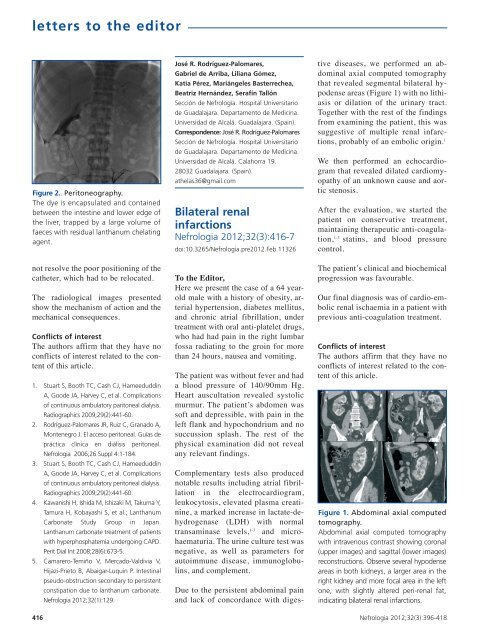
Figure 1. Abdominal axial computed<br />
<strong>to</strong>mography.<br />
Abdominal axial computed <strong>to</strong>mography<br />
with intravenous contrast showing coronal<br />
(upper images) and sagittal (lower images)<br />
reconstructions. Observe several hypodense<br />
areas in both kidneys, a larger area in <strong>the</strong><br />
right kidney and more focal area in <strong>the</strong> left<br />
one, with slightly altered peri-renal fat,<br />
indicating bilateral renal infarctions.<br />
416 <strong>Nefrologia</strong> 2012;32(3):396-418
<strong>letters</strong> <strong>to</strong> <strong>the</strong> edi<strong>to</strong>r<br />
1. Hazanov N, Somin M, Attali M, Beilinson<br />
N, Thaler M, Mouallem M, et al. Acute<br />
renal embolism. Forty-four cases of renal<br />
infarction in patients with atrial fibrillation.<br />
Medicine (Baltimore) 2004;83:292.<br />
2. Korzets Z, Plotkin E, Bernheim J, Zissin R.<br />
The clinical spectrum of acute renal<br />
infarction. Isr Med Assoc J 2002;4:781.<br />
3. Lessman RK, Johnson SF, Coburn JW,<br />
Kaufman JJ. Renal artery embolism: clinical<br />
features and long-term follow-up of 17<br />
cases. Ann Intern Med 1978;89:477.<br />
Marta Cuberes-Izquierdo 1 ,<br />
Nerea Yanguas-Barea 2 , Olga Mar<strong>to</strong>rell-Almau 3 ,<br />
Ángel Gamen-Pardo 1 , Eduardo Parra-Moncasi 1 ,<br />
Raquel Artal-Sánchez 4 ,<br />
Rosa Cozcolluela-Cabrejas 2<br />
1<br />
Sección de Nefrología.<br />
Hospital Reina Sofía de Tudela. Navarra. (Spain).<br />
2<br />
Servicio de Radiodiagnóstico.<br />
Hospital Reina Sofía de Tudela. Navarra. (Spain).<br />
3<br />
Servicio de Urgencias.<br />
Hospital Reina Sofía de Tudela. Navarra. (Spain).<br />
4<br />
Servicio de O<strong>to</strong>rrinolaringología.<br />
Hospital Reina Sofía de Tudela. Navarra. (Spain).<br />
Correspondence: Marta Cuberes Izquierdo<br />
Sección de Nefrología.<br />
Hospital Reina Sofía de Tudela, Carretera<br />
Tarazona Km 3. 31500 Tudela. (Spain).<br />
zairadc@hotmail.com<br />
Severe<br />
hypertriglyceridaemia.<br />
Treatment with<br />
plasmapheresis<br />
<strong>Nefrologia</strong> 2012;32(3):417-8<br />
doi:10.3265/<strong>Nefrologia</strong>.pre2012.Feb.11394<br />
Very little experience has been gained<br />
in <strong>the</strong> treatment of HTG with apheresis,<br />
although <strong>the</strong> few studies in <strong>the</strong><br />
medical literature describing <strong>the</strong> treatment<br />
of this pathology with apheresis<br />
have obtained very positive results. 2,3<br />
The current guidelines of <strong>the</strong> American<br />
Society for Apheresis (ASFA) consider<br />
this a category III practice, and have approved<br />
its use in <strong>the</strong> case of HTG and<br />
in <strong>the</strong> presence or possibility of severe<br />
pancreatitis, which is quite probable<br />
when triglyceride (TG) levels exceed<br />
2000mg/dl, and always when <strong>the</strong> patient<br />
does not respond <strong>to</strong> normal medical<br />
treatment. There are few comparative<br />
studies, but <strong>the</strong>y have shown that<br />
1-3 sessions of plasmapheresis in patients<br />
with pancreatitis and HTG can reduce<br />
symp<strong>to</strong>ms by 46%-80%, <strong>the</strong> same<br />
results as for drug treatment. 4 In a study<br />
of 8 patients with recurring pancreatitis<br />
undergoing chronic treatment with<br />
plasmapheresis, <strong>the</strong> frequency of pancreatitis<br />
was reduced by 67% when TG<br />
levels were maintained below<br />
150mg/dl, thus preventing patient hospitalisations<br />
and reducing health costs.<br />
For filtration techniques, we can use<br />
double filtration or cascade filtration,<br />
where one filter separates blood from<br />
<strong>the</strong> plasma, which is <strong>the</strong>n passed<br />
through a second filter with a smaller<br />
pore size that does not allow <strong>the</strong> passage<br />
of molecules with a larger molecular<br />
weight; in this case, TG.5 In <strong>the</strong> DALI<br />
(Direct Absorption of Lipoproteins) system,<br />
<strong>the</strong> TG are directly absorbed from<br />
<strong>the</strong> blood using a filter that consists of<br />
modified polyacrylate ligands immobilized<br />
on a polyacrylamide matrix.<br />
Here we discuss <strong>the</strong> case of a 45 yearold<br />
male with no relevant medical<br />
his<strong>to</strong>ry and no symp<strong>to</strong>ms, but whose<br />
labora<strong>to</strong>ry tests revealed a TG value<br />
of 7916mg/dl. The patient was admitted<br />
<strong>to</strong> our department for <strong>the</strong>rapeutic<br />
and preventative plasmapheresis<br />
against pancreatitis. Only two sessions<br />
were administered. We used an<br />
apheresis moni<strong>to</strong>r that first passed<br />
<strong>the</strong> blood through a plasma separating<br />
filter, and <strong>the</strong>n <strong>the</strong> plasma was<br />
passed through ano<strong>the</strong>r filter that<br />
trapped TG from plasma using hydrophobic<br />
interactions, finally returning<br />
<strong>the</strong> treated plasma <strong>to</strong> <strong>the</strong> patient.<br />
This procedure does not require<br />
plasma or albumin supplements. The<br />
plasma volume treated was 2.5 litres,<br />
calculated by patient weight and<br />
haema<strong>to</strong>crit values, with a mean time<br />
per session of approximately 1 hour<br />
and 45 minutes. After <strong>the</strong> first session,<br />
TG levels decreased <strong>to</strong><br />
1500mg/dl. After <strong>the</strong> second session,<br />
<strong>the</strong> value was 267mg/dl (Table 1 and<br />
Table 2). The patient was <strong>the</strong>n discharged<br />
with prescriptions for rosuvastatin<br />
at 10mg/24h and fenofibrate<br />
at 145mg/24h. Currently, <strong>the</strong> patient<br />
is asymp<strong>to</strong>matic, with good lipid control<br />
under medical treatment, and<br />
does not require hospitalisation despite<br />
such high levels of TG.<br />
With this case, we wish <strong>to</strong> awaken interest<br />
amongst nephrologists in understanding<br />
and implementing apheresis<br />
techniques. This is ano<strong>the</strong>r type of extracorporeal<br />
purification that can obtain<br />
positive clinical results, avoiding<br />
unnecessary health costs and hospitalisations,<br />
as in our case.<br />
To <strong>the</strong> To <strong>the</strong> Edi<strong>to</strong>r,<br />
The application of apheresis treatments<br />
is gaining more importance in nephrological<br />
practice. In patients with metabolic<br />
diseases, clear indications exist<br />
for apheresis procedures, such as in familial<br />
hypercholesterolaemia. 1 However,<br />
in o<strong>the</strong>r diseases, this type of treatment<br />
is applied only as an alternative<br />
when normal <strong>the</strong>rapies fail <strong>to</strong> garner a<br />
response, such as in primary hypertriglyceridaemia<br />
(HTG).<br />
Table 1. Total cholesterol, triglycerides, HDL, and LDL levels after <strong>the</strong> first<br />
apheresis session<br />
Start 1 hour End<br />
Total cholesterol (mg/dl) 1104 980 675<br />
Triglycerides (mg/dl) 7916 2940 1500<br />
HDL (mg/dl) 63 57 50<br />
LDL (mg/dl) 447 347 327<br />
HDL: high-density lipoprotein; LDL: low-density lipoprotein.<br />
<strong>Nefrologia</strong> 2012;32(3):396-418<br />
417