Objective assessment of limb tissue elasticity - Rehabilitation ...
Objective assessment of limb tissue elasticity - Rehabilitation ...
Objective assessment of limb tissue elasticity - Rehabilitation ...

Create successful ePaper yourself
Turn your PDF publications into a flip-book with our unique Google optimized e-Paper software.
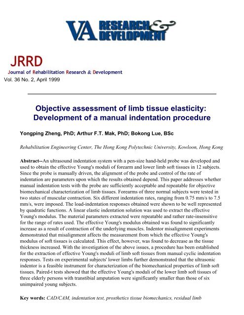
Vol. 36 No. 2, April 1999<br />
<strong>Objective</strong> <strong>assessment</strong> <strong>of</strong> <strong>limb</strong> <strong>tissue</strong> <strong>elasticity</strong>:<br />
Development <strong>of</strong> a manual indentation procedure<br />
Yongping Zheng, PhD; Arthur F.T. Mak, PhD; Bokong Lue, BSc<br />
<strong>Rehabilitation</strong> Engineering Center, The Hong Kong Polytechnic University, Kowloon, Hong Kong<br />
Abstract--An ultrasound indentation system with a pen-size hand-held probe was developed and<br />
used to obtain the effective Young's moduli <strong>of</strong> forearm and lower <strong>limb</strong> s<strong>of</strong>t <strong>tissue</strong>s in 12 subjects.<br />
Since the probe is manually driven, the alignment <strong>of</strong> the probe and control <strong>of</strong> the rate <strong>of</strong><br />
indentation are parameters upon which the results obtained depend. This paper addresses whether<br />
manual indentation tests with the probe are sufficiently acceptable and repeatable for objective<br />
biomechanical characterization <strong>of</strong> <strong>limb</strong> <strong>tissue</strong>s. Forearms <strong>of</strong> three normal subjects were tested in<br />
two states <strong>of</strong> muscular contraction. Six different indentation rates, ranging from 0.75 mm/s to 7.5<br />
mm/s, were imposed. The load-indentation responses obtained were shown to be well represented<br />
by quadratic functions. A linear elastic indentation solution was used to extract the effective<br />
Young's modulus. The material parameters extracted were repeatable and rather rate-insensitive<br />
for the range <strong>of</strong> rates used. The effective Young's modulus obtained was found to significantly<br />
increase as a result <strong>of</strong> contraction <strong>of</strong> the underlying muscles. Indentor misalignment experiments<br />
demonstrated that misalignment affects the measurement from which the effective Young's<br />
modulus <strong>of</strong> s<strong>of</strong>t <strong>tissue</strong>s is calculated. This effect, however, was found to decrease as the <strong>tissue</strong><br />
thickness increased. With the investigation <strong>of</strong> the above issues, a procedure has been established<br />
for the extraction <strong>of</strong> effective Young's moduli <strong>of</strong> <strong>limb</strong> s<strong>of</strong>t <strong>tissue</strong>s from manual cyclic indentation<br />
responses. Tests on experimental subjects' lower <strong>limb</strong>s further demonstrated that the ultrasonic<br />
indentor is a feasible instrument for characterization <strong>of</strong> the biomechanical properties <strong>of</strong> <strong>limb</strong> s<strong>of</strong>t<br />
<strong>tissue</strong>s. Paired-t tests showed that the effective Young's moduli <strong>of</strong> the lower <strong>limb</strong> s<strong>of</strong>t <strong>tissue</strong>s <strong>of</strong><br />
three elderly persons with transtibial amputation were significantly smaller than those <strong>of</strong> six<br />
unimpaired young subjects.<br />
Key words: CAD/CAM, indentation test, prosthetics <strong>tissue</strong> biomechanics, residual <strong>limb</strong>
<strong>assessment</strong>, s<strong>of</strong>t <strong>tissue</strong> biomechanics, <strong>tissue</strong> mechanics, ultrasound instrumentation.<br />
INTRODUCTION<br />
Palpation on the skin is widely used clinically to assess the biomechanical characteristics <strong>of</strong> the<br />
residual <strong>limb</strong> <strong>tissue</strong> <strong>of</strong> persons with amputation, due to the lack <strong>of</strong> an easy-to-use quantitative tool<br />
to assess the biomechanical properties <strong>of</strong> <strong>limb</strong> <strong>tissue</strong>s. Palpation produces a subjective <strong>assessment</strong><br />
and, therefore, requires substantial clinical experience. In addition, the qualitative nature <strong>of</strong> such<br />
<strong>assessment</strong>s makes the accumulation <strong>of</strong> knowledge difficult and teaching/learning imprecise.<br />
During the last decade, computer-aided design and computer-aided manufacturing (CAD/CAM)<br />
technologies were introduced to prosthetic socket design (1-9). However, exactly how a socket is<br />
designed still depends on the experience <strong>of</strong> the prosthetist and his or her subjective <strong>assessment</strong> <strong>of</strong><br />
the patient's residual <strong>limb</strong> <strong>tissue</strong>s. The quantitative biomechanical properties <strong>of</strong> s<strong>of</strong>t <strong>tissue</strong>s have<br />
not been included and used in current commercial CAD/CAM socket design systems.<br />
Finite element (FE) analysis has been used by several research groups to study the interface<br />
stresses between the surface <strong>of</strong> the residual <strong>limb</strong> <strong>tissue</strong> and the socket (10-34). Biomechanical<br />
properties <strong>of</strong> the residual <strong>limb</strong> <strong>tissue</strong>s are required inputs for residual <strong>limb</strong> FE models. Finite<br />
element methods, together with information on <strong>limb</strong> <strong>tissue</strong> properties, can be integrated into CAD/<br />
CAM socket design systems to realize the full potential <strong>of</strong> CAD/CAM techniques.<br />
Among the various mechanical materials testing methods that have been utilized, indentation<br />
testing is probably the most popular technique for determining the in-vivo mechanical behavior <strong>of</strong><br />
skin and <strong>limb</strong> s<strong>of</strong>t subcutaneous <strong>tissue</strong>s. In 1912, Schade developed an indentation apparatus to<br />
study the creep properties <strong>of</strong> skin and the changes that result from edematous conditions (35).<br />
Since then, a number <strong>of</strong> indentation apparata have been developed to investigate human s<strong>of</strong>t<br />
<strong>tissue</strong>s in vivo for various purposes (19,20,23,26-30,36-48). Indentation testing is an effective and<br />
relatively straightforward way to assess biomechanical properties <strong>of</strong> the skin and subcutaneous<br />
<strong>tissue</strong>s under compression. The test itself very much resembles the situation <strong>of</strong> palpation.<br />
However, most <strong>of</strong> the indentation apparata reported in the literature were developed for research<br />
purposes and are not yet ready for routine clinical application. In addition, since the <strong>tissue</strong><br />
thickness was not monitored, the load-indentation data represented both the geometric effects as<br />
well as the material properties <strong>of</strong> the <strong>limb</strong> s<strong>of</strong>t <strong>tissue</strong>s.<br />
An ultrasound indentation apparatus was developed by the investigators for the <strong>assessment</strong> <strong>of</strong><br />
the mechanical properties <strong>of</strong> <strong>limb</strong> s<strong>of</strong>t <strong>tissue</strong>s (49-53). The indentation apparatus consists <strong>of</strong> a pensize,<br />
hand-held indentation probe (Figure 1).
Figure 1.<br />
Demonstration <strong>of</strong> the indentation test using the pen-size hand-held indentor on the forearm <strong>of</strong> a<br />
test subject.<br />
A cylindrical flat-ended ultrasound transducer with a diameter <strong>of</strong> 9.0 mm and a frequency <strong>of</strong> 5<br />
MHz is at the tip <strong>of</strong> the indentor and serves as the indentor probe. The thickness and deformation<br />
<strong>of</strong> the s<strong>of</strong>t <strong>tissue</strong> layer are determined from the ultrasound echo signal. A compressive 10 N load<br />
cell is connected in series with the ultrasound transducer to record the corresponding force<br />
response. A schematic <strong>of</strong> the ultrasound indentation system is shown in Figure 2. The accuracy <strong>of</strong><br />
the deformation and thickness determined by the system was better than 0.02 mm, and the<br />
accuracy <strong>of</strong> the force result was better than 0.003 N (53). The sampling rate was 12.5 data points<br />
per second.<br />
Figure 2.
A schematic showing the ultrasound indentation system.<br />
Hayes et al. presented a rigorous mathematical solution to the <strong>elasticity</strong> problem <strong>of</strong> indentation<br />
<strong>of</strong> a thin, elastic, homogenous layer <strong>of</strong> material bonded to a rigid half-space (54). The solution<br />
yields an expression for calculation <strong>of</strong> the Young's modulus from the indentation loaddeformation<br />
data:<br />
where P is the applied force, w the indentation depth, a the radius <strong>of</strong> the indentor, ν Poisson's<br />
ratio <strong>of</strong> the <strong>tissue</strong>, h the <strong>tissue</strong> thickness, and κ a scaling factor. The scaling factor κ provides a<br />
theoretical correction for the finite thickness <strong>of</strong> the elastic layer, and it depends on both the aspect<br />
ratio a/h and Poisson's ratio ν. Equation 1 was first developed for the study <strong>of</strong> articular cartilage,<br />
and was later used to extract Young's modulus <strong>of</strong> <strong>limb</strong> s<strong>of</strong>t <strong>tissue</strong>s (19,20,24). When the aspect<br />
ratio a/h is small enough (i.e., the <strong>tissue</strong> thickness is large enough compared with the radius <strong>of</strong> the<br />
indentor), the scaling factor κ tends to be 1. In this case, the load-indentation response is<br />
independent to <strong>tissue</strong> thickness h. Some investigators calculated Young's modulus <strong>of</strong> lower <strong>limb</strong><br />
s<strong>of</strong>t <strong>tissue</strong>s using Equation 1 with this assumption (30). Since the s<strong>of</strong>t <strong>tissue</strong>s are assumed to be<br />
homogenous in this solution, the modulus extracted using Equation 1 can be regarded as an<br />
effective Young's modulus or effective elastic modulus for the full-thickness bulk <strong>tissue</strong>. Equation<br />
1 assumes the indentor is perpendicular to the skin surface and to the underlying bony interface.<br />
Numerical methods have also been developed to extract from the indentation data parameters<br />
governing the biomechanical responses <strong>of</strong> the <strong>limb</strong> <strong>tissue</strong>s (19,20,23,24,26,27,47). The ultrasound<br />
indentation system allows P, w, and h to be determined experimentally.<br />
As indentation with the hand-held probe is manually driven, the rate <strong>of</strong> indentation cannot be<br />
controlled precisely. Misalignment <strong>of</strong> the indentor is also difficult to avoid when the indentation<br />
is manual or automated, without a priori knowledge <strong>of</strong> underlying skeletal geometry, orthogonal<br />
alignment is not always easy to achieve. The objective <strong>of</strong> the work reported in this paper is to<br />
address whether indentation testing, using the ultrasound hand-held indentation probe, provides a<br />
feasible approach for objective biomechanical characterization <strong>of</strong> <strong>limb</strong> <strong>tissue</strong>s. In particular, the<br />
issues <strong>of</strong> the indentor misalignment, the rate-sensitivity, and the nonlinearity <strong>of</strong> the <strong>tissue</strong><br />
properties were examined.<br />
The validity <strong>of</strong> the load, deformation, and <strong>tissue</strong> thickness determined by the ultrasound indentor<br />
has been previously verified in a mechanical testing machine (52). To test the feasibility <strong>of</strong> using<br />
the new approach, fresh fish <strong>tissue</strong>s and porcine <strong>tissue</strong>s have been used at the beginning <strong>of</strong> the<br />
study. Results <strong>of</strong> the misalignment studies are presented here. To assess the adequacy <strong>of</strong> the<br />
ultrasound indentor as a clinically feasible procedure, we investigated the material properties <strong>of</strong><br />
forearm s<strong>of</strong>t <strong>tissue</strong>s with the underlying muscles in either relaxed or contracted states.<br />
Additionally, the material properties <strong>of</strong> lower <strong>limb</strong> s<strong>of</strong>t <strong>tissue</strong>s at bony areas <strong>of</strong> subjects with and<br />
without amputation were investigated. These data are intrinsically important and relevant to the
design <strong>of</strong> prosthetic sockets.<br />
METHODS<br />
Experiments on Indentor Misalignment<br />
Using the ultrasound indentation apparatus, in vivo, indentation tests were conducted manually<br />
by pressing the probe into the skin and s<strong>of</strong>t <strong>tissue</strong>s <strong>of</strong> the subjects. The apparatus was designed so<br />
that indentation testing would be performed without any complicated attachment jig; thus, making<br />
it clinically convenient for residual <strong>limb</strong> <strong>assessment</strong>. Instead <strong>of</strong> assuming the displacement <strong>of</strong> the<br />
indentor to be the deformation <strong>of</strong> the s<strong>of</strong>t <strong>tissue</strong> layer as used in most <strong>of</strong> the previous indentation<br />
apparata, the <strong>tissue</strong> deformation was determined, in this study, from the ultrasound echo reflected<br />
from the bone-s<strong>of</strong>t <strong>tissue</strong> interface. To obtain the largest reflected ultrasound signal, the indentor<br />
should be perpendicular to the underlying bone surface. If the probe was not perpendicular to the<br />
bone, misalignment would also influence the received ultrasound signal, from which the original<br />
<strong>tissue</strong> thickness and the subsequent <strong>tissue</strong> deformation are derived.<br />
An in vitro experiment on a porcine <strong>tissue</strong> layer was performed to study the effect <strong>of</strong><br />
misalignment on the ultrasound reflection signals. The hand-held indentation probe was fixed in<br />
line with the load cell <strong>of</strong> a Hounsfield material-testing machine. A fresh porcine <strong>tissue</strong> layer with<br />
dimensions roughly <strong>of</strong> 100×100×25 mm was placed in a container. It was filled with saline<br />
solution and was installed on a slanting device, with which the orientation <strong>of</strong> the bottom <strong>of</strong> the<br />
<strong>tissue</strong> layer could be adjusted. The indentation was conducted at the center <strong>of</strong> the layer. Thickness<br />
<strong>of</strong> the entire <strong>tissue</strong> layer underneath the testing site was 24.5 mm. The probe was first driven by<br />
the machine to touch the surface <strong>of</strong> the <strong>tissue</strong> layer with a very small preload <strong>of</strong> 0.02 N. Then the<br />
ultrasound echo waves, reflected from the bottom interface <strong>of</strong> the porcine <strong>tissue</strong> layer with probe<br />
misalignment up to 12.5°, were recorded and compared.<br />
The second experiment concerning misalignment was performed in vivo on the left forearm <strong>of</strong><br />
an unimpaired young male subject. Three sites with different thickness <strong>of</strong> s<strong>of</strong>t <strong>tissue</strong>s over the<br />
radius were selected as shown in Figure 3. The subject sat in front <strong>of</strong> a table and put his left arm<br />
on the surface <strong>of</strong> the table with the hand and fingers relaxed. Two groups <strong>of</strong> tests were conducted<br />
at each testing site. In the first group <strong>of</strong> tests, the probe was aligned to maintain a maximal<br />
ultrasound echo signal reflected from the bone surface. Five trials were recorded at each site. In<br />
the second group <strong>of</strong> tests, the orientation <strong>of</strong> the probe was adjusted while still maintaining a small<br />
yet acceptable amplitude <strong>of</strong> ultrasound reflection signal for signal processing. The amplitude in<br />
this case was approximately 15 dB less than the maximum amplitude obtained for the maximal<br />
signal alignment at the same site, and the orientation <strong>of</strong> the probe corresponded to a misalignment<br />
<strong>of</strong> approximately 5° along the lateral-medial direction, and approximately 10° along the proximaldistal<br />
direction. The degree <strong>of</strong> misalignment was measured by an electrogoniometer (Model<br />
XM110, Biometrics Ltd., Cwmfelinfach, Gwent, UK). A total <strong>of</strong> 20 trials was performed along<br />
different directions at each testing site.
Figure 3.<br />
The three testing sites for the in-vivo experiments <strong>of</strong> indentor misalignment.<br />
Experiments on Indentation Rate and Muscular Contraction<br />
Three unimpaired young men, 29, 30, and 32 years old, were involved in this series <strong>of</strong> tests. The<br />
test site was at the s<strong>of</strong>t <strong>tissue</strong>s covering the left radius, about 10 cm distal from the elbow joint.<br />
The <strong>tissue</strong>s tested involved skin, fat, and the underlying muscle. Figure 1 shows the test<br />
configuration used.<br />
The subject was instructed to sit in front <strong>of</strong> a table and put his left arm on the supporting surface.<br />
The angle <strong>of</strong> the elbow was approximately 100°, the angle between the upper arm and the<br />
supporting surface was approximately 30°, and the angle between the upper arm and the shoulder<br />
was approximately 90°. The tests were conducted in two states <strong>of</strong> muscular activation. In the first<br />
group <strong>of</strong> tests, the subject was instructed to relax his wrist and fingers and put his palm and<br />
fingers on the supporting surface along the same direction <strong>of</strong> the forearm. In the second group <strong>of</strong><br />
trials, the subject was instructed to exert a 5-kg gripping force with his left hand during the test,<br />
and then to relax for about 2 minutes. The gripping force was monitored by a device that was<br />
developed in our Center for grip training. Figure 4 shows the experimental setup for this test.<br />
Subjects were asked to maintain a stable muscular activation state during the test by monitoring<br />
the LED readout <strong>of</strong> the gripping device.
Figure 4.<br />
The experimental setup used to study the effects <strong>of</strong> muscular contraction on the respective <strong>tissue</strong><br />
mechanical properties.<br />
The indentation was performed with the ultrasound hand-held probe. A couplant gel was used to<br />
acoustically couple the ultrasound transducer with the subject's s<strong>of</strong>t <strong>tissue</strong>s. By monitoring the<br />
indentation measurement data shown on the screen <strong>of</strong> the computer, the maximum indentation<br />
was controlled and limited to within approximately 30 percent <strong>of</strong> the initial thickness <strong>of</strong> the s<strong>of</strong>t<br />
<strong>tissue</strong>s. The maximum indentation load was limited to 5 N. Such indentation on the forearm s<strong>of</strong>t<br />
<strong>tissue</strong>s with the two muscular states could be accepted by the subjects without obvious<br />
discomfort. Before each group <strong>of</strong> tests, three to four trial tests were performed at the test site to<br />
maximize and stabilize the ultrasound reflection signal from the surface <strong>of</strong> the bone; the s<strong>of</strong>t<br />
<strong>tissue</strong>s were also preconditioned during this process (55). From the trial tests, the load cell was<br />
<strong>of</strong>fset-adjusted so that its reading was zero N when no force was applied. To reduce effects other<br />
than indentation rate by averaging, up to six trials were conducted at a similar indentation rate for<br />
each test. The rate was estimated by monitoring the force and deformation curves shown on the<br />
screen. Six different indentation rates, ranging from 0.75 mm/s to 7.5 mm/s, were imposed at each<br />
site for each muscular contraction state. Figures 5a and 5b demonstrate two typical indentation<br />
sequences with a rate <strong>of</strong> approximately 0.75 mm/s and 7.5 mm/s, respectively. These indentation<br />
rates were regarded as within the general range that manual indentation could be imposed. When<br />
the indentation rate was lower than 0.75 mm/s, it was difficult to control the loading and<br />
unloading sequence smoothly by hand. This may be observed to some degree from the indentation<br />
responses shown in Figure 5a. On the other hand, when the indentation rate was higher than 7.5<br />
mm/s, the skin surface did not follow the probe during the unloading phases. The repeatability<br />
tests were conducted on one subject in a relaxed muscular state using a rate <strong>of</strong> approximately 4<br />
mm/s. Six tests were measured there and each test comprised up to six trials. Their results were
used to verify the repeatability <strong>of</strong> tests and to investigate how many trials were adequate for a<br />
single test.<br />
Figure 5.<br />
Typical indentation responses recorded on the forearm <strong>of</strong> subject #1 with the forearm musculature<br />
relaxed. Indentation rates <strong>of</strong> approximately (a) 0.75 mm/s and (b) 7.5 mm/s were used. The<br />
original thickness <strong>of</strong> the s<strong>of</strong>t <strong>tissue</strong> layer was 19.6 mm.<br />
Experiments on Lower Limbs<br />
Six unimpaired subjects and three subjects with transtibial amputation participated in this series<br />
<strong>of</strong> tests. The mean age <strong>of</strong> the subjects without amputation was 21.8, and that <strong>of</strong> subjects with<br />
amputation was 63.8. The residual <strong>limb</strong>s <strong>of</strong> the test subjects were <strong>of</strong> cylindrical shape and free <strong>of</strong><br />
edema. The subjects had worn their prostheses successfully for at least 1 year and were not further<br />
categorized by the level <strong>of</strong> amputation and the period <strong>of</strong> post-amputation. The sockets the subjects<br />
wore were the patella tendon bearing (PTB) type or other PTB variants.<br />
A previous study documented the indentation properties <strong>of</strong> several pressure-tolerant areas <strong>of</strong>
esidual <strong>limb</strong>s <strong>of</strong> persons with transtibial amputation (44). However, little attention has been paid<br />
to the pressure sensitive or bony-prominent area. Figure 6 shows the locations on the lower leg at<br />
which the indentation tests were performed. In the study, the location <strong>of</strong> the sites was mainly<br />
identified by palpation. The four sites at which the tests were performed were the tibial tubercule,<br />
the anterolateral tibial tubercule, the fibula head, and the anterodistal tibia. Both the amputated<br />
side and the contralateral sound side <strong>of</strong> subjects were tested. In the cases <strong>of</strong> the subjects without<br />
amputation, both the right and left legs were tested.<br />
Figure 6.<br />
The testing regions used in experiments on lower <strong>limb</strong> <strong>tissue</strong>s' material properties.<br />
The location <strong>of</strong> the anterodistal tibial test site was different in each <strong>of</strong> the subjects, depending on<br />
the length <strong>of</strong> their residual <strong>limb</strong>. The anterodistal tibia was defined as the most anterior and distal<br />
bone prominence <strong>of</strong> the tibia. After selecting the anterodistal site, the distance from the center <strong>of</strong><br />
the patella to the anterodistal tibia was recorded. The same distance at the sound side was used to<br />
locate the corresponding site. For the unimpaired subjects, the anterodistal tibia was defined at<br />
about 16.5 cm from the center <strong>of</strong> the patella. The dimension <strong>of</strong> 16.5 cm was the mean distance for<br />
the subjects with amputation.<br />
During the test, the subjects sat on an adjustable height chair. The condition <strong>of</strong> their (residual)<br />
<strong>limb</strong> s<strong>of</strong>t <strong>tissue</strong> was examined and recorded. The dimensions <strong>of</strong> their (residual) <strong>limb</strong>s were<br />
measured including length and circumference at different levels. By adjusting the height <strong>of</strong> the<br />
chair, the angle between the lower leg and the thigh was set at 90°, with the lower leg placed<br />
perpendicular to the floor. A goniometer was used to monitor the knee angle. Following a similar<br />
testing procedure described previously for the forearm tests, two trials were performed at each<br />
site, noting that the extracted effective Young's modulus was quite repeatable, based on the results<br />
<strong>of</strong> the preliminary studies. The maximum force applied was kept below 4 N to avoid discomfort<br />
to the subjects. Finite element analysis showed that the pressures applied to these areas <strong>of</strong> the<br />
residual <strong>limb</strong>-socket interface were in a similar range (32).<br />
Preliminary Observations<br />
Nonlinear behavior and hysteresis were observed in the load-indentation curves recorded at the<br />
various sites and indentation rates tested. Figure 7 shows the experimental data points measured
for a typical load-indentation response test <strong>of</strong> forearm s<strong>of</strong>t <strong>tissue</strong>s.<br />
Figure 7.<br />
Typical load-indentation measurements obtained from a forearm test subject, and the<br />
corresponding quadratic regression curve fit to the data.<br />
The data points <strong>of</strong> the loading phase were labeled with square marks, and those <strong>of</strong> the unloading<br />
phase were labeled with cross marks. It was found that the following quadratic function could<br />
well represent the load-indentation relationship with the coefficient <strong>of</strong> determination R 2 generally<br />
greater than 0.9:<br />
where P is the indentation load, w the indentation depth, C 0 , C 1 , and C 2 the coefficients that<br />
represent the indentation behavior <strong>of</strong> the <strong>tissue</strong>. The data points <strong>of</strong> the loading and unloading<br />
phase were both involved in this curve fitting process. The derived coefficients represented not<br />
only the material properties <strong>of</strong> the s<strong>of</strong>t <strong>tissue</strong>, but also other geometric factors, such as the<br />
indentor diameter and the <strong>tissue</strong> thickness, visco<strong>elasticity</strong>, and stiffness.<br />
Data Reduction Methods<br />
The effective Young's modulus was calculated using Equation 1, and the Poisson's ratio K was<br />
assumed to be 0.45. The diameter <strong>of</strong> the indentor (i.e., the ultrasound transducer) was 9.0 mm in<br />
this study. With these constants, Equation 1 can be written in the form:
where K(h), with a dimension <strong>of</strong> length, is a factor only depending on the thickness h <strong>of</strong> the s<strong>of</strong>t<br />
<strong>tissue</strong> if a constant Poisson's ratio ν is assumed. Using the value <strong>of</strong> κ(a/h,ν) determined by Hayes<br />
et al. (54), the relationship between K(h) and thickness h can be derived. K(h) can be interpolated<br />
for an arbitrary value <strong>of</strong> h.<br />
A typical indentation response is shown in Figure 7. In this specific test, five loading-unloading<br />
cycles were manually performed within a testing period <strong>of</strong> about 17 seconds. The loadindentation<br />
curve shows the typical nonlinear and viscoelastic characteristics <strong>of</strong> the s<strong>of</strong>t <strong>tissue</strong>s.<br />
During the indentation test with the probe manually driven, the indentation rate could only be<br />
approximately controlled. The experimental data points in the loading phases with the indentation<br />
load greater than 0.5 N and less than 75 percent <strong>of</strong> the maximum value <strong>of</strong> 5.0 N were selected for<br />
the following data analysis to estimate the effective Young's modulus. It was noted that the<br />
indentation rate was relatively more difficult to control outside this range. The selected data<br />
points within such limits were plotted in Figure 8 with different symbols representing the data<br />
points <strong>of</strong> different cycles. With the limitations on the selection <strong>of</strong> data points, the nonlinear<br />
properties <strong>of</strong> the load-indentation response in Figure 7 have been reduced. The data point with<br />
the first 0.5 N load reading was defined as the initial state. The s<strong>of</strong>t <strong>tissue</strong> thickness at this state<br />
was defined as the initial thickness. A linear regression was used to fit the experimental data. The<br />
slope <strong>of</strong> the curve was the mean ratio <strong>of</strong> load to indentation (i.e., P/w), which is related to the<br />
biomechanical properties <strong>of</strong> the s<strong>of</strong>t <strong>tissue</strong> according to Equation 3. The small <strong>of</strong>fset was a result<br />
<strong>of</strong> the linear regression <strong>of</strong> the mildly nonlinear force-deformation data. For the forearm test shown<br />
in Figures 7 and 8, the initial thickness h was determined as 16.2 mm with a preload <strong>of</strong> 0.5 N, K<br />
(h) was calculated to be 15.6 (mm), and the average ratio <strong>of</strong> the applied load to the indentation<br />
depth P/w obtained from the linear regression was 0.35 N/mm. The effective Young's modulus<br />
was then calculated to be 22.6 kPa using Equation 3.
Figure 8.<br />
The selected data points used for the estimation <strong>of</strong> the effective Young's modulus <strong>of</strong> <strong>tissue</strong>.<br />
Different symbols represent the data points <strong>of</strong> different cycles. The data in the loading phases<br />
with the indentation load greater than 0.5 N and less than 75 percent <strong>of</strong> the maximum value were<br />
selected. The plotted data were re-scaled from the raw data assuming the 0.5 N preload point to be<br />
the reference state.<br />
RESULTS<br />
Indentor Misalignment<br />
Table 1 presents the effective Young's modulus and thickness <strong>of</strong> forearm s<strong>of</strong>t <strong>tissue</strong>s at the three<br />
testing sites shown in Figure 3 calculated using Equation 3 and the measurements acquired with<br />
two different alignments <strong>of</strong> the probe. The measured <strong>tissue</strong> thickness varied slightly between the<br />
two different alignment cases at all three sites. However, the resulting effective Young's modulus<br />
computed, decreased at all three test sites as the probe misalignment increased. This misalignment<br />
effect was the greatest at the site with the thinnest s<strong>of</strong>t <strong>tissue</strong>.<br />
Table 1.<br />
Effective Young's moduli and thicknesses <strong>of</strong> the forearm s<strong>of</strong>t<br />
<strong>tissue</strong>s at the three testing sites with two different alignment<br />
states <strong>of</strong> the probe.<br />
Site 1 Site 2 Site 3<br />
Modulus (kPa) Mean SD Mean SD Mean SD
Test A 20.39 1.81 37.41 1.81 34.74 1.94<br />
Test B 19.35 0.76 33.96 1.98 21.33 0.91<br />
Thickness (mm)<br />
Test A 17.50 0.13 15.14 0.21 8.80 0.12<br />
Test B 17.63 0.18 15.61 0.20 8.94 0.13<br />
A: Aligning the probe with the amplitude <strong>of</strong> the ultrasound reflection signals<br />
being maximal. Test B: Aligning the probe with a minimal acceptable<br />
ultrasound reflection signal, corresponding to a misalignment <strong>of</strong><br />
approximately ±10° to the normal to the radial shaft along the proximal-distal<br />
direction, and <strong>of</strong> approximately ±5° to the normal to the radial shaft along the<br />
medial-lateral direction.<br />
Figure 9 shows the ultrasound echo signals recorded after reflection from the bottom interface<br />
<strong>of</strong> the fresh porcine <strong>tissue</strong> specimen. A slight time shift <strong>of</strong> the ultrasound signal <strong>of</strong> up to<br />
approximately 2 percent was observed as the probe misalignment increased from 0° to 10°. This<br />
time shift would cause a small error in the measured thickness, but would not affect the derived<br />
indentation. The amplitude <strong>of</strong> the echo signal was significantly reduced as the degree <strong>of</strong><br />
misalignment increased from 0° to 15°. These characteristics can be used to empirically maintain<br />
a reference alignment <strong>of</strong> the probe during the manually activated indentation. These phenomena<br />
can be ascribed to the behavior <strong>of</strong> ultrasonic signal propagation. When the surface <strong>of</strong> the<br />
ultrasound transducer is not parallel to the reflecting interface, the signal is not entirely spectrally<br />
reflected, and thus, is not completely received by the transducer. An additional propagation path<br />
would be induced, and rough osseous reflecting surfaces cause wavelet model cancellation.<br />
Figure 9.<br />
Recorded ultrasound echo signals, which were reflected from the bottom interface <strong>of</strong> a layer <strong>of</strong><br />
fresh porcine <strong>tissue</strong>, showing the various degrees <strong>of</strong> indentor misalignment used.
Indentation Rate and Muscular Contraction<br />
Figure 10 shows the comparison between the regression curves <strong>of</strong> the forearm indentation<br />
responses recorded from the three subjects with the underlying muscles both relaxed and<br />
contracted. Differences among individuals can be noted from the curves. Significant changes in<br />
the measured indentation responses were observed for the subjects as a function <strong>of</strong> the underlying<br />
state <strong>of</strong> their muscular activation.<br />
Figure 10.<br />
Comparison <strong>of</strong> the regression curves <strong>of</strong> the load-indentation data among subjects. The indentation<br />
data were taken on the forearm under two different muscular contractile states. Subjects 1,2, and 3<br />
were the three subjects in this series <strong>of</strong> tests.<br />
Figure 11 shows the thicknesses <strong>of</strong> the s<strong>of</strong>t <strong>tissue</strong> layers, measured at the forearm test sites, <strong>of</strong><br />
the three subjects for two different states <strong>of</strong> muscular contraction. The mean <strong>tissue</strong> thickness and<br />
respective standard deviations were calculated for each subject from a total <strong>of</strong> 36 trials each. The<br />
s<strong>of</strong>t <strong>tissue</strong> thicknesses were found to increase by 25, 26, and 11 percent for subjects #1, #2, and<br />
#3, respectively, as they contracted their underlying muscles.
Figure 11.<br />
Original thickness <strong>of</strong> s<strong>of</strong>t <strong>tissue</strong>s at the indentation sites <strong>of</strong> the three subjects under two different<br />
muscular contraction states.<br />
In a study <strong>of</strong> measurement, a total <strong>of</strong> six tests were conducted, in which six trials were<br />
performed for each test. The percentage standard deviations in the calculated values <strong>of</strong> the<br />
effective Young's modulus, E, among the six tests were all less than 3 percent. The percentage<br />
standard deviations <strong>of</strong> the original measured <strong>tissue</strong> thickness were found to be less than 2 percent.<br />
The small variations obtained suggested that the extracted material parameters were repeatable.<br />
The mean percentage standard deviation <strong>of</strong> the effective Young's modulus, E, obtained from the<br />
tests <strong>of</strong> the three subjects under two muscular contraction states using six different indentation<br />
rates was 8.4 percent. The maximum value (13.0 percent) and the minimum value (3.2 percent)<br />
were observed from subject #3 and subject #1 with muscle contracted, respectively. The effect <strong>of</strong><br />
variations in indentation rate on the values extracted for the effective Young's modulus <strong>of</strong> the<br />
subjects, therefore, was not dramatic for the range <strong>of</strong> indentation rates tested, that is, from 0.75<br />
mm/s (rate A) to 7.5 mm/s (rate F), as shown in Figure 12. The effective Young's modulus <strong>of</strong><br />
<strong>limb</strong> s<strong>of</strong>t <strong>tissue</strong>s thus appears to be reasonably rate-insensitive within this range.
Figure 12.<br />
The relationship between effective Young's modulus, E, <strong>of</strong> forearm s<strong>of</strong>t <strong>tissue</strong> and indentation<br />
rate. The error bar represents the standard deviation <strong>of</strong> the four trials. "A" to "F" represent the six<br />
different indentation rates. "4-r" indicates the tests <strong>of</strong> subject #4 with the forearm and the hand<br />
relaxed, and "4-c" indicates the tests with the hand exerting a 5-kg gripping force.<br />
Figure 13 shows the effective Young's moduli <strong>of</strong> the forearm s<strong>of</strong>t <strong>tissue</strong>s significantly increased<br />
from 14.0±5.0 kPa to 58.8±1.7 kPa as a result <strong>of</strong> contraction <strong>of</strong> the underlying muscles. These<br />
results were calculated from the mean values from the tests <strong>of</strong> the three subjects with their<br />
relevant muscles relaxed and contracted respectively. This shows that the subjects' s<strong>of</strong>t forearm<br />
<strong>tissue</strong>s became significantly stiffer when the underlying muscles were contracted.<br />
Figure 13.<br />
Variation <strong>of</strong> the effective Young's modulus, E, <strong>of</strong> the forearm s<strong>of</strong>t <strong>tissue</strong> as a function <strong>of</strong> the
underlying muscular contractile state. The error bar represents the standard deviation <strong>of</strong> the<br />
results <strong>of</strong> the six groups <strong>of</strong> tests using different indentation rates.<br />
Lower Limb Tests<br />
Figure 14 represents the variation <strong>of</strong> the effective Young's modulus <strong>of</strong> the lower <strong>limb</strong> s<strong>of</strong>t<br />
<strong>tissue</strong>s measured at different sites on both <strong>limb</strong>s for the test groups with and without amputation.<br />
Generally, the effective Young's moduli <strong>of</strong> the sound <strong>limb</strong>s <strong>of</strong> the subjects with amputation were<br />
greater than those obtained for their residual <strong>limb</strong>s. However, the difference was rather small<br />
except at the anterodistal tibia. For the tibial tuberosity, anterolateral epicondyle, and fibular head,<br />
paired-t comparisons between the measurements on the residual <strong>limb</strong>s and on the sound <strong>limb</strong>s did<br />
not demonstrate significant difference. For the site over the anterodistal tibia, however, paired-t<br />
tests showed a significant difference between the effective Young's moduli <strong>of</strong> the residual <strong>limb</strong>s<br />
<strong>of</strong> the subjects with amputation and that <strong>of</strong> their contralateral sound <strong>limb</strong>s. It may also be seen in<br />
Figure 14 that the effective Young's moduli <strong>of</strong> the young unimpaired subjects were higher than<br />
those <strong>of</strong> the elderly subjects with amputation. A similar result was reported earlier by other<br />
investigators using an electromechanical indentation system (44).<br />
Figure 14.<br />
The comparison <strong>of</strong> the effective Young's moduli obtained for the subjects with and without<br />
amputation.<br />
DISCUSSION<br />
Indentation Response<br />
A newly developed ultrasound apparatus was used to obtain the indentation responses <strong>of</strong> forearm<br />
s<strong>of</strong>t <strong>tissue</strong>s. Three unimpaired forearms were tested with two muscular contraction states.<br />
Quadratic functions were found to be adequate to represent the load-indentation responses. Silver-<br />
Thorn used a third order polynomial to fit the load-indentation response recorded on the belowknee<br />
residual <strong>limb</strong>s within the confinement <strong>of</strong> a prosthetic socket (23). It should be noted that the
indentation responses <strong>of</strong> the s<strong>of</strong>t <strong>tissue</strong> not only depended on the mechanical behavior <strong>of</strong> the s<strong>of</strong>t<br />
<strong>tissue</strong>s, but also on the s<strong>of</strong>t <strong>tissue</strong> thickness, the initial state <strong>of</strong> the s<strong>of</strong>t <strong>tissue</strong>, the boundary<br />
interface conditions, and the indentor geometry.<br />
S<strong>of</strong>t Tissue Thickness<br />
Tissue thickness is an important parameter in determination <strong>of</strong> the material properties <strong>of</strong> the<br />
<strong>tissue</strong> from indentation response measurements, especially when the indentor diameter and the<br />
<strong>tissue</strong> thickness are comparable (54). In the literature, there have been reports on the use <strong>of</strong><br />
CATSCAN, X-rays, and MRIs to estimate <strong>tissue</strong> thicknesses at indentation sites on test<br />
subjects' (residual) <strong>limb</strong>s (19,26,30). Ultrasound has also been used to measure the thickness <strong>of</strong><br />
s<strong>of</strong>t <strong>tissue</strong>s (42,44). In the current study, s<strong>of</strong>t <strong>tissue</strong> thickness was estimated continuously during<br />
indentation using ultrasonic pulse/echo analysis technique. Thickness measurement is an inherent<br />
result <strong>of</strong> the present indentation technique and can be readily incorporated into data reduction and<br />
analysis procedures. Significant changes <strong>of</strong> the s<strong>of</strong>t <strong>tissue</strong> thickness were found due to changes <strong>of</strong><br />
underlying musculature activation states. The thickness <strong>of</strong> forearm bulk s<strong>of</strong>t <strong>tissue</strong>s was found to<br />
increase by up to 26 percent as test subjects contracted their underlying muscles, exerting a 5 kg<br />
palmar prehension gripping force.<br />
Repeatability<br />
The extracted material parameters obtained in this study were repeatable at a given site for a<br />
given individual with a given state <strong>of</strong> muscular activation. The percentage standard deviation <strong>of</strong><br />
the measured <strong>tissue</strong> thickness was less than 2 percent. The effective Young's modulus extracted<br />
using the linear elastic model showed a percentage standard deviation <strong>of</strong> less than 3 percent.<br />
Effects <strong>of</strong> Indentation Rate<br />
It is known that the mechanical behavior <strong>of</strong> <strong>limb</strong> s<strong>of</strong>t <strong>tissue</strong>s generally shows time-dependent<br />
phenomena (e.g., hysteresis). The influences <strong>of</strong> indentation rate on <strong>tissue</strong> mechanical response<br />
were reported by Reynolds and by Torres-Moreno (19,30). The loading rates studied by Reynolds<br />
were 0.3, 0.8, and 1.3 mm/s. Those investigated by Torres-Moreno were 9.9, 14.2, and 19.8 mm/s.<br />
Their indentors, however, were attached to the prosthetic socket, and were driven by mechanical<br />
actuators into test subjects' s<strong>of</strong>t <strong>tissue</strong> through ports in the sockets. In the current study, the<br />
indentation probe was manually driven, and the mean indentation rates used in the current study<br />
ranged from 0.75 to 7.5 mm/s. This range <strong>of</strong> indentation rates was approximately in the middle<br />
between that used by Reynolds and that used by Torres-Moreno. This was found to be the<br />
manageable range <strong>of</strong> rates imposable by hand. The Young's moduli determined from the<br />
ultrasonic indentation probe measurements were found to be relatively insensitive to the<br />
indentation rate, with a percentage standard deviation <strong>of</strong> less than 10 percent for the range <strong>of</strong><br />
indentation rates imposed. Torres-Moreno demonstrated that the effect <strong>of</strong> indentation rate was<br />
different at different testing sites, and also with a different load level (30). Reynolds reported that<br />
the effect <strong>of</strong> indentation rate on the indentation response was more serious when greater loads<br />
were applied (19). In the experiments <strong>of</strong> Torres-Moreno and Reynolds, the average maximal<br />
pressure applied to obtain a certain amount <strong>of</strong> deformation was much greater (about 5 times) than<br />
that used in the current study because s<strong>of</strong>t <strong>tissue</strong>s were confined by the socket in their studies.<br />
This might also be one <strong>of</strong> the reasons why hysteresis observed in Torres-Moreno's results was<br />
generally more serious than that observed in the current study. In the confined tests, the skin<br />
would be drawn sideways along the socket wall by the indentation, and additional in<strong>elasticity</strong>
might be induced. The lower load levels used in concert with a 0.5 N preload may have reduced<br />
the <strong>tissue</strong> measurement sensitivities to indentation rate observed in the current study to some<br />
degree. Further studies on rate sensitivity under a broader range <strong>of</strong> conditions, such as higher<br />
loading levels, <strong>tissue</strong> socket confinement, and so forth, warrant further investigation using the<br />
current ultrasonic measurement technique.<br />
Effect <strong>of</strong> Muscle Contraction<br />
Tissue mechanical response measurements and the associated material parameters derived<br />
therefrom varied significantly with changes in the contractile state <strong>of</strong> the underlying musculature.<br />
The effective Young's modulus <strong>of</strong> proximal forearm <strong>tissue</strong> significantly increased from 14.0±5.0<br />
kPa to 58.8±1.7 kPa as a result <strong>of</strong> contraction <strong>of</strong> the underlying muscles. The two states can be<br />
easily differentiated by the obtained effective Young's moduli.<br />
Effect <strong>of</strong> Poisson's Ratio Assumed<br />
In the current study, a Poisson's ratio <strong>of</strong> 0.45 was assumed for the s<strong>of</strong>t <strong>tissue</strong> in calculation <strong>of</strong><br />
Young's modulus for the <strong>tissue</strong> tested. If the Poisson's ratio was assumed to be 0.3 or 0.5, instead<br />
<strong>of</strong> 0.45, the extracted effective Young's modulus increased by 19 percent, or decreased by 8.6<br />
percent, respectively, for a <strong>tissue</strong> thickness <strong>of</strong> 20 mm. This effect becomes more serious when the<br />
<strong>tissue</strong> thickness decreased (53). In most indentation studies on skin and subcutaneous <strong>tissue</strong>s,<br />
investigators have assigned a Poisson's ratio within the range <strong>of</strong> from 0.45 to 0.5 to simulate the<br />
"nearly incompressible" behavior <strong>of</strong> the s<strong>of</strong>t <strong>tissue</strong>s as a whole (19,23,25,30,39,44,47). The<br />
Poisson's ratio <strong>of</strong> the <strong>limb</strong> <strong>tissue</strong>s in vivo merits further investigation.<br />
Effects <strong>of</strong> Indentor Misalignment<br />
The misalignment <strong>of</strong> the indentor is an important issue for indentation testing. Previous in-vitro<br />
experiments on a fresh fish <strong>tissue</strong> layer demonstrated that the indentation responses can be<br />
influenced by the misalignment <strong>of</strong> the indentor, especially at sites with s<strong>of</strong>t <strong>tissue</strong> thickness<br />
comparable to the diameter <strong>of</strong> the indentor (53). It was observed with up to 12.5° misalignment <strong>of</strong><br />
the indentor, the effect on the indentation response decreased as the <strong>tissue</strong> thickness increased,<br />
and became almost negligible when the thickness was greater than double that <strong>of</strong> the indentor<br />
diameter. Similar results were obtained from the in-vivo experiment on test subjects' forearms in<br />
this study. The effective Young's modulus obtained with approximately 5-10° <strong>of</strong> misalignment<br />
was smaller than that obtained with the maximum strength signal alignment. This effect was the<br />
most pronounced when the <strong>tissue</strong> thickness was comparable to the indentor diameter. Fortunately,<br />
alignment <strong>of</strong> the ultrasonic indentation probe for maximum echo signal strength is readily<br />
accomplished. The amplitude <strong>of</strong> the ultrasonic echo signal decreases as the marked degree <strong>of</strong><br />
misalignment <strong>of</strong> the indentor is increased, while the time shift <strong>of</strong> the ultrasonic signal increased<br />
only slightly. The sensitivity <strong>of</strong> the ultrasonic amplitude signal to misalignment can be used as<br />
constructively as feedback for initial determination and subsequent maintenance <strong>of</strong> correct<br />
indentor alignment during in-vivo indentation testing.<br />
Results <strong>of</strong> Lower Limb S<strong>of</strong>t Tissues<br />
The experiments on the lower <strong>limb</strong>s <strong>of</strong> subjects with and without transtibial amputation<br />
demonstrated that the ultrasound indentation probe was feasible for the biomechanical <strong>assessment</strong><br />
<strong>of</strong> <strong>limb</strong> s<strong>of</strong>t <strong>tissue</strong>s in vivo. No significant difference was established by paired-t comparisons<br />
between the measurements at the sites <strong>of</strong> the right <strong>limb</strong>s and those <strong>of</strong> left <strong>limb</strong>s <strong>of</strong> unimpaired
subjects. However, results showed that the effective Young's moduli <strong>of</strong> lower <strong>limb</strong> s<strong>of</strong>t <strong>tissue</strong>s <strong>of</strong><br />
the six unimpaired young subjects were significantly greater at the four testing sites than those <strong>of</strong><br />
the three elderly subjects with amputation. Similar results have been obtained previously by using<br />
a mechanically driven indentor (44). The variation <strong>of</strong> the effective Young's modulus was due<br />
mainly to age rather than to amputation. This statement was supported by the finding that the<br />
difference between the effective Young's moduli <strong>of</strong> the sound <strong>limb</strong> <strong>tissue</strong>s and those <strong>of</strong> the<br />
amputated sides <strong>of</strong> the same subjects were quite small. The age dependence <strong>of</strong> the biomechanical<br />
properties <strong>of</strong> biological s<strong>of</strong>t <strong>tissue</strong>s is previously documented (55). Paired-t tests demonstrated<br />
that the effective Young's moduli <strong>of</strong> the residual <strong>limb</strong> <strong>tissue</strong>s were significantly smaller than those<br />
<strong>of</strong> their sound sides at only the site <strong>of</strong> anterodistal tibia. Significant differences were not observed<br />
at other three sites. The s<strong>of</strong>t <strong>tissue</strong>s at the anterodistal tibia <strong>of</strong> the residual <strong>limb</strong> apparently have<br />
degraded at a higher rate than the corresponding site on the sound side. The previous study<br />
demonstrated that the Young's modulus <strong>of</strong> the residual <strong>limb</strong> <strong>tissue</strong>s in three load-tolerant areas<br />
became slightly greater when it was tested at approximately 6 months after the subject began<br />
wearing a prosthesis compared with that measured approximately 3 weeks post-amputation (44).<br />
However, It is not clear from these two studies whether the changes in the biomechanical<br />
properties were due to the biomechanical interactions <strong>of</strong> the residual <strong>limb</strong> with the prosthesis or<br />
simply due to a natural course <strong>of</strong> further <strong>tissue</strong> reconfiguration post-amputation regardless <strong>of</strong><br />
whether or not the residual <strong>limb</strong> was loaded. Further investigation <strong>of</strong> long-term monitoring <strong>of</strong> the<br />
biomechanical properties <strong>of</strong> residual <strong>limb</strong>s would be necessary to answer this question. An easyto-use<br />
approach like the one introduced in this paper may facilitate this kind <strong>of</strong> research.<br />
Limitations and Extensions <strong>of</strong> the Theoretical Model<br />
The data reduction method used in this paper is based on Hayes' solution <strong>of</strong> the indentation<br />
problem (54), which assumes that an infinite plane <strong>of</strong> homogenous isotropic tested material<br />
bonded at the bottom to an infinite rigid planar surface, is orthogonally indented at the material<br />
upper surface by a frictionless, cylindrical indentor. In reality, in this application, since the<br />
reflecting bone surface below the s<strong>of</strong>t <strong>tissue</strong> is not flat, nor is it large enough to satisfy Hayes'<br />
assumed boundary condition, the determined s<strong>of</strong>t <strong>tissue</strong> thickness can only be treated as an<br />
effective thickness. Therefore, the effect <strong>of</strong> the shape and size <strong>of</strong> the underlying osseous reflecting<br />
surface on the <strong>tissue</strong> material properties extracted, warrants further investigation, both<br />
numerically and experimentally. Friction between the indentor surface and the skin surface can be<br />
neglected, since a couplant gel was used to couple the ultrasound signal and the s<strong>of</strong>t <strong>tissue</strong>s.<br />
Infinitesimal deformation is assumed in the mathematical solution proposed by Hayes et al. (54).<br />
This assumption was also not satisfied in the indentation tests performed in this study on subjects'<br />
skin and subcutaneous <strong>tissue</strong>s. If the indentor diameter and the s<strong>of</strong>t <strong>tissue</strong> thickness were<br />
comparable, the effects <strong>of</strong> the large deformation should be taken into account when using Hayes'<br />
solution. To reduce the large-deformation effect, an indentor with a small diameter can be used.<br />
However, the material properties measured in this case may represent the mechanical behavior <strong>of</strong><br />
the superficial layer <strong>of</strong> <strong>limb</strong> s<strong>of</strong>t <strong>tissue</strong>s rather than that <strong>of</strong> the entire layer. A finite element<br />
analysis for a large-deformation indentation problem has been presented (56). It showed that the<br />
scaling factor κ in Equation 1 increased as indentation depth increased. For a <strong>tissue</strong> layer with 20<br />
mm in thickness and an indentor with 9 mm in diameter, κ increased by 5.5 percent from 1.28 to<br />
1.35 when the indentation depth changed from 0.1 percent to 10 percent <strong>of</strong> <strong>tissue</strong> thickness.<br />
Simply using Hayes' result to calculate Young's modulus for a large indentation depth may
produce an error in the result, especially for a large aspect ratio (a/h). The results <strong>of</strong> this<br />
numerical study can be used to correct the error caused by a large-deformation problem when<br />
extracting the effective Young's modulus from indentation responses.<br />
The mechanical properties <strong>of</strong> <strong>limb</strong> s<strong>of</strong>t <strong>tissue</strong>s are generally nonlinear and viscoelastic.<br />
This could be observed to some degree in the load-indentation response presented in<br />
Figure 7. To obtain the effective Young's modulus, the nonlinearity and visco<strong>elasticity</strong><br />
were reduced in the data analysis used in this study by excluding the data points near the<br />
peak and valley <strong>of</strong> the indentation cycles and by the linear regression <strong>of</strong> the data series.<br />
Such a simplified material parameter may be particularly useful for comparative studies.<br />
For more precise descriptions <strong>of</strong> biomechanical properties <strong>of</strong> s<strong>of</strong>t <strong>tissue</strong>s, a quasilinear<br />
viscoelastic theory has been used to study the nonlinear and time-dependent behavior <strong>of</strong><br />
the <strong>limb</strong> s<strong>of</strong>t <strong>tissue</strong>s (52,53,55). Linear and nonlinear modulus and time constant <strong>of</strong> s<strong>of</strong>t<br />
<strong>tissue</strong>s were extracted from the cyclic load-indentation response by a curve-fitting<br />
procedure.<br />
CONCLUSION<br />
In this paper, the feasibility <strong>of</strong> using a hand-held indentation probe for the objective <strong>assessment</strong><br />
<strong>of</strong> <strong>limb</strong> s<strong>of</strong>t <strong>tissue</strong> <strong>elasticity</strong> has been investigated. Issues relevant to the extraction <strong>of</strong> the effective<br />
Young's modulus were addressed, including test repeatability, indentor misalignment, indentation<br />
rate sensitivity, assumption <strong>of</strong> Poisson's ratio, and the issue <strong>of</strong> large deformation. Results showed<br />
that the extracted Young's moduli <strong>of</strong> <strong>limb</strong> s<strong>of</strong>t <strong>tissue</strong>s under similar conditions were well<br />
repeatable. The effects <strong>of</strong> misalignment increased as <strong>tissue</strong> thickness decreased, especially when<br />
the thickness was smaller than the indentor diameter. Sensitivity <strong>of</strong> the amplitude <strong>of</strong> ultrasound<br />
signal to the misalignment could be used as an indication <strong>of</strong> correct indentor alignment. The<br />
Young's moduli determined using the current data reduction approach were relatively insensitive<br />
to the indentation rate with a percentage standard deviation <strong>of</strong> less than 10 percent for the range <strong>of</strong><br />
rates appropriate for manual indentation. The assumption <strong>of</strong> a constant Poisson's ratio <strong>of</strong> s<strong>of</strong>t<br />
<strong>tissue</strong>s at various sites, states <strong>of</strong> muscular activities and pathological conditions, and for different<br />
subjects might cause an uncertainty to the extracted Young's modulus. The error in the use <strong>of</strong> the<br />
Hayes's indentation model due to large deformation can be corrected by the results <strong>of</strong> the finite<br />
element analysis. However, other issues, such as curvatures <strong>of</strong> the <strong>limb</strong> surface and underlying<br />
bone should be further investigated to examine their effects on the extracted Young's modulus.<br />
With the consideration <strong>of</strong> the above issues, a procedure has been established to extract the<br />
effective Young's modulus from manual cyclic indentation responses. The experiments on<br />
forearms with underlying muscle relaxed and contracted and the tests on the lower <strong>limb</strong>s <strong>of</strong><br />
subjects with and without amputation demonstrated that the new approach was feasible for the<br />
measurement <strong>of</strong> effective Young's modulus <strong>of</strong> <strong>limb</strong> s<strong>of</strong>t <strong>tissue</strong>s. We expect that such a relatively<br />
simple approach can be used in the field <strong>of</strong> prosthetics to objectively assess the mechanical<br />
properties <strong>of</strong> residual <strong>limb</strong> s<strong>of</strong>t <strong>tissue</strong>s for socket design. The material properties obtained by this
approach can be used as an objective reference for prosthetists, as an input to the CAD/CAM<br />
socket design systems, or for the finite element modeling <strong>of</strong> socket-residual <strong>limb</strong> interface.<br />
REFERENCES<br />
1. Boone DA, Harlan J. Considerations for computer interface design for prosthetics and<br />
orthotics: a user's perspective. Proceedings <strong>of</strong> the 7th World Congress <strong>of</strong> ISPO, Chicago;<br />
1992. p. 157.<br />
2. Boone DA, Harlan JS, Burgess EM. Automated fabrication <strong>of</strong> mobility aids: review <strong>of</strong> the<br />
AFMA process and VA/Seattle ShapeMaker s<strong>of</strong>tware design. J Rehabil Res Dev 1994; 31<br />
(1):42-9.<br />
3. Dean D, Saunders CG. A s<strong>of</strong>tware package for design and manufacture <strong>of</strong> prosthetic<br />
sockets for transtibial amputees. IEEE Trans Biomed Eng 1985;32(4):257-62.<br />
4. Ellepola W, Sheredos SJ. Report on the evaluation <strong>of</strong> the VA/Seattle below-knee<br />
prosthesis. J Rehabil Res Dev 1993;30(2):260-6.<br />
5. Engsberg JR, Clynch GS, Lee AG, Allan JS, Harder JA. A CAD/CAM method for custom<br />
below-knee sockets. Prosthet Orthot Int 1992;16:183-8.<br />
6. Houston VL, Burgess EM, Childress DS, et al. Automated fabrication <strong>of</strong> mobility aids<br />
(AFMA): below-knee CASD/CAM testing and evaluation program results. J Rehabil Res<br />
Dev 1992;29(4):78-124.<br />
7. Lilja M, Öberg T. Volumetric determinations with CAD/CAM in prosthetics and<br />
orthotics: errors <strong>of</strong> measurement. J Rehabil Res Dev 1995;32(2):141-8.<br />
8. Saunders CG, Foort J, Bannon M, Dean D, Panych L. Computer-aided design <strong>of</strong><br />
prosthetic sockets for below-knee amputees. Prosthet Orthot Int 1985;9:17-22.<br />
9. Torres-Moreno R, Morrison JB, Cooper D, Saunders CG, Foort J. A computer-aided<br />
socket design procedure for above-knee prostheses. J Rehabil Res Dev 1992;29(3):35-44.<br />
10. Brennan JM, Childress DS. Finite element and experimental investigation <strong>of</strong> above- knee<br />
amputee <strong>limb</strong>/prosthesis systems: a comparative study. Advances Bioeng ASME 1991;<br />
BED-20:547-50.<br />
11. Huang DT, Mak AFT. The effects <strong>of</strong> sliding between muscle groups on the stress<br />
distribution within below-knee stumps. Proceedings <strong>of</strong> the 8th International Conference<br />
on Biomedical Engineering, Singapore; 1994. p. 348-50.<br />
12. Krouskop TA, Muilenberg AL, Dougherty DR, Winningham DJ. Computer-aided design<br />
<strong>of</strong> a prosthetic socket for an above-knee amputee. J Rehabil Res Dev 1987;24(2):31-8.<br />
13. Lee VSP, Solomonidis SE, Paul JP. Mechanical behaviour <strong>of</strong> amputee stump/socket using<br />
finite element analysis. Proceedings <strong>of</strong> the International Society <strong>of</strong> Biomechanics XIV<br />
Congress, Paris; 1993. p. 774-5.<br />
14. Lee VSP, Solomonidis SE, Spence WD, Paul JP, A study <strong>of</strong> the biomechanics <strong>of</strong> the<br />
residual <strong>limb</strong>/prosthesis interface in trans-femoral amputees. Abstracts <strong>of</strong> the 8th World<br />
Congress <strong>of</strong> ISPO, Melbourne, Australia; 1995. p. 79.<br />
15. Mak AFT, Yu YM, Hong ML, Chan C. Finite element models for analyses <strong>of</strong> stresses<br />
within above-knee stumps. Proceedings <strong>of</strong> the 7th World Congress <strong>of</strong> ISPO, Chicago;<br />
1992. p. 147.<br />
16. Mak AFT, Huang DT. Stresses within the above-knee stump <strong>tissue</strong>s due to relative
motions between the femur and the prosthetic socket. Proceedings <strong>of</strong> the 2nd World<br />
Congress <strong>of</strong> Biomechanics, Amsterdam; 1994.<br />
17. Quesada PM, Skinner HB. Analysis <strong>of</strong> a below-knee patellar tendon-bearing prosthesis: a<br />
finite element study. J Rehabil Res Dev 1991;28(3):1-12.<br />
18. Quesada PM, Skinner HB. Finite element analysis <strong>of</strong> the effects <strong>of</strong> prosthesis model<br />
alterations on socket/stump interface stresses. Proceedings <strong>of</strong> the 7th World Congress <strong>of</strong><br />
ISPO, 1992. p. 275.<br />
19. Reynolds D. Shape design and interface load analysis for below-knee prosthetic sockets<br />
(dissertation). University <strong>of</strong> London; 1988.<br />
20. Reynolds D, Lord M. Interface load analysis for computer-aided design <strong>of</strong> below-knee<br />
prosthetic sockets. Med Biol Eng Comput 1992;1:89-96.<br />
21. Sanders JE. Ambulation with a prosthetic <strong>limb</strong>: mechanical stress in amputated <strong>limb</strong><br />
<strong>tissue</strong>s (dissertation). University <strong>of</strong> Washington; 1991.<br />
22. Sanders JE, Daly CH. Normal and shear stresses on a residual <strong>limb</strong> in a prosthetic socket<br />
during ambulation: comparison <strong>of</strong> finite element results with experimental measurements.<br />
J Rehabil Res Dev 1993;30(2):191-204.<br />
23. Silver-Thorn MB. Prediction and experimental verification <strong>of</strong> residual <strong>limb</strong>/prosthetic<br />
socket interface pressures for below-knee amputees (dissertation). Northwestern<br />
University, Illinois; 1991.<br />
24. Silver-Thorn MB, Childress DS. Use <strong>of</strong> a generic, geometric finite element model <strong>of</strong> the<br />
below-knee residual <strong>limb</strong> and prosthetic socket to predict interface pressures. Proceedings<br />
<strong>of</strong> the 7th World Congress <strong>of</strong> ISPO, Chicago; 1992. p. 272.<br />
25. Silver-Thorn MB, Childress DS. Parametric analysis using the finite element method to<br />
investigate prosthetic interface stresses for person with trans-tibial amputation. J Rehabil<br />
Res Dev 1996; 33(3):227-38.<br />
26. Steege JW, Schnur DS, van Vorhis RL, Rovick J. Finite element analysis as a method <strong>of</strong><br />
pressure prediction in the below-knee socket. Proceedings <strong>of</strong> the 10th Annual RESNA<br />
Conference, San Jose; 1987. p. 814-6.<br />
27. Steege JW, Schnur DS, Childress DS. Prediction <strong>of</strong> pressure at the below-knee socket<br />
interface by finite element analysis. Symposium on the Biomechanics <strong>of</strong> Normal and<br />
Pathological Gait, Boston, AMSE, WAM; 1987. p. 39-43.<br />
28. Steege JW, Childress DS. Finite element modeling <strong>of</strong> the below-knee socket and <strong>limb</strong>:<br />
phase II. Modeling and Control Issues in Biomechanical System Symposium, ASME<br />
WAM; 1988. p. 121-9.<br />
29. Steege JW, Silver-Thorn MD, Childress DS. Design <strong>of</strong> prosthetic sockets using finite<br />
element analysis. Proceedings <strong>of</strong> the 7th World Congress <strong>of</strong> ISPO, Chicago; 1992. p. 273.<br />
30. Torres-Moreno R. Biomechanical analysis <strong>of</strong> the interaction between the above-knee<br />
residual <strong>limb</strong> and the prosthetic socket, (dissertation). University <strong>of</strong> Strathclyde, Glasgow,<br />
UK; 1991.<br />
31. Zachariah SG, Sanders JE. Interface mechanics in lower-<strong>limb</strong> external prosthetics: a<br />
review <strong>of</strong> finite element models. IEEE Trans Rehabil Eng 1996;4(4):288-302.<br />
32. Zhang M. Biomechanics <strong>of</strong> the residual <strong>limb</strong> and prosthetic socket interface in belowknee<br />
amputees (dissertation). University <strong>of</strong> London; 1995.<br />
33. Zhang M, Lord M, Turner-Smith AR, Roberts VC. Development <strong>of</strong> a non-linear finite<br />
element modelling <strong>of</strong> the below-knee prosthetic socket interface. Med Eng Phys 1995;17<br />
(8):559-66.<br />
34. Zhang M, Mak AFT. A finite element analysis <strong>of</strong> the load transfer between an above-knee
esidual <strong>limb</strong> and its prosthetic socket--roles <strong>of</strong> interface friction and distal-end boundary<br />
conditions. IEEE Trans Rehabil Eng 1996;4(4):337-46.<br />
35. Schade H. Untersuchungen zur organfunction des bindegewebes. Ztschr Exper Path<br />
Therapis 1912;11:369-99.<br />
36. Bader DL, Bowker P. Mechanical characteristics <strong>of</strong> skin and underlying <strong>tissue</strong>s in vivo.<br />
Biomaterials 1983;4:305-8.<br />
37. Dikstein S, Hartzshtark A. In vivo measurement <strong>of</strong> some elastic properties <strong>of</strong> human skin.<br />
In: Marks R, Payne PA, editors. Bioengineering and skin. Lancaster: MTP Press; 1981. p.<br />
45-53.<br />
38. Ferguson-Pell M, Hagisawa S, Masiello RD. A skin indentation system using a pneumatic<br />
bellows. J Rehabil Res Dev 1994;31(1):15-9.<br />
39. Horikawa M, Ebihara S, Sakai F, Akiyama M. Non-invasive measurement method for<br />
hardness in muscular <strong>tissue</strong>s. Med Biol Eng Comput 1993;31:623-7.<br />
40. Kirk E, Kvorning SA. Quantitative measurements <strong>of</strong> the elastic properties <strong>of</strong> the skin and<br />
subcutaneous <strong>tissue</strong> in young and old individuals. J Gerontol 1949;4:273-84.<br />
41. Kirk E, Chieffi M. Variations with age in <strong>elasticity</strong> <strong>of</strong> skin and subcutaneous <strong>tissue</strong> in<br />
human individuals. J Gerontol 1962:17:373-80.<br />
42. Kydd WL, Daly CH, Nansen D. Variation in the response to mechanical stress <strong>of</strong> human<br />
s<strong>of</strong>t <strong>tissue</strong>s as related to age. J Prosthet Dent 1974;32(5):493-500.<br />
43. Lewis HE, Mayer J, Pandiscio AA. Recording skinfold calipers for the determination <strong>of</strong><br />
subcutaneous edema. J Lab Clin Med 1965;66(1):154-60.<br />
44. Mak AFT, Liu GHW, Lee SY. Biomechanical <strong>assessment</strong> <strong>of</strong> below-knee residual <strong>limb</strong><br />
<strong>tissue</strong>. J Rehabil Res Dev 1994;31(3):188-98.<br />
45. Torres-Moreno R, Solomonidis SE, Jones D. Three-dimensional finite element analysis <strong>of</strong><br />
the above-knee residual <strong>limb</strong>. Proceedings <strong>of</strong> the 7th World Congress <strong>of</strong> ISPO, Chicago;<br />
1992. p. 274.<br />
46. Vannah WM, Childress DS. An investigation <strong>of</strong> the three-dimensional mechanical<br />
response <strong>of</strong> bulk muscular <strong>tissue</strong>: experimental methods and results. In: Spilker RL,<br />
Simon BR, editors. Computational methods in bioengineering; 1988. p. 493-503.<br />
47. Vannah WM, Childress DS. Indentor tests and finite element modeling <strong>of</strong> bulk muscular<br />
<strong>tissue</strong> in vivo. J Rehabil Res Dev 1996;33(3):239-52.<br />
48. Ziegert JC, Lewis JL. In-vivo mechanical properties <strong>of</strong> s<strong>of</strong>t <strong>tissue</strong> covering bony<br />
prominences. J Biomech Eng 1978;100:194-201.<br />
49. Mak AFT, Zheng YP. Biomechanical <strong>assessment</strong> <strong>of</strong> stump <strong>tissue</strong>s using a force transducer<br />
in series with an ultrasound thickness gage (Abstract). Abstracts <strong>of</strong> the 8th World<br />
Congress <strong>of</strong> ISPO. Melbourne, Australia; 1995. p. 127.<br />
50. Zheng YP, Mak AFT. Development <strong>of</strong> an ultrasound indentation system for<br />
biomechanical properties <strong>assessment</strong> <strong>of</strong> s<strong>of</strong>t <strong>tissue</strong>s in vivo. Proceedings <strong>of</strong> the 17th<br />
Annual International Conference <strong>of</strong> the IEEE Engineering in Medicine and Biology<br />
Society, Montreal, Canada; 1995. p. 1599-600.<br />
51. Zheng YP, Mak AFT. Determination <strong>of</strong> the in-vivo incremental modulus <strong>of</strong> s<strong>of</strong>t <strong>tissue</strong>s<br />
using an ultrasound indentation system. 2nd Medical Engineering Week <strong>of</strong> the World,<br />
Taiwan; May 26-30, 1996.<br />
52. Zheng YP, Mak AFT. An ultrasound indentation system for biomechanical properties<br />
<strong>assessment</strong> <strong>of</strong> s<strong>of</strong>t <strong>tissue</strong>s in vivo. IEEE Trans Biomed Eng 1996;43(9):912-8.<br />
53. Zheng YP. Development <strong>of</strong> an ultrasound indentation system for biomechanical properties<br />
<strong>assessment</strong> <strong>of</strong> <strong>limb</strong> s<strong>of</strong>t <strong>tissue</strong>s in vivo (dissertation). Hong Kong Polytechnic University;
1997.<br />
54. Hayes WC, Keer LM, Herrmann G, Mockros LF. A mathematical analysis for indentation<br />
tests <strong>of</strong> articular cartilage. J Biomech 1972;5:541-51.<br />
55. Fung YC. Bio-viscoelastic solids. In: Biomechanics: mechanical properties <strong>of</strong> living<br />
<strong>tissue</strong>s. New York: Springer-Verlag; 1981. p. 196-260.<br />
56. Zhang M, Zheng YP, Mak AFT. Estimating the effective Young's modulus <strong>of</strong> s<strong>of</strong>t <strong>tissue</strong>s<br />
from indentation tests--nonlinear finite element analysis <strong>of</strong> effects <strong>of</strong> friction and large<br />
deformation. Med Eng Phys 1997;19:512-7.<br />
Back to Top