

CT Protocols: (Brain, ENT, Spine, Vascular) - Department of Radiology
CT Protocols: (Brain, ENT, Spine, Vascular) - Department of Radiology
CT Protocols: (Brain, ENT, Spine, Vascular) - Department of Radiology

You also want an ePaper? Increase the reach of your titles
YUMPU automatically turns print PDFs into web optimized ePapers that Google loves.
8 Revised 7/22/09 (Gentry/Ranallo)<br />
Adult Head: Helical Scan with Angled Axial Reformations (Protocol # 1.2)<br />
Billing: 1. <strong>CT</strong> Head without, or with, or without and with<br />
2. Contrast if used<br />
Setup: 1. Use this protocol when the head cannot be properly positioned for a routine helical<br />
head scan. Example: when you cannot move the patient’s head into proper position<br />
(trauma, cervical collar, rigid neck).<br />
2. Supine, AP and lateral scouts, no gantry angle<br />
3. Start the scans at C2 and scan through the top <strong>of</strong> the head<br />
4. Do not send the source images to PACS (Only send the 2D-reformations)<br />
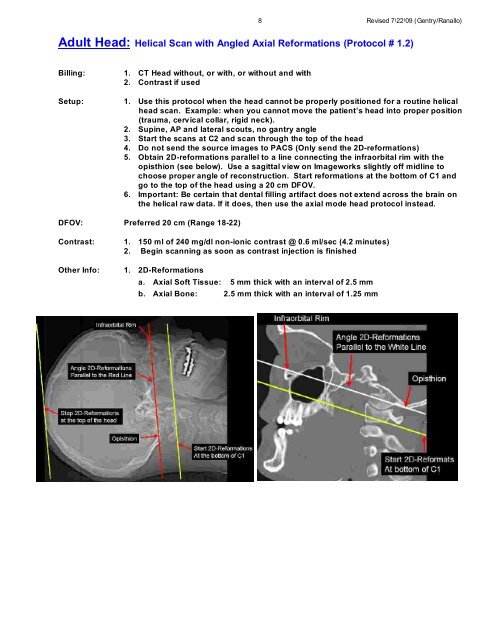
5. Obtain 2D-reformations parallel to a line connecting the infraorbital rim with the<br />
opisthion (see below). Use a sagittal view on Imageworks slightly <strong>of</strong>f midline to<br />
choose proper angle <strong>of</strong> reconstruction. Start reformations at the bottom <strong>of</strong> C1 and<br />
go to the top <strong>of</strong> the head using a 20 cm DFOV.<br />
6. Important: Be certain that dental filling artifact does not extend across the brain on<br />
the helical raw data. If it does, then use the axial mode head protocol instead.<br />
DFOV: Preferred 20 cm (Range 18-22)<br />
Contrast: 1. 150 ml <strong>of</strong> 240 mg/dl non-ionic contrast @ 0.6 ml/sec (4.2 minutes)<br />
2. Begin scanning as soon as contrast injection is finished<br />
Other Info: 1. 2D-Reformations<br />
a. Axial S<strong>of</strong>t Tissue: 5 mm thick with an interval <strong>of</strong> 2.5 mm<br />
b. Axial Bone: 2.5 mm thick with an interval <strong>of</strong> 1.25 mm


