

Ankle and Foot 47 - Department of Radiology - University of ...
Ankle and Foot 47 - Department of Radiology - University of ...
Ankle and Foot 47 - Department of Radiology - University of ...

Create successful ePaper yourself
Turn your PDF publications into a flip-book with our unique Google optimized e-Paper software.
<strong>47</strong> <strong>Ankle</strong> <strong>and</strong> <strong>Foot</strong> 2215 <strong>47</strong><br />
A<br />
B<br />
C<br />
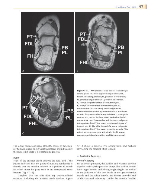
Figure <strong>47</strong>-11. MRI <strong>of</strong> normal ankle tendons in the oblique<br />
coronal plane. FDL, flexor digitorum longus tendon; FHL,<br />
flexor hallucis longus tendon; PB, peroneus brevis tendon;<br />
PL, peroneus longus tendon; PT, posterior tibial tendon.<br />
A, Through the posterior facet <strong>of</strong> the subtalar joint.<br />
B, Through the middle facet <strong>of</strong> the subtalar joint. ST,<br />
sustentaculum tali. A&N (artery <strong>and</strong> nerve) points to<br />
the dotted circle surrounding the neurovascular bundle that<br />
includes the posterior tibial artery <strong>and</strong> nerve. C, Through the<br />
talonavicular joint. At this level, the PT tendon has divided<br />
into separate slips. The white line with the round end points<br />
to the portion <strong>of</strong> the PT that inserts onto the medial pole <strong>of</strong><br />
the navicular (N). The white line with the square end points<br />
to the portion <strong>of</strong> the PT that passes under the navicular. This<br />
patient has an os peroneum, which is why the PL tendon<br />
appears enlarged <strong>and</strong> gray at this level (dark gray arrow).<br />
The lack <strong>of</strong> edematous signal along the course <strong>of</strong> the extensor<br />
hallucis longus on T2-weighted images should reassure<br />
the radiologist there is no pathologic process.<br />
Injury<br />
Tears <strong>of</strong> the anterior ankle tendons are rare, <strong>and</strong> if the<br />
patient indicates that the point <strong>of</strong> maximal tenderness is<br />
directly over the anterior tendons, it is prudent to search<br />
for other causes for pain, such as an unsuspected stress<br />
fracture (Fig. <strong>47</strong>-12).<br />
Ganglion cysts can arise from any synovium-lined<br />
structure, including the anterior ankle tendons. Figure<br />
<strong>47</strong>-13 shows a synovial cyst arising from <strong>and</strong> partially<br />
enveloping the anterior tibial tendon.<br />
• Posterior Tendons<br />
Normal Anatomy<br />
For anatomic purposes, the Achilles <strong>and</strong> plantaris tendons<br />
together make up the posterior group. The Achilles tendon<br />
is the largest tendon in the body, originating in the midcalf<br />
at the junction <strong>of</strong> the two heads <strong>of</strong> the gastrocnemius<br />
muscle <strong>and</strong> the soleus muscle, <strong>and</strong> inserts onto the back<br />
<strong>of</strong> the calcaneal tuberosity. Unlike the anterior, medial,<br />
Ch0<strong>47</strong>-A05375.indd 2215<br />
9/9/2008 5:33:35 PM


