

Ankle and Foot 47 - Department of Radiology - University of ...
Ankle and Foot 47 - Department of Radiology - University of ...
Ankle and Foot 47 - Department of Radiology - University of ...

Create successful ePaper yourself
Turn your PDF publications into a flip-book with our unique Google optimized e-Paper software.
2280 VII Imaging <strong>of</strong> the Musculoskeletal System<br />
A<br />
B<br />
C<br />
E<br />
D<br />
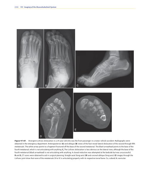
Figure <strong>47</strong>-87. Divergent Lisfranc dislocation in a 34-year-old who was the front passenger in a motor vehicle accident. Radiographs were<br />
obtained in the emergency department. Anteroposterior (A) <strong>and</strong> oblique (B) views <strong>of</strong> the foot reveal lateral dislocation <strong>of</strong> the second through fifth<br />
metatarsals. The white arrow points to a fragment fractured <strong>of</strong>f the base <strong>of</strong> the second metatarsal. The black arrowhead points to the base <strong>of</strong> the<br />
fourth metatarsal, which is not articulating with anything. C, The Lisfranc dislocation is less obvious on the lateral view, although the base <strong>of</strong> the<br />
fourth metatarsal (black arrowhead) is not articulating with anything. A closed reduction was attempted at the bedside but was unsuccessful.<br />
D <strong>and</strong> E, CT scans were obtained to aid in surgical planning. Straight axial (long-axis) (D) <strong>and</strong> coronal oblique (long-axis) (E) images through the<br />
Lisfranc joint show that none <strong>of</strong> the metatarsals (II to V) is articulating properly with its respective tarsal bone. Cu, cuboid; N, navicular.<br />
Ch0<strong>47</strong>-A05375.indd 2280<br />
9/9/2008 5:35:29 PM


