

Ankle and Foot 47 - Department of Radiology - University of ...
Ankle and Foot 47 - Department of Radiology - University of ...
Ankle and Foot 47 - Department of Radiology - University of ...

Create successful ePaper yourself
Turn your PDF publications into a flip-book with our unique Google optimized e-Paper software.
A<br />
B<br />
<strong>47</strong> <strong>Ankle</strong> <strong>and</strong> <strong>Foot</strong> 2263 <strong>47</strong><br />
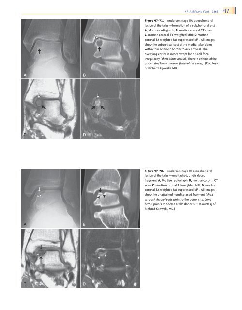
Figure <strong>47</strong>-71. Anderson stage IIA osteochondral<br />
lesion <strong>of</strong> the talus—formation <strong>of</strong> a subchondral cyst.<br />
A, Mortise radiograph; B, mortise coronal CT scan;<br />
C, mortise coronal T1-weighted MRI; D, mortise<br />
coronal T2-weighted fat-suppressed MRI. All images<br />
show the subcortical cyst <strong>of</strong> the medial talar dome<br />
with a thin sclerotic border (black arrows). The<br />
overlying cortex is intact except for a small focal<br />
irregularity (short white arrow). There is edema <strong>of</strong> the<br />
underlying bone marrow (long white arrow). (Courtesy<br />
<strong>of</strong> Richard Kijowski, MD.)<br />
C<br />
D<br />
Figure <strong>47</strong>-72. Anderson stage III osteochondral<br />
lesion <strong>of</strong> the talus—unattached, undisplaced<br />
fragment. A, Mortise radiograph; B, mortise coronal CT<br />
scan; C, mortise coronal T1-weighted MRI; D, mortise<br />
coronal T2-weighted fat-suppressed MRI. All images<br />
show the unattached nondisplaced fragment (short<br />
arrows). Arrowheads point to the donor site. Long<br />
arrow points to edema at the donor site. (Courtesy <strong>of</strong><br />
Richard Kijowski, MD.)<br />
A<br />
B<br />
C<br />
D<br />
Ch0<strong>47</strong>-A05375.indd 2263<br />
9/9/2008 5:35:02 PM


