

Ankle and Foot 47 - Department of Radiology - University of ...
Ankle and Foot 47 - Department of Radiology - University of ...
Ankle and Foot 47 - Department of Radiology - University of ...

Create successful ePaper yourself
Turn your PDF publications into a flip-book with our unique Google optimized e-Paper software.
2262 VII Imaging <strong>of</strong> the Musculoskeletal System<br />
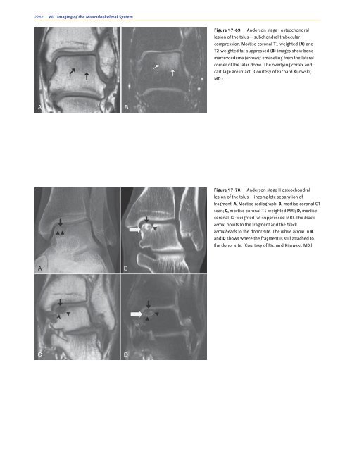
Figure <strong>47</strong>-69. Anderson stage I osteochondral<br />
lesion <strong>of</strong> the talus—subchondral trabecular<br />
compression. Mortise coronal T1-weighted (A) <strong>and</strong><br />
T2-weighted fat-suppressed (B) images show bone<br />
marrow edema (arrows) emanating from the lateral<br />
corner <strong>of</strong> the talar dome. The overlying cortex <strong>and</strong><br />
cartilage are intact. (Courtesy <strong>of</strong> Richard Kijowski,<br />
MD.)<br />
A<br />
B<br />
Figure <strong>47</strong>-70. Anderson stage II osteochondral<br />
lesion <strong>of</strong> the talus—incomplete separation <strong>of</strong><br />
fragment. A, Mortise radiograph; B, mortise coronal CT<br />
scan; C, mortise coronal T1-weighted MRI; D, mortise<br />
coronal T2-weighted fat-suppressed MRI. The black<br />
arrow points to the fragment <strong>and</strong> the black<br />
arrowheads to the donor site. The white arrow in B<br />
<strong>and</strong> D shows where the fragment is still attached to<br />
the donor site. (Courtesy <strong>of</strong> Richard Kijowski, MD.)<br />
A<br />
B<br />
C<br />
D<br />
Ch0<strong>47</strong>-A05375.indd 2262<br />
9/9/2008 5:35:01 PM


