

Ankle and Foot 47 - Department of Radiology - University of ...
Ankle and Foot 47 - Department of Radiology - University of ...
Ankle and Foot 47 - Department of Radiology - University of ...

You also want an ePaper? Increase the reach of your titles
YUMPU automatically turns print PDFs into web optimized ePapers that Google loves.
2312 VII Imaging <strong>of</strong> the Musculoskeletal System<br />
A B C<br />
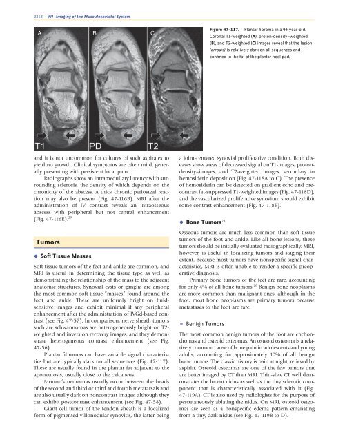
Figure <strong>47</strong>-117. Plantar fibroma in a 44-year-old.<br />
Coronal T1-weighted (A), proton-density–weighted<br />
(B), <strong>and</strong> T2-weighted (C) images reveal that the lesion<br />
(arrows) is relatively dark on all sequences <strong>and</strong><br />
confined to the fat <strong>of</strong> the plantar heel pad.<br />
<strong>and</strong> it is not uncommon for cultures <strong>of</strong> such aspirates to<br />
yield no growth. Clinical symptoms are <strong>of</strong>ten mild, generally<br />
presenting with persistent local pain.<br />
Radiographs show an intramedullary lucency with surrounding<br />
sclerosis, the density <strong>of</strong> which depends on the<br />
chronicity <strong>of</strong> the abscess. A thick chronic periosteal reaction<br />
may also be present (Fig. <strong>47</strong>-116B). MRI after the<br />
administration <strong>of</strong> IV contrast reveals an intraosseous<br />
abscess with peripheral but not central enhancement<br />
(Fig. <strong>47</strong>-116E). 29<br />
Tumors<br />
• S<strong>of</strong>t Tissue Masses<br />
S<strong>of</strong>t tissue tumors <strong>of</strong> the feet <strong>and</strong> ankle are common, <strong>and</strong><br />
MRI is useful in determining the tissue type as well as<br />
demonstrating the relationship <strong>of</strong> the mass to the adjacent<br />
anatomic structures. Synovial cysts or ganglia are among<br />
the most common s<strong>of</strong>t tissue “masses” found around the<br />
foot <strong>and</strong> ankle. These are uniformly bright on fluidsensitive<br />
images <strong>and</strong> exhibit minimal if any peripheral<br />
enhancement after the administration <strong>of</strong> IVGd-based contrast<br />
(see Fig. <strong>47</strong>-57). In comparison, nerve sheath tumors<br />
such are schwannomas are heterogeneously bright on T2-<br />
weighted <strong>and</strong> inversion recovery images, <strong>and</strong> they demonstrate<br />
heterogeneous contrast enhancement (see Fig.<br />
<strong>47</strong>-56).<br />
Plantar fibromas can have variable signal characteristics<br />
but are typically dark on all sequences (Fig. <strong>47</strong>-117).<br />
These are usually found in the plantar fat adjacent to the<br />
aponeurosis, usually close to the calcaneus.<br />
Morton’s neuromas usually occur between the heads<br />
<strong>of</strong> the second <strong>and</strong> third or third <strong>and</strong> fourth metatarsals <strong>and</strong><br />
are also usually dark on noncontrast images, although they<br />
can exhibit postcontrast enhancement (see Fig. <strong>47</strong>-58).<br />
Giant cell tumor <strong>of</strong> the tendon sheath is a localized<br />
form <strong>of</strong> pigmented villonodular synovitis, the latter being<br />
a joint-centered synovial proliferative condition. Both diseases<br />
show areas <strong>of</strong> decreased signal on T1-images, protondensity–images,<br />
<strong>and</strong> T2-weighted images, secondary to<br />
hemosiderin deposition (Fig. <strong>47</strong>-118A to C). The presence<br />
<strong>of</strong> hemosiderin can be detected on gradient echo <strong>and</strong> precontrast<br />
fat-suppressed T1-weighted images (Fig. <strong>47</strong>-118D),<br />
<strong>and</strong> the vascularized proliferative synovium should exhibit<br />
some contrast enhancement (Fig. <strong>47</strong>-118E).<br />
• Bone Tumors 28<br />
Osseous tumors are much less common than s<strong>of</strong>t tissue<br />
tumors <strong>of</strong> the foot <strong>and</strong> ankle. Like all bone lesions, these<br />
tumors should be initially evaluated radiographically. MRI,<br />
however, is useful in localizing tumors <strong>and</strong> staging their<br />
extent. Because most tumors have nonspecific signal characteristics,<br />
MRI is <strong>of</strong>ten unable to render a specific preoperative<br />
diagnosis.<br />
Primary bone tumors <strong>of</strong> the feet are rare, accounting<br />
for only 4% <strong>of</strong> all bone tumors. 20 Benign bone neoplasms<br />
are more common than malignant ones, although in the<br />
foot, most bone neoplasms are primary tumors because<br />
metastases to the foot are rare.<br />
• Benign Tumors<br />
The most common benign tumors <strong>of</strong> the foot are enchondromas<br />
<strong>and</strong> osteoid osteomas. An osteoid osteoma is a relatively<br />
common cause <strong>of</strong> bone pain in adolescents <strong>and</strong> young<br />
adults, accounting for approximately 10% <strong>of</strong> all benign<br />
bone tumors. The classic history is pain at night, relieved by<br />
aspirin. Osteoid osteomas are one <strong>of</strong> the few tumors that<br />
are better imaged by CT than MRI. Thin-slice CT well demonstrates<br />
the lucent nidus as well as the tiny sclerotic component<br />
that is characteristically associated with it (Fig.<br />
<strong>47</strong>-119A). CT is also used by radiologists for the purpose <strong>of</strong><br />
percutaneously ablating the nidus. On MRI, osteoid osteomas<br />
are seen as a nonspecific edema pattern emanating<br />
from a tiny, dark nidus (see Fig. <strong>47</strong>-119B to D).<br />
Ch0<strong>47</strong>-A05375.indd 2312<br />
9/9/2008 5:36:21 PM


