

Bone Tumors: In 1 Simple Chart
Bone Tumors: In 1 Simple Chart
Bone Tumors: In 1 Simple Chart

Create successful ePaper yourself
Turn your PDF publications into a flip-book with our unique Google optimized e-Paper software.
<strong>Bone</strong> <strong>Tumors</strong>:<br />
<strong>In</strong> 1 <strong>Simple</strong> <strong>Chart</strong><br />
<strong>Bone</strong> <strong>Tumors</strong><br />
<strong>In</strong> 1 <strong>Simple</strong> <strong>Chart</strong><br />
with<br />
PowerPoint<br />
<strong>In</strong>teractivity<br />
©Ken L Schreibman, PhD/MD 2010<br />
Download this entire slideshow from<br />
When running this on your own computer<br />
you can jump from slide to slide using<br />
these buttons at bottom of each slide:<br />
Last slide<br />
viewed<br />
Overview<br />
slide<br />
The<br />
<strong>Chart</strong><br />
schreibman.info<br />
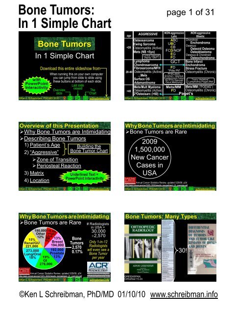
age<br />
40<br />
AGGRESSIVE<br />
Osteosarcoma<br />
Ewing Sarcoma<br />
Osteomyelitis (Active)<br />
Mets (NB 60)<br />
©Ken L Schreibman, PhD/MD 2010<br />
page 1 of 31<br />
NON-aggressive<br />
Lytic<br />
ABC<br />
UBC<br />
CB<br />
FCD/NOF<br />
EG<br />
FD<br />
GCT<br />
FD<br />
“Pitts Pit”<br />
PHALANX:<br />
Enchon, Glomus<br />
Epidermoid,Felon<br />
GCTTS,Sarcoid<br />
Gout,Met(lung)<br />
Mets/MM<br />
FD<br />
NON-aggressive<br />
Blastic<br />
Chondroid:<br />
Enchondroma<br />
Osseous:<br />
Osteoid Osteoma<br />
Osteoblastoma<br />
Osseous & Chondroid:<br />
Osteochondroma<br />
<strong>Bone</strong> <strong>In</strong>farct<br />
Enchondroma<br />
Stress Fracture<br />
Osteomyelitis (Chronic)<br />
“Ivory Vertebra”:<br />
Lymphoma,Paget,Met<br />
Mets/MM (“POEMS”)<br />
Osteomyelitis (Chronic)<br />
Paget’s<br />
schreibman.info<br />
Overview of this Presentation<br />
‣Why <strong>Bone</strong> <strong>Tumors</strong> are <strong>In</strong>timidating<br />
‣Describing <strong>Bone</strong> <strong>Tumors</strong><br />
1) Patient’s Age<br />
2) “Aggressive”<br />
‣Zone of Transition<br />
‣Periosteal Reaction<br />
3) Matrix<br />
4) Location<br />
©Ken L Schreibman, PhD/MD 2010<br />
Building the<br />
<strong>Bone</strong> Tumor <strong>Chart</strong><br />
Underlined Text =<br />
PowerPoint <strong>In</strong>teractivity<br />
schreibman.info<br />
Why<strong>Bone</strong><strong>Tumors</strong>are<strong>In</strong>timidating<br />
‣<strong>Bone</strong> <strong>Tumors</strong> are Rare<br />
2009<br />
1,500,000<br />
New Cancer<br />
Cases in<br />
USA<br />
Annual Cancer Statistics Review, updated 5/29/09, p34<br />
http://seer.cancer.gov/csr/1975_2006/results_merged/sect_01_overview.pdf<br />
©Ken L Schreibman, PhD/MD 2010<br />
schreibman.info<br />
Why<strong>Bone</strong><strong>Tumors</strong>are<strong>In</strong>timidating<br />
‣<strong>Bone</strong> <strong>Tumors</strong> are Rare<br />
15%<br />
Renal/GU<br />
180,000<br />
Other<br />
12%<br />
221,000<br />
273,000<br />
Lung/Oral<br />
18%<br />
13%<br />
Breast<br />
194,000<br />
192,000<br />
Prostate<br />
13%<br />
19%<br />
GI<br />
276,000<br />
<strong>Bone</strong><br />
<strong>Tumors</strong><br />
2,570<br />
0.17%<br />
# Radiologists<br />
in USA =<br />
30,000<br />
2,570<br />
Only 1-in-12<br />
Radiologists<br />
will even see a<br />
<strong>Bone</strong> Tumor<br />
per year<br />
<strong>Bone</strong> <strong>Tumors</strong>: Many Types<br />
30!<br />
Annual Cancer Statistics Review, updated 5/29/09, p34<br />
http://seer.cancer.gov/csr/1975_2006/results_merged/sect_01_overview.pdf<br />
©Ken L Schreibman, PhD/MD 2010<br />
www.acr.org/SecondaryMainMenu<br />
Categories/SocioeconomicResearc<br />
h/PracticeofRadiologyintheUS.aspx<br />
schreibman.info<br />
GREENSPAN:<br />
OrthoRad 15.24<br />
©Ken L Schreibman, PhD/MD 01/10/10 www.schreibman.info
<strong>Bone</strong> <strong>Tumors</strong>:<br />
<strong>In</strong> 1 <strong>Simple</strong> <strong>Chart</strong><br />
Why<strong>Bone</strong><strong>Tumors</strong>are<strong>In</strong>timidating<br />
‣<strong>Bone</strong> <strong>Tumors</strong> are Rare <br />
Don’t see enough to be confident <br />
‣Many types of <strong>Bone</strong> <strong>Tumors</strong> <br />
‣Have Confusing (similar) Names<br />
30%<br />
“Osteosarcoma” <br />
<strong>In</strong>cidence <strong>Bone</strong> <strong>Tumors</strong><br />
25%<br />
by age<br />
“Osteochondroma” <br />
20%<br />
15%<br />
‣Occur in children<br />
10%<br />
Essentially only 2 <br />
5%<br />
bone malignancies<br />
0%<br />
85<br />
occur in children<br />
http://seer.cancer.gov/statfacts/html/bones.html<br />
©Ken L Schreibman, PhD/MD 2010<br />
schreibman.info<br />
<strong>Bone</strong> <strong>Tumors</strong>: by Age<br />
page 2 of 31<br />
20<br />
<strong>Bone</strong> <strong>Tumors</strong>: by Age<br />
20<br />
40<br />
Overview of this Presentation<br />
‣Why <strong>Bone</strong> <strong>Tumors</strong> are <strong>In</strong>timidating<br />
‣Describing <strong>Bone</strong> <strong>Tumors</strong><br />
1) Patient’s Age<br />
40<br />
Multiple Myeloma, Metastases<br />
©Ken L Schreibman, PhD/MD 2010<br />
schreibman.info<br />
Overview of this Presentation<br />
‣Why <strong>Bone</strong> <strong>Tumors</strong> are <strong>In</strong>timidating<br />
‣Describing <strong>Bone</strong> <strong>Tumors</strong><br />
1) Patient’s Age<br />
2) “Aggressive”<br />
vs “Non-aggressive”<br />
(NOT “Malignant” vs “Benign”)<br />
‣ Zone of Transition<br />
‣ Periosteal Reaction<br />
Not everything that looks aggressive is malignant<br />
(e.g. osteomyelitis)<br />
2 Cases: Destructive lesions distal fibula<br />
Benign?<br />
Malignant?<br />
Can’t tell with radiographs…<br />
Thus we use the term<br />
“Aggressive”<br />
©Ken L Schreibman, PhD/MD 2010<br />
schreibman.info<br />
©Ken L Schreibman, PhD/MD 2010<br />
schreibman.info<br />
©Ken L Schreibman, PhD/MD 01/10/10 www.schreibman.info
<strong>Bone</strong> <strong>Tumors</strong>:<br />
<strong>In</strong> 1 <strong>Simple</strong> <strong>Chart</strong><br />
Aggressive vs Non-aggressive<br />
Zone of Transition<br />
Periosteal Reactions<br />
page 3 of 31<br />
Aggressive vs Non-aggressive<br />
Zone of Transition<br />
Grow Slowly<br />
‣ “Narrow”<br />
‣ “Geographic”<br />
‣ “Well Defined”<br />
Can Outline Lesion<br />
with Sharp Pencil<br />
Sclerotic Margins<br />
Grows VERY Slowly!<br />
©Ken L Schreibman, PhD/MD 2010<br />
schreibman.info<br />
©Ken L Schreibman, PhD/MD 2010<br />
schreibman.info<br />
Aggressive vs Non-aggressive<br />
Zone of Transition<br />
Grow Slowly<br />
‣ “Narrow”<br />
‣ “Geographic”<br />
‣ “Well Defined”<br />
Can Outline Lesion<br />
with Sharp Pencil<br />
Sclerotic Margins<br />
Grows VERY Slowly!<br />
Aggressive vs Non-aggressive<br />
Zone of Transition<br />
Grow Slowly<br />
‣ “Narrow”<br />
‣ “Geographic”<br />
‣ “Well Defined”<br />
Can Outline Lesion<br />
with Sharp Pencil<br />
Sclerotic Margins<br />
Grows VERY Slowly!<br />
Asymptomatic, incidental finding<br />
©Ken L Schreibman, PhD/MD 2010<br />
schreibman.info<br />
©Ken L Schreibman, PhD/MD 2010<br />
schreibman.info<br />
Aggressive vs Non-aggressive<br />
Zone of Transition<br />
Grow Slowly<br />
‣ “Narrow”<br />
‣ “Geographic”<br />
‣ “Well Defined”<br />
Can Outline Lesion<br />
with Sharp Pencil<br />
©Ken L Schreibman, PhD/MD 2010<br />
schreibman.info<br />
Aggressive vs Non-aggressive<br />
Zone of Transition<br />
Grow Rapidly<br />
“Wide”<br />
“Permeative”<br />
“Ill Defined”<br />
“Moth Eaten”<br />
Cannot tell where<br />
Lesion ends and<br />
Normal <strong>Bone</strong> begins<br />
©Ken L Schreibman, PhD/MD 2010<br />
Grow Slowly<br />
‣ “Narrow”<br />
‣ “Geographic”<br />
‣ “Well Defined”<br />
Can Outline Lesion<br />
with Sharp Pencil<br />
Sclerotic Margins<br />
Grows VERY Slowly!<br />
schreibman.info<br />
©Ken L Schreibman, PhD/MD 01/10/10 www.schreibman.info
<strong>Bone</strong> <strong>Tumors</strong>:<br />
<strong>In</strong> 1 <strong>Simple</strong> <strong>Chart</strong><br />
Aggressive vs Non-aggressive<br />
Zone of Transition<br />
Grow Rapidly<br />
“Wide”<br />
“Permeative”<br />
“Ill Defined”<br />
“Moth Eaten”<br />
Cannot tell where<br />
Lesion ends and<br />
Normal <strong>Bone</strong> begins<br />
Grows Rapidly<br />
“Wide”<br />
“Permeative”<br />
“Ill Defined”<br />
“Moth Eaten”<br />
page 4 of 31<br />
Aggressive vs Non-aggressive<br />
Zone of Transition<br />
Cannot tell where<br />
Lesion ends and<br />
Normal <strong>Bone</strong> begins<br />
Grows Slowly<br />
‣ “Narrow”<br />
‣ “Geographic”<br />
‣ “Well Defined”<br />
Can Outline Lesion<br />
with Sharp Pencil<br />
W,S 16yoF<br />
©Ken L Schreibman, PhD/MD 2010<br />
schreibman.info<br />
©Ken L Schreibman, PhD/MD 2010<br />
schreibman.info<br />
Aggressive vs Non-aggressive<br />
Periosteal Reaction<br />
Grows Rapidly Grows Slowly<br />
TOO<br />
COMPLI-<br />
CATED<br />
Aggressive vs Non-aggressive<br />
Simplifying Periosteal Reaction<br />
Grows Rapidly<br />
“<strong>In</strong>terrupted”<br />
Grows Slowly<br />
‣“Solid”<br />
Smooth<br />
Continuous<br />
©Ken L Schreibman, PhD/MD 2010<br />
schreibman.info<br />
©Ken L Schreibman, PhD/MD 2010<br />
schreibman.info<br />
Aggressive vs Non-aggressive<br />
Simplifying Periosteal Reaction<br />
Grows Slowly<br />
‣“Solid”<br />
Smooth<br />
Continuous<br />
Looks like<br />
Healing Callus<br />
<strong>Bone</strong><br />
Model<br />
Fx<br />
F,A 2moM<br />
©Ken L Schreibman, PhD/MD 2010<br />
1m later<br />
schreibman.info<br />
Aggressive vs Non-aggressive<br />
Simplifying Periosteal Reaction<br />
Grows Slowly<br />
‣“Solid”<br />
Smooth<br />
Continuous<br />
<strong>Bone</strong><br />
Model<br />
HOA HPOA<br />
Hypertrophic<br />
Osteo- Pulmonary<br />
Arthropathy<br />
Osteo-<br />
Arthropathy<br />
V,T 49yoM<br />
©Ken L Schreibman, PhD/MD 2010<br />
Stable over 1y<br />
schreibman.info<br />
©Ken L Schreibman, PhD/MD 01/10/10 www.schreibman.info
<strong>Bone</strong> <strong>Tumors</strong>:<br />
<strong>In</strong> 1 <strong>Simple</strong> <strong>Chart</strong><br />
Aggressive vs Non-aggressive<br />
Simplifying Periosteal Reaction<br />
Grows Rapidly<br />
“<strong>In</strong>terrupted”<br />
May grow so<br />
rapidly it doesn’t<br />
have time to<br />
ossify<br />
(Unossified<br />
periosteum is not<br />
radiopaque)<br />
©Ken L Schreibman, PhD/MD 2010<br />
schreibman.info<br />
©Ken L Schreibman, PhD/MD 2010<br />
page 5 of 31<br />
Aggressive vs Non-aggressive<br />
Simplifying Periosteal Reaction<br />
Grows Rapidly<br />
“<strong>In</strong>terrupted”<br />
Lamellated<br />
Onionskin<br />
Grows… ossifies…<br />
Grows… ossifies…<br />
Grows… ossifies…<br />
Courtesy of James Choi, MD<br />
schreibman.info<br />
Aggressive vs Non-aggressive<br />
Simplifying Periosteal Reaction<br />
Grows Rapidly<br />
“<strong>In</strong>terrupted”<br />
Lamellated<br />
Onionskin<br />
Spiculated<br />
Hair-on-end<br />
©Ken L Schreibman, PhD/MD 2010<br />
schreibman.info<br />
Aggressive vs Non-aggressive<br />
Simplifying Periosteal Reaction<br />
Grows Rapidly<br />
“<strong>In</strong>terrupted”<br />
Lamellated<br />
Onionskin<br />
Spiculated<br />
Sunburst<br />
Codman’s<br />
Triangles<br />
(Growing so rapidly,<br />
has time to ossify<br />
only at corners)<br />
©Ken L Schreibman, PhD/MD 2010<br />
schreibman.info<br />
Aggressive vs Non-aggressive<br />
Simplifying Periosteal Reaction<br />
Grows Rapidly Grows Slowly<br />
“<strong>In</strong>terrupted” ‣“Solid”<br />
Lamellated Smooth<br />
Onionskin<br />
Continuous<br />
Spiculated<br />
Looks like<br />
Sunburst<br />
Healing Callus<br />
Codman’s<br />
Triangles<br />
8w post Fx, ORIF<br />
=more mature callus<br />
©Ken L Schreibman, PhD/MD 2010<br />
3w post Fx, ORIF<br />
=very early callus<br />
S,C 15yoM<br />
schreibman.info<br />
©Ken L Schreibman, PhD/MD 01/10/10 www.schreibman.info
<strong>Bone</strong> <strong>Tumors</strong>:<br />
<strong>In</strong> 1 <strong>Simple</strong> <strong>Chart</strong><br />
Overview of this Presentation<br />
‣Why <strong>Bone</strong> <strong>Tumors</strong> are <strong>In</strong>timidating<br />
‣Describing <strong>Bone</strong> <strong>Tumors</strong><br />
1) Patient’s Age<br />
2) “Aggressive”<br />
vs “Non-aggressive”<br />
‣ Zone of Transition<br />
‣ Periosteal Reaction<br />
‣ Cortical Destruction<br />
page 6 of 31<br />
Aggressive vs Non-aggressive<br />
Cortical Destruction<br />
Cortex Absent =<br />
Aggressive<br />
Similar lytic lesions<br />
Both have well<br />
defined, sclerotic,<br />
medullary borders<br />
IR<br />
Cortex <strong>In</strong>tact =<br />
Non-aggressive<br />
©Ken L Schreibman, PhD/MD 2010<br />
schreibman.info<br />
©Ken L Schreibman, PhD/MD 2010<br />
schreibman.info<br />
Overview of this Presentation<br />
‣Why <strong>Bone</strong> <strong>Tumors</strong> are <strong>In</strong>timidating<br />
‣Describing <strong>Bone</strong> <strong>Tumors</strong><br />
1) Patient’s Age<br />
2) “Aggressive”<br />
vs “Non-aggressive”<br />
‣ Zone of Transition<br />
‣ Periosteal Reaction Radiographs<br />
‣ Cortical Destruction<br />
MRI<br />
‣ Soft Tissue Extension<br />
2 Cases: Aggressive lesions distal fibula<br />
Benign?<br />
Malignant?<br />
Can’t tell with radiographs…<br />
Thus we use the term<br />
“Aggressive”<br />
Periosteal<br />
Reaction<br />
Cortical<br />
Destruction<br />
©Ken L Schreibman, PhD/MD 2010<br />
schreibman.info<br />
©Ken L Schreibman, PhD/MD 2010<br />
schreibman.info<br />
2 Cases: Aggressive lesions distal fibula<br />
T2<br />
Soft Tissue<br />
Extension<br />
T1<br />
Active Osteomyelitis Chronic Osteo.<br />
Aggressive vs Non-aggressive<br />
T2<br />
Two<br />
YEARS<br />
later <br />
H,M 13yoF<br />
©Ken L Schreibman, PhD/MD 2010<br />
schreibman.info<br />
©Ken L Schreibman, PhD/MD 2010<br />
schreibman.info<br />
©Ken L Schreibman, PhD/MD 01/10/10 www.schreibman.info
<strong>Bone</strong> <strong>Tumors</strong>:<br />
<strong>In</strong> 1 <strong>Simple</strong> <strong>Chart</strong><br />
<strong>Bone</strong> Matrix: 4 Types<br />
Chondroid<br />
“rings&arcs”<br />
Osseous<br />
“cloud-like”<br />
“amorphous”<br />
<strong>Bone</strong> Matrix: 4 Types<br />
Chondroid<br />
“rings&arcs”<br />
page 7 of 31<br />
Calcified Uterine Fibroid<br />
▓Fibrous<br />
“Ground<br />
Glass”<br />
©Ken L Schreibman, PhD/MD 2010<br />
Myositis Ossificans<br />
◦None<br />
Purely Lytic<br />
Not<br />
necessarily<br />
cystic<br />
Multiple Myeloma<br />
schreibman.info<br />
Enchondroma Calcified Uterine Fibroid<br />
©Ken L Schreibman, PhD/MD 2010<br />
schreibman.info<br />
<strong>Bone</strong> Matrix: 4 Types<br />
Osseous<br />
“cloud-like”<br />
“amorphous”<br />
<strong>Bone</strong> Matrix: 4 Types<br />
▓Fibrous<br />
“Ground<br />
Glass”<br />
F,C 8yoF<br />
Osteogenic Sarcoma<br />
©Ken L Schreibman, PhD/MD 2010<br />
Myositis Ossificans<br />
H,S 15yoM<br />
schreibman.info<br />
©Ken L Schreibman, PhD/MD 2010<br />
Fibrous Dysplasia<br />
B,C 53yoF<br />
schreibman.info<br />
<strong>Bone</strong> Matrix: 4 Types<br />
◦None<br />
Purely Lytic<br />
<strong>Bone</strong> Matrix: 4 Types<br />
◦None<br />
Purely Lytic<br />
CT<br />
T1<br />
Not necessarily cystic<br />
S,N 62yoM<br />
©Ken L Schreibman, PhD/MD 2010<br />
Multiple Myeloma<br />
schreibman.info<br />
Not necessarily cystic<br />
<strong>In</strong>traosseous Lipoma<br />
G,B 18yoF<br />
©Ken L Schreibman, PhD/MD 2010<br />
schreibman.info<br />
©Ken L Schreibman, PhD/MD 01/10/10 www.schreibman.info
<strong>Bone</strong> <strong>Tumors</strong>:<br />
<strong>In</strong> 1 <strong>Simple</strong> <strong>Chart</strong><br />
Overview of this Presentation<br />
‣Why <strong>Bone</strong> <strong>Tumors</strong> are <strong>In</strong>timidating<br />
‣Describing <strong>Bone</strong> <strong>Tumors</strong><br />
1) Patient’s Age<br />
2) “Aggressive”<br />
3) Matrix<br />
4) Location<br />
Which bone?<br />
Some tumors have propensity for certain bones<br />
Which part of the bone?<br />
MANY tumors characteristically occur at the:<br />
Epiphysis / Metaphysis / Diaphysis<br />
©Ken L Schreibman, PhD/MD 2010<br />
schreibman.info<br />
age<br />
40<br />
AGGRESSIVE<br />
Osteosarcoma<br />
Ewing Sarcoma<br />
©Ken L Schreibman, PhD/MD 2010<br />
page 8 of 31<br />
NON-aggressive<br />
Lytic<br />
NON-aggressive<br />
Blastic<br />
schreibman.info<br />
Osteogenic Sarcoma<br />
Pt Age: 10-20 years<br />
(when growth spurt occurs)<br />
Location: Metaphyseal<br />
(where growth occurs)<br />
‣Distal Femur<br />
‣Proximal Tibia<br />
(where most growth occurs)<br />
Matrix: Osseous<br />
“osteo-genic”: makes bone<br />
Need to eval for “skip mets”<br />
MR entire length of bone<br />
©Ken L Schreibman, PhD/MD 2010<br />
schreibman.info<br />
Osteogenic Sarcoma<br />
Pt Age: 10-20 years<br />
Location: Metaphyseal<br />
Matrix: Osseous<br />
F,C 8yoF<br />
T1<br />
©Ken L Schreibman, PhD/MD 2010<br />
MRI is useful for staging<br />
the extent of the tumor…<br />
T1<br />
T2<br />
schreibman.info<br />
Osteogenic Sarcoma<br />
F,C 8yoF<br />
©Ken L Schreibman, PhD/MD 2010<br />
MRI is useful for staging<br />
the extent of the tumor…<br />
Radiographs show us<br />
what we need to know to<br />
diagnose type of tumor!<br />
Skeletally immature<br />
Aggressive lesion<br />
‣Wide zone of trans.<br />
‣Sunburst periost.<br />
Osseous matrix<br />
Metaphyseal<br />
---------------------------------<br />
Osteogenic Sarcoma!<br />
schreibman.info<br />
Osteogenic Sarcoma<br />
Sometimes osteosarc is<br />
growing SO quickly it<br />
doesn’t have time to<br />
form an osseous matrix<br />
R,T 11yoF<br />
Patient presents<br />
with hair-on-end<br />
periosteal reaction<br />
©Ken L Schreibman, PhD/MD 2010<br />
After 2 months of <br />
chemotherapy<br />
tumor growth has<br />
slowed enough to<br />
form osseous matrix<br />
schreibman.info<br />
©Ken L Schreibman, PhD/MD 01/10/10 www.schreibman.info
<strong>Bone</strong> <strong>Tumors</strong>:<br />
<strong>In</strong> 1 <strong>Simple</strong> <strong>Chart</strong><br />
Ewing Sarcoma<br />
Pt Age: 5-25 years<br />
Tumor of <strong>Bone</strong> Marrow<br />
Location: Diaphyseal<br />
Flat <strong>Bone</strong>s<br />
Matrix: Permeative<br />
‣Cortical Destruction<br />
‣Aggressive<br />
Periosteal Reaction<br />
Soft Tissue Extension<br />
>> <strong>Bone</strong> Extent<br />
©Ken L Schreibman, PhD/MD 2010<br />
schreibman.info<br />
Ewing Sarcoma<br />
H,M 13yoF<br />
©Ken L Schreibman, PhD/MD 2010<br />
page 9 of 31<br />
T2<br />
Soft Tissue<br />
Extension<br />
T1<br />
T2<br />
>> <strong>Bone</strong><br />
Extent<br />
schreibman.info<br />
Ewing Sarcoma common in pelvis<br />
Air in<br />
colon<br />
Things can<br />
hide in the<br />
pelvis<br />
S,B 6yoM<br />
©Ken L Schreibman, PhD/MD 2010<br />
Air in<br />
colon?<br />
3 months later…<br />
schreibman.info<br />
Ewing Sarcoma common in pelvis<br />
T2fs<br />
Things can<br />
hide in the<br />
pelvis<br />
S,B 6yoM<br />
©Ken L Schreibman, PhD/MD 2010<br />
Unlike in the extremities<br />
where radiographs are key,<br />
the usefulness of<br />
radiographs in the<br />
pelvis is limited.<br />
<strong>In</strong> the pelvis,<br />
cross-sectional<br />
imaging is crucial,<br />
preferably with MRI.<br />
schreibman.info<br />
age<br />
40<br />
AGGRESSIVE<br />
Osteosarcoma<br />
Ewing Sarcoma<br />
Osteomyelitis (Active)<br />
Mets (NB
<strong>Bone</strong> <strong>Tumors</strong>:<br />
<strong>In</strong> 1 <strong>Simple</strong> <strong>Chart</strong><br />
Cortical Desmoid: Do Not Touch!<br />
CT<br />
‣Just a little periosteal reaction<br />
MEDIAL posterior femoral condyle<br />
Tug lesion: Adductor longus insertion<br />
Q,M 17yoM<br />
Cross-sectional imaging doesn’t really help<br />
T2fs<br />
©Ken L Schreibman, PhD/MD 2010<br />
Medial gastrocnemius origin<br />
schreibman.info<br />
age<br />
40<br />
AGGRESSIVE<br />
Osteosarcoma<br />
Ewing Sarcoma<br />
Osteomyelitis (Active)<br />
Mets (NB 60)<br />
©Ken L Schreibman, PhD/MD 2010<br />
page 10 of 31<br />
NON-aggressive NON-aggressive<br />
Lytic<br />
Blastic<br />
ALWAYS consider Lymphoma!<br />
Lymphoma is 29x more common<br />
than all <strong>Bone</strong> <strong>Tumors</strong> combined<br />
Lymphoma = 74,490<br />
180,000<br />
Other<br />
12%<br />
15%<br />
Renal/GU<br />
221,000<br />
273,000<br />
Lung/Oral<br />
18%<br />
13%<br />
Breast<br />
194,000<br />
192,000<br />
Prostate<br />
13%<br />
19%<br />
GI<br />
276,000<br />
New Cancer Cases, USA, 2009<br />
<strong>Bone</strong><br />
<strong>Tumors</strong><br />
2,570<br />
0.17%<br />
schreibman.info<br />
Lymphoma<br />
Tumor of bone marrow<br />
Can be lytic or blastic<br />
R,H 22yoM<br />
T1<br />
©Ken L Schreibman, PhD/MD 2010<br />
Resembles Ewing<br />
T1<br />
T2fs<br />
schreibman.info<br />
Why Age is Important<br />
Location: Diaphyseal<br />
Soft tissue extension<br />
Age: < 20<br />
Ewing Sarcoma<br />
Age: 20-40<br />
Lymphoma<br />
Age: > 40<br />
Metastases<br />
Multiple Myeloma<br />
©Ken L Schreibman, PhD/MD 2010<br />
schreibman.info<br />
Chondrosarcoma<br />
Cartilage malignancy<br />
Matrix: Chondroid<br />
Location: Ends of bones<br />
Pelvis<br />
Soft tissues<br />
Chondrosarcoma<br />
Cartilage malignancy<br />
Matrix: Chondroid<br />
Chondroid<br />
“rings&arcs”<br />
Radiographs<br />
S,B 39yoM<br />
©Ken L Schreibman, PhD/MD 2010<br />
schreibman.info<br />
©Ken L Schreibman, PhD/MD 2010<br />
schreibman.info<br />
©Ken L Schreibman, PhD/MD 01/10/10 www.schreibman.info
<strong>Bone</strong> <strong>Tumors</strong>:<br />
<strong>In</strong> 1 <strong>Simple</strong> <strong>Chart</strong><br />
Chondrosarcoma<br />
Cartilage malignancy<br />
Matrix: Chondroid<br />
Chondroid<br />
“rings&arcs”<br />
Radiographs<br />
CT<br />
Chondrosarcoma<br />
Cartilage malignancy<br />
Matrix: Chondroid<br />
T2fs<br />
page 11 of 31<br />
T1fs<br />
+Gd<br />
Chondroid<br />
MRI<br />
T2: Bright<br />
Gd: peripheral<br />
enhancement<br />
Bulk of tumor<br />
doesn’t enhance<br />
S,B 39yoM<br />
S,B 39yoM<br />
©Ken L Schreibman, PhD/MD 2010<br />
schreibman.info<br />
©Ken L Schreibman, PhD/MD 2010<br />
schreibman.info<br />
Chondrosarcoma<br />
Cartilage malignancy<br />
Matrix: Chondroid<br />
‣Normal cartilage has no blood supply<br />
<strong>In</strong>jured cartilage doesn’t regrow<br />
‣Chondrosarcoma: poor blood supply<br />
Shows very little Gd enhancement<br />
Doesn’t respond to chemotherapy<br />
‣Treatment: Complete tumor resection<br />
S,B 39yoM<br />
©Ken L Schreibman, PhD/MD 2010<br />
schreibman.info<br />
Chondrosarcoma<br />
30yoF 1 year history heal pain<br />
Chondrosarcoma<br />
T1<br />
other side<br />
1 week later<br />
T2fs<br />
T1fs<br />
+Gd<br />
W,A 30yoF<br />
©Ken L Schreibman, PhD/MD 2010<br />
schreibman.info<br />
©Ken L Schreibman, PhD/MD 2010<br />
schreibman.info<br />
©Ken L Schreibman, PhD/MD 01/10/10 www.schreibman.info
<strong>Bone</strong> <strong>Tumors</strong>:<br />
<strong>In</strong> 1 <strong>Simple</strong> <strong>Chart</strong><br />
age<br />
40<br />
Mets/Mult Myeloma<br />
Osteomyelitis (Active)<br />
2ºOsteosarc (>60)<br />
©Ken L Schreibman, PhD/MD 2010<br />
schreibman.info<br />
©Ken L Schreibman, PhD/MD 2010<br />
schreibman.info<br />
Fibrosarcoma<br />
T1<br />
MRI is useful for staging<br />
the extent of the tumor…<br />
T2fs<br />
age<br />
60)<br />
©Ken L Schreibman, PhD/MD 2010<br />
schreibman.info<br />
Osteogenic Sarcoma<br />
Pt Age: 10-20 years<br />
Location: Metaphyseal<br />
Matrix: Osseous<br />
4 Subtypes:<br />
1) Conventional<br />
2) Telangiectatic<br />
Surface Osteosarcomas<br />
3) PERIosteal<br />
4) PARosteal<br />
©Ken L Schreibman, PhD/MD 2010<br />
schreibman.info<br />
Osteogenic Sarcoma<br />
1) Conventional<br />
Pt Age: 10-20 years<br />
Location: Metaphyseal<br />
Matrix: Osseous<br />
2) Telangiectatic<br />
Highly vascular/bloody<br />
Very aggressive<br />
Nearly purely lytic<br />
Usually present after<br />
pathologic fracture<br />
Diffuse metastases<br />
©Ken L Schreibman, PhD/MD 2010<br />
schreibman.info<br />
©Ken L Schreibman, PhD/MD 01/10/10 www.schreibman.info
<strong>Bone</strong> <strong>Tumors</strong>:<br />
<strong>In</strong> 1 <strong>Simple</strong> <strong>Chart</strong><br />
Osteogenic Sarcoma<br />
Surface Osteosarcoma<br />
Pt Age: 20-30 years<br />
Good prognosis if<br />
marrow not involved,<br />
can resect tumor.<br />
If spreads to marrow,<br />
conventional OS.<br />
3) PERIosteal<br />
Looks like aggressive<br />
periosteal reaction<br />
Location: Long bones<br />
©Ken L Schreibman, PhD/MD 2010<br />
schreibman.info<br />
page 13 of 31<br />
Periosteal Osteosarcoma<br />
Aggressive<br />
Periosteal<br />
Reaction<br />
tibia 11yoM<br />
©2004 Radiological Society of North America<br />
Murphey M D et al.<br />
Radiology 2004;<br />
233:129-138<br />
Periosteal Osteosarcoma<br />
Aggressive<br />
Periosteal<br />
Reaction<br />
tibia 34yoF<br />
©2004 Radiological Society of North America<br />
Soft<br />
Tissue<br />
Extension<br />
Sparing<br />
<strong>Bone</strong><br />
Marrow<br />
Murphey M D et al.<br />
Radiology 2004;<br />
233:129-138<br />
Osteogenic Sarcoma<br />
Surface Osteosarcoma<br />
Pt Age: 20-30 years<br />
Good prognosis if<br />
marrow not involved,<br />
can resect tumor<br />
If spreads to marrow,<br />
conventional OS.<br />
3) PERIosteal<br />
Looks like aggressive<br />
periosteal reaction<br />
4) PARosteal<br />
©Ken L Schreibman, PhD/MD 2010<br />
schreibman.info<br />
Osteogenic Sarcoma<br />
Surface Osteosarcoma<br />
4) PARosteal<br />
Pt Age: 20-30 years<br />
Location: Back of<br />
Femoral Condyles<br />
Arise from cortex,<br />
grow outward<br />
Do NOT contain<br />
normal marrow<br />
(As opposed to<br />
osteochondroma)<br />
CT<br />
Osteogenic Sarcoma<br />
T1<br />
T2fs<br />
MRI: No Marrow<br />
involvement<br />
CT<br />
©Ken L Schreibman, PhD/MD 2010<br />
schreibman.info<br />
©Ken L Schreibman, PhD/MD 2010<br />
schreibman.info<br />
©Ken L Schreibman, PhD/MD 01/10/10 www.schreibman.info
<strong>Bone</strong> <strong>Tumors</strong>:<br />
<strong>In</strong> 1 <strong>Simple</strong> <strong>Chart</strong><br />
age<br />
40<br />
AGGRESSIVE<br />
Osteosarcoma<br />
Ewing Sarcoma<br />
Osteomyelitis (Active)<br />
Mets (NB 60)<br />
©Ken L Schreibman, PhD/MD 2010<br />
NON-aggressive<br />
Lytic<br />
NON-aggressive<br />
Blastic<br />
schreibman.info<br />
Adamantinoma<br />
‣VERY RARE<br />
0.1% Primary <strong>Bone</strong> <strong>Tumors</strong><br />
Pt Age: 30-50<br />
Matrix: Permeative<br />
Location: TIBIA (90%)<br />
Diaphyseal<br />
Anterior Cortex<br />
Soft Tissue Mass:<br />
Likely Malignant<br />
©Ken L Schreibman, PhD/MD 2010<br />
page 14 of 31<br />
schreibman.info<br />
Adamantinoma<br />
‣VERY RARE<br />
0.1% Primary <strong>Bone</strong> <strong>Tumors</strong><br />
Pt Age: 30-50<br />
Matrix: Permeative<br />
Location: TIBIA (90%)<br />
Diaphyseal<br />
Anterior Cortex<br />
Soft Tissue Mass:<br />
Likely Malignant<br />
Adamantinoma<br />
‣VERY RARE<br />
0.1% Primary <strong>Bone</strong> <strong>Tumors</strong><br />
Pt Age: 30-50<br />
Matrix: Permeative<br />
Location: TIBIA (90%)<br />
Diaphyseal<br />
Anterior Cortex<br />
Soft Tissue Mass:<br />
Likely Malignant<br />
©Ken L Schreibman, PhD/MD 2010<br />
schreibman.info<br />
©Ken L Schreibman, PhD/MD 2010<br />
schreibman.info<br />
©Ken L Schreibman, PhD/MD 01/10/10 www.schreibman.info
<strong>Bone</strong> <strong>Tumors</strong>:<br />
<strong>In</strong> 1 <strong>Simple</strong> <strong>Chart</strong><br />
AGGRESSIVE NON-aggressive<br />
age<br />
Lytic<br />
Osteosarcoma ABC<br />
Ewing Sarcoma UBC<br />
60)<br />
©Ken L Schreibman, PhD/MD 2010<br />
NON-aggressive<br />
Blastic<br />
schreibman.info<br />
©Ken L Schreibman, PhD/MD 2010<br />
page 15 of 31<br />
A) Aneurysmal <strong>Bone</strong> Cyst<br />
Pt Age: < 20<br />
Matrix: None (Cyst)<br />
Only tumor named for x-ray appearance<br />
Aneurysmal & Cystic<br />
“AVM of <strong>Bone</strong>”<br />
MRI: fluid/fluid level<br />
Location: Metaphyseal<br />
Posterior Spine<br />
Hands<br />
Pelvis<br />
schreibman.info<br />
A) Aneurysmal <strong>Bone</strong> Cyst<br />
A) Aneurysmal <strong>Bone</strong> Cyst<br />
CT<br />
©Ken L Schreibman, PhD/MD 2010<br />
‣Aneurysmal<br />
‣Multi-septated<br />
looks like<br />
soap bubbles<br />
schreibman.info<br />
N,N 15yoF<br />
©Ken L Schreibman, PhD/MD 2010<br />
T2<br />
fluid/fluid level<br />
‣Aneurysmal<br />
‣Multi-septated<br />
looks like<br />
soap bubbles<br />
schreibman.info<br />
A) Aneurysmal <strong>Bone</strong> Cyst<br />
A) Aneurysmal <strong>Bone</strong> Cyst<br />
T2<br />
fluid/fluid level<br />
fluid/fluid level<br />
D,R 12yoM<br />
©Ken L Schreibman, PhD/MD 2010<br />
‣Aneurysmal<br />
‣Multi-septated<br />
looks like<br />
soap bubbles<br />
schreibman.info<br />
T2<br />
©Ken L Schreibman, PhD/MD 2010<br />
‣Aneurysmal<br />
‣Multi-septated<br />
looks like<br />
soap bubbles<br />
schreibman.info<br />
©Ken L Schreibman, PhD/MD 01/10/10 www.schreibman.info
<strong>Bone</strong> <strong>Tumors</strong>:<br />
<strong>In</strong> 1 <strong>Simple</strong> <strong>Chart</strong><br />
B) Unicameral (<strong>Simple</strong>) <strong>Bone</strong> Cyst<br />
Uni-cameral: Latin “one” - “chamber”<br />
(in US we have bi-cameral legislature)<br />
Pt Age:< 20<br />
Matrix: None (True Cyst)<br />
Location:Metaphyseal<br />
>50% Proximal Humerus<br />
20-30% Proximal Femur<br />
50% - <strong>In</strong>cidental Finding<br />
50% - Pathologic Fx<br />
“Fallen Fragment”<br />
©Ken L Schreibman, PhD/MD 2010<br />
schreibman.info<br />
page 16 of 31<br />
B) Unicameral (<strong>Simple</strong>) <strong>Bone</strong> Cyst<br />
Normal Side<br />
D,C 5yoM<br />
©Ken L Schreibman, PhD/MD 2010<br />
schreibman.info<br />
B) Unicameral (<strong>Simple</strong>) <strong>Bone</strong> Cyst<br />
Fractures tend to heal<br />
Fracture healed<br />
B) Unicameral (<strong>Simple</strong>) <strong>Bone</strong> Cyst<br />
Cysts tend to recur<br />
Although UBCs<br />
arise from<br />
metaphysis…<br />
end of bone<br />
grows away<br />
from cyst…<br />
Cyst?<br />
so cyst<br />
becomes<br />
diaphyseal<br />
D,C 5yoM<br />
©Ken L Schreibman, PhD/MD 2010<br />
after 1 month…<br />
after 3 months…<br />
schreibman.info<br />
6m later…<br />
©Ken L Schreibman, PhD/MD 2010<br />
12m later… 18m later<br />
schreibman.info<br />
B) Unicameral (<strong>Simple</strong>) <strong>Bone</strong> Cyst<br />
MRI shows cyst extent T1 T2fs<br />
T1<br />
B) Unicameral (<strong>Simple</strong>) <strong>Bone</strong> Cyst<br />
T2fs<br />
18m later<br />
©Ken L Schreibman, PhD/MD 2010<br />
<strong>Simple</strong> cyst<br />
schreibman.info<br />
M,T 4yoM<br />
©Ken L Schreibman, PhD/MD 2010<br />
Fallen<br />
Fragment<br />
after 3 months<br />
schreibman.info<br />
©Ken L Schreibman, PhD/MD 01/10/10 www.schreibman.info
<strong>Bone</strong> <strong>Tumors</strong>:<br />
<strong>In</strong> 1 <strong>Simple</strong> <strong>Chart</strong><br />
B) Unicameral (<strong>Simple</strong>) <strong>Bone</strong> Cyst<br />
page 17 of 31<br />
B) Unicameral (<strong>Simple</strong>) <strong>Bone</strong> Cyst<br />
T2fs<br />
<strong>Simple</strong> cyst with<br />
hemorrhage<br />
fluid-fluid level<br />
©Ken L Schreibman, PhD/MD 2010<br />
P,D 6yoM<br />
schreibman.info<br />
H,T 18yoM<br />
©Ken L Schreibman, PhD/MD 2010<br />
schreibman.info<br />
C) Chondroblastoma<br />
Pt Age: Skeletally immature<br />
Location: Epiphyseal<br />
Matrix: Chondroid<br />
(No matrix if not calcified)<br />
Benign…<br />
Aggressive appearance!<br />
‣Periosteal Reaction<br />
‣Surrounding Edema<br />
<strong>Bone</strong> Marrow<br />
Soft Tissues<br />
©Ken L Schreibman, PhD/MD 2010<br />
schreibman.info<br />
C) Chondroblastoma<br />
T2fs Pt Age: 10 – 30yo<br />
Location: Epiphyseal<br />
Matrix: Chondroid<br />
(No matrix if not calcified)<br />
Benign…<br />
Aggressive appearance!<br />
‣Periosteal Reaction<br />
‣Surrounding Edema<br />
<strong>Bone</strong> Marrow<br />
Soft Tissues<br />
©Ken L Schreibman, PhD/MD 2010<br />
16yoM<br />
schreibman.info<br />
C) Chondroblastoma<br />
Cartilage-sensitive sequence<br />
C) Chondroblastoma<br />
Epiphyseal mass, skeletally immature<br />
Aggressive appearance<br />
‣Edema in surrounding marrow & tissues<br />
T1 IR Cartilage sequence<br />
Cartilage<br />
unfused physis<br />
Articular Cartilage<br />
©Ken L Schreibman, PhD/MD 2010<br />
16yoM<br />
schreibman.info<br />
B,Q 15yoM<br />
©Ken L Schreibman, PhD/MD 2010<br />
schreibman.info<br />
©Ken L Schreibman, PhD/MD 01/10/10 www.schreibman.info
<strong>Bone</strong> <strong>Tumors</strong>:<br />
<strong>In</strong> 1 <strong>Simple</strong> <strong>Chart</strong><br />
D) Fibrous Cortical Defect<br />
Non-Ossifying Fibroma (NOF)<br />
THE most common bone lesion<br />
‣Occurs up to 40% ALL children<br />
(75% occur 10 – 20 years old)<br />
‣Regress after skeletal maturity<br />
Asymptomatic, incidental finding<br />
(e.g. on knee MR for ACL tear)<br />
If >50% bone diameter Fx<br />
Location: Metaphysis<br />
Femur & Tibia<br />
©Ken L Schreibman, PhD/MD 2010<br />
schreibman.info<br />
©Ken L Schreibman, PhD/MD 2010<br />
page 18 of 31<br />
D) Fibrous Cortical Defect<br />
Non-Ossifying Fibroma (NOF)<br />
Radiographic appearance:<br />
Characteristic& Diagnostic<br />
‣If asymptomatic, no<br />
further workup is needed<br />
Eccentric, sub-cortical<br />
‣Cortex thinned, expanded<br />
Sclerotic margin<br />
‣Scalloped<br />
Multi-loculated<br />
F,M 18yoF<br />
schreibman.info<br />
D) Fibrous Cortical Defect<br />
Non-Ossifying Fibroma (NOF)<br />
Radiographic appearance:<br />
IR<br />
T1<br />
No aggressive characteristics<br />
Characteristic& Diagnostic<br />
‣If asymptomatic, no<br />
further workup is needed<br />
Eccentric, sub-cortical<br />
‣Cortex thinned, expanded<br />
Sclerotic margin<br />
‣Scalloped<br />
Multi-loculated<br />
©Ken L Schreibman, PhD/MD 2010<br />
B,J 19yoM<br />
schreibman.info<br />
D) Fibrous Cortical Defect<br />
Non-Ossifying Fibroma (NOF)<br />
9yo 11yo 13yo<br />
G,M 9yoF<br />
©Ken L Schreibman, PhD/MD 2010<br />
Fx<br />
Healing<br />
Callus<br />
schreibman.info<br />
©Ken L Schreibman, PhD/MD 01/10/10 www.schreibman.info
<strong>Bone</strong> <strong>Tumors</strong>:<br />
<strong>In</strong> 1 <strong>Simple</strong> <strong>Chart</strong><br />
E) Eosinophilic Granuloma<br />
Non-neoplastic proliferation histiocytes<br />
Langerhans Cell Histiocytosis<br />
Pt Age: typically 3yo)<br />
Triad: skull lesions, exophthalmos, DI<br />
‣Letterer-Siwe (
<strong>Bone</strong> <strong>Tumors</strong>:<br />
<strong>In</strong> 1 <strong>Simple</strong> <strong>Chart</strong><br />
F) Fibrous Dysplasia<br />
Pt Age:
<strong>Bone</strong> <strong>Tumors</strong>:<br />
<strong>In</strong> 1 <strong>Simple</strong> <strong>Chart</strong><br />
age<br />
40<br />
AGGRESSIVE<br />
Osteosarcoma<br />
Ewing Sarcoma<br />
Osteomyelitis (Active)<br />
Mets (NB 60)<br />
©Ken L Schreibman, PhD/MD 2010<br />
NON-aggressive<br />
Lytic<br />
ABC<br />
UBC<br />
CB<br />
FCD/NOF<br />
EG<br />
FD<br />
GCT<br />
NON-aggressive<br />
Blastic<br />
schreibman.info<br />
Giant Cell Tumor<br />
Pt Age: Skeletally Mature<br />
(as opposed to Chondroblastoma)<br />
‣THE most common bone tumor<br />
in young adults 20-40yo<br />
Location: Subarticular<br />
‣Arise from Metaphysis<br />
‣Extend across fused<br />
Growth Plate<br />
Matrix: Purely Lytic<br />
‣Narrow Zone of Transition<br />
‣NO SCLEROTIC MARGIN<br />
©Ken L Schreibman, PhD/MD 2010<br />
page 21 of 31<br />
schreibman.info<br />
Giant Cell Tumor<br />
Pt Age: Skeletally T2fs MatureT1<br />
(as opposed to Chondroblastoma)<br />
‣THE most common bone tumor<br />
in young adults 20-40yo<br />
Location: Subarticular<br />
‣Arise from Metaphysis<br />
‣Extend across fused<br />
Growth Plate<br />
Matrix: Purely Lytic<br />
‣Narrow ‣BenignZone of Transition<br />
‣NO ‣Locally SCLEROTIC Aggressive MARGIN<br />
©Ken L Schreibman, PhD/MD 2010<br />
V,R 21yoM<br />
schreibman.info<br />
Giant Cell Tumor<br />
‣Benign<br />
‣Locally Aggressive<br />
©Ken L Schreibman, PhD/MD 2010<br />
B,J 25yoM<br />
schreibman.info<br />
Giant Cell Tumor<br />
T1 T2fs T1fs+Gd<br />
Giant Cell Tumor<br />
Pt Age: Skeletally Mature<br />
Physis fused<br />
16yoM<br />
18yoM<br />
‣Solid & Cystic components<br />
‣Histologically, similarities GCT↔ABC<br />
©Ken L Schreibman, PhD/MD 2010<br />
B,J 25yoM<br />
schreibman.info<br />
©Ken L Schreibman, PhD/MD 2010<br />
C,A 18yoM<br />
schreibman.info<br />
©Ken L Schreibman, PhD/MD 01/10/10 www.schreibman.info
<strong>Bone</strong> <strong>Tumors</strong>:<br />
<strong>In</strong> 1 <strong>Simple</strong> <strong>Chart</strong><br />
Why Age is Important<br />
Location: Subarticular<br />
Matrix: Purely Lytic<br />
Age:< 20 (skeletally immature)<br />
Chondroblastoma<br />
Age:20-40 (skeletally mature)<br />
Giant Cell Tumor<br />
Age:> 40<br />
Metastases<br />
Multiple Myeloma<br />
©Ken L Schreibman, PhD/MD 2010<br />
GCT<br />
V,R 21yoM<br />
schreibman.info<br />
Why Age is Important<br />
Location: Subarticular<br />
Matrix: Purely Lytic<br />
Age:< 20 (skeletally immature)<br />
Chondroblastoma<br />
1<br />
Age:20-40 Lung (skeletally mature)<br />
Cancer<br />
Giant Cell Tumor<br />
Age:> 40<br />
Metastases<br />
Multiple Myeloma<br />
©Ken L Schreibman, PhD/MD 2010<br />
page 22 of 31<br />
Met<br />
C,G 61yoM<br />
schreibman.info<br />
age<br />
40<br />
AGGRESSIVE<br />
Osteosarcoma<br />
Ewing Sarcoma<br />
Osteomyelitis (Active)<br />
Mets (NB 60)<br />
©Ken L Schreibman, PhD/MD 2010<br />
NON-aggressive<br />
Lytic<br />
ABC<br />
UBC<br />
CB<br />
FCD/NOF<br />
EG<br />
FD<br />
GCT<br />
FD<br />
“Pitts Pit”<br />
NON-aggressive<br />
Blastic<br />
schreibman.info<br />
Herniation Pit of the Femoral Neck<br />
aka “Pitt’s Pit”<br />
Michael Pitt, et.al. AJR 1982<br />
vol 138, 6, p 1115-1121<br />
Round lucency<br />
Thin sclerotic rim<br />
Proximal Superior<br />
‣Anterior<br />
courtesy Donna Blankenbaker, MD<br />
<strong>In</strong>cidental finding ⅓ patients<br />
Mechanical, not neoplastic<br />
©Ken L Schreibman, PhD/MD 2010<br />
schreibman.info<br />
age<br />
40<br />
AGGRESSIVE<br />
Osteosarcoma<br />
Ewing Sarcoma<br />
Osteomyelitis (Active)<br />
Mets (NB 60)<br />
©Ken L Schreibman, PhD/MD 2010<br />
NON-aggressive<br />
Lytic<br />
ABC<br />
UBC<br />
CB<br />
FCD/NOF<br />
EG<br />
FD<br />
GCT<br />
FD<br />
“Pitts Pit”<br />
PHALANX:<br />
Enchon, Glomus<br />
Epidermoid,Felon<br />
GCTTS,Sarcoid<br />
Gout,Met(lung)<br />
NON-aggressive<br />
Blastic<br />
schreibman.info<br />
Lytic Lesion: Distal Phalanx<br />
Enchondroma Giant Cell Tumor<br />
‣Lytic: phalanges Tendon Sheath<br />
‣Pathologic Fx (Localized PVNS)<br />
Glomus Tumor Felon<br />
‣Nail bed<br />
(Fingertip infection)<br />
Dorsal<br />
Sarcoidosis<br />
Epidermoid<br />
<strong>In</strong>clusion Cyst Gout<br />
‣Puncture Metastases<br />
Volar<br />
‣Lung<br />
©Ken L Schreibman, PhD/MD 2010<br />
schreibman.info<br />
©Ken L Schreibman, PhD/MD 01/10/10 www.schreibman.info
<strong>Bone</strong> <strong>Tumors</strong>:<br />
<strong>In</strong> 1 <strong>Simple</strong> <strong>Chart</strong><br />
Enchondroma<br />
Benign rests of hyaline cartilage<br />
‣Common<br />
Often discovered incidentally<br />
‣Typically asymptomatic<br />
‣50% small tubular bones<br />
Mostly lytic<br />
Pathologic Fracture<br />
©Ken L Schreibman, PhD/MD 2010<br />
schreibman.info<br />
page 23 of 31<br />
Glomus Tumor<br />
Benign vascular tumor<br />
(neuromyoarterial apparatus)<br />
Subungual, erodes bone<br />
Dorsal cortex distal phalanx<br />
Age: 30 – 50 ( 3x> )<br />
Triad<br />
‣Sensitivity to cold<br />
‣Localized tenderness<br />
‣Severe intermittent<br />
pain<br />
S,D 37yoF<br />
©Ken L Schreibman, PhD/MD 2010<br />
schreibman.info<br />
Glomus Tumor<br />
Dorsal cortex distal phalanx<br />
Epidermoid <strong>In</strong>clusion Cyst<br />
Implantation of epidermal elements<br />
‣Amputation<br />
‣Puncture (seamstress)<br />
Volar cortex distal phalanx<br />
©Ken L Schreibman, PhD/MD 2010<br />
S,D 53yoM<br />
schreibman.info<br />
T1fs<br />
T1fs+Gd<br />
©Ken L Schreibman, PhD/MD 2010<br />
T1fs+Gd<br />
T2fs<br />
schreibman.info<br />
Epidermoid <strong>In</strong>clusion Cyst<br />
Gout<br />
M,B 78yoM<br />
©Ken L Schreibman, PhD/MD 2010<br />
schreibman.info<br />
©Ken L Schreibman, PhD/MD 2010<br />
schreibman.info<br />
©Ken L Schreibman, PhD/MD 01/10/10 www.schreibman.info
<strong>Bone</strong> <strong>Tumors</strong>:<br />
<strong>In</strong> 1 <strong>Simple</strong> <strong>Chart</strong><br />
age<br />
40<br />
AGGRESSIVE<br />
Osteosarcoma<br />
Ewing Sarcoma<br />
Osteomyelitis (Active)<br />
Mets (NB 60)<br />
©Ken L Schreibman, PhD/MD 2010<br />
NON-aggressive<br />
Lytic<br />
ABC<br />
UBC<br />
CB<br />
FCD/NOF<br />
EG<br />
FD<br />
GCT<br />
FD<br />
NON-aggressive<br />
Blastic<br />
“Pitts Pit”<br />
PHALANX:<br />
Enchon, Glomus<br />
Epidermoid,Felon<br />
GCTTS,Sarcoid<br />
Gout,Met(lung)<br />
Mets/MM Mets/MM (“POEMS”)<br />
FD<br />
schreibman.info<br />
POEMS syndrome<br />
Polyneuropathy<br />
Organomegaly<br />
Endocrinopathy<br />
Monoclonal gammopathy<br />
Skin abnormalities<br />
(Sclerotic bone lesions)<br />
Medial Clavicle<br />
Pelvis<br />
page 24 of 31<br />
http://www.scielo.br/scielo.php?pid=S0100-39842008000600002&script=sci_arttext&tlng=en<br />
©Ken L Schreibman, PhD/MD 2010<br />
schreibman.info<br />
age<br />
40<br />
AGGRESSIVE<br />
Osteosarcoma<br />
Ewing Sarcoma<br />
Osteomyelitis (Active)<br />
Mets (NB 60)<br />
©Ken L Schreibman, PhD/MD 2010<br />
NON-aggressive<br />
Lytic<br />
ABC<br />
UBC<br />
CB<br />
FCD/NOF<br />
EG<br />
FD<br />
GCT<br />
FD<br />
NON-aggressive<br />
Blastic<br />
“Pitts Pit”<br />
PHALANX:<br />
Enchon, Glomus<br />
Epidermoid,Felon<br />
GCTTS,Sarcoid<br />
Gout,Met(lung)<br />
Mets/MM Mets/MM (“POEMS”)<br />
FD Osteomyelitis (Chronic)<br />
Paget’s<br />
schreibman.info<br />
Paget’s Disease<br />
Becoming less common<br />
Three Phases<br />
‣Lytic: Wedge with sharp borders<br />
“Blade of grass”, “Candle flame”<br />
‣Mixed: <strong>Bone</strong> destruction & formation<br />
‣Blastic: Cortical/Trabecular thickening<br />
2 Osteosarcoma<br />
©Ken L Schreibman, PhD/MD 2010<br />
B,S 83yoF<br />
schreibman.info<br />
age<br />
40<br />
AGGRESSIVE<br />
Osteosarcoma<br />
Ewing Sarcoma<br />
Osteomyelitis (Active)<br />
Mets (NB 60)<br />
©Ken L Schreibman, PhD/MD 2010<br />
NON-aggressive<br />
Lytic<br />
ABC<br />
UBC<br />
CB<br />
FCD/NOF<br />
EG<br />
FD<br />
GCT<br />
FD<br />
“Pitts Pit”<br />
PHALANX:<br />
Enchon, Glomus<br />
Epidermoid,Felon<br />
GCTTS,Sarcoid<br />
Gout,Met(lung)<br />
Mets/MM<br />
FD<br />
NON-aggressive<br />
Blastic<br />
“Ivory Vertebra”:<br />
Lymphoma,Paget,Met<br />
Mets/MM (“POEMS”)<br />
Osteomyelitis (Chronic)<br />
Paget’s<br />
schreibman.info<br />
Ivory Vertebra<br />
‣Lymphoma<br />
‣Paget<br />
‣Blastic Met<br />
Breast<br />
Prostate<br />
‣Treated Met<br />
‣Chronic Osteo<br />
‣(Sarcoid) rare<br />
K,K 76yoM<br />
©Ken L Schreibman, PhD/MD 2010<br />
schreibman.info<br />
©Ken L Schreibman, PhD/MD 01/10/10 www.schreibman.info
<strong>Bone</strong> <strong>Tumors</strong>:<br />
<strong>In</strong> 1 <strong>Simple</strong> <strong>Chart</strong><br />
age<br />
40<br />
AGGRESSIVE<br />
Osteosarcoma<br />
Ewing Sarcoma<br />
Osteomyelitis (Active)<br />
Mets (NB 60)<br />
©Ken L Schreibman, PhD/MD 2010<br />
NON-aggressive<br />
Lytic<br />
ABC<br />
UBC<br />
CB<br />
FCD/NOF<br />
EG<br />
FD<br />
GCT<br />
FD<br />
“Pitts Pit”<br />
PHALANX:<br />
Enchon, Glomus<br />
Epidermoid,Felon<br />
GCTTS,Sarcoid<br />
Gout,Met(lung)<br />
Mets/MM<br />
FD<br />
NON-aggressive<br />
Blastic<br />
<strong>Bone</strong> <strong>In</strong>farct<br />
Enchondroma<br />
Stress Fracture<br />
Osteomyelitis (Chronic)<br />
“Ivory Vertebra”:<br />
Lymphoma,Paget,Met<br />
Mets/MM (“POEMS”)<br />
Osteomyelitis (Chronic)<br />
Paget’s<br />
schreibman.info<br />
page 25 of 31<br />
Enchondroma<br />
Benign rests of hyaline cartilage<br />
‣Common<br />
Often discovered incidentally<br />
‣Typically asymptomatic<br />
can be painful (40%)<br />
Pathologic Fracture<br />
‣50% long tubular bones<br />
Metaphyseal<br />
Chondroid matrix<br />
©Ken L Schreibman, PhD/MD 2010<br />
schreibman.info<br />
Enchondroma<br />
Benign rests of hyaline cartilage<br />
‣Common<br />
Often discovered incidentally<br />
‣Typically asymptomatic<br />
can be painful (40%)<br />
Pathologic Fracture<br />
‣50% long tubular bones<br />
Metaphyseal<br />
Chondroid matrix<br />
Enchondroma<br />
Benign rests of hyaline cartilage<br />
‣Common<br />
Often discovered incidentally<br />
‣Typically asymptomatic<br />
can be painful (40%)<br />
Pathologic Fracture<br />
‣50% long tubular bones<br />
Metaphyseal<br />
Chondroid matrix<br />
‣50% small tubular bones<br />
Mostly lytic<br />
©Ken L Schreibman, PhD/MD 2010<br />
schreibman.info<br />
©Ken L Schreibman, PhD/MD 2010<br />
schreibman.info<br />
Chondrosarcoma<br />
Malignant tumor of cartilage<br />
Pelvis<br />
Ends of bones<br />
‣Presents with PAIN!<br />
99% Painful<br />
40% Enchondromas<br />
‣Low Grade difficult to<br />
differentiate from benign<br />
Radiologist<br />
Pathologist<br />
‣30% - Low Grade<br />
©Ken L Schreibman, PhD/MD 2010<br />
schreibman.info<br />
©Ken L Schreibman, PhD/MD 2010<br />
Chondrosarcoma<br />
Histopathology<br />
1: Low Grade<br />
2: <strong>In</strong>termediate<br />
3: High Grade<br />
Cellularity:<br />
markedly<br />
increased<br />
Nuclei Size:<br />
markedly<br />
increased<br />
schreibman.info<br />
©Ken L Schreibman, PhD/MD 01/10/10 www.schreibman.info
<strong>Bone</strong> <strong>Tumors</strong>:<br />
<strong>In</strong> 1 <strong>Simple</strong> <strong>Chart</strong><br />
IR<br />
©Ken L Schreibman, PhD/MD 2010<br />
Chondrosarcoma<br />
Histopathology<br />
1: Low Grade<br />
2: <strong>In</strong>termediate<br />
3: High Grade<br />
Cellularity:<br />
markedly<br />
increased<br />
Nuclei Size:<br />
markedly<br />
increased T1fs + Gd<br />
schreibman.info<br />
©Ken L Schreibman, PhD/MD 2010<br />
page 26 of 31<br />
Chondrosarcoma<br />
Histopathology<br />
1: Low Grade<br />
2: <strong>In</strong>termediate<br />
3: High Grade<br />
Cellularity:<br />
slightly<br />
increased<br />
Nuclei Size:<br />
slightly<br />
increased<br />
schreibman.info<br />
Enchondroma Chondrosarcoma<br />
Histopathology<br />
0.5: Borderline<br />
1: Low Grade<br />
2: <strong>In</strong>termediate<br />
3: High Grade<br />
Histologically:<br />
resembles<br />
enchondroma<br />
Radiologically:<br />
aggressive<br />
©Ken L Schreibman, PhD/MD 2010<br />
schreibman.info<br />
Enchondroma<br />
B,B 42yoF<br />
©Ken L Schreibman, PhD/MD 2010<br />
Chondrosarcoma<br />
T1 T2fs<br />
schreibman.info<br />
Enchondroma Chondrosarcoma<br />
How do you distinguish between them?<br />
Very difficult, sometimes you can’t <br />
Clues:<br />
Some Enchon All Chondrosarc<br />
Hot on BS<br />
Hot on BS<br />
Enchondroma<br />
Chondrosarcoma<br />
Pt was very happy with outcome!<br />
She’s now pain free<br />
She’s doesn’t have cancer<br />
40% Enchon<br />
Painful<br />
This pt had pain<br />
uncontrollable with<br />
oral narcotics<br />
©Ken L Schreibman, PhD/MD 2010<br />
All Chondrosarc<br />
Painful (never incidental)<br />
schreibman.info<br />
B,B 42yoF<br />
This pt had pain<br />
uncontrollable with<br />
oral narcotics<br />
©Ken L Schreibman, PhD/MD 2010<br />
Histopathology:<br />
No malignant cells<br />
So was this:<br />
Enchondroma?<br />
0.5 Borderline<br />
Chondrosarcoma?<br />
schreibman.info<br />
©Ken L Schreibman, PhD/MD 01/10/10 www.schreibman.info
<strong>Bone</strong> <strong>Tumors</strong>:<br />
<strong>In</strong> 1 <strong>Simple</strong> <strong>Chart</strong><br />
age<br />
40<br />
AGGRESSIVE<br />
Osteosarcoma<br />
Ewing Sarcoma<br />
Osteomyelitis (Active)<br />
Mets (NB 60)<br />
©Ken L Schreibman, PhD/MD 2010<br />
NON-aggressive<br />
Lytic<br />
ABC<br />
UBC<br />
CB<br />
FCD/NOF<br />
EG<br />
FD<br />
GCT<br />
FD<br />
“Pitts Pit”<br />
PHALANX:<br />
Enchon, Glomus<br />
Epidermoid,Felon<br />
GCTTS,Sarcoid<br />
Gout,Met(lung)<br />
Mets/MM<br />
FD<br />
NON-aggressive<br />
Blastic<br />
<strong>Bone</strong> <strong>In</strong>farct<br />
Enchondroma<br />
Stress Fracture<br />
Osteomyelitis (Chronic)<br />
“Ivory Vertebra”:<br />
Lymphoma,Paget,Met<br />
Mets/MM (“POEMS”)<br />
Osteomyelitis (Chronic)<br />
Paget’s<br />
schreibman.info<br />
age<br />
40<br />
AGGRESSIVE<br />
Osteosarcoma<br />
Ewing Sarcoma<br />
Osteomyelitis (Active)<br />
Mets (NB 60)<br />
©Ken L Schreibman, PhD/MD 2010<br />
page 27 of 31<br />
NON-aggressive<br />
Lytic<br />
ABC<br />
UBC<br />
CB<br />
FCD/NOF<br />
EG<br />
FD<br />
GCT<br />
FD<br />
“Pitts Pit”<br />
PHALANX:<br />
Enchon, Glomus<br />
Epidermoid,Felon<br />
GCTTS,Sarcoid<br />
Gout,Met(lung)<br />
Mets/MM<br />
FD<br />
NON-aggressive<br />
Blastic<br />
Chondroid:<br />
Enchondroma<br />
Osseous:<br />
Osteoid Osteoma<br />
Osteoblastoma<br />
<strong>Bone</strong> <strong>In</strong>farct<br />
Enchondroma<br />
Stress Fracture<br />
Osteomyelitis (Chronic)<br />
“Ivory Vertebra”:<br />
Lymphoma,Paget,Met<br />
Mets/MM (“POEMS”)<br />
Osteomyelitis (Chronic)<br />
Paget’s<br />
schreibman.info<br />
Osteoid Osteoma (Osteoblastoma)<br />
Pt Age: < 30<br />
‣Presents with PAIN!<br />
98% Painful<br />
Night pain, Rx NSAID<br />
Matrix: Lucent Nidus<br />
Location:<br />
‣Diaphyseal<br />
Surrounding Sclerosis<br />
‣<strong>In</strong>tra-capsular<br />
No Sclerosis<br />
‣Posterior Elements (OB)<br />
Painful Scoliosis<br />
©Ken L Schreibman, PhD/MD 2010<br />
schreibman.info<br />
Osteoid Osteoma<br />
Radiographs<br />
‣Cortical thickening<br />
<strong>Bone</strong> Scan<br />
‣Hot all 3 phases<br />
Flow Blood Pool Delayed<br />
©Ken L Schreibman, PhD/MD 2010<br />
W,N 13yoM<br />
schreibman.info<br />
Osteoid Osteoma<br />
Radiographs<br />
‣Cortical thickening<br />
MR<br />
‣Edema<br />
‣Enhancement<br />
‣ Nidus<br />
Pain<br />
Marker<br />
<br />
Marrow<br />
Edema<br />
Cortical<br />
Thicken<br />
<br />
Sub-Q<br />
Edema<br />
Osteoid Osteoma<br />
CT:Gold Standard for OO<br />
‣Diagnosis<br />
Lucent Nidus<br />
Central Dot Calcium<br />
‣Rx<br />
CT<br />
Guided<br />
RF<br />
Ablation<br />
T2fs T1fs +Gd T1fs IR +Gd<br />
W,N 13yoM<br />
©Ken L Schreibman, PhD/MD 2010<br />
schreibman.info<br />
©Ken L Schreibman, PhD/MD 2010<br />
W,N 13yoM<br />
schreibman.info<br />
©Ken L Schreibman, PhD/MD 01/10/10 www.schreibman.info
<strong>Bone</strong> <strong>Tumors</strong>:<br />
<strong>In</strong> 1 <strong>Simple</strong> <strong>Chart</strong><br />
Osteoblastoma (= Osteoid Osteoma)<br />
Term osteoblastoma<br />
used for:<br />
‣Larger lesions<br />
( > ≈1cm)<br />
‣Lesion in spine<br />
posterior elements<br />
Painful scoliosis<br />
(Typically<br />
scoliosis is<br />
painless)<br />
©Ken L Schreibman, PhD/MD 2010<br />
S,T 16yoM<br />
schreibman.info<br />
©Ken L Schreibman, PhD/MD 2010<br />
page 28 of 31<br />
Osteoblastoma (= Osteoid Osteoma)<br />
<strong>Bone</strong> Scan<br />
(Posterior)<br />
SPECT<br />
(Sagittal)<br />
SPECT<br />
(Axial)<br />
S,T 16yoM<br />
schreibman.info<br />
Osteoblastoma (= Osteoid Osteoma)<br />
CT:Gold Standard<br />
‣Diagnosis<br />
Lucent Nidus<br />
Central Dot Calcium<br />
Osteoblastoma<br />
Young woman<br />
with back pain<br />
‣Fetus is an<br />
incidental finding<br />
Years earlier…<br />
with back pain<br />
S,T 16yoM<br />
©Ken L Schreibman, PhD/MD 2010<br />
schreibman.info<br />
©Ken L Schreibman, PhD/MD 2010<br />
schreibman.info<br />
©Ken L Schreibman, PhD/MD 01/10/10 www.schreibman.info
<strong>Bone</strong> <strong>Tumors</strong>:<br />
<strong>In</strong> 1 <strong>Simple</strong> <strong>Chart</strong><br />
age<br />
40<br />
AGGRESSIVE<br />
Osteosarcoma<br />
Ewing Sarcoma<br />
Osteomyelitis (Active)<br />
Mets (NB 60)<br />
©Ken L Schreibman, PhD/MD 2010<br />
NON-aggressive<br />
Lytic<br />
ABC<br />
UBC<br />
CB<br />
FCD/NOF<br />
EG<br />
FD<br />
GCT<br />
FD<br />
“Pitts Pit”<br />
PHALANX:<br />
Enchon, Glomus<br />
Epidermoid,Felon<br />
GCTTS,Sarcoid<br />
Gout,Met(lung)<br />
Mets/MM<br />
FD<br />
NON-aggressive<br />
Blastic<br />
Chondroid:<br />
Enchondroma<br />
Osseous:<br />
Osteoid Osteoma<br />
Osteoblastoma<br />
Osseous & Chondroid:<br />
Osteochondroma<br />
<strong>Bone</strong> <strong>In</strong>farct<br />
Enchondroma<br />
Stress Fracture<br />
Osteomyelitis (Chronic)<br />
“Ivory Vertebra”:<br />
Lymphoma,Paget,Met<br />
Mets/MM (“POEMS”)<br />
Osteomyelitis (Chronic)<br />
Paget’s<br />
schreibman.info<br />
©Ken L Schreibman, PhD/MD 2010<br />
page 29 of 31<br />
Osteochondroma<br />
Most Common Benign Neoplasm of <strong>Bone</strong><br />
‣“Exostosis”<br />
Pedunculated (stalk)<br />
Sessile (flat)<br />
‣Cartilage Cap<br />
seen only on MR<br />
‣Point away from joint<br />
‣Continuity with<br />
underlying bone<br />
Cortex continuous with cortex<br />
Marrow continuous with marrow<br />
<strong>Bone</strong><br />
Model<br />
schreibman.info<br />
Osteochondroma<br />
Most Common Benign Neoplasm of <strong>Bone</strong><br />
‣“Exostosis”<br />
Pedunculated (stalk)<br />
Sessile (flat)<br />
‣Cartilage Cap<br />
seen only on MR<br />
‣Point away from joint<br />
‣Continuity with<br />
underlying bone<br />
Cortex continuous with cortex<br />
Marrow continuous with marrow<br />
©Ken L Schreibman, PhD/MD 2010<br />
P,N 20yoF<br />
schreibman.info<br />
©Ken L Schreibman, PhD/MD 01/10/10 www.schreibman.info
<strong>Bone</strong> <strong>Tumors</strong>:<br />
<strong>In</strong> 1 <strong>Simple</strong> <strong>Chart</strong><br />
Osteochondroma<br />
Most Common Benign Neoplasm of <strong>Bone</strong><br />
‣“Exostosis”<br />
Pedunculated (stalk)<br />
Sessile (flat)<br />
‣Cartilage Cap<br />
seen only on MR<br />
‣Point away from joint<br />
‣Continuity with<br />
underlying bone<br />
Cortex continuous with cortex<br />
Marrow continuous with marrow<br />
©Ken L Schreibman, PhD/MD 2010<br />
schreibman.info<br />
©Ken L Schreibman, PhD/MD 2010<br />
page 30 of 31<br />
Osteochondroma<br />
Most Common Benign Neoplasm of <strong>Bone</strong><br />
‣“Exostosis”<br />
Pedunculated (stalk)<br />
Sessile (flat)<br />
‣Cartilage Cap<br />
seen only on MR<br />
‣Point away from joint<br />
‣Continuity with<br />
underlying bone<br />
Cortex continuous with cortex<br />
Marrow continuous with marrow<br />
K,J 11yoM<br />
schreibman.info<br />
Osteochondroma<br />
T1<br />
T2fs<br />
Cartilage Cap<br />
No aggressive characteristics<br />
Cortex continuous with cortex<br />
Marrow continuous with marrow<br />
K,J 11yoM<br />
©Ken L Schreibman, PhD/MD 2010<br />
schreibman.info<br />
Osteochondroma<br />
Most Common Benign Neoplasm of <strong>Bone</strong><br />
Malignant Transformation to Chondrosarcoma<br />
‣Solitary: 1%<br />
‣Multiple Hereditary Exostoses (MHE): 10-30%<br />
‣Signs of malignant transformation:<br />
Growth of lesion after skeletal maturity<br />
(can grow during childhood)<br />
Cartilage cap > 1cm<br />
(can be 2-3cm during childhood)<br />
©Ken L Schreibman, PhD/MD 2010<br />
schreibman.info<br />
Osteochondroma<br />
Malignant Transformation to Chondrosarcoma<br />
Osteochondroma<br />
Malignant Transformation to Chondrosarcoma<br />
Chondroid matrix<br />
Aggressive appearance<br />
T2fs<br />
CT: Tissue Window<br />
T1fs+Gd<br />
CT: <strong>Bone</strong> Window<br />
Hot on BS<br />
c/w Chondrosarc<br />
©Ken L Schreibman, PhD/MD 2010<br />
Funny shaped femurs<br />
MHE?<br />
schreibman.info<br />
©Ken L Schreibman, PhD/MD 2010<br />
schreibman.info<br />
©Ken L Schreibman, PhD/MD 01/10/10 www.schreibman.info
<strong>Bone</strong> <strong>Tumors</strong>:<br />
<strong>In</strong> 1 <strong>Simple</strong> <strong>Chart</strong><br />
Osteochondroma<br />
Most Common Benign Neoplasm of <strong>Bone</strong><br />
Malignant Transformation to Chondrosarcoma<br />
‣Solitary: 1%<br />
‣Multiple Hereditary Exostoses (MHE): 10-30%<br />
‣Signs of malignant transformation:<br />
Growth of lesion after skeletal maturity<br />
(can grow during childhood)<br />
Cartilage cap > 1cm<br />
(can be 2-3cm during childhood)<br />
Can cause mechanical problems<br />
©Ken L Schreibman, PhD/MD 2010<br />
schreibman.info<br />
page 31 of 31<br />
Osteochondroma<br />
Can cause mechanical problems<br />
Multiple miscarriages<br />
T2fs<br />
W,M 25yoF<br />
©Ken L Schreibman, PhD/MD 2010<br />
schreibman.info<br />
T1<br />
Osteochondroma<br />
Can cause mechanical problems<br />
Osteochondroma<br />
Can cause mechanical problems<br />
Mass effect on right<br />
subclavian artery<br />
Rubbing, causing<br />
Pes Anserine Bursitis<br />
T2fs<br />
K,T 18yoM<br />
©Ken L Schreibman, PhD/MD 2010<br />
MRA<br />
schreibman.info<br />
H,P 11yoF T1 PDfs<br />
©Ken L Schreibman, PhD/MD 2010<br />
schreibman.info<br />
Osteochondroma<br />
T1<br />
vs PARosteal Osteosarcoma<br />
Cortex continuous with cortex<br />
Marrow continuous with marrow<br />
T1<br />
Cartilage<br />
Cap<br />
W,K 17yoM<br />
©Ken L Schreibman, PhD/MD 2010<br />
T2fs<br />
NO cortex/marrow<br />
continuity<br />
schreibman.info<br />
age<br />
40<br />
AGGRESSIVE<br />
Osteosarcoma<br />
Ewing Sarcoma<br />
Osteomyelitis (Active)<br />
Mets (NB 60)<br />
©Ken L Schreibman, PhD/MD 2010<br />
NON-aggressive<br />
Lytic<br />
ABC<br />
UBCB<br />
CB<br />
FCD/NOFD<br />
EG<br />
FD<br />
GCT<br />
FD<br />
“Pitts Pit”<br />
PHALANX:<br />
Enchon, Glomus<br />
Epidermoid,Felon<br />
GCTTS,Sarcoid<br />
Gout,Met(lung)<br />
Mets/MM<br />
FD<br />
NON-aggressive<br />
Blastic<br />
Chondroid:<br />
Enchondroma<br />
Osseous:<br />
Osteoid Osteoma<br />
Osteoblastoma<br />
Osseous & Chondroid:<br />
Osteochondroma<br />
<strong>Bone</strong> <strong>In</strong>farct<br />
Enchondroma<br />
Stress Fracture<br />
Osteomyelitis (Chronic)<br />
“Ivory Vertebra”:<br />
Lymphoma,Paget,Met<br />
Mets/MM (“POEMS”)<br />
Osteomyelitis (Chronic)<br />
Paget’s<br />
schreibman.info<br />
©Ken L Schreibman, PhD/MD 01/10/10 www.schreibman.info


