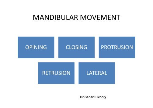
MANDIBULAR MOVEMENT
MANDIBULAR MOVEMENT
MANDIBULAR MOVEMENT
Create successful ePaper yourself
Turn your PDF publications into a flip-book with our unique Google optimized e-Paper software.
<strong>MANDIBULAR</strong> <strong>MOVEMENT</strong><br />
OPINING CLOSING PROTRUSION<br />
RETRUSION<br />
LATERAL<br />
Dr Sahar Elkholy
Factors that regulate the mandibular<br />
movements:<br />
• 1- TEMPRO<strong>MANDIBULAR</strong> JOINT.<br />
• 1- TEMPRO<strong>MANDIBULAR</strong> JOINT.<br />
• 2-MUSCLES.<br />
• 3-AXIS OF ROTATION.<br />
• 4-OPPOSING TEETH CONTACT..
Temporomandibular Joint (TMJ)<br />
Composed of<br />
Condyle<br />
Mandibular fossa<br />
Articular capsule<br />
Synovial tissue<br />
Articular disc<br />
Ligaments<br />
3
Fig. from atlas of anatomy
C0NDYLE<br />
• It about two centimeters wide mediolaterally<br />
and one centimetermthik anteroposteriorly.<br />
• Its superior surface is convex from back to<br />
front.<br />
• Medial and lateral ends of condyle called<br />
poles
Fig. from atlas of anatomy
Articular Capsule<br />
Ligamentous capsule surrounds<br />
the joint<br />
Attached to the neck of the<br />
condyle and around the border<br />
of the articular surface of the<br />
temporal bone<br />
anterolateral aspect of the<br />
capsule may thicken form the<br />
Temporomandibular ligament<br />
function as stabilising structure<br />
7
Fig. from atlas of anatomy
Synovial tissue<br />
Synovial cell and connective tissue<br />
covering the lower and upper-joint<br />
spaces<br />
Synovial fluid, acts as a lubricant and<br />
may participated in nutritional and<br />
metabolic interchange for central part.<br />
9
Temporomandibular<br />
ligament<br />
Ligaments<br />
extend from base of<br />
zygomatic process of<br />
the temporal bone<br />
downward and oblique<br />
to the neck of the<br />
condyle<br />
10
Ligaments<br />
Stylomandibular<br />
ligament<br />
From styloid process<br />
and runs downward<br />
and forward to attach<br />
broadly on the inner<br />
aspect of the angle of<br />
mandible<br />
11
Ligaments<br />
<br />
Sphenomandibular ligament<br />
arising from the angular<br />
spine of sphenoid bone<br />
and petrotympanic<br />
fissure, ending at lingula<br />
of mandible<br />
12
MUSCLES
MASTICATORY MUSCLES<br />
MASSETER TEMPORAL MEDIAL PTERYGOID LATERAL PTERYGOID
Fig. from atlas of anatomy
Temporal<br />
Massete<br />
r<br />
Medial petrygoid<br />
lateral petrygoid
superior lateral petrygoid<br />
superior lateral petrygoid<br />
inferior lateral petrygoid
Slide 19<br />
D1<br />
D2<br />
superior lateral petrygoid<br />
Dr.Sahar, 3/18/2007<br />
inferior lateral petrygoid<br />
Dr.Sahar, 3/18/2007
Masster muscle<br />
• Superficial layer<br />
– O : lower border of malar<br />
bone, Zygomatic arch &<br />
zygomatic process of maxilla<br />
– R : Downward and Backward<br />
– I : Angle of mandible and<br />
inferior half of the lateral side<br />
of mandible<br />
20
Masster muscle<br />
• Deep layer<br />
– O : Internal surface of<br />
zygomatic arch<br />
– R : Downward (vertical)<br />
– I : Ramus of mandible and<br />
base of coronoid process<br />
– 50 degree between 2 layers<br />
21
Temporalis muscle<br />
• 3 bundles<br />
– Anterior bundle (vertical fibre)<br />
–Action: Mandible elevator<br />
(Close jaws), crushing and<br />
chewing at C.O.<br />
–Inaction: Mandible depression<br />
(except Max. Opening and<br />
Opening against resistance)<br />
22
Temporalis muscle<br />
– Posterior bundle (Horizontal<br />
bundle)<br />
Action: Mand. retraction<br />
and positioner<br />
Inaction: Mand. depression<br />
and protrusion<br />
– Intermediate bundle<br />
Action: Protrisive movement<br />
23
Med. Pterygoid muscle<br />
O : Pterygoid fossa and medial<br />
surf. of the lateral pterygoid<br />
plate<br />
I : Inf. + Post. border of ramus<br />
and angle of mand.<br />
R : Downward and Backward<br />
N : Medial Pterygoid nerve<br />
24
• Superior head<br />
Lat. Pterygoid muscle<br />
O: Wing of sphenoid and<br />
infratemporal crest<br />
R: Downward and Backward<br />
• Inferior head<br />
O: Lateral surf. of lateral<br />
pterygoid plate<br />
R: Upward and backward<br />
25
Lat. Pterygoid muscle<br />
Insertion of superior and inferior<br />
heads<br />
– Ant. portion of the condylar<br />
neck (pterygoid fovea)<br />
–<br />
Ant. surface of the articular<br />
capsule<br />
– Ant. Border of the disk<br />
Function<br />
– Open the jaws, protrude and<br />
lateral movement with moving<br />
disk forward<br />
26
• Superior head<br />
Lat. Pterygoid muscle<br />
Synergistic with elevator<br />
group of muscle for closing<br />
and clenching<br />
• Inferior head<br />
Synergistic with suprahyoid<br />
group of muscle for opening<br />
jaw<br />
• Nerve supply<br />
Lateral pterygoid nerve<br />
27
• Superior head<br />
Lat. Pterygoid muscle<br />
Synergistic with elevator<br />
group of muscle for closing<br />
and clenching<br />
• Inferior head<br />
Synergistic with suprahyoid<br />
group of muscle for opening<br />
jaw<br />
• Nerve supply<br />
Lateral pterygoid nerve<br />
28
AXIS OF ROTATION
• HORIZONTAL AXIS<br />
HORIZONTAL AXIS
• VERTICAL AXIS<br />
VERTICAL AXIS
SAGITAL AXIS<br />
SAGITALAL AXIS
Opposing Tooth Contact<br />
33
• The movements of the condyle during<br />
mandibular movement are either :<br />
• - Rotation (in lower compartment)<br />
• Rotational movements take place in the<br />
lower compartment of the T.M.J. between<br />
the superior surface of the condyle and the<br />
inferior surface of the articular disc<br />
•
• b- Translation (in upper compartment)<br />
Translation or gliding movements of the<br />
mandible takes place in the upper<br />
compartment of the T.M.J. between the<br />
superior surface of the articular disc as it<br />
moves with the condyle and the inferior<br />
surface of the glenoid fossa.
<strong>MANDIBULAR</strong> <strong>MOVEMENT</strong>S<br />
• 1-Opening and closing: (depression and<br />
elevation of the mandible):<br />
This movement starts from the rest position<br />
to the maximum opening position. At the<br />
beginning of the opening movement, there is<br />
a rotation in the lower compartment of the<br />
TMJ.
• The mandible can make pure rotational<br />
movements through an arc of 10-20 mm with<br />
further opening a gliding movement occurs in<br />
the upper compartment.<br />
• For the closing movement, the mandible<br />
moves from the maximum opening position<br />
with a reverse movement back to the rest<br />
position.
2- Forward and backward (protrusive<br />
and retrusive)<br />
• The condyles together with their articular<br />
discs move as one unit downwards and<br />
forwards along the glenoid fossa and the<br />
articular eminence.<br />
• Protrusive movements are brought about by<br />
the contraction of the external pterygoid<br />
muscles on each side.
Christensen’s phenomenon<br />
• When the mandible moves forward to an<br />
edge to edge position a separation occurs<br />
distally between the distal arches or<br />
occlusion rims. This distal separation of teeth<br />
is the result of the forward and downward<br />
glide of the condyle on the articular<br />
eminence.
3- Lateral movement (sideways)<br />
• The right and left lateral movements of the<br />
mandible form the rest position and back<br />
again to the same position are asymmetric.<br />
When the mandible moves towards the right<br />
side, the condyle on this side rotates mainly<br />
with a very slight bodily lateral translation<br />
(Bennett movement).
INCISAL PATH<br />
• The incisal path forms an angle with the<br />
horizontal plane that differs form one person<br />
to another with an average 10 degree. The<br />
deeper the overbite, the more is the incisal<br />
angle. While the wider the overget, the less is<br />
the incisal angle.
Condylar Path<br />
• A-The lateral condylar path is the path<br />
followed by the condyle in the glenoid fossa<br />
when a lateral movement is made.<br />
• B-Protrusive condylar path<br />
• B-Protrusive condylar path is the path<br />
followed by the condyle when the mandible<br />
is moved forward from its centric position.
• It varies in individuals and also in the same<br />
individual from the left to the right side. It<br />
ranges between 30°-40°.
Importance of studying mandibular<br />
movements<br />
• 1- designing, selection, and adjustment of<br />
articulator.<br />
• 2-eveloping tooth form for dental restorations.-<br />
• 3-Understanding the basic principles of occlusion.<br />
• 4-Diagnosis and treatment of TMJ disturbances .<br />
• 5-Proper selection of teeth.<br />
• 6-Arrangement of artificial teeth .
<strong>MANDIBULAR</strong> POSITION
a-REST POSITION<br />
• 1- It is the position of the mandible related to<br />
the cranium in the resting state. Muscles and<br />
joints determine this position. It is a<br />
physiological position.<br />
• 2- All mandibular movements start from and<br />
end at this position.
est vertical dimension
Slide 50<br />
D3<br />
rest vertical dimension<br />
Dr.Sahar, 4/22/2007
-Intercuspal position (ICP) (Centric<br />
occlusion)<br />
• 1- It is the position of the mandible<br />
determined by bilateral maximal contact<br />
(intercuspation of natural teeth). It is the most<br />
important contact position for stomatognathic<br />
functional position.
• 2- This position is used during chewing<br />
and swallowing to stabilize the mandible,<br />
and allow the suprahyoid muscles to pull<br />
the hyoid bone and trachea upward and<br />
forward to prevent inhalation of food,<br />
drinks and saliva
• 3- Setting of artificial teeth and construction<br />
of artificial occlusal surfaces are planned at<br />
this intercuspal position if the remaining<br />
natural teeth are enough to determine this<br />
position correctly.
Importance of studying mandibular<br />
movements<br />
1- Designing, selection, and adjustment of articulator.<br />
2-Developing tooth form for dental restorations.<br />
3-Understanding the basic principles of occlusion.<br />
4-Diagnosis and treatment of TMJ disturbances<br />
5-Proper selection of artificial teeth.<br />
6-Arrangement of artificial teeth.


