steroidal glycosides from the underground parts of allium ursinum l ...
steroidal glycosides from the underground parts of allium ursinum l ...
steroidal glycosides from the underground parts of allium ursinum l ...

You also want an ePaper? Increase the reach of your titles
YUMPU automatically turns print PDFs into web optimized ePapers that Google loves.
Acta Poloniae Pharmaceutica ñ Drug Research, Vol. 63 No. 3 pp. 219ñ223, 2006<br />
ISSN 0001ñ6837<br />
Polish Pharmaceutical Society<br />
STEROIDAL GLYCOSIDES FROM THE UNDERGROUND PARTS<br />
OF ALLIUM URSINUM L. AND THEIR CYTOSTATIC AND<br />
ANTIMICROBIAL ACTIVITY<br />
DANUTA SOBOLEWSKA 1* , ZBIGNIEW JANECZKO 1 , WANDA KISIEL 2 , IRMA PODOLAK 1 ,<br />
AGNIESZKA GALANTY 1 and DANUTA TROJANOWSKA 3<br />
1<br />
Department <strong>of</strong> Pharmacognosy, Collegium Medicum, Jagiellonian University,<br />
9 Medyczna Str., 30-688 KrakÛw, Poland<br />
2<br />
Department <strong>of</strong> Phytochemistry, Institute <strong>of</strong> Pharmacology, Polish Academy <strong>of</strong> Sciences,<br />
12 SmÍtna Str., 31-343 KrakÛw, Poland<br />
3<br />
Department <strong>of</strong> Pharmaceutical Microbiology, Collegium Medicum, Jagiellonian University,<br />
9 Medyczna Str., 30-688 KrakÛw, Poland<br />
Abstract: The aim <strong>of</strong> this study was <strong>the</strong> isolation and structural elucidation <strong>of</strong> <strong>steroidal</strong> <strong>glycosides</strong> <strong>from</strong> <strong>the</strong><br />
<strong>underground</strong> <strong>parts</strong> <strong>of</strong> ramson Allium <strong>ursinum</strong> L. The structures <strong>of</strong> <strong>the</strong> isolated compounds were established<br />
based upon chromatographic methods and 1D- and 2D-NMR, MS and IR analyses. The mixture <strong>of</strong> two <strong>steroidal</strong><br />
saponins: (25R)-spirost-5-en-3b-ol tetrasaccharide and (25R)-spirost-5, 25(27)-dien-3b-ol tetrasaccharide,<br />
along with a 3-hydroxypregna-5,16-dien-20-one glycoside were identified. The results <strong>of</strong> in vitro cytotoxic<br />
activity <strong>of</strong> <strong>the</strong> mixture <strong>of</strong> spirostanol saponins against cell lines melanoma B16 and sarcoma XC and human<br />
fibroblasts HSF are also reported. The spirostanol saponins mixture was investigated to determine its in vitro<br />
antimicrobal activity against Trichophyton mentagrophytes and Microsporum canis.<br />
Keywords: Allium <strong>ursinum</strong>, <strong>steroidal</strong> saponins, cytotoxicity<br />
Allium <strong>ursinum</strong> L. (Alliaceae), commonly<br />
called ramson or wild garlic, is a perennial plant<br />
widely distributed in Europe and Asia, which develops<br />
a characteristic garlic-like smell and flavor. The<br />
aerial <strong>parts</strong> and bulbs <strong>of</strong> ramson have long been used<br />
in European traditional medicine as an antihypertensive,<br />
antia<strong>the</strong>rosclerotic, antimicrobial, antidiarrhoeal<br />
and antiphlogistic agents (1). Pharmacological<br />
studies revealed its inhibitory activity in vitro<br />
on 5-lipoxygenase, cyclooxygenase, angiotensin I-<br />
converting enzyme and thrombocyte aggregation<br />
(2). It was also demonstrated that chlor<strong>of</strong>orm and<br />
acetone/chlor<strong>of</strong>orm extracts might reduce serum<br />
cholesterol level primarily by inhibition <strong>of</strong> cholesterol<br />
syn<strong>the</strong>sis (3). Reports on chemical composition<br />
<strong>of</strong> ramson indicated <strong>the</strong> presence <strong>of</strong> many sulfur<br />
compounds, including cysteine sulfoxides, thiosulfinates,<br />
sulfides, ajoenes, vinyldithiins, as well as<br />
flavonoids and lectins (4-6). In a search for biologically<br />
active compounds <strong>from</strong> traditional medicinal<br />
plants we isolated two spirostanol saponosides <strong>from</strong><br />
<strong>the</strong> <strong>underground</strong> <strong>parts</strong> <strong>of</strong> Allium <strong>ursinum</strong>. Structure<br />
elucidation was accomplished mainly on <strong>the</strong> basis <strong>of</strong><br />
2D proton-proton and proton-carbon shift correlation<br />
spectroscopy. The cytotoxic and antifungal<br />
activities <strong>of</strong> <strong>the</strong>se compounds are also presented. In<br />
a previous paper on ramson, we have reported <strong>the</strong><br />
isolation and structural elucidation <strong>of</strong> a 3-hydroxypregna-5,16-dien-20-on<br />
tetraglycoside (7). Now its<br />
structure is reviewed.<br />
EXPERIMENTAL<br />
Materials and methods<br />
General<br />
IR: Specord M 80 (Carl Zeiss Jena) spectrometer.<br />
FAB-MS: Finnigan MAT 95 mass spectrometer,<br />
glycerol as <strong>the</strong> matrix, Cs ions accelerated at 13<br />
keV. Optical rotation: Polamat-A polarimeter.<br />
NMR: Bruker AMX 500 spectrometer in C 5 D 5 N<br />
solution, using TMS as an internal standard. 1 H<br />
NMR spectra were recorded at 500.13 MHz and 13 C<br />
NMR spectra at 125.77 MHz. TLC: precoated<br />
Merck Kieselgel 60 plates with CHCl 3 -MeOH- H 2 O<br />
(23:12:2, v/v/v), CHCl 3 -AcOH-H 2 O (3:3.5:0.5,<br />
v/v/v) and CHCl 3 -EtOAc (4:1, v/v) as mobile phases.<br />
The spots were detected by spraying with 10%<br />
H 2 SO 4 followed by heating for saponins and<br />
sapogenins, and with aniline phthalate for sugars.<br />
* Correspondence to: Dr D. Sobolewska, Department <strong>of</strong> Pharmacognosy, Collegium Medicum, Jagiellonian University,<br />
9 Medyczna Str., 30ñ688 KrakÛw, Poland. Eñmail: dsobolew@cmñuj.krakow.pl<br />
219
220 DANUTA SOBOLEWSKA et al.<br />
GLC: Chromatograph N-504; glass column (0.3 x<br />
220 cm) filled with Chromosorb W-AW-DMCS<br />
(2% HI-EFF-3BP).<br />
Plant material<br />
The <strong>underground</strong> <strong>parts</strong> <strong>of</strong> Allium <strong>ursinum</strong> were<br />
collected in September in Czaslaw, Poland. A<br />
voucher specimen was deposited in <strong>the</strong> Herbarium<br />
<strong>of</strong> <strong>the</strong> Department <strong>of</strong> Pharmacognosy, Collegium<br />
Medicum, Jagiellonian University.<br />
Extraction and isolation<br />
Air-dried powdered <strong>underground</strong> <strong>parts</strong> (578 g)<br />
<strong>of</strong> Allium <strong>ursinum</strong> were extracted with hot methanol<br />
(3 x 1 L MeOH; 3h), and concentrated to dryness. The<br />
dry residue (32 g) was dissolved in water and partitioned<br />
successively with n-hexane and n-BuOH. The<br />
butanol-soluble portion was subjected to chromatographic<br />
separation on a silica gel column. The column<br />
was eluted with CHCl 3 -MeOH-H 2 O (23:12:2) to yield<br />
compounds 1 (21 mg) and 2 (10 mg).<br />
Compound 1. Colorless needles, m.p. 212-216 O C,<br />
[α] D<br />
24<br />
= -92.5 O (c 0.1; MeOH), IR (KBr) ν cm- 1 :<br />
3420, 1049 (OH); 880, 900, 921 (C-22-spiro), 13 C-<br />
NMR (pyridine ñ d 5 ) δ (ppm): see Table 1. 1 H NMR<br />
(pyridine ñ d 5 ) δ (ppm): 6.38 (1H, br s, Rha 1 ), 5.81<br />
(1H, br s, Rha 2 ), 6.26 (1H, br s, Rha 3 ), 5.32 (1H, H-<br />
6), 4.92 (1H, Glc), 4.53 (1H, H-16), 1.76 (3H, d, J =<br />
6.1 Hz), 1.59 (3H x 2, d, J = 5.7 Hz), 1.04 (3H, s,<br />
Me-19), 0.82 (3H, s, Me-18).<br />
Positive ion FAB-MS (1a) m/z 1014.5 [M+H] +,<br />
868.1 = [M + H ñ 147] + , 721.9 = [M + H ñ 147-<br />
146] + , 413.6 = [M + H ñ 2 x 147 ñ 146 ñ 161] + ,<br />
(1b) m/z 1013.0 [M+H] + , 866.5 = [M + H ñ 147] + ,<br />
720.5 = [M + H ñ 147-146] + , 412.0 = [M + H ñ 2 x<br />
147 ñ 146 ñ 161] + .<br />
Acid hydrolysis <strong>of</strong> 1<br />
Compound 1 (3 mg) was hydrolyzed with 5%<br />
methanolic HCl (5 mL) for 4 h and diluted with<br />
water (5 mL). Methanol was evaporated under<br />
reduced pressure and <strong>the</strong> water phase was partitioned<br />
with diethyl e<strong>the</strong>r (3 x 5 mL). The e<strong>the</strong>r layers<br />
containing <strong>the</strong> aglycones were concentrated to<br />
dryness. Prior to analysis <strong>the</strong> resulting dry residue<br />
was dissolved in chlor<strong>of</strong>orm.<br />
Cytotoxic assay<br />
The cytotoxic activity <strong>of</strong> 1 (in DMSO) was<br />
tested against murine cancer cell lines melanoma<br />
B16 and sarcoma XC and human fibroblasts HSF<br />
which were grown in MEM medium supplemented<br />
with 10% heat inactivated calf serum and a mixture<br />
<strong>of</strong> antibiotics (37 O C, 5% CO 2 ). Cells were transferred<br />
into 24-well microtiter plates (density 1,5×10 4<br />
cells/well) and preincubated for 24 h to allow stabilization.<br />
The compound was added to <strong>the</strong> wells at<br />
different concentrations (1 to 12 µg mL- 1 ). The cells<br />
were incubated for 24 or 48 h. The viability <strong>of</strong> <strong>the</strong><br />
cells was tested using trypan blue dye. Cytotoxic<br />
activity was measured as a percentage <strong>of</strong> dead cells.<br />
Antimycotic evaluation<br />
Evaluation <strong>of</strong> pure 1 was performed following<br />
<strong>the</strong> conventional disc assay procedure for activity<br />
against Trichophyton mentagrophytes and Microsporum<br />
canis. The tested strains were grown on Sabourand<br />
broth. Paper disc with <strong>the</strong> solution <strong>of</strong> 1 (2-400<br />
µg/mL) was put in <strong>the</strong> middle <strong>of</strong> <strong>the</strong> plate with <strong>the</strong><br />
tested strain (10 6 cells/mL). The incubation was carried<br />
out at 27 O C for 5-7 days. Growth inhibition<br />
zone was observed after this time.<br />
RESULTS AND DISCUSSION<br />
The dry MeOH extract <strong>of</strong> bulbs and roots <strong>of</strong><br />
Allium <strong>ursinum</strong> was partitioned successively<br />
between n-hexane, n-butanol and water. The n-<br />
BuOH-soluble portion was subjected to silica gel<br />
column chromatography to afford 1 and 2.<br />
Compound 1 was obtained as colorless needles;<br />
mp. 212-216 O 24<br />
C, [α] D = -92.5 O (c 0.1; MeOH). It<br />
responded positively to <strong>the</strong> Liebermann-Burchard<br />
and negatively to <strong>the</strong> Ehrlich reagent. On acid<br />
hydrolysis with 5% HCl in MeOH, 1 afforded diosgenin<br />
which was identified by cochromatography<br />
(TLC) with an au<strong>the</strong>ntic sample. The sugar residues<br />
present in <strong>the</strong> acidic hydrolysate <strong>of</strong> 1 were identified<br />
as rhamnose and glucose in <strong>the</strong> ratio 3:1 based on<br />
TLC and GLC analysis. This was fur<strong>the</strong>r confirmed<br />
by FAB-MS fragmentation ions. The IR spectrum <strong>of</strong><br />
1 showed a strong absorption at 3420 and 1049 cm- 1<br />
due to OH groups and an olefinic group at 1629.8<br />
cm- 1 ; moreover bands characteristic <strong>of</strong> a spirostane<br />
ring at 880, 900, 921 cm- 1 were also seen.<br />
Positive ion FAB-MS spectrum <strong>of</strong> 1 displayed,<br />
however, two quasi-molecular ion peaks at 1014.5<br />
[M+H] + and 1013.0 [M+H] + , thus indicating <strong>the</strong><br />
presence <strong>of</strong> two compounds differing by two mass<br />
units, with molecular formulae C 51 H 82 O 20 and<br />
C 51 H 80 O 20 , respectively. These compounds, which<br />
were denoted as 1a and 1b, could not be separated<br />
by column chromatography. Therefore, <strong>the</strong>y were<br />
analyzed as a mixture by means <strong>of</strong> one- and twodimensional<br />
NMR spectroscopy. The nature <strong>of</strong> <strong>the</strong><br />
sugar residues and <strong>the</strong> sequence <strong>of</strong> <strong>the</strong> saccharide<br />
chains <strong>of</strong> 1a and 1b were determined using a combi-
Steroidal <strong>glycosides</strong> <strong>from</strong> <strong>the</strong> <strong>underground</strong> <strong>parts</strong>... 221<br />
nation <strong>of</strong> 1 H-NMR, 13 C-NMR, DEPT 135 O , 1 H- 1 H<br />
COSY, HETCOR and HMBC. The 1 H-NMR spectrum<br />
displayed signals <strong>of</strong> four anomeric protons at δ<br />
4.92, 5.81, 6.26 and 6.38 ppm and <strong>of</strong> three secondary<br />
methyl groups characteristic <strong>of</strong> 6-deoxypentose at<br />
1.76, 1.59 and 1.59 ppm.<br />
The sugar spin system was assigned on <strong>the</strong><br />
basis <strong>of</strong> 1 H- 1 H-COSY and HETCOR. The position<br />
<strong>of</strong> interglycosidic linkages were determined using<br />
<strong>the</strong> HMBC spectrum. The significant glycosylation<br />
shifts showed that two terminal rhamnoses were<br />
linked to <strong>the</strong> C-2 position <strong>of</strong> <strong>the</strong> glucopyranose and<br />
C-4 position <strong>of</strong> <strong>the</strong> inner rhamnopyranose. The α<br />
configuration <strong>of</strong> <strong>the</strong> rhamnopyranosyl units were<br />
established <strong>from</strong> signals ascribable to <strong>the</strong>ir C 5 data<br />
(70.43 ppm, 69.57 ppm and 68.35 ppm). This was<br />
fur<strong>the</strong>r confirmed by <strong>the</strong> three-bond coupled strong<br />
HMBC correlations <strong>from</strong> <strong>the</strong> anomeric proton to <strong>the</strong><br />
C-3 and C-5 carbons (8). The absence <strong>of</strong> 13 C glycosylation<br />
shift for two rhamnosyl residues suggested that<br />
<strong>the</strong>y were <strong>the</strong> terminal units. The FAB-MS spectrum<br />
(positive ions) <strong>of</strong> 1 exhibited fragment ions at m/z<br />
868.1 = [M + H ñ 147] + , 721.9 = [M + H ñ 147-146] + ,<br />
413.6 = [M + H ñ 2 x 147 ñ 146 ñ 161] + and 866.5 =<br />
[M + H ñ 147] + , 720.5 = [M + H ñ 147-146] + , 412.0 =<br />
[M + H ñ 2 x 147 ñ 146 ñ 161] + supporting <strong>the</strong> sugar<br />
sequence <strong>of</strong> 1a and 1b, respectively.<br />
The spectroscopic results suggested that <strong>the</strong>se<br />
compounds must be monodesmosidic saponins. The<br />
signal ascribable to C-3 shifted downfield by<br />
approximately +7 ppm and <strong>the</strong> signals due to C-2<br />
and C-4 shifted to higher field by -1 ppm and -3<br />
ppm, respectively, indicating that <strong>the</strong> sugar chain<br />
was bound to C-3 position <strong>of</strong> <strong>the</strong> aglycones.<br />
Table 1. 13 C NMR chemical shift assignments <strong>of</strong> compounds 1a and 1b.<br />
Aglycones<br />
Sugar moiety<br />
1a 1b 1a and 1b<br />
13<br />
C chemical chemical chemical<br />
shift (δ ppm) shift (δ ppm) shift (δ ppm)<br />
Cñ1 37.55 37.55<br />
Glc<br />
Cñ2 30.21 30.21<br />
Cñ1 100.41<br />
Cñ2 78.16<br />
Cñ3 78.04 78.04<br />
Cñ3 77.77<br />
Cñ4 39.03 39.03<br />
Cñ4 77.90<br />
Cñ5 140.88 140.88<br />
Cñ5 77.04<br />
Cñ6 121.82 121.82<br />
Cñ6 61.31<br />
Cñ7 32.35 32.35<br />
Rha 1 (1g2) Glc<br />
Cñ8 31.74 31.74<br />
(terminal)<br />
Cñ9 50.36 50.36<br />
Cñ1 102.20<br />
Cñ10 37.19 37.19<br />
Cñ2 72.52<br />
Cñ11 21.15 21.15<br />
Cñ3 72.90<br />
Cñ4 74.05<br />
Cñ12 39.91 39.85<br />
Cñ5 69.57<br />
Cñ13 40.51 40.51<br />
Cñ6 18.67<br />
Cñ14 56.69 56.69<br />
Cñ15 32.25 32.21<br />
Rha 2 (1g4) Glc<br />
(inner)<br />
Cñ16 81.15 81.15<br />
Cñ1 102.28<br />
Cñ17 62.96 62.96<br />
Cñ2 72.90<br />
Cñ18 16.36 16.36<br />
Cñ3 73.29<br />
Cñ19 19.44 19.44<br />
Cñ4 80.45<br />
Cñ20 42.02 41.91<br />
Cñ5 68.39<br />
Cñ6 18.45<br />
Cñ21 15.05 15.01<br />
Cñ22 109.30 109.48<br />
Rha 3 (1g4) Rha 2<br />
Cñ23 31.88 29.0<br />
(terminal)<br />
Cñ24 29.31 33.26<br />
Cñ1 103.31<br />
Cñ2 72.66<br />
Cñ25 30.64 144.48<br />
Cñ3 72.90<br />
Cñ26 66.91 65.05<br />
Cñ4 74.18<br />
Cñ27 17.35 108.73<br />
Cñ5 70.42
222 DANUTA SOBOLEWSKA et al.<br />
Moreover, <strong>the</strong> 1D- and 2D-NMR spectra indicated<br />
that compounds 1a and 1b had identical sugar moieties,<br />
but different aglycones (see Table 1). These<br />
two sapogenins were overlapped to produce a single<br />
spot on <strong>the</strong> TLC plate, which was identified as diosgenin.<br />
The 1 H-NMR spectrum showed signals characteristic<br />
<strong>of</strong> four <strong>steroidal</strong> methyl groups; two<br />
appeared as singlets at δ 0.82 ppm (3H, s, Me-18)<br />
and 1.04 ppm (3H, s, Me-19), and <strong>the</strong> o<strong>the</strong>r two as<br />
doublets at δ 1.09 ppm (d; J=6.8) and 0.69 ppm (d;<br />
J=5.2). The quaternary carbon atom C-5 (140.88<br />
ppm) and <strong>the</strong> methine carbon atom C-6 (121.82<br />
ppm) were easily assigned based on 13 C-NMR and<br />
DEPT 135 O spectra. The 13 C chemical shifts <strong>of</strong> 1b<br />
were almost identical to those <strong>of</strong> 1a except for <strong>the</strong><br />
chemical shifts <strong>of</strong> F-ring and C-20. The ketal carbon<br />
signal at δ 109.3 ppm (1a) and 109.5 ppm (1b) were<br />
assigned to <strong>the</strong> spiroketal carbon atom (C-22) with<br />
two oxygens attached. A feature differing 1b <strong>from</strong><br />
1a was <strong>the</strong> substitution <strong>of</strong> <strong>the</strong> C-25 ñ methyl group<br />
by <strong>the</strong> exocyclic methylene group. Two carbonyl<br />
resonances at δ 144.5 ppm and d 108.7 ppm were<br />
assigned to carbon atoms C-25 and C-27 <strong>of</strong> 1b,<br />
respectively (9). Four signals at δ 41.9, 29.0, 33.3<br />
and 65.0 ppm were assigned to <strong>the</strong> C-20, C-23, C-24<br />
and C-26 positions, respectively. The 13 C-NMR and<br />
1<br />
H-NMR spectra implied that <strong>the</strong> 27-Me orientation<br />
<strong>of</strong> 1a was equatorial. The obtained data confirmed<br />
that <strong>the</strong> aglycone <strong>of</strong> 1a was diosgenin ñ (25R)-<br />
spirost-5-en-3b-ol, while <strong>the</strong> sapogenin <strong>of</strong> 1b was<br />
(25R)- spirost-5, 25(27)-dien-3b-ol.<br />
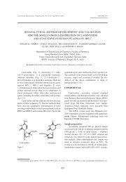
On <strong>the</strong> basis <strong>of</strong> <strong>the</strong> foregoing data <strong>the</strong> structures<br />
<strong>of</strong> <strong>the</strong> two compounds are proposed to be: (25R)-<br />
spirost-5-en-3b-ol 3-O-α-rhamnopyranosyl-(1g4)-αrhamnopyranosyl-(1g4)-<br />
[a-rhamnopyranosyl-(1g2)]-<br />
glucopyranoside (1a) and (25R)-spirost-5,25(27)-<br />
dien-3β-ol 3-O-α-rhamnopyranosyl-(1g4)-α-rham-<br />
Table 2. 13 C NMR chemical shift assignments <strong>of</strong> sugar moiety <strong>of</strong><br />
compound 2.<br />
Sugar moiety<br />
13<br />
C (δ ppm)<br />
Glc<br />
Cñ1 100.40<br />
Cñ2 78.14<br />
Cñ3 77.76<br />
Cñ4 77.97<br />
Cñ5 77.05<br />
Cñ6 61.24<br />
Rha 1 (1g2) Glc<br />
(terminal)<br />
Cñ1 102.14<br />
Cñ2 72.54<br />
Cñ3 72.88<br />
Cñ4 74.04<br />
Cñ5 69.57<br />
Cñ6 18.66<br />
Rha 2 (1g4) Glc<br />
(inner)<br />
Cñ1 102.25<br />
Cñ2 72.93<br />
Cñ3 73.30<br />
Cñ4 80.43<br />
Cñ5 68.35<br />
Cñ6 18.45<br />
Rha 3 (1g4) Rha 2<br />
(terminal)<br />
Cñ1 103.30<br />
Cñ2 72.68<br />
Cñ3 72.93<br />
Cñ4 74.16<br />
Cñ5 70.43<br />
Cñ6 18.91<br />
Figure 1. The structures <strong>of</strong> 1a and 1b.
Steroidal <strong>glycosides</strong> <strong>from</strong> <strong>the</strong> <strong>underground</strong> <strong>parts</strong>... 223<br />
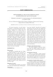
Figure 2. The structure <strong>of</strong> 2.<br />
nopyranosyl-(1g4)- [α-rhamnopyranosyl-(1g2)]-<br />
glucopyranoside (1b). A survey <strong>of</strong> <strong>the</strong> literature<br />
showed that <strong>the</strong> compound identical with 1a was identified<br />
in Disocorea cayenensis (10) and Paris polyphylla<br />
var. yunnanensis (11).<br />
The NMR data <strong>of</strong> 2 were in full agreement with<br />
<strong>the</strong> signals <strong>of</strong> <strong>the</strong> pregnadienolone tetraglycoside previously<br />
isolated by us <strong>from</strong> Allium <strong>ursinum</strong> (7). The<br />
13<br />
C-NMR spectrum due to <strong>the</strong> sugar chain <strong>of</strong> 2 was<br />
almost superimposable on that <strong>of</strong> 1 suggesting that <strong>the</strong><br />
tetrasaccharide sequence <strong>of</strong> 2 was <strong>the</strong> same as that <strong>of</strong><br />
1. The structure <strong>of</strong> sugar residue now was revised (see<br />
Table 2). On <strong>the</strong> basis <strong>of</strong> spectroscopic evidence <strong>the</strong><br />
structure <strong>of</strong> compound 2 was determined as 3-O-αrhamnopyranosyl-(1g4)-α-<br />
rhamnopyranosyl-(1g4)<br />
-[α-rhamnopyranosyl-(1g2)]-glucopyranoside 3-<br />
hydroxy-pregna-5, 16-dien-20-one.<br />
Cytotoxic activity <strong>of</strong> 1 (a mixture <strong>of</strong> 1a and 1b)<br />
on melanoma B16, sarcoma XC and human fibroblasts<br />
HSF was evaluated. Compound 1 was found<br />
active against murine melanoma B16 and sarcoma<br />
XC. The compound exhibited 100% effect at 2 µg<br />
mL- 1 on both strains. It showed no activity towards<br />
human fibroblasts HSF at concentrations below 3 µg<br />
mL- 1 . With regard to antifungal properties against<br />
Trichophyton mentagrophytes and Microsporum<br />
canis <strong>the</strong> saponin was active at concentration <strong>of</strong> 400<br />
µg mL -1 . Our previous studies have shown <strong>the</strong> activity<br />
<strong>of</strong> compound 1 against Candida glabrata and<br />
Candida parapsilosis (MIC values 200 mg mL -1 and<br />
250 mg mL -1 , respectively) [12]. Compound 1 did<br />
not show any toxicity against Pseudomonas aeruginosa<br />
and Aspergillus niger at concentrations up to<br />
400 mg mL- 1 , in <strong>the</strong> disk assay study. This low<br />
fungistatic activity is most probably due to a relatively<br />
long sugar chain. Dioscin, <strong>the</strong> analogue <strong>of</strong> 1a<br />
which possesses shorter sugar chain (-1 Rha)<br />
showed stronger activity (between 12.5 and 25 mg<br />
mL -1 ) against Candida [10].<br />
REFERENCES<br />
1. Treben M.: Apteka Pana Boga. Porady i praktyka<br />
stosowania ziÛ≥ leczniczych. Natur-Produkt<br />
TOM-MARK, Warszawa 1992.<br />
2 Sendl A., Elbl G., Steinke B., Redl K., Breu W.,<br />
Wagner H.: Planta Med. 58, (1992).<br />
3 Sendl A., Schliack M., Loeser R., Stanislaus F.,<br />
Wagner H.: A<strong>the</strong>rosclerosis 94, 79 (1992).<br />
4 Wagner H., Sendl A.: Dtsch. Apoth. Ztg 33,<br />
1809 (1990).<br />
5 Carotenuto A., De Feo V., Fattorusso E.,<br />
Lanzotti V., Magno S., Cicala C.: Phytochem.<br />
41, 531 (1996).<br />
6 Kaku H., Goldstein I.J., Van-Damme E.J.,<br />
Peumans W.J.: Carbohydr. Res. 229, 347<br />
(1992).<br />
7 Janeczko Z., Sobolewska D., Podolak I.: Acta<br />
Pol. Pharm. Drug Res. 57, 131 (2000).<br />
8 Yokosuka A., Mimaki Y., Sashida Y.: J. Nat.<br />
Prod. 65, 1293 (2002).<br />
9 Agrawal P.K., Jain D.C., Gupta R.K., Thakur<br />
R.S.: Phytochem. 24, 2479 (1985).<br />
10 Sautour M., Mitaine-Offer A-C., Miyamoto T.,<br />
Dongmo A., Lacaille-Dubois M-A. Planta Med.<br />
70, 90 (2004).<br />
11 Matsuda H., Pongpiriyadacha Y., Morikawa T.,<br />
Kishi A., Kataoka S., Yoshikawa M.: Bioorg.<br />
Med. Chem. Lett. 13, 1101 (2003).<br />
12 Sobolewska D., Janeczko Z., Galanty A.,<br />
Trojanowska D.: 3 rd International Symposium on<br />
Natural Drugs. Proceedings. Naples, 2-4<br />
October 2003.<br />
Received: 31.03.2006