Exploring effects of different nonsteroidal antiinflammatory drugs...
Exploring effects of different nonsteroidal antiinflammatory drugs...
Exploring effects of different nonsteroidal antiinflammatory drugs...

You also want an ePaper? Increase the reach of your titles
YUMPU automatically turns print PDFs into web optimized ePapers that Google loves.
Acta Poloniae Pharmaceutica ñ Drug Research, Vol. 64 No. 3 pp. 211ñ216, 2007 ISSN 0001-6837<br />
Polish Pharmaceutical Society<br />
EXPLORING EFFECTS OF DIFFERENT NONSTEROIDAL<br />
ANTIINFLAMMATORY DRUGS ON LIPID PEROXIDATION.<br />
PART II. 4-HNE PROFILE<br />
SANTANU CHAKRABORTY, KUNAL ROY and CHANDANA SENGUPTA*<br />
Division <strong>of</strong> Medicinal and Pharmaceutical Chemistry, Department <strong>of</strong> Pharmaceutical Technology,<br />
Jadavpur University, Kolkata ñ 700 032, India<br />
Abstract: Non-steroidal anti-inflammatory <strong>drugs</strong> (NSAIDs) are common in alleviating pain, pyrexia and<br />
inflammation, in patients with rheumatoid arthritis and osteoarthritis. As these <strong>drugs</strong> are associated with high<br />
incidence <strong>of</strong> gastrointestinal ulceration, bleeding and kidney damage which may be linked with lipid<br />
peroxidation, our study was aimed to examine lipid peroxidation induction capacity <strong>of</strong> NSAIDs (dicl<strong>of</strong>enac<br />
sodium, ibupr<strong>of</strong>en, flurbipr<strong>of</strong>en, paracetamol, nimesulide, celecoxib and indomethacin) by determining<br />
4-hydroxy-2-nonenal (4-HNE) concentration as an index <strong>of</strong> lipid peroxidation and to see the suppressive<br />
potential <strong>of</strong> ascorbic acid on NSAID induced lipid peroxidation. The results suggest that dicl<strong>of</strong>enac sodium,<br />
ibupr<strong>of</strong>en, flurbipr<strong>of</strong>en, paracetamol, nimesulide and celecoxib exerted mild antioxidant activity. Indomethacin<br />
exerted statistically significant increase in 4-HNE content, indicating statistically significant peroxidation<br />
activity. Ascorbic acid could significantly reduce indomethacin-induced lipid peroxidation.<br />
Keywords: 4-HNE, lipid peroxidation, NSAID, dicl<strong>of</strong>enac sodium, ibupr<strong>of</strong>en, flurbipr<strong>of</strong>en, paracetamol,<br />
nimesulide, celecoxib, indomethacin<br />
Lipid peroxidation is the oxidative deterioration<br />
<strong>of</strong> polyunsaturated lipids (1) resulting in<br />
the generation <strong>of</strong> stereospecific endoperoxides and<br />
hydroperoxides by enzymatic and non-enzymatic<br />
involvement <strong>of</strong> ìreactive oxygen speciesî (ROS).<br />
These species resulting from the exposure <strong>of</strong> larger<br />
variety <strong>of</strong> <strong>drugs</strong>, chemicals and environmental<br />
agents or by normal metabolic process can easily<br />
initiate peroxidation <strong>of</strong> membrane lipids, leading to<br />
the accumulation <strong>of</strong> lipid peroxides and free<br />
radicals, which are believed to be a primary factor<br />
in various untoward <strong>effects</strong> <strong>of</strong> <strong>drugs</strong> and in various<br />
diseases (2-4). Different adverse reactions due to<br />
some <strong>drugs</strong> have been reported to be mediated<br />
through drug induced lipid peroxidation mechanism<br />
(1).<br />
The non-steroidal anti-inflammatory <strong>drugs</strong><br />
(NSAIDs) are commonly used in alleviating pain,<br />
pyrexia, inflammation, and in patients with rheumatoid<br />
arthritis and osteoarthritis. The <strong>drugs</strong> are associated<br />
with high incidence <strong>of</strong> gastrointestinal ulceration<br />
(5), bleeding (6), and kidney damage (7, 8).<br />
(E)-(±)-4-hydroxy-2-nonenal (more commonly<br />
referred to as 4-hydroxynonenal, 4-HNE, or<br />
HNE) is found throughout animal tissues, and in<br />
higher quantities during oxidative stress due to the<br />
increase in the lipid peroxidation chain reaction<br />
(9). 4-HNE shows a variety <strong>of</strong> cytotoxic <strong>effects</strong><br />
such as inhibition <strong>of</strong> DNA, RNA and protein<br />
synthesis, cell cycle arrest, mitochondrial<br />
dysfunction, induction <strong>of</strong> cataracts <strong>of</strong> the lens and<br />
neuronal apoptosis (10-12). Intracellularly, 4-HNE<br />
reacts rapidly with thiol groups <strong>of</strong> GSH and<br />
cysteine and with lysine and histidine residues <strong>of</strong><br />
proteins (12, 13). 4-HNE-modified proteins have<br />
been detected in pathological disorders such as<br />
chronic liver diseases (14-16), Alzheimerís disease<br />
(17) and atherosclerotic lesions (18). Furthermore,<br />
plasma levels <strong>of</strong> 4-HNE increase in patients with<br />
rheumatoid arthritis (19).<br />
Lipid peroxidation is known as a common<br />
mechanism <strong>of</strong> toxic manifestations <strong>of</strong> many <strong>drugs</strong><br />
and attempt has been made to explore lipid<br />
peroxidation induction capacity <strong>of</strong> NSAIDs (20). In<br />
the ongoing effort <strong>of</strong> the authors in this direction,<br />
presently an attempt has been made to study the<br />
potential <strong>of</strong> ascorbic acid as possible suppressor <strong>of</strong><br />
NSAID-induced lipid peroxidation (if any), taking<br />
4-HNE as a model marker to provide a scope <strong>of</strong><br />
further investigation on possible consideration <strong>of</strong><br />
this as prospective chemoprotective supplement for<br />
co-therapy with <strong>drugs</strong>. The present study explores<br />
the effect <strong>of</strong> NSAIDs (dicl<strong>of</strong>enac sodium, ibupr<strong>of</strong>en,<br />
flurbipr<strong>of</strong>en, paracetamol, nimesulide, celecoxib<br />
* Corresponding author: E-mail: csgjupt@yahoo.com<br />
211
212 SANTANU CHAKRABORTY et al.<br />
and indomethacin) on 4-HNE content <strong>of</strong> the goat<br />
liver tissue.<br />
MATERIALS AND METHODS<br />
The study had been performed on goat (Capra<br />
capra) liver using 4-HNE content as laboratory<br />
marker <strong>of</strong> lipid peroxidation. Goat liver was selected<br />
because <strong>of</strong> its easy availability and close similarity<br />
to the human liver, in its lipid pr<strong>of</strong>ile (21). The work<br />
was carried out according to the guideline <strong>of</strong> the<br />
Institutional Animal Ethical Committee.<br />
Preparation <strong>of</strong> tissue homogenate<br />
Liver was perfused with normal saline through<br />
hepatic portal vein. Liver was harvested and its<br />
lobes were briefly dried between filter papers (to<br />
remove excess <strong>of</strong> blood) and were thin-cut with a<br />
sharp blade. These small pieces were then<br />
transferred to the glass-teflon homogenizing tube to<br />
prepare homogenate (1 g/mL) in phosphate buffer<br />
saline (PBS) (pH = 7.4) under cold conditions. It<br />
was centrifuged at 2000 rpm for 10 min and then the<br />
supernatant was collected and finally suspended in<br />
PBS to contain approximately 0.8-1.5 mg <strong>of</strong> protein<br />
in 0.1 mL <strong>of</strong> suspension to perform in vitro<br />
experiments.<br />
Incubation <strong>of</strong> tissue homogenate with drug and/<br />
or ascorbic acid<br />
For each drug, the tissue homogenate was<br />
divided into four <strong>different</strong> parts <strong>of</strong> 50 mL each in a<br />
glass stoppered 250 mL conical flasks. The first<br />
portion was kept as the control (C) while the second<br />
portion was treated with drug (D). The third portion<br />
was treated with both drug and ascorbic acid (A).<br />
After treatment with drug and/or antioxidant, liver<br />
homogenates were stirred for 1 h at 15 O C on a<br />
mechanical shaker and then incubated at 15 O C for 4 h<br />
along with the control sample.<br />
Effective concentration <strong>of</strong> <strong>drugs</strong> and ascorbic<br />
acid<br />
Considering therapeutic dose <strong>of</strong> <strong>drugs</strong> per<br />
average weight <strong>of</strong> human liver (i.e., 1500 g),<br />
dicl<strong>of</strong>enac sodium (0.03 mg/g <strong>of</strong> liver homogenate),<br />
ibupr<strong>of</strong>en (0.267 mg/g <strong>of</strong> liver homogenate),<br />
flurbipr<strong>of</strong>en (0.03 mg/g <strong>of</strong> liver homogenate),<br />
paracetamol (0.33 mg/g <strong>of</strong> liver homogenate),<br />
nimesulide (0.067 mg/g <strong>of</strong> liver homogenate),<br />
celecoxib (0.067 mg/g <strong>of</strong> liver homogenate),<br />
indomethacin (0.016 mg/g <strong>of</strong> liver homogenate) and<br />
ascorbic acid (0.17 mg/g <strong>of</strong> liver homogenate) were<br />
taken to carry out the experiments.<br />
Estimation <strong>of</strong> 4-HNE level from tissue<br />
homogenate<br />
The estimation was done at 2 and 4 h <strong>of</strong><br />
incubation, and it was repeated in replicates<br />
consisted <strong>of</strong> 5 animals. In each case, three samples<br />
<strong>of</strong> 2 mL <strong>of</strong> incubation mixture were treated with 1.5<br />
mL <strong>of</strong> 10% TCA solution, and centrifuged at 3000<br />
rpm for 30 min. Then 2 mL <strong>of</strong> the filtrate was treated<br />
with 1 mL <strong>of</strong> 2,4-dinitrophenylhydrazine (DNPH<br />
100 mg/100 mL <strong>of</strong> 0.5 M HCl), and kept for 1 h at<br />
room temperature. Then, the samples were extracted<br />
with hexane, and the extract was evaporated at 40 O C.<br />
After cooling to room temperature, 2 mL <strong>of</strong><br />
methanol was added to each sample, and the<br />
absorbance was measured at 350 nm against<br />
methanol as blank (22). The 4-HNE content values<br />
were determined from the standard curve. The<br />
standard calibration curve was drawn based on the<br />
following procedure. A series <strong>of</strong> dilutions <strong>of</strong> 4-HNE<br />
in solvent (phosphate buffer, pH = 7.4) were<br />
prepared. Pure 4-HNE was used as primary<br />
standard. 4-HNE (1 mg; 6.4 µM) was dissolved in<br />
methanol (6.4 mL) to give a 1 mM solution. From<br />
this primary standard 4-HNE solution, 300 µL was<br />
diluted with 4.70 mL <strong>of</strong> methanol. Similarly, 400 µL<br />
was diluted with 4.60 mL <strong>of</strong> methanol, 500 µL <strong>of</strong><br />
was diluted with 4.50 mL <strong>of</strong> methanol, and 600 µL<br />
was diluted with 4.40 mL <strong>of</strong> methanol. From each<br />
diluted solution <strong>of</strong> 4-HNE, 2 mL was pipetted out,<br />
and transferred into stoppered glass tube. 1 mL <strong>of</strong><br />
DNPH solution was added to all the samples and<br />
kept at room temperature for 1 h. Each sample was<br />
extracted three times with 2 mL <strong>of</strong> hexane. All<br />
extracts were collected in a glass-stoppered test<br />
tubes. After that, the extract was evaporated to<br />
dryness under argon at 40 O C and the residue was<br />
reconstituted in 1 mL <strong>of</strong> methanol. The absorbance<br />
was measured at 350 nm in a 1 cm quartz cuvette<br />
using methanol as a blank. The best fit equation is:<br />
nanomoles <strong>of</strong> 4-HNE = (A 350 ñ 0.005603185)/<br />
0.003262215, where A 350 = absorbance at 350 nm,<br />
r = 0.999, SEE = 0.007<br />
Statistical analysis<br />
Analysis <strong>of</strong> variance (ANOVA) and multiple<br />
comparisons were done to check statistical<br />
significance <strong>of</strong> the results. Any two means not<br />
included in same parenthesis are statistically<br />
significantly <strong>different</strong> at p = 0.05. The interpretation<br />
<strong>of</strong> the results is supported by Studentís ëtí test<br />
and also by a statistical multiple comparison<br />
analysis using a least significant <strong>different</strong> procedure<br />
(23, 24).
<strong>Exploring</strong> <strong>effects</strong> <strong>of</strong> <strong>different</strong> <strong>nonsteroidal</strong> <strong>antiinflammatory</strong> <strong>drugs</strong>... 213<br />
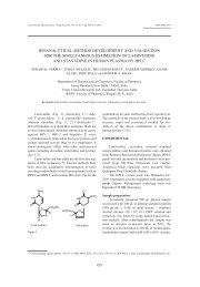
Figure 1. Average percent changes (± standard error) in 4-HNE pr<strong>of</strong>ile after treatment with <strong>different</strong> NSAIDs [(a) dicl<strong>of</strong>enac sodium, (b)<br />
ibupr<strong>of</strong>en, (c) flurbipr<strong>of</strong>en, (d) paracetamol, (e) nimesulide, (f) celecoxib, (g) indomethacin] and ascorbic acid (D, DA, and A indicate:<br />
drug-treated, drug and antioxidant-treated, and antioxidant-treated groups, respectively).
214 SANTANU CHAKRABORTY et al.<br />
RESULTS<br />
The average percent changes in 4-HNE content<br />
with respect to the control <strong>of</strong> the corresponding<br />
periods <strong>of</strong> incubation <strong>of</strong> <strong>different</strong> animal sets for<br />
<strong>different</strong> NSAIDs are shown in Figure 1. The results<br />
were analyzed using ANOVA and least significant<br />
difference procedure, which reveals that there was<br />
no statistically significant difference between the<br />
drug-treated group and the group treated with both<br />
drug and antioxidant except in the case <strong>of</strong><br />
indomethacin.<br />
DISCUSSION<br />
From the obtained data shown in Figure 1, it is<br />
evident that 4-HNE levels decrease significantly<br />
with respect to control at both the periods <strong>of</strong><br />
incubation (2 h and 4 h) after the treatment <strong>of</strong> the<br />
liver homogenates with dicl<strong>of</strong>enac sodium,<br />
ibupr<strong>of</strong>en, flurbipr<strong>of</strong>en, paracetamol, nimesulide<br />
and celecoxib. This is due to the antioxidant action<br />
<strong>of</strong> the <strong>drugs</strong>. It was previously reported that<br />
dicl<strong>of</strong>enac sodium has the ability to scavange the<br />
stable free radical 1,1-diphenyl-2-picrylhydrazyl<br />
(DPPH) (25), peroxynitrite (26) and has a<br />
significant cytoprotective effect (27).<br />
Ibupr<strong>of</strong>en acts as an antioxidant, and thus<br />
makes a new therapeutic avenue for the treatment <strong>of</strong><br />
Alzheimerís disease (28, 29). Flurbipr<strong>of</strong>en (30),<br />
paracetamol (31), nimesulide (32, 33) and celecoxib<br />
(34) have also antioxidative activity. In our earlier<br />
finding, we have suggested (20) that at 2 h and 4 h<br />
<strong>of</strong> incubation, celecoxib raises the malondialdehyde<br />
(MDA) level with respect to the control value,<br />
representing its significant lipid peroxidation<br />
activity. It may be due to the fact that celecoxib may<br />
have a prominent role for the formation <strong>of</strong> MDA in<br />
liver homogenate but not for the 4-HNE generation.<br />
The results <strong>of</strong> average percent changes <strong>of</strong> 4-HNE<br />
levels after treatment <strong>of</strong> liver homogenate with<br />
celecoxib are very close to the control value<br />
[-0.24(± 0.013) and -0.35(± 0.011) at 2 h and 4 h,<br />
respectively], suggesting its limited antioxidant<br />
activity.<br />
An increase in 4-HNE levels <strong>of</strong> the drug-treated<br />
group suggests the occurrence <strong>of</strong> lipid peroxidation.<br />
4-HNE is the end product produced by the ω-3 and<br />
ω-6 polyunsaturated fatty acids (35). This process <strong>of</strong><br />
lipid peroxidation especially occurs in the presence<br />
<strong>of</strong> some metal ions like Fe 2+ and other prooxidants<br />
(36). The increase in 4-HNE content with respect to<br />
control when the tissue homogenates were treated<br />
with indomethacin, indicates the prooxidant effect <strong>of</strong><br />
the indomethacin (37, 38). Lipid peroxidation<br />
mediated by oxygen radicals, especially hydroxyl<br />
radicals, plays a crucial role in the development <strong>of</strong><br />
the gastric mucosal injury induced by indomethacin<br />
(38). Recent report suggests that indomethacin<br />
causes mitochondrial injury by dissipating the<br />
mitochondrial transmembrane potential (MTP) and<br />
inducing mitochondrial permeability transition pore<br />
(PTP), which liberates cytochrome C. This enzyme<br />
generates ROS, thereby triggering cellular lipid<br />
peroxidation, resulting in cellular apoptosis (39).<br />
Structural interpretation <strong>of</strong> prooxidant activity <strong>of</strong><br />
indomethacin cannot be easily explained unless one<br />
carries out mechanistic study which is a scope <strong>of</strong><br />
<strong>different</strong> paper.<br />
At pH 7.4 (phosphate buffer), 99.95% <strong>of</strong><br />
vitamin C remains present as ascorbic acid<br />
monoanion ñ AscH ñ ; 0.05% as free acid ñ AscH 2 and<br />
0.004% as dianion Asc 2ñ (40). Thus, the antioxidant<br />
chemistry <strong>of</strong> vitamin C is the chemistry <strong>of</strong> AscH ñ .<br />
AscH ñ reacts (AscH ñ + Rï → Ascï ñ + RH) rapidly<br />
with HOï, ROï (tert-butyl alkoxyl radical), ROOï<br />
(alkyl peroxyl radical, e.g. CH 3 OOï), GSï<br />
(glutathiyl radical), UHï ñ (urate radical), TOï<br />
(tocopheroxyl radical), O 2 ï ñ /HO 2 ï and similar<br />
oxidants making it an outstanding donor<br />
antioxidant. AscH ñ donates a hydrogen atom (Hï or<br />
H + + e ñ ) to an oxidizing radical to produce the<br />
resonance-stabilized tricarbonyl ascorbate free<br />
radical. AscHï is not protonated in biological system<br />
and is present as Ascï ñ . The dismutation reaction<br />
(2Ascï ñ + H + → AscH ñ + dihydroascorbate) is the<br />
principal route to the elimination <strong>of</strong> the Ascï ñ in<br />
vitro. However, it is thought that reducing enzymes<br />
are involved in the removal <strong>of</strong> this radical, resulting<br />
in the recycling <strong>of</strong> ascorbate in vivo (41). Our study<br />
also reveals that ascorbic acid is capable <strong>of</strong><br />
significantly minimizing the 4-HNE levels when<br />
used alone or together with the <strong>drugs</strong>.<br />
CONCLUSION<br />
From the present study it is suggested that<br />
dicl<strong>of</strong>enac sodium, ibupr<strong>of</strong>en, flurbipr<strong>of</strong>en,<br />
paracetamol, nimesulide and celecoxib exert<br />
statistically significant mild antioxidant activity. By<br />
contrast, indomethacin is involved in oxidative<br />
processes. Ascorbic acid shows its antioxidant<br />
potential. These observations imply that the adverse<br />
<strong>effects</strong> <strong>of</strong> dicl<strong>of</strong>enac sodium, ibupr<strong>of</strong>en, flurbipr<strong>of</strong>en,<br />
paracetamol, nimesulide and celecoxib may<br />
not be linked through free radical mediated<br />
processes, whereas adverse <strong>effects</strong> <strong>of</strong> indomethacin<br />
may be linked with its lipid peroxidation activity.
<strong>Exploring</strong> <strong>effects</strong> <strong>of</strong> <strong>different</strong> <strong>nonsteroidal</strong> <strong>antiinflammatory</strong> <strong>drugs</strong>... 215<br />
The concept <strong>of</strong> antioxidant co-therapy may also be<br />
exploited during future formulation design with an<br />
aim <strong>of</strong> reducing this drug-induced toxicity.<br />
Moreover, lipid peroxidation induction capacity <strong>of</strong> a<br />
drug may be tested at the individual level to<br />
determine the extent <strong>of</strong> risk from a drug in case <strong>of</strong> a<br />
particular individual in view <strong>of</strong> variable in vivo<br />
antioxidant defence and accordingly, the decision<br />
about safe use <strong>of</strong> drug and necessary coadministration<br />
<strong>of</strong> ascorbic acid may be taken.<br />
However, further extensive study is required to<br />
advance such hypothesis.<br />
Acknowledgment<br />
The authors thank to Nicholas Piramal India<br />
Ltd.; Mumbai; India for providing dicl<strong>of</strong>enac<br />
sodium and paracetamol, Albert David Ltd. Kolkata;<br />
India for providing gift samples <strong>of</strong> ibupr<strong>of</strong>en and<br />
nimesulide and Abbott India Ltd. Goa, Wintac Ltd.<br />
Bangalore and Cipla Ltd. Mumbai, India for<br />
providing flurbipr<strong>of</strong>en, indomethacin and celecoxib,<br />
respectively.<br />
REFERENCES<br />
1. Halliwell B., Gutteridge J.M.C.: Free Radicals<br />
in Biology and Medicine, 2 nd edition, Oxford<br />
University Press, Oxford 1989.<br />
2. Esterbauer H., Zollner H., Schaur R.J.:<br />
Biochemistry 1, 311 (1988).<br />
3. Aust S.D., Chignell C.F., Bray T.M., Kalyanaraman<br />
B., Mason R.P.: Toxicol. Appl.<br />
Pharmacol. 120, 168 (1993).<br />
4. Roy K., Saha A., Chakraborty S., Sengupta C.:<br />
Indian J. Pharm. Sci. 62, 46 (2000).<br />
5. Allison M.C., Howatson A.G., Torrance C.J.,<br />
Lee F.D., Russel R.I.: N. Engl. J. Med. 327, 749<br />
(1992).<br />
6. Pilotto A., Franceschi M., Leandro G., Dimario<br />
F., Valerio G.: Eur. J. Gastroenterol. Hepatol. 9,<br />
951 (1997).<br />
7. Pirson Y., Van Y., Persele D.E., Strihou C.:<br />
Am. J. Kidney Dis. 8, 337 (1986).<br />
8. Clive D.M., St<strong>of</strong>f J.S.: N. Engl. J. Med. 563,<br />
310 (1984).<br />
9. Esterbauer H., Schaur R.J., Zollner H.: Free<br />
Radic. Biol. Med. 11, 81 (1991).<br />
10. Eckl P.M., Ortner A., Esterbauer H.: Mutation<br />
Res. 290, 183 (1993).<br />
11. Mattson M.P.: Trends Neurosci. 21, 53 (1998).<br />
12. Spitz D.R., Sullivan S.J., Malcolm R.R.,<br />
Roberts R.J.: Free Radic. Biol. Med. 11, 415<br />
(1991).<br />
13. Uchida K., Szweda L.I., Chae H.Z., Stadtman<br />
E.R.: Proc. Natl. Acad. Sci. USA 90, 8742<br />
(1993).<br />
14. Paradis V., Kollinger M., Fabre M., Holstege<br />
A., Poynard T., Bedossa P.: Hepatology 26, 135<br />
(1997).<br />
15. Yoritaka A., Hattori N., Uchida K., Tanaka M.,<br />
Stadtman E.R., Mizuno Y.: Proc. Natl. Acad.<br />
Sci. USA 93, 2696 (1996).<br />
16. Luo X., Pitkanen S., Kassovska B.S., Robinson<br />
B.H., Lehotay D.C.: J. Clin. Invest. 12, 2877<br />
(1997).<br />
17. Sayre L.M., Zelasko D.A., Harris P.L.R., Perry<br />
G., Salomon R.G., Smith M.A.: J. Neurochem.<br />
68, 2092 (1997).<br />
18. Yla H.S., Palinski W. Rosenfeld M.E.,<br />
Parthasarathi S., Carew T.E., Butler S.,<br />
Witztum J.L., Steinberg D.: J. Clin. Invest. 84,<br />
1086 (1989).<br />
19. Selley M.L., Bourne D.J., Bartlett M.R.,<br />
Tymms K.E.: Annu. Rheum. Dis. 51, 481<br />
(1992).<br />
20. Chakraborty S., Kar S.K., Roy K., Sengupta C.:<br />
Acta Pol. Pharm. 63, 83 (2006).<br />
21. Hilditch T.P., Williams P.N.: in The Chemical<br />
Constituents <strong>of</strong> Natural Fats, pp. 13, 100, 128,<br />
Chapman & Hall, London 1964.<br />
22. Kinter M., Punchard N.A., Kelly G.J.: in Free<br />
Radicals ñ A Practical Approach, p. 136,<br />
Oxford University Press, Oxford 1996.<br />
23. Snedecor G.W., Cochran W.G.: in Statistical<br />
Methods, p. 301, Oxford & IBH Publishing Co.<br />
Pvt. Ltd., New Delhi 1967.<br />
24. Bolton S., Gennaro A.R.: In Remington: The<br />
Science and Practice <strong>of</strong> Pharmacy, p. 111, 19 th<br />
ed., vol. I, Mack Publishing Company, Pennsylvannia<br />
1995.<br />
25. George L. E.: Arch. Biochem. Biophys. 82, 70<br />
(1959).<br />
26. Takayama F., Egashira T., Yamanaka Y.: Jpn. J.<br />
Pharmacol. 64, 71 (1994).<br />
27. Mouithys M.A.M., Zheng S.X., Deby D.G.P.,<br />
Deby C.M., Lamy M.M., Reginster J.Y.,<br />
Herotin Y.E.: Free. Radic. Res. 33, 607 (2000).<br />
28. Dokmeci D.: Folia Med. 46, 5 (2004).<br />
29. Lambat Z., Conrad N., Anoopkumar D.S.,<br />
Walker R.B., Daya S.: Metab. Brain Dis. 15,<br />
249 (2000).<br />
30. LaLonde C., Knox J., Daryani R., Zhu D.G.,<br />
Demling R.H., Neumann M.: Surgery 109, 645<br />
(1991).<br />
31. Marit S.N., Halvorsen B., Rosvold O., Rustan<br />
A.C., Drevon C.A.: Arter. Thromb. Vasc. Biol.<br />
15, 1338 (1995).
216 SANTANU CHAKRABORTY et al.<br />
32. Bishnoi M., Patil C.S., Kumar A., Kulkarni<br />
S.K.: Exp. Clin. Pharmacol. 27, 465 (2005).<br />
33. Khanduja K.L., Sohi K.K., Pathak C.M.,<br />
Kaushik G.: Life Sci. 78, 1662 (2006).<br />
34. Ajith T.A., Subin J.P., Jacob J., Sanjay P.S.,<br />
Babitha N.V.: Clin. Exp. Pharmacol. Physiol.<br />
32, 888 (2005).<br />
35. Parola M., Belloma G., Robino G., Barrera G.,<br />
Diazani M.U.: Antioxid. Redox Signal 1, 255<br />
(1999).<br />
36. Gutteridge J.M.C., Halliwell B: Antioxidants in<br />
Nutrition, Health and Disease, p. 12, Oxford<br />
University Press, Oxford 1994.<br />
37. Takeuchi K., Ueshima K., Hironaka Y., Fujioka<br />
Y., Matsumoto J., Okabe S.: Digestion 49, 175<br />
(1991).<br />
38. Natio Y., Yoshikawa T., Yoshida N., Kondo<br />
M.: Dig. Dis. Sci. 43, 30 (1998).<br />
49. Nagano Y., Matsui H., Muramatsu M., Shimokawa<br />
O., Shibahara T., Yanaka A., Nakahara A.<br />
Matsuzaki Y., Tanaka N., Nakamura Y.: Dig.<br />
Dis. Sci. 50, S76 (2005).<br />
40. Buettner G.R., Jurkiewicz B.A.: Radic. Res.<br />
145, 532 (1996).<br />
41. Hossain M.A., Asada K.: J. Biol. Chem. 260,<br />
12920 (1985).<br />
Received: 20.09.2006