Phys. Rev. Lett., 90, 028304 (2003); PDF file 500KB.
Phys. Rev. Lett., 90, 028304 (2003); PDF file 500KB.
Phys. Rev. Lett., 90, 028304 (2003); PDF file 500KB.

You also want an ePaper? Increase the reach of your titles
YUMPU automatically turns print PDFs into web optimized ePapers that Google loves.
VOLUME <strong>90</strong>, NUMBER 2<br />
PHYSICAL REVIEW LETTERS week ending<br />
17 JANUARY <strong>2003</strong><br />
Bragg Rods and Multiple X-Ray Scattering in Random-Stacking Colloidal Crystals<br />
A.V. Petukhov, 1, * I. P. Dolbnya, 2 D. G. A. L. Aarts, 1 G. J. Vroege, 1 and H. N.W. Lekkerkerker 1<br />
1 van ’t Hoff Laboratory for <strong>Phys</strong>ical and Colloid Chemistry, Debye Institute, University of Utrecht,<br />
Padualaan 8, 3508 TB Utrecht, The Netherlands<br />
2 DUBBLE CRG/ESRF, The Netherlands Organisation for Scientific Research, ESRF BP220, F-38043, Grenoble Cedex, France<br />
(Received 15 May 2002; published 17 January <strong>2003</strong>)<br />
Synchrotron small-angle x-ray diffraction images of random-stacking-induced Bragg scattering rods<br />
are obtained in a wide range of wave vectors from a single colloidal crystal. The results reveal a strong<br />
multiple scattering effect, which leads to new features in the diffraction pattern — secondary Bragg<br />
rods. We argue that dynamic x-ray diffraction is rather common for high-quality colloidal photonic<br />
crystals and should be taken into account.<br />
DOI: 10.1103/<strong>Phys</strong><strong>Rev</strong><strong>Lett</strong>.<strong>90</strong>.<strong>028304</strong><br />
PACS numbers: 82.70.Dd, 42.70.Qs, 61.10.–i<br />
The spontaneous formation of crystals in dispersions of<br />
colloidal spheres [1,2] remains an intriguing phenomenon,<br />
which has aroused renewed interest because of its<br />
photonic applications [3]. Modern microscopy [4–7] allows<br />
3D imaging of colloidal crystals giving mainly<br />
information on local structure and ordering. Scattering<br />
techniques are more appropriate for the study of longrange<br />
order in colloidal crystals. Until recently this involved<br />
primarily light scattering [2,8–11], which limits<br />
the range of scattering vectors severely and is known to be<br />
extremely prone to multiple scattering. In the x-ray field a<br />
new era has spawned by the advent of synchrotron radiation,<br />
which offers high-quality x-ray beams and has led<br />
to the development of 2D x-ray detectors. These now<br />
permit small-angle x-ray diffraction (SAXD) on colloidal<br />
single crystals [12] and allow us to achieve a resolution<br />
sufficient to determine long-range order parameters in<br />
high-quality crystals [13]. So far, the so-called kinematic<br />
theory, where it is assumed that diffraction is weak, has<br />
been used [12–15] to describe x-ray diffraction patterns<br />
from colloidal crystals. However, already long ago it was<br />
realized that in perfect crystals with long-range periodic<br />
positional order, the kinematic approach fails and one has<br />
to use the more complicated dynamic theory [16], which<br />
accounts for mutual interactions between the incident and<br />
all the diffracted waves. While for SAXD in colloidal<br />
crystals the applicability of the kinematic approach is<br />
hardly ever questioned, our results indicate a significant<br />
effect of dynamic x-ray diffraction, which reveals itself<br />
in our crystals in an unusual way as novel features in the<br />
diffraction pattern.<br />
Calculations [17] indicate that colloidal hard spheres<br />
should favor a face centered cubic crystal as a closepacked<br />
structure, but in practice a random hexagonal<br />
close-packing (rhcp) structure is rather more common<br />
[5,9–11,13,14] either as a transient state or possibly<br />
even permanently (factors such as polydispersity may<br />
readily influence the very small free energy differences<br />
involved). Because of the irregular stacking of hexagonal<br />
layers in three possible lateral positions, some features in<br />
the reciprocal lattice of rhcp are smeared out into Bragg<br />
scattering rods [10,13,14,18] in the direction perpendicular<br />
to the hexagonal layers [Fig. 1(a)]. Periodicities common<br />
to all layers lead to sharp Bragg spots.<br />
We have been able to image Bragg scattering rods directly<br />
in a single diffraction pattern within a wide range<br />
of diffraction wave vectors. Moreover, the combination of<br />
our crystals, showing long-range spatial order [13], and<br />
the high-quality synchrotron beam allow us to observe<br />
multiple scattering in a spectacular way. Because of the<br />
random stacking, multiple scattering here leads to qualitatively<br />
different features (secondary Bragg rods) opposed<br />
to a mere change in intensities of spots expected<br />
for ordinary crystals with full 3D periodicity.<br />
The same set of samples and a similar x-ray diffraction<br />
setup as those described in [13] were used. In brief,<br />
samples were prepared by filling flat glass capillaries<br />
with dilute colloid/polymer suspensions with 5% volume<br />
fraction of sterically stabilized silica hard spheres [19] of<br />
radius R 112 nm; effectively about 10%–15% of the<br />
volume was filled by penetrable random polymer coils of<br />
14 nm radius of gyration. The polymer induces a weak<br />
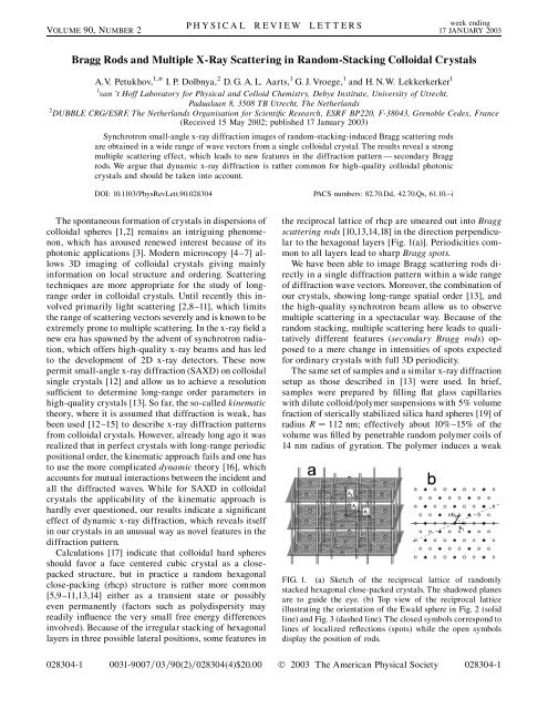
FIG. 1. (a) Sketch of the reciprocal lattice of randomly<br />
stacked hexagonal close-packed crystals. The shadowed planes<br />
are to guide the eye. (b) Top view of the reciprocal lattice<br />
illustrating the orientation of the Ewald sphere in Fig. 2 (solid<br />
line) and Fig. 3 (dashed line). The closed symbols correspond to<br />
lines of localized reflections (spots) while the open symbols<br />
display the position of rods.<br />
<strong>028304</strong>-1 0031-<strong>90</strong>07=03=<strong>90</strong>(2)=<strong>028304</strong>(4)$20.00 © <strong>2003</strong> The American <strong>Phys</strong>ical Society <strong>028304</strong>-1
VOLUME <strong>90</strong>, NUMBER 2<br />
PHYSICAL REVIEW LETTERS week ending<br />
17 JANUARY <strong>2003</strong><br />
FIG. 2. (a) Diffraction pattern measured for the crystal orientation<br />
corresponding to the Ewald sphere position illustrated<br />
by the solid line in Fig. 1(b). The direct beam is absorbed by a<br />
small beam stop in the middle of the detector. (b) Magnified<br />
view of the structure factor S q pro<strong>file</strong> within the area marked<br />
on panel (a). Arrows point to the secondary Bragg rods in<br />
between the sharp Bragg spots.<br />
depletion attraction between the silica spheres promoting<br />
spontaneous formation of large single crystals [13] (up<br />
to about 1 mm along the capillary walls and usually<br />
filling the 0.2 mm space between the flat walls of the<br />
capillary allowing for single-crystal diffraction measurements).<br />
The experiments were performed about 18 months<br />
after crystallization at the BM26 ‘‘DUBBLE’’ beam line<br />
at the European synchrotron radiation facility in<br />
Grenoble, France. Diffraction induced by an x-ray<br />
beam with wavelength 1:24 A was recorded at 8 m<br />
distance from the sample.<br />
The diffraction wave vector q k 0 k must lie on<br />
the so-called Ewald sphere since the wave vectors, k 0<br />
and k, of the incident and diffracted waves have the same<br />
length of 2 = . For colloidal crystals q k 0 so that<br />
diffraction is observed only at small angles and the diffraction<br />
vector q is practically normal to k 0 . The relevant<br />
part of the Ewald sphere is then extremely flat [20]. The<br />
diffraction vector q can be written in terms of three basis<br />
vectors q hb 1 kb 2 lb 3 introduced in Fig. 1(a),<br />
where h and k are integers due to the in-plane periodicity.<br />
Bragg spots are observed for h k divisible by 3 and<br />
integer values of l. For Bragg rods h k is not divisible<br />
by 3 and l is any real number.<br />
To directly image the Bragg rods on the detector, the<br />
Ewald sphere must produce a vertical cut through the<br />
reciprocal lattice of Fig. 1(a). In our flat capillaries one<br />
can find crystals with various orientations suggesting that<br />
the crystals did not nucleate at the glass wall but rather at<br />
the top interface of the concentrated sediment. In our<br />
previous work [13] a crystal with hexagonal planes<br />
(nearly) parallel to the flat capillary walls was studied.<br />
Now we have chosen a crystal with hexagonal planes<br />
making an angle of about 60 with respect to the capillary<br />
wall, enabling us to send the incoming x-ray beam<br />
parallel to the crystal planes. Careful orientation of the<br />
sample leads to a diffraction pattern I q as in Fig. 2(a).<br />
For this orientation the Ewald sphere cuts through the<br />
reciprocal lattice as shown by the solid line in Fig. 1(b).<br />
Figure 2(b) shows a magnified view of the structure factor<br />
S q I q =F q , where F q is the form factor determined<br />
from scattering in a dilute suspension of colloidal<br />
particles. X-ray scattering is observed on the detector<br />
along many lines, which originate from the Bragg rods<br />
of the reciprocal lattice and (each third line) from the<br />
localized spots of Fig. 1(a). However, some scattering is<br />
also observed in between the spots, which is not expected<br />
from the reciprocal lattice [Fig. 1(a)]. As discussed below,<br />
this scattering is a fingerprint of dynamic diffraction.<br />
Dynamic diffraction takes place when the interaction<br />
of the incident wave with the sample is no longer weak<br />
and one has to take into account that the diffracted waves<br />
deplete the incident beam and become in turn sources of<br />
secondary diffraction. The complexity of the theoretical<br />
modeling of such dynamic interactions raises significantly<br />
upon increasing the number of mutually interacting<br />
waves [21]. An even more complicated description<br />
is developed for visible light waves in photonic materials,<br />
where the refractive index contrast is large and the effect<br />
of diffraction is not weak even within one period of the<br />
structure [22,23]. Almost all theories deal with dynamic<br />
diffraction in crystals with full 3D periodicity.<br />
The effect of stacking faults lifting periodicity in one<br />
direction has been investigated in Ref. [22] and the photonic<br />
gap for visible light at normal and grazing incidence<br />
is found to broaden. Despite the significant progress of<br />
the theories, none of them can be applied to describe our<br />
data shown in Fig. 2. Here one has to consider not only<br />
multibeam diffraction into many Bragg spots, but also<br />
diffraction into a continuum of plane waves (induced via<br />
Bragg rods).<br />
To demonstrate that in our system diffraction enters the<br />
dynamic regime, one can estimate the strength of diffraction<br />
using the much simpler kinematic approach. The<br />
power dP sc d =d I 0 d of the wave scattered by a<br />
single colloidal particle into a small element d of the<br />
solid angle can then be described by the differential<br />
scattering cross section d =d r 2 0 Z2 F q , where I 0<br />
is the intensity of the incident wave at the position of the<br />
particle, r 0 e 2 = mc 2 is the Thompson radius, and Z is<br />
the excess number of electrons in the colloidal particle<br />
relative to an equivalent volume of solvent. The form<br />
factor is normalized such that F q ! 0 1, and it<br />
equals F q 9 sinqR qR cosqR 2 = qR 6 [24] for a<br />
sphere of radius R with a uniform distribution of the<br />
electron density. Under the conditions of our experiment<br />
(specific weight [19] of silica particles and the solvent<br />
cyclohexane are 1.7 and 0:77 g=ml, respectively, corresponding<br />
to a refractive index contrast of n 2:1<br />
10 6 for 10 keV x rays), R the total small-angle scattering<br />
cross section d =d d of one sphere is about<br />
<strong>028304</strong>-2 <strong>028304</strong>-2
VOLUME <strong>90</strong>, NUMBER 2<br />
PHYSICAL REVIEW LETTERS week ending<br />
17 JANUARY <strong>2003</strong><br />
12 nm 2 , i.e., only 3 10 4 of its geometrical cross section<br />
R 2 . Thus, a single particle only weakly interacts<br />
with the x-ray wave.<br />
However, the situation may change drastically if silica<br />
spheres form a single crystal possessing long-range order<br />
and the incident synchrotron x-ray beam provides conditions<br />
for coherent interference on large distances<br />
[13,25]. If for a sharp hkl reflection with h k divisible<br />
by 3 the Bragg condition is fulfilled (i.e., it is crossed by<br />
the Ewald sphere), the weak waves scattered by individual<br />
spheres interfere constructively and the diffracted<br />
power grows quadratically,<br />
P hkl L=L 2 hkl P 0 ; (1)<br />
with the distance L traveled by the beam. Here P 0 is the<br />
total power of the incident beam and the characteristic<br />
length L hkl is determined by [16]<br />
L<br />
2 2 hkl n 2 sph d =d hkl ; (2)<br />
where n sph is the number density of spherical particles.<br />
Assuming<br />
p<br />
a close-packed crystal structure with n sph<br />
1= 4 2 R<br />
3<br />
and collecting all the numbers in Eq. (2) for<br />
the lowest order (001) reflection seen in Fig. 2, one finds<br />
L 001 0:11 mm, i.e., about half the crystal size along<br />
the beam. Clearly, the diffracted power is then comparable<br />
to P 0 and diffraction switches from the kinematic to<br />
the dynamic regime. Interestingly, the dependence of<br />
L hkl on the particle size R cancels in Eq. (2) since<br />
d =d /R 6 and n sph / R 3 . Thus, for larger spheres<br />
dynamic diffraction can be observed for a smaller number<br />
of lattice periods. This factor leads to a principal<br />
difference between atomic and colloidal crystals in requirements<br />
of their perfectness to observe dynamic diffraction.<br />
While the former one requires perfect order over<br />
10 5 lattice constants, in the latter positional order over<br />
as little as a few hundreds of lattice periods can break up<br />
the kinematic description of x-ray diffraction.<br />
In contrast to a sharp reflection like (001), in a Bragg<br />
rod the scattering amplitudes of different hexagonal<br />
planes have additional stacking-dependent phase shifts.<br />
This significantly reduces the intensity diffracted in one<br />
particular direction and spreads the diffraction intensity<br />
along the Bragg rod. However, if the crystal possesses<br />
long-range in-plane order along the beam, the scattering<br />
amplitudes within each layer interfere constructively<br />
leading to a similar quadratic dependence of the scattered<br />
power with the distance L. For example, to evaluate the<br />
power P 0;1<br />
10l<br />
scattered into a piece of the low-order 10l<br />
rod between l 0 and l 1, one has to integrate the form<br />
factor F q together with the structure factor S rod q<br />
arising from interference between contributions of randomly<br />
stacked planes. One then finds that within the<br />
kinematic theory P 0;1<br />
10l<br />
grows as in Eq. (1) with the<br />
characteristic length L 0;1<br />
10l<br />
0:15 mm, i.e., the power<br />
scattered into the low-order 10l rod grows nearly as<br />
fast as the power diffracted into the (001) reflection since<br />
scattering along the whole rod is possible at this sample<br />
orientation. The estimates thus show that the incident<br />
x-ray beam is quickly depleted by scattering into the<br />
low-order spots and rods, which become in turn sources<br />
of strong secondary diffraction. Scattering into the 10l<br />
Bragg rods is able to compete with diffraction into the<br />
sharp (001) reflection and the appearance of secondary<br />
Bragg rods in Fig. 2 is therefore not surprising.<br />
To reduce the effect of multiple scattering via rods, a<br />
new crystal orientation has been chosen such that the<br />
incident x-ray beam is again parallel to the hexagonal<br />
planes of the crystal but the Ewald sphere intersects the<br />
reciprocal lattice differently [as sketched in Fig. 1(b) by<br />
the dashed line]. In this case the Ewald sphere misses<br />
many rods of low order but does cross the 32l and 64l<br />
rods. The scattering into these high-order rods is then<br />
much weaker, mainly due to the rapid decay of the form<br />
factor F q . Consequently, the probability of multiple<br />
scattering via Bragg rods is significantly reduced and<br />
the lines of diffraction spots 00l and 96l , l integer,<br />
are clearly visualized and free of scattering between the<br />
spots. This observation thus confirms the origin of the<br />
secondary Bragg rods in Fig. 2, which arise due to dynamic<br />
diffraction via the primary Bragg rods of Fig. 1(a).<br />
The estimates given above show that the presence of longrange<br />
order along the beam is essential for the transition<br />
into the dynamic regime. This intraplanar order is complementary<br />
to the interplanar long-range order found<br />
earlier [13]. Development of appropriate theory is, however,<br />
needed in order to exploit dynamic diffraction for a<br />
detailed quantitative structural characterization.<br />
The diffraction intensity along the Bragg rods 32l<br />
and 64l in Fig. 3 is seen to smoothly vary and display<br />
a periodic modulation with minima at integer values<br />
of l and broad maxima in between them. This pro<strong>file</strong> of<br />
the structure factor along the rod is typical for an rhcp<br />
crystal with stacking parameter 0:5. Calculations<br />
[10,13,14,18] show that for
VOLUME <strong>90</strong>, NUMBER 2<br />
PHYSICAL REVIEW LETTERS week ending<br />
17 JANUARY <strong>2003</strong><br />
are narrower and new maxima develop at integer<br />
values of l. For >0:6 the broad maxima split into<br />
two. Note that diffraction into the (001) spots in Fig. 3<br />
is still very strong and might affect the distribution of<br />
the x-ray power over the sharp reflections as well as along<br />
the rods. However, since this can change the wave vector<br />
only by an integer times the b 3 vector, these multiple<br />
scattering events should not significantly change the<br />
intensity pro<strong>file</strong> within one period of the structure factor<br />
along the rod, which is equal to b 3 .<br />
In conclusion, this <strong>Lett</strong>er presents the first direct images<br />
of Bragg scattering rods in small-angle synchrotron<br />
x-ray diffraction obtained from a colloidal single crystal<br />
still retaining its random-stacking structure 18 months<br />
after crystallization. Moreover, we observed a strong<br />
effect of multiple scattering, which reveals itself in the<br />
diffraction pattern as secondary Bragg rods. Simple estimates<br />
show that, in contrast to common belief, dynamic<br />
x-ray diffraction should be rather typical for crystals<br />
consisting of highly ordered (sub)micrometer colloidal<br />
spheres and has to be taken into account. In our previous<br />
work [13], the assignment of the rhcp structure for oneyear-old<br />
crystals was based on comparison of intensities<br />
of different reflections, which might be somewhat affected<br />
by the dynamic diffraction. The present results<br />
unambiguously confirm the rhcp structure because multiple<br />
scattering cannot broaden sharp reflections into<br />
Bragg rods. Since dynamic diffraction is likely to redistribute<br />
the diffracted power towards weaker reflections,<br />
one should be aware that the amplitude of particle excursions<br />
evaluated from the Debye-Waller factor [15] and<br />
the spatial extent of the positional order [13] in colloidal<br />
crystals could be underestimated.<br />
The authors thank David van der Beek for his assistance<br />
in the x-ray diffraction experiment, Alexander<br />
Moroz and Wim Bras for useful discussions, and the<br />
Netherlands Organisation for the Advancement of<br />
Research (NWO) for providing us with the possibility<br />
of performing measurements at DUBBLE.<br />
*Corresponding author.<br />
Electronic address: a.v.petukhov@chem.uu.nl<br />
[1] P. N. Pusey and W. van Megen, Nature (London) 320,340<br />
(1986).<br />
[2] Zh. Cheng et al., <strong>Phys</strong>. <strong>Rev</strong>. <strong>Lett</strong>., 88, 015501 (2002).<br />
[3] Y. A. Vlasov, X.-Z. Bo, J. C. Sturm, and D. J. Norris,<br />
Nature (London) 414, 289 (2001); A. Blanco et al.,<br />
Nature (London) 405, 437 (2000).<br />
[4] A. van Blaaderen, R. Ruel, and P. Wiltzius, Nature<br />
(London) 385, 321 (1997).<br />
[5] N. A. M. Verhaegh, J. S. van Duijneveldt, A. van<br />
Blaaderen, and H. N.W. Lekkerkerker, J. Chem. <strong>Phys</strong>.<br />
102, 1416 (1995).<br />
[6] U. Gasser et al., Science 292, 258 (2001).<br />
[7] M. S. Elliot, S. B. Haddon, and W. C. K. Poon, J. <strong>Phys</strong>.<br />
Condens. Matter 13, L553 (2001).<br />
[8] S. I. Henderson and W. van Megen, <strong>Phys</strong>. <strong>Rev</strong>. <strong>Lett</strong>. 80,<br />
877 (1998).<br />
[9] J. Zhu et al., Nature (London) 387, 883 (1997).<br />
[10] Ch. Dux and H. Versmold, <strong>Phys</strong>. <strong>Rev</strong>. <strong>Lett</strong>. 78, 1811<br />
(1997).<br />
[11] W. K. Kegel and J. K. G. Dhont, J. Chem. <strong>Phys</strong>. 112, 3431<br />
(2000).<br />
[12] W. Vos, M. Megens, C. M. van Kats, and P. Bosecke,<br />
Langmuir 13, 6004 (1997); J. E. G. J. Wijnhoven,<br />
L. Bechger, and W. L. Vos, Chem. Mater. 13, 4486 (2001).<br />
[13] A.V. Petukhov et al., <strong>Phys</strong>. <strong>Rev</strong>. <strong>Lett</strong>. 88, 208301 (2002).<br />
[14] H. Versmold et al., J. Chem. <strong>Phys</strong>. 116, 2658 (2002).<br />
[15] M. Megens and W. L. Vos, <strong>Phys</strong>. <strong>Rev</strong>. <strong>Lett</strong>. 86, 4855<br />
(2001).<br />
[16] R.W. James, The Optical Principles of the Diffraction of<br />
X-Rays (Cornell University Press, Ithaca, NY, 1965);<br />
J. M. Cowley, Diffraction <strong>Phys</strong>ics (North-Holland,<br />
Amsterdam, 1981).<br />
[17] P. G. Bolhuis, D. Frenkel, S.-C. Mau, and D. A. Huse,<br />
Nature (London) 388, 235 (1997).<br />
[18] O. S. Edwards and H. Lipson, Proc. R. Soc. London A<br />
180, 268 (1941); A. J. C. Wilson, ibid. 180, 277 (1941);<br />
X-Ray Optics (Methuen & Co. Ltd., London, 1949).<br />
[19] N. A. M. Verhaegh, D. Asnaghi, and H. N.W. Lekkerkerker,<br />
<strong>Phys</strong>ica (Amsterdam) 264A, 64 (1999); E. H. A.<br />
de Hoog et al., Langmuir 17, 5486 (2001).<br />
[20] The extremely small but finite curvature of the Ewald<br />
sphere can lead to asymmetry of the diffraction pattern<br />
for highly ordered crystals, which are slightly tilted from<br />
a low-index orientation [13]. Although we have tried to<br />
minimize the tilt angle, some asymmetry can still be<br />
seen in Figs. 2 and 3 , which points to the presence of<br />
long-range order within hexagonal planes [13].<br />
[21] Q. Shen, in Methods in Materials Research, editedby<br />
E. Kaufmann et al. (John Wiley & Sons, New York,<br />
2000).<br />
[22] V. Yannopapas, N. Stefanou, and A. Modinos, <strong>Phys</strong>. <strong>Rev</strong>.<br />
<strong>Lett</strong>., 86, 4811 (2001).<br />
[23] A. Moroz, <strong>Phys</strong>. <strong>Rev</strong>. <strong>Lett</strong>., 83, 5274 (1999).<br />
[24] L. A. Feigin and D. I. Svergun, Structure Analysis by<br />
Small-Angle X-Ray and Neutron Scattering (Plenum<br />
Press, New York, 1987).<br />
[25] The requirements of crystal quality and the beam coherence<br />
are crucial in the longitudinal direction (along the<br />
beam) while they are less important in the transverse<br />
direction [13].<br />
<strong>028304</strong>-4 <strong>028304</strong>-4