Download Supporting Information (PDF)
Download Supporting Information (PDF)
Download Supporting Information (PDF)

Create successful ePaper yourself
Turn your PDF publications into a flip-book with our unique Google optimized e-Paper software.
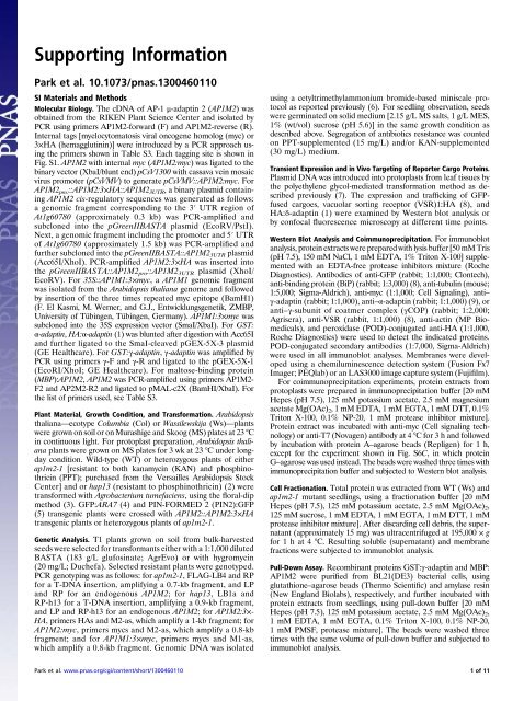
<strong>Supporting</strong> <strong>Information</strong><br />
Park et al. 10.1073/pnas.1300460110<br />
SI Materials and Methods<br />
Molecular Biology. The cDNA of AP-1 μ-adaptin 2 (AP1M2) was<br />
obtained from the RIKEN Plant Science Center and isolated by<br />
PCR using primers AP1M2-forward (F) and AP1M2-reverse (R).<br />
Internal tags [myelocytomatosis viral oncogene homolog (myc) or<br />
3×HA (hemagglutinin)] were introduced by a PCR approach using<br />
the primers shown in Table S3. Each tagging site is shown in<br />
Fig. S1. AP1M2 with internal myc (AP1M2:myc) was ligated to the<br />
binary vector (XbaI/blunt end) pCsV1300 with cassava vein mosaic<br />
virus promoter (pCsVMV) to generate pCsVMV::AP1M2:myc. For<br />
AP1M2 pro: ::AP1M2:3×HA::AP1M2 3UTR , a binary plasmid containing<br />
AP1M2 cis-regulatory sequences was generated as follows:<br />
a genomic fragment corresponding to the 3′ UTR region of<br />
At1g60780 (approximately 0.3 kb) was PCR-amplified and<br />
subcloned into the pGreenIIBASTA plasmid (EcoRV/PstI).<br />
Next, a genomic fragment including the promoter and 5′ UTR<br />
of At1g60780 (approximately 1.5 kb) was PCR-amplified and<br />
further subcloned into the pGreenIIBASTA::AP1M2 3UTR plasmid<br />
(Acc65I/XhoI). PCR-amplified AP1M2:3×HA was inserted into<br />
the pGreenIIBASTA::AP1M2 pro ::AP1M2 3UTR plasmid (XhoI/<br />
EcoRV). For 35S::AP1M1:3×myc, aAP1M1 genomic fragment<br />
was isolated from the Arabidopsis thaliana genome and followed<br />
by insertion of the three times repeated myc epitope (BamH1)<br />
(F. El Kasmi, M. Werner, and G.J., Entwicklungsgenetik, ZMBP,<br />
University of Tübingen, Tübingen, Germany). AP1M1:3×myc was<br />
subclonedintothe35Sexpressionvector(SmaI/XbaI).ForGST:<br />
α-adaptin, HA:α-adaptin (1) was blunted after digestion with Acc65I<br />
and further ligated to the SmaI-cleaved pGEX-5X-3 plasmid<br />
(GE Healthcare). For GST:γ-adaptin, γ-adaptin was amplified by<br />
PCR using primers γ-F and γ-R and ligated to the pGEX-5X-1<br />
(EcoRI/XhoI; GE Healthcare). For maltose-binding protein<br />
(MBP):AP1M2, AP1M2 was PCR-amplified using primers AP1M2-<br />
F2andAP2M2-R2andligatedtopMAL-c2X(BamHI/XbaI).For<br />
the list of primers used, see Table S3.<br />
Plant Material, Growth Condition, and Transformation. Arabidopsis<br />
thaliana—ecotype Columbia (Col) or Wassilewskija (Ws)—plants<br />
were grown on soil or on Murashige and Skoog (MS) plates at 23 °C<br />
in continuous light. For protoplast preparation, Arabidopsis thaliana<br />
plants were grown on MS plates for 3 wk at 23 °C under longday<br />
condition. Wild-type (WT) or heterozygous plants of either<br />
ap1m2-1 [resistant to both kanamycin (KAN) and phosphinothricin<br />
(PPT); purchased from the Versailles Arabidopsis Stock<br />
Center] and or hap13 (resistant to phosphinothricin) (2) were<br />
transformed with Agrobacterium tumefaciens, using the floral-dip<br />
method (3). GFP:ARA7 (4) and PIN-FORMED 2 (PIN2):GFP<br />
(5) transgenic plants were crossed with AP1M2::AP1M2:3×HA<br />
transgenic plants or heterozygous plants of ap1m2-1.<br />
Genetic Analysis. T1 plants grown on soil from bulk-harvested<br />
seeds were selected for transformants either with a 1:1,000 diluted<br />
BASTA (183 g/L glufosinate; AgrEvo) or with hygromycin<br />
(20 mg/L; Duchefa). Selected resistant plants were genotyped.<br />
PCR genotyping was as follows: for ap1m2-1, FLAG-LB4 and RP<br />
for a T-DNA insertion, amplifying a 0.7-kb fragment, and LP<br />
and RP for an endogenous AP1M2; for hap13, LB1a and<br />
RP-h13 for a T-DNA insertion, amplifying a 0.9-kb fragment,<br />
and LP and RP-h13 for an endogenous AP1M2; forAP1M2:3×-<br />
HA, primers HAs and M2-as, which amplify a 1-kb fragment; for<br />
AP1M2:myc, primers mycs and M2-as, which amplify a 0.8-kb<br />
fragment; and for AP1M1:3×myc, primers mycs and M1-as,<br />
which amplify a 0.8-kb fragment. Genomic DNA was isolated<br />
using a cetyltrimethylammonium bromide-based miniscale protocol<br />
as reported previously (6). For seedling observation, seeds<br />
were germinated on solid medium [2.15 g/L MS salts, 1 g/L MES,<br />
1% (wt/vol) sucrose (pH 5.6)] in the same growth condition as<br />
described above. Segregation of antibiotics resistance was counted<br />
on PPT-supplemented (15 mg/L) and/or KAN-supplemented<br />
(30 mg/L) medium.<br />
Transient Expression and in Vivo Targeting of Reporter Cargo Proteins.<br />
Plasmid DNA was introduced into protoplasts from leaf tissues by<br />
the polyethylene glycol-mediated transformation method as described<br />
previously (7). The expression and trafficking of GFPfused<br />
cargoes, vacuolar sorting receptor (VSR)1:HA (8), and<br />
HA:δ-adaptin (1) were examined by Western blot analysis or<br />
by confocal fluorescence microscopy at different time points.<br />
Western Blot Analysis and Coimmunoprecipitation. For immunoblot<br />
analysis, protein extracts were prepared with lysis buffer [50 mM Tris<br />
(pH 7.5), 150 mM NaCl, 1 mM EDTA, 1% Triton X-100] supplemented<br />
with an EDTA-free protease inhibitors mixture (Roche<br />
Diagnostics). Antibodies of anti-GFP (rabbit; 1:1,000; Clontech),<br />
anti-binding protein (BiP) (rabbit; 1:3,000) (8), anti-tubulin (mouse;<br />
1:5,000; Sigma-Aldrich), anti-myc (1:1,000; Cell Signaling), anti–<br />
γ-adaptin (rabbit; 1:1,000), anti–α-adaptin (rabbit; 1:1,000) (9), or<br />
anti–γ-subunit of coatmer complex (γCOP) (rabbit; 1:2,000;<br />
Agrisera), anti-VSR (rabbit, 1:1,000) (8), anti-actin (MP Biomedicals),<br />
and peroxidase (POD)-conjugated anti-HA (1:1,000,<br />
Roche Diagnostics) were used to detect the indicated proteins.<br />
POD-conjugated secondary antibodies (1:7,000, Sigma-Aldrich)<br />
were used in all immunoblot analyses. Membranes were developed<br />
using a chemiluminescence detection system (Fusion Fx7<br />
Imager; PEQlab) or an LAS3000 image capture system (Fujifilm).<br />
For coimmunoprecipitation experiments, protein extracts from<br />
protoplasts were prepared in immunoprecipitation buffer [20 mM<br />
Hepes (pH 7.5), 125 mM potassium acetate, 2.5 mM magnesium<br />
acetate Mg(OAc) 2 ,1mMEDTA,1mMEGTA,1mMDTT,0.1%<br />
Triton X-100, 0.1% NP-20, 1 mM protease inhibitor mixture].<br />
Protein extract was incubated with anti-myc (Cell signaling technology)<br />
or anti-T7 (Novagen) antibody at 4 °C for 3 h and followed<br />
by incubation with protein A–agarose beads (Repligen) for 1 h,<br />
except for the experiment shown in Fig. S6C, in which protein<br />
G–agarose was used instead. The beads were washed three times with<br />
immunoprecipitation buffer and subjected to Western blot analysis.<br />
Cell Fractionation. Total protein was extracted from WT (Ws) and<br />
ap1m2-1 mutant seedlings, using a fractionation buffer [20 mM<br />
Hepes (pH 7.5), 125 mM potassium acetate, 2.5 mM Mg(OAc) 2 ,<br />
125mMsucrose,1mMEDTA,1mMEGTA,1mMDTT,1mM<br />
protease inhibitor mixture]. After discarding cell debris, the supernatant<br />
(approximately 15 mg) was ultracentrifuged at 195,000 × g<br />
for 1 h at 4 °C. Resulting soluble (supernatant) and membrane<br />
fractions were subjected to immunoblot analysis.<br />
Pull-Down Assay. Recombinant proteins GST:γ-adaptin and MBP:<br />
AP1M2 were purified from BL21(DE3) bacterial cells, using<br />
glutathione–agarose beads (Thermo Scientific) and amylase resin<br />
(New England Biolabs), respectively, and further incubated with<br />
protein extracts from seedlings, using pull-down buffer [20 mM<br />
Hepes (pH 7.5), 125 mM potassium acetate, 2.5 mM Mg(OAc) 2 ,<br />
1 mM EDTA, 1 mM EGTA, 0.1% Triton X-100, 0.1% NP-20,<br />
1 mM PMSF, protease mixture]. The beads were washed three<br />
times with the same volume of pull-down buffer and subjected to<br />
immunoblot analysis.<br />
Park et al. www.pnas.org/cgi/content/short/1300460110<br />
1of11
Semiquantitative RT-PCR Analysis of Transcripts. To determine the<br />
transcript level in the mutant plants, total RNA was isolated from<br />
the 7-d-old seedlings grown on plates using an RNeasy Mini Kit<br />
(Qiagen). Total RNA (2 μg) was reverse-transcribed using<br />
SuperScript II (Invitrogen) with random primers mixed with oligo<br />
(dT) primers. Equal amount of cDNA was used for PCR with<br />
gene specific primers listed in Table S3. PCR products were analyzed<br />
by ethidium bromide staining.<br />
Anti–γ-Adaptin Antibody Generation. To generate anti–γ-adaptin<br />
antibody, the full-length γ-adaptin was expressed as a fusion<br />
protein with MBP using the pMAL-c2 vector (New England<br />
Biolabs). Recombinant MBP:γ-adaptin was expressed in E. coli<br />
BL21(DE3) strain and purified using amylose resin (New England<br />
Biolabs). MBP:γ-adaptin was used to immunize rabbits and antibody<br />
was affinity-purified using recombinant proteins. To confirm<br />
the specificity of the anti–γ-adaptin antibody, protein extracts<br />
from protoplasts transformed with HA:γ-adaptin were used in<br />
Western blot analysis (1).<br />
Immunohistochemistry. Immunostaining of root tissues was performed<br />
as reported previously (10). In brief, 5-d-old seedlings were<br />
fixed in 4% (wt/vol) paraformaldehyde and labeled with the indicated<br />
antibodies. For immunofluorescence, anti-HA (mouse;<br />
1:1,000; BAbCO), anti-γCOP (rabbit; 1:600; Agrisera), anti-syntaxin<br />
of plants 61 (SYP61) (rabbit; 1:600; a gift from N. Raikhel, University<br />
of Califonia Riverside, Riverside, CA), anti-clathrin heavy<br />
chain (CHC) (rabbit; 1:1,000) (11), anti-KNOLLE (rabbit; 1:4,000)<br />
(10), anti-tubulin (rat; 1:1,000; Abcam), or anti-PIN2 (rabbit, 1:600)<br />
(5) antibodies were used. As the secondary antibody, FITC-labeled<br />
anti-mouse IgG (1:600; Dianova), Cy3-labeled anti-rabbit IgG<br />
(1:600; Dianova), Alexa488-labeled anti-rabbit IgG (1:600; Invitrogen),<br />
or FITC-labeled anti-rat IgG (1:600; Dianova) were used.<br />
Chemical Treatment. Five-day-old seedlings were treated with<br />
50 μM brefeldin A (BFA) (50 mM stock solution in 1:1 DMSO/<br />
EtOH; Invitrogen) or 20 μM wortmannin (20 mM stock solution<br />
in DMSO) for 1 h, followed by fixation or immediate observation.<br />
For FM4-64, 2 μM FM4-64 was added to 5-d-old seedlings<br />
(4 mM stock solution in H 2 O; Invitrogen). For simultaneous<br />
treatment with BFA and FM4-64, BFA was followed by addition<br />
of FM4-64. For BFA washout, BFA-treated seedlings for 1 h<br />
were transferred to new liquid medium and fixed 30 min after the<br />
transfer. For concanamycin A (ConcA) treatment, protoplasts<br />
isolated from WT seedlings were treated at indicated concentrations<br />
(1 mM stock solution in DMSO, Invitrogen). For cycloheximide<br />
(CHX) treatment, 50 μM CHX was added to<br />
seedlings for 1 h (50 mM stock solution in H 2 O; Sigma-Aldrich).<br />
Ultrastructure Analysis. Root tips of 5-d-old seedlings were highpressure-frozen<br />
in 1-hexadecene (Merck Sharp and Dohme)<br />
using a Bal-Tec HPM 010 high-pressure freezer (Balzers). Frozen<br />
samples were freeze-substituted in acetone containing 2.5% (wt/<br />
vol) osmium tetroxide (2 d at −90 °C, 6 h at −60 °C, 6 h at −30 °C,<br />
and 1 h at 0 °C) and finally embedded in epoxy resin (Roth).<br />
Ultrathin sections were stained with 2% (wt/vol) uranyl acetate in<br />
50% ethanol for 10–20 min and lead citrate (3–5 min).<br />
Software. Sequences were analyzed using Vector NTI (Invitrogen)<br />
or CLC DNA Workbench 5. Images were processed using<br />
Adobe Photoshop CS3 and Adobe Illustrator CS3. Quantification<br />
of confocal laser scanning microscopy (CLSM) images was<br />
done using Pearson and Spearman correlation coefficients (PSC)<br />
colocalization plugin of ImageJ [National Institutes of Health<br />
(NIH)] after adjusting the background level to 5 in five independently<br />
taken images according to instructions in the paper<br />
by French et al. (12). Signal intensity in immunoblot analyses<br />
were determined using ImageJ (NIH). Pixel count in live seedlings<br />
was done using Leica LAS in three or four independently<br />
taken images.<br />
1. Lee GJ, et al. (2007) EpsinR2 interacts with clathrin, adaptor protein-3, AtVTI12, and<br />
phosphatidylinositol-3-phosphate. Implications for EpsinR2 function in protein trafficking<br />
in plant cells. Plant Physiol 143(4):1561–1575.<br />
2. Johnson MA, et al. (2004) Arabidopsis hapless mutations define essential gametophytic<br />
functions. Genetics 168(2):971–982.<br />
3. Clough SJ, Bent AF (1998) Floral dip: A simplified method for Agrobacteriummediated<br />
transformation of Arabidopsis thaliana. Plant J 16(6):735–743.<br />
4. Lee GJ, Sohn EJ, Lee MH, Hwang I (2004) The Arabidopsis rab5 homologs rha1 and<br />
ara7 localize to the prevacuolar compartment. Plant Cell Physiol 45(9):1211–1220.<br />
5. Abas L, et al. (2006) Intracellular trafficking and proteolysis of the Arabidopsis auxinefflux<br />
facilitator PIN2 are involved in root gravitropism. Nat Cell Biol 8(3):249–256.<br />
6. Assaad FF, Huet Y, Mayer U, Jürgens G (2001) The cytokinesis gene KEULE encodes<br />
a Sec1 protein that binds the syntaxin KNOLLE. J Cell Biol 152(3):531–543.<br />
7. Jin JB, et al. (2001) A new dynamin-like protein, ADL6, is involved in trafficking from the<br />
trans-Golgi network to the central vacuole in Arabidopsis. Plant Cell 13(7):1511–1526.<br />
8. Kim H, Park M, Kim SJ, Hwang I (2005) Actin filaments play a critical role in vacuolar<br />
trafficking at the Golgi complex in plant cells. Plant Cell 17(3):888–902.<br />
9. Song K, et al. (2012) An A/ENTH domain-containing protein functions as an adaptor<br />
for clathrin-coated vesicles on the growing cell plate in Arabidopsis root cells. Plant<br />
Physiol 159(3):1013–1025.<br />
10. Lauber MH, et al. (1997) The Arabidopsis KNOLLE protein is a cytokinesis-specific<br />
syntaxin. J Cell Biol 139(6):1485–1493.<br />
11. Song J, Lee MH, Lee GJ, Yoo CM, Hwang I (2006) Arabidopsis EPSIN1 plays an<br />
important role in vacuolar trafficking of soluble cargo proteins in plant cells via<br />
interactions with clathrin, AP-1, VTI11, and VSR1. Plant Cell 18(9):2258–2274.<br />
12. French AP, Mills S, Swarup R, Bennett MJ, Pridmore TP (2008) Colocalization of<br />
fluorescent markers in confocal microscope images of plant cells. Nat Protoc 3(4):619–628.<br />
Park et al. www.pnas.org/cgi/content/short/1300460110<br />
2of11
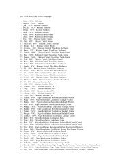
Fig. S1. Alignment of amino acid sequences of the Arabidopsis mu adaptins. Amino acid sequences of AP1M1 (At1g10730), AP1M2 (At1g60780), AP2M<br />
(At5g46630), AP3M (At1g56590), and AP4M (At4g24550) were compared using CLUSTAL Omega. Note that AP1M1 and AP1M2 share 90% sequence identity<br />
(analyzed in CLUSTAL Omega). Red and green triangles indicate the insertion sites of three times repeated HA (3×HA) and myc epitope in AP1M2, respectively.<br />
Blue triangle indicates the insertion site of three times repeated myc (3×myc) epitope in AP1M1.<br />
Park et al. www.pnas.org/cgi/content/short/1300460110<br />
3of11
Fig. S2. Genetic analysis of T-DNA insertional AP1M2 mutants and phenotype of mature ap1m2-1 mutant grown in a closed container. (A and B) Reciprocal<br />
crosses of ap1m2 heterozygotes with the respective WT (Ws, Wassilewskija or Col, Columbia) revealed reduced transmission of ap1m2 mutant alleles ap1m2-1<br />
[KAN resistance (KAN R )] (A) orhap13 [PPT resistance (PPT R )] (B) through the pollen, resulting in only 9% or 5% ap1m2 homozygous seedlings upon selfing of<br />
heterozygotes, respectively. N, total number of seedlings analyzed. (C) Complementation tests. Transgenic plants each carrying a different single T-DNA insertion<br />
of CsVMV::AP1M2:myc (Hyg R )orAP1M2::AP1M2:3×HA (PPT R ) were crossed with ap1m2-1 heterozygous plants. F2 progenies (N, number of seedlings)<br />
were analyzed for the phenotypic segregation and genotyped. Note that 2% ap1m2-1 homozygous seedlings corresponds to the expected value if 75% of<br />
approximately 9% ap1m2-1 homozygous seedlings from selfed ap1m2-1 heterozygous plants are rescued by the transgene (A). R, resistant; S, sensitive. (D and E)<br />
Immunoblot analysis of CsVMV::AP1M2:myc and AP1M2::AP1M2:3×HA plants. (D) Two independent lines of CsVMV::AP1M2:myc harboring a single T-DNA<br />
insertion were crossed with ap1m2-1 heterozygous plants. Total protein of nontransformed (N), ap1m2-1 homozygous, and rescued seedlings was subjected to<br />
immunoblot analysis with anti-myc or anti-BiP antiserum. T, transgene. (E) Total protein of nontransformed (N) and T1 plants harboring AP1M2::AP1M2:3×HA<br />
was subjected to immunoblot analysis with anti-HA or anti-tubulin antiserum. Plants bearing a single T-DNA insertion (#1 and #4) were crossed with the<br />
ap1m2-1 heterozygous plants for complementation test (C). Molecular mass markers are shown at the left. (F) Semiquantitative RT-PCR analysis to compare<br />
the level of transgenically expressed epitope-tagged AP1M2 in rescued mutant lines to that of endogenous AP1M2 in WT. Note that the level of AP1M2:3×HA<br />
and AP1M2:myc is comparable to that of WT. 18S rRNA (18S) was used as loading control. Molecular mass markers are shown at the left. (G) Phenotype of<br />
mature ap1m2-1 mutant grown in a closed container. Note that the mutant presents ill-developed anthers. (Scale bar: 1 cm.)<br />
Park et al. www.pnas.org/cgi/content/short/1300460110<br />
4of11
Fig. S3. Expression profile of the Arabidopsis mu adaptins. Expression levels of AP1M1 (magenta), AP1M2 (turquoise), AP2M (green), AP3M (red), and AP4M<br />
(blue) were compared using the AtGenExpress Visualization Tool (www.weigelworld.org/resources/microarray/AtGenExpress) (1).<br />
1. Schmid M, et al. (2005) A gene expression map of Arabidopsis thaliana development. Nat Genet 37(5):501–506.<br />
Fig. S4. T-DNA insertion AP1M1 mutant does not show any discernible morphological change. (A) Diagram of the T-DNA insertion in AP1M1, ap1m1<br />
(SALK_027224). Primers used in Fig. 1E are indicated. (B) Images of ap1m1 homozygous seedlings grown on an MS plate for 7 d. Note that seedlings of the<br />
ap1m1 mutant look normal, in contrast to the ap1m2-1 mutant. (Scale bar: 1 cm.)<br />
Park et al. www.pnas.org/cgi/content/short/1300460110<br />
5of11
Fig. S5. Localization of tagged AP1M2 in seedling roots and the interaction of γ-adaptin–containing AP1 complex with clathrin and VSR. (A) After BFA<br />
treatment, AP1M2:3×HA in ap1m2-1 homozygous background was counterstained with anti-ARF1 (Upper) or anti-γCOP (Lower) antiserum. Note that<br />
AP1M2:3×HA colocalized with ARF1 in endosomal BFA compartments surrounded by γCOP-positive Golgi stacks. (B and C) AP1M2:3×HA in ap1m2-1 homozygous<br />
background was counterstained with anti-SYP61 (B) or anti-CHC (C) antiserum. Note that the tagged AP1M2 does not accumulate at the cell plate,<br />
which is positively labeled with anti-SYP61 antiserum (B). Note partial colocalization of AP1M2:3×HA with clathrin (C). (D) AP1M2:3×HA and the multivesicular<br />
body (MVB) marker GFP:ARA7/RAB-F2b were examined for colocalization. AP1M2:3×HA was detected by immunostaining with anti-HA antibody and GFP<br />
signals of GFP:ARA7/RAB-F2b were observed directly. AP1M2:3×HA-positive endosomes were insensitive to wortmannin, in contrast to the swollen ARA7/<br />
RabF2b-labeled prevacuolar compartment/multivesicular body/late endosome (PVC/MVB/LE) (white arrows). The extent of colocalization was quantified using<br />
the linear Pearson correlation coefficient (r p ) and the nonlinear Spearman rank correlation coefficient (r s ), and the resulting scatterplot images are shown on<br />
the right (C and D). A value of 1.0 means complete colocalization of two fluorescent signals. Note partial colocalization of AP1M2:3×HA with clathrin, as<br />
quantified by r p /r s = 0.71/0.53 and no localization of AP1M2:3×HA at the PVC/MVB labeled with GFP:ARA7/RAB-F2b, as indicated by r p /r s = 0.19/0.34. Insets<br />
show boxed areas at higher magnification. (Scale bars: 5 μm.) (E) Purified recombinant GST or GST:γ-adaptin protein was precipitated with glutathione beads<br />
after incubation with plant extracts from WT seedlings and followed by immunoblot analysis. Note that CHC is detected in the precipitate but not in the<br />
control precipitate done with GST alone. The Coomassie-stained membrane (CBS) showed bands of GST (asterisk) and GST:γ-adaptin (double asterisks). (F) Pull<br />
down of HA-tagged VSR1 from protoplasts, using recombinant MBP fused to AP1M2; MBP alone was used as control. The CBS showed bands of MBP (asterisk)<br />
and MBP:AP1M2 (double asterisks). Molecular mass markers are indicated in kilodaltons (kDa).<br />
Park et al. www.pnas.org/cgi/content/short/1300460110<br />
6of11
Fig. S6. Specificity determination of anti–γ-adaptin antiserum and no interaction of AP1M2 with δ-adaptin. (A) Total protein extracts from protoplasts<br />
transformed with HA:γ-adaptin (HA:γ) were analyzed by immunoblotting using anti-HA antibody and anti–γ-adaptin antiserum. N, nontransformed protoplasts.<br />
Asterisk, endogenous γ-adaptin; double asterisks, transiently expressed HA:γ-adaptin. A, Lower shows the CBS. (B) Fractionation analysis. Total protein<br />
was extracted from WT (Con) or ap1m2-1 mutant seedlings, followed by microultracentrifugation. Note that total protein level of γ-adaptin in ap1m2-1 mutant<br />
is highly reduced by approximately 80% compared with the WT and that less γ-adaptin is detected in the membrane (M) fraction of the ap1m2-1 mutant<br />
compared with WT. Actin and VSR were detected as controls of soluble (S) and membrane fractions, respectively. Rel. of adaptins, relative signal intensity: total<br />
(T) signal of ap1m2-1 is expressed as percentage of control (Con, WT; 100%). The relative signal intensity of soluble and membrane fraction in each case is<br />
expressed as percentage of the total signal. (C) Protein extract [input (IN)] from rescued protoplasts (CsVMV::AP1M2:myc in ap1m2-1 mutant background) that<br />
had been transformed with 35S::HA:δ-adaptin was subjected to immunoprecipitation (IP) with anti-myc antibody and the immunoprecipitate was analyzed by<br />
immunoblotting using anti-HA, anti-myc, or anti–γ-adaptin antiserum. Note that HA:δ-adaptin is not immunoprecipitated with AP1M2, in contrast to<br />
γ-adaptin. Molecular mass markers are indicated in kilodaltons (kDa).<br />
Park et al. www.pnas.org/cgi/content/short/1300460110<br />
7of11
Fig. S7. Vacuolar and secretory pathways are impaired in the ap1m2 mutants. (A) Trafficking of vacuolar cargo. Protoplasts from nontransformed, ap1m2-1,<br />
and two independent rescued plants (CsVMV::AP1M2:myc in ap1m2-1) were transformed with Sporamin:GFP (Spo:GFP). At 24 or 36 h after transformation,<br />
total protein extracts from protoplasts were subjected to immunoblot analysis with anti-GFP antiserum. Asterisk, full-length Spo:GFP; closed triangle, processed<br />
Spo:GFP. (B) Trafficking of secretory cargo. Secretory GFP (secGFP) was transformed into protoplasts from the indicated plants. At 24 h after transformation,<br />
total protein extracts from protoplasts (C) and the incubation medium (M) were subjected to immunoblot analysis with anti-GFP antiserum. BiP was used as<br />
a control for nonspecific leaking of cellular proteins. (C) Protoplasts transformed with Spo:GFP or AtβFructosidase4:GFP (AtβFruc4:GFP) were treated with<br />
ConcA at the indicated concentrations. At 24 h after transformation, total protein extracts from protoplasts were subjected to immunoblot analysis with anti-<br />
GFP antiserum. Asterisk, full-length Spo:GFP; double asterisks, full length AtβFruc4:GFP; closed triangle, processed forms of AtβFruc4:GFP and Spo:GFP. Note<br />
that ConcA treatment inhibits the delivery of Spo:GFP, but not of AtβFruc4:GFP, to the vacuole. Both precursor and mature Spo:GFP are secreted from ConcAtreated<br />
protoplasts into the medium. Endoplasmic reticulum (ER)-localized BiP was used as control for intact cells. (D) Localization of soluble vacuolar and<br />
secretory proteins in ap1m2-1 mutant protoplasts. Protoplasts from WT or ap1m2-1 mutant plants were transformed with the indicated constructs and<br />
subcellular localization of the proteins was examined. Note that AALP:GFP and invertase:GFP accumulate in the ER, as indicated by their colocalization with<br />
BiP:mCherry:HDEL in the ap1m2-1 protoplasts. (Scale bars: 10 μm.)<br />
Park et al. www.pnas.org/cgi/content/short/1300460110<br />
8of11
Fig. S8. Endocytosis and recycling of PIN2 occur normally in the ap1m2 mutants but vacuolar traffic of PIN2:GFP is impaired in the mutant. (A and B) No<br />
obvious defect in endocytosis in ap1m2-1 mutant plants. (A) WT, ap1m2-1, and hap13 mutant seedlings were treated with FM4-64 alone or together with BFA.<br />
Note intracellular FM4-64–positive endosomes in the seedling roots of all three genotypes. As in WT, BFA compartments were observed in ap1m2 seedling<br />
roots after BFA treatment, indicating that endocytosis of FM4-64 occurs normally in ap1m2 mutants. (B) Measurement of FM4-64 internalization in WT and<br />
ap1m2-1 seedling roots. Pixels of intracellular FM4-64 signal taken 5 min after uptake were counted from three different images. The mean value of counted<br />
pixels per square micron was calculated. Error bars indicate SD. (C) Normal recycling of PIN2 in ap1m2-1 plants. WT and ap1m2-1 homozygous seedlings were<br />
fixed and stained with anti-PIN2 antiserum. For BFA washout (BFAw), seedlings were incubated in BFA-free medium for 30 min after 1 h BFA treatment. Note<br />
the disappearance of the BFA compartment and restoration of PIN2 polar localization at the plasma membrane after BFA washout, indicating that PIN2<br />
recycling occurs normally in ap1m2-1 seedling roots compared with WT roots. Gray arrows indicate the direction of gravity. (D and E) Measurement of PIN2:GFP<br />
internalization in ap1m2-1 mutant seedling roots. PIN2:GFP was observed after incubating 4-d-old seedlings with 50 μM CHX for 1 h, followed by darkness<br />
(Dark) for 30 min. Note appearance of PIN2:GFP-labeled intracellular endosomes in ap1m2-1, similar to the level in WT, which also presents weak, but discernible,<br />
vacuolar signals of PIN2:GFP. Control (Con) images show PIN2:GFP in WT or ap1m2-1 under normal growth condition. (E) Measurement of PIN2:GFP<br />
internalization in WT and ap1m2-1 seedling roots. Pixels of PIN2:GFP signal were counted from four different images. The mean value of counted pixels per<br />
square micron was calculated. Error bars indicate SD. (F) Confocal images showing impaired delivery of PIN2:GFP to the vacuole in ap1m2-1. Seedlings were<br />
treated with 50 μM CHX for 1 h followed by darkness (Dark) for 30 min and BFA treatment. Note that PIN2:GFP is observed in both the vacuole (indicated by<br />
arrows) and BFA bodies (indicated by arrowheads) in WT, whereas PIN2:GFP is mainly observed in BFA bodies in the ap1m2-1 mutant. (G) No alteration of<br />
vacuolar morphology in the ap1m2-1 and hap13 mutants. Vacuolar tonoplasts were observed 5 h after application of FM4-64. Note normal-looking tonoplasts<br />
in the mutants. Blue, DAPI staining of chromatin in C. (Scale bars: 5 μm).<br />
Park et al. www.pnas.org/cgi/content/short/1300460110<br />
9of11
Table S1.<br />
Genetic analyses of ap1m1 ap1m2 double mutants<br />
Parental genotype Unfertilized ovules, % (N) Fertilized ovules, % (N)<br />
Col 2 (5) 98 (185)<br />
ap1m1/AP1M1 ap1m2-1/AP1M2 32 (206) 68 (416)<br />
ap1m1/AP1M1 hap13/AP1M2 20 (143) 80 (591)<br />
N, number of ovules analyzed in F1 progeny of self-fertilized plants.<br />
Table S2.<br />
Genetic analyses of ap1m1 ap1m2 double mutants<br />
Female X male (parental genotypes) PPT R KAN R , % (N)* PPT S KAN S ,% (N) †<br />
Col X ap1m1/AP1M1 ap1m2-1/AP1M2 2 (4) 98 (182)<br />
ap1m1/AP1M1 ap1m2-1/AP1M2 X Col 50 (82) 50 (82)<br />
Col X ap1m1/AP1M1 hap13/AP1M2 0 (0) 100 (92)<br />
ap1m1/AP1M1 hap13/AP1M2 X Col 26 (18) 74 (52)<br />
N, number of seedlings analyzed in F1 progeny from crosses.<br />
*Seedlings resistant to both PPT and KAN.<br />
† Seedlings sensitive to either PPT or KAN.<br />
Park et al. www.pnas.org/cgi/content/short/1300460110 10 of 11
Table S3.<br />
List of primers used<br />
Genes Primer name Direction Sequence (5′–3′)<br />
Constructs<br />
AP1M2 cassette AP1M2 promoter-5 Forward ATTGCCCATGATTCATTTGTA<br />
AP1M2 promoter-3 Reverse TTTTTTCTCGAGGTTCTTCCCTAAGAGTTTCGAT<br />
AP1M2 3′ UTR-5 Forward AAAAAAGATATCTGCACATTTAGTTTCATCGTC<br />
AP1M2 3′ UTR-3 Reverse TTTTTTCTGCAGGCCCCAAAAAACAAAACCA<br />
AP1M2:3×HA* M2-s Forward AAAAAACTCGAGATGGCAGGAGCTGCCTCC<br />
M-HA3 Reverse AGGAACATCGTATGGGTACATCATTCTATATGCATCAGTCTT<br />
M-HA5 Forward AAGACTGATGCATATAGAATGATGTACCCATACGATGTTCCT<br />
HA-M3 Reverse TGGAGGTCTCTGTGTAACTTCAGCGTAATCTGGAACGTCATA<br />
HA-M5 Forward TATGACGTTCCAGATTACGCTGAAGTTACACAGAGACCTCCA<br />
M2-as Reverse TTTTTTGGATCCTTACACGAGTCTTAGTTCGTA<br />
AP1M2:myc † AP1M2-F Forward CCCTCTAGAATGGCAGGGGCGGCCTCAGCG<br />
Internal-MYC-3 Reverse ACTAAGGGAAAAGCCGAACAAAAGCTTATTTCTGAAGAAGAT<br />
CTTATTGATTTG<br />
Internal-MYC-5 Forward GATGTCCTCCAAATCAATAAGATCTTCTTCAGAAATAAGCTTTT<br />
GTTCGGCTTTTCCCTT<br />
AP1M2-R Reverse GGGCTCGAGTTACACGAGTCTTAGTTCGTA<br />
AP1M1:3xmyc M1-s Forward AAAAAA CTCGAGATGGCAGGGGCGGCCTCA<br />
M1-as Reverse TTTTTTCCCGGGTTACATGAGTCTCAGTTCGTA<br />
GST:g-Ad γ-F Forward TGCTCTAGAATGAATCCCTTTTCTTC GGTA<br />
γ-R Reverse CCGCTCGAGTCACAACCCGCGAGGGAAGTTG<br />
MBP:AP1M2 AP1M2-F2 Forward CGCGGATC ATGGCAGGGGCGGCCTCAGCG<br />
AP1M2-R2 Reverse TGCTCTAGATTACACGAGTCTTAGTTCGTA<br />
Genotyping<br />
ap1m2-1 allele FLAG_LB4 Forward CGTGTGCCAGGTGCCCACGGAATAGT<br />
LP Forward ATGTCCTAACGGTGGGGTTAG<br />
RP Reverse CCGAAGCAAGAATTCTCAGTG<br />
hap13 allele LB1a Forward GCCTTTTCAGAAATGGATAAATAGCCTTGCTTC<br />
RP-h13 Reverse TTTTTTGGATCCTTACACGAGTCTTAGTTCGTA<br />
ap1m1 allele salk LB1.3 Forward ATTTTGCCGATTTCGGAAC<br />
Salk_027224-LP Forward TAGTGACAGCCATTGGAGGTC<br />
Salk_027224-RP Reverse TTTCGCAGAATAATGCAAAGC<br />
HA HAs Forward GTTCCTGACTATGCGGGCTAT<br />
myc mycs Forward ATGGAGCAAAAGCTTATTTCT<br />
RT-PCR<br />
AP1M2-RT-F Forward TCGAGGAGTTAGAAGAGGAACA<br />
AP1M2-RT-R Reverse CGTTCGTTGCGGTGCTTCGT<br />
AP1M1-RT-F Forward GAGGAGTGAGGGGTTAAAGTTT<br />
AP1M1-RT-R Reverse TCAATCTCAACACTTGTTGCATA<br />
18S RNA-F Forward ATGATAACTCGACGGATCGC<br />
18S RNA-R Reverse CCTCCAATGGATCCTCGTTA<br />
*AP1M2:3×HA was generated by a two-step PCR amplification: a fragment was amplified with primers M2-s and M-HA3 from AP1M2, a fragment with primers<br />
M-HA5 and HA-M3 from 3×HA and a fragment with primers HA-M5 and M2-as from AP1M2. The final PCR amplification of the three PCR products was done<br />
with primers M2-s and M2-as.<br />
† AP1M2:myc was generated by a two-step PCR amplification: a N-terminal fragment was amplified with primers AP1M2-F and internal-MYC-3 from AP1M2 and<br />
a C-terminal fragment with primers internal-MYC-5 and AP1M2-R from AP1M2. Subsequent PCR was done with the two PCR products and primers AP1M2-F<br />
and AP1M2-R.<br />
Park et al. www.pnas.org/cgi/content/short/1300460110 11 of 11