Modeling the cell division cycle: cdc2 and cyclin interactions
Modeling the cell division cycle: cdc2 and cyclin interactions
Modeling the cell division cycle: cdc2 and cyclin interactions
Create successful ePaper yourself
Turn your PDF publications into a flip-book with our unique Google optimized e-Paper software.
Proc. Nati. Acad. Sci. USA<br />
Vol. 88, pp. 7328-7332, August 1991<br />
Cell Biology<br />
<strong>Modeling</strong> <strong>the</strong> <strong>cell</strong> <strong>division</strong> <strong>cycle</strong>: <strong>cdc2</strong> <strong>and</strong> <strong>cyclin</strong> <strong>interactions</strong><br />
(maturation promoting factor/metaphase arrest/weel/<strong>cdc2</strong>5)<br />
JOHN J. TYSON<br />
Department of Biology, Virginia Polytechnic Institute <strong>and</strong> State University, Blacksburg, VA 24061<br />
Communicated by David M. Prescott, May 20, 1991 (receivedfor review January 23, 1991)<br />
ABSTRACT The proteins <strong>cdc2</strong> <strong>and</strong> <strong>cyclin</strong> form a heterodimer<br />
(maturation promoting factor) that controls <strong>the</strong> major<br />
events of <strong>the</strong> <strong>cell</strong> <strong>cycle</strong>. A ma<strong>the</strong>matical model for <strong>the</strong> <strong>interactions</strong><br />
of <strong>cdc2</strong> <strong>and</strong> <strong>cyclin</strong> is constructed. Simulation <strong>and</strong> analysis<br />
of <strong>the</strong> model show that <strong>the</strong> control system can operate in three<br />
modes: as a steady state with high maturation promoting factor<br />
activity, as a spontaneous oscillator, or as an excitable switch.<br />
We associate <strong>the</strong> steady state with metaphase arrest in unfertilized<br />
eggs, <strong>the</strong> spontaneous oscillations with rapid <strong>division</strong><br />
<strong>cycle</strong>s in early embryos, <strong>and</strong> <strong>the</strong> excitable switch with growthcontrolled<br />
<strong>division</strong> <strong>cycle</strong>s typical of nonembryonic <strong>cell</strong>s.<br />
Passage through <strong>the</strong> <strong>cell</strong> <strong>cycle</strong> is marked by a temporally<br />
organized sequence of events including DNA replication,<br />
mitosis, <strong>and</strong> <strong>the</strong> appearance of certain <strong>cell</strong>-<strong>cycle</strong> specific<br />
proteins <strong>and</strong> enzymatic activities (1). In most populations of<br />
proliferating <strong>cell</strong>s, <strong>the</strong> processes of growth <strong>and</strong> <strong>division</strong> occur<br />
simultaneously <strong>and</strong> are coordinated by some mechanism that<br />
monitors <strong>the</strong> nucleocytoplasmic ratio of a <strong>cell</strong> <strong>and</strong> triggers<br />
<strong>cell</strong> <strong>division</strong> at a characteristic value of this ratio (2-4). In<br />
contrast, during oogenesis <strong>the</strong> developing egg accumulates<br />
great quantities of maternal cytoplasm while undergoing a<br />
reductive nuclear <strong>division</strong>, so <strong>the</strong> nucleocytoplasmic ratio<br />
becomes abnormally small. After fertilization <strong>the</strong> developing<br />
embryo undergoes many <strong>cycle</strong>s of DNA syn<strong>the</strong>sis <strong>and</strong> nuclear<br />
<strong>division</strong> in <strong>the</strong> absence of <strong>cell</strong> growth, to bring <strong>the</strong><br />
nucleocytoplasmic ratio back to values typical of somatic<br />
<strong>cell</strong>s. The <strong>division</strong> <strong>cycle</strong>s of an early embryo are extremely<br />
rapid (30 min in frog embryos) until <strong>the</strong> midblastula transition<br />
(MBT) (5, 6). Fur<strong>the</strong>rmore, if <strong>the</strong> nucleus is removed from a<br />
fertilized frog egg, <strong>the</strong> enucleated <strong>cell</strong> continues to undergo<br />
periodic cortical contractions at 30-min intervals, as if it were<br />
trying to divide (7). Enucleated sea urchin eggs even undergo<br />
cleavage <strong>and</strong> develop into abnormal blastulas (8). As Mazia<br />
(9) puts it, <strong>the</strong> <strong>cell</strong> <strong>cycle</strong> is really a <strong>cell</strong> "bi<strong>cycle</strong>;" <strong>the</strong> two<br />
wheels are <strong>the</strong> growth <strong>cycle</strong> <strong>and</strong> <strong>the</strong> <strong>division</strong> <strong>cycle</strong>, which<br />
normally turn in a 1:1 ratio but may turn independently.<br />
The mitotic <strong>cycle</strong>s in both embryonic <strong>and</strong> somatic <strong>cell</strong>s<br />
appear to be controlled by <strong>the</strong> activity of an enzyme, maturation<br />
promoting factor (MPF), that peaks abruptly at metaphase<br />
(10-14). MPF is a heterodimer composed of <strong>cyclin</strong> (Mr<br />
= 45,000) <strong>and</strong> a protein kinase (Mr = 34,000) (15, 16). The<br />
protein kinase is sometimes called p34, in reference to its<br />
apparent molecular weight, <strong>and</strong> sometimes called <strong>cdc2</strong>, in<br />
reference to <strong>the</strong> gene (<strong>cdc2</strong>) that codes for <strong>the</strong> protein in fission<br />
yeast.<br />
The interplay between <strong>cyclin</strong> <strong>and</strong> <strong>cdc2</strong> in generating MPF<br />
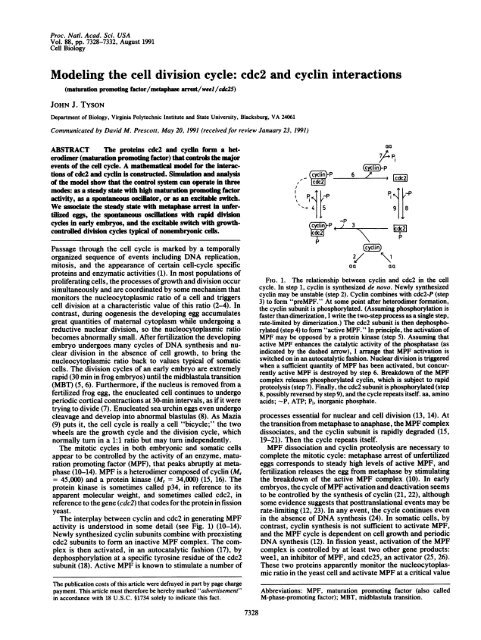
activity is understood in some detail (see Fig. 1) (10-14).<br />
Newly syn<strong>the</strong>sized <strong>cyclin</strong> subunits combine with preexisting<br />
<strong>cdc2</strong> subunits to form an inactive MPF complex. The complex<br />
is <strong>the</strong>n activated, in an autocatalytic fashion (17), by<br />
dephosphorylation at a specific tyrosine residue of <strong>the</strong> <strong>cdc2</strong><br />
subunit (18). Active MPF is known to stimulate a number of<br />
The publication costs of this article were defrayed in part by page charge<br />
payment. This article must <strong>the</strong>refore be hereby marked "advertisement"<br />
in accordance with 18 U.S.C. §1734 solely to indicate this fact.<br />
I<br />
Ga<br />
- yrnP p~~~~~~~~~~~~~~~~~~~~~~. 0<br />
p<br />
-<br />
aB ~ -/ 2,/<br />
oa<br />
FIG. 1. The relationship between <strong>cyclin</strong> <strong>and</strong> <strong>cdc2</strong> in <strong>the</strong> <strong>cell</strong><br />
<strong>cycle</strong>. In step 1, <strong>cyclin</strong> is syn<strong>the</strong>sized de novo. Newly syn<strong>the</strong>sized<br />
<strong>cyclin</strong> may be unstable (step 2). Cyclin combines with <strong>cdc2</strong>-P (step<br />
3) to form "preMPF." At some point after heterodimer formation,<br />
<strong>the</strong> <strong>cyclin</strong> subunit is phosphorylated. (Assuming phosphorylation is<br />
faster than dimerization, I write <strong>the</strong> two-step process as a single step,<br />
rate-limited by dimerization.) The <strong>cdc2</strong> subunit is <strong>the</strong>n dephosphorylated<br />
(step 4) to form "active MPF." In principle, <strong>the</strong> activation of<br />
MPF may be opposed by a protein kinase (step 5). Assuming that<br />
active MPF enhances <strong>the</strong> catalytic activity of <strong>the</strong> phosphatase (as<br />
indicated by <strong>the</strong> dashed arrow), I arrange that MPF activation is<br />
switched on in an autocatalytic fashion. Nuclear <strong>division</strong> is triggered<br />
when a sufficient quantity of MPF has been activated, but concurrently<br />
active MPF is destroyed by step 6. Breakdown of <strong>the</strong> MPF<br />
complex releases phosphorylated <strong>cyclin</strong>, which is subject to rapid<br />
proteolysis (step 7). Finally, <strong>the</strong> <strong>cdc2</strong> subunit is phosphorylated (step<br />
8, possibly reversed by step 9), <strong>and</strong> <strong>the</strong> <strong>cycle</strong> repeats itself. aa, amino<br />
acids; -P, ATP; Pi, inorganic phosphate.<br />
processes essential for nuclear <strong>and</strong> <strong>cell</strong> <strong>division</strong> (13, 14). At<br />
<strong>the</strong> transition from metaphase to anaphase, <strong>the</strong> MPF complex<br />
dissociates, <strong>and</strong> <strong>the</strong> <strong>cyclin</strong> subunit is rapidly degraded (15,<br />
19-21). Then <strong>the</strong> <strong>cycle</strong> repeats itself.<br />
MPF dissociation <strong>and</strong> <strong>cyclin</strong> proteolysis are necessary to<br />
complete <strong>the</strong> mitotic <strong>cycle</strong>: metaphase arrest of unfertilized<br />
eggs corresponds to steady high levels of active MPF, <strong>and</strong><br />
fertilization releases <strong>the</strong> egg from metaphase by stimulating<br />
<strong>the</strong> breakdown of <strong>the</strong> active MPF complex (10). In early<br />
embryos, <strong>the</strong> <strong>cycle</strong> ofMPF activation <strong>and</strong> deactivation seems<br />
to be controlled by <strong>the</strong> syn<strong>the</strong>sis of <strong>cyclin</strong> (21, 22), although<br />
some evidence suggests that posttranslational events may be<br />
rate-limiting (12, 23). In any event, <strong>the</strong> <strong>cycle</strong> continues even<br />
in <strong>the</strong> absence of DNA syn<strong>the</strong>sis (24). In somatic <strong>cell</strong>s, by<br />
contrast, <strong>cyclin</strong> syn<strong>the</strong>sis is not sufficient to activate MPF,<br />
<strong>and</strong> <strong>the</strong> MPF <strong>cycle</strong> is dependent on <strong>cell</strong> growth <strong>and</strong> periodic<br />
DNA syn<strong>the</strong>sis (12). In fission yeast, activation of <strong>the</strong> MPF<br />
complex is controlled by at least two o<strong>the</strong>r gene products:<br />
weel, an inhibitor of MPF, <strong>and</strong> <strong>cdc2</strong>5, an activator (25, 26).<br />
These two proteins apparently monitor <strong>the</strong> nucleocytoplasmic<br />
ratio in <strong>the</strong> yeast <strong>cell</strong> <strong>and</strong> activate MPF at a critical value<br />
Abbreviations: MPF, maturation promoting factor (also called<br />
M-phase-promoting factor); MBT, midblastula transition.<br />
p<br />
aa<br />
7328
Cell Biology: Tyson<br />
of this ratio, although it is not clear at present just how <strong>the</strong><br />
(size) control works.<br />
The model summarized in Fig. 1 is adapted from many<br />
sources (12, 14, 16, 27-29), but it is still a highly simplified<br />
view of <strong>cdc2</strong>-<strong>cyclin</strong> <strong>interactions</strong>. It concentrates on dephosphorylation<br />
of tyrosine-15 but ignores <strong>the</strong> activating phosphorylation<br />
of threonine-167 (14, 30, 47). It attributes <strong>cyclin</strong><br />
degradation to phosphate labeling instead of conjugation with<br />
ubiquitin (48), <strong>and</strong> it ignores <strong>the</strong> apparent stimulatory effect<br />
of active MPF on <strong>cyclin</strong> degradation (29).<br />
Despite such simplifications <strong>and</strong> omissions, <strong>the</strong> model in<br />
Fig. 1 is a reasonable "first approximation" to <strong>the</strong> <strong>cell</strong>-<strong>cycle</strong><br />
regulatory network. How good is this picture? Can it account<br />
for <strong>the</strong> coordination of <strong>cell</strong> growth <strong>and</strong> <strong>division</strong> during <strong>the</strong><br />
normal (somatic) <strong>cell</strong> <strong>cycle</strong>? How is <strong>the</strong> nucleocytoplasmic<br />
ratio measured <strong>and</strong> how is this information communicated to<br />
<strong>the</strong> <strong>cyclin</strong>-<strong>cdc2</strong> mitotic-triggering complex? Can <strong>the</strong> same<br />
model also account for metaphase arrest of unfertilized eggs,<br />
for rapid <strong>cycle</strong>s of DNA syn<strong>the</strong>sis <strong>and</strong> <strong>cell</strong> <strong>division</strong> (without<br />
<strong>cell</strong> growth) during <strong>the</strong> embryonic <strong>cell</strong> <strong>cycle</strong>, <strong>and</strong> for <strong>the</strong><br />
autonomous <strong>cyclin</strong>g of MPF activity in <strong>the</strong> absence of DNA<br />
syn<strong>the</strong>sis or <strong>cell</strong> <strong>division</strong> in enucleated embryos?<br />
To answer <strong>the</strong>se questions I frame <strong>the</strong> model in Fig. 1 in<br />
precise ma<strong>the</strong>matical equations (Table 1) <strong>and</strong> investigate <strong>the</strong><br />
properties of <strong>the</strong>se equations. This approach makes evident<br />
<strong>the</strong> precise consequences of <strong>the</strong> assumptions about <strong>cdc2</strong>-<br />
<strong>cyclin</strong> <strong>interactions</strong> implicit in Fig. 1. To <strong>the</strong> extent that <strong>the</strong>se<br />
consequences are in accord with observations, we gain<br />
confidence in our underst<strong>and</strong>ing of <strong>cell</strong>-<strong>cycle</strong> regulation.<br />
Where <strong>the</strong> consequences are out of accord, we have a<br />
framework in which to analyze <strong>and</strong> compare alternative<br />
assumptions about <strong>the</strong> control system.<br />
Steady States <strong>and</strong> Oscillations<br />
Solutions of <strong>the</strong> equations in Table 1 depend on <strong>the</strong> values<br />
assumed by <strong>the</strong> 10 parameters in <strong>the</strong> model (Table 2). Nothing<br />
is known experimentally about appropriate values for <strong>the</strong>se<br />
parameters, so I can demonstrate at present only that <strong>the</strong>re<br />
exist numerical values of <strong>the</strong> parameters for which <strong>the</strong> model<br />
exhibits dynamical behavior reminiscent of <strong>cell</strong>-<strong>cycle</strong> control.<br />
In this report I focus on two parameters: k4, <strong>the</strong> rate constant<br />
describing <strong>the</strong> autocatalytic activation ofMPF by dephosphorylation<br />
of <strong>the</strong> <strong>cdc2</strong> subunit, <strong>and</strong> k6, <strong>the</strong> rate constant describing<br />
breakdown of <strong>the</strong> active <strong>cdc2</strong>-<strong>cyclin</strong> complex. Depending<br />
on <strong>the</strong> values of k4 <strong>and</strong> k6 (Fig. 2), <strong>the</strong>re are regions of stable<br />
steady-state behavior (regions A <strong>and</strong> C) <strong>and</strong> a region of<br />
spontaneous limit-<strong>cycle</strong> oscillations (region B); see <strong>the</strong> Appendix<br />
for details. In region A, MPF is maintained primarily in<br />
Table 1. Kinetic equations governing <strong>the</strong> <strong>cyclin</strong>-<strong>cdc2</strong> <strong>cycle</strong> in<br />
Fig. 1<br />
d[C2]/dt = k6[M]<br />
- k8[-P][C2] + k4[CP]<br />
d[CP]/dt = -k3[CP][Y] + k8[-P][C2] k4[CP] -<br />
d[pM]/dt = k3[CP][Y]<br />
- [pM]F([M]) + k[--P][M]<br />
d[M]/dt = [pM]F([M]) k5[-P][M] kWM]<br />
- -<br />
d[Y]/dt = k1[aa] - k2[Y] - k3[CP][Y]<br />
d[YP]/dt = k6[M] - k7[YP]<br />
t, time; ki, rate constant for step i (i = 1, . . ., 9); aa, amino acids.<br />
The concentrations [aa] <strong>and</strong> [-PI are assumed to be constant. There<br />
are six time-dependent variables: <strong>the</strong> concentrations of <strong>cdc2</strong> ([C2]),<br />
<strong>cdc2</strong>-P ([CP]), preMPF = P-<strong>cyclin</strong>-<strong>cdc2</strong>-P ([pM]), active MPF =<br />
P-<strong>cyclin</strong>-<strong>cdc2</strong> ([M]), <strong>cyclin</strong> ([YI]), <strong>and</strong> <strong>cyclin</strong>-P ([YP]). The activation<br />
of step 4 by active MPF is described by <strong>the</strong> function F([M]) = k4' +<br />
k4([M]/[CT])2, where k4' is <strong>the</strong> rate constant for step 4 when [active<br />
MPF] = 0 <strong>and</strong> k4 is <strong>the</strong> rate constant when [active MPF] = [CT],<br />
where [CT] = total <strong>cdc2</strong>. I assume k4 >> k4'. This form of F([M]) is<br />
only one of many possible ways to describe <strong>the</strong> autocatalytic<br />
feedback of active MPF on its own production.<br />
Proc. NaMl. Acad. Sci. USA 88 (1991) 7329<br />
Table 2. Parameter values used in <strong>the</strong> numerical solution of <strong>the</strong><br />
model equations<br />
Parameter Value Notes<br />
kl[aa]/[CT] 0.015 min1 *<br />
k2 0 t<br />
k3[CT] 200 min' *<br />
k4 10-1000 min- (adjustable)<br />
k4'<br />
0.018 mink5[-P]<br />
0<br />
/C6 0.1-10 min- (adjustable)<br />
k7 0.6 min-1 t<br />
k8[-P] >>kg §<br />
kg >>k6 §<br />
*It is assumed that [CT] = [C2] + [CP] + [pM] + [Ml = constant.<br />
For growing <strong>cell</strong>s, this implies that <strong>cdc2</strong> protein is continuously<br />
syn<strong>the</strong>sized to maintain a constant concentration of <strong>cdc2</strong> subunits<br />
(31).<br />
tIn <strong>the</strong> absence of evidence to <strong>the</strong> contrary, it is assumed that newly<br />
syn<strong>the</strong>sized <strong>cyclin</strong> is stable (k2 = 0). If k2 # 0, <strong>the</strong> behavior of <strong>the</strong><br />
model is basically unchanged, as long as k2 > kg» k6. This<br />
allows us to neglect <strong>the</strong> first differential equation in Table 1 (i.e.,<br />
d[C2]/dt = 0) <strong>and</strong><br />
=<br />
[C2] (k9/k8[-P])[CP]
7330 Cell Biology: Tyson<br />
1000r<br />
E 1001<br />
10I<br />
0.1 1.0<br />
k6 min1<br />
FIG. 2. Qualitative behavior of <strong>the</strong> <strong>cdc2</strong>-<strong>cyclin</strong> model of <strong>cell</strong><strong>cycle</strong><br />
regulation. The control parameters are k4, <strong>the</strong> rate constant<br />
describing <strong>the</strong> maximum rate of MPF activation, <strong>and</strong> k6, <strong>the</strong> rate<br />
constant describing dissociation of <strong>the</strong> active MPF complex. Regions<br />
A <strong>and</strong> C correspond to stable steady-state behavior of <strong>the</strong> model;<br />
region B corresponds to spontaneous limit <strong>cycle</strong> oscillations. In <strong>the</strong><br />
stippled area <strong>the</strong> regulatory system is excitable. The boundaries<br />
between regions A, B, <strong>and</strong> C were determined by integrating <strong>the</strong><br />
differential equations in Table 1, for <strong>the</strong> parameter values in Table 2.<br />
Numerical integration was carried out by using Gear's algorithm for<br />
solving stiff ordinary differential equations (32). The "developmental<br />
path" 1 ... 5 is described in <strong>the</strong> text.<br />
so k6 abruptly increases 2-fold. Continued <strong>cell</strong> growth causes<br />
k6(t) again to decrease, <strong>and</strong> <strong>the</strong> <strong>cycle</strong> repeats itself. The<br />
interplay between <strong>the</strong> control system, <strong>cell</strong> growth, <strong>and</strong> DNA<br />
replication generates periodic changes in k6(t) <strong>and</strong> periodic<br />
bursts of MPF activity with a <strong>cycle</strong> time identical to <strong>the</strong><br />
mass-doubling time of <strong>the</strong> growing <strong>cell</strong>.<br />
Figs. 2 <strong>and</strong> 3 demonstrate that, depending on <strong>the</strong> values of<br />
k4 <strong>and</strong> k6, <strong>the</strong> <strong>cell</strong> <strong>cycle</strong> regulatory system exhibits three<br />
10<br />
Proc. Natl. Acad. Sci. USA 88 (1991)<br />
different modes of control. For small values of k6, <strong>the</strong> system<br />
displays a stable steady state of high MPF activity, which I<br />
associate with metaphase arrest of unfertilized eggs. For<br />
moderate Values of k6, <strong>the</strong> system executes autonomous<br />
oscillations reminiscent of rapid <strong>cell</strong> <strong>cyclin</strong>g in early embryos.<br />
For large values of k6, <strong>the</strong> system is attracted to an<br />
excitable steady state of low MPF activity, which corresponds<br />
to interphase arrest of resting somatic <strong>cell</strong>s or to<br />
growth-controlled bursts of MPF activity in proliferating<br />
somatic <strong>cell</strong>s.<br />
Midblastula Traiisiton<br />
A possible developmental scenario is illustrated by <strong>the</strong> path<br />
1 ... 5 in Fig. 2. Upon fertilization, <strong>the</strong> metaphase-arrested<br />
egg (at point 1) is stimulated to rapid <strong>cell</strong> <strong>division</strong>s (2) by an<br />
increase in <strong>the</strong> activity of <strong>the</strong> enzyme catalyzing step 6 (28).<br />
During <strong>the</strong> early embryonic <strong>cell</strong> <strong>cycle</strong>s (2-+ 3), <strong>the</strong> regulatory<br />
system is executing autonomous oscillations, <strong>and</strong> <strong>the</strong> control<br />
parameters, k4 <strong>and</strong> k6, increase as <strong>the</strong> nuclear genes coding<br />
for <strong>the</strong>se enzymes are activated. At midblastula (3), autonomous<br />
oscillations cease, <strong>and</strong> <strong>the</strong> regulatory system enters<br />
<strong>the</strong> excitable domain. Cell <strong>division</strong> now becomes growth<br />
controlled. As <strong>cell</strong>s grow, k6 decreases (inhibitor diluted)<br />
<strong>and</strong>/or k4 increases (activator accumulates), which drives <strong>the</strong><br />
regulatory system back into domain B (4 -S 5). The subsequent<br />
burst of MPF activity triggers mitosis, causes k6 to<br />
increase (inhibitor syn<strong>the</strong>sis) <strong>and</strong>/or k4 to decrease (activator<br />
degradation), <strong>and</strong> brings <strong>the</strong> regulatory system back into <strong>the</strong><br />
excitable domain (5 -* 4).<br />
Although <strong>the</strong>re is a clear <strong>and</strong> abrupt leng<strong>the</strong>ning of inter<strong>division</strong><br />
times at MBT, <strong>the</strong>re is no visible increase in <strong>cell</strong><br />
volume immediately <strong>the</strong>reafter (6, 20), so how can we entertain<br />
<strong>the</strong> idea that <strong>cell</strong> <strong>division</strong> becomes growth controlled<br />
after MBT? In <strong>the</strong> paradigm ofgrowth control of <strong>cell</strong> <strong>division</strong>,<br />
<strong>cell</strong> "size" is never precisely specified, because no one<br />
knows what molecules, structures, or properties are used by<br />
<strong>cell</strong>s to monitor <strong>the</strong>ir size. Thus, even though post-MBT <strong>cell</strong>s<br />
0.4<br />
a 100<br />
b<br />
C<br />
r k6' min-1<br />
0 20 40 60 80 100 0 20 40 60 80 100<br />
t, min t, min<br />
0 100 200 300 400 500<br />
t, min<br />
FIG. 3. Dynamical behavior of <strong>the</strong> <strong>cdc2</strong>-<strong>cyclin</strong> model. The curves are total <strong>cyclin</strong> ([YT] = [Y] + [YP] + [pM] + [M]) <strong>and</strong> active MPF [Ml<br />
relative to total <strong>cdc2</strong> ([CT] = [C2] + [CP] + [pM] + [MI). The differential equations in Table 1, for <strong>the</strong> parameter values in Table 2, were solved<br />
numerically by using a fourth-order Adams-Moulton integration routine (33) with time step = 0.001 min. (The adequacy of <strong>the</strong> numerical<br />
integration was checked by decreasing <strong>the</strong> time step <strong>and</strong> also by comparison to solutions generated by Gear's algorithm.) (a) Limit <strong>cycle</strong><br />
oscillations for k4 = 180 min-', k6 = 1 min- (point x in Fig. 2). A "limit <strong>cycle</strong>" solution of a set of ordinary differential equations is a periodic<br />
solution that is asymptotically stable with respect to small perturbations in any of <strong>the</strong> dynamical variables. (b) Excitable steady state for k4 =<br />
180 min 1, k6 = 2 min' (point + in Fig. 2). Notice that <strong>the</strong> ordinate is a logarithmic scale. The steady state of low MPF activity ([M]/[CT]<br />
= 0.0074, [YT]/[CT] = 0.566) is stable with respect to small perturbations of MPF activity (at 1 <strong>and</strong> 2) but a sufficiently large perturbation of<br />
[Ml (at 3) triggers a transient activation of MPF <strong>and</strong> subsequent destruction of <strong>cyclin</strong>. The regulatory system <strong>the</strong>n recovers to <strong>the</strong> stable steady<br />
state. (c) Parameter values as in b except that k6 is now a function of time (oscillating near point + in Fig. 2). See text for an explanation of<br />
<strong>the</strong> rules for k6(Q). Notice that <strong>the</strong> period between <strong>cell</strong> <strong>division</strong>s (bursts in MPF activity) is identical to <strong>the</strong> mass-doubling time (Td = 116 min<br />
in this simulation). Simulations with different values of Td (not shown) demonstrate that <strong>the</strong> period between MPF bursts is typically equal to<br />
<strong>the</strong> mass-doubling time-i.e., <strong>the</strong> <strong>cell</strong> <strong>division</strong> <strong>cycle</strong> is growth controlled under <strong>the</strong>se circumstances. Growth control can also be achieved<br />
(simulations not shown), holding k6 constant, by assuming that k4 increases with time between <strong>division</strong>s <strong>and</strong> decreases abruptly after an MPF<br />
burst.
may show no visible increase in volume, <strong>the</strong>re may be within<br />
<strong>cell</strong>s certain biochemical changes that are interpreted as<br />
growth. For instance, RNA syn<strong>the</strong>sis turns on abruptly at<br />
MBT (5), <strong>and</strong> <strong>the</strong>re is a dramatic rise in protein syn<strong>the</strong>sis<br />
based on newly transcribed nuclear mRNA (34). A protein<br />
coded by this RNA ra<strong>the</strong>r than by maternal message could<br />
serve as a proxy for <strong>cell</strong> size, diluting out (or inactivating) <strong>the</strong><br />
enzyme catalyzing step 6. Alternatively, in an activatoraccumulation<br />
model, one of <strong>the</strong> post-MBT proteins could be<br />
<strong>the</strong> enzyme catalyzing step 4.<br />
<strong>cdc2</strong>5 <strong>and</strong> weed<br />
The parameters, k4 <strong>and</strong> k6, that govern <strong>the</strong> developmental<br />
path shown in Fig. 2 are rate constants determined by <strong>the</strong><br />
concentrations of proteins that serve as an activator <strong>and</strong> an<br />
inhibitor, respectively, of MPF activity. The rate of activation<br />
of MPF is often associated with <strong>the</strong> level of expression<br />
of <strong>the</strong> <strong>cdc2</strong>5 gene (26, 35, 36), suggesting that k4 be set<br />
proportional to <strong>the</strong> concentration of <strong>cdc2</strong>5 protein. This<br />
association is encouraged by recent observations that <strong>cdc2</strong>5<br />
levels in fission yeast <strong>cell</strong>s increase 3- to 4-fold during<br />
interphase <strong>and</strong> <strong>the</strong>n drop abruptly at <strong>cell</strong> <strong>division</strong> (35).<br />
Exactly such behavior of k4 can lead to growth-controlled<br />
<strong>cycle</strong>s like those in Fig. 3c (simulations not shown).<br />
Deactivation of MPF is often associated with <strong>the</strong> level of<br />
expression of wee] (10-12, 25). Recent evidence that weel<br />
can phosphorylate tyrosine residues (37) bolsters <strong>the</strong> suspicion<br />
that weel inhibits MPF by catalyzing step 5 <strong>and</strong>/or step<br />
8 in Fig. 1. But, if this is <strong>the</strong> role of weel, it is hard to<br />
underst<strong>and</strong> how changing levels of expression of wild-type<br />
wee] can alter size at <strong>division</strong>, because, in <strong>the</strong> ma<strong>the</strong>matical<br />
model, <strong>the</strong> magnitudes of k5 <strong>and</strong> k8 have very little effect on<br />
<strong>the</strong> dynamical behavior of <strong>the</strong> regulatory system. In contrast,<br />
size at <strong>division</strong> (in <strong>the</strong> model) is sensitively dependent on <strong>the</strong><br />
magnitude of k6. However, we cannot associate weel with<br />
step 6 because dysfunctional mutants at <strong>the</strong> locus controlling<br />
step 6 should block <strong>cell</strong>s in mitosis, <strong>and</strong> this is clearly not <strong>the</strong><br />
case for wee] mutations. It remains an open question to<br />
reformulate Fig. 1 so that wee] plays a more significant role<br />
in <strong>the</strong> control of <strong>division</strong>.<br />
Discussion<br />
Cell Biology: Tyson<br />
To develop a precise ma<strong>the</strong>matical model of <strong>cdc2</strong>-<strong>cyclin</strong><br />
<strong>interactions</strong>, I have made many specific assumptions, some<br />
of which are crucial to <strong>the</strong> model <strong>and</strong> o<strong>the</strong>rs inconsequential.<br />
Critical steps in <strong>the</strong> mechanism are <strong>the</strong> autocatalytic dephosphorylation<br />
of <strong>the</strong> <strong>cdc2</strong> subunit (step 4) <strong>and</strong> breakdown of <strong>the</strong><br />
active MPF complex (step 6). On <strong>the</strong> o<strong>the</strong>r h<strong>and</strong>, inhibition<br />
of MPF by rephosphorylating <strong>the</strong> <strong>cdc2</strong> subunit (step 5) does<br />
not seem to be particularly significant or even necessary in<br />
this model. In all calculations reported here, k5 = 0 but similar<br />
results are obtained for nonzero values of k5. In particular,<br />
<strong>the</strong> "<strong>cycle</strong> control mode" (region A, B, or C in Fig. 2) is<br />
insensitive to <strong>the</strong> value of k5 within broad limits.<br />
Similarly, I have assumed that newly syn<strong>the</strong>sized <strong>cyclin</strong> is<br />
stable (k2 = 0), but <strong>the</strong> behavior of <strong>the</strong> model is not much<br />
changed when k2 $ 0, as long as proteolysis is considerably<br />
slower than dimerization (k2
7332 Cell Biology: Tyson<br />
y does not appear in Eqs. 1-3. Second, because k3[CTJ>><br />
max{ki[aa]/[CT], k2, k6}, w changes very rapidly compared to<br />
changes in v, so w = v as long as 0 < v < 1. Thus, <strong>the</strong><br />
<strong>cdc2</strong>-<strong>cyclin</strong> model reduces to a pair of nonlinear ordinary<br />
differential equations<br />
du/dt = k4(v - u)(a + u2) - k6u<br />
dv/dt = (k1[aa]/[CT]) - k6u.<br />
Equations similar to Eqs. 5 <strong>and</strong> 6 were derived by Norel <strong>and</strong><br />
Agur (43) on <strong>the</strong> basis of different assumptions about <strong>cdc2</strong>-<br />
<strong>cyclin</strong> <strong>interactions</strong>. In <strong>the</strong>ir study of <strong>the</strong> <strong>cell</strong> <strong>cycle</strong> of P.<br />
polycephalum, Tyson <strong>and</strong> Kauffman (44) formulated a hypo<strong>the</strong>tical<br />
model of a "mitotic oscillator" that is identical to<br />
Eqs. 5 <strong>and</strong> 6: simply define u = Y <strong>and</strong> v = X + Y in <strong>the</strong>ir<br />
equation 3. From this point of view, our current underst<strong>and</strong>ing<br />
of <strong>cell</strong>-<strong>cycle</strong> regulation can be expressed as a modified<br />
version of <strong>the</strong> "Brusselator," a famous <strong>the</strong>oretical model of<br />
chemical oscillations (45).<br />
Equations like Eqs. 5 <strong>and</strong> 6 are best analyzed by phase<br />
plane techniques (46). In <strong>the</strong> u-v plane (Fig. 4), I plot <strong>the</strong> locus<br />
of points where du/dt = 0,<br />
v = u + k6u/{k4(a + u2)} ("u-nullclinel) [71<br />
<strong>and</strong> <strong>the</strong> locus of points where dv/dt = 0,<br />
u = kl[aa]/k6[CTJ ("v-nullcline'). [81<br />
The v-nullcline is just a vertical line. The u-nullcline is<br />
N-shapedwith a local maximum near u = V T4, v =<br />
k6/2Vrk4'k4 <strong>and</strong> a local minimum near u = k6/k4, v =<br />
2\/~7k4. (These estimates are reasonably accurate as long as<br />
k4'k6 < 0.025.) To ensure that v < 1, we insist that k6 <<br />
2Vk4'k4, which implies that k4 must exceed 10 k6. The<br />
requirements that 400 -k4' < 10 k6 < k4 are satisfied by <strong>the</strong><br />
parameter values in Table 2.<br />
Steady states, where both du/dt = 0 <strong>and</strong> dv/dt = 0, lie at<br />
intersections of <strong>the</strong> nullclines. The nuliclines may intersect in<br />
three qualitatively different ways (Fig. 4), which correspond<br />
to <strong>the</strong> three characteristic modes of <strong>the</strong> control system:<br />
Mode A. Stable steady state with high MPF activity, when<br />
k1[aa]/k6[CT] > N/6/.<br />
Mode B. Unstable steady state (spontaneous oscillations),<br />
when<br />
Vk4'l4 < k1[aa/k6[CT] < k<br />
0.4<br />
0.2<br />
0.1 0.2<br />
FIG. 4. The phase plane for Eqs. 5 <strong>and</strong> 6. Steady states occur at<br />
<strong>the</strong> intersections of <strong>the</strong> solid curve (<strong>the</strong> u-nullcline) <strong>and</strong> dashed lines<br />
(<strong>the</strong> v-nuilcline for three different cases). A, stable steady state with<br />
high MPF activity; B, unstable steady state (limit <strong>cycle</strong> oscillations);<br />
C, excitable steady state with low MPF activity.<br />
A<br />
U<br />
[51<br />
[6]<br />
Proc. Natl. Acad. Sci. USA 88 (1991)<br />
Mode C. Stable steady state with low MPF activity, when<br />
kj[aa]/k6[CT]


