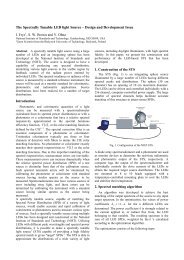
Here - PMOD/WRC
Here - PMOD/WRC
Here - PMOD/WRC

Create successful ePaper yourself
Turn your PDF publications into a flip-book with our unique Google optimized e-Paper software.
Spatial Anisotropy of the Exciton Resonance in CaF 2 at 112 nm and its<br />
Relation to Optical Anisotropy in the DUV<br />
M. Richter, A. Gottwald<br />
Physikalisch-Technische Bundesanstalt, Berlin, Germany<br />
M. Letz<br />
Schott Glas, Mainz, Germany<br />
Abstract. Excimer lasers are widely used for industrial<br />
applications such as photolithography of semiconductor<br />
devices. The basic optical material for the excimer-laser<br />
wavelengths in the deep ultraviolet is calcium fluoride<br />
which shows an unexpected optical anisotropy at 157 nm.<br />
In this context, we have measured the spatial anisotropy of<br />
the exciton resonance at 112 nm by precision reflection<br />
measurements using dispersed synchrotron radiation on<br />
oriented samples of high purity. The results are discussed<br />
in terms of the complex dynamic dielectric function and<br />
explain the optical anisotropy by the interaction of<br />
radiation with the cubic structure of a perfect crystal.<br />
Introduction<br />
Fluoride crystals are strongly ionic and have the largest<br />
band gaps known for crystalline solids. As a consequence,<br />
they are transparent even in the wavelength region of deep<br />
ultraviolet (DUV) radiation and used for DUV optics.<br />
Recently, the effect of an optical anisotropy for calcium<br />
fluoride (CaF 2 ) has been observed at 157 nm which is of<br />
great importance for DUV lithography (Burnett et al.<br />
2001). Already the measured weak birefringence of ∆n/n ≈<br />
10 -6 of the refractive index considerably influences the<br />
design of imaging optics.<br />
Due to its cubic symmetry, it is generally presumed that<br />
the CaF 2 crystal shows isotropic optical properties.<br />
However, strongly ionic crystals show deep excitonic<br />
bound states. The most pronounced one in CaF 2 , the<br />
Γ-exciton, is known to arise at 11.1 eV, yielding to a strong<br />
and narrow absorption structure at a wavelength of about<br />
112 nm. Close to such a strong absorption line, an optical<br />
anisotropy can result from a slight deviation of the cubic<br />
symmetry, due to the incident radiation field even at<br />
optical wavelengths much larger than the lattice constant.<br />
The effect of optical anisotropy in the vicinity of a narrow<br />
absorption line was formulated by Ginzburg 1958. The<br />
effect is well established at optical wavelengths and<br />
usually called ‘spatial dispersion induced birefringence’.<br />
In this work we describe a direct measurement of the<br />
spatial anisotropy of the narrow absorption line at 112 nm<br />
(Letz et al. 2003). The experiments were performed at the<br />
UV and VUV beamline for detector calibration and<br />
reflectometry in the Radiometry Laboratory of the<br />
Physikalisch-Technische Bundesanstalt at the electron<br />
storage ring BESSY II on extremely pure CaF 2 samples<br />
with surface orientations in the (111) and (100) directions<br />
of the crystals. The reflectance of the samples was<br />
measured in a near-normal incidence geometry.<br />
Results<br />
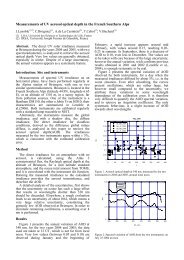
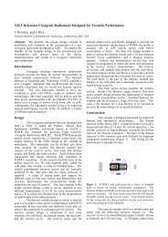
The results of the reflection measurements are shown in<br />
Fig. 1. When comparing the data of the two different<br />
samples, a small but significant shift of about 0.2 nm<br />
becomes apparent, the resonance structure of the sample<br />
with (111) surface orientation being shifted towards<br />
smaller wavelengths. To verify the experimental<br />
significance of this small shift, the reproducibility of the<br />
wavelength was tested by simultaneously recording<br />
resonance absorption lines of Ar in a gas cell.<br />
Figure 1. Experimental data (symbols) and fit (lines) of the<br />
normal-incidence reflectance around the Γ-exciton of CaF 2 for a<br />
(100) and (111) surface (Letz et al. 2003).<br />
For interpreting our experimental data with regard to<br />
exciton position and lifetime, we applied a least squares<br />
fitting algorithm. The resulting fit curves are also shown in<br />
Fig. 1. For the exciton positions one obtains for the<br />
different crystal orientations:<br />
(100): ηω 0 = 11.11(1) eV λ 0 = 111.6(7) nm<br />
(111): ηω 0 = 11.13(1) eV λ 0 = 111.4(7) nm<br />
Discussing the results in terms of the complex dynamic<br />
dielectric function it can be demonstrated that the direction<br />
of the shift for the exciton resonance as measured in the<br />
present work is consistent with the sign of the optical<br />
anisotropy as measured by Burnett et al. 2001 at 157 nm.<br />
References<br />
Burnett, J.H., Levine, Z.H., Shirley, E.L., Phys. Rev. B, 64,<br />
241102R, 2001.<br />
Ginzburg, V.L., JETP, 34, 1593, 1958.<br />
Letz, M., Parthier, L., Gottwald, A., Richter, M., Phys. Rev. B, 67,<br />
233101, 2003<br />
Proceedings NEWRAD, 17-19 October 2005, Davos, Switzerland 145