Here - PMOD/WRC
Here - PMOD/WRC
Here - PMOD/WRC

You also want an ePaper? Increase the reach of your titles
YUMPU automatically turns print PDFs into web optimized ePapers that Google loves.
NEWRAD<br />
PROCEEDINGS OF THE<br />
9 TH INTERNATIONAL CONFERENCE<br />
ON NEW DEVELOPMENTS AND<br />
APPLICATIONS IN OPTICAL RADIOMETRY<br />
17-19 OCTOBER, 2005, DAVOS, SWITZERLAND<br />
EDITED BY<br />
JULIAN GRÖBNER<br />
PHYSIKALISCH-METEOROLOGISCHES OBSERVATORIUM DAVOS,<br />
WELTSTRAHLUNGSZENTRUM<br />
DAVOS, SWITZERLAND, SEPTEMBER 2005
Scientific Programme Committee:<br />
Andrew Wallard, BIPM, France<br />
Tony Bittar, IRL/CRI, New Zealand<br />
James Butler, NASA, USA<br />
Antonio Corrons, IFA/CSIC, Spain<br />
Peter Foukal, Heliophysics, USA<br />
Nigel Fox, NPL, UK<br />
Claus Fröhlich, <strong>PMOD</strong>/<strong>WRC</strong>, Switzerland<br />
Martin Huber, PSI, Switzerland<br />
Erkki Ikonen, MIKES, Finland<br />
Jürgen Metzdorf, PTB, Germany<br />
Al Parr, NIST, USA<br />
Maria Luisa Rastello, IEN, Italy<br />
Gerhard Ulm, PTB, Germany<br />
Local Organising Committee:<br />
Physikalisch-Meteorologisches Observatorium Davos,<br />
Weltstrahlungszentrum<br />
Julian Gröbner<br />
Werner Schmutz<br />
Sonja Degli Esposti<br />
Wolfgang Finsterle<br />
Christoph Wehrli<br />
ISBN-10 3-033-00570-5<br />
ISBN-13 978-3-033-00570-9
NEWRAD 2005 Scientific Program<br />
Day Chair Session<br />
Monday Nigel Fox 1 Absolute Radiometry Posters<br />
Erkki Ikonen/Jürgen Metzdorf 2 UV, Vis, and IR Radiometry Posters<br />
Jürgen Metzdorf 3 Photolithography and UV Processing Posters<br />
Tuesday Peter Foukal/Claus Fröhlich 4 Remote Sensing Posters<br />
Antonio Corrons 5 Photometry and Colorimetry Posters<br />
Maria Luisa Rastello 6 Novel Techniques Posters<br />
Wednesday Tony Bittar 7 International Comparisons Posters<br />
Tony Bittar/Martin Huber 8 Radiometric and Photometric Sources Posters<br />
Gerhard Ulm 9 Realisation of Scales Posters<br />
Sunday, October 16, 2005<br />
17:30 - Opening Reception in the foyer Aspen of the Conference Centre<br />
Participant registration<br />
Monday, October 17, 2005<br />
8:00 Participant registration<br />
8:45 Welcome Werner Schmutz, <strong>PMOD</strong>/<strong>WRC</strong><br />
8:55 Information Julian Gröbner, <strong>PMOD</strong>/<strong>WRC</strong><br />
Session 1 Chair – Nigel Fox<br />
Absolute Radiometry<br />
9:00 Claus Fröhlich, <strong>PMOD</strong> 1-1 Invited Talk, Absolute Accuracy of Total Solar Irradiance<br />
Measurements in Space (page 17)<br />
9:30 Eric Usadi, NPL 1-2 Reflecting Cavity Blackbodies for Radiometry (page 19)<br />
9:50 Joaquin Campos-Acosta, CSIC 1-3 Low-uncertainty absolute radiometric calibration of a<br />
CCD (page 21)<br />
10:10 Jeanne Houston, NIST 1-4 NIST Reference Cryogenic Radiometer Designed for<br />
Versatile Performance (page 23)<br />
10:30 Refreshment break / Poster installation<br />
Session 2 Chair – Erkki Ikonen UV, Vis, and IR Radiometry<br />
11:10 Mario Blumthaler, UIIMP 2-1 Invited Talk , QA/QC of Spectral Solar UV irradiance<br />
measurements (page 59)<br />
11:40 Gerhard Ulm, PTB 2-2 The Metrology Light Source – the new dedicated electron<br />
storage ring of PTB (page 61)<br />
12:00 Jarle Gran, JV 2-3 Fractional self-calibration of silicon photodiodes<br />
(page 63)<br />
12:20 Lunch Break / Poster Session 1,2,3<br />
Session 2 Chair – Erkki Ikonen UV, Vis, and IR Radiometry<br />
14:30 Peter Meindl, PTB 2-4 The UV Spectral Responsivity Scale of the PTB<br />
(page 69)<br />
14:50 Ping-shine Shaw, NIST 2-5 A method of characterizing narrow-band filtered<br />
radiometers using synchrotron radiation (page 73)<br />
Proceedings NEWRAD, 17-19 October 2005, Davos, Switzerland 3
15:10 Terubumi Saito, AIST 2-6 Characterization of Photoconductive Diamond Detectors<br />
as a Candidate of FUV/VUV Transfer Standard Detectors<br />
(page 67)<br />
15:30 Laurent Vuilleumier, SMA 2-7 Operational mode uncertainty for broadband erythemal<br />
UV radiometers (page 71)<br />
15:50 Refreshment break<br />
Session 2 Chair – Jürgen Metzdorf UV, Vis, and IR Radiometry<br />
16:20 George Eppeldauer, NIST 2-8 Development of a Versatile Radiometer-Photometer<br />
System (page 75)<br />
16:40 Lasse Ylianttila, STUK 2-9 Temperature effects of PTFE diffusers (page 65)<br />
Session 3 Chair – Jürgen Metzdorf Photolithography and UV Processing<br />
17:00 Frank Scholze, PTB 3-1 Improvements in EUV reflectometry at PTB (page 139)<br />
17:20 Rob Vest, NIST 3-2 Measuring pulse energy with solid state photodiodes<br />
(page 141)<br />
17:40 End of Session<br />
Tuesday, October 18, 2005<br />
Session 4 Chair – Peter Foukal Remote sensing<br />
8:30 Greg Kopp, LASP 4-1 Invited Talk, Solar Radiometry from SORCE (page 151)<br />
9:00 Jack Xiong, NASA/GSFC 4-2 Inter-comparison Study of Terra and Aqua MODIS<br />
Reflective Solar Bands Using On-orbit Lunar<br />
Observations (page 153)<br />
9:20 Hugh Kieffer, CR 4-3 A Model of the Spectral Irradiance of the Moon for<br />
Calibration of Earth-orbiting Spacecraft Instruments<br />
(page 159)<br />
9:40 Victor Sapritsky, VNIIOFI 4-4 On measuring the radiant properties of objects of<br />
observations for the Global Earth Observation System of<br />
Systems (GEOSS) (page 157)<br />
10:00 Refreshment break<br />
Session 4 Chair – Claus Fröhlich Remote sensing<br />
10:30 Peter Foukal, Heliophysics 4-5 Keynote Talk, The Solar Bolometric Imager – Recent<br />
Results and Future Plans (page 155)<br />
11:00 Anton D. Nikolenko, INP 4-6 Procedures of absolute calibration for the Space Solar<br />
Patrol instrumentation at the Synchrotron radiation<br />
source (page 163)<br />
11:20 Chris Gueymard, SCS 4-7 Determination of Aerosol Optical Depth and Ångström’s<br />
Wavelength Exponent Using Sunphotometers and<br />
Spectroradiometers: A Preliminary Assessment<br />
(page 161)<br />
11:40 Lunch Break / Poster Session 4,5,6<br />
Session 5 Chair – Antonio Corrons Photometry and Colorimetry<br />
14:00 Georg Sauter, PTB 5-1 Invited Talk, Review on new developments in<br />
Photometry (page 185)<br />
14:30 Christian Monte, BAM 5-2 Linking Fluorescence Measurements to Radiometric<br />
Units (page 187)<br />
4
14:50 Hiroshi Shitomi, AIST 5-3 Photoluminescence from White Reference Materials for<br />
Spectral Diffuse Reflectance upon Exposure to the<br />
Radiation Shorter than 400 nm (page 189)<br />
15:10 Yuqin Zong, NIST 5-4 A Simple Stray-light Correction Matrix for Array<br />
Spectrometers (page 191)<br />
15:30 Refreshment break<br />
Session 6 Chair – Maria Luisa Rastello Novel Techniques<br />
16:00 Petri Kärhä, TKK 6-1 Determination of luminous intensity of light-emitting<br />
diodes with modified inverse-square law (page 211)<br />
16:20 Mike Shaw, NPL 6-2 The Measurement of Surface and Volume Fluorescence<br />
at NPL (page 215)<br />
16:40 Antti Lamminpää, TKK 6-3 Characterization of germanium photodiodes and trap<br />
detector (page 209)<br />
17:00 Valentina Schettini, IEN 6-4 Single-photon source heralding efficiency and detection<br />
efficiency metrology at 1550 nm using periodically poled<br />
lithium niobate (page 213)<br />
17:20 End of Session<br />
19:00 Evening Dinner at Schatzalp<br />
Wednesday, October 19, 2005<br />
Session 7 Chair – Tony Bittar<br />
International Comparisons<br />
8:30 Nigel Fox, NPL 7-1 Invited Talk,<br />
9:00 Joachim Hussong, ATLAS 7-2 Comparison of Measurements of Spectral Irradiance<br />
(UV/VIS) by an International Round Robin Test<br />
(page 235)<br />
9:20 Emma Woolliams, NPL 7-3 The CCPR K1-a Key Comparison of Spectral Irradiance<br />
250 – 2500 nm: Measurements, Analysis and Results<br />
(page 233)<br />
Session 8 Chair – Tony Bittar<br />
Radiometric and Photometric Sources<br />
9:40 Joe Rice, NIST 8-1 Hyperspectral Image Projectors for Radiometric<br />
Applications (page 253)<br />
10:00 Steven Brown, NIST 8-2 Spatial Light Modulator-based Advanced Radiometric<br />
Sources (page 259)<br />
10:20 Refreshment break<br />
Session 8 Chair – Martin Huber Radiometric and Photometric Sources<br />
10:40 Yoshiro Yamada, AIST 8-3 Invited Talk, Application of Metal (Carbide)-Carbon<br />
Eutectic Fixed Points in Radiometry (page 249)<br />
11:10 Klaus Anhalt, PTB 8-4 Thermodynamic temperature determinations of Co-C,<br />
Pd-C, Pt-C and Ru-C eutectic fixed points cells<br />
(page 257)<br />
11:30 David Lowe, NPL 8-5 Reproducible Metal-Carbon Eutectic Fixed-Points<br />
(page 255)<br />
11:50 Peter Rosenkranz, BEV 8-6 On Estimation of Distribution Temperature (page 251)<br />
12:10 Lunch Break / Poster Session 7,8,9<br />
Session 9 Chair – Gerhard Ulm<br />
Realisation of Scales<br />
Proceedings NEWRAD, 17-19 October 2005, Davos, Switzerland 5
14:00 Howard Yoon, NIST 9-1 Invited Talk, The Realization and the Dissemination of<br />
Thermodynamic Temperature Scales (page 299)<br />
14:30 Rene Monshouwer, NMI 9-2 New method for the primary realization of the spectral<br />
irradiance scale from 400 to 900 nm (page 305)<br />
14:50 Uwe Arp, NIST 9-3 From X-rays to T-rays: Synchrotron Source-based<br />
Calibrations at SURF III (page 309)<br />
15:10 Mathias Richter, PTB 9-4 The PTB High-Accuracy Spectral Responsivity Scale in<br />
the VUV and X-Ray Range (page 303)<br />
15:30 Refreshment break<br />
Session 9 Chair – Gerhard Ulm Realisation of Scales<br />
16:00 Theo Theocharous, NPL 9-5 The establishment of the NPL infrared relative spectral<br />
responsivity scale using cavity pyroelectric detectors.<br />
(page 301)<br />
16:20 Jimmy Dubard, LNE 9-6 Infrared Radiometry at LNE: characterization of a<br />
pyroelectric detector used for relative spectral<br />
responsivity measurement (page 311)<br />
16:40 Sergey Mekhontsev, NIST 9-7 Preliminary Realization of a Spectral Radiance Scale in<br />
the Range of 2.5 m to 20 m (page 315)<br />
17:00 Pedro Corredera, CSIC 9-8 An integrated sphere radiometer as a solution for high<br />
power laser calibrations in fibre optics (page 307)<br />
17:20 Mart Noorma, NIST 9-9 Transfer standard pyrometers for radiance temperature<br />
measurements below the freezing temperature of silver<br />
at NIST (page 313)<br />
17:40 Chairman SC_NEWRAD Closing words<br />
17:50 End of Session/ End of Conference<br />
6
NEWRAD 2005 Scientific Program-Posters<br />
Day<br />
Monday<br />
Tuesday<br />
Wednesday<br />
Session<br />
1 Absolute Radiometry<br />
2 UV, Vis, and IR Radiometry<br />
3 Photolithography and UV Processing<br />
4 Remote Sensing<br />
5 Photometry and Colorimetry<br />
6 Novel Techniques<br />
7 International Comparisons<br />
8 Radiometric and Photometric Sources<br />
9 Realisation of Scales<br />
Session 1<br />
Absolute Radiometry<br />
1. Measurement of the absorptance of a cryogenic radiometer cavity in the visible and near<br />
infrared (NIR) (page 25)<br />
M. López, H. Hofer, S. Kück<br />
2. Grooves for Emissivity and Absorptivity Enhancement in High Performance Cavity<br />
Sources and Radiometers (page 27)<br />
E. Usadi, R. Montgomery<br />
3. Pulsed UV spectroradiometry with the primary standard synchrotron radiation sources of<br />
high intensity (page 29)<br />
S. Anevsky, V. Ivanov, O. Minaeva, V. Sapritsky, Y. Zolotarevsky<br />
4. New apparatus for the spectral radiant power calibration at CMS of Taiwan (page 31)<br />
H.-L. Yu, S.-W. Hsu<br />
5. VUV and Soft X-ray metrology stations at the International Siberian Synchrotron<br />
Radiation center (page 33)<br />
V.I. Buhiyarov, N.G. Gavrilov, N.A. Gentselev, B.G. Goldenberg, G.N. Kulipanov, A.A.<br />
Legkodymov, V.V. Lyakh, O.I. Meshkov, A.I. Nizovsky, A.D. Nikolenko, V.F. Pndyurin, I.V.<br />
Poletaev, E. P. Ya. V. Rakshun, Semenov, M.A. Kholopov, V.A. Chenov, M.A. Sheromov<br />
6. Experimental study of application of the self-calibration method to certify absolute<br />
spectral sensitivity of a scintillation counter in the soft X-ray range. (page 35)<br />
A.D. Nikolenko, V.F. Pindyurin, V.A. Chernov, V.V. Lyakh<br />
7. The metrology line on the SOLEIL synchrotron facility (page 37)<br />
M. Lièvre, M-C. Lépy, B. Rougié, J-R. Filtz, J. Bastie, M. Idir, T. Moreno, P. Mercede<br />
8. Cryogenic radiometer developments at NPL (page 39)<br />
J. Ireland, M.G. White, N.P. Fox<br />
9. Energy Calorimeter for Pulsed Laser Radiometry (page 41)<br />
D. Fukuda, K. Amemiya, A. Kamimura, S. Kimura<br />
10. Characterization of new trap detectors as transfer standards (page 43)<br />
J-M Coutin, F. Chandoul, J. Bastie<br />
11. Measurement of Small Aperature Areas (page 45)<br />
J.A. Fedchak, A.C. Carter, R. Datla<br />
12. Low-Background Temperature Calibration of Infrared Blackbodies (page 47)<br />
A.C. Carter, R.U. Datla, T.M. Jung, A.W. Smith, J.A. Fedchak<br />
13. Cryogenic Radiometers and Absolute Radiometry Instrumentation (page 49)<br />
S.R. Lorentz<br />
14. Consistency of radiometric temperature measurement with a local realization of the ITS-<br />
90 from 1700°C to 2900°C (page 51)<br />
Ch.W. Park, D.-H. Lee, B.-H. Kim, S.-N. Park<br />
Proceedings NEWRAD, 17-19 October 2005, Davos, Switzerland 7
15. Project of absolute measuring instrument of power of the basis of high temperature<br />
superconducting thermometer for soft X-ray radiation (page 53)<br />
A.D. Nikolenko, V.F. Pindyurin, I.A. Khrebtov, V.G. Malyarov, D.A. Khokhlov, K.V. Ivanov<br />
16. WRR to SI Intercomparisons 1991-2005 (page 55)<br />
W. Finsterle, S. Möbus, C. Wehrli, I. Rüedi, and W. Schmutz<br />
Session 2<br />
UV, VIS, IR Radiometry<br />
17. Nonlinearity Measurement of UV Detectors using Light Emitting Diodes in an Integrating<br />
Sphere (page 77)<br />
D.-J. Shin, D.-H. Lee, G.-R. Jeong, Y.-J. Cho, S.-N. Park, I.-W. Lee<br />
18. Radiometric investigation of a compact integrating sphere based spectral radiance<br />
transfer standard (page 79)<br />
D.R. Taubert, J. Hollandt, A. Gugg-Helminger<br />
19. Accurate and independent spectral response scale based on silicon trap detectors<br />
spectrally invariant detectors (page 81)<br />
J. Gran, Aa. S. Sudbo<br />
20. Characterization of detectors for extreme UV radiation (page 83)<br />
F. Scholze, R. Klein, R. Müller<br />
21. Realization of high accuracy spectrophotometric system as national standard of regular<br />
spectral transmittance coefficient (page 85)<br />
V. Skerovic, P. Vukadin, V. Zarubica, Lj. Zekovic<br />
22. Mutual comparison of detectors spectral responsivity to prove stated measurement<br />
uncertainty (page 87)<br />
V. Slerovic, P. Vukadin, V. Zarubica<br />
23. Calibration of Space Instrumentation in the Vacuum Ultraviolet (page 89)<br />
M. Richter, A. Gottwald, W. Paustian, F. Scholze, R. Thornagel, G. Ulm<br />
24. A comparison of the performance of a photovoltaic HgCdTe detector with that of large<br />
area single pixel QWIPs for infrared radiometric applications (page 91)<br />
J. Ishii, E. Theocharous<br />
25. Stray-light correction of array spectroradiometers using tunable pulsed and cw lasers<br />
(page 93)<br />
A. Sperling, O. Larionov, U. Grusemann, S. Winter<br />
26. Characterization the performance of UV radiometers monitoring UV disinfection devices<br />
(page 95)<br />
W. Heering, H.-P. Daub<br />
27. Optical Radiation Action Spectra for Safety Regulations and Their Realization Using<br />
Integral Detector Measurement Devices (page 97)<br />
A. Gugg-Helminger<br />
28. On potential discrepancies between goniometric and sphere-based spectral diffuse<br />
reflectance (page 101)<br />
F. Manoocheri, S. Holopainen, S. Nevas, E. Ikonen<br />
29. Correcting for bandwidth effects in monochromator measurements (page 103)<br />
E.R. Woolliams, M.G. Cox, P.M. Harris, H.M. Pegrum<br />
30. Establishment of Fiber Optic Measurement Standards at KRISS (page 105)<br />
S.K. Kim, D.H. Lee, D.H. Lee, H.S. Moon, S.N. Park, J.C. Seo<br />
31. Detector-based NIST-traceable Validation and Calibration of Infrared Collimators<br />
(page 107)<br />
H.W. Yoon, G.P. Eppeldauer, J.P. Rice, J. Brady<br />
32. Long-term calibration of a New Zealand erythemal sensor network (page 109)<br />
J.D. Hamlin, K.M. Nield, A. Bittar<br />
8
33. Drift in the absolute responsivities of solid-state photodetectors at two NMIs (page 111)<br />
K.M. Nield, J.D. Hamlin, A. Bittar, P.B. Lukins<br />
34. Radiance source for CCD low-uncertainty absolute radiometric calibration (page 113)<br />
A. Ferrero, J. Campos, A. Pons<br />
35. Improved NIR spectral resonsivity scale of the PTB and implications for radiation<br />
thermometry (page 115)<br />
A.E. Klinkmüller, P. Meindl, U. Johannsen, N. Noulkhow, L. Werner<br />
36. Characterization of a portable, fiber-optic coupled spectroradiometer as a transfer<br />
radiometer for the calibration of the Robotic Lunar Observatory (page 117)<br />
B.C. Johnson, S.W. Brown, J.J. Butler, M. Hom, B. Markham, S.F. Biggar, T.C. Stone<br />
37. Synchrotron radiation based irradiance calibration of deuterium lamps from 200 nm to<br />
400 nm at SURF III (page 119)<br />
P.-S. Shaw, U. Arp, R.D. Saunders, D.J. Shin, H.W. Yoon, Ch.E. Gibson, Z. Li, K.R. Lykke<br />
38. The Spectral Irradiance and Radiance responsivity Calibrations using Uniform Sources<br />
(SIRCUS) facility at NIST (page 121)<br />
S.W. Brown, J.P. Rice, G.P. Eppeldauer, J. Houston, J. Zhang, K.R. Lykke<br />
39. NIST BXR I Calibration (page 123)<br />
A. Smith, T. Jung, J. Fedchak, A. Carter, R. Datla<br />
40. NIST BXR II Infrared Transfer Radiometer (page 125)<br />
T. Jung, A. Smith, J. Fechak, A. Carter, R. Datla<br />
41. Spectral responsivity interpolation of silicon CCDs (page 129)<br />
A. Ferrero, J. Campos, A. Pons<br />
42. Monochromator Based Calibration of Radiance Mode Filter Radiometers for<br />
Thermodynamic Temperature Measurement (page 131)<br />
R. Gobel, M. Stock, Y. Yamada<br />
43. Study of the Infrared Emissivity of Fixed-Point Blackbody Cavities (page 133)<br />
L. Hanssen, S. Mekhontsev, V. Khromchenko, A. Prokhorov, J. Zeng<br />
44. Radiometric Stability of a Calibration Transfer Standard Spectroradiometer and a<br />
Spectralon Sphere Illuminated Internally or Externally (page 135)<br />
D.F. Heath, M.G. Kowalewski<br />
Session 3<br />
Photolithography and UV Processing<br />
45. Vacuum ultraviolet quantum efficiency of large-area SiC photodiodes (page 143)<br />
R.E. Vest, S. Aslam<br />
46. Spatial Anisotropy of the Exciton Resonance in CaF 2 at 112 nm and its Relation to Optical<br />
Anisotropy in the DUV (page 145)<br />
M. Richter, A. Gottwald, M. Letz<br />
47. Comparison of spectral irradiance responsivity scales of TKK and NIST in the UVA region<br />
(page 147)<br />
J. Envall, B.C. Johnson, P. Kärhä, T. Larason, E. Ikonen<br />
Proceedings NEWRAD, 17-19 October 2005, Davos, Switzerland 9
Session 4<br />
Remote Sensing<br />
48. Using a Blackbody to Determine the Longwave Responsivity of Shortwave Solar<br />
Radiometers for thermal offset error connection (page 165)<br />
I. Reda, J. Hickey, D. Myers, T. Stoffel, S. Wilcox, C. Long, E.G. Dutton, D. Nelson,<br />
J.J. Michalsky<br />
49. On the drifts exhibited by cryogenically cooled InSb infrared filtered detectors end their<br />
importance to the ATSR-2 and Landsat-5 earth observation mission. (page 167)<br />
E. Theocharous<br />
50. On-Orbit Characterization of Terra MODIS Solar Diffuser Bi-directional Reflectance Factor<br />
(BRF) (page 169)<br />
X. Xiong, V.V. Salomonson, J. Sun, J. Esposito, X. Xie, W.L. Barnes, B. Guenther<br />
51. Space solar patrol mission for monitoring of the extreme UV and X-ray radiation of the<br />
Sun (page 171)<br />
I. Afanas’ev, S. Avakyan, N. Voronin<br />
52. Multi-channel ground-based and airborne infrared radiometers (page 173)<br />
G. Brogniez, M. Legrand, B. Damiri, I. Behnert, J.-P. Buis<br />
53. Thermal Modelling and Digital Servo Algorithm for the next generation of Differential<br />
Absolute Radiometer (page 175)<br />
S. Mekaoui, S. Dewitte<br />
54. Calibration of Filter Radiometers for Weathering and Photostability Tests (page 177)<br />
A. Schönlein<br />
55. Radiometric Calibration of a Coastal Ocean Hyperspectral Imager Using a Blue Enhanced<br />
Integrating Sphere (page 179)<br />
D.R. Korwan, J.H. Bowles, W.A. Snyder, M.R. Corson, C.O. Davis<br />
56. A Verification of the Ozone Monitoring Instrument Calibration Using Antarctic Radiances<br />
(page 181)<br />
G. Jaross, J. Warner, R.P. Cebula, A. Kashlinsky<br />
Session 5<br />
Photometry and Colorimetry<br />
57. New robot-based gonioreflectometer for measuring spectral diffuse reflection (page 193)<br />
D. Hünerhoff, U. Grusemann, A. Höpe<br />
58. Rotational radiance invariance of diffuse reflection standards (page 195)<br />
A. Höpe<br />
59. Minimizing Uncertainty for Traceable Fluorescence Measurements – The BAM Reference<br />
Fluorometer (page 197)<br />
C. Monte, W. Pilz, U. Resch-Genger<br />
60. Development of a total luminous flux measurement facility for LEDs at the National<br />
Metrology Institute of Japan (page 199)<br />
K. Godo, T. Saito, H. Shitomi, T. Zama, I. Saito<br />
61. Determination of the diffuser reference plane for accurate photometric and radiometric<br />
measurements (page 201)<br />
J. Hovila, P. Manninen, L. Seppälä, P. Kärhä, L. Ylianttila, E. Ikonen<br />
62. Development of a luminance Standard (page 203)<br />
A. Corróns, J. Fontecha<br />
63. Measuring photon quantities in the International System of Units (page 205)<br />
M.L. Rastello<br />
10
Session 6<br />
Novel Techniques<br />
64. The evaluation of a pyroelectric detector with a carbon multi-walled nanotube coating in<br />
the infrared. (page 217)<br />
E. Theocharous, R. Deshpande, A.C. Dillon, J. Lehman<br />
65. UV detector calibration based on an IR reference and frequency doubling (page 219)<br />
J. Hald, J.C. Petersen<br />
66. Non selective thermal detector for low lower measurements (page 221)<br />
F. Durantel, D. Robbes, B. Guillet, J. Bastie<br />
67. Calibration of Current-to-voltage Converters for Radiometric Applications at Picoampere<br />
Level (page 223)<br />
P. Sipilä, R. Rajala, P. Kärhä, A. Manninen, E. Ikonen<br />
68. Simplified diffraction effects for laboratory and celestial thermal sources (page 225)<br />
E.L. Shirley<br />
69. Widley Tunable Twin-Beam Light Source based on Quasi-Phased-Matched Optical<br />
Parametric Generator as a Spectral Responsivity Comparator between InGaAs and Si<br />
Photodiodes (page 227)<br />
D.-H. Lee, S.-N. Park, S.K. Kim, J.-Y. Lee, S.-K. Choi, H.-S. Park, C.-Y. Park<br />
70. Measurement of quantum efficiency using the correlated photon technique (page 229)<br />
J.Y. Cheung, P.J. Thomas, C.J. Chunnilall, J.R. Mountford, N.P. Fox<br />
Session 7<br />
International Comparisons<br />
71. APMP PR-S1 comparison on irradiance responsivity of UVA detectors (page 237)<br />
G. Xu, X. Huang, Y. Liu<br />
72. Comparison Measurements of Spectral Diffuse Reflectance (page 239)<br />
S. Nevas, S. Holopainen, F. Manoocheri, E. Ikonen, Y. Liu, T.H. Lang, G. Xu<br />
73. Comparison of mid-infrared absorptance scales at NMIJ and NIST (page 241)<br />
J. Ishii, L.M. Hanssen<br />
74. Comparison of photometer calibrations at six different facilities of PTB and NIST<br />
(page 243)<br />
S. Winter, D. Lindner, A. Sperling, G. Sauter, S. Brown, T. Larason, Y. Zong, Y. Ohno<br />
75. A Comparison of Re-C, Pt-C, and Co-C fixed-point cells between NIST and NMIJ<br />
(page 245)<br />
N. Sasajima, F. Sakuma, Y. Yamada, H.W. Yoon, C.E. Gibson, V. Khromchenko<br />
Session 8<br />
Radiometric and Photometric Sources<br />
76. Novel subtractive band-pass filters based on coloured glasses. (page 261)<br />
E. Theocharous<br />
77. Analysis of the Uncertainty Propagation through Fitting Spectral Irradiance Data<br />
(page 263)<br />
S. Nevas, A. Lamminpää, P. Kärhä, E. Ikonen<br />
78. Absolute linearity measurements on a PbS detector in the infrared (page 265)<br />
E. Theocharous<br />
79. Comparison of two methods for spectral irradiance scale transfer (page 267)<br />
K.M. Nield, J.D. Hamlin, A. Bittar<br />
80. Precision Extended-Area Low Temperature Blackbody BB100-V1 for IR Calibrations in<br />
Medium Background Environment (page 269)<br />
S. Ogarev, M. Samoylov, V. Sapritsky, A. Panfilov, S. Koichi, T. Kawashima<br />
Proceedings NEWRAD, 17-19 October 2005, Davos, Switzerland 11
81. Flash Measurement System for the 250 – 2100 nm Wavelength Range (page 271)<br />
J. Blanke, G. Mathe<br />
82. A normal broadband irradiance (Heat Flux) calibration facility (page 273)<br />
M. Ballico, E. Atkinson<br />
83. A laser-based radiance source for calibration of radiation thermometers (page 275)<br />
M. Ballico, P.B. Lukins<br />
84. Furnace for High-Temperature Metal (Carbide)-Carbon Eutectic Fixed-Point (page 277)<br />
B. Khlevnoy, M. Sakharov, S. Ogarev, V. Sapritsky, Y. Yamada, K. Anhalt<br />
85. Investigations of Impurities in TiC-C Eutectic System as a Fixed Point (page 279)<br />
A. Bourdakin, M. Sakharov, B. Khlevnoy, S. Ogarev, V. Sapritsky, A. Elyutin<br />
86. Determining the temperature of a blackbody based on a spectral comparison with a fixed<br />
temperature blackbody (page 281)<br />
T. Zama, I. Saito<br />
87. High-temperature fixed-point radiators: The effect of the heat exchange between cavity<br />
and furnace tube on the effective emissity of the radiator (page 283)<br />
P. Bloembergen, Y. Yamada, B.B. Khlevnoy, P. Jimeno Largo<br />
88. Long-term experience in using deuterium lamp systems as secondary standards of UV<br />
spectral irradiance (page 285)<br />
P. Sperfeld, J. Metzdorf, S. Pape<br />
89. High-temperature fixed-point radiators: The effect of the heat exchange within the cavity<br />
and between cavity and furnace tube on the temperature drop across the black will of the<br />
cavity (page 287)<br />
P. Bloembergen, Y. Yamada, P. Jimeno Largo, B.B. Khlevnoy<br />
90. A Comparison of Co-C, Pd-C, Pt-C, Ru-C and Re-C eutectic fixed points independently by<br />
three different institutes (page 289)<br />
K. Anhalt, J. Hartmann, D. Lowe, G. Machin, M. Sadli, Y. Yamada, P. Bloembergen<br />
91. Irradiance measurements of Re-C, TiC-C and ZrC-C fixed point blackbodies (page 291)<br />
K. Anhalt, P. Sperfeld, J. Hartmann, M. Sakharov, B. Khlevnoy, S. Ogarev, V. Sapritsky<br />
92. Evaluation and improvement of the performance of a commercially available detector<br />
stabilized radiance sphere source (page 293)<br />
E.F. Zalewski, S.F. Biggar<br />
93. The Spectrally Tunable LED light Source – Design and Development Issue (page 295)<br />
I. Fryc, S.W. Brown, Y. Ohno<br />
12
Session 9<br />
Realisation of scales<br />
94. New photometric standards development at NIM-Romania (page 317)<br />
M. Simionescu, A. Seucan, J. Bastie, A. Stepnik<br />
95. Distribution temperature scale realization at NIM-Romania (page 319)<br />
M. Simionescu, A Seucan, B. Rougie<br />
96. Gloss standard calibration by spectrophotometer reflectance measurement-suggested<br />
method (page 321)<br />
A. Andersson<br />
97. Absolute cryogenic radiometry in the extreme ultraviolet at NIST (page 323)<br />
R.E. Vest, S. Grantham, C. Tarrio<br />
98. Detailed Comparison of Illuminance Scale Realizations of KRISS and TKK (page 325)<br />
S.-N. Park, D.-H. Lee, Y.-W. Kim, I.-W. Lee, E. Ikonen, M. Noorma, F. Manoocheri<br />
99. Radiometric Temperature Comparisons of Three NIST Gold Freezing-point Blackbodies<br />
(page 327)<br />
N. Sasajima, C.E. Gibson, V. Khromchenko, R.D. Saunders, H.W. Yoon<br />
100. Characterization of Thermopile for Laser Power Measurements (page 329)<br />
A.K. Türkoglu<br />
101. Detector Based Traceability Chain Established at the UME (page 331)<br />
F. Sametoglu, O. Bazkir, O. Celikel<br />
102. Comparison of the PTB Radiometric Scales for UV Source Calibration (page 333)<br />
R. Friedrich, J. Hollandt, W. Paustian, M. Richter, P. Sperfeld, R. Thornagel<br />
103. Spectral Responsivity Scale between 1 m and 19 m at the NIST (page 335)<br />
G.P. Eppeldauer, J. Zeng, L.M. Hanssen<br />
104. Realization of the NIST total Spectral Radiant Flux Scale (page 337)<br />
Y. Ohno, Y. Zong<br />
105. Goniometric Realisation of Reflectance Scales in the Ultra Violet to Near Infrared<br />
(page 339)<br />
M.J. Shaw, C.J. Chunnilall<br />
Proceedings NEWRAD, 17-19 October 2005, Davos, Switzerland 13
Session 1<br />
Absolute Radiometry<br />
Proceedings NEWRAD, 17-19 October 2005, Davos, Switzerland 15
Absolute Accuracy of Total Solar Irradiance<br />
Measurements in Space<br />
C. Fröhlich<br />
Abstract. Accurate measurements of total solar irradiance (TSI) from space with electrically calibrated radiometers<br />
started on NIMBUS-7 launched in November 1978. Since then a set of TSI measurements is available<br />
which covers now more than 25 years. During this period results from at least two independent experiments<br />
on different spacecraft are available. Comparing the measurements made by the different radiometers, HF on<br />
NIMBUS 7, ACRIM I on SMM, ERBE on ERBS, ACRIM II on UARS, VIRGO on SOHO and ACRIM III on<br />
ACRIMSat, the differences in the absolute value decreased from more than ±0.3% to within about ±0.15% in<br />
the course of the years indicating an improvement of the scale representation and its transfer to space. This<br />
confirmed also the stated uncertainty of the individual radiometers of less than about 0.2%. With the launch<br />
of SORCE in 2003 this situation changed drastically as the radiometer TIM showed results which were more<br />
than 0.3% lower than those of the other experiments simultaneously in space, although its stated uncertainty<br />
is estimated to be of the order of a few 0.01%. This major difference initiated a workshop which was organized<br />
by NIST at Gaithersburg in July 2005 to discuss the problem in terms of absolute uncertainty estimates and<br />
how this difference could possibly be resolved. This presentation will summarize the outcome of this workshop<br />
and report on the conclusions. More specifically, the results of the detailed uncertainty analysis of all current<br />
space experiments will be discussed in some detail.<br />
C. Fröhlich: Physikalisch-Meteorologisches Observatorium<br />
Davos, World Radiation Center, CH 7260 Davos, Switzerland<br />
E-mail: cfrohlich@pmodwrc.ch<br />
1<br />
Proceedings NEWRAD, 17-19 October 2005, Davos Switzerland 17
Reflecting Cavity Blackbodies for Radiometry<br />
E. Usadi<br />
National Physical Laboratory, Middlesex, UK<br />
Abstract Nearly 20 years ago Quinn and Martin<br />
presented a novel blackbody design with an absorbing end<br />
face and highly reflecting side walls. This blackbody had<br />
the considerable advantage of being less sensitive to<br />
temperature gradients along the side walls (potentially of<br />
several degrees Celsius) compared to an all-black cavity.<br />
Recent work at NPL has developed this concept further,<br />
laying out the conceptual framework in simple terms,<br />
analyzing potential pitfalls such as diffraction, and<br />
describing a design which closely matches the theoretical<br />
best performance.<br />
Summary<br />
Blackbody sources with reflecting side walls are, when<br />
designed correctly, less sensitive than absorbing cavities to<br />
thermal gradients along the cavity length. This fact offers<br />
benefits for primary standard instruments, since these tend<br />
to be long and require the highest and most uniform<br />
emissivities. Furthermore, since a reflecting cavity<br />
design allows thermal engineering requirements to be<br />
relaxed, mass and size reductions for high emissivity<br />
transfer standard sources for both laboratory and space<br />
flight applications is possible.<br />
The rationale for having a blackbody with reflecting<br />
rather than absorbing side walls was established in 1986<br />
[Quinn]. The argument can be summarised as follows.<br />
Suppose the rear wall of the cavity is thermally uniform<br />
and has an emissivity ε 0 . <strong>Here</strong> “rear wall” refers to the<br />
section of the cavity interior which can be directly viewed<br />
by a detector. Suppose further that a temperature gradient<br />
exists along the length of the cavity, i.e. along the side<br />
walls. Then as Quinn and Martin show (assuming, for<br />
simplicity, an arbitrarily small cavity aperture) the<br />
deviation from 1 of the effective emissivity of a reflecting<br />
side wall cavity is (n ε 1 ) times that of an absorbing side<br />
wall cavity. <strong>Here</strong> n is the number of reflections a typical<br />
ray emitted by the rear wall experiences within the cavity<br />
before impinging again on the rear wall; ε 1 is the<br />
emissivity of the reflecting side wall, equal to one minus<br />
the wall reflectivity and hence much less than one. Thus<br />
for small n, the reflecting side wall cavity has an effective<br />
emissivity closer to one than does an absorbing side wall<br />
cavity.<br />
This argument breaks down as the thermal gradient<br />
along the cavity length approaches zero. An isothermal<br />
cavity with absorbing walls has a higher emissivity than<br />
one with partly reflecting walls. This paper will discuss,<br />
for the first time, the conditions for which a reflecting<br />
cavity is advantageous. We will answer the question: for<br />
what thermal gradients does the reflecting cavity<br />
outperform the absorbing cavity?<br />
Since the optimal reflecting cavity reflects radiation<br />
emitted by the cavity rear plate back onto the plate after<br />
one bounce, the optimal design is a spherical reflector with<br />
a high emissivity rear plate positioned to overlap with the<br />
center of the sphere. The output aperture is a hole in the<br />
reflector, so that only the absorbing plate is directly visible<br />
to a radiometer. Radiation emitted by the plate either<br />
directly exits the aperture or returns to the plate after a<br />
single reflection from the cavity wall.<br />
An estimate of the cavity absorptivity, and hence, by<br />
invoking the Kirchoff Law and the reciprocity theorem, the<br />
cavity emissivity, can easily be made by calculating the<br />
fractional loss of diffuse radiation from the plate through<br />
the aperture. For a fixed aperture size, increasing the<br />
distance from the plate to the aperture increases the<br />
emissivity. However, as this distance is also the radius of<br />
the spherical reflector, the size of a high emissivity<br />
blackbody can easily become unwieldy. In earlier designs,<br />
a “ Christmas tree ” shaped reflector was used to<br />
approximate the spherical reflector. This paper will<br />
describe our recent work, employing better ray tracing<br />
techniques than originally available, which establishes an<br />
optimum shape for a reflecting cavity which maintains<br />
practical dimensions.<br />
An absorbing cavity absorbs and, therefore emits,<br />
radiation over a full hemisphere with an approximately<br />
lambertian distribution. This ensures that diffraction at<br />
the blackbody aperture can be neglected. For a reflecting<br />
cavity, however, the directional emissivity is sensitive to<br />
the reflector design and may be very far from lambertian.<br />
In this case diffraction cannot be neglected. Recent work<br />
on diffraction in radiometry has shown that the Fresnel and<br />
paraxial approximations used in nearly all diffraction loss<br />
calculations to date may introduce errors of several tenths<br />
of a percent [Edwards]. Therefore careful design which<br />
either creates lambertian emission over the full hemisphere<br />
or enables an accurate diffraction loss calculation is critical<br />
for reflecting cavity blackbodies. This paper will discuss<br />
the diffraction problem in some detail and include design<br />
solutions which minimise or eliminate it.<br />
Acknowledgments The author would like to thank the<br />
Department of Trade and Industry of the UK for supporting this<br />
work.<br />
References<br />
Quinn, T. J., J. E. Martin, A Black-body cavity for total radiation<br />
thermometry, Metrologia, 23, 111-114, 1986.<br />
Edwards, P. J., Diffraction Theory and Radiometry, PhD Thesis,<br />
Imperial College, 2004.<br />
Proceedings NEWRAD, 17-19 October 2005, Davos, Switzerland 19
Low-uncertainty absolute radiometric calibration of a CCD<br />
A. Ferrero, J. Campos and A. Pons<br />
Instituto de Física Aplicada, CSIC, Madrid, Spain<br />
Abstract. A low-uncertainty absolute radiometric<br />
calibration system for CCDs has been developed. The<br />
calibration experimental setup and procedure are shown.<br />
The calibration procedure has been carried out by direct<br />
substitution. The CCD responsivity is also shown. The<br />
uncertainty budget has been analyzed in detail. The<br />
obtained uncertainty contribution apart from the reference<br />
radiometer uncertainty (0.29%, k=1) is about 0.18%.<br />
Introduction<br />
In addition to their wide use as imaging systems,<br />
CCD cameras would be very interesting measurement<br />
instruments for optical radiation if the measurement<br />
uncertainty was kept low 1 . For CCD calibration, it is<br />
necessary bearing in mind, unlike a photodiode calibration,<br />
that it is a two dimensional calibration and the added<br />
charge transfer and electronic problems. The<br />
low-uncertainty calibration of a CCD requires the previous<br />
design of a stable and uniform radiance source, whose<br />
radiance could be measured with low uncertainty. The<br />
designed source was an externally dye laser illuminated<br />
integrating sphere 2 . The radiance measurement was carried<br />
out by a silicon radiometer.<br />
In this work, the calibration procedure is explained,<br />
and the results for an absolute radiometric calibration for<br />
an interline Sony ICX414AL having 640 x 480 pixels<br />
(9.9μm x 9.9μm) are shown. It is integrated in a camera,<br />
model Imager Compact, that has got a 12 bits A/D<br />
converter.<br />
Experimental setup and procedure<br />
The source consists of an integrating sphere externally<br />
illuminated by a power-stabilized dye laser. It was proved<br />
that this source is uniform and lambertian 2 . A rotating<br />
diffuser is located before the sphere’s entrance port in<br />
order to reducing the speckle noise of the laser radiation 3,4 .<br />
For the calibration, it is not possible to locate the CCD at<br />
the plane of the exit port plane, because it is necessary to<br />
avoid radiation from direct incidence and first reflection.<br />
The exit port-CCD distance where there is enough<br />
uniformity for the calibration was studied. For this study<br />
was considered the shadow of the CCD mount over the<br />
own CCD, the radiation from direct incidence and first<br />
reflection, the field uniformity and the variation of the<br />
responsivity at different solid angles. As the pixels have<br />
different responsivity at different solid angles, it is<br />
necessary to know the maximum solid angle that assures a<br />
responsivity variation with respect to normal incidence<br />
smaller than the wanted measurement uncertainty. For<br />
determining this solid angle, the response of every pixel<br />
was evaluated increasing the exit port-CCD distance,<br />
finding that pixels’ relative response becomes constant at a<br />
given distance. This distance defines the calibration solid<br />
angle.<br />
The calibration is carried out by direct substitution,<br />
using a 10 ms exposure time. It was proved that at this<br />
exposure time there were not linearity problems produced<br />
by the relation between the exposure time and the readout<br />
time 5 . We consider the best CCD alignment the position<br />
where the most uniform response across the whole CCD is<br />
obtained. As reference radiometer was employed a silicon<br />
photodiode of proven linearity with an aperture. This<br />
photodiode was calibrated respect to the Spectral<br />
Responsivity Scale of Instituto de Física Aplicada (IFA) 6 ,<br />
that has got a standard uncertainty of 0.29%.<br />
The responsivity measurement equation is<br />
( Ni<br />
N<br />
Ri<br />
=<br />
C t<br />
(1)<br />
where N i is the pixel i response, N o,i is the pixel i dark<br />
signal, t exp is the exposure time, r rad is the response of the<br />
radiometer, R rad is the responsivity of the radiometer, and<br />
C NL ,i is a response non linearity correction factor. We have<br />
calculated C NL,i by studying, at constant irradiance, the<br />
variation of (N i -N o,i )/t exp for several exposure times. Then<br />
the relation between a relative responsivity and N i is<br />
obtained. So, R i is a corrected responsivity, and has to be a<br />
constant for a given wavelength.<br />
The calibration procedure was carried out measuring R i<br />
for several wavelengths. We did this procedure three times<br />
in order to calculate the procedure uncertainty. N i and E i<br />
were averaged to minimize temporal noise. The higher N i<br />
value, the lower CCD relative uncertainty. The calibration<br />
wavelengths were chosen according to the slope and<br />
curvature of a typical R i , bearing in mind a potential<br />
interpolation.<br />
(counts μs -1 W -1 m 2 )<br />
15<br />
14<br />
13<br />
12<br />
11<br />
10<br />
9<br />
8<br />
7<br />
Results<br />
R<br />
r<br />
o, i<br />
)<br />
NL,<br />
i exp rad<br />
rad<br />
450 500 550 600 650 700<br />
Wavelength (nm)<br />
Figure 1: Average spectral responsivity of CCD Sony<br />
ICX414AL.<br />
In figure 1 the averaged spectral responsivity across the<br />
Proceedings NEWRAD, 17-19 October 2005, Davos, Switzerland 21
whole CCD in the range (460 nm - 700 nm) is shown.<br />
The averaged responsivity value is very similar to the<br />
responsivity values of all the pixels. In fact, the relative<br />
standard deviation of the pixels’ responsivity is between<br />
about 0.4% (at 550 nm) and about 1% (at 460 nm).<br />
A maximum in responsivity is observed at about 500<br />
nm, and an inflexion point at about 600 nm. All the<br />
wavelengths were obtained using only two dies (550 nm -<br />
580 nm, 620 nm - 700 nm) and argon lines (458 nm-514.6<br />
nm).<br />
The total uncertainty never reaches 0.4% (k=1). It was<br />
evaluated from the field non uniformity (0.012%,<br />
calculated from a lambertian disc model for the exit port 2 ),<br />
the repeatability of the calibration procedure (around 0.1%,<br />
accounting repeatability of alignment, radiometer response<br />
and CCD response), the reference radiometer uncertainty<br />
(0.29%), and the CCD uncertainty, that is estimated<br />
bearing in mind the charge transfer noise, the quantization<br />
noise and the residual non linearity noise after the<br />
correction by C NL,I (each of these contributions to the CCD<br />
uncertainty is shown in figure 2). The larger contribution<br />
to the CCD uncertainty is the charge transfer noise. The<br />
higher N i , the lower the CCD relative uncertainty.<br />
It is useful to determine a function that relates the CCD<br />
relative uncertainty to N i in order to know it for each N i .<br />
This uncertainty goes from 0.6% (few counts) to 0.1%<br />
(close to the count saturation level). Therefore, it is<br />
important to carry out the calibration at a high count level.<br />
CCD uncertainty (%)<br />
1<br />
0,1<br />
0,01<br />
Nonlinearity correction uncertainty<br />
Charge transfer noise<br />
Quantization noise<br />
Total CCD uncertainty<br />
0 500 1000 1500 2000 2500 3000 3500 4000<br />
N (counts)<br />
Figure 2: CCD uncertainty budget.<br />
The total uncertainty of the absolute calibration and the<br />
quoted contributions are shown in figure 3. The obtained<br />
uncertainty contribution apart from the reference<br />
radiometer uncertainty is about 0.18%, so the calibration<br />
procedure can be considered good.<br />
Uncertainty (%)<br />
0,40<br />
0,35<br />
0,30<br />
0,25<br />
0,20<br />
0,15<br />
0,10<br />
0,05<br />
0,00<br />
Conclusions<br />
Figure 3: Calibration uncertainty budget.<br />
A CCD absolute radiometric calibration system has been<br />
developed. We have analyzed in detail and quoted the<br />
uncertainty sources that contribute to the total uncertainty.<br />
The obtained uncertainty contribution apart from the<br />
reference radiometer uncertainty (0.29%, k=1) is about<br />
0.18%.<br />
Acknowledgments This work has been supported by the<br />
thematic network DPI2002-11636-E and the project<br />
DPI2001-1174-C02-01.<br />
References<br />
Procedure (repeatability) Field uniformity<br />
Reference radiometer CCD Total<br />
450 500 550 600 650 700<br />
Wavelength (nm)<br />
1. J. Campos, Radiometric calibration of<br />
charge-coupled-device video cameras, Metrologia37,<br />
459-464, 2000.<br />
2. A. Ferrero, J. Campos and A. Pons, Radiance source for<br />
CCD absolute radiometric calibration, NewRad (sent),<br />
Davos, 2005.<br />
3. S. Lowenthal, D. Joyeux and H. Arsenault, Relation<br />
entre le deplacement fini d'un diffuseur mobile, eclaire<br />
par un laser, et le rapport signal sur bruit dans<br />
l'eclairement observe a distance finie ou dans un plan<br />
image, Optics Communications, 2, No. 4, 184-188,<br />
1970.<br />
4. E. Schroeder, Elimination of granulation in laser beam<br />
projections by means of moving diffusers, Optics<br />
Communications, 3, No. 1, 68-72, 1970.<br />
5. A. Ferrero, J. Campos and A. Pons, Violation of the<br />
radiant exposure reciprocity law in interline CCDs, In<br />
preparation, 2005.<br />
6. J. Campos, A. Pons and P. Corredera, Spectral<br />
Responsivity Scale in the Visible Range based on<br />
single silicon photodiodes, Metrologia, 40, S181-S184,<br />
2003.<br />
22
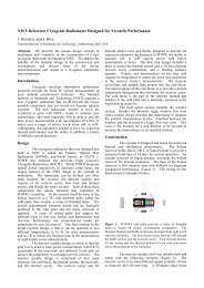
NIST Reference Cryogenic Radiometer Designed for Versatile Performance<br />
J. Houston, and J. Rice<br />
National Institute of Standards and Technology, Gaithersburg, MD, USA<br />
Abstract. We describe the unique design concept of<br />
modularity and versatility in the construction of a new<br />
cryogenic radiometer developed at NIST. We address the<br />
benefits of the modular design in the construction and<br />
development and discuss some of the device<br />
characterizations and results of a cryogenic radiometer<br />
intercomparison.<br />
Introduction<br />
Cryogenic electrical substitution radiometers<br />
presently provide the basis for optical measurements in<br />
most national measurement institutes. The National<br />
Institute of Standards and Technology (NIST) required a<br />
new cryogenic radiometer that would provide the lowest<br />
possible uncertainty and yet would not become quickly<br />
obsolete. The new radiometer needed to have the<br />
versatility to grow with NIST’s needs, to embrace new<br />
technologies, and most especially still be able to provide<br />
laser power measurements with uncertainties of 0.01% or<br />
better over a range of power levels from μW to mW.<br />
Additionally, the radiometer needed to have an improved<br />
thermal performance and the ability to calibrate a variety<br />
of different optical detectors.<br />
Design<br />
The cryogenic radiometer that was designed and<br />
built at NIST is called the Primary Optical Watt<br />
Radiometer (POWR), previously known as HACR 2.<br />
POWR has replaced the previous High Accuracy<br />
Cryogenic Radiometer (HACR). While POWR measures<br />
optical power responsivity of detectors using lasers, the<br />
unique element of the NIST design is the concept of<br />
modularity. The radiometer can be divided into three<br />
main sections: the cryostat, the detector module that<br />
consists of the receiver cavity, heat sink, and thermal<br />
anchor, and finally the optics section. Each of these three<br />
components has design elements that contribute to<br />
POWR’s versatility. In the cryostat itself the base of the<br />
helium reservoir is a 381 mm diameter plate, called the<br />
cold plate, that contains a series of threaded holes. The<br />
critical experimental elements mount and are thermally<br />
anchored to the cold plate and are easily removed or<br />
replaced. A series of wiring paths that support the<br />
different types of four-wire heaters and thermometry and<br />
two-wire thermometry are connectorized inside the<br />
radiometer for ease of removal. One final element in the<br />
actual cryostat design is that it can be operated at two<br />
different temperatures, at 4.2 K for most measurements<br />
and at 2 K to reduce the thermal noise in the measurement<br />
of significantly lower optical powers.<br />
The detector module design is critical in that the<br />
goal was to achieve laser power measurements with 0.01%<br />
uncertainty at the microwatt and milliwatt levels and yet<br />
achieve the modularity to exchange receiver cavities. The<br />
detector module design is comprised of four main<br />
elements: the cold block, the thermal anchor, the heat sink,<br />
and the receiver cavity. The receiver cavity and the<br />
thermal anchor were specifically designed to provide the<br />
expected radiometric performance of POWR, the ability to<br />
measure μW to mW optical power with 0.01%<br />
uncertainties or better. The heat sink design includes a<br />
place to mount the thermal anchor and a 20 mm diameter<br />
receiver cavity combination, and a limiting entrance<br />
aperture. Heaters and thermometers on the heat sink<br />
regulate its temperature to reduce the noise and uncertainty<br />
in the receiver cavity’s measurements. The receiver<br />
cavity/heat sink module then mounts into the cold block.<br />
The main purpose of the cold block is to provide a helium<br />
temperature background that surrounds the receiver cavity.<br />
The cold block is the part of the detector module that<br />
attaches to the cold plate and is thermally anchored to the<br />
liquid helium reservoir.<br />
The front optics section includes the window<br />
section. Besides the Brewster angle window, this front<br />
optics section design provides the opportunity to measure<br />
the window transmittance in-situ. Attached between the<br />
window and the cryostat is a large, five-way cross. This<br />
cross is the location for a trap detector to be inserted to<br />
measure the transmittance of an installed window.<br />
Construction<br />
The cryostat is designed and tested for improved<br />
thermal and mechanical performance. The helium<br />
reservoir holds almost 100 L of liquid helium to provide a<br />
measured hold time of 14 days for normal operation. An<br />
annular reservoir of liquid nitrogen surrounds the helium<br />
reservoir for thermal insulation. The base of the helium<br />
reservoir is 304 stainless steel with helicoils for longterm<br />
mechanical performance (Figure 1). The wiring for the<br />
thermometry is phosphor bronze.<br />
Figure 1. POWR’s cold plate provides a series of threaded<br />
holes to mount all critical experimental components. The<br />
Detector Module and baffle section are mounted from right to left<br />
onto the cold plate. The laser beam enters from the left through<br />
the baffle section into the receiver cavity. Four thermal anchors<br />
for the wiring and two thermal anchors for the wire connectors<br />
are on the perimeter of the cold plate.<br />
The detector module itself was initially built and<br />
tested with the following elements (Figure 2). The<br />
receiver cavity is an electroformed copper cylinder, 20 mm<br />
in diameter and 150 mm long. A 30-degree slant closes<br />
Proceedings NEWRAD, 17-19 October 2005, Davos, Switzerland 23
off the back end of the cavity. The cavity is coated with a<br />
specular black paint. One heater is noninductivally<br />
wrapped on the closed end of the cavity, with another two<br />
chip heaters attached to the back slant. The germanium<br />
resistance thermometer is located on the cylinder barrel.<br />
All wiring from the detector module ends in connectors to<br />
provide easy detachment. A Kapton thermal anchor<br />
attaches the receiver cavity to the heat sink. Both the heat<br />
sink and the cold block are made of OFHC copper that is<br />
plated first with nickel and then gold. All surfaces are<br />
highly polished and reflective to reduce any radiative<br />
effects.<br />
Figure 2. The Detector Module is on the right half with the<br />
receiving cavity mounted onto the heat sink and surrounded by<br />
the cold block. On the left side is the baffle section with the off<br />
axis parabolic mirror and silicon photodiode combination.<br />
While the majority of the detector section itself<br />
remained unchanged, the receiver cavity and some<br />
thermometry evolved until POWR achieved the desired<br />
performance. The receiver cavity evolved until the<br />
performance was achieved in Detector Module 3.<br />
Additionally the wiring was changed to provide the<br />
capability for a feed-forward temperature control loop<br />
algorithm.<br />
One element designed into the measurement is<br />
the determination of the light scatter magnitude. This<br />
was achieved by placing a baffle section attached to the<br />
cold block located directly in front of the detector module,<br />
and additional baffles along the interior optical path. The<br />
baffle section collects the scattered radiation with an off<br />
axis parabolic mirror that surrounds the laser beam and<br />
reflects the scattered radiation into a silicon photodiode<br />
for measurement.<br />
The evolving detector module demonstrates the<br />
benefits of the modular design. While the construction<br />
and wiring of each detector module required days of work,<br />
the actual exchange of detector modules in the cryostat<br />
itself took less than one day. A connectorized detector<br />
module was easily replaced in the cryostat.<br />
Measurements<br />
The detector modules after being painted and<br />
built were measured outside the cryostat for reflectance<br />
(from which absorptance is inferred) at three different<br />
wavelengths. The measurements were done against NIST<br />
PTFE reflectance standards. The final cavity had<br />
absorptances of 0.999995 at 633 nm. Because the<br />
radiometer is versatile, the detector module can easily be<br />
removed and tested over time to see if the paint and<br />
absorptance is stable.<br />
Once Detector Module 3 was installed into the<br />
cryostat two types of measurements commenced, first the<br />
characterization of the electrical to optical equivalence and<br />
second the calibration of traps. A full discussion of<br />
POWRs characterization is to be discussed in a future<br />
paper. The calibration of the traps was performed in two<br />
different configurations, one with the traps on a separate<br />
translation stage in front of the window, and a second with<br />
the traps in the optical plane of the receiver cavity on the<br />
translation stage that moves the radiometer itself. In the<br />
end the second setup was used. The same traps measured<br />
by POWR were measured by other cryogenic radiometers<br />
at NIST and the calibrations agreed within 0.01% to 0.02%<br />
which is well within the three radiometers uncertainties<br />
(Table 1). A full discussion of this intercomparison will<br />
be published later.<br />
Table 1. Results of a comparison of three cryogenic<br />
radiometers measuring trap detector, presented in terms of<br />
the difference from the POWR-measured responsivity of a<br />
silicon photodiode trap detector.<br />
Cryogenic 488 nm 514 nm 633 nm<br />
radiometer<br />
L1 ACR -0.021% -0.002% -0.011%<br />
LOCR -0.005% 0.020% 0.001%<br />
The radiometer Brewster-angle window was<br />
measured in-situ, but not in vacuum. The Brewster-angle<br />
window was optimized for 633 nm and maintained in that<br />
alignment for the calibrations. The transmittances at each<br />
wavelength were measured before and after the trap<br />
calibrations. For the window transmittance measurement<br />
the trap was inserted inside the five way cross and aligned<br />
to the laser beam. The trap was removed for POWRs<br />
calibration cycles. A typical window transmittance was<br />
0.999921 with an uncertainty of .0012% at 633 nm.<br />
Conclusion<br />
A new NIST reference cryogenic radiometer,<br />
POWR, has been developed. The goal in the<br />
development of the POWR was to have the versatility in<br />
design so that detector modules could be exchanged while<br />
providing optical power measurements at uncertainties of<br />
0.01% or better. Final characterization measurements on<br />
Detector Module 3 (Rice, et al.) are soon to be completed,<br />
but the exchanging of the first three detector modules and<br />
the performance of the third module shows that POWR has<br />
achieved this goal. In the future, as technology changes<br />
or there are special requests for measurements at different<br />
power levels or wavelengths, POWR will be able to meet<br />
the needs.<br />
Acknowledgements. We would like to thank Steven<br />
Lorentz and Joe O’Connell for their contributions in the<br />
construction of POWR. Responsivity measurements from<br />
the L1 ACR were provided by Steve Brown and Keith<br />
Lykke, and for the LOCR by David Livigni. We would<br />
like to thank Albert C. Parr, Chief of the Optical<br />
Technology Division at NIST, for his continued support of<br />
this project.<br />
24
Measurement of the absorptance of a cryogenic radiometer cavity in the visible and near infrared<br />
(NIR)<br />
M. López 1,2 , H. Hofer 2 and S. Kück 2<br />
1 Centro Nacional de Metrología, 76241 Querétaro, México<br />
2 Physikalisch-Technische Bundesanstalt, Braunschweig 381116, Germany<br />
Abstract. Results of the measurement of the absorptance of<br />
a LaseRad cavity used as an absorber in the cryogenic<br />
radiometer in the Physikalisch-Technische Bundesanstalt<br />
(PTB) are presented. The measurements were carried out at<br />
several laser wavelengths in the visible and near infrared<br />
(NIR); at 633 nm, 1280 nm - 1360 nm and 1480 nm –<br />
1620 nm. The absorptance of 0.999885 ± 3.0 × 10 -6<br />
measured at 633 nm matches very well with the value of<br />
0.999879 ± 1.0 × 10 -5 reported by the manufacturer<br />
(Cambridge Research & Instrumentation, Inc.) . In the NIR<br />
the absorptance is approx. 1.1 × 10 -4 ± 3 × 10 -6 lower than at<br />
633 nm, which is significant for high accuracy<br />
measurements. In the wavelength range from 1280 nm to<br />
1620 nm, the absorptance varies by 19 × 10 -6 which is<br />
almost negligible for this wavelength range.<br />
1. Introduction<br />
In the last decade, the demand for calibration services in the<br />
field of optical communication through optical fiber had<br />
increased substantially and consequently also the need of<br />
lower measurement uncertainties. Therefore, the PTB and<br />
also other National Metrology Institutes (NMIs) work on<br />
transfer standards (Ge and InGaAs photodiodes) calibrated<br />
directly against the Cryogenic Radiometer (CR), the<br />
primary standard for optical power measurement, in the<br />
near infrared (mainly around 1300 nm and 1550 nm) [1-4].<br />
Thus, uncertainties below 4×10 -4 have been obtained in<br />
these wavelength ranges. In general, two correction factors<br />
contribute to the CR measurement accuracy: the non-ideal<br />
absorption coefficient α of the cavity and the non-ideal<br />
transmittance τ of the Brewster-angle window. Usually, the<br />
absorption coefficient at 632.8 nm reported by the<br />
manufacturer is used in the PTB and other NMIs and<br />
considered to be constant for other wavelength ranges [1-4].<br />
However, to achieve lower measurement uncertainties, it is<br />
necessary to know the exact spectral distribution of both<br />
coefficients.<br />
In this paper, we report the absorption coefficient<br />
measurements of the LaseRad cavity carried out at several<br />
wavelengths: 632.8 nm, 1280 nm – 1360 nm and 1480 nm –<br />
1620 nm. The cavity under measurement was purchased by<br />
the PTB from the manufacturer of our CR, Cambridge<br />
Research & Instrumentation (CRI), separately for this<br />
purpose. It has the same characteristics as the cavity<br />
contained in the CR; it is constructed from an oxygen-free<br />
high-conductivity copper (OFHC) tube and blackened with<br />
Chemglaze Z-302 black paint [5].<br />
2. Measurement method<br />
The absorption coefficient of the cavity is determined from<br />
the measurement of the diffuse reflection ρ by the simple<br />
formula α = 1 – ρ. The measurement is carried out using a<br />
four-ports integrating sphere and a photodetector (see<br />
Figure 1). In the visible wavelength range, a He-Ne laser<br />
operating at 632.8 is used as radiation source. The beam<br />
irradiates a 2-mm diameter circular aperture and is imaged<br />
1:1 by a 200 mm focal length lens - passing through the<br />
sphere - into the cavity. To reduce the fluctuation of the<br />
laser power, an external stabilizer and a monitor detector are<br />
used. An attenuator and a polarizer are used to maintain the<br />
power level and the linear polarization of the laser beam.<br />
The detector placed on the sphere for the measurement at<br />
632.8 nm is a Si detector of 5-mm diameter (Hamamatsu<br />
S1227 66BR). In the IR, two tunable diode laser sources<br />
(Agilent 81600B) were used, whose wavelength were<br />
adjusted from 1280 nm to 1360 nm and from 1480 nm to<br />
1620 nm, respectively. The outputs of the laser sources are<br />
fiber-optic connectors; therefore an external collimator with<br />
a fiber-optic pigtail is used to collimate the laser beam. The<br />
laser stabilizer, used during the measurement at 632.8 nm, is<br />
not needed for the measurement in the IR, instead, the<br />
collimator was placed in front of the aperture and polarizer,<br />
see also Figure 1. An InGaAs photodiode (Telcom<br />
35PD5M) of 5-mm diameter placed on the sphere carries<br />
out the measurement of the reflected fluxes.<br />
The reflectance measurement of the cavity is done as<br />
follows: in a first step, a laser beam illuminates the cavity<br />
attached to the sample port. The reflected flux is diffusely<br />
emitted from the cavity and is collected by the integrating<br />
sphere finally generating a signal S c is generated by the<br />
photodetector. In this scheme the white standard is attached<br />
to the supplementary port. In a second step, the cavity and<br />
the white standard interchange their ports from where a<br />
second signal is generated S s . A third signal S 0 is measured<br />
by taking off the white standard from the sample port. From<br />
the ratio between those signals one can get the reflection<br />
coefficient of the cavity, ρ c :<br />
Sc<br />
− S0<br />
ρ =<br />
c<br />
S<br />
⋅<br />
c<br />
− S0 ρ<br />
s<br />
δ → ρc<br />
= ⋅<br />
(1)<br />
S − S ρ<br />
S − S δ<br />
s<br />
0<br />
s<br />
s<br />
where ρ s is the reflection of the white standard and δ is the<br />
correction factor due to possible changes of the geometrical<br />
conditions of the sphere between the two measurement<br />
processes. In our case, these conditions remain unchanged,<br />
so δ = 1.<br />
0<br />
Proceedings NEWRAD, 17-19 October 2005, Davos, Switzerland 25
Transimpedance<br />
amplifier<br />
Digital-voltmeter<br />
Integrating<br />
sphere<br />
Photodetector (Si, InGaAs)<br />
Monitor detector<br />
Polariser<br />
Attenuator<br />
Radiometer<br />
cavity<br />
Baffle<br />
White<br />
standard<br />
Beam<br />
splitter<br />
Lens<br />
Laser<br />
-stabilizer<br />
Aperture<br />
Fiber optic<br />
He-Ne<br />
Laser<br />
Polariser<br />
Collimator<br />
Tunable laser<br />
1260 nm – 1360nm<br />
1460 nm – 1570 nm<br />
Aperture<br />
Figure1. Experimental set-up used to measure the diffuse reflectance of the radiometer cavity.<br />
3. Measurement results<br />
Figure 2 shows the results of the absorption coefficient<br />
measurements of the cavity. At 632.8 nm the absorption<br />
coefficient measured is 0.999885, which matches very well<br />
with the value reported by the manufacturer (0.999879). At<br />
the infrared wavelengths, the absorption coefficient varies<br />
from 0.999765 to 0.999785 between the wavelength ranges<br />
of 1280 nm – 1360 nm and 1480 nm – 1620 nm,<br />
respectively. The deviation observed for these ranges is<br />
19 × 10 –6 , which means that in this spectral range the value<br />
of the absorption coefficient is practically flat. Thus, for the<br />
whole NIR wavelength range, in principle a value of<br />
0.999777 ± 0.000014 (k=1) can be used. Although no<br />
significant difference in the absorptance within the infrared<br />
spectral range investigated is observed, the mean value for<br />
the NIR range is about 1.1 × 10 -4 lower than the value at 633<br />
nm. This difference can be significant in the total correction<br />
factor of the CR, especially when one wish to reach<br />
uncertainties lower than 10 -4 .<br />
Absorption coefficient<br />
0.99990<br />
0.99988<br />
0.99986<br />
0.99984<br />
0.99982<br />
0.99980<br />
0.99978<br />
Absorption coefficient<br />
0.99980<br />
0.99978<br />
0.99976<br />
1250 1300 1350 1400 1450 1500 1550 1600 1650<br />
Wavelength (nm)<br />
The values of the absorption coefficient of a radiometer<br />
cavity in the visible and near infrared are presented. The<br />
results indicate that in the NIR, the absorption coefficient of<br />
the cavity is 1.1 × 10 -4 lower than in the visible wavelength<br />
(632.8 nm).The characterization of the cavity will allow us<br />
to correct the absorption coefficient of the LaseRad cavity<br />
of the PTB cryogenic radiometer in the NIR. Thus, we<br />
expect to reach uncertainties lower than 3×10 -4 for these<br />
wavelength ranges.<br />
Acknowledgments The authors would like to thank the Deutscher<br />
Akademischer Austauschdienst (DAAD) for the support granted<br />
to this project through the Ph.D. scholarship A/03/18297. We also<br />
wish to thank the 4.52 Reflection measurements work group of the<br />
PTB for the white standard calibration.<br />
References<br />
[1] K. D. Stock and H. Hofer 1993 Metrologia 30 291-296<br />
[2] E. G. Atkinson and D. J. Butler 1998 Metrologia 35 241-245<br />
[3] L. Werner, R. Friedrich, U. Johannsen and A. Steiger 2000<br />
Metrologia 37 523-526<br />
[4] P. Corredera, J. Campos, M. L. Hernanz, J.L. Fontecha, A.<br />
Pons and A. Corróns 1998 Metrologia 35 273-277<br />
[5] Product of Lord Corporation, Industrial Coatings Division,<br />
2000 West Grandview Boulevard, Erie, PA 16514-0038<br />
0.99976<br />
600 800 1000 1200 1400 1600<br />
Wavelength (nm)<br />
Figure 2. Absorption coefficients of the cavity measured in the<br />
visible and near infrared wavelengths. The error bars correspond<br />
to the standard uncertainty of the measurement. Open circle:<br />
manufacturer result.<br />
4. Conclusions<br />
26
Grooves for Emissivity and Absorptivity Enhancement in High Performance<br />
Cavity Sources and Radiometers<br />
E. Usadi, and R. Montgomery<br />
National Physical Laboratory, Middlesex, UK<br />
Abstract The emissivity of a flat emitting plate, and<br />
similarly the absorptivity of a flat absorbing plate, are well<br />
known to be enhanced by machining grooves into the flat<br />
surface. In addition to minimising retroreflection from<br />
cavities with flat end plates, grooves increase the number<br />
of absorbing surfaces a typical ray hits before exiting such<br />
a cavity. However two factors, generally overlooked in<br />
groove design, may become important for the design of<br />
ultrahigh emissivity or absorptivity cavities: (1) axial rays<br />
intersect the grooved surface off-normal, where the surface<br />
reflectivity is potentially much higher than at normal<br />
incidence, and (2) the groove angle may be difficult to<br />
define accurately, especially after the application of<br />
absorbing paint. This paper will describe ray tracing<br />
work showing that small changes in the groove angle can<br />
lead to dramatic changes in cavity emissivity or<br />
absorptivity (up to several percent) for relatively highly<br />
reflecting surface coatings. While this is not normally a<br />
problem for common black coatings and for radiation<br />
within the visible and IR spectrum below 100 microns, it<br />
may become an important correction for longer<br />
wavelengths where many common coatings become more<br />
highly reflecting and specular, especially at off-normal<br />
incidence. Since a room temperature thermal source has a<br />
significant fraction of emission at these long wavelengths,<br />
this may lead to an overall correction at the 1 – 100 ppm<br />
level when measuring the radiance of such sources.<br />
Acknowledgments The authors would like to thank the<br />
Department of Trade and Industry of the UK for supporting this<br />
work.<br />
Proceedings NEWRAD, 17-19 October 2005, Davos, Switzerland 27
PULSED UV SPECTRORADIOMETRY<br />
WITH THE PRIMARY STANDARD<br />
SYNCHROTRON RADIATION SOURCES<br />
OF HIGH INTENSITY<br />
S. Anevsky, V. Ivanov, O. Minaeva, V. Sapritsky, Y. Zolotarevsky<br />
All-Russian Research Institute for Optical and Physical Measurements<br />
(VNIIOFI)<br />
Ozernay St., 46, Moscow, 119361, Russia<br />
Principal Contact: S. Anevsky<br />
Phone: 007-095-437-3183<br />
E-mail: Anevsky @ vniiofi.ru<br />
VNIIOFI has developed table-top electron accelerators – primary synchrotron<br />
radiation sources as national standards of spectral radiance and irradiance in the<br />
range of vacuum and air UV. The creation of pulsed synchrotron radiation sources<br />
with strong magnetic field for UV spectroradiometry is based on the use of available<br />
magnetic field with induction of 10-18 T and pulse duration 3-300 µs . The small<br />
duration of synchrotron radiation pulse permit to use the strong magnetic field and<br />
to operate with low vacuum level. The value of optimal radius is about few<br />
centimeters and particles energy is about 30-100 MeV for UV spectral region. The<br />
high level of UV spectral flux makes pulsed synchrotron radiation source quite<br />
useful for calibration of the secondary standard plasma sources as capillary<br />
discharge with evaporated walls and plasma focus. The synchrotron radiation<br />
source beam diagnostics includes the measurements of orbit radius, electrons<br />
energy, particles number and radial, axial, phase dimensions of electron bunch.<br />
VNIIOFI for many years attempted to create and improve special small-size<br />
standard synchrotron radiation sources available for metrological laboratory based<br />
on technique of strong magnetic field generation: uniron low-inductive magnet<br />
system. The further development of synchrotron radiation sources is connected with<br />
optimization acсelerator parameters in order to rich spectral range of EUV about<br />
13.5 nm in regime of 1 Hz frequency.<br />
Proceedings NEWRAD, 17-19 October 2005, Davos, Switzerland 29
New apparatus for the spectral radiant power calibration at CMS in Taiwan<br />
Hsueh-Ling Yu and Shau-Wei Hsu<br />
Center for Measurement Standards/ITRI, Hsinchu, Taiwan, R.O.C.<br />
Abstract. A monochromator-based cryogenic<br />
radiometer (CR) facility has been established at the<br />
Center for Measurement Standards (CMS) in<br />
Taiwan, for calibrating the spectral radiant power<br />
responsivity of standard detectors. The design<br />
features of the new apparatus and uncertainty<br />
analysis are discussed. Presently the Si and Ge<br />
detectors can be calibrated in the spectral ranges<br />
350 nm to 1100 nm and 800 nm to 1700 nm,<br />
respectively. The measured results of the Si and<br />
Ge detectors using the new apparatus were<br />
compared with the results in the NPL calibration<br />
reports to check the performance of the new<br />
apparatus. The agreement is better than 0.5 % for<br />
the Ge detector and 0.2 % for the Si detector.<br />
1. Introduction<br />
A new facility for the monochromator-based<br />
cryogenic radiometer [1,2] has been set up in the<br />
CMS in Taiwan. A monochromator-based<br />
cryogenic radiometer, rather than laser-based one,<br />
was established to calibrate the responsivity<br />
because of cost, convenience, and the flexibility<br />
of future applications. Instead of rotating the<br />
bellows to translate the CR and the transfer<br />
detectors as presented in Refs. 1 and 2, a linear<br />
translation stage is utilized to move the CR and<br />
the transfer detector to the focal position of the<br />
monochromatoe-source. The design of the new<br />
apparatus allows three detectors to be calibrated<br />
in a single measurement process to save time and<br />
liquid helium (LHe). The 1σ uncertainty of the<br />
Si detectors is approximately 0.2 % and that of<br />
the Ge detectors is 0.3 %.<br />
2. Design features<br />
Figure 1 presents the schematic overview of the<br />
new apparatus. The whole apparatus can be<br />
grouped into three categories - the CR, the<br />
detector stage, and the light source. The CR<br />
was manufactured by L-1 Standards and<br />
Technology, Inc. The hold time of LHe is well<br />
longer than 80 hours and the LN 2 for the cryostat<br />
can be refilled automatically. The relative<br />
standard uncertainty of the CR in radiant power<br />
measurement is less than 0.02 % for the<br />
monochromator-source.<br />
The detector stage was originally designed by<br />
CMS and manufactured by L-1 Inc. The<br />
transfer detectors were placed on a holder inside<br />
a six-way reducing cross and connected to the<br />
feedthrough. It allows three detectors<br />
(depending on the size of the transfer detector) to<br />
be placed inside the cross. A thermistor was<br />
mounted on the holder to monitor the ambient<br />
temperature around the transfer detector. It<br />
showed that the ambient temperature was<br />
approximately 21.5°C during measurements.<br />
The welded bellows and linear translation stage<br />
were utilized to move the CR and the transfer<br />
detector to the focal position of the<br />
monochromator-source with an overall traveling<br />
length of around 31 cm. The X- and Z-axes of<br />
the XYZ manipulator were used to locate the<br />
center of and test the uniformity of the transfer<br />
detector. A flat fused quartz entrance window<br />
is placed in front of the welded bellows to<br />
eliminate the uncertainty form the window<br />
transmission.<br />
A quartz- tungsten-halogen (QTH) lamp and an<br />
Xe lamp were used to cover the wavelength<br />
ranges from 400 nm to 1700 nm and below<br />
400 nm, respectively. The F number of the<br />
monochromator is 4. After mirrors M1 to M4,<br />
the F number of the monochromator beam was<br />
about 13. Therefore, it could be absorbed<br />
totally by the CR (F/8). An aperture with a<br />
diameter of 1.9 mm was placed in front the exit<br />
port of the monochromator to make the spot<br />
diameter of the monochromator beam<br />
approximately 3 mm at its focal position.<br />
3. Measured results and uuncertainty<br />
analysis<br />
To verify the performance of the new apparatus,<br />
the measurements of the spectral responsivity on<br />
two NPL-calibrated Si and Ge detectors were<br />
carried out. The measured procedures involved<br />
the photocurrent measurements of the detectors,<br />
radiant power measurements with the CR, and<br />
then obtaining the spectral radiant power<br />
responsivities of the detectors. The measured<br />
results of Si and Ge detectors against the<br />
apparatus were compared with the results in their<br />
NPL calibration reports. The discrepancy is<br />
Proceedings NEWRAD, 17-19 October 2005, Davos Switzerland 31
Detector stage<br />
Figure 1. Schematic diagram of CMS new monochromator-based cryogenic radiometer for spectral radiant power calibration<br />
less than 0.5 % for the Ge detector and 0.2 % for<br />
Si detector. The error sources include the<br />
bandwidth effect, wavelength accuracy, and stray<br />
light of the monochromator; the uniformity,<br />
polarization effect, and output current accuracy<br />
of the transfer detector; the measured accuracy of<br />
the radiant power of the CR. Table 1 lists the<br />
uncertainty budget and the conditions of the<br />
estimation.<br />
Table 1. Uncertainty budget of the monochromator-based<br />
spectral radiant power responsivity calibration<br />
⎯⎯⎯⎯⎯⎯⎯⎯⎯⎯⎯⎯⎯⎯⎯⎯⎯⎯⎯⎯⎯⎯⎯⎯⎯<br />
Source<br />
relative standard uncertainty<br />
⎯⎯⎯⎯⎯⎯⎯⎯⎯⎯⎯⎯⎯⎯⎯⎯⎯⎯⎯⎯⎯⎯⎯⎯⎯<br />
Power measurement CR 0.02 %<br />
(monochromator-source)<br />
Bandwidth effect 0.0001 % (Si; 400 nm-1000 nm)<br />
(Δ=1.5nm for Si 0.01 % (Si; other range)<br />
Δ=3.5nm for Ge) 0.001 % (Ge; 800 nm-1500 nm)<br />
0.04 % (Ge; other range)<br />
Wavelength accuracy 0.03 % (Si; 400 nm-1000 nm)<br />
(±0.1nm for Si 0.2 % (Si; other range)<br />
±0.2nm for Ge) 0.05 % (Ge; 800 nm-1500 nm)<br />
0.3 % (Ge; other range)<br />
Uniformity 0.02 % (Si)<br />
(0.3 mm deviation from 0.08 % (Ge)<br />
the center of the diode)<br />
Polarization 0.01 %<br />
(F/13, 0.2° incident angle)<br />
Current measurement 0.0005 %<br />
Repeatability 0.05 % (Si; 400 nm-1100 nm)<br />
0.1 % (Si;
VUV and Soft X-ray metrology stations at the International Siberian<br />
Synchrotron Radiation centre<br />
V. I. Buhtiyarov, N. G. Gavrilov, N. A. Gentselev, B. G. Goldenberg, G. N. Kulipanov, A. A. Legkodymov, V.<br />
V. Lyakh, O. I. Meshkov, A. I. Nizovsky, A. D. Nikolenko, 1 V. F. Pindyurin, I. V. Poletaev, E. P.<br />
Ya. V. Rakshun, Semenov, M. A. Kholopov, V. A. Chernov, M. A. Sheromov<br />
Budker Institute of Nuclear Physics, 630090 Novosibirsk, Russian Federation<br />
At present, there are two experimental stations under<br />
construction at the International Siberian Synchrotron<br />
Radiation centre (Novosibirsk, Russia). They are intended<br />
for metrology work in the VUV and soft X-ray spectral<br />
ranges with the application of SR from the storage rings of<br />
VEPP-3 and VEPP-4.<br />
The soft X-ray metrology station (the spectral range is from<br />
80 to 5000 eV) at the storage ring of VEPP-3 makes it<br />
possible to perform very different works for time and<br />
absolute spectral calibration of various X-ray detectors as<br />
well as absolute and relative certification of elements of<br />
X-ray optics (multi-layer mirrors, multi-layer lattices,<br />
absorption film filters etc). The first line of the station has<br />
been commissioned already. Certification of power of the<br />
incoming “white” SR beam is under way now. The<br />
monochromator is being prepared for commissioning. Its<br />
design allows one to use both multi-layer mirrors as wall as<br />
crystals as x-ray optical spectral elements. The first<br />
experiment is supposed to deal with experimental<br />
investigation of possibility of broadening the<br />
self-calibration method [1] in order to use it later to calibrate<br />
scintillation counters. Another task of the first experiment<br />
is a preliminary test of elements of the Space Solar Patrol<br />
(SSP, made by SOI, St. Petersburg) [2] within the<br />
framework of ISTC project #2500, which is devoted to SSP<br />
calibration.<br />
The VUV and soft X-ray metrology station with the<br />
application of SR from the VEPP-4 storage ring is being<br />
developed at the moment. In contrast to the similar station at<br />
VEPP-3, the spectral range of this station (from 10 to 1000<br />
eV) allows one to perform calibration measurements in the<br />
VUV range. Due to partial overlapping of the spectral<br />
ranges of these stations one can check at one station a<br />
calibration done at the other. The metrology station of<br />
VEPP-4 is to be equipped with two monochromators. Those<br />
are a lattice one (the spectral range is 10 to 100eV) and a<br />
two-mirror monochromator based on multi-layer mirrors<br />
(the spectral range is 80 to 1500 eV). Since the experimental<br />
space of the station is rather large (about 0.7х05х0.5 m 3 ) it<br />
will be possible to perform calibration of quite big-sized<br />
space equipment). The first series of prospective<br />
experimental works is to be devoted to absolute calibration<br />
of the equipment of the space solar patrol. The station is<br />
supposed to be commissioned in the first half of the year<br />
2006.<br />
The SR beamlines of both the stations have been made by a<br />
technology without windows. They are equipped with a fast<br />
closing system for the case of a vacuum damage at the<br />
station and with differential vacuum pump-down. That<br />
makes it possible to investigate unbakeable samples in the<br />
experimental spaces at an operational pressure up to 10 -4 Pa.<br />
This report presents the optical schemes of both the stations,<br />
their main parameters and design photon flows for different<br />
operation regimes.<br />
1. Krumrey M., Tegeler E., Nuclear Instruments and<br />
Methods in Physics Research A288 (1990) 114-118<br />
2. Avakyan S., at al. Sensors, Systems and Next<br />
Generation Satellites, Proc. SPIE, v. 3870, pp. 451-461,<br />
1999.<br />
1 A.D.Nikolenko@inp.nsk.su<br />
Proceedings NEWRAD, 17-19 October 2005, Davos, Switzerland 33
Experimental study of application of the self-calibration method to certify<br />
absolute spectral sensitivity of a scintillation counter in the soft X-ray range.<br />
A.D. Nikolenko 1 , V. F. Pindyurin, V. A. Chernov, V.V. Lyakh.<br />
Budker Institute of Nuclear Physics, 630090 Novosibirsk, Russian Federation<br />
The self-calibration method earlier has been successfully<br />
developed by the PTB team (Berlin, Germany) for absolute<br />
certification of spectral sensitivity of semiconductor diodes<br />
[1, 2]. In these works, parameters required to determine<br />
spectral sensitivity of a detector (the dead layer on the<br />
surface of detector and sensitive area thickness) were found<br />
via measuring the spectral response of the detector at<br />
different angles of radiation incidence on the receiving<br />
surface of the detector.<br />
In the case of absolute calibration of a scintillation counter<br />
(SC), the spectral sensitivity of the detector is described<br />
with a slightly more complicated model. Beside the dead<br />
layer thickness, the model includes the spectral efficiency of<br />
the scintillator, the efficiency of collection of visible<br />
photons on the photo-cathode, the photo-cathode efficiency<br />
and particularities of signal amplification by the dynode<br />
system of the photomultiplier. The present work describes<br />
the preliminary measurement of the spectral sensitivity of a<br />
SC based on a photomultiplier with guaranteed one-electron<br />
peak FEU-130 with a YAP scintillator. The dead layer<br />
thickness of the scintillator was determined in the same way<br />
as in [1, 2]. The efficiency of registration of X-ray photons<br />
that passed the dead layer of scintillator is found by the form<br />
of the amplitude spectrum of anode pulses of the<br />
photomultiplier.<br />
The evident explanation of this technique briefly can be<br />
described as follows: x-ray quantum absorbed by the<br />
scintillator, is generated the light flash, which is registered<br />
by the photoelectron multiplier (PEM). In case of soft x-ray<br />
photon the number of the visible photons in each flare is not<br />
big (from units up to several tens depending on energy of<br />
x-ray photon and characteristics of scintillator) and<br />
consequently the significant part of useful pulses of PEM is<br />
situated in the one-electron peak. It means, that the average<br />
number of optical photons, be registered by the PEM from<br />
the such flash, is less then 1. Nevertheless, in a amplitude<br />
spectrum of pulse signal of PEM, we can observe not only<br />
one-electron peak, but also the effect of presence of twoand<br />
three-electron peaks. Using the relative intensity of<br />
these peaks we can to calculate the probability of<br />
registration of every x-ray photon which has penetrated<br />
through a dead layer and was registered in the scintillator.<br />
This method does not allow one to separate the contribution<br />
of each of the SC parameters that influence its efficiency but<br />
it gives the absolute efficiency of a particular SC. The<br />
advantage of this method is that all the measurements are<br />
relative ones and can be performed on any available source<br />
of monochromatic soft X-radiation. A disadvantage of the<br />
method is its low accuracy (estimated to be not better than<br />
10%).<br />
The experimental checking of this method were performed<br />
at the Soft X-ray metrology station at the VEPP-3 storage<br />
ring for the few different energy of photons from 100 to<br />
1500 eV. In the near future, as expect, the station is to be<br />
equipped with reference detectors with a calibrated spectral<br />
sensitivity, which will allow one to check correctness of<br />
calibration.<br />
1. Krumrey M., Tegeler E., Rev. Sci. Instrum. 1992 63,<br />
797-801<br />
2. Krumrey M., Tegeler E., Nuclear Instruments and<br />
Methods in Physics Research A288 (1990) 114-118<br />
1 A.D.Nikolenko@inp.nsk.su<br />
Proceedings NEWRAD, 17-19 October 2005, Davos, Switzerland 35
The metrology line on the SOLEIL synchrotron facility<br />
M. Lièvre, M-C. Lépy, B. Rougié, J-R. Filtz, J. Bastie<br />
LNE, Paris, France<br />
M. Idir, T. Moreno, P. Mercere<br />
SOLEIL, Gif sur Yvette, France<br />
Abstract. In the new SOLEIL synchrotron facility one<br />
line will be devoted to metrology, providing 3 branches for<br />
hard-X, X-UV and UV-VUV radiation applications. So<br />
metrology between about 5 eV to 14 keV (250 nm down to<br />
0.09 nm) will benefit from this calculable radiation source.<br />
The UV-VUV branch is expected towards the end of 2006.<br />
Introducing SOLEIL<br />
SOLEIL (Source Optimisée de Lumière d’Energie<br />
Intermédiaire du LURE = Optimized source of mid-level<br />
energy light in LURE) is a 3rd generation 2.75 GeV<br />
synchrotron. Building began in 2000, and the first lines are<br />
to open end of 2006, followed by 14 others between 2006<br />
and 2010 - potentially 40. It is designed and managed from<br />
the beginning to host international teams.<br />
This synchrotron facility built in the Optics Valley<br />
addresses new or increasing needs in X to UV applications<br />
such as X lasers or deep UV lithography. It will inherit and<br />
amplify the high level-activity of the former LURE<br />
synchrotron.<br />
The metrology line partners<br />
There will be 3 main users of this metrology line: SOLEIL<br />
itself for its own instruments testing, the French Agency<br />
for Nuclear Energy (CEA) and the National Test and<br />
Measurement Laboratory (LNE). LNE has taken over the<br />
former BNM since beginning of 2005, including now the<br />
BNM previous members. The 3 LNE laboratories involved<br />
in the metrology line are the LNE-LNHB, the LNE-INM<br />
and the former LNE.<br />
Radiation metrology benefits from the<br />
synchrotron radiation (SR) characteristics<br />
Situation in France<br />
Up to now, different laboratories (now members of LNE)<br />
have performed a metrological activity with SR. INM<br />
carried on its first SR applications in 1974, establishing an<br />
electric arc as a secondary standard by comparing both<br />
radiations. LNHB characterized and calibrated X detectors.<br />
And outside metrological laboratories, SR enabled several<br />
metrological achievements such as optical components and<br />
detectors qualification for hot plasma X diagnosis.<br />
Situation in other countries<br />
In Germany, PTB runs a well-known and important<br />
facility at BESSY II, providing monochromatic radiation<br />
between 3 eV and 15 keV. Due to the increasing demand<br />
for SR access, the MLS (Metrology Light Source) new<br />
facility is expected to complete BESSY II in 2008, with a<br />
spectrum ranging from infrared to deep UV.<br />
In the US, NIST uses the SURF III facility in<br />
Gaithersburg as a calculable source in the 50 to 260 nm<br />
range, and as a UV source calibration to a cryogenic<br />
radiometer in the 125 to 320 nm range.<br />
In Russia, the VEPP facility in Novosibirsk provides<br />
radiation between 120 and 400 nm for calibration of lamps<br />
and detectors.<br />
Other facilities are used for radiometry in China and<br />
Japan.<br />
One of the main issues for radiometry with SR is access to<br />
the beam: not enough available time or too scattered. And<br />
it is worse with operating requirements often incompatible<br />
with other more widespread SR applications.<br />
Characteristics of the SOLEIL SR<br />
The synchrotron radiation shows a broad continuous<br />
spectrum, and its characteristics (irradiance, polarization,<br />
coherence, beam profile and pulse shape) are calculable<br />
with the Schwinger theory, with an accuracy only limited<br />
by the determination accuracy of the influence parameters,<br />
which can be very good. The output power can be adjusted<br />
on a 12 orders of magnitude scale.<br />
The SR emits pulses with a 50 ps width every 140 ns<br />
typically. Polarization can be adjusted from linear to<br />
circular.<br />
Metrology line characteristics<br />
SOLEIL committed the “Optics” special team to initiate<br />
and assist the users in designing the metrology line. The<br />
general requirements are a well-collimated beam, a high<br />
spectral purity (almost no harmonics), very low scattering,<br />
and a large spectral range, especially for absolute<br />
calibration of detectors, relying on the possibility to<br />
calculate the exact incident power.<br />
The “Sources and Accelerators” special team will provide<br />
measurements and develop methods to perfectly control<br />
the key machine parameters, in agreement with the<br />
metrology users requirements.<br />
The 8-mrad beam available at the end of the magnet will<br />
be spatially split into 3 branches, each of them devoted to a<br />
specific energy (spectral) range.<br />
We will focus now on the VUV branch, and mention only<br />
for completeness sake the hard and soft X-ray lines.<br />
Proceedings NEWRAD, 17-19 October 2005, Davos, Switzerland 37
UV-VUV branch<br />
The design is not yet definitive. This branch is now<br />
optimized for the 7 to 70 eV (18 to 180 nm) range. The<br />
beam will pass through a monochromator yielding a<br />
simulated photon flux above 1.5.10 8 photons/s, with a peak<br />
of 10 11 photons/s at 50 nm, respectively above 1 nW and<br />
300 nW. At the output focus, the beam has a rectangular<br />
shape of 45 x 600 µm. The spectral width varies between<br />
20 and 120 pm.<br />
X-UV branch<br />
This branch will be operated between 30 eV and 2 keV,<br />
with a focused or collimated output beam, and a harmonic<br />
rejection rate of 0.1 %.<br />
Hard-X branch<br />
This branch will be operated between 0.5 and 12 keV, with<br />
a focused or collimated output beam.<br />
Uncertainties<br />
The main interest of SR in radiometry is the possibility to<br />
calculate the incident flux from the measurement of<br />
independent operating parameters. At BESSY, the<br />
combined uncertainty has been assessed to 0.2% at 1 eV<br />
and 0.35% at 5 keV. The dominant term belongs to the<br />
geometrical factors in the optical range, and to the<br />
electrical ring parameters in the X range.<br />
SOLEIL, private report, September 2004<br />
BNM, CEA, Laboratoire National Henri Becquerel, Intérêt du<br />
rayonnement synchrotron pour la métrologie – première<br />
approche, in French technical note LNHB/00/21 2ème<br />
version, 2000.<br />
Bastie, J, Coutin, J-M, Rougié, B, CNAM-INM, Contribution du<br />
BNM-INM à la préparation du projet métrologique sur<br />
SOLEIL, in French technical note, 2001<br />
Klein, R, Ulm, G, PTB, Abo-Bakr, M, Budz, P,<br />
Bürkmann-Gehrlein, Krämer, D, Rahn, J, Wüstefeld, G,<br />
Bessy Gmbh, The metrology Light Source of the Physikalisch<br />
Technische Bundesanstalt in Berlin-Adlershof, in the<br />
Proceedings of EPAC 2004, Lucerne, Switzerland, 2004<br />
Klein, R, Thornagel, R, Ulm, G, PTB, BESSY II operated as a<br />
primary source standard, in the Proceedings of EPAC 2004,<br />
Lucerne, Switzerland, 2004<br />
Wende, B, PTB, Radiometry with synchrotron radiation,<br />
Metrologia, 1995/1996, 32, 419-424<br />
Furst, M.L, Graves R.M., Hamilton, A, Hughey, L.R, Madden,<br />
R.P, Vest R.E. NIST Gaithersburg, MD, Trzeciak, W.S,<br />
Bosch, R.A, Synchrotron Radiation Center, Stoughton, WI,<br />
Greenler, L, Robl, P, Wahl, D, Physical Sciences Laboratory,<br />
Stoughton, WI, The Conversion of SURF II to SURF III, in<br />
the proceedings of the 1999 Particle Accelerator Conference,<br />
New-York, USA, 1999<br />
Of course there are other contributions to the final<br />
uncertainty in a complete calibration process. In the case<br />
of the already running synchrotron calibration facilities,<br />
the final uncertainty amounts generally to a few percent. It<br />
has not yet been assessed at SOLEIL.<br />
Expectation in UV Metrology<br />
The SOLEIL metrology line is expected to bring a<br />
breakthrough in UV calibrations through the realization of<br />
standard references for detector sensitivity and energetic<br />
source properties (radiance, irradiance, radiant intensity).<br />
It will of course enhance comparison possibilities with<br />
other international realizations of the same quantities, and<br />
the transfer to end-users. It is true that the main demand on<br />
UV radiometric calibration is still in the less energetic<br />
range (above 200 nm), where applications such as therapy<br />
or diagnostic will bring more stringent demand on<br />
accuracy. However the emerging applications in biology,<br />
industry or scientific research (astronomy, plasma,<br />
fluorescence) and above all in photolithography are<br />
driving calibration needs toward the VUV (vacuum<br />
ultraviolet) range. In this range only will the SR prove<br />
more efficient than laser or high temperature blackbody<br />
sources.<br />
References<br />
Lépy, M.C., Rougié, B., Lièvre, M., Stemmler, P., Marmoret, R.,<br />
Idir, M., Moreno, T., Polack, F., Ligne de métrologie sur<br />
38
Cryogenic radiometer developments at NPL<br />
J. Ireland, M.G. White and N.P. Fox,<br />
National Physical Laboratory, Teddington, UK<br />
Abstract Cryogenic radiometers are widely used as<br />
the primary standard of choice for optical radiometric<br />
measurements. This has led to increased demand for<br />
designs optimised for specific applications. NPL has<br />
pioneered the field of cryogenic radiometry, both in<br />
terms of design and application, from its first<br />
successful implementation in the 1970’s through to<br />
the wide range of instruments and applications<br />
available today.<br />
This paper will review some of these developments<br />
and how they have progressed into the designs of<br />
today. Particular emphasis will be given to a new<br />
instrument optimised for the low powers ~ 1 µW<br />
which typify the output of monochromators whilst<br />
maintaining the flexibility of mechanical cooling i.e.<br />
no liquid cryogens.<br />
Introduction<br />
Since the first cryogenic radiometer of Quinn and<br />
Martin, designed to measure total radiation from a<br />
black body for determining the Stefan-Boltzmann<br />
constant (1), there have been numerous designs and<br />
applications (2). NPL has been at the forefront of the<br />
technology and has been the source of many designs<br />
over the years, originally exploited by Oxford<br />
Instruments Ltd and more recently by NPL directly.<br />
One of the trends in recent years has been the drive<br />
towards the lower cost more flexible operation of<br />
mechanical cooling engines, coupled with the desire<br />
for greater operational flexibility. This has been<br />
achieved through the use of lower power<br />
monochromator based radiation sources, eliminating<br />
the use of more expensive laser sources in<br />
wavelength regions outside of the visible spectrum.<br />
NPL has been engaged in the parallel development<br />
and operation of a number of cryogenic radiometers;-<br />
1/ The Absolute Radiation Detector, ARD for a new<br />
radiometric determination of the Stefan Boltzmann<br />
constant (and consequently Boltzmann constant for a<br />
potential future redefinition of the Kelvin). (3)<br />
2/ The mechanical cooled (~15 K) cryogenic<br />
radiometer for laser based radiometry. (4)<br />
3/ A space based cryogenic radiometer for<br />
measurements of Total Solar Irradiance and Earth<br />
reflected spectral radiance. (5)<br />
4/ A mechanically cooled (4 K) cryogenic radiometer<br />
for lower power, monochromator based applications.<br />
This paper will concentrate on this latter<br />
development.<br />
Summary<br />
The new radiometer uses a 4 K mechanical cooling<br />
engine to directly cool a “helium pot” mounted on the<br />
second stage of the cold head. This helium pot is<br />
connected to an external helium gas reservoir and the<br />
gas is cooled to the liquid phase and collected in the<br />
pot, providing a thermal buffer to which the reference<br />
block is mounted via a heat link. By pumping on the<br />
helium pot we can achieve a base temperature of<br />
around 2 K and an operating temperature of around<br />
3.7 K.<br />
Custom built ac resistance bridge electronics are<br />
being developed to optimise the control of<br />
temperature stability of the reference block. The three<br />
reference resistors in the bridge are mounted directly<br />
on the reference block alongside the temperature<br />
sensor, to minimise noise. The radiation cavity, also<br />
of novel design, will be operated in active mode (with<br />
a constant power). The radiometer is designed so that<br />
the cavity and heat link are modular, and can easily<br />
be changed, so that the sensitivity can be optimised<br />
for specific applications. The cavity temperature will<br />
also be monitored using an ac resistance bridge.<br />
The cryostat is mounted on a rotating arm so that<br />
either the cavity or the detectors to be calibrated can<br />
be moved into the path of the beam. The detectors are<br />
mounted modularly to the vacuum chamber, allowing<br />
interchange of detectors when cold.<br />
The design objectives are for a measurement of<br />
optical radiant power at the 1 µW level with<br />
uncertainties of
Energy Calorimeter for Pulsed Laser Radiometry<br />
Daiji Fukuda, Kuniaki Amemiya, Asuka Kamimura, and Shinji Kimura<br />
National Metrology Institute of Japan, AIST, Tsukuba, Japan<br />
Abstract.<br />
We have developed an energy calorimeter that can<br />
determine absolute energy of single-shot laser pulses. The<br />
calorimeter is composed of a U-shaped absorption cavity<br />
and semiconductor-based thermocouples. The energy<br />
sensitivity is calibrated by inducing energy to an electrical<br />
heater on the cavity. To reduce a standard deviation, an<br />
optimal filtering method was used for the sensitivity<br />
calibration. As a result, we obtained that the energy<br />
sensitivity is 9.42 mV/J for 10 mJ energy with the low<br />
relative standard deviation of 0.02 %.<br />
Introduction<br />
The pulsed laser sources are now widely used in<br />
industry, and its precise determination of the laser energy<br />
is more important. In 2005, National Metrology Institute of<br />
Japan will start an energy sensitivity calibration service for<br />
laser energy meters as a special test. To realize this, we<br />
have developed a laser energy calorimeter. This abstract<br />
describes some characteristics of the energy calorimeter<br />
and a calibration method of the energy sensitivity.<br />
Configuration<br />
Figure 1 shows the absorption cavity of the laser energy<br />
calorimeter. The cavity is fabricated with free oxygen<br />
copper with a thickness of 0.2 mm. A bottom of the cavity<br />
is sloped with an angle of 60 degrees, on which a glass<br />
volume absorber of NG-1 (shott glass) with a thickness of<br />
0.5 mm is attached. A single shot laser pulse is irradiated<br />
through an entrance, and the most of energy is absorbed in<br />
the glass volume. Some energy, which is reflected at the<br />
glass surface, is absorbed at the cavity wall by multiple<br />
reflections. The inside of the cavity is painted with velvet<br />
Nextel coating to increase the absorptance of the cavity. In<br />
this configuration, the total reflectance of the cavity for<br />
633 nm is less than 0.02 %, which is measured by an<br />
integrating sphere.<br />
The resulting temperature increase from the energy<br />
absorption is detected with bismuth telluride thermocouples<br />
of seventy-two pairs at the front region of the<br />
cavity. The temperature sensitivity of the thermocouple per<br />
pair is approximately 200 µV/K, which is seven times<br />
lager than that of metal-based thermocouples.<br />
Sensitivity calibration<br />
The sensitivity of the calorimeter is calibrated with an<br />
electrical heater on the absorption cavity. A rectangular<br />
electrical pulse of the pulse width ∆ t is generated with a<br />
constant voltage source and a gate switching circuit, and it<br />
is induced to the heater, as shown in fig. 2. The electrical<br />
energy E produced by Joule heating is written as<br />
2<br />
E = V ∆t<br />
/ R,<br />
where V is the voltage of the source and R is the<br />
resistance of the heater. The time-dependent temperature<br />
change of the cavity is recorded with a nanovoltmeter as<br />
the output voltage of the thermocouples.<br />
To derive the energy sensitivity from the time-dependent<br />
output voltage signals, a theory that is based on the First<br />
Low of Thermodynamics by West is frequently used. In<br />
the theory, the output signals are divided into some<br />
regions; an initial rating period, a relaxation period, and a<br />
final rating period. The sensitivity is determined with the<br />
induced energy and a corrected voltage rise that is<br />
calculated with time integration and the voltage difference<br />
during these periods. More detail process is given by<br />
Zhang. This theory is very useful to determine the<br />
sensitivity of the calorimeter, however, intrinsically noise<br />
characteristics of the calorimeter are not considered in the<br />
theory. Thus, we tried to improve the theory by combining<br />
the optimal filtering method. In our method, a noise<br />
property of the calorimeter is considered, that would be<br />
expected to greatly reduce the standard deviation of the<br />
energy sensitivity.<br />
In the present method, we firstly prepare a model signal<br />
waveform and a voltage noise spectrum of the calorimeter.<br />
We define these Fourier transformations as<br />
M ( f ) and N ( f ), respectively. Then a relative pulse height<br />
PH for measured waveform D (t)<br />
, which is a signal for<br />
each single energy shot, is calculated using a least square<br />
method at a frequency region as the following formula.<br />
2<br />
2<br />
= D(<br />
f ) M ( f ) M ( f )<br />
PH ∫<br />
df<br />
M ( f ) N(<br />
f )<br />
∫<br />
N(<br />
f )<br />
df<br />
Finally, the corrected voltage rise for D (t)<br />
is obtained by<br />
multiplying PH by the corrected voltage rise for the model<br />
waveform M (t)<br />
. More detailed methods are described by<br />
Figure 1. Schematic drawings of the absorption cavity of the<br />
energy calorimeter. Left is the front view and right is the side<br />
view. Pulsed laser enters like an arrow of broken line.<br />
Szymkowiak and Fukuda.<br />
Figure 2. Experimental setup for energy sensitivity<br />
determination. Rectangular-shape electrical pulses are generated,<br />
and are induced the heater. The resulting temperature rise is<br />
Proceedings NEWRAD, 17-19 October 2005, Davos, Switzerland 41
measured with BiTe thermopiles.<br />
Experimental Results<br />
We calibrated the energy sensitivity of the calorimeter<br />
for the electrically induced energy from 1 mJ to 100 mJ<br />
using the method mentioned above. At each energy stage,<br />
we obtained five waveforms of D (t)<br />
which has 1024 data<br />
points with the sampling time ∆t of 0.6 s. A fall time<br />
constant of the calorimeter is 55 s, thus the calorimeter will<br />
completely return to an equilibrium state after an energy<br />
shot during the waveform measurement. As to the<br />
calculation process of the corrected voltage rise, we set the<br />
relaxation period and the final rating period to 18 s and 39<br />
s, respectively.<br />
The energy sensitivity obtained by our method was 9.42<br />
mV/J, and was well agreed with the value obtained by<br />
West’s method, as shown in fig. 3. For the measured<br />
energy range, the nonlinearity of the sensitivity is less than<br />
0.1 %, which is very useful for the laser energy calibration.<br />
Figure 4 shows the relative standard deviation of the<br />
energy sensitivity for each electrical energy point obtained<br />
by both methods. In the presents method the deviation<br />
decreased by ten percents, which is very desirable for<br />
precise energy determination.<br />
E-mail: d.fukuda@aist.go.jp<br />
Figure 4. Relative standard deviations of the energy sensitivity.<br />
The deviations increased at lower energy, however, the deviations<br />
for the present method improved by ten percents in this energy<br />
range.<br />
Conclusion<br />
We developed a laser energy calorimeter that is intended<br />
as a primary standard for the calibration of the laser energy<br />
meters. The sensitivity of the calorimeter was well defined<br />
using the optimal filtering method and was found to be<br />
9.42 mV/J. The standard deviation by our method was<br />
reduced by ten percents compared to West’s method. The<br />
nonlinearity of the energy sensitivity was less than 0.1 %<br />
in the energy range from 10 mJ to 100 mJ. These<br />
performances are very desirable to calibrate laser energy<br />
meters with a low measurement calibration uncertainty.<br />
Acknowledgments The authors would like to thank Dr.<br />
Koyanagi for encouraging this research.<br />
Figure 3. Measurement results for energy sensitivity of the<br />
calorimeter obtained by West’s method and present method. Bars<br />
indicate the standard deviation (1σ) for five measurements.<br />
References<br />
West, E. D., A Reference Calorimeter for Laser Energy<br />
Measurements, in J. of Res. of the National Bureau of<br />
Standards, 76A, No. 1, pp. 13-26, 1972.<br />
Franzen, D. L., Absolute reference calorimeter for measureing<br />
high power laser pulses, in Appl. Optics, Vol. 15, No. 12, pp.<br />
3115-3122, 1976.<br />
Zhan, Z. M., Thermal Modeling and Analysis of Laser<br />
Calorimters, in J. of Thermo. And Heat Trans., Vol. 10, No. 2,<br />
pp. 350-356, 1996.<br />
Szymkowiak, A.E., Signal-processing for microcalorimters, J. of<br />
Low Temp. Phys., vol. 93, no. 3-4, pp. 281-285, 1993.<br />
Fukuda, Daiji, Absolute energy reference calorimeter with BiTe<br />
thermocouples for measuring laser pulses, in preparation.<br />
42
Characterization of new trap detectors as transfer standards<br />
J-M. Coutin, F. Chandoul, and J Bastie<br />
LNE-INM / CNAM, 292 rue Saint-Martin, 75003 Paris, France<br />
Abstract. The French radiometric references are based<br />
on a cryogenic radiometer working at a restricted number<br />
of laser wavelengths. The traceability to the cryogenic<br />
radiometer is implemented by using transfer detectors<br />
linked in absolute spectral responsivity to the cryogenic<br />
radiometer. To improve this traceability, we have been<br />
developing new trap detectors build with new Silicon<br />
HAMAMATSU photodiodes (part number Si-S8552 and<br />
Si-S855).<br />
of the trap. Due to their larger area and their wider FOV,<br />
these detectors can be used much more easily at the exit of<br />
the monochromator, in our set-up for spectral responsivity<br />
measurements.<br />
Introduction<br />
The French national standard laboratory uses an<br />
electrically calibrated cryogenic radiometer as the basis for<br />
its optical radiation measurement scales. The purpose of<br />
the work in progress is to improve the traceability to the<br />
cryogenic radiometer of the various radiometric and<br />
photometric quantities measured. This traceability is<br />
implemented by using transfer detectors linked in absolute<br />
spectral responsivity to the cryogenic radiometer.<br />
The cryogenic radiometer allows direct calibration of<br />
detectors in absolute spectral responsivity with a relative<br />
standard uncertainty in the range of 1 part in 10 4 , but only<br />
for a few laser wavelengths. In order to derive from these<br />
punctual spectral measurements all the radiometric and<br />
photometric quantities maintained by the laboratory, it is<br />
necessary to interpolate and eventually extrapolate the<br />
spectral responsivity of detectors used as transfer standards<br />
within their spectral range.<br />
In order to facilitate and optimize the extrapolation and<br />
interpolation method, we use as transfer detectors<br />
large-area reflection silicon trap detectors. The objective is<br />
to take advantage of the main property of trap detectors :<br />
this type of detectors has an absolute spectral responsivity<br />
curve that can be modelled over the spectral range of<br />
useful wavelengths.<br />
Description of the trap detectors<br />
In our laboratory, we use as secondary standards trap<br />
detectors, which are the only detectors able to maintain<br />
and transfer the accuracy reached with the cryogenic<br />
radiometer. These traps are three element photodiode<br />
reflectance traps, built with three 10x10 mm 2 Silicon<br />
photodiodes having very high internal quantum efficiency<br />
[1] . But due to their geometry, they have a small area and a<br />
very limited field-of-view (FOV), so it is difficult to use<br />
them with large or not parallel beam of radiation.<br />
In order to take advantage of the properties of trap<br />
detectors and minimize the geometrical limitation, we are<br />
studying large-area reflection silicon trap detectors, where<br />
three large-area Silicon photodiodes with an active area of<br />
18x18 mm 2 are used. Figure 1 shows the scheme of the<br />
trap detectors we developed 2 , and the arrangement of the<br />
three photodiodes inside the trap : the incident beam<br />
(arrows on figure 1) is absorbed 5 times before coming out<br />
Figure 1. Scheme of principle of a large-area reflectance trap.<br />
The first prototypes of large-area trap detectors that we<br />
studied are made with large-area windowless<br />
HAMAMATSU photodiodes (part number S3204-09) 3 . But<br />
the photodiodes mounted in these trap detectors are not<br />
suitable for the realization of trap detectors, because their<br />
reflection factor is near zero in the visible range. In<br />
addition, the measured quantum efficiency of this type of<br />
trap detector is not sufficiently close to the unity to<br />
develop easily a valuable model for the spectral<br />
responsivity.<br />
In order to continue our study and improve the first<br />
results, we investigated the large-area Silicon windowless<br />
photodiodes proposed by HAMAMATSU. We found out<br />
new Silicon photodiodes of type S8552 that seems to be<br />
more appropriate to the problem. Furthermore, these<br />
photodiodes are designed to provide optimal performance<br />
in the VUV range and offer more stable sensitivity even<br />
after long exposure to VUV radiation, compared with<br />
conventional type. So we are now studying new trap<br />
detectors made with these photodiodes in order to make<br />
new standard detectors over the spectral range 190 nm –<br />
1000 nm.<br />
Characterization of the trap detectors<br />
We are studying two types of new trap detectors. The<br />
first type is made with 10x10 mm 2 Silicon photodiodes<br />
(part number S8552) to be used as secondary standards<br />
detectors, and especially in the VUV range. In Figure 2 are<br />
shown the results of the calibration of this detector in<br />
spectral responsivity at some laser wavelengths. The<br />
measurements were made by comparison with the<br />
cryogenic radiometer, with an uncertainty of the order of<br />
1.5 parts in 10 4 4 . The results are very encouraging for the<br />
use of this detector as secondary standard, because the<br />
Proceedings NEWRAD, 17-19 October 2005, Davos, Switzerland 43
variations of the spectral sensitivity, according to the<br />
wavelength, are relatively linear (better than 1 part in 10 3 )<br />
on the whole of the measurement points. In addition, the<br />
values obtained for the spectral sensitivity are very close to<br />
the theoretical curve of trap detectors, which is obtained<br />
for quantum efficiency equal to one.<br />
Spectral responsivity (A/W)<br />
0.52<br />
0.50<br />
0.48<br />
0.46<br />
0.44<br />
0.42<br />
0.40<br />
0.38<br />
0.36<br />
0.34<br />
Calibration of trap detector P-04-I<br />
Quantum efficiency =1<br />
linear regression<br />
450 475 500 525 550 575 600 625 650<br />
Wavelength (nm)<br />
Figure 1. Results of the calibration of a small-area trap detector<br />
made with S8552 photodiodes<br />
The second type of trap detectors we developed are<br />
made with large-area windowless HAMAMATSU<br />
photodiodes (part number S8553), to use them as transfer<br />
detectors for radiometric and photometric measurements.<br />
The absolute responsivity measurements are carried out at<br />
some laser wavelengths by comparison with the secondary<br />
standard trap detector described above, with an uncertainty<br />
of the order of 2 parts in 10 4 . The results are similar to thus<br />
obtained with the small-area trap detector made with the<br />
same type of photodiodes.<br />
To take into account the possible variations of local<br />
responsivity, and to become closer to the using conditions,<br />
which correspond to a surface of the detector much more<br />
irradiated, than during the calibration with laser beam, the<br />
spectral responsivity has been measured in various points<br />
around the centre of the active area of the detector, for two<br />
perpendicular positions of the detector. The results are<br />
shown in figure 3 : the modification of the method of<br />
calibration allowed to underline a variation of the spectral<br />
sensitivity between the two measurement positions of the<br />
detector, which is in the order of 4 parts in 10 4 . But the<br />
relative uncertainty on the global value of the spectral<br />
sensitivity, for each laser wavelength, is in the order of 1<br />
part in 10 4 . The resulting global uncertainty on the<br />
calibration is then 2 to 3 parts in 10 4 that is quite<br />
acceptable.<br />
To use the detector as standard transfer detector, it is<br />
necessary to determine the spectral responsivity of the trap<br />
detector over it using spectral range, and not only at laser<br />
wavelengths. The absolute values are then interpolated and<br />
extrapolated by comparing the trap detector to a<br />
non-selective cavity shape pyroelectric detector, on the<br />
spectral responsivity measurements set-up.<br />
Conclusion<br />
The determination of the absolute spectral responsivity,<br />
by using the pyroelectric detector as means of interpolation<br />
and extrapolation, can not be obtained with an uncertainty<br />
better than 1 and 2 parts in 10 -3 that does not allow yet to<br />
improve the whole radiometric measurements notably. At<br />
present time, we are continuing the characterization of<br />
these detectors and are trying to find a mathematical model<br />
for the spectral responsivity that can permit to improve the<br />
uncertainty on the absolute values of spectral responsivity.<br />
The aim of this study is to obtain transfer detectors based<br />
on these new large-area trap detectors, to be able to<br />
maintain and transfer accurately spectral responsivity<br />
measurements with an uncertainty better than 5 parts in 10 4<br />
References<br />
[1] Fox N., Trap Detectors and their Properties, Metrologia, vol.28,<br />
1991, p.197-201<br />
[2] The mechanical parts of these trap detectors were bought at the<br />
AS RANTELL Company, in Estonia. The mechanical design was<br />
studied by the Metrology Research Institute of the Helsinki<br />
University of Finland in collaboration with the department of<br />
Physics of the University of Tartu in Estonia<br />
3 Coutin J.-M., Tayeb F., Bastie J. Development of large area trap<br />
detectors for improving spectral responsivity measurement of<br />
detectors at the BNM-INM, Proceedings of the 25th Session of<br />
the CIE, San Diego, USA, 25 June - 2 July; 2003, Vol.1, pages<br />
D2-50,D2-53 (CIE 152:2003 ISBN 3 901 906 21 5)<br />
4 Coutin J.M., Touayar O., Bastie J., The using conditions of the<br />
BNM-INM cryogenic radiometer as the basis for the French<br />
optical radiation measurement scales, 24th session of the CIE,<br />
Varsovie, 24-30 Juin 1999, Proceedings vol. 1 part. 1, p. 729-732.<br />
relative difference / average responsivity<br />
Local responsivity variations : détecteur P-04-H<br />
6.0E-04<br />
4.0E-04<br />
2.0E-04<br />
0.0E+00<br />
-2.0E-04<br />
-4.0E-04<br />
-6.0E-04<br />
-8.0E-04<br />
-1.0E-03<br />
-1.2E-03<br />
Position 2<br />
-2.5 -2 -1.5 -1 -0.5 0 0.5 1 1.5 2 2.5<br />
Position 1<br />
454 nm<br />
472 nm<br />
488 nm<br />
514 nm<br />
543 nm<br />
612 nm<br />
Figure 3. Local variations of the spectral responsivity obtained<br />
for the two measurement positions of the large-area trap detector<br />
-1.4E-03<br />
beam position on the detector (mm)<br />
633 nm<br />
44
Measurement of Small Aperture Areas<br />
J. A. Fedchak, A. C. Carter, and R. Datla<br />
National Institute of Standards and Technology, Gaithersburg, MD, USA<br />
1. Introduction<br />
Precise and accurate knowledge of small aperture areas is<br />
critical to blackbody radiance temperature calibrations<br />
performed by the Low Background Infrared (LBIR) calibration<br />
facility at the National Institute of Standards and<br />
Technology (NIST). The uncertainty in the geometric size<br />
and shape of an aperture is directly related to the uncertainty<br />
of the calibration. We are presently developing a<br />
technique of measuring aperture areas with diameters as<br />
small as to 0.05 mm. We require a standard uncertainty to<br />
better than 0.1 %.<br />
An instrument for determining the absolute geometric aperture<br />
area has been established at NIST (Fowler and<br />
Litorja 2003). The technique is based on diffraction corrected<br />
optical edge detection and utilizes a high-quality<br />
microscope, an interferometer referenced X-Y stage, and a<br />
CCD camera. Presently this instrument is limited to circular<br />
apertures of diameter greater than 0.35 mm. The measurement<br />
accuracy is better than 0.01 % for apertures<br />
greater than 3.5 mm, and is better than 0.1 % for aperture<br />
diameters larger than 0.35 mm. This instrument is highly<br />
commissioned and a single measurement can take many<br />
hours.<br />
The LBIR facility has developed an in situ technique to<br />
radiometrically deduce the size of small apertures. The<br />
diffraction corrected signals due to radiation passing<br />
through small apertures is compared to those of larger apertures<br />
that have known aperture areas (Smith et al. 2003).<br />
This measurement is performed during a blackbody calibration<br />
and does not necessarily represent the geometric<br />
aperture area. It does, however, provide the user of the<br />
calibrated blackbody with an effective area for computing<br />
radiance.<br />
Presently, we are developing a technique to determine the<br />
geometric area of small apertures. This is also a relative<br />
comparison technique. An integrating sphere is used as a<br />
Lambertian source. The aperture of interest is directly<br />
mounted to the integrating sphere. A detector, mounted<br />
some distance from the sphere, is used to monitor the optical<br />
flux passing through the aperture. Since the aperture<br />
is directly illuminated by a Lambertian source, diffraction<br />
corrections should be minimal. By comparing the detector<br />
signals between a large aperture with a known area to that<br />
of a smaller aperture with an unknown area, we can obtain<br />
precise and accurate measurements of the area or radius of<br />
a small aperture. In this paper we discuss the current progress<br />
and development of this aperture area measurement<br />
technique. A primary goal of this study is to understand the<br />
relationship between the effective aperture area, as seen by<br />
a detector, and the geometric aperture area. We are particularly<br />
interested in apertures with diameters between<br />
0.350 mm and 0.050 mm, as these areas cannot be determined<br />
using the NIST absolute instrument.<br />
2. Apparatus and Experimental Technique<br />
In our approach we mount the aperture directly to an integrating<br />
sphere. The sphere is illuminated by a high-power<br />
LED that has a spectral output in the visible. A photodiode<br />
is also mounted to the sphere to monitor the optical intensity<br />
within the sphere. Throughout this paper, this photodiode<br />
will be referred to as the monitor detector. The aperture<br />
is the only open port on the sphere. A portion of the<br />
flux passing through the aperture is measured by a silicon<br />
photodiode aligned with the aperture and the center of the<br />
sphere. This detector, which will henceforth be referred to<br />
as the signal detector, is mounted on a 60 cm translation<br />
stage. The encoder on the translation stage has a displacement<br />
measurement accuracy of 0.1 µm. Data is collected as<br />
a function of the separation between the aperture and detector.<br />
Presently the signal detector is a 1 mm 2 photodiode.<br />
Other detector sizes or types may be used in the future.<br />
Thus far, only green LEDs with a nominal wavelength of<br />
530 nm have been used to illuminate the sphere. Approximately<br />
1 W of optical power is produced by the LED.<br />
Careful baffling inside the integrating sphere insures that<br />
the light will reflect at least 3 times before exiting the aperture.<br />
Most of the optical radiation in the field of view of<br />
the signal detector will have undergone many reflections<br />
within the sphere. The electric power to the LED is electronically<br />
chopped. A lock-in amplifier technique is used<br />
to filter the signal from both the signal and monitor detectors.<br />
Reflections of the LED light from surfaces outside of<br />
the sphere may also be detected. A stationary baffle placed<br />
about 8 cm in front of the detector eliminates most of these<br />
reflections. In addition, a 3 mm aperture 50 mm in front of<br />
the signal detector limits the detector field of view to approximately<br />
5°.<br />
A set of circular apertures with diameters ranging from<br />
0.05 mm to 5 mm is used to test the system as it is developed.<br />
These are photo-chemically etched metal apertures;<br />
the edge is defined by 8 µm – 13 µm thick nickel plating.<br />
The radii of the apertures with diameters larger than<br />
0.35 mm were determined using the absolute instrument<br />
described above. These apertures are typical of those used<br />
by the users of the LBIR facility. The apertures are attached<br />
to the sphere using blackened aperture mounts.<br />
These have a 35° knife edge which faces the signal detector.<br />
3. Data and Analysis<br />
The optical intensity on the signal detector is related to the<br />
Proceedings NEWRAD, 17-19 October 2005, Davos, Switzerland 45
aperture area by I = π·L·F·A, where L is the radiance in the<br />
sphere illuminating the aperture, A is the aperture area, and<br />
F is the geometric configuration factor between the detector<br />
and aperture. A normalized signal is determined by<br />
dividing the signal produced by the signal detector by that<br />
of the monitor detector. The ratio I/L is proportional to the<br />
normalized signal. The configuration factor, F, is a<br />
function of the detector area, the aperture area, and the<br />
separation, d, between the signal detector and aperture. In<br />
addition, a correction should be applied which accounts for<br />
any diffraction due to the aperture or baffling. Since we<br />
will be concerned with the ratio of normalized signals between<br />
two different apertures, many of these terms, such as<br />
the detector area that appears in F, cancel or are negligible.<br />
Assuming diffraction corrections to be negligible, I/L<br />
should be proportional to 1/d 2 for large separations.<br />
Data is taken as a function of the separation between the<br />
detector and aperture. The absolute distance between the<br />
aperture and the detector is difficult to measure. Therefore<br />
the normalized signal is fit to a/d 2 , where d=s+b, s is the<br />
position read from the encoder on the translation stage, and<br />
b is a fit parameter which represents the distance between<br />
the zero position of the encoder and the aperture. The amplitude<br />
of the fit, a, is proportional to aperture area. The<br />
ratio of the aperture areas between any two apertures is<br />
determined from the fit parameter a (e.g., a 1 /a 2 ). There are<br />
several advantages to this analysis technique. Random<br />
errors are minimized by taking many data points. Some<br />
systematic errors are also eliminated. For example, the<br />
distance between the zero position and aperture may depend<br />
on the aperture mount, making comparison between<br />
two different apertures difficult. However, since any offset<br />
is determined by the fit, knowledge of the absolute separation<br />
is unnecessary. In addition, deviations from a 1/d 2 fit<br />
serves as a guide to further improvements in the apparatus.<br />
For example, stray reflections striking the signal detector<br />
may cause the signal to vary as something other than 1/d 2 ,<br />
indicating that changes in baffling, or some other component<br />
of the experiment, may be necessary.<br />
Preliminary data has been taken for the aperture set discussed<br />
in the preceding section. Aperture area ratios are<br />
determined between any two apertures by taking the ratio<br />
of the fit parameters, a, as discussed above. To facilitate a<br />
comparison to the aperture radii determined using the<br />
NIST instrument, we compare the square root of the aperture<br />
area ratio to the radii ratio calculated from the measurements<br />
of the absolute instrument. Radii ratios between<br />
apertures of similar diameter are likely to have smaller<br />
systematic uncertainty than those between large and small<br />
apertures. For example, the radii ratio between a 5 mm to<br />
3.5 mm diameter aperture or a 0.6 mm to 0.35 mm diameter<br />
aperture may have smaller systematic uncertainties than<br />
the ratio of the 5 mm to 0.35 mm diameter aperture. We<br />
expect this because configuration factor or diffraction corrections<br />
should nearly cancel for apertures of similar diameter.<br />
In the current experimental configuration, we find<br />
aperture radii ratios typically compare to within 0.5% of<br />
those calculated from the NIST absolute instrument for<br />
apertures of similar diameter. The diameter of the largest<br />
aperture in the test set is 5 mm and the smallest aperture<br />
with a known area has approximately a 0.35 mm diameter.<br />
The measured radii ratio between these two apertures<br />
compares to within 1% of that determined with the absolute<br />
instrument.<br />
While this is still below the ultimate goal of developing an<br />
aperture area measurement instrument capable measuring<br />
small apertures to within 0.1%, it is clear that there are<br />
many ways to improve accuracy. Structure seen in the residuals<br />
from the fitting procedure, as well as considerations<br />
derived from ray-tracing, indicates that some improvement<br />
can be achieved by adjusting the size and position<br />
of the first baffle as well as reducing the field of view<br />
of the detector. Accuracy may also be improved by using a<br />
more sophisticated configuration factor in the fit. Improvements<br />
in the optical alignment, as well as reducing<br />
stray and retro-reflections should also improve performance.<br />
4. Conclusion<br />
We are presently developing an instrument to measure the<br />
areas of small apertures. While the goal of 0.1% agreement<br />
has not been achieved to present date, it is believed that it<br />
will be reached in the near future based on the current results<br />
and the opportunity to resolve the perceived set of<br />
experimental deficiencies. Currently we can only test our<br />
method down the 0.35 mm diameter aperture limit of the<br />
NIST absolute instrument. Our ultimate goal is to measure<br />
apertures as small as 0.05 mm with an uncertainty of 0.1%.<br />
Systematic uncertainties will have to be well understood to<br />
measure the area of apertures with diameters as small as<br />
0.05 mm.<br />
In the future, we would also like to use an infrared source<br />
to illuminate an infrared integrating sphere for similar<br />
testing. Most of the radiometric calibrations performed by<br />
the LBIR facility cover a wavelength range of 2 µm to 25<br />
µm. Configuration factors and diffraction corrections used<br />
in the calibrations are computed using the geometric area<br />
of the apertures. The geometric areas, or radii, are measured<br />
using techniques that employ optical wavelengths.<br />
The technique described in this paper will not only allow<br />
us to measure the geometric aperture area, but will also<br />
allow us to understand the difference between geometric<br />
areas and effective aperture areas at optical and infrared<br />
wavelengths.<br />
References<br />
Fowler, J., M. Litorja, Geometric measurements of circular apertures<br />
for radiometry at NIST, Metrologia, 40, S9-S12, 2003.<br />
Smith A. W., A. C. Carter, S. R. Lorentz, T. M. Jung, R. V. Datla,<br />
Radiometrically deducing aperture sizes, Metrologia, 40,<br />
S13-S16, 2003.<br />
46
Low-Background Temperature Calibration of Infrared Blackbodies<br />
Adriaan C Carter, Raju U. Datla<br />
National Institute of Standards and Technology, Gaithersburg, MD, United States<br />
Timothy M. Jung, Allan W. Smith, James A. Fedchak<br />
Jung Research and Development Corporation, Washington, DC, United States<br />
Introduction<br />
The stability of blackbody sources and their ability to be<br />
modeled accurately make them well suited for radiometric<br />
calibration activity. However, the implementation of the<br />
blackbody in industry or the laboratory may, and often<br />
does result in blackbody performance that deviates from<br />
ideal. The Low Background Infrared (LBIR) facility at the<br />
National Institute of Standards and Technology (NIST)<br />
was established in 1989 to fulfill the national need for<br />
calibrations in the infrared wavelength region in a low<br />
20 K to 80 K background environment (Datla et al.). The<br />
calibration capability is based on the ability to measure<br />
infrared power absolutely using Absolute Cryogenic Radiometers<br />
(ACRs) (Foukal et al., Datla et al.). Four ACRs<br />
are maintained at the LBIR facility, two of one model, the<br />
ACR I, for measuring powers from 5 nW to 250 µW, and<br />
two of a more sensitive model, the ACR II, for measuring<br />
powers from 40 pW to 100 µW (Carter et al.). Power<br />
measurements of 500 pW with 4 pW standard deviation<br />
(Type A uncertainty) are now routinely achieved with the<br />
ACR II. The four ACRs are periodically compared against<br />
the NIST primary national optical power measurement<br />
standard, the High Accuracy Cryogenic Radiometer<br />
(HACR) for verification of their accuracy (Gentile et al.).<br />
The results of the most recent intercomparisons are presented<br />
below.<br />
Since 2001, 8 blackbodies were calibrated against the<br />
ACRs, which demonstrated a significant spread in blackbody<br />
performance. These 8 blackbodies incorporated 5<br />
different blackbody designs. Some blackbodies were designed<br />
with the goal of achieving near perfect blackbody<br />
performance. These blackbodies showed excellent performance<br />
and were easy to calibrate. Blackbodies that<br />
demonstrated relatively poor calibrations were typically<br />
designed with priority given to other performance requirements<br />
such as temperature ramp rate, spectral filter<br />
use between the cavity opening and the defining aperture<br />
of the system, or very low power output. This does not<br />
mean that blackbodies can not be designed to meet both<br />
industrial process requirements and high calibration quality.<br />
In most cases simple changes to those with poor calibration<br />
performance could have resolved the most significant<br />
problems without changing the desired industrial process<br />
performance. These issues are discussed to show that the<br />
quality of a delivered blackbody calibration report is a<br />
result of the combined performance of the customers<br />
blackbody and the ACR together, not just the optimal performance<br />
capabilities of the LBIR calibration facility.<br />
Low Background Calibration Facilities<br />
The LBIR facility performs calibration activity in a high<br />
vacuum, low temperature 20 K to 80 K background environment<br />
created in either one of two cryogenic-vacuum<br />
chambers. Both chambers are approximately 2 m in length<br />
and have cryo-shrouds that enclose a cylindrical volume<br />
that is about 1.5 m in length and about 0.5 m diameter. At<br />
test conditions the chambers are typically operated at<br />
1.3*10 -7 Pascal and the closed-cycle He refrigerators used<br />
to cool the cryo-shrouds to anywhere between 15 K and<br />
80 K. Inside the chambers, the ACRs are mounted on liquid<br />
He cryostats which are cooled to 2 K by pumping the<br />
liquid He down to 2670 Pascal.<br />
Absolute Cryogenic Radiometer Performance<br />
A QED-150 trap detector was used as the transfer standard<br />
in this study in a way that was similar to previous<br />
intercomparisons (Lorentz et al.). This is a three Si photodiode<br />
reflectance trap detector. The optical path within the<br />
detector includes five reflections so that the total reflection<br />
at 632.8 nm should be less than 0.4%. To determine an<br />
absolute responsivity, the optical power from an intensity<br />
stabilized He-Ne laser was measured using first the NIST<br />
primary optical power measurement standard, the High<br />
Accuracy Cryogenic Radiometer (HACR) and then again<br />
later by the newer HACR II. The external responsivity of<br />
the trap was measured to be .502448 A/W by the HACR at<br />
the beginning of intercomparison effort, and 0.502457<br />
A/W by the HACR II at the end. The uncertainty associated<br />
with the calibrated responsivity of each measurement<br />
is 0.02 %.<br />
The light source for the intercomparison was a polarized,<br />
stabilized He-Ne laser with a maximum power output of<br />
1.5 mW. The laser was transmitted into the cryogenic-vacuum<br />
chamber through a Brewster angle window<br />
and a 1 cm diameter hole in the cryo-shroud. The ACR<br />
under test was placed 1 m from the 1 cm hole in the cryo-<br />
-shroud so that the ACR only received 20 µW to 30 µW of<br />
background radiation through the hole in the shroud. A<br />
QED-150 trap detector that was calibrated with the HACR<br />
was used to measure the laser power just in front of the<br />
Brewster angle window. The trap detector was then removed<br />
from the beam path to allow the power to be<br />
measured by the ACR inside the chamber. The effective<br />
transmission of the HeNe laser from trap detector location<br />
in front of the Brewster window to the location of the ACR<br />
1.5 m away was measured in a separate experiment. Table<br />
1 shows the intercomparison results as the ratio of radiometric<br />
power measured in the calibrated QED-150 trap<br />
to that measured in the ACR. In summary, the intercomparison<br />
shows that on average the LBIR ACRs agree with<br />
HACR to 0.003 %, with a standard deviation of 0.08 %<br />
(Type A). The total uncertainty of the individual intercomparisons<br />
is dominated by the Brewster window transmission<br />
measurements uncertainty. Greater detail of the intercomparison<br />
effort will be presented along with the evaluation<br />
of the non-equivalence in the ACRs.<br />
Proceedings NEWRAD, 17-19 October 2005, Davos, Switzerland 47
Table 1 Ratio of radiometric power measured in the calibrated<br />
QED-150 trap to that measured in the ACR.<br />
ACR Name R trap/ACR Total Uncertainty<br />
ACR I(a) 1.000658 .036%<br />
ACR I(b) 1.000532 .038%<br />
ACR II(a) 0.999753 .060%<br />
ACR II(b) 0.998922 .040%<br />
Blackbody Calibrations and Overall<br />
Performance<br />
Using these ACRs, the performance of the 8 blackbodies<br />
calibrated since 2001 can be accurately assessed and compared.<br />
The blackbodies calibrated at the LBIR facility are<br />
typically cryogenic-vacuum blackbodies that operate with<br />
cavity temperatures over the range of 180 K to 800 K. All<br />
but one of the blackbodies tested used aperture wheels to<br />
vary the size of the defining aperture in front to the blackbody<br />
cavity. All of the blackbodies had space between the<br />
defining aperture and the cavity for a shutter, 1 or 2 filters<br />
wheels, or a chopper. Calibrations of the blackbodies were<br />
typically performed entirely within the cryogenic vacuum<br />
chambers at 1.3*10 -7 Pascal in a 20 K to 30 K background<br />
environment.<br />
The blackbodies were calibrated for radiance temperature<br />
using the Stefan-Boltzmann law, length measurements<br />
of the optical geometry of the calibration configuration and<br />
power measurements made by the ACRs. Expected ACR<br />
signals were computed from a simple geometric optical<br />
model that uses the radii of the blackbody and ACR defining<br />
apertures, the distance between those apertures, and the<br />
contact thermometry on the blackbody cavity. Then, all of<br />
the defining and non-limiting apertures and baffles are<br />
then used to compute diffraction corrections (Shirley et al.)<br />
for the power measurements actually made by the ACR.<br />
The geometrically modeled and diffraction corrected<br />
power measurements are then compared to make a radiance<br />
temperature calibration curve for that blackbody.<br />
The results of the calibrations for the 5 different blackbody<br />
designs show an informative pattern of blackbody<br />
behavior. Blackbodies that were designed for rapid temperature<br />
ramping demonstrated the most problems. The<br />
blackbody cavity that demonstrated the largest difference<br />
between measured radiance temperature and the contact<br />
thermometry showed a temperature error that varied from<br />
6 % at 200 K to 3 % at 600 K. It is unlikely that this was a<br />
sensor calibration error because there were two co-located<br />
sensors that measured nearly the same value and an independent<br />
experiment confirmed the radiance temperature<br />
difference. By comparison, blackbody cavities that were<br />
designed to have good thermal conductivity within the<br />
cavity showed agreement between radiance temperature<br />
and contact thermometry that varied from 0.1 % to 0.5 %<br />
at all temperatures.<br />
Often evidence of thermal problems within a blackbody<br />
cavity is demonstrated by the temperatures sensors themselves.<br />
In one particular blackbody, the thin walls of the<br />
cavity conducted so little heat that the cavity temperature<br />
as measured by the contact thermometry dropped by 0.3 K<br />
when the shutter was opened. Furthermore, two calibrated<br />
sensors at dissimilar locations showed a temperature difference<br />
of 6 K at a cavity temperature of 400 K. The obvious<br />
and subtle causes of these and other calibration issues<br />
will be presented in more detail.<br />
References<br />
Datla, R. U., Croarkin, M. C., Parr, A. C., Cryogenic Blackbody<br />
Calibrations at the National Institute of Standards and Technology,<br />
Journal of Research of the National Institute of Standards<br />
and Technology, 99, 77-87, 1994.<br />
Foukal, P. V., Hoyt C. C., Kochling, H., Miller, P. J., Cryogenic<br />
absolute radiometers as laboratory irradiance standards, remote<br />
sensing detectors, and pyroheliometers, Applied Optics,<br />
29, 988-993, 1990.<br />
Datla, R. U., Stock, K., Parr, A. C., Hoyt, C. C., Miller, P. J.<br />
Foukal, P. V., Characterization of an absolute cryogenic radiometer<br />
as a standard detector for radiant power measurements,<br />
Applied Optics, 31, 7219-7225, 1992.<br />
Carter, A. C., Lorentz, S. R., Jung, T. M., Datla, R. U., ACR II:<br />
Improved Absolute Cryogenic Radiometer for Low Background<br />
Infrared Calibrations, Applied Optics, 44, 871-875,<br />
2005.<br />
Gentile, T. R., Houston, J. M., Cromer, C. L., Realization of a<br />
scale of absolute spectral response using the National Institute<br />
of Standards and Technology high-accuracy cryogenic radiometer,<br />
Applied Optics, 35, 4392-4403, 1996.<br />
Lorentz, S. R., Datla, R. U., Intercomparison between the NIST<br />
LBIR Absolute Cryogenic Radiometer and an Optical Trap<br />
Detector, Metrologia 30, 341-344. 1993.<br />
Shirley, E. L., Terraciano, M. L., Two innovations in diffraction<br />
calculations for cylindrically symmetrical systems, Applied<br />
Optics, 40, 4463-4472, 2001.<br />
48
Cryogenic Radiometers and Absolute Radiometry Instrumentation<br />
Steven R. Lorentz<br />
L-1 Standards and Technology, Inc., 10097 Tyler Place, Suite 1, Ijamsville, Maryland, USA<br />
Email: lorentz@L-1.biz<br />
Abstract<br />
L-1 Standards and Technology, Inc. possesses<br />
significant expertise in absolute radiometry<br />
applicable for standards laboratories, test facilities<br />
and space-based measurements. Our cryogenic<br />
radiometers are used by 16 national laboratories<br />
around the world to provide the primary standard for<br />
radiometric power measurement scales from the<br />
deep-UV to the far-IR, and can be customized to<br />
perform specific measurements such as cryogenic<br />
blackbody calibrations, detector calibrations, laser<br />
power, synchrotron radiation, and hard radiation<br />
(gamma, x-ray, and particle). L-1 delivers complete<br />
solutions to exceed customers’ goals for achieving<br />
true state-of-the-art absolute radiometric standards.<br />
L-1 Standards and Technology, Inc. acquired the<br />
cryogenic radiometer line of Cambridge Research &<br />
Instrumentation, Inc. in 2001. This product line<br />
includes the LaserRad-II, CryoRad (ACR), and the<br />
CryoRad-II primary standard electrical-substitution<br />
radiometers. CRI was a pioneer in commercializing<br />
the field of primary optical power measurements,<br />
giving laboratories around the world access to<br />
state-of-the-art instruments for use as national<br />
standards. L-1 continues to manufacture these<br />
instruments, while introducing versions with even<br />
lower noise, wider dynamic range and longer helium<br />
hold times. The CryoRad-IIC delivered recently to<br />
NIST, has a noise floor of ~0.5 nW with a natural<br />
time constant of 2 s and a liquid helium hold-time of<br />
over 80 hours. The uncertainty in the optical power<br />
measurement scale at 1 mW for this radiometer is<br />
0.005%.<br />
L-1 is in the process of bringing two new cryogenic<br />
radiometer products to market. One is a significant<br />
upgrade of the existing cryogenic radiometer<br />
electronics called TC-04. The new electronics will<br />
replace the TC-02 system currently in use. The<br />
communication interface is 100MBaud Ethernet. The<br />
new system increases power resolution from 10 pW<br />
to 100 fW with a proportional decrease in<br />
measurement noise. All components will be<br />
temperature controlled to better than 0.1 K. The<br />
uncertainty of the power measurement scale will be<br />
Consistency of radiometric temperature measurement with a local<br />
realization of the ITS-90 from 1700 º C to 2900 º C<br />
Chul-Woung Park, Dong-Hoon Lee, Bong-Hak Kim, and Seung-Nam Park<br />
Division of Optical Metrology, Korea Research Institute of Standards and Science (KRISS), Korea<br />
Abstract. We measured temperatures of a blackbody<br />
with a filter radiometer to confirm the consistency of<br />
radiometric temperature measurement with the local<br />
realization of the ITS-90 at KRISS from 1700 °C to 2900<br />
°C. The resulting temperature differences are within a<br />
range from 1 °C to 2 °C, which is less than the<br />
measurement uncertainty as well as our calibration and<br />
measurement capability of luminous intensity that is 0.5 %<br />
(k = 2) at 2900 °C.<br />
I. Introduction<br />
Since high temperature blackbodies were used as the<br />
spectral radiance and irradiance sources for spectroradiometer<br />
calibration, several NMIs compared filter<br />
radiometer measurements with the international<br />
temperature scale ITS-90 using blackbodies [1-3].<br />
Measurement of the thermodynamic temperature of<br />
blackbodies with a filter radiometer enables us to confirm<br />
its consistency with the ITS-90 and to establish a<br />
detector-based irradiance scale [4]. Although the filter<br />
radiometry requires more advanced realization technique,<br />
it can reduce errors caused by the extrapolation of the<br />
ITS-90 above 1084.62 °C. Furthermore, if the ITS-90 and<br />
the filter radiometry are realized simultaneously within one<br />
laboratory, a high level of confidence in the radiometric<br />
scale as well as in the ITS-90 can be achieved by periodic<br />
comparison [2].<br />
In this paper, we present the comparison results of the<br />
ITS-90 with radiometric temperature measurements at a<br />
high temperature blackbody from 1700 °C to 2900 °C. The<br />
difference between the two measurement methods is<br />
discussed considering the major uncertainty components of<br />
the radiometric temperature measurement and the ITS-90<br />
realization in KRISS.<br />
II. Experimental setup<br />
The experimental setup consists of a high temperature<br />
blackbody, a filter radiometer, and the KRISS reference<br />
pyrometer, as shown Fig.1. A graphite tube with an inner<br />
diameter of 36 mm and a length of 280 mm acts as a<br />
heating element in the blackbody (Thermogage). The<br />
aperture of the graphite tube is limited by a blackbody<br />
aperture with a diameter of 6 mm. The radiometer and the<br />
pyrometer are installed on a linear translation stage. The<br />
filter radiometer is a photometer supplied by LMT with a<br />
temperature controller. An external precision aperture is<br />
installed to define the active area size of the radiometer.<br />
The size of the precision aperture is measured by the laser<br />
spot scanning method [5] with a relative uncertainty of<br />
8·10 -5 . The distance between the radiometer aperture and<br />
the blackbody aperture is fixed to 1 m. The distance of the<br />
pyrometer from the blackbody is adjusted so that the<br />
pyrometer has the same field of view as the radiometer.<br />
The pyrometer has two spectral bands determined by<br />
interference filters with a bandwidth of 10 nm at a peak<br />
wavelength of 650 nm and 850 nm, respectively. With the<br />
650-nm filter, an additional band suppression filter (CVI<br />
KG5) is used to reject the possible leakage light at longer<br />
wavelengths [6]. At both spectral bands, the pyrometer is<br />
scaled in reference to a copper point blackbody according<br />
to the definition of the ITS-90.<br />
The absolute spectral responsivity of the radiometer is<br />
measured from 360 nm to 825 nm by the Spectral<br />
Responsivity Comparator (SRC) at KRISS. A trap detector<br />
(QED-200, Grasby Optronics) and a pyroelectric detector<br />
used as a transfer standard in the SRC are calibrated<br />
against the KRISS absolute cryogenic radiometer. The<br />
relative standard uncertainty of the radiometer is 0.3 % in<br />
terms of the illuminance responsivity.<br />
Control<br />
Pyrometer<br />
DVM<br />
Gas<br />
GPIB<br />
Figure 1. Schematic diagram of experimental setup for<br />
comparison between the detector-based radiometric temperature<br />
measurement and the source-based ITS-90.<br />
III. Results<br />
High Temp.<br />
Blackbody<br />
Power<br />
Supply<br />
PID Control PC<br />
Temperature measurement is performed at each<br />
temperature setting after confirming stabilization of the<br />
blackbody with the reference pyrometer. The pyrometric<br />
temperature at each setting is determined by taking an<br />
equally weighted average of temperatures obtained with<br />
the two spectral bands.<br />
Temperatures measured by the filter radiometer are<br />
calculated from the simplified equation [3]<br />
I D<br />
=<br />
D/A<br />
G ε<br />
Water<br />
∫ ∞<br />
0<br />
Blackbody<br />
Aperture<br />
S ( λ ) L ( λ , T ) d λ<br />
Reference<br />
KRISS<br />
Pyrometer<br />
, where the photocurrent I D is given by an integral of the<br />
blackbody radiance L(λ,T) multiplied with the absolute<br />
spectral responsivity of the radiometer S(λ). The geometric<br />
factor G is given by the aperture areas and the distance<br />
between the apertures. The blackbody emissivity ε is<br />
assumed to be unity.<br />
Figure 2 shows the temperature difference between the<br />
local ITS-90 realized by the pyrometer and the filter<br />
radiometric measurement in a temperature range from<br />
1700 °C to 2900 °C. The differences lie in a range from 1<br />
°C to 2 °C. Even if the ITS-90 is smaller that the<br />
radiometric measurement, the differences are less than the<br />
comparison uncertainty (k = 2) denoted by the error bars in<br />
Fig. 2. Furthermore, the comparison shows that the relative<br />
difference of the radiometer and the ITS-90 at 2900 °C in<br />
PC<br />
Photometer<br />
with Aperture<br />
Current meter<br />
Proceedings NEWRAD, 17-19 October 2005, Davos, Switzerland 51
terms of the illuminance responsivity is less than 0.5 %,<br />
which is declared in our calibration and measurement<br />
capability (CMC) of the luminous intensity.<br />
ITS-90 - RTM( o C)<br />
2<br />
0<br />
-2<br />
-4<br />
-6<br />
-8<br />
1500 2000 2500 3000<br />
Temperature( o C)<br />
Figure 2. Temperature difference between the ITS-90 and the<br />
radiometric temperature measurement (RTM) at different<br />
temperatures.<br />
Table 1. Uncertainty budget of comparison of the ITS-90 and the<br />
radiometric temperature measurement at 2700 °C.<br />
Uncertainty<br />
Sources<br />
Components<br />
( o C; k=2)<br />
the ITS-90 Temperature scale 0.68<br />
Radiometric<br />
temperature<br />
measurement<br />
Spectral responsivity 1.07<br />
Infrared leakage 0.17<br />
Distance 0.35<br />
Blackbody aperture 0.05<br />
Radiometer aperture 0.04<br />
Signal measurement 0.02<br />
Comparison Stability 0.1<br />
using blackbody Uniformity 0.15<br />
Combined uncertainty (k=2) 2.1<br />
To understand the difference and draw out ideas to<br />
improve the consistency, the comparison uncertainty at<br />
2700 °C is evaluated as shown in Table 1, where the<br />
uncertainty from the ITS-90 realization, the radiometric<br />
temperature measurement, and the comparison process<br />
using the blackbody are considered. The uncertainty of the<br />
ITS-90 is estimated by the local realization of the ITS-90<br />
and the measurement deviation of the two spectral bands<br />
of the pyrometer. Taking the uncertainty components such<br />
as the cooper fixed point blackbody, and the spectral<br />
characteristics, non-linearity of the pyrometer into account<br />
according to the standard method described in the<br />
reference [7], we estimated the ITS-90 realization<br />
uncertainty. The expanded uncertainty (k=1) increases<br />
from 0.1 °C at the copper fixed point to 0.7 °C at 3000 °C.<br />
The uncertainty of the ITS-90 is smaller than that of<br />
the radiometric temperature measurement, in which the<br />
spectral responsivity uncertainty is the dominant<br />
uncertainty component. Although the temperature<br />
differences shown in Fig. 2 are within the error bars, the<br />
uncertainty evaluation seems to be somehow optimistic. A<br />
systematic error might be mainly related to our lower<br />
assignment in scaling the spectral responsivity and the<br />
illuminance responsivity, which is observed also in the key<br />
comparisons such as CCPR-K.2.b [8] and CCPR-K3.b.2<br />
[9]. Additionally, since the silicon-based photometer can<br />
response up to 1100 nm and the spectral emission of the<br />
blackbody is more dominant in the infrared, the calibration<br />
range of the radiometer spectral responsivity needs to be<br />
extended. The uncertainty of the infrared leakage in Table<br />
1 is estimated by assuming a uniform leakage in the order<br />
of 10 -6 from 825 nm to 1100 nm. The infrared leakage<br />
uncertainty become more significant as the temperature<br />
deceases, resulting in 0.5 °C at 1700 °C. We expect that an<br />
improvement in the spectral responsivity measurement of<br />
the radiometer can resolve the systematic error below 2600<br />
°C. Above 2600 °C, however, we address the pyrometer<br />
linearity as an important correction factor to be considered,<br />
which has been only measured below the irradiance level<br />
corresponding to 2600 °C.<br />
IV. Conclusion<br />
Consistency of radiometric temperature<br />
measurements with the local realization of the ITS-90 at<br />
KRISS is confirmed by comparing the blackbody<br />
temperatures measured with a reference pyrometer and a<br />
filter radiometer in a range from 1700 °C to 2900 °C. The<br />
differences are within 2 °C, which is less than the<br />
measurement uncertainty as well as our calibration and<br />
measurement capability of luminous intensity that is 0.5 %.<br />
More accurate calibration of the spectral responsivity of<br />
the radiometer in the IR range up to 1100 nm is expected<br />
to resolve the systematic error below 2600 °C. Above 2600<br />
°C, it is necessary to check nonlinearity of the pyrometer.<br />
Acknowledgement. We are grateful to Dr.<br />
Howard Yoon and Dr. Robert D. Saunders at NIST for<br />
their continuous encouragement and valuable discussions.<br />
References<br />
[1] Khlevnoy B. B., Harrison N. J., Rogers L. J., Pollard D. F.,<br />
Fox N. P., Sperfeld P., Fischer J.,Friedrich R., Metzdorf J.,<br />
Seidel J., Samoylov M. L., Stolyarevskaya R. I.,<br />
Khromchenko V. B.,Ogarev S. A., and Sapritsky V. I.,<br />
Metrologia, 40, S39-S44, 2003.<br />
[2] Yoon H. W., Sperfeld P., Galal Yousef S., and Metzdorf J.,<br />
Metrologia, 37, 377-380, 2000.<br />
[3] Sperfeld, P., Raatz, K.-H., Nawo, B., Moller, W., and<br />
Metzdorf, J. Metrologia, 32, 435-439, 1995/6.<br />
[4] Yoon H. W., Gibson C. E., and Barnes P. Y., Appl. Opt., 41,<br />
5879-5890, 2002.<br />
[5] Lassila, A., Toivanen, P., and Ikonen, E., Measure. Sci.<br />
Technology, 8, 973-977, 1997.<br />
[6] Hahn, J. W., Park, S. N., Lee, E. S., and Rhee, C.,<br />
Temperature: Its Measurement and Control in Science and<br />
Industry 6, 227-235, 1992.<br />
[7] Fisher, J., Battuello, M., Sadli, M., Ballico, M., Park, S. N.,<br />
Saunders, P., Zundong, Y., Carol Johnson, B., Van der Ham,<br />
E., Sakuma, F., Machin, G., Fox, N., Li, W., Ugur, S. and<br />
Matveyev, M. Temperature: Its Measurement and Control in<br />
Science and Industry, 7, 631-638, 2003.<br />
[8] Goebel, R., and Stock, M., Metrologia, 41/1A, 2004.<br />
[9] Ikonen, E., and Holvila, J., Metrologia, 41/1A, 2004.<br />
52
Project of absolute measuring instrument of power on the basis of high-temperature<br />
superconducting thermometer for soft X-ray radiation.<br />
A.D. Nikolenko 1 , V. F. Pindyurin.<br />
Budker Institute of Nuclear Physics, 630090 Novosibirsk, Russian Federation<br />
Khrebtov I.A, Malyarov V.G., Khokhlov D.A., Ivanov K.V.<br />
S.I.Vavilov State Optical Institute, 199034, St.Petersburg, Russian Federation<br />
The present report considers the design and main attainable<br />
parameters of an absolute radiometer with electrical<br />
substitution of power on the base of high- temperature<br />
superconducting film bolometer cooled by the liquid<br />
nitrogen. This radiometer will be created in the framework<br />
of the ISTC project #2920 in the Vavilov State Optical<br />
Institute (St. Petersburg, Russia). As is planned, this<br />
instrument will be used as the reference detector on the<br />
"Soft X-ray metrology" station at the VEPP-3 storage ringin<br />
of the Siberian Synchrotron Radiation Center (Budker<br />
Institute of Nuclear Physics, Novosibirsk, Russia) for<br />
measurements of the soft X-ray photon flux with power<br />
circa 1mkW with an accuracy of 1%.<br />
1 A.D.Nikolenko@inp.nsk.su<br />
Proceedings NEWRAD, 17-19 October 2005, Davos, Switzerland 53
WRR to SI intercomparisons 1991 - 2005<br />
W. Finsterle, S. Möbus, C. Wehrli, I. Rüedi, and W. Schmutz<br />
<strong>PMOD</strong>/<strong>WRC</strong>, Dorfstr. 33, CH-7260 Davos Dorf, Switzerland<br />
Abstract. Since 1977 the World Radiometric Reference<br />
(WRR) has served as the primary standard for Solar<br />
Irradiance Wm -2 (Fröhlich et al. 1977). The WRR is<br />
realized by the World Standard Group (WSG) of<br />
pyrheliometers. In order to check the compatibility of the<br />
WRR with the SI system of units, the WSG was compared<br />
to cryogen standards in 1991, 1995, (Romero et al. 1991,<br />
1995) and 2005 (Möbus 2005). In this poster we present an<br />
overview of these comparison. Due to a re-determination<br />
of the area of the radiometric aperture used by Romero et<br />
al. (1991, 1995) their results had to be corrected by<br />
roughly 0.1%. The corrected results still agree with the<br />
assumption WRR/SI=1. Together with the latest results of<br />
the 2005 campaign the high stability of the WRR and<br />
excellent reproducibility of the comparisons to within a<br />
few hundred ppm are shown.<br />
<br />
Acknowledgments We thank Claus Fröhlich for helpful<br />
discussions as well as Peter Blattner of metas and Malcolm White<br />
of NPL for their support of the 2005 comparisons.<br />
References<br />
Fröhlich, C., et al., World Radiometric Reference, in<br />
WMO/CIMO Abridged Final Report of the 7 th session,<br />
WMO-No. 490, 97-100, 1977<br />
Möbus, S., Experimenteller Vergleich von Primärstandards für<br />
die optische Strahlungsmessung, Diploma thesis,<br />
Fachhochschle Ravensburg-Weingarten and <strong>PMOD</strong>/<strong>WRC</strong>,<br />
2005<br />
Romero, J., Fox, N. P., Fröhlich, C., First comparison of the Solar<br />
and an SI Radiometric Scale, Metrologia, 28, 125-128, 1991<br />
Romero, J., Fox, N. P., Fröhlich, C., Improved comparison of the<br />
World Radiometric Reference and the SI radiometric scale,<br />
1995: Metrologia, 32, 523-524, 1995<br />
<br />
<br />
Proceedings NEWRAD, 17-19 October 2005, Davos, Switzerland 55
Session 2<br />
UV, Vis, and IR Radiometry<br />
Proceedings NEWRAD, 17-19 October 2005, Davos, Switzerland 57
QA/QC of Spectral Solar UV irradiance measurements<br />
M. Blumthaler, and J. Schreder<br />
Innsbruck Medical University, Austria<br />
A. F. Bais, and S. Kazadzis<br />
Aristotle University of Thessaloniki, Greece<br />
J. Gröbner<br />
Physikalisch-Meteorologisches Observatorium Davos,<br />
Switzerland<br />
G. Seckmeyer<br />
University of Hannover, Germany<br />
A. R. Webb<br />
University of Manchester, U.K.<br />
Abstract. With a transportable spectroradiometer system<br />
routine quality assurance of spectrally resolved ultraviolet<br />
irradiance measurements were performed at 26 UV<br />
monitoring sites in Europe. 20 out of the 26 visited sites<br />
agreed to within 5% in the UVB and in the UVA range<br />
with the travelling unit. The results so far have shown the<br />
unique possibilities offered by such a travelling unit for<br />
providing on-site quality assurance of solar ultraviolet<br />
irradiance measurements.<br />
Introduction<br />
Solar UV irradiance is measured in Europe at more than 25<br />
stations with spectroradiometers since several years. Most<br />
of the data are transferred to the European UV database for<br />
spectral UV irradiance, which is operated by the Finish<br />
Meteorological Institute and which was initiated within the<br />
EU-funded project EDUCE (European database for Ultraviolet<br />
radiation climatology and evaluation) from 2000 to<br />
2003. In order to assure the quality of these data, several<br />
activities have been carried out at the measurement sites<br />
and at the data base. Routine quality control at the site is<br />
based on a careful schedule for frequent absolute calibration<br />
of the spectroradiometer by use of calibrated lamps.<br />
Furthermore, several different methods for quality control<br />
are in use by the local operators, mainly the regular comparison<br />
of weighted spectral data with collocated broadband<br />
detectors in the UVB and in the UVA wavelength<br />
range, and the comparison of measurements under clear<br />
sky conditions with results from radiative transfer model<br />
calculations. Guidelines for this quality control of UV<br />
monitoring are published by WMO (Webb, 2001).<br />
A new report about the state of the art for quality assurance<br />
in monitoring solar ultraviolet radiation is published by<br />
WMO (Webb, 2003). Until recently, the standard method<br />
for quality assurance of spectral UV measurements was the<br />
direct intercomparison of the instruments. The spectroradiometers<br />
from different parts in Europe were gathered<br />
at one location and operated there for several days in an<br />
synchronous mode, so that the individual results could be<br />
intercompared, even when the weather conditions were<br />
changing during the scanning time (i.e. Bais et al., 2001).<br />
A new approach for quality assurance at the site of the UV<br />
measurements was undertaken with a travelling spectroradiometer<br />
system, which has been developed and validated<br />
within the frame of the EU-founded project<br />
QASUME (Quality Assurance of Spectral Ultraviolet<br />
Measurements in Europe through the development of a<br />
transportable unit). The aim of the project is to establish a<br />
reliable unit which can be transported to any UV monitoring<br />
site in Europe to provide an assessment of the UV<br />
measurements performed by the local site instrument. This<br />
on-site quality assurance exercise should be viewed as an<br />
alternative to the intercomparisons performed previously.<br />
The advantages of the proposed approach are that local<br />
monitoring instruments do not need to be transported and<br />
are used in their natural environment during the intercomparison;<br />
furthermore, a site can be visited at regular<br />
intervals to check its stability over extended time periods.<br />
While this is a more realistic evaluation of a monitoring<br />
site, it places strict criteria on the performance and<br />
operation of the travelling instrument that must be proven<br />
to be stable at a level against which all other instruments<br />
will be judged.<br />
Validation<br />
The validation phase of the travelling unit (B5503) was<br />
scheduled for the first year of the project (2002), and consisted<br />
of an intercomparison with six qualified spectroradiometers<br />
followed by site visits to each of the home<br />
sites of these instruments. The summary of the results are<br />
shown in Figure 1. The absolute offset between five out of<br />
six instruments and the travelling unit are between –5 and<br />
Ratio to travelling unit B5503<br />
1.2<br />
ATI<br />
1.1<br />
1.0<br />
0.9<br />
0.8<br />
300 350 400<br />
1.2<br />
FIJ<br />
1.1<br />
1.0<br />
0.9<br />
0.8<br />
300 350 400<br />
1.4<br />
GBM<br />
1.3<br />
1.2<br />
1.1<br />
1.0<br />
300 350 400<br />
Wavelength [nm]<br />
1.2<br />
GRT<br />
1.1<br />
1.0<br />
0.9<br />
Intercomparison<br />
Home site<br />
0.8<br />
300 350 400<br />
1.2<br />
NLR<br />
1.1<br />
1.0<br />
0.9<br />
0.8<br />
300 350 400<br />
1.2<br />
DEH<br />
1.1<br />
1.0<br />
0.9<br />
0.8<br />
300 350 400<br />
Wavelength [nm]<br />
Figure 1 Mean spectral ratios between six spectroradiometers<br />
and the travelling unit B5503. The blue curves were measured<br />
during the joint intercomparison at JRC, the red (dashed) ones<br />
were obtained at the home sites of each instrument.<br />
+5% and are in good agreement with the differences in the<br />
respective irradiance standards to which each instrument is<br />
referenced. The consistency between the intercomparison<br />
and the home site measurements is good, with four out of<br />
six site visits being within 2%. The deviations seen relative<br />
Proceedings NEWRAD, 17-19 October 2005, Davos, Switzerland 59
Table 1: Summary of the quality assurance of spectral UV measurements. The ratio to the travelling unit B5503 was calculated from<br />
the mean of all simultaneously measured spectra in the wavelength range 305 to 315 nm (UVB) and above 315 nm (UVA-visible).<br />
Site<br />
No. of spectra /<br />
Ratio to B5503<br />
Variability in %<br />
No. of days<br />
UVB / UVA-Vis. 5-95 th percentile<br />
Austria, Innsbruck 53 / 3 1.00 / 0.98 4<br />
Austria, Wien 45 / 4 1.08 / 1.04 8<br />
Belgium, Brussels, 1 51 / 3 1.05 / 1.03 5<br />
Belgium, Brussels, 2 52 / 3 0.97 / 0.96 9<br />
Belgium, Brussels, 3 52 / 3 0.95 / 0.94 7<br />
Czech, Hradec Kralove 52 / 3 0.98 / 0.97 5<br />
EU - JRC, Ispra 1094 / 54 1.01 / 0.99 4<br />
Finland, Jokioinen 228 / 8 1.03 / 1.02 6<br />
Finland, Sodankyl 67 / 3 1.05 / 1.03 4<br />
France, Briancon 65 / 3 0.90 / 0.87 5<br />
France, Lille 55 / 3 1.01 / 0.99 8<br />
Germany, Hanover 13 / 3 0.96 / 0.96 8<br />
Germany, Lindenberg 41 / 2 0.98 / 0.96 6<br />
Germany, Neuherberg 59 / 5 1.05 / 1.01 10<br />
Great Britain, Manchester 64 / 4 1.28 / 1.27 14<br />
Great Britain, Reading 79 / 4 0.99 / 0.98 8<br />
Greece, Thessaloniki 116 / 8 1.04 / 1.03 8<br />
Italy, Lampedusa 59 / 4 1.05 / 1.04 8<br />
Italy, Rome 53 / 3 0.98 / 0.95 5<br />
Netherlands, Bilthoven 133 / 7 1.04 / 1.03 6<br />
Norway, Oslo 82 / 4 0.96 / 0.95 4<br />
Norway, Trondheim 75 / 4 0.98 / 0.99 9<br />
Poland, Warsaw 69 / 4 0.99 / 0.97 8<br />
Portugal, Lisbon 86 / 4 1.21 / 1.16 11<br />
Spain, El Arenosillo 95 / 5 0.95 / 0.89 8<br />
Sweden, Norrköping 107 / 5 1.03 / 1.03 10<br />
to the GBM instrument are unresolved but are thought to<br />
be due to problems related with the local site instrument.<br />
Based on these measurements and on a comprehensive uncertainty<br />
estimate of all relevant parameters affecting the<br />
measurements, the conclusion is that the travelling unit is<br />
able to provide quality assurance of spectral ultraviolet<br />
measurements in the range 300 to 400 nm with an expanded<br />
uncertainty of 5%.<br />
Results <br />
Between May 2003 and September 2004, the travelling<br />
unit visited 19 further UV monitoring sites in Europe and<br />
performed routine quality assurance of each local site spectroradiometer.<br />
Each visit lasted between 3 and 5 days so as<br />
to gather simultaneous UV measurements between the<br />
local instrument and the travelling unit during at least two<br />
nonrainy days from morning to evening (solar zenith angle<br />
below 85°). The measurements were normalised to a<br />
common slit width of 1 nm using the publicly available<br />
SHICRivm software [Slaper et al., 1995] and used successfully<br />
in previous intercomparisons. This procedure also<br />
allowed to determine the spectral wavelength shift of each<br />
local instrument.<br />
Combined with the results from the validation round in<br />
2002, a total of 26 UV monitoring sites were visited, of<br />
which two were visited twice. These quality assurance<br />
results are summarized in Table1. The results of each site<br />
visit are reported in individual reports which are available<br />
from the project WEB-page (http://lap.physics.auth.gr/<br />
qasume) and also as separately published documents (see<br />
Gröbner et al., 2003a, b and 2004).<br />
These results show that the majority of the instruments performed<br />
well during the intercomparison period with differences<br />
to the travelling unit of 5% or less. Even though the<br />
results from such a short intercomparison period cannot be<br />
extrapolated to the whole measurement series available at<br />
each site, it is hoped that these results are still representative<br />
for the overall performance of these site instruments.<br />
<br />
Acknowledgments: The financial support by the European<br />
Commission through the project QASUME (EVR1-CT2001<br />
-40011) is acknowledged. We wish to thank all institutes which<br />
participated and hosted the site intercomparisons.<br />
References<br />
Bais, et al., The SUSPEN intercomparison of ultraviolet spectroradiometers,<br />
J. Geophys. Res., 106, 12509-12526, 2001.<br />
Gröbner, et al., Quality Assurance of Spectral Ultraviolet<br />
Measurements in Europe through the development of a<br />
transportable unit (QASUME); Report of visits Round 2002,<br />
EUR 20991 EN, European Commission, 2003a.<br />
Gröbner, et al., Quality Assurance of Spectral Ultraviolet<br />
Measurements in Europe through the development of a<br />
transportable unit (QASUME); Report of visits Round 2003,<br />
EUR 20992 EN, European Commission, 2003b.<br />
Gröbner, et al., Quality Assurance of Spectral Ultraviolet<br />
Measurements in Europe through the development of a<br />
transportable unit (QASUME); Report of visits Round 2004,<br />
EUR 21398 EN, European Commission, 2004.<br />
Slaper, H., H. Reinen, M. Blumthaler, M. Huber, and F. Kuik,<br />
Comparing ground-level spectrally resolved UV<br />
measurements from various instruments: a technique resolving<br />
effects of wavelength shifts and slitwidths, Geophys. Res. Lett.,<br />
22, 2721-2724, 1995.<br />
Webb, A., Guidelines for Site Quality Control of UV Monitoring,<br />
World Meteorological Organization, TD 884, GAW-report No.<br />
126, 2001.<br />
Webb, A., Quality Assurance in Monitoring Solar Ultraviolet<br />
Radiation: the State of the Art, World Meteorological<br />
Organization,TD 1180, GAW-report No. 146, 2003.<br />
60
NEWRAD 2005<br />
The Metrology Light Source – the new dedicated electron storage ring of<br />
PTB<br />
R. Klein, G. Ulm<br />
Physikalisch-Technische Bundesanstalt, Berlin, Germany<br />
BESSY MLS Team<br />
BESSY GmbH, Berlin, Germany<br />
Abstract<br />
The Physikalisch-Technische Bundesanstalt (PTB) has<br />
started the construction of a low-energy electron storage<br />
ring in the close vicinity of BESSY II where PTB operates<br />
a laboratory for X-ray radiometry. The new 600 MeV<br />
storage ring, named ‘Metrology Light Source’ (MLS) [1],<br />
will be mainly dedicated to metrology and technological<br />
development in the UV and VUV spectral range and thus<br />
will fill the gap in the spectral range that has opened up<br />
since the shut-down of BESSY I. Moreover, the MLS will<br />
deliver intense radiation in the IR and FIR/THz spectral<br />
range. The MLS can be operated with parameters<br />
optimized for special calibration tasks, which is rarely<br />
possible at a multi-user facility such as BESSY II. The<br />
MLS is designed in close co-operation with the BESSY<br />
GmbH, construction has started in the autumn of 2004 and<br />
user operation is scheduled to begin in 2008.<br />
Introduction<br />
PTB, the German National Metrology Institute, has been<br />
using synchrotron radiation for photon metrology in the<br />
spectral range from the UV to X-rays at the electron<br />
storage rings BESSY I and BESSY II for more than 20<br />
years to fulfill its mission to realize and disseminate the<br />
legal radiometric units in this spectral range for Germany.<br />
Bending magnets which emit synchrotron radiation with<br />
well-defined properties are an ideal source for radiometry,<br />
especially when the storage ring can be operated as a<br />
primary source standard [2, 3, 4] which allows radiometry,<br />
limited so far by the utilization of black-body radiation to<br />
the infrared, visible and near UV spectral region, to be<br />
extended to higher photon energies up to the X-ray region<br />
[5]. At BESSY II, PTB operates a laboratory [4, 6], the<br />
PTB laboratory for UV and VUV radiometry [7], located<br />
at the BESSY I electron storage ring, was shut down at the<br />
end of the operation of BESSY I in 1999. Since then, PTB<br />
has lacked a dedicated source for metrology in the<br />
UV/VUV spectral region. This shortcoming will soon be<br />
overcome with the beginning of operation of the MLS.<br />
MLS Layout<br />
The MLS has been designed by the BESSY GmbH to meet<br />
PTB’s demands for a UV/VUV source with high stability<br />
and reproducibility and to complement the spectral region<br />
covered by the BESSY II electron storage ring at the lower<br />
photon energy range. The main storage ring parameters are<br />
summarized in table 1. Injection will be from a 100 MeV<br />
microtron, the electron energy can be ramped up into the<br />
range from 200 MeV to 600 MeV, i. e. the characteristic<br />
photon energy can be tuned in the range from 11.6 eV up<br />
to 314 eV. The available spectra from the bending magnets<br />
as well as from an undulator perfectly complement the<br />
spectra available at BESSY II at the low energy side (fig.<br />
2). The variation of the electron energy allows the high<br />
energy end of the bending magnet spectrum to be adjusted<br />
in such a way, that radiation that would lead to higher<br />
diffraction orders and stray light in a monochromator is<br />
suppressed. The electron beam current can be adjusted in a<br />
range of more than 11 orders of magnitude, i.e. from a<br />
single electron (1 pA) up to 200 mA, in order to comply<br />
with the dynamic range of a device to be calibrated. The<br />
MLS can also be operated in a special mode for the<br />
generation of coherent synchrotron radiation with greatly<br />
enhanced power in the FIR/THz spectral region.<br />
Figure 1: Aerial view of the location of the MLS in the close<br />
vicinity of BESSY II.<br />
Table 1: Main parameters of the storage ring<br />
parameter<br />
value<br />
lattice structure<br />
DBA<br />
circumference<br />
48 m<br />
electron energy<br />
200 MeV to 600 MeV<br />
electron beam current 1 pA to 200 mA<br />
magn. induction of 0.43 T to 1.3 T<br />
bending magnet<br />
char. photon energy 11.6 eV to 314 eV<br />
nat. emittance (600 MeV) 100 nm rad<br />
injection energy<br />
100 MeV<br />
Proceedings NEWRAD, 17-19 October 2005, Davos, Switzerland 61
wavelength / nm<br />
U180 @ MLS<br />
band width<br />
U49 @ E/ E= 10 -2<br />
BESSY II<br />
photon flux / s -1<br />
MLS<br />
W180 @ MLS<br />
BESSY II<br />
WLS<br />
7T<br />
200 MeV 600 MeV<br />
PTB special<br />
900 MeV 1700 MeV<br />
far IR and IR VIS UV VUV / soft X-rays X-rays<br />
photon energy E / eV<br />
Figure 2: Spectrum of the radiation from MLS and BESSY II.<br />
Instrumentation and tasks<br />
Figure 3 illustrates the planned instrumentation at the MLS,<br />
a list of the beamlines is given in table 2.<br />
The spectral photon flux of synchrotron radiation from<br />
bending magnets can be precisely calculated from<br />
Schwinger’s theory [5], given that all parameters entering<br />
the equation are known. At the MLS, PTB will install<br />
equipment for the measurement of these parameters with<br />
high accuracy. The storage ring parameters taken into<br />
account in the calculation are the electron beam current,<br />
the electron energy, the magnetic induction at the radiation<br />
source point and the vertical beam size and divergence.<br />
The electron energy will be measured by Compton<br />
backscattering of laser photons (beamline 2), the other<br />
parameters will be determined in a similar way as done at<br />
BESSY II [2, 3]. The relative uncertainty in the calculation<br />
of the spectral photon flux will be below 0.04 % for<br />
photon energies below 100 eV and will then gradually<br />
increase for increasing photon energies. For 1000 eV<br />
photons, the relative uncertainty will be 0.17 %.<br />
PTB will use the storage ring as a primary source standard<br />
for the calibration of energy-dispersive detectors (beamline<br />
4) and radiation sources (beamline 5). For these<br />
applications, it is essential that the electron energy and<br />
electron beam current allow adjustment as required by the<br />
current calibration task in order to achieve low relative<br />
uncertainties. In combination with a monochromator beam<br />
line as a source of monochromatic radiation of high<br />
spectral purity, the storage ring is also used for<br />
detector-based radiometry and reflectometry (beamlines 6,<br />
7). IR (beamline 9) and FIR/THz radiation (beamline 8)<br />
will be available for radiometry, photon metrology or<br />
analytics.<br />
Besides bending magnet radiation, highly intense<br />
undulator radiation in the spectral range from the IR to the<br />
EUV will be available for high accuracy radiometry based<br />
on a cryogenic radiometer (beamline 3) or high flux<br />
experiments (beamline 1 and 2).<br />
Figure 3: Planned instrumentation at the MLS.<br />
Table 2: List of planned beamlines and experimental stations at<br />
the MLS<br />
1 deflected undulator radiation<br />
2 direct undulator radiation / IR /<br />
Compton-backscattering<br />
3 UV/VUV monochromator for undulator radiation<br />
4 direct bending magnet radiation<br />
5 UV/VUV monochromator (source calibration)<br />
6 EUV plane-grating monochromator<br />
7 UV/VUV monochromator (detector calibration)<br />
8 FIR/THz beamline<br />
9 IR beamline<br />
10 diagnostics frontend<br />
Summary<br />
The parameters of the MLS, especially the electron beam<br />
current and the electron energy, can be varied in a wide<br />
range in order to create measurement conditions that are<br />
tailor-made for specific calibration tasks. All storage ring<br />
parameters can be precisely measured, which enables PTB<br />
to operate the storage ring as a primary source standard.<br />
Bending magnet radiation with characteristic energies<br />
ranging from 11.6 eV up to 314 eV will be available and<br />
so will be undulator radiation from the IR up to the EUV<br />
spectral region.<br />
The MLS complements the measurement potential<br />
available at BESSY II in the lower energy range and thus<br />
enables PTB to use synchrotron radiation from the THz up<br />
to the hard X-ray region for high-accuracy photon<br />
metrology, especially radiometry.<br />
References<br />
1. Klein, R. et al., Proc. of EPAC04, Lucerne, Switzerland,<br />
2290–2292, 2004.<br />
2. Klein, R., R. Thornagel, G. Ulm, Proc. of EPAC04,<br />
Lucerne, Switzerland, 273–275, 2004.<br />
3. Thornagel, R., et al., Metrologia, 38, 385-389, 2001.<br />
4. Klein, R., et al., Synchrotron Rad. News 15, no. 1, 23-29,<br />
2002.<br />
5. Ulm, G., Metrologia, 40, S101–S106, 2003.<br />
6. Ulm, G., et al., Proc. SPIE, 3444, 610–621, 1998.<br />
7. Ulm, G., et al., Rev. Sci. Instrum., 66, 2244–2247, 1995.<br />
62
NEWRAD 2005<br />
Fractional self-calibration of silicon photodiodes<br />
J. Gran<br />
JV, Kjeller, Norway<br />
T. E. Hanssen<br />
AME, Horten, Norway<br />
A. Hallén<br />
KTH, Kista, Sweden<br />
J. C. Petersen<br />
DFM, Lyngby, Denmark<br />
Abstract. We describe the first results aiming at using<br />
specially designed silicon photodiodes as primary<br />
standards in optical radiometry. Currently the most<br />
accurate measurements of optical power are obtained using<br />
a cryogenic radiometer (CR), where the principle of<br />
operation is by electrical substitution. The CR is a rather<br />
complex and expensive device, and therefore simpler more<br />
straightforward methods are desirable. We propose to use<br />
specially designed silicon photodiodes as a primary<br />
standard, where one half of the surface will be used for<br />
calibration purposes only whereas the other half is used for<br />
measurements. By biasing the detector we eliminate the<br />
internal losses in the detector and by measuring the<br />
reflectance of the surface of the detector we estimate the<br />
two loss mechanisms separately and thus expresses the<br />
responsivity of the detector in terms of fundamental<br />
constants. The first design and initial measurements of the<br />
detectors is reported.<br />
Introduction<br />
The principles for the establishment of a new primary<br />
standard based on specially designed silicon photodiodes<br />
is presented. The method is a modified method of the<br />
self-calibration method established by Geist and Zalewski<br />
in 1980. The basic idea is that the responsivity, R, of an<br />
ideal photodiode can be described by fundamental<br />
constants and the wavelength of the radiation while<br />
deviations from ideal performance is described by the two<br />
loss mechanisms, reflectance from the surface and<br />
non-perfect internal quantum efficiency (IQE). The<br />
reflectance and the IQE are estimated separately in<br />
different relative experiments. Unfortunately, the initial<br />
methods turned out to change the IQE resulting in a<br />
decreased interest for using the method. The method<br />
proposed here is expected not to have the disadvantages<br />
experienced by the original self-calibration method. The<br />
initial measurements and characterizations of the detectors<br />
are presented.<br />
Theory<br />
The responsivity of a semiconductor photodiode can be<br />
expressed as<br />
( 1−<br />
ρ(<br />
λ)<br />
)( 1−<br />
δ ( ))<br />
R ( λ)<br />
= eλ<br />
hc<br />
λ , (1)<br />
where e is the elementary charge, h is Planck’s constant, c<br />
is the speed of light in vacuum, and λ is the vacuum<br />
wavelength of the radiation. These quantities form the<br />
ideal term of a quantum detector. The reflectance is given<br />
by ρ(λ), and the internal quantum deficiency (IQD) by δ(λ).<br />
From (1) it is seen that by reducing the reflectance and the<br />
quantum deficiency to negligible levels the silicon<br />
photodiode would become a primary standard detector,<br />
because the spectral response R(λ) is then expressed in<br />
terms of fundamental constants and the wavelength.<br />
In a photodiode, one electron hole pair is created per<br />
incident photon and transported through the diode by its<br />
built in potential at the pn junction of the diode. The finite<br />
lifetime for electrons and holes makes it unlikely that all of<br />
them reach the connectors of the diode and an IQE less<br />
than one is expected and observed. In the proposed method<br />
a potential is created throughout the whole diode in order<br />
to reduce transport time of electrons and holes and thus<br />
practically eliminate the recombination probability. Since<br />
it is impossible to create fully transparent electrodes over a<br />
wide spectral range we avoided the problem by depositing<br />
a semitransparent electrode on a fraction (half) of the<br />
surface and thereby eliminated the IQD on this part. We<br />
thereby find the IQD on the uncoated surface. A picture of<br />
the detector is shown in fig.1.<br />
Figure 1. Picture of the Si detector. A semitransparent layer<br />
of gold is deposited on the left half of the surface. The<br />
Proceedings NEWRAD, 17-19 October 2005, Davos, Switzerland 63
ight half is uncoated.<br />
Measurements<br />
So far, only a limited number of measurements have<br />
been made. We will present the preliminary results and<br />
some preliminary conclusions. More extensive<br />
measurements are needed to make definite suggestions and<br />
conclusions.<br />
A measurement sequence determining responsivitiy change<br />
with applied oxide bias at selected wavelengths is<br />
presented in fig. 2. The responsivity change saturates with<br />
oxide bias approaching 30 V. The saturation level is a<br />
measure of the IQD caused by recombination losses<br />
between the oxide and the pn junction of the diode.<br />
to the reflectance from the surface of the detector caused<br />
most likely by differences in the oxide thickness. In the<br />
next generation of these detectors a different process for<br />
the growth of the oxide layer is needed.<br />
The detectors and the thin layer of gold have a relatively<br />
large surface. A criterion to succeed with the proposed<br />
method is that the gold bias saturates the IQE uniformly.<br />
We measured the difference in uniformity with 30 V gold<br />
bias and without bias. The results are presented in fig. 4<br />
and shows that the change is relatively uniform taken into<br />
account that a semiconductor laser was used in the<br />
characterization.<br />
Change in responsivity [%]<br />
5<br />
4<br />
3<br />
2<br />
1<br />
400<br />
420<br />
440<br />
460<br />
0<br />
0 10 20 30 40<br />
Applied bias of gold [V]<br />
Figure 2. The responsivity change with applied oxide bias<br />
at selected wavelengths between 400 and 460 nm is<br />
presented.<br />
The uniformity of the detector responsivity is critical in<br />
order to achieve the highest possible accuracy. The<br />
uniformity of the detector measured with a semiconductor<br />
laser at 405 nm. Simultaneously, the spatial reflectance<br />
was measured allowing decomposition of the loss<br />
mechanisms and determination of the source of the<br />
nonuniformity. In fig. 3 the measured uniformity (a) and<br />
spatial reflectance (b) is presented.<br />
Figure 4. The uniformity change with an applied oxide<br />
bias of 30 V.<br />
Preliminary conclusions<br />
So far, promising results of the principles have been<br />
obtained. The responsivity changes with oxide bias have<br />
been as predicted. The uniformity of the detectors was<br />
poorer than expected, making the ones tested so far<br />
unsuited as single detector standards. However, since the<br />
reason for the nonuniformity is variations in the<br />
reflectance, mounting two detectors in a wedge trap<br />
configuration is likely to make the detectors suitable for<br />
calibration purposes as well. It is highly likely that the<br />
uniformity can be improved in the next generation of<br />
detectors. More extensive measurements and comparison<br />
to the CR will be presented at the conference. In addition,<br />
necessary corrections and caution with the method will be<br />
presented.<br />
Acknowledgments This project is partly financed by<br />
the Nordic Innovations Center (NICe).<br />
(a)<br />
Figure 3. The detector uniformity (a) and spatial<br />
reflectance (b) is presented.<br />
We note that the nonuniformity as shown in fig. 3 (a) is in<br />
the order of 4 %. This high nonuniformity does not make<br />
the detector suited as a standard. The uniformity<br />
measurement shows that in areas where the responsivity is<br />
high we have a dip in the reflectance and vice versa.<br />
Therefore, the reason for the nonuniformity is mainly due<br />
(b)<br />
References<br />
Zalewski, E. F., Geist, J., Silicon photodiode absolute spectral<br />
response self-calibration, Appl. Optics, 19, 1214 – 1216, 1980.<br />
Verdebout, J., Booker R. L., Degradation of native oxide<br />
passivated silicon photodiodes by repeated oxide bias, J. Appl.<br />
Phys., 55, 406-412, 1984.<br />
Gentile T. R., Houston J. M., Cromer C. L., Realization of a scale<br />
of absolute spectral response using the National Institute of<br />
Standards and Technology high-accuracy cryogenic<br />
radiometer, Appl. Optics, 35, 4392-4403, 1996.<br />
Haapalinna A., Kärhä P., Ikonen E., Spectral reflectance of<br />
silicon photodiodes, Appl. Optics, 37, 729-732, 1998.<br />
64
Temperature effects of PTFE diffusers<br />
L. Ylianttila<br />
STUK, Helsinki, Finland<br />
J. Schreder<br />
CMS, Kirchbichl, Austria<br />
Abstract. Poly(tetrafluoroethylene) (PTFE) is the most<br />
commonly used diffuser material in ultraviolet irradiance<br />
measurements. The temperature sensitivities of five PTFE<br />
diffusers were measured over a broad temperature range.<br />
The transmittance change varied from -0.015%/ ° C to<br />
-0.1%/°C. At 19°C there was an unexpected abrupt change<br />
in transmittance ranging from 1% to 3%. This change is<br />
due to the change of the crystal structure of PTFE at 19°C.<br />
Temperature sensitivity decreases significantly the<br />
accuracy of high precision measurements, especially if the<br />
temperature of the diffusers is not stabilized.<br />
Introduction<br />
In optical radiation measurements diffusers are used for<br />
input optics in order to achieve a uniform spatial<br />
responsivity and a specific angular response, i.e. cosine<br />
response in irradiance measurements. In ultraviolet (UV)<br />
measurements the most commonly used diffuser material is<br />
poly(tetrafluoroethylene), PTFE. PTFE is better known by<br />
trade name Teflon ® . PTFE has good chemical resistance, it<br />
is light- and weather-resistant and has no absorption of<br />
water. These properties make PTFE an attractive material<br />
for outdoor use.<br />
The effect of temperature on the UV-transmittance of<br />
five PTFE diffusers and one quartz diffuser was examined<br />
[Ylianttila and Schreder, 2005].<br />
Materials and methods<br />
The temperature sensitivities of six different diffusers<br />
were tested. One of the diffusers was a quartz diffuser, so<br />
that the effect of the temperature change on the PTFE<br />
diffuser and on the fiber-end could be distinguished. The<br />
tested diffusers included three Schreder UV-J1002 and<br />
UV-J1003 type PTFE diffusers [Schreder et. al., 1999], a<br />
standard PTFE plane diffuser of a Bentham DM 150<br />
spectroradiometer, a PTFE dome diffuser of an Optronic<br />
742 spectroradiometer and a a quartz diffuser of an<br />
Optronic 742 spectroradiometer. The Schreder UV-J1002<br />
and UV-J1003 diffusers have the same diffuser material<br />
and design. Due to the similarity they are from now on<br />
addressed as Schreder J1002 diffusers. They were also the<br />
only diffusers with a protective quartz dome. The thickness<br />
of the PTFE layer in the diffusers varied, the Optronic<br />
dome diffuser was the thinnest; 0.2 mm, the standard plane<br />
diffuser was 0.6 mm thick and the thickness of the<br />
Schreder J1002 diffuser was approx. 2.5 mm. The<br />
majority of tests were made with the Schreder J1002<br />
diffuser (both STUK and CMS) and the other diffusers<br />
were tested to obtain additional data for comparison.<br />
The experiment was done by controlling the<br />
temperature of the diffuser and measuring a stable<br />
reference lamp. The changes in transmittance were<br />
obtained by comparing the lamp measurements to the<br />
measurements done at reference temperature. A more<br />
detailed description of the test set-up is presented in<br />
[Ylianttila and Schreder, 2005].<br />
Results<br />
The transmittance change as a function of temperature<br />
from all Schreder J1002 tests is presented in figure 1. The<br />
transmittance is normalized to 1 at 26°C. The wavelength<br />
used in the measurements is 400 nm, except for the first<br />
CMS measurements, which were made at 320 nm. The<br />
transmittance of the Schreder J1002 diffuser decreased by<br />
0.1%/°C (1%/10°C) when the temperature was increased.<br />
At 19 ° C there was a sudden change of 3% in the<br />
transmittance. There was some spread ( ± 1 ° C) in the<br />
transition temperature and there could be some hysteresis<br />
in the exact position of the transition temperature.<br />
Difference [%]<br />
2<br />
1<br />
0<br />
-1<br />
-2<br />
-3<br />
-4<br />
-10 0 10 20 30 40 50<br />
Temperature [°C]<br />
Figure 1. The change of transmittance of the Schreder J1002<br />
PTFE diffuser as a function of temperature. The measurements<br />
done at CMS are those connected by the thin line. The<br />
transmittance is normalized to 1 at 26 ° C. The phase shift<br />
temperatures of PTFE are marked as vertical dashed lines.<br />
At CMS the measurements were made with a wider<br />
wavelength range of 280 nm - 600 nm. The transmittance<br />
change at different wavelengths is presented in figure 2.<br />
The results have been normalized at 24.5°C. The 420 nm<br />
data have also the error bars (1σ) drawn. Below 27°C only<br />
minimal wavelength dependency can be seen. Above 27°C<br />
there is a slight wavelength dependency. No wavelength<br />
dependency could be distinguished from the measurements<br />
made at STUK due to the smaller wavelength range (300<br />
nm - 400 nm).<br />
Figure 2. The change of transmittance of the Schreder J1002<br />
Proceedings NEWRAD, 17-19 October 2005, Davos, Switzerland 65
Change [%]<br />
2<br />
1<br />
0<br />
-1<br />
-2<br />
-3<br />
-4<br />
PTFE diffuser for different wavelengths. The error bars (1σ) are<br />
given for the 420 nm data. At temperatures above 27°C a clear<br />
wavelength depending difference can be seen.<br />
Change [%]<br />
2<br />
b) Optronic PTFE diffuser<br />
1<br />
-0.015% / °C<br />
0<br />
-1<br />
-2<br />
-3<br />
0 10 20 30 40 50<br />
Temperature [°C]<br />
Figure 3. The transmittance change of a Bentham plane PTFE<br />
diffuser and Optronic PTFE diffuser.<br />
Change [%]<br />
2<br />
1<br />
0<br />
-1<br />
-2<br />
-3<br />
The temperature response was similar with the other PTFE<br />
diffusers as can be seen in figure 3. The effects of<br />
temperature change were slightly smaller; the<br />
transmittance decreased by 0.045%/°C (1%/22°C) and the<br />
transmittance change at 19°C was 2%. The Optronic 742’s<br />
PTFE diffuser had the smallest temperature sensitivity of<br />
PTFE diffusers; the transmittance decreased only by<br />
0.015%/°C (1%/66°C) and the transmittance changed only<br />
by 1% at 19 ° C. The quartz diffuser had a different<br />
temperature response; the transmittance increased by<br />
0.035%/°C (1%/33°C) and there were no sudden changes<br />
in transmittance in this temperature range.<br />
Discussion<br />
320 nm<br />
520 nm<br />
580 nm<br />
420 nm<br />
0 10 20 30 40 50<br />
Temperature [°C]<br />
a) Bentham plane PTFE diffuser<br />
-0.045% / °C<br />
0 10 20 30 40 50<br />
Temperature [°C]<br />
The drastic change in transmittance at 19°C is due to the<br />
properties of PTFE. The crystal structure of PTFE changes<br />
from phase II to phase IV at 19°C [Clark 1999, Briscoe et.<br />
al. 2003, Yamamoto and Hara, 1986]. This change has an<br />
influence e.g. in the density and the thermal expansion<br />
coefficient of PTFE. PTFE has another crystal structure<br />
change from phase IV to phase I at 30°C, but there was no<br />
clear change in transmittance at this temperature. The<br />
absence of the transmittance change at 19°C in the quartz<br />
diffuser measurement shows that the transmittance change<br />
is not coming from the quartz fibre.<br />
The thickness of the PTFE layer seems to increase the<br />
temperature effects. With the thin Optronic 742 diffuser<br />
both the abrupt change at 19°C and the linear change of<br />
transmittance as a function of temperature are much<br />
smaller than with the thicker Schreder J1002 diffuser. A<br />
reason for this could be that the phase shift changes the<br />
absorbance or diffuse propagation properties of PTFE.<br />
Another reason for the difference could be the type of<br />
PTFE used in the diffusers. Different materials can be<br />
inserted into PTFE to change its properties, e.g. wear<br />
resistance. The exact type of PTFE used in the tested<br />
diffusers is not known.<br />
These results strongly suggest that, when a PTFE<br />
diffuser ’ s temperature is near 19 ° C, the measurement<br />
uncertainty is increased. In a temperature regulated<br />
laboratory room this is hardly a problem, as the ambient<br />
room temperature is usually kept above 20°C. In outdoor<br />
measurements, especially in the monitoring of solar UV<br />
radiation, ambient temperatures of 19°C can’t be avoided.<br />
One option is to characterize the temperature effect of the<br />
diffuser and monitor the temperature of the diffuser and<br />
correct the measurement data accordingly. The possible<br />
temperature difference between the PTFE diffuser and the<br />
temperature sensor (the temperature sensor can ’ t be<br />
attached directly to the optical part of the diffuser) and<br />
small temperature drifts near the transition temperature 19°<br />
C would still be problematic. The other option is to<br />
stabilize the temperature of the diffuser. By setting the<br />
stabilization temperature above ambient temperature, the<br />
condensation of water into the surface of the diffuser is<br />
also prevented.<br />
The effect of the temperature sensitivity of PTFE<br />
diffusers have been detected in sky measurements in<br />
optimum conditions [McKenzie et. al., 2005]. Therefore<br />
the diffuser has to be temperature stabilized and the<br />
temperature has to be monitored, if the uncertainty of solar<br />
UV measurements is required to stay within the state of the<br />
art limits (± 5%).<br />
References<br />
Briscoe, B.J., H. Mahgerefteh, and S. Suga, The effect of gamma<br />
irradiation on the pressure dependence of the room<br />
temperature transition in PTFE, Polymer, 44, 783-791, 2003.<br />
Clark E.S., The molecular conformations of<br />
polytetrafluoroethylene: forms II and IV, Polymer, 40,<br />
4659-4665, 1999.<br />
McKenzie, R., J. Badosa, M. Kotkamp, and P. Johnson, Effects of<br />
the temperature dependency in PTFE diffusers on observed<br />
UV irradiances, Geophys. Res. Lett., 32, L06808, 2005.<br />
Schreder, J., M. Blumthaler and M. Huber, Design of an input<br />
opticfor solar UV-measurements, Internet Photochemistry &<br />
Photobiology, Issue: Protection against the hazards of UV,<br />
http://www.photobiology.com/UVR98/index.htm, 1999.<br />
Yamamoto, T. and T. Hara, X-ray and Monte Carlo studies on the<br />
19°C transition of poly(tetrafluoroethylene), Polymer, 27,<br />
986-992, 1986.<br />
Ylianttila, L. and J. Schreder, Temperature effects of PTFE<br />
diffusers, Opt. Mater., in press.<br />
66
Characterization of Photoconductive Diamond Detectors as a Candidate of FUV<br />
/VUV Transfer Standard Detectors<br />
T. Saito, and I. Saito<br />
Electromagnetic Waves Division, NMIJ, AIST, Tsukuba, Japan<br />
K. Hayashi,<br />
Electronics Research Laboratory, Kobe Steel, Ltd., Kobe, Japan<br />
H. Ishihara,<br />
Technical R & D Center, Iwasaki Electric Co., Ltd., Gyoda, Japan<br />
Abstract. Photoconductive diamond detectors consisting<br />
of highly oriented film were characterized in temporal<br />
response, spectral responsivity and its spatial uniformity as<br />
a candidate of FUV/VUV transfer standard detectors. The<br />
results showed that the diamond detectors have a superior<br />
stability under 172 nm- irradiation but need to be<br />
improved for better carrier collection efficiency,<br />
uniformity and temporal response possibly due to<br />
photo-carrier traps.<br />
Objectives<br />
As a candidate for transfer standard detectors in the far<br />
ultraviolet (FUV) and vacuum ultraviolet region (VUV),<br />
we characterize photoconductive diamond detectors in<br />
temporal response, spectral response, and spatial<br />
uniformity.<br />
Since diamonds are known to have low positive electron<br />
affinity or negative electron affinity, we investigate how<br />
much photoemission current contributes to the output<br />
signal.<br />
Experimental<br />
Devices under test were photoconductive detectors<br />
using highly oriented diamond (HOD) films [1-5]. The<br />
HOD films consist of azimuthally oriented (001) facets<br />
with typical area size of several µm 2 and a thickness of 10<br />
µm. The HOD films were deposited on Si (001) substrates<br />
by microwave plasma chemical vapor deposition (CVD).<br />
A pair of interdigitated Pt electrodes were fabricated on<br />
the HOD film surface. Each electrode stripe was<br />
approximately 200-nm thick and 10-µm wide. The gap<br />
between the interdigitated electrodes was 30 µm, and the<br />
total detector area was approximately 40 mm 2 .<br />
Stability of the diamond detectors was tested by<br />
monitoring their photocurrent output under the<br />
illumination by a xenon excimer lamp whose emission is<br />
almost concentrated in the 172 nm-line and irradiance is<br />
about 37 mW/cm 2 .<br />
The spectral responsivity measurements were carried<br />
out in two wavelength regions, 10-60 nm and 190-1000<br />
nm at the NMIJ spectral responsivity calibration facilities<br />
based on rare gas ionization chamber[6-7] and electrical<br />
substitution radiometer[8] respectively. In addition,<br />
uniformity and temporal response were characterized in<br />
the latter spectral range.<br />
The VUV system mainly consists of the electron storage<br />
ring, TERAS as a synchrotron radiation source,<br />
pre-focusing mirror optics, a toroidal grating<br />
monochromator, post-focusing mirror optics.<br />
The photocurrents were measured in two electrical<br />
circuits to distinguish photoemission contribution as<br />
shown in Figure 1. The first circuit is for photocurrent<br />
measurement with the switch (indicated by SW) closed<br />
(This will be referred to as the PC configuration). The<br />
second one is a circuit with the switch opened to measure<br />
the photoemission current (This will be referred to as the<br />
PE configuration). In the PC configuration, the measured<br />
current can contain both, photoemission current and<br />
photoconductive current, depending on the applied<br />
voltage.<br />
Figure 1. Schematic diagram of measurement<br />
configuration. A: electgrometer, SW: switch, U: applied<br />
voltage, i i : internal photoconductive current, i e : external<br />
photoemission current.<br />
The longer wavelength (190-1000 nm) system mainly<br />
consists of deuterium/halogen lamps and a double grating<br />
monochromator and computer-controlled detector stage.<br />
Uniformity measurements were performed using a<br />
monochromatized beam with the beam spot size of 1 mm<br />
and the full-angle divergence of 20 mrad by controlling<br />
the lateral position of the detector stage.<br />
Results and Summary<br />
Successful operation of the photoconductive detectors<br />
was demonstrated in the VUV region. Detectors, which<br />
Proceedings NEWRAD, 17-19 October 2005, Davos, Switzerland 67
had been accommodated to industrial use [3], did not show<br />
any degradation during 700 hour-irradiation of the intense<br />
xenon excimer lamp radiation (37 mW/cm 2 @172 nm) as<br />
shown in Figure 2.<br />
Relative<br />
Photocurrent<br />
1.2<br />
1<br />
0.8<br />
0.6<br />
0.4<br />
0.2<br />
0<br />
Irradiated by<br />
Xe Excimer Lamp (172 nm)<br />
0 100 200 300 400 500 600 700 800<br />
Time /hour<br />
Figure 2. Stability of a diamond photoconductive detector<br />
under irradiation by xenon excimer lamp with irradiance level of<br />
about 37 mW/cm 2 at 172 nm.<br />
Temporal measurement results for the longer<br />
wavelength region (190 − 1000 nm) showed a long time<br />
drift of the baseline that was not observed during an<br />
intense radiation, implying the presence of deep level<br />
carrier traps.<br />
Spectral responsivities of the detectors increased with<br />
biasing voltages, but were still below those with perfect<br />
carrier collection efficiency and varied from one specimen<br />
to another even in the same batch implying the different<br />
densities of carrier traps.<br />
All the detectors tested showed a gradual change toward<br />
a certain direction in their spatial uniformity, which was<br />
found to correlate with the facet size.<br />
In the VUV measurements, photoemission current<br />
contribution was observed by comparison using positive<br />
and negative applied voltages, or directly (See Figure 3).<br />
Spectral responsivity including both (photoconductive and<br />
photoemission) current components becomes almost<br />
constant in the 10-40 nm wavelength range. The<br />
photoemission current contribution to the total output<br />
current can be dominant (at least 40-60 nm).<br />
It was concluded that the successful operation<br />
throughout the FUV/VUV region and superior radiation<br />
hardness against 172 nm radiation were observed but the<br />
carrier collection efficiency, temporal response and spatial<br />
uniformity of the present detectors should be improved for<br />
the radiometric purposes. Optimizing the facet size or the<br />
device structure is expected to improve such shortcomings<br />
and might give sufficient properties required for<br />
FUV/VUV transfer standard detectors considering the<br />
proven superior radiation hardness.<br />
Acknowledgments The authors are grateful to the AIST<br />
accelerator group, especially, Dr. S. Goko and Dr. H. Toyokawa<br />
for the operation of TERAS specialized for the measurements.<br />
Spectral Responsivity [A/W]<br />
10 -2 PC(-20V) - PC(+20V)<br />
PC(-20V)<br />
PC(+20V)<br />
PE(0V)<br />
10 -3<br />
10 -4<br />
10 20 30 40 50 60<br />
Figure 3. Spectral responsivities in the VUV obtained in the PC<br />
configuration. Applied voltages, U, are -20 V (filled squares) and<br />
+20 V (filled circles). The spectrum obtained in the PE<br />
configuration with U=0 (open squares) is also shown for<br />
comparison with the difference spectrum (filled triangles)<br />
subtracting the data for +20 V in the PC configuration from the<br />
data for -20 V in the PC configuration.<br />
References<br />
Wavelength [nm]<br />
1 Hayashi, K., Yokota, Y., Tachibana, T., Kobashi, K., Achard, J.,<br />
Gicquel, A., Olivero, C., Castex, M., Treshchalov, A.,<br />
Temporal response of UV sensors made of highly oriented<br />
diamond films by 193 and 313 nm laser pulses, Diamond and<br />
Related Materials 10 , 1794-1798, 2001.<br />
2 Hayashi, K., Yokota, Y., Tachibana, T., Kobashi, K., Achard, J.,<br />
Gicquel, A., Olivero, C., Castex, M., Treshchalov, A.,<br />
Ultraviolet sensors using highly-oriented diamond films Proc.<br />
of the Seventh Applied Conference/ Third Frontier Carbon<br />
Technology Joint Conference (ADC/FCT 2003), Murakawa,<br />
M., Miyoshi, M., Koga, Y., Schafer, L., Tzeng, Y. (Editors),<br />
130-135, 2003.<br />
3 Uchida, K., Ishihara, H., Nippashi, K., Matsuoka, M.,<br />
Hayashi, K., Measurement of vacuum ultraviolet radiation<br />
with diamond photo sensors, J. Light & Vis. Eng. 28, 97-100,<br />
2004.<br />
4 Saito, T., Hayashi K., Spectral responsivity measurements of<br />
photoconductive diamond detectors in the vacuum ultraviolet<br />
region distinguishing between internal photocurrent and<br />
photoemission current Appl. Phys. Lett. 86, 122113-1-3, 2005.<br />
5 Saito, T., Hayashi K., Ishihara H., Saito, I., Characterization<br />
in temporal response, spectral responsivity and its spatial<br />
uniformity of photoconductive diamond detectors, submitted<br />
to Proc. of ICNDST (Int. Conf. on New Diamond Science and<br />
Technology) -10, 2005.<br />
6 Saito, T., Onuki, H., Detector calibration in the 10-60 nm<br />
spectral range at the Electrotechnical Laboratory, J. Optics 24,<br />
23-30, 1993.<br />
7 Saito, T., Onuki, H., Detector calibration in the wavelength<br />
region 10 nm to 100 nm based on a windowless rare gas<br />
ionization chamber, Metrologia 32, 525-529, 1995/96.<br />
8 Saito, T., Saito, I., Yamada, T., Zama, T., Onuki, H., UV<br />
detector calibration based on ESR using undulator radiation , J.<br />
Electron and Spectroscopy and Related Phonomena 80,<br />
397-400, 1996.<br />
68
The UV Spectral Responsivity Scale of the PTB<br />
P. Meindl, A. E. Klinkmüller, L. Werner, U. Johannsen, and K. Grützmacher<br />
Physikalisch-Technische Bundesanstalt, Institut Berlin, Abbestraße 2-12, 10587 Berlin, Germany<br />
Abstract. A cryogenic radiometer based calibration<br />
facility utilizing monochromatized light of an argon arc<br />
plasma has been taken into operation. The rather high<br />
radiance of the plasma source allows to improve the<br />
accuracy of the UV spectral responsivity scale between<br />
200 nm and 410 nm. A relative standard uncertainty<br />
between 0.1 % and 0.2 % was achieved. Three detectors<br />
can be calibrated simultaneously at a high degree of<br />
automation in order to make a relatively high throughput<br />
of secondary standard calibrations possible. Comparison<br />
with the PTB laser based cryogenic radiometer at five<br />
laser lines yielded excellent agreement.<br />
Introduction<br />
UV radiation is widely used in research and industry for<br />
lithography, non-destructive testing, water purification,<br />
environmental monitoring, atmospheric research, material<br />
curing, phototherapy and space-based astrophysical<br />
observation. The applications require the calibration of<br />
detectors with sufficient accuracy.<br />
The PTB realisation of the spectral responsivity is based<br />
on cryogenic radiometers as primary detector standards.<br />
In the UV, it has proven a challenge to improve the<br />
accuracy of calibration because of the properties of<br />
available detectors. The spectral responsivity of Si trap<br />
detectors in the spectral range between 450 nm and<br />
950 nm can be realized with uncertainties well below<br />
0.1 % by calibration against a cryogenic radiometer at<br />
laser wavelengths and by interpolation of the spectral<br />
responsivity using a physical model. Due to the lack of a<br />
sufficiently accurate physical model for detectors in the<br />
ultraviolet spectral range, a calibration at laser<br />
wavelengths combined with the interpolation method does<br />
not lead to sufficiently low uncertainties. Consequently, a<br />
calibration with a light source tuneable over the whole<br />
wavelength range is mandatory. Thus a monochromator<br />
based design has been chosen for an improved UV<br />
calibration facility. A decisive property for attaining high<br />
radiant power at the detector is the spectral radiance of the<br />
light source.<br />
Apparatus<br />
The apparatus is a cryogenic radiometer calibration<br />
facility which contains a plasma arc as a light source<br />
combined with a monochromator for wavelength<br />
selection. The entire optical path is set up in gas- and<br />
light-tight monolithic boxes of black anodized aluminium.<br />
The cryogenic radiometer of type CryoRad II was<br />
manufactured by Cambridge Research & Instrumentation.<br />
This radiometer has a specified wavelength range between<br />
0.2 µm and 50 µm and a time constant of 3.5 s.<br />
A high-current argon arc plasma is used as the light source<br />
[1]. The argon gas of the plasma is electrically heated at a<br />
pressure of 2·10 5 Pa within a 100 mm long bore of 4 mm<br />
diameter. The discharge is driven by a stabilized direct<br />
current of 150 A. The heating power amounts to about<br />
40 kW. The radiation of the argon plasma shows no line<br />
structure except between 350 nm and 400 nm where a<br />
slight line structure is superimposed on an intense<br />
continuum background (ratio 1:10).<br />
A prism-grating double monochromator is used for<br />
selecting the wavelength. The rotation angles of the prism<br />
and the grating are measured by using absolute angle<br />
encoders. By this absolute measurement, the uncertainty<br />
of the wavelength determination can be attained<br />
completely, and the wavelength calibration can be<br />
obtained easily by using laser light at one known<br />
wavelength.<br />
UV enhanced optics are essential for sufficient radiant<br />
power throughput even at 200 nm. The mirrors used have<br />
a special UV enhanced AlMgF 2 coating with more than<br />
91 % reflectivity at 200 nm. The bandwidth is 1 nm, and<br />
Fig. 1 shows the measured radiant power for this<br />
bandwidth. The available radiant power of 1.5 µW/nm at<br />
200 nm is greater than the radiant power at synchrotron<br />
UV radiation facilities [2], and in contrast to synchrotron<br />
sources, the radiant power of the argon plasma source<br />
increases above 200 nm, up to about 7 µW/nm around<br />
300 nm.<br />
Power / µW<br />
8<br />
6<br />
4<br />
2<br />
0<br />
200 250 300 350 400<br />
Wavelength / nm<br />
Figure 1. Measured radiant power at a bandwidth of 1 nm. The<br />
wavelength dependent power reaching the detector is shown.<br />
Measurements and Discussion<br />
Detectors are calibrated against the cryogenic radiometer<br />
at the same position in the beam path. Since the cryogenic<br />
radiometer measures the absolute radiant power, the<br />
responsivity of the secondary standard can be determined.<br />
A monitor detector can be used to correct for power<br />
variations between the radiometer and the detector<br />
measurement. Polarisation effects are corrected by<br />
measuring at three different angles of detector rotation.<br />
Detectors are calibrated at controlled temperature.<br />
Si photodiodes and trap detectors have been calibrated<br />
between 235 nm and 410 nm and PtSi photodiodes and<br />
trap detectors between 200 nm and 410 nm. The relative<br />
uncertainties of the Si detector calibrations are around<br />
0.1 % whereas the relative uncertainties of the calibration<br />
of PtSi detectors are between 0.1 % and 0.2 % except at<br />
certain wavelength regions with a high spectral gradient<br />
of the responsivity (see Fig. 2). Therefore, PtSi detectors<br />
are used as secondary standards only at wavelengths<br />
below 250 nm.<br />
Proceedings NEWRAD, 17-19 October 2005, Davos, Switzerland 69
Relative Uncertainty<br />
0.4%<br />
0.3%<br />
0.2%<br />
0.1%<br />
0.0%<br />
200 250 300 350 400<br />
Wavelength / nm<br />
Figure 2. Relative standard uncertainties of the calibration of the<br />
spectral responsivities of trap detectors consisting of Si<br />
photodiodes (solid line) and PtSi photodiodes (dashed lines).<br />
The calibrations were compared with measurements of the<br />
spectral responsivity at the laser based radiation<br />
thermometry cryogenic radiometer of the PTB [3] at five<br />
laser wavelengths (see Fig. 3), and good agreement was<br />
found. However, relative differences between the<br />
calibrations at both facilities are larger for PtSi detectors.<br />
This is caused by the poorer properties of these detectors.<br />
Rel. Difference of Spectral<br />
Responsivity<br />
0.4%<br />
0.2%<br />
0.0%<br />
-0.2%<br />
wavelengths before and after a calibration. Each<br />
calibration is corrected individually for the window<br />
transmittance at the assigned wavelength.<br />
The wavelength uncertainty can become the dominant<br />
contribution to the uncertainty at certain wavelength<br />
regions with a larger spectral gradient of the spectral<br />
responsivity. This is only the case for PtSi detectors<br />
around 285 nm and 375 nm (see peaks in Fig. 2).<br />
Especially above 250 nm, Si photodiode detectors should<br />
be preferred.<br />
Conclusion<br />
The new calibration facility is now used to realize the<br />
PTB scale of the spectral responsivity between 200 nm<br />
and 410 nm. The accuracy of 0.1 % to 0.2 % of this<br />
facility has been proven. Since the apparatus offers a good<br />
throughput for high-accuracy calibrations it is also used to<br />
monitor the travelling detector standards during the CCPR<br />
key comparison K2.c.<br />
References<br />
[1] Grützmacher, K., Wall-stabilized arc-plasma source for<br />
radiometric applications in the range 200 nm to 10 µm,<br />
Metrologia, 37, 465-468, 2000.<br />
[2] Shaw, P.-S., et al., The new ultraviolet spectral responsivity<br />
scale based on cryogenic radiometry at Synchrotron<br />
Ultraviolet Radiation Facility III, Rev. Sci. Instrum., Vol.<br />
72, No. 5, 2242-2247, 2001.<br />
[3] Werner, L., J. Fischer, U. Johannsen, and J. Hartmann,<br />
Accurate determination of the spectral responsivity of<br />
silicon trap detectors between 238 nm and 1015 nm using<br />
a laser-based cryogenic radiometer, Metrologia, 37, 279-<br />
284, 2000.<br />
-0.4%<br />
200 250 300 350 400<br />
Wavelength / nm<br />
Figure 3. Relative differences of Si trap detector calibrations at<br />
the PTB radiation thermometry cryogenic radiometer at five<br />
laser wavelengths and at the new monochromator based<br />
cryogenic radiometer. The uncertainty bars indicate the standard<br />
uncertainties of the relative difference.<br />
The new realisation of the UV spectral responsivity scale<br />
was found to agree well with the previous realisation at<br />
the PTB while being more accurate. Main contributions to<br />
the uncertainty of the calibrations are the noise and drift<br />
of the cryogenic radiometer, the fluctuations of the radiant<br />
power, stray light, and the uncertainty of the correction for<br />
the window transmittance.<br />
The temporal drift of the cryogenic radiometer is<br />
independent of the radiant power and varies from<br />
measurement to measurement. Typical drift rates are not<br />
larger than 20 nW / 10 min presuming stable background<br />
conditions. The drift cannot be corrected completely.<br />
Being independent of the incident power, the relative<br />
significance of this contribution increases with decreasing<br />
power. This underlines the importance of having sufficient<br />
radiant power especially at short wavelengths around<br />
200 nm.<br />
The contribution to the relative uncertainty caused by the<br />
temporal instability of the argon plasma light source<br />
amounts to about 3·10 -4 to 7·10 -4 .<br />
The stray light was measured at different wavelengths.<br />
The largest uncertainty of 4·10 -4 was found at the shortest<br />
wavelength. The window transmittance is measured at all<br />
70
Operational mode uncertainty for broadband erythemal UV radiometers<br />
L.Vuilleumier<br />
MeteoSwiss, Payerne, Switzerland<br />
J. Gröbner<br />
Physikalisch-Meteorologisches Observatorium Davos, World Radiation Centre, Davos Dorf, Switzerland<br />
Abstract. Recommendations for measuring UV erythemal<br />
irradiance with broadband radiometers require the instruments<br />
to be calibrated by comparison to a traceable<br />
absolute spectral irradiance reference, taking into account<br />
the difference between the instrument spectral response and<br />
the theoretical CIE erythemal response [e.g., WMO, 1996].<br />
Three instruments were calibrated according to these recommendations<br />
by two different centers and were subsequently<br />
measuring side by side in operational mode during<br />
a test period. The reproducibility of observations made<br />
according to the recommendations could then be checked,<br />
and was found to be compatible with the usually quoted<br />
uncertainty of 5-10%.<br />
Introduction<br />
In the framework of the Swiss Atmospheric Radiation<br />
Monitoring program (CHARM) of MeteoSwiss, UV erythemally-weighted<br />
broadband irradiance is measured using<br />
SolarLight 501A UV broadband radiometers (biometers).<br />
Three instruments (SL1903, SL1904 and SL1905) were<br />
chosen to be used as reference at MeteoSwiss based on the<br />
availability of past characterizations and stability. One instrument<br />
(SL1903) was sent for characterization to the<br />
European Reference Centre for Ultraviolet Radiation<br />
measurements (ECUV) from the Joint Research Centre at<br />
Ispra, Italy, while the two others were sent to the U.S. Central<br />
UV Calibration Facility (CUCF) at Boulder, U.S.A.<br />
After characterization at the calibration centers, the three<br />
biometers were installed in parallel at the Payerne site for<br />
measurement of global UV radiation between 31 August<br />
and 5 October 2004. The signals were sampled constantly<br />
at a 1 Hz frequency with 1-min averages recorded.<br />
Results from the comparisons between the three biometers<br />
for 14 clear-sky days are reported here. The uncertainty<br />
of well characterized biometers is estimated to be on<br />
the order of 5-10% [Lantz et al, 1999]. The goal of the<br />
analysis reported here is to verify whether the agreement<br />
between biometers characterized at different centers and<br />
operating in standard network measurements conditions is<br />
compatible with the stated uncertainty<br />
Spectral characterization of the biometers<br />
The three biometers were initially characterized by the<br />
manufacturer prior to 1997 and underwent a second characterization<br />
at a Swiss facility (Novartis) between the end<br />
of 1998 and the beginning of 1999. They were characterized<br />
a third time before their calibration at CUCF or ECUV<br />
in 2004. Figure 1 shows the normalized spectral characterizations<br />
of the three biometers. The different characterizations<br />
give similar results between 290 and 330 nm, while<br />
some differences are present at wavelengths shorter than<br />
290 nm and longer than 330 nm. Closer inspection reveals<br />
Response<br />
References filter transmission function<br />
SL1903 original<br />
SL1903 Novartis (1999)<br />
SL1903 ECUV (2004)<br />
SL1904 original<br />
SL1904 Novartis (1999)<br />
SL1904 CUCF (2004)<br />
SL1905 original<br />
SL1905 Novartis (1998)<br />
SL1905 CUCF (2004)<br />
CIE Erythemal<br />
10 −1<br />
x10 −2<br />
10 −2 1.8<br />
1.6<br />
1.4<br />
10 −3<br />
1.2<br />
1.1<br />
1.0<br />
10 −4<br />
0.9<br />
320 321 322 323 324 325<br />
260 280 300 320 340 360 380<br />
10 0 Wavelength (nm)<br />
Figure 1, Comparison of filter transmission function for UV reference<br />
biometers (note: the dotted blue curve – original SL1903 –<br />
is covered by the dotted green curve – original SL1905 – and the<br />
dashed blue curve – Novartis SL1903 – is covered by the dashed<br />
red curve – Novartis SL1904).<br />
that there may also be substantial differences in the middle<br />
region. The 320-325 nm close-up part of Figure 1 shows<br />
that the responses of SL1903 and SL1905 according to the<br />
2004 characterizations differ by about 40% in this region<br />
Characterizations made by a given center at a given<br />
time are usually very similar, while the largest differences<br />
occur for characterizations made by different centers or at<br />
different times. The 3 original characterizations by the<br />
manufacturer yield almost identical results, except for<br />
SL1905 at wavelengths shorter than 275 nm. Similarly, the<br />
second set of characterizations by Novartis also yields<br />
strikingly similar results, although SL1905, characterized<br />
earlier than SL1903 and SL1904, seems to have a lower<br />
response in the central region. Finally, in the third set of<br />
characterizations, SL1904 and SL1905, which are characterized<br />
by CUCF, give similar results but substantially different<br />
from the previous characterizations, in the wavelength<br />
range above 330 nm, while SL1903, characterized<br />
by ECUV, is similar to the Novartis characterization in the<br />
range above 330 nm, and similar to the original characterization<br />
in the range below. Such differences may be due to<br />
instrument ageing characteristics or the influence of environmental<br />
conditions [Huber et al., 2003]. However, in<br />
light of the substantial differences that can result from<br />
small wavelength shifts and of the inconsistencies in the<br />
evolution of the biometers characteristics, it remains to be<br />
determined whether the observed differences result from<br />
Proceedings NEWRAD, 17-19 October 2005, Davos, Switzerland 71
instrument differences or from differences in the methods<br />
of the reference centers. In another study, Schreder et al.<br />
[2004] observed differences up to 20% between biometers<br />
spectral characterizations by different centers.<br />
Comparison of biometer results<br />
Beyond spectral and angular response, the biometers<br />
were also calibrated against traceable absolute spectral<br />
irradiance references at ECUV or CUCF. Because of the<br />
mismatch between the filter response and the theoretical<br />
erythemal function, the calibration factor is expressed as a<br />
function of total ozone column (O 3 ) and solar zenith angle<br />
(θ z ), and is determined using both spectral characterization<br />
and absolute calibration. UV erythemal irradiances compared<br />
below are obtained using these calibration functions.<br />
The ratio between the pairs SL1903/SL1904, and<br />
SL1905/SL1904 are shown on Figure 2 and 3 as function<br />
of cos(θ z ) and O 3 . In these figures, each black dot represents<br />
the ratio of the observations from two biometers for<br />
concurrent measurements. The semi-transparent surface is a<br />
fit of a two-dimensional polynomial function of θ z and O 3 .<br />
the calibrated data. All the raw signal data agreed within a<br />
couple percent for the SL1903/SL1904 pair and within 3<br />
percent for the SL1903/SL1904 pair, indicating that the<br />
spectral characteristics of the three instruments should be<br />
similar. Thus, for these comparisons, the calibration process<br />
did not decrease the θ z and O 3 dependence of the ratio.<br />
Figure 3, Same as Figure 2 for the ratio between calibrated measurements<br />
of biometers SL1905 and SL1904.<br />
Figure 2, Ratio between calibrated measurements of biometers<br />
SL1903 and SL1904; upper panel: data and fit function vs.<br />
cos(θ z ) and O 3 ; lower panel: curves from fit function at O 3 = 250,<br />
275, and 300 DU and data measured around 275 DU.<br />
The comparison on Figure 2 and 3 show that UV erythemal<br />
irradiance measured by the three instruments are<br />
within about 7-8% at the ozone and solar angle conditions<br />
where they disagree most. The agreement between the instruments<br />
calibrated by the same center (SL1905/SL1904)<br />
is significantly better than the agreement between the<br />
instruments calibrated at different centers<br />
(SL1903/SL1904). In addition, for the latter, a hump is<br />
present in the ratio for cos(θ z ) between 0.3 an 0.4. This<br />
hump does not correspond to difference between<br />
instruments and seems to be an artifact of the calibration.<br />
The raw data signals of the instruments before applying<br />
the calibration functions were also compared. While some<br />
difference in absolute normalization existed, the shape of<br />
the (θ z ,O 3 ) dependence exhibited less variability than for<br />
Conclusions<br />
Comparisons of UV erythemal irradiances measured<br />
concurrently by broadband radiometers recently calibrated<br />
at two different reference centers showed an agreement that<br />
is compatible with the stated uncertainty of about 5-10%.<br />
Comparisons of the raw data signals for a range of solar<br />
zenith angle and total ozone column suggest that the three<br />
tested instruments have very similar spectral characteristics.<br />
Successive characterizations by independent centers revealed<br />
differences between the filter transmission functions,<br />
but these do not show a coherent evolution. They may<br />
originate in different ageing characteristics of the individual<br />
instruments or in differences between the methods used<br />
during the characterizations.<br />
References<br />
Huber, M., M. Blumthaler, and J. Schreder, Solar UV measurements<br />
with Robertson-Berger type instruments: Influence of<br />
the detector’s internal humidity status, Agric. For. Meteorol.,<br />
120, 39-43, 2003, doi:10.1016/j.agrformet.2003.08.010.<br />
Lantz K. O., P. Disterhoft, J. J. DeLuisi, E. Early, A. Thompson,<br />
D. Bigelow and J. Slusser, Methodology for deriving clear-sky<br />
erythemal calibration factors for UV broadband radiometers of<br />
the US Central UV Calibration Facility, J. Atmospheric and<br />
Oceanic Technol., 16, 1736-1752, 1999.<br />
Schreder J., J. Gröbner, A. Los and M. Blumthaler, Intercomparison<br />
of monochromatic source facilities for the determination<br />
of the relative spectral response of erythemal broadband<br />
filter radiometers, Opt. Lett. 29, 1455-1457, 2004.<br />
WMO, WMO-UMAP Workshop on Broad-Band UV Radiometers,<br />
WMO Global Atmosphere Watch No. 120, WMO TD-No.<br />
894, 1996.<br />
72
A method of characterizing narrow-band filtered radiometers using synchrotron<br />
radiation<br />
Ping-Shine Shaw, Uwe Arp, and Keith R. Lykke<br />
National Institute of Standards and Technology, Gaithersburg, MD, USA<br />
Abstract. Synchrotron radiation is a primary<br />
source standard for a broad spectral range from<br />
x-rays to infrared [1,2] because synchrotron<br />
radiation can be accurately calculated by using<br />
the Schwinger equation and three parameters of<br />
the storage ring. In terms of spectral range,<br />
synchrotron radiation complements another<br />
primary standard source, black body sources,<br />
which are also calculable by using the Planck<br />
equation and temperature and emissivity. Beyond<br />
that, the two standard sources exhibit quite<br />
different characteristics. Black body source<br />
radiation is emitted in all directions uniformly<br />
with the same spectral distribution, whereas<br />
synchrotron radiation is highly concentrated in<br />
the direction parallel to the electron beam orbital<br />
plane. The angular divergence in this direction is<br />
also a strong function of the wavelength, i.e.,<br />
narrower for shorter wavelength radiation and<br />
wider for longer wavelength radiation. Shown in<br />
Fig.1 is the calculated angular distribution for the<br />
storage ring of Synchrotron Ultraviolet Radiation<br />
Facility (SURF III). Typically, this phenomenon<br />
is treated as a complication and has to be dealt<br />
with carefully for radiometric applications.<br />
<strong>Here</strong>, we propose a new technique by taking<br />
advantage of the calculable angular distribution<br />
of synchrotron radiation for the characterization<br />
of narrow-band filtered radiometers. For this<br />
technique, the narrow-band filtered radiometer<br />
under study will be used to scan the synchrotron<br />
radiation in the direction perpendicular to the<br />
orbital plane and the signal from the filtered<br />
radiometer versus the emitting angle of the<br />
synchrotron radiation is measured. Because the<br />
measured angular width depends on wavelength,<br />
one can deduce the mean wavelength of the<br />
filtered radiometer. In addition, because the<br />
incident synchrotron radiation can be accurately<br />
calculated, one can also derive the absolute<br />
integral response of the filtered radiometer.<br />
Currently, measurement of such quantities of a<br />
filtered radiometer requires very fine, and time<br />
consuming, spectral scanning of the filtered<br />
radiometer using a monochromator or, better,<br />
with a tunable laser and the transfer of the<br />
irradiance scale to the filtered radiometer. With<br />
this technique, the measurement is simple and<br />
fast. Furthermore, it can be used for any filtered<br />
radiometers in a broad spectral range from VUV<br />
to IR. We will provide detailed mathematical<br />
basis for this technique and the challenges<br />
involved in such measurements. Preliminary<br />
experimental results will be presented.<br />
References<br />
1. D.L. Ederer, E.B. Saloman, S.C. Ebner, and<br />
R.P. Madden, The use of synchrotron radiation as<br />
an absolute source of VUV radiation, J. Res. Natl.<br />
Bur. Stand. (U.S.) 79a, 761 (1975).<br />
2. M. Stock, J. Fischer, R. Friedrich, H.J. Jung, R.<br />
Thornagel, G. Ulm, and B. Wende, Present state<br />
of the comparison between radiometric scales<br />
based on three primary standards, Metrologia 30,<br />
439 (1993).<br />
Proceedings NEWRAD, 17-19 October 2005, Davos, Switzerland 73
3.00E-08<br />
Nomalized radiant flux<br />
2.50E-08<br />
2.00E-08<br />
1.50E-08<br />
1.00E-08<br />
5.00E-09<br />
100 nm<br />
200 nm<br />
400 nm<br />
600 nm<br />
800 nm<br />
1000 nm<br />
0.00E+00<br />
-10 -5 0 5 10<br />
Angle (mrad)<br />
Figure 1 Calculated angular distribution of synchrotron radiation for different<br />
wavelengths using typical parameters of SURF III with 380 MeV electron energy and<br />
84 cm of electron beam orbital radius.<br />
74
Development of a Versatile Radiometer-Photometer System<br />
George Eppeldauer 1 , Richard Austin 2 , and Charles Lustenberger 2<br />
1 National Institute of Standards and Technology<br />
Gaithersburg, Maryland 20899, USA<br />
2 Gamma Scientific, San Diego, California 92123, USA<br />
Abstract. A new generation radiometer-photometer system<br />
has been developed in a National Institute of Standards and<br />
Technology (NIST) and Gamma Scientific (GS) cooperation<br />
to satisfy the increased requirements in the dissemination of<br />
NIST responsivity scales. The NIST responsivity scales have<br />
been extended from the silicon wavelength range to<br />
2500 nm and in addition to the traditional spectral power<br />
responsivity calibrations, spectral irradiance and radiance<br />
responsivity calibrations are performed as well. The new<br />
generation radiometers and photometers can be utilized to<br />
maintain the NIST spectral responsivity scales and also to<br />
propagate the extended NIST scales to field applications<br />
with a minimal increase in the measurement uncertainty.<br />
Mechanical and optical design<br />
The new radiometer-photometer system has a versatile<br />
measuring head design. The front geometry of the heads can<br />
be simply modified to measure radiant power, irradiance,<br />
radiance, or the photometric equivalents, luminous flux,<br />
illuminance, and luminance. The different head<br />
constructions are made using the combination of apertures,<br />
diffusers, filters, filter combinations, and a variety of<br />
detectors depending on the head design for a given<br />
application. In addition to silicon photodiodes, nitride<br />
passivated silicon (for the ultraviolet), InGaAs, and<br />
extended-InGaAs photodiodes (for the near-infrared) have<br />
been applied. Detectors with large area and spatially uniform<br />
responsivity are used for power mode measurements where<br />
the detectors are underfilled with the incident radiation<br />
(beam). In addition to diffuser-input irradiance meters that<br />
can measure different source sizes [1], irradiance meters<br />
without diffusers have been developed to measure either<br />
“point sources” (where the detector is overfilled with the<br />
radiation from a small and distant source) or collimators. In<br />
these irradiance meters, the field-of-view is carefully<br />
designed and can be determined from goniometrical tests.<br />
An ultraviolet (UV) filter radiometer has been designed with<br />
interference filter and passivated silicon photodiode. It has a<br />
bandwidth of 25 nm with high ultraviolet and infrared<br />
blocking and can measure the irradiance produced by the<br />
365 nm Hg line of UV collimators. Also, a versatile<br />
photometer has been developed where the attachable input<br />
optics can convert the illuminance measuring head into a<br />
luminance meter. The aperture plane and the outside surface<br />
of the front ring are in the same reference plane of the<br />
illuminance meter. With the luminance head attached,<br />
measurement field-of-view angles can be changed by<br />
choosing one of the six different hole-sizes in the<br />
mirror-aperture wheel. A second imaging optics is used in<br />
the input optics to produce efficient blocking for radiation<br />
outside of the target area and to maintain a constant<br />
luminance responsivity throughout the focus range of the<br />
camera lens at the front. This device can also measure<br />
radiant power, irradiance, and radiance when the photopic<br />
filter is removed.<br />
Design of heating and cooling<br />
For lower measurement uncertainty, both the filters and<br />
the detectors can be temperature controlled independently<br />
from each other using thermoelectric (TE) coolers/heaters.<br />
Usually, detectors are cooled to increase their shunt<br />
resistance and filters are heated to avoid condensation.<br />
Thermistor sensors are attached to the temperature<br />
controlled components. One stage TE coolers are used for<br />
heating and 1 to 4 stage TE coolers can cool the detectors<br />
down to -85 degree C. The heat dissipation performance of<br />
the measuring heads is matched to the required cooling<br />
temperature. The thermal design of extended-InGaAs<br />
radiometers is challenging because the coldest possible<br />
detector temperature is needed with high temperature<br />
stability to produce high detector shunt resistance and low<br />
measurement uncertainty. When detectors are applied that<br />
have high shunt resistance at room temperature (such as<br />
selected silicon photodiodes), both detector and filter(s) can<br />
be temperature controlled together. The common (heated)<br />
temperature-control is frequently applied in photometers<br />
and in other filter-radiometers where the controlled<br />
temperature can be slightly higher than the ambient<br />
temperature. The spectral responsivity of the detector-filter<br />
combinations is optimized for the selected constant<br />
temperature [2].<br />
Electronic design<br />
Pre-amplifiers are mounted to the back of the measuring<br />
heads that are matched to the detector electrical<br />
characteristics to optimize the signal gain, loop gain, and the<br />
closed loop voltage gain at the signal frequency [3]. In DC<br />
measurements, usually the optimization is simplified for<br />
selecting (and/or cooling) the detectors for high shunt<br />
resistance. However, only the optimization of these three<br />
gain characteristics makes it possible to measure modulated<br />
(chopped) radiation with high sensitivity and low<br />
measurement uncertainty. Consequently, all three gain<br />
characteristics were optimized versus frequency for the<br />
radiometers that measure AC radiation. The selected<br />
optimum chopping frequency for AC radiation<br />
measurements is between 8 Hz and 10 Hz. This is a high<br />
enough signal frequency to get rid of the 1/f noise of the<br />
applied operational amplifiers. Also, the upper roll-off<br />
frequencies of the signal gain characteristics were tuned to<br />
80 Hz (at 10 9 V/A signal gain selection) or higher using gain<br />
switching reed relays of low capacitance instead of the<br />
traditionally used rotary switches. For the increased roll-off<br />
Proceedings NEWRAD, 17-19 October 2005, Davos, Switzerland 75
frequencies, the chopping frequency is low enough to obtain<br />
constant signal gains for all signal gain selections at the<br />
chopping frequency.<br />
Low noise and low input current operational amplifiers<br />
with high upper roll-off frequency are used in the current<br />
measuring pre-amplifiers. The operational amplifiers and<br />
their feedback resistors have not been temperature controlled.<br />
Instead, light shutters are applied and the output (voltage)<br />
signal is measured with the shutter on and off. The<br />
temperature dependent (dark) output (offset) signal is<br />
subtracted from the sum of the output signal (to be<br />
measured) and the (dark) output signal. The feedback<br />
resistors used to have temperature coefficients of<br />
10 ppm/degree C. The resistance uncertainties for the decade<br />
nominal values are 0.01 %. Using the low uncertainty<br />
feedback resistors and the output (dark) offset subtracting<br />
method, the photocurrent-to-voltage conversion uncertainty<br />
is 0.01 % without performing any signal gain linearity<br />
calibrations or corrections for the pre-amplifiers. The signal<br />
gain selections over eight decades can be either manual or<br />
remote controlled.<br />
A carefully designed wiring, grounding, and shielding<br />
system for the measuring head and the control unit (that<br />
includes the power supply, the temperature controller(s), and<br />
the remote gain control circuit) minimizes noise and 60 Hz<br />
pickup.<br />
Radiometer tests<br />
Photodiode shunt resistances were measured to<br />
determine the three fundamental gains of photocurrent<br />
measuring detector-preamplifier circuits. A noise floor of<br />
3 fA was measured on the prototype radiometer at 10 10 V/A<br />
signal gain selection when a 20 Gohm shunt resistance<br />
silicon photodiode was applied. The electrical bandwidth of<br />
this DC measurement was 0.3 Hz. The 6 Mohm<br />
shunt-resistance of a 5 mm diameter InGaAs photodiode<br />
measured at room-temperature was increased to 42 Mohm<br />
when the detector was cooled to 4 degree C. This low<br />
temperature decreased the 180 fA (room temperature) noise<br />
floor to 25 fA at the 10 10 V/A signal gain selection of the<br />
InGaAs irradiance meter.<br />
The three fundamental gains are always optimized for<br />
the signal frequency using partial or full frequency<br />
compensations in the current measuring analog control loop<br />
of the preamplifier. In order to obtain the best results,<br />
photodiodes with the lowest junction capacitance are<br />
selected and the external feedback capacitors of the<br />
operational amplifier are changed. Because of the variation<br />
of the feedback component values (including stray<br />
capacitance as well), the 3 dB upper roll-off points of the<br />
different gain selections cannot be controlled to the same<br />
frequency. This results in significant responsivity differences<br />
at decade signal gain switching when the chopping<br />
frequency is on the slope of the signal gain versus frequency<br />
characteristics. To avoid non-decade signal gain ratios at the<br />
chopping frequency, the responsivity calibration results are<br />
usually reported at DC (zero Hz). The solution to obtain<br />
exact decade ratios for the different signal gain selections at<br />
a given signal frequency is to measure the frequency<br />
dependent gain (responsivity) curves first and then to apply<br />
correction, based on the measured data.<br />
Operational and performance tests have been worked out<br />
for the modular radiometers/photometers to minimize errors<br />
and unacceptable performance during fabrication. First,<br />
operational DC tests are made where the detector shunt<br />
resistance and then the dark output offset voltages are<br />
measured at all gain selections. Thereafter, the dark output<br />
voltage is measured with an AC measuring digital voltmeter<br />
at all gain selections to check for oscillations. After the dark<br />
tests, the detector is exposed to different optical radiation<br />
levels and it is checked if decade changes are obtained in the<br />
output voltages when the signal gains are gradually changed<br />
from the maximum to the minimum. These photodiode and<br />
preamplifier tests are followed by the cooling tests where the<br />
biasing current is selected for the target resistance of the<br />
NTC thermistor and the voltage drops on the thermistor are<br />
measured at both the ambient and the controlled<br />
temperatures. The voltage drop on the thermistor should<br />
remain constant during operation indicating that the<br />
temperature of the monitored component is stable versus<br />
time.<br />
References<br />
[1] G.Eppeldauer, M. Rácz, and T. Larason, Optical<br />
characterization of diffuser-input standard irradiance meters,<br />
SPIE Proc. Vol. 3573, pp. 220-224, 1998.<br />
[2] George P. Eppeldauer and Miklós Rácz, Design and<br />
characterization of a photometer-colorimeter standard, Applied<br />
Optics, Vol. 43, No. 13, pp. 2621-2631, 2004.<br />
[3] George P. Eppeldauer, Editor, Optical radiation measurement<br />
with selected detectors and matched electronic circuits between<br />
200 nm and 20 µm, NIST Technical Note 1438, April 2001, U.S.<br />
Government Printing Office, Washington, D.C.<br />
76
Nonlinearity Measurement of UV Detectors using Light Emitting Diodes in<br />
an Integrating Sphere<br />
Dong-Joo Shin, Dong-Hoon Lee, Gi-Ryong Jeong, Yong-Jae Cho,<br />
Seung-Nam Park, and In-Won Lee<br />
Division of Optical Metrology, Korea Research Institute of Standards and Science (KRISS),<br />
PO Box 102, Yuseong, Daejeon 305-600, Korea<br />
Abstract. We measured the nonlinearity of UV detectors<br />
using a linearity tester which consists of four UV light<br />
emitting diodes (LEDs) and an integrating sphere. The<br />
tester measures photocurrent ratios of a detector by flux<br />
addition for a range over 6 decades with high speed and<br />
accuracy. The nonlinearity of irradiance responsivity is<br />
determined from the measured of photocurrent ratios by<br />
interpolation. We observed a clear dependence of<br />
nonlinearity upon the size of detector aperture.<br />
I. Introduction<br />
In the ultraviolet (UV) radiometry, most detectors are<br />
calibrated for their spectral irradiance responsivity.<br />
Nonlinearity of a detector is the property that the<br />
responsivity does not remain constant when the signal is<br />
higher than a certain level. An established method to<br />
measure the nonlinearity of detectors is the flux addition<br />
method using lamps or lasers as light source, which<br />
requires no reference standard [1,2]. However, this method<br />
measures the nonlinearity of photocurrent ratios rather than<br />
that of detector responsivity. Another method for directly<br />
measuring the nonlinearity of responsivity is to use neutral<br />
filters with a laser [3], but this method requires a calibrated<br />
reference detector.<br />
We present in this paper a new flux-addition type<br />
linearity tester, in which the detector under test is<br />
illuminated by LEDs inside an integrating sphere. The<br />
linearity tester using LEDs as light source is recently<br />
reported with its advantages of fast and accurate<br />
measurement in a wide range [4]. The additional use of<br />
integrating sphere eliminates the need of light shielding<br />
and flux alignment, resulting in the increased practicability<br />
and operator’s safety especially in the UV radiometry. We<br />
used this linearity tester to measure the nonlinearity of a Si<br />
photodiode with different aperture sizes at a UV<br />
wavelength around 400 nm. We show how the nonlinearity<br />
of irradiance responsivity can be determined from the<br />
measured photocurrent ratios, and present also the<br />
measured effect of aperture size on the detector<br />
nonlinearity.<br />
II. Experimental setup<br />
Figure 1 shows schematically the experimental setup.<br />
LEDs are installed in pair on an integrating sphere and<br />
driven independently by two current sources (Keithley,<br />
Model 2400). For high irradiance level, more than one<br />
LED can be simultaneously driven by each current source,<br />
which we designate collectively as LED A or LED B. In<br />
the experiment, we used four UV LEDs (Opto Technology,<br />
Model OTLH-360-UV); two for LED A and two for LED<br />
B. Note that this scalability of irradiance level simply by<br />
changing the number of LEDs is one of the important<br />
practical advantages the LED-based linearity tester provide.<br />
The peak wavelength and the spectral bandwidth of the<br />
LEDs are measured to be 396 nm and 20 nm, respectively.<br />
Current Source<br />
Current Source<br />
Computer<br />
Ammeter<br />
Aperture<br />
LED A<br />
Detector<br />
Integrating Sphere<br />
LED B<br />
Figure 1. Schematic setup of the linearity tester using LEDs in an<br />
integrating sphere.<br />
The integrating sphere is made of synthetic resin with a<br />
diameter of 40.0 cm, and its inside is uniformly coated<br />
with polytetrafluoro-ethylene (PTFE). Using two kinds of<br />
jig, PTFE powder is formed into the pentagonal and<br />
hexagonal shape plates, which build, similar to a football, a<br />
sphere surface with the outside diameter of 40.0 cm. The<br />
surfaces of jigs are roughened to an average roughness of<br />
(2.8 E 0.015) µm for enhancing the diffuse reflection of<br />
the PTFE plate surface. The plates have a density of 1.2<br />
g/cm 3 and a thickness of 1.2 cm, and are glued to the inside<br />
wall of the sphere with vacuum grease. Figure 2 shows the<br />
formed PTFE plates and the hemisphere coated with such<br />
PTFE plates.<br />
(a)<br />
(b)<br />
Figure 2. Photographs of the PTFE plates formed to a<br />
pentagonal and hexagonal shape (a) and the hemisphere coated<br />
with such PTFE plates (b).<br />
The photodiode under test (UDT Sensors Inc., Model<br />
UV-100) is positioned on a hole at the top of the<br />
integrating sphere (see Fig. 1). In order to define the active<br />
area irradiated, an aperture of known diameter is placed in<br />
front of the detector. The photocurrent of the photodiode is<br />
measured with a calibrated ammeter (Keithley, Model<br />
6485). All instruments are interfaced with a computer.<br />
The procedure of nonlinearity measurement in detail is<br />
described in the reference [4]. We performed the<br />
measurement for different active aperture areas with a<br />
diameter of 11.28 mm (no aperture), 8 mm, 5 mm, and 2<br />
mm. For the apertures with diameter not smaller than 5<br />
mm, two LEDs, i.e. one for LED A and one for LED B,<br />
were sufficient for measuring the detector nonlinearity,<br />
where the injection current for each LED is varied from 0.1<br />
mA to 300 mA. For the 2-mm diameter aperture, however,<br />
two additional LEDs were required, i.e. two for LED A and<br />
two for LED B, so that the injection current is varied up to<br />
Proceedings NEWRAD, 17-19 October 2005, Davos, Switzerland 77
600 mA for each set. In both cases, the resulting<br />
photocurrent of the photodiode spanned from 10 -10 A to 10 -<br />
4<br />
A over six decades. We repeated the measurement three<br />
times for each aperture size with different initial<br />
photocurrents of 1.0·10 -10 A, 3.0·10 -10 A, and 5.0·10 -10 A.<br />
III. Results<br />
Figure 3 shows the measured photocurrent ratios<br />
(i A+B )/(i A + i B ) for different aperture diameters as a function<br />
of the photocurrent level i A . <strong>Here</strong>, i A , i B , and i A+B are the<br />
photocurrent of the photodiode irradiated by LED A, LED<br />
B, and both of them, respectively. Each symbols show the<br />
measured values, while the lines are the numerically<br />
interpolated values. From the measured photocurrent ratios<br />
in Fig. 3, the nonlinearity of the detector responsivity is<br />
determined as described below.<br />
be regarded as linear (R = R 0 ). Numerically interpolating<br />
the data sets of photocurrents and relative<br />
responsivities ( i , we obtain the relative<br />
n,<br />
Rn<br />
( in<br />
))<br />
responsivity as a function of photocurrent. Finally, we<br />
obtain the nonlinearity of the detector responsivity NL as a<br />
function of photocurrent i:<br />
R(<br />
i)<br />
NL(<br />
i)<br />
= − 1.<br />
(4)<br />
R<br />
0<br />
Figure 4 shows the resulting nonlinearity of the irradiance<br />
responsivity for the photodiode with different aperture<br />
diameters. The result shows clearly that the photocurrent<br />
level at which the nonlinearity occurs decreases, as the<br />
aperture size decreases. The nonlinearity of the photodiode<br />
with the 2-mm diameter aperture become nonlinear to NL<br />
= 1.0·10 -4 at a photocurrent of 3.0·10 -7 A, while the same<br />
nonlinearity occurred at 3.0·10 -5 A without aperture.<br />
Mean Ratio ( r = i A+B<br />
/ ( i A<br />
+ i B<br />
) )<br />
1.0<br />
0.9<br />
0.8<br />
0.7<br />
0.6<br />
1E-10 1E-9 1E-8 1E-7 1E-6 1E-5 1E-4 1E-3<br />
Photocurrent (i n<br />
= i A<br />
= i B<br />
) (A)<br />
Figure 3. Plot of the measured photocurrent ratios (i A+B )/(i A + i B )<br />
for different aperture diameters (: no aperture, : 8 mm, : 5<br />
mm, : 2 mm) and the numerically interpolated values (lines) as<br />
a function of the single photocurrent level i A .<br />
According to the measurement procedure [4], the<br />
single photocurrents i A and i B are adjusted to be nearly the<br />
same and the combined photocurrent i A+B is set as the<br />
single photocurrent level in the next step, so that the<br />
current ratio r n in the n-th measurement step can be written<br />
as following:<br />
iA<br />
B<br />
i<br />
n+<br />
1<br />
r<br />
n<br />
=<br />
+ = for iA<br />
= iB<br />
= in<br />
and in+<br />
1<br />
iA+<br />
B.<br />
(1)<br />
( i + i ) 2×<br />
i<br />
=<br />
A<br />
B<br />
n<br />
n<br />
As the initial value i 0 , the lowest photocurrent can be<br />
chosen so that the repetition runs until the maximal<br />
injection currents for LEDs are achieved. Because the<br />
detector responsivity R n in each step is defined as the ratio<br />
of the photocurrent i n and the irradiance E n , and the<br />
irradiance is doubled in each step, i.e. E n+1 = 2E n , we can<br />
also write Eq. (1) as following:<br />
Rn+ 1<br />
⋅ En+<br />
1<br />
Rn+<br />
1<br />
r<br />
n<br />
= =<br />
(2)<br />
2Rn<br />
⋅ En<br />
Rn<br />
From Eq. (2), we can calculate the relative responsivity of<br />
the detector from the photocurrent ratios r n as<br />
Rn<br />
( in<br />
) = rn<br />
−1 ⋅ rn<br />
−2<br />
⋅ ⋅ ⋅ r0<br />
⋅ R0<br />
( i0<br />
) ,<br />
(3)<br />
where R 0 is the constant relative responsivity in the linear<br />
range of low photocurrent. We repeat the calculation of the<br />
relative responsivity R n according to Eq. (3) beginning<br />
with different initial values i 0 , which are photocurrent<br />
values around 1.0∙10 -8 A in Fig. 3, such as 1.0∙10 -8 A,<br />
3.0∙10 -8 A, and 5.0∙10 -8 A. In this range, the detector can<br />
Nonlinearity ( NL = R(i)/ R 0<br />
- 1 )<br />
0.0<br />
-0.1<br />
-0.2<br />
-0.3<br />
-0.4<br />
-0.5<br />
0.0000<br />
-0.0002<br />
-0.0004<br />
-0.0006<br />
-0.0008<br />
-0.0010<br />
1E-8 1E-7 1E-6 1E-5 1E-4<br />
1E-8 1E-7 1E-6 1E-5 1E-4<br />
Photocurrent (A)<br />
Aperture diameter<br />
: No aperture<br />
: 8 mm<br />
: 5 mm<br />
: 2 mm<br />
Figure 4. Nonlinearity of the irradiance responsivity for the<br />
photodiode with different aperture diameters, determined from<br />
the measured nonlinearity of the photocurrent ratios.<br />
IV. Conclusion<br />
We developed a linearity tester using LEDs in an<br />
PTFE coated integrating sphere. The use of integrating<br />
sphere enhanced the practicality and safety of the LEDbased<br />
linearity tester in the UV radiometry. The feasibility<br />
of the tester is demonstrated by measuring the nonlinearity<br />
of a Si photodiode with different aperture sizes at a<br />
wavelength around 400 nm. We introduced a method to<br />
determine the nonlinearity of irradiance responsivity from<br />
the nonlinearity of photocurrent ratios measured by flux<br />
addition. With the presented methodology, we expect to be<br />
able to accurately calibrate a UV detector even in the<br />
nonlinear region for applications at high irradiance level.<br />
References<br />
[1] L. P. Boivin, Automated absolute and relative spectral<br />
linearity measurements on photovoltaic detectors, Metrologia,<br />
30, 355-360, 1993.<br />
[2] J. Fischer, L Fu, Photodiode nonlinearity measurement with<br />
an intensity stabilized laser as a radiation source, Appl. Opt. 32,<br />
4187-4190, 1993.<br />
[3] T. Kubarsepp, A. Haapalinna, P. Karha, E Ikkonen, Nonlinearity<br />
measurements of silicon photodetectors, Appl. Opt.<br />
37, 2716-2722, 1998.<br />
[4] D. J. Shin, D. H. Lee, C. W. Park, S. N. Park, A novel linearity<br />
tester for optical detectors using high-brightness light emitting<br />
diodes, Metrologia, 42, 154-158, 2005.<br />
78
Radiometric investigation of a compact integrating sphere based spectral<br />
radiance transfer standard<br />
D.R. Taubert, J. Hollandt<br />
Physikalisch-Technische Bundesanstalt, Abbestraße 2-12, D-10587 Berlin, Germany<br />
A. Gugg-Helminger<br />
Gigahertz-Optik GmbH, Puchheim, Germany<br />
Introduction<br />
In recent years, to fulfil the requirements of DIN EN<br />
ISO/IEC 17025, the traceability of measurements to a<br />
primary standard has become mandatory, ensuring the<br />
comparability of inter-laboratory measurements and of<br />
measurements performed over a long time period. This is<br />
also the case in the wide and ever increasing field of<br />
optical radiation measurements. To meet the demand of<br />
traceability, there is a need for highly accurate and yet<br />
simple to use robust radiometric transfer standards.<br />
New Spectral Radiance Transfers Standards<br />
One of the most frequently used radiometric quantity in<br />
the field of optical radiation measurements is the spectral<br />
radiance, being of importance for manufacturers of<br />
radiation sources as well as for laboratories in the<br />
calibration of their spectrometers and detection systems.<br />
The well established transfer standard for spectral radiance<br />
is the tungsten ribbon lamp. Yet of proven very high<br />
stability, this type of standard is suitable only for a small<br />
group of experienced users, i.e. national metrology<br />
institutes. For the broad community of industrial users the<br />
delicate to handle tungsten ribbon lamps are less suitable.<br />
The most promising candidates for user friendly spectral<br />
radiance transfer standards have shown to be integrating<br />
sphere based sources. In contrast to the fragile and<br />
cumbersome to align tungsten ribbon lamps, they are<br />
rather robust and offer a comparatively large and<br />
homogeneous radiating area. The emitted radiation is in<br />
good approximation lambertian and unpolarized. An<br />
important feature of this type of radiance standard is that,<br />
by adapting its design, the spectral radiance magnitude can<br />
be matched to specific calibration demands. For example,<br />
the calibration of the detection channel of a fluorometer<br />
requires a spectral radiance three to four orders of<br />
magnitudes lower than that of a tungsten ribbon lamp.<br />
Integrating sphere radiator design<br />
As part of the project “Development, Characterization<br />
and Dissemination of Fluorescence Standards for the<br />
Traceable Qualification of Fluorometers” the PTB and<br />
Gigahertz-Optik GmbH in close cooperation have<br />
developed and characterized a new type of integrating<br />
sphere based spectral radiance transfer standard. An image<br />
and a schematic view of the design are shown in Figure 1.<br />
The basic design consists of two hemispheres made of<br />
OP.DI.MA and separated by a nearly opaque wall with two<br />
OP.DI.MA surfaces. The 5 W, constant current driven,<br />
quartz halogen lamp integrated in the back hemisphere<br />
illuminates trough 16 holes in the wall the front<br />
hemisphere. The front hemisphere has a 20 mm diameter<br />
aperture as the radiating area.<br />
monitor detector (optional)<br />
aluminium baffle<br />
5 W halogen lamp<br />
OP.DI.MA sphere<br />
Figure 1. Compact and large radiating area integrating sphere<br />
based spectral radiance standard (left) and a schematic view of<br />
the design (right).<br />
Radiometric Characterization<br />
26,5 mm<br />
53,5 mm<br />
To investigate and optimize the radiometric properties<br />
of this new type of integrating sphere radiator, PTB has set<br />
up a new spectral radiance comparator facility the Spectral<br />
Radiance Comparator II. This facility is designed for low<br />
intensity measurements and traceable spectral radiance<br />
calibrations in the wavelength range from 250 nm up to<br />
2.5 µm as they are needed i.e. for radiometric traceability<br />
in fluorometry [1,2].<br />
The customer relevant and most important radiometric<br />
parameter investigated and optimized were:<br />
• the distribution of the spectral radiance over the<br />
radiating area<br />
• the angular distribution of the spectral radiance<br />
• the mid- and long-term stability<br />
• the influence of ambient condition changes on<br />
the spectral radiance<br />
• the absolute spectral radiance in the UV, VIS<br />
and NIR<br />
An optimizing process of the wall structure resulted in an<br />
excellent homogeneity of the radiating area, both in the<br />
visible and in the near infrared. Within an area of 10 mm<br />
in diameter, the relative variation of the spectral radiance<br />
is less the 5·10 -3 . Measurements of the horizontal and<br />
vertical angular emission distribution proved that these<br />
sources are in very good approximation lambertian<br />
radiators: relative changes of the radiance for a ± 10°<br />
change of the observation direction to the optical axis are<br />
within 5·10 -3 .<br />
M6<br />
20 mm<br />
Proceedings NEWRAD, 17-19 October 2005, Davos, Switzerland 79
Detailed and comparative results with a tungsten ribbon<br />
lamp will be presented. Furthermore, the determination of<br />
the degree of polarization of the emitted radiation in the<br />
wavelength range from 450 nm up 1100 nm resulted in<br />
less than 4·10 -3 , hence compared to tungsten ribbon lamps,<br />
these integrating spheres can be considered sources of<br />
unpolarized radiation. The temporal stability of the<br />
spectral radiance has been assessed by calibrating two<br />
integrating spheres of the same lot against a stable<br />
tungsten ribbon lamp in regular time intervals of about<br />
100 hours. The overall time of continuous run for the<br />
integrating spheres was more than 1000 hours. The<br />
observed relative change of the spectral radiance over<br />
100 hours of continuous operation was found to be about<br />
1%. Within the calibration uncertainty, no change of the<br />
spectral radiance could be observed for ambient<br />
temperatures changes from 20 °C up to 30 °C.<br />
The presented results indicate that these integrating<br />
sphere based radiators are suitable for a wide spread, easy<br />
to handle, day to day use as transfer standards for<br />
disseminating the radiometric base unit spectral radiance<br />
in the wavelength range from 350 nm up to 2 µm.<br />
Acknowledgments This work has been funded as part of<br />
the project VI A2 -18 “Development, Characterization and<br />
Dissemination of Fluorescence Standards for the traceable<br />
Qualification of Fluorometers” by the Federal Ministry of<br />
Economics and Labour of Germany.<br />
References<br />
J. Hollandt, R.D. Taubert, J. Seidel, U. Resch-Genger,<br />
A. Gugg-Helminger, D. Pfeifer, C. Monte, W. Pilz, Traceability<br />
in Fluorometry—Part I: Physical Standards, Journal of<br />
Fluorescence, in press<br />
U. Resch-Genger, D. Pfeifer, C. Monte, W. Pilz, A. Hoffmann,<br />
M Spieles, K. Rurack, J. Hollandt, R.D. Taubert,<br />
B. Schönenberger, P. Nording, Traceability in Fluorometry—P<br />
art II: Spectral Fluorescence Standards, Journal of<br />
Fluorescence, in press<br />
80
Accurate and independent spectral response scale based on silicon trap<br />
detectors spectrally invariant detectors<br />
J. Gran,<br />
Justervesenet (JV), Kjeller, Norway<br />
Aa. S. Sudbø<br />
University of Oslo, University Graduate Center - UniK, Kjeller, Norway<br />
Abstract. The established model for silicon detector<br />
responsivity is exploited to realize a very accurate<br />
independent spectral response scale with the aid of a<br />
spectrally invariant but noisy detector. Two different<br />
approaches are considered to estimate the parameters in the<br />
responsivity model, one based on a purely relative<br />
measurement between the silicon trap detector and the<br />
spectrally invariant detector, and one combining the<br />
traditional self-calibration procedure and the purely<br />
relative measurement to form a hybrid self-calibration<br />
method.<br />
Introduction<br />
Geist and Zalewski demonstrated in 1980 how silicon<br />
photodiodes can be used as an absolute measurements<br />
standard. Unfortunately, their earliest proposed methods<br />
turned out to change the detectors responsivity and hence<br />
made them unacceptable as a measurement standard. Since<br />
the introduction of the cryogenic radiometer in the mid<br />
eighties there has been little work of improving the<br />
self-calibration method. Large technological advances<br />
have improved the detectors and made the internal<br />
quantum efficiency more ideal. In addition, the fast<br />
technological advances of integrated circuit technology<br />
and computers have improved the availability of<br />
computational resources significantly, enabling more<br />
accurate calculations of the detectors and opened the way<br />
for improved models. In principle, it is these two<br />
improvements that are exploited in this new approach to<br />
the self-calibration procedure, which replaces the previous<br />
oxide bias method.<br />
Theory<br />
A silicon photodiode can be modelled as a modified<br />
ideal quantum detector, where the two loss mechanisms,<br />
reflectance from the surface and internal losses, are taken<br />
into account. The responsivity is modelled as<br />
( 1<br />
(<br />
)<br />
)( 1<br />
( ))<br />
R ( )<br />
= e<br />
hc<br />
, (1)<br />
where e is the elementary charge, h is Planck’s constant, c<br />
is the speed of light in vacuum, and is the vacuum<br />
wavelength of the radiation. These quantities form the<br />
ideal term of a quantum detector. The spectrally dependent<br />
reflectance is given by (), and the quantum deficiency is<br />
given by (). Our approach to get a primary realisation is<br />
to estimate the reflectance and the internal quantum<br />
deficiency (IQD) separately and through that establish the<br />
link between the responsivity and fundamental constants.<br />
Reflectance<br />
Fresnel’s equations allow us to calculate the spectral<br />
reflectance over the whole spectrum with an uncertainty of<br />
less than 1% from a single measurement, when the<br />
spectrally dependent refractive indices, polarisation, angle<br />
of incidence and oxide thickness is known. In a trap<br />
detector the geometry is fixed, and the spectral reflectance<br />
of a trap detector can be expressed by the oxide thickness<br />
only. In addition, the trap configuration reduces the<br />
reflectance by two orders of magnitude making the<br />
responsivity less sensitive to the uncertainty in the oxide<br />
thickness estimate.<br />
Internal quantum deficiency<br />
The IQD model () is based on the assumption that<br />
the recombination probability for a generated electron hole<br />
pair (e-h) depends only upon the depth, x, into the detector<br />
where it is created. The quantum deficiency is found by<br />
integrating the product of the normalised distribution of<br />
photons and their recombination probability over the depth<br />
of the detector. The spectral dependence comes from the<br />
normalised distribution of photons given from the highly<br />
spectral dependent absorption coefficient. These values are<br />
given by the extinction coefficient of the refractive index<br />
of silicon. The recombination probability function used in<br />
our model is shown in fig. 1.<br />
d f<br />
T<br />
Figure 1. The recombination probability as a function of<br />
generation depth of electron – hole pairs.<br />
The important property of the model is that the<br />
recombination probability of (e-h) pairs created at the pn<br />
junction (T) is zero. In the presented work this property is<br />
exploited by two different methods. In the purely relative<br />
method we estimated the IQD by measuring the silicon<br />
trap detector relative response to a spectrally invariant<br />
cavity pyroelectric detector (CPD) and fitted the<br />
recombination probability to the measurements. A fitted<br />
scaling constant is needed to convert the relative<br />
D<br />
R<br />
b<br />
d r<br />
x<br />
Proceedings NEWRAD, 17-19 October 2005, Davos, Switzerland 81
measurement to an independent spectral response scale.<br />
The property of the model ensures correct scaling.<br />
In our second method we combine the traditional<br />
self-calibration procedure and the purely relative method to<br />
a hybrid self-calibration method. The classical reverse bias<br />
experiment at 25 V was used to find the parameters d r and<br />
D by fitting the responsivity change over the whole<br />
spectral range. The relative method was used to find the<br />
parameters that describe the losses in the front (d f , T) and<br />
at the back of the detector (R, b) in two different but<br />
similar experiments. The uncertainty of this method is<br />
lower than the purely relative method, but it requires 3<br />
separate experiments each determining 2 parameters in the<br />
model.<br />
Results and uncertainty<br />
The uncertainty in both approaches is calculated from<br />
the observed variance in the input measurement, brought<br />
through the calculations to the output covariance in the<br />
responsivity. The combined uncertainties are shown in fig.<br />
2. The calculations are valid for random variables and are<br />
based upon the observed variance in these random<br />
variables, which requires a number of observations of<br />
those variables. Very low uncertainties are achieved by<br />
both methods.<br />
Conclusions<br />
Two different methods for using silicon photodiode trap<br />
detectors to realize spectral response scales have been<br />
investigated. In the purely relative method the uncertainty<br />
is limited by the signal noise ratio in the relative<br />
measurement, whereas in the hybrid method the<br />
uncertainty is limited by the uniformity and reflectance of<br />
(a)<br />
the trap detectors. The best responsivity scale is observed<br />
Combined uncertainty [%]<br />
0.25<br />
0.2<br />
Comb Unc Hybrid<br />
Relative method<br />
0.15<br />
0.1<br />
0.05<br />
0<br />
350 550 750 950<br />
Wavelength [nm]<br />
Figure 2. The calculated combined uncertainties in<br />
responsivity for the two methods are compared. The<br />
uncertainty is given with 1 level of confidence.<br />
The fitting of a phododiode model to a set of responsivity<br />
measurements yields strongly correlated responsivity<br />
values. One interesting observation is the difference<br />
between the correlation coefficients of the fit function and<br />
the responsivity values in the relative method. The relation<br />
between the fit function F() and the responsivity R() is<br />
given as<br />
() R()k<br />
<br />
F = , (2)<br />
where k is a fitted scaling constant and is the optical<br />
wavelength. The correlation matrices for the fit function<br />
and the responsivities for a set of wavelengths are<br />
compared for the relative method in fig. 3. Whereas the<br />
correlation matrix of fit function values has a diagonal-like<br />
character, the responsivity values are strongly correlated.<br />
The responsivity values for the hybrid method have even<br />
stronger correlations than for the relative method.<br />
to have a combined uncertainty of less than 0.04 %<br />
between 530 and 950 nm at the 1 level of confidence.<br />
Figure 3. The correlation coefficients for the fit function<br />
(a) and the responsivity (b). <br />
References<br />
Zalewski, E. F., Geist, J., Silicon photodiode absolute spectral<br />
response self-calibration, Appl. Optics, 19, 1214 – 1216, 1980.<br />
Verdebout, J., Booker R. L., Degradation of native oxide<br />
passivated silicon photodiodes by repeated oxide bias, J. Appl.<br />
Phys., 55, 406-412, 1984.<br />
Gentile T. R., Houston J. M., Cromer C. L., Realization of a scale<br />
of absolute spectral response using the National Institute of<br />
Standards and Technology high-accuracy cryogenic<br />
radiometer, Appl. Optics, 35, 4392-4403, 1996.<br />
Haapalinna A., Kärhä P., Ikonen E., Spectral reflectance of<br />
silicon photodiodes, Appl. Optics, 37, 729-732, 1998.<br />
Gran J., Sudbø Aa. S., Absolute calibration of silicon photodiodes<br />
by purely relative measurements, Metrologia, 41, 204-212,<br />
2004.<br />
Lira I., Curve adjustment by the least-squares method,<br />
Metrologia, 37, 677-681, 2000.<br />
<br />
82<br />
(b)
NEWRAD 2005:<br />
Characterization of detectors for extreme UV radiation<br />
F. Scholze, R. Klein, R. Müller<br />
Physikalisch-Technische Bundesanstalt, Abbestraße 2-12, 10587 Berlin, Germany<br />
Abstract. Photodiodes are used as easy-to-operate<br />
detectors in the extreme ultraviolet (EUV) spectral range.<br />
The Physikalisch-Technische Bundesanstalt calibrates<br />
photodiodes in the spectral range from 1 nm to 30 nm with<br />
an uncertainty of the spectral responsivity of 0.3% or better.<br />
For the dissemination of these high-accuracy calibrations,<br />
we investigated the stability and linearity of silicon n-on-p<br />
junction photodiodes under intense EUV irradiation in<br />
ultra high vacuum. We used quasi-monochromatic direct<br />
undulator radiation or focused radiation from a bending<br />
magnet to achieve high radiant power. The maximum<br />
current in linear operation (1% relative saturation) ranges<br />
from about 3 mA for 6 mm photon beam diameter to<br />
0.2 mA for a 0.25 mm diameter spot. The corresponding<br />
irradiance increases from 30 mW/cm² for 6 mm photon<br />
beam size to about 2 W/cm² for a 0.25 mm beam. Diodes<br />
with diamond-like carbon as well as TiSiN top layer<br />
proved to be stable up to a radiant exposure of about<br />
100 kJ/cm 2 . The observed changes of the responsivity<br />
could be explained as the result of carbon contamination of<br />
the surface and changes in the electrical behaviour at the<br />
interface between top layer and sensitive silicon region.<br />
Under UHV conditions, no indication of oxidation of the<br />
surface was found.<br />
Introduction<br />
The Physikalisch- Technische Bundesanstalt (PTB)<br />
calibrates radiation detectors in the EUV spectral range<br />
with an uncertainty of the spectral responsivity of 0.3% or<br />
better i . Photodiodes are used as easy-to-operate reference<br />
detectors. The calibrations at PTB are based on the<br />
comparison of the photodiodes to a cryogenic electrical<br />
substitution radiometer radiometer (ESR) ii as primary<br />
detector standard using monochromatized synchrotron<br />
radiation iii , a quasi DC- radiation with a rather low radiant<br />
power of about 1 µW. At the customer, these diodes may<br />
be used for strongly pulsed radiation and very different<br />
radiant power iv . We present here investigations of the<br />
stability and linearity of the diode signal.<br />
Experimental<br />
At present, mainly silicon n-on-p junction photodiodes<br />
of the types AXUV (nitrided siliconoxide passivation<br />
layer) and SXUV (metal silicide passivation) form<br />
International Radiation Detectors or Au/GaAsP Schottky<br />
diodes (e.g. G1127 from Hamamatsu) are used for EUV<br />
detection. PTB uses the EUV radiometry beamline with<br />
the electrical substitution radiometer SYRES I ii as the<br />
primary detector standard to provide calibrations of<br />
radiation detectors over a broad spectral range from 1 nm<br />
up to 30 nm with low relative uncertainty (0.3 % at<br />
13.5 nm, k=1) i . Typical results are shown in Fig. 1. The<br />
Schottky diodes have the advantage of a very low dark<br />
current und thus better signal-to-noise for low power<br />
applications. They are, however, less homogeneous because<br />
of the strong absorption in the Au- top layer (dashed<br />
line in Fig. 1). The AXUV type silicon diode shows<br />
superior homogeneity and spectrally flat response. Therefore,<br />
this type seems to be suitable as a reference detector.<br />
We, however, showed that AXUV diodes degrade during<br />
storage even with no irradiation at all v , see Fig. 2.<br />
Figure 1. Spectral responsivity of three photodiodes. The upper<br />
curve (open circles) is an AXUV type diode. Data for an SXUV<br />
type diode are shown with diamonds and for a GaAsP/Au<br />
Schottky diode by solid circles. The lines represent model<br />
calculations vi . For the Schottky diode, the dashed line represents<br />
the responsivity that would result if the charge collection<br />
efficiency were ideal and only the Au contact layer absorption<br />
taken into account.<br />
Figure 2. Ratio of spectral responsivity of an AXUV diode, six<br />
months after the initial calibration, to the responsivity measured<br />
initially (closed circles), and for an SXUV100 diode with TiSiN<br />
passivation, measured at the same times (open circles). The error<br />
bars represent the uncertainty of a single measurement with the<br />
extension factor k=2. The dashed line represents the<br />
transmittance of 0.11 µg/cm² oxygen.<br />
Proceedings NEWRAD, 17-19 October 2005, Davos, Switzerland 83
The stability of silicon n-on-p junction photodiodes<br />
under intense EUV irradiation in ultra high vacuum (UHV)<br />
has also been investigated vii . Diodes with diamond-like<br />
carbon as well as TiSiN top layer proved to be stable up to<br />
a radiant exposure of about 100 kJ/cm 2 . Although the<br />
responsivity remained unchanged within a few percent<br />
over the complete spectral range from 2 nm to 20 nm, the<br />
low uncertainty in the responsivity measurement allowed a<br />
significant spectral dependence to be revealed, see Fig. 3.<br />
Using a model for the spectral responsivity vi , the observed<br />
changes of the responsivity could be explained as the<br />
result of carbon contamination of the surface, yielding a<br />
decrease in responsivity and changes in the electrical<br />
behaviour at the interface between top layer and sensitive<br />
silicon region. It appears that depending on the type of the<br />
top layer, part of this layer becomes either more sensitive<br />
or more non-sensitive, and also exhibits a small change in<br />
the charge collection efficiency at the interface. Under the<br />
UHV conditions of our experiment, no indication of<br />
oxidation of the surface was found.<br />
Figure 4. Relation between aperture diameter and current at 1%<br />
saturation as obtained from the relation between aperture size and<br />
serial resistance as measured down to 0.5 mm aperture size. The<br />
data for aperture sizes below 0.25 mm are calculated.<br />
PTB at BESSY provides EUV detector calibrations<br />
with low uncertainty using an ESR and radiation of about<br />
1µW radiant power with high spectral purity and temporal<br />
stability. Reliable information on linearity and stability for<br />
the extension of the detector calibration to higher radiant<br />
power can be obtained at hig-flux beamlines with radiant<br />
power in the mW-range.<br />
References<br />
Figure 3. Ratio of responsivity after irradiation to the initial<br />
value for two SXUV type diodes. The diode coated with DLC<br />
(open circles) was exposed to 69 kJ/cm² and the diode with TiSiN<br />
(closed circles) to 143 kJ/cm². The dashed line shows absorptance<br />
of 0.57 µg/cm² carbon. The solid lines show the change in<br />
responsivity due an increase of the charge collection efficiency at<br />
the top silicon interface by 0.5 % absolutely. The error bars<br />
represent the uncertainty of a single measurement with the<br />
extension factor k=2.<br />
The linearity of the photodiodes was tested in quasi-<br />
DC illumination using direct undulator radiation for<br />
different photon beam spot sizes viii . A systematic and significant<br />
variation of the maximum external photocurrent<br />
with the photon beam spot size is shown. The maximum<br />
current in linear operation (1% relative saturation) decreesed<br />
from about 3 mA for 6 mm photon beam diameter to<br />
0.2 mA for a 0.25 mm diameter. The corresponding<br />
irradiance increased from 30 mW/cm² for the 6 mm<br />
aperture to about 2 W/cm² for a 0.25 mm aperture, see<br />
Fig. 4. The behaviour is explained by a change in the<br />
effective serial resistance with photon beam size. The<br />
values derived from the saturation measurement vary between<br />
65 Ω for a 6 mm beam and 540 Ω for 0.25 mm. This<br />
effect can be attributed to the finite conductivity of the thin<br />
front contact layer carrying the current to the electrode.<br />
For spot sizes smaller than the diode’s active area, the<br />
serial resistance scales logarithmically with the spot size.<br />
i<br />
Scholze, F., Tümmler, J., Ulm, G., High-accuracy radiometry in the<br />
EUV range at the PTB soft X-ray radiometry beamline,<br />
Metrologia 40, S224-S228, 2003<br />
ii<br />
Rabus H., Persch V., and Ulm G., Synchrotron-Radiation Operated<br />
Cryogenic Electrical-Substitution Radiometer as High-Accuracy<br />
Primary Detector Standard in the Ultraviolet, Vacuum Ultraviolet<br />
and Soft X-ray Spectral Ranges Appl. Opt., 36, 5421-5440, 1997<br />
iii<br />
Scholze, F., Beckhoff, B., Brandt, G., Fliegauf, R., Klein, R.,<br />
Meyer, B., Rost, D., Schmitz, D., Veldkamp, M., The new<br />
PTB-beamlines for high-accuracy EUV reflectometry at BESSY<br />
II, Proc. SPIE 4146, 72 – 82, 2000<br />
iv Schürmann, M.C., Mißalla, T., Mann, K., Kranzusch, S., Klein, R.,<br />
Scholze, F., Ulm, G., Lebert, R., Juschkin, L., Tools for<br />
EUVL-source characterization and optimization, Proc. SPIE 5037,<br />
378-388, 2003<br />
v<br />
Scholze, F., Brandt, G., Müller, P., Meyer, B., Scholz, F.,<br />
Tümmler, J., Vogel, K., Ulm, G., High-accuracy detector<br />
calibration for EUV metrology at PTB, Proc. SPIE 4688, 680-689,<br />
2002<br />
vi<br />
Scholze, F., Rabus, H., Ulm, G., Mean energy required to produce<br />
an electron-hole pair in silicon for photons of energies between 50<br />
and 1500 eV, J. Appl. Phys. 84, 2926-2939, 1998<br />
vii<br />
Scholze, F., Klein, R, Bock, T., Irradiation Stability of Silicon<br />
Photodiodes for Extreme-Ultraviolet Radiation, Appl. Opt. 42,<br />
5621-5626, 2003<br />
viii<br />
Scholze, F., Klein, R., Müller, R., Linearity of silicon photodiodes<br />
for EUV radiation, Proc. SPIE 5374, 926-934, 2004<br />
84
NEWRAD 2005: Preparing Final Camera-Ready<br />
Realization of high accuracy spectrophotometric system as national standard<br />
of regular spectral transmittance coefficient<br />
V. Skerovic, P. Vukadin, V. Zarubica<br />
Bureau for measures and precious metals, Belgrade, Serbia and Montenegro<br />
Lj. Zekovic<br />
Physical faculty, University of Belgrade, Belgrade, Serbia and Montenegro<br />
Abstract. The national standard spectrophotometric<br />
system developed in the Laboratory for photometry and<br />
radiometry of the Bureau of Measures and Precious Metals<br />
(ZMDM) is used for realization of regular spectral<br />
transmittance scale in spectral region 360 nm to 1000 nm.<br />
Evaluation of the performances of the system shows that<br />
the measurements on neutral density filters can be<br />
performed with the uncertainties in range from 0.1 % to<br />
2 % (k = 2), depending on transmittance level and<br />
wavelength. System verification was performed according<br />
to NIST SRM reference standards. The validation of the<br />
method is expected with the results of EUROMET key<br />
comparison of regular spectral transmittance, in which, the<br />
Laboratory for photometry of ZMDM was included<br />
through the - bilateral comparison of regular spectral<br />
transmittance measurements with INM (EUROMET<br />
project No 766). Realization of the system and the method<br />
used in that bilateral comparison, together with its<br />
metrological characterization and measurement uncertainty<br />
evaluation, are presented in this paper.<br />
Introduction<br />
With realization of primary standard spectrophotometric<br />
system and realization by definition of the value of regular<br />
spectral transmittance coefficient [1], basic conditions for<br />
achieving measurement unity in the field of<br />
spectrophotometry are accomplished.<br />
At the beginning of the year 2004, the bilateral<br />
comparison of spectral transmittance measurements,<br />
between Laboratory for photometry of the ZMDM and<br />
Laboratory for spectrophotometry of INM, was performed.<br />
It is officially recognized as EUROMET Project No 766.<br />
This Project is included as part of the EUROMET key<br />
comparisons of regular spectral transmittance<br />
measurements.<br />
In this paper, measurement procedure, together with<br />
measurement results and uncertainty evaluation, regarding<br />
measurements performed in Laboratory for photometry as<br />
part of that project, are presented.<br />
Design of the Spectrophotometric System<br />
A single beam instrument with automated exchange of<br />
measured samples and compensation detector, based on<br />
double monochromator, is build up on the optical table, in<br />
the Laboratory for photometry (Figure 1).<br />
The image of the halogen light source is projected on<br />
the entrance slit of the monochromator with quartz lens<br />
objective with f/5.5. In the plane of the exit slit of the<br />
monochromator, 3 mm in diameter, circular aperture is<br />
placed, so that, in the focal plane of the optical system, the<br />
regular circular spot may be obtained. All-mirror off-axes<br />
(off-axes angle ≤ 6°) optical system consists of one plain<br />
mirror and one spherical mirror of large diameter (152 mm,<br />
f/2 mirror relative aperture) projecting image of circular<br />
aperture at focal plane with 1:1.1 ratio. Beam splitter is<br />
used to divert approximately 10 % of the radiant flux on to<br />
the compensation detector. Stepp motor driven translation<br />
stage has linear resolution of 10 µm. One plane and one<br />
spherical mirror have a role to refocus the spot from the<br />
Figure 1. The scheme of spectrophotometric system.<br />
Power<br />
supply<br />
Lig ht sourc e<br />
Transimpedans<br />
amplifier<br />
Digital voltmeter<br />
Monochromator<br />
Photodetector<br />
Translation<br />
stage<br />
measured sample on to the detector surface with 1:1 ratio.<br />
Both detectors are silicon photodiodes Hammatsu S1337<br />
1010BQ. Signals from photodiodes are measured through<br />
transimpendance amplifier, with scanner and digital<br />
voltmeter. The system is fully automated [2].<br />
In the first realization of the system, refocusing optical<br />
system was achromatic objective, what results that the<br />
system was liable to interreflections [1]. The effect was<br />
larger measurement uncertainty and limited wavelength<br />
range. In order to extend the wavelength range to UV, and<br />
to reduce the measurement uncertainty, changes in<br />
refocusing optical system (Figure 1.) was made.<br />
Description of measuring technique<br />
Compensation<br />
detector<br />
Measurement procedure is automatic exchange of<br />
measured sample in the path of the light beam. Each time,<br />
signal from main and compensation detectors are measured<br />
simultaneously, ten times each detector. Average value of<br />
those readings was taken into account. At every<br />
wavelength measurements were preformed for sample and<br />
the reference (air), and before each of that, the<br />
measurement of dark current were made and correction<br />
Proceedings NEWRAD, 17-19 October 2005, Davos, Switzerland 85
applied.<br />
Reproducibility of measurements was determined up on<br />
the sample of 30 independent measurements of each<br />
sample for every wavelength. All measurements were<br />
preformed during two months.<br />
Method of characterization of the<br />
spectrophotometer<br />
The aim of the project of realization of the national<br />
spectrophotometric system is to develop the system that<br />
can provide realization of the unit of regular spectral<br />
transmittance coefficient at uncertainty level that can fulfill<br />
requirements for accuracy in spectrophotometric<br />
measurements. This goal can be achieved only with careful<br />
interpretation of spectrophotometric data, which means the<br />
evaluation of influential parameters and correction of<br />
systematic errors wherever it is possible.<br />
Design of the optical system defines the light beam<br />
geometry, whose influence on measurements is manifested<br />
as beam-displacement errors, interreflection errors and<br />
obliquity errors (path-length error and Fresnel-reflectance<br />
error) [3]. Uncertainties arising from these effects are<br />
evaluated, and for pat-length error, the correction is<br />
applied. Interreflection error is evaluated by the method of<br />
transmittance measurements on tilted samples in polarized<br />
light [4].<br />
Nonlinearity of the system is determined by double<br />
aperture method [5]. Wavelength accuracy [6], and stray<br />
light effects are discussed also. Detailed analysis of all<br />
these parameters is reported in this paper.<br />
Uncertainty of measurement<br />
Uncertainties given in table 1 are average values for<br />
wavelength range of 500 nm to 1000 nm, for 4 nm<br />
bandwidth measurements for the filter of nominal<br />
transmittance of 10 %. The numbers in table are relative<br />
values.<br />
Table 1. Uncertainty budget.<br />
Type A Type B Uncertainty<br />
Parameter<br />
in spectral<br />
transmittanc<br />
e<br />
Repeatability 0.000603 0.000603<br />
Non linearity 0.0005 0.00045<br />
Wavelength<br />
0.1 nm 0.00003<br />
setting<br />
Beam<br />
0.00004 0.00004<br />
displacement<br />
Inter-reflectio<br />
- -<br />
n<br />
Obliquity<br />
Corrected 0.00006<br />
effect<br />
Polarization - -<br />
Drift 0.0004<br />
RMS total 0.000856<br />
In this paper, also, measurement uncertainty evaluation<br />
for the first measurement setup, with implementation of<br />
interreflection correction, is given, together with results of<br />
validation measurements made on NIST standard filters<br />
[7].<br />
Measurement on NIST standard filters is repeated with<br />
changed (improved) system after bilateral comparison, and<br />
the results are compared with previous.<br />
Conclusion<br />
Validation measurements will show, that in booth cases,<br />
average difference between results obtained by<br />
measurement for NIST filters and their values from the<br />
certificate, is within the estimated measurement uncertainty,<br />
which indicate that the measurement uncertainty is well<br />
determined.<br />
Agreement between the results from measurements<br />
made on same filters with two different measurement<br />
setups, represent real test for reproducibility of the method.<br />
EUROMET key comparison of regular spectral<br />
transmittance measurements, when finalized, should<br />
support validation of our spectrophotometric<br />
measurements. These results will be reference for some<br />
future improvements of our spectrophotometric system.<br />
Acknowledgments The authors wish to thank to all<br />
associates from department for metrology of the Physical faculty<br />
of Belgrade, who was engaged in the project of realization of our<br />
spectrophotometric system.<br />
References<br />
[1] Skerovic, V., Vukadin, P., Zarubica, V., Zekovic, Lj.,<br />
Realization of primary spectrophotometric system, Proc.<br />
Fifth General Conference of the Balkan Physical Union.<br />
[2] Skerovic, V., Vukadin, P., Zarubica, Application of the<br />
concept of virtual instrument in realization of primary<br />
spectrophotoradiometric system, Proc. XLVII ETRAN<br />
Conference.<br />
[3] Mielenz, K. D., Physical Parameters in High-Accuracy<br />
Spectrophotometry, JOURNAL OF RESEARCH of the<br />
National Bureau of Standards – A. Physics and<br />
Chemistry, Vol. 76A, No. 5 (1972) 455-467.<br />
[4] Mielenz, K. D., Mavrodineanu, R., Reflection Correction For<br />
High-Accuracy Transmittance Measurements on Filter<br />
Glasses, JOURNAL OF RESEARCH of the National<br />
Bureau of Standards – A. Physics and Chemistry, Vol.<br />
77A, No. 6 (1973) 699-703.<br />
[5] Mielenz, K. D., Eckerle, K., Spectrophotometer linearity<br />
testing usingthe double-aperture method, Applied Optics,<br />
Vol. 11, 2294 (1972).<br />
[6] Manoochehri, F., Ikonen, E., High-accuracy spectrometer for<br />
measurement of regular spectral transmittance, Applied<br />
Optics, Vol. 34 (1995) 3686-3692.<br />
[7] Mavrodineanu, R., Burke, R. W., Baldwin, J. R., Smith, M. V.,<br />
Messman, J. D., Travis, J. C., Colbert, J. C., Glass Filters<br />
as a Standard Reference Material for Spectrophotometry<br />
- Selection, Preparation, Certification, and Use of SRM<br />
930 and SRM 1930, NIST Special Publication 2260-166<br />
(1994).<br />
Specific uncertainties associated to each filter (nominal<br />
transmittances in range from 0.1 % to 90 %) on every<br />
wavelength are given with results of measurements,<br />
according to EUROMET`s “Technical protocol for key<br />
comparison of regular spectral transmittance<br />
measurements”.<br />
86
NEWRAD 2005: Preparing Final Camera-Ready<br />
Mutual comparison of detectors spectral responsivity to prove stated<br />
measurement uncertainty<br />
V. Skerovic, P. Vukadin, V. Zarubica<br />
Bureau for measures and precious metals, Belgrade, Serbia and Montenegro<br />
Abstract. Realization of the scale of spectral responsivity<br />
of the detectors in the Bureau of Measures and Precious<br />
Metals (ZMDM) is based on silicon detectors traceable to<br />
BNM-INM. For a laboratory, which do not has own scale<br />
realization, and establishes its traceability to other NMI,<br />
the main goal is to develop appropriate method for tracing<br />
the value of spectral responsivity. In order to provide<br />
objective information about calibration and measurement<br />
capabilities in that field, the method for proving stated<br />
measurement uncertainty based on mutual comparison of at<br />
least three detectors, is developed. In this paper, method of<br />
mutual comparison is presented, and the results with<br />
measurement uncertainty analysis are given.<br />
Introduction<br />
Tracing the value of the unit based on the realization of<br />
foreign Laboratory (NMI), is the case in the most of the<br />
smaller countries NMI′s. In order to evaluate measurement<br />
uncertainty of tracing the value, received measurement<br />
uncertainty must be integrated with uncertainty of the<br />
realized method of comparison. However, in such cases of<br />
tracing the value of the unit in multiple steps, stated<br />
measurement uncertainty will not always represent real<br />
truth about calibration and measurement capabilities.<br />
Based on long tradition in realization and tracing the<br />
values of light quantities based on International Mean of<br />
the candela and lumen conserved in the BIPM [1], the<br />
method of proving measurement uncertainty by means of<br />
mutual comparisons is developed in the Laboratory for<br />
photometry and radiometry of ZMDM.<br />
Realization of the method for comparison of<br />
spectral responsivity of silicon detectors<br />
Measurement system [2, 3] for comparison of spectral<br />
responsivity of silicon detectors is shown in Figure 1.<br />
Figure 1. System of spectral comparisons. SISFRMS, light<br />
source; SFRMS, measurement station; RD, monitor<br />
detector; PN, translation stage.<br />
System is based on double monochromator, halogen<br />
light source with 3 mW optical power at 550 nm. Beam<br />
divergence (full angle) is f/6, and spot size at focal point of<br />
the system is approximately 4 mm with bandwidth of 4 nm.<br />
Three detectors (Hammatsu S1337 1010BQ) were<br />
calibrated at the end of year 2003 in BNM-INM.<br />
Calibrations were made in spectral range from 300 nm to<br />
1000 nm at 23 different wavelengths. Bandwidth was 3 nm,<br />
and spot size ≈5 nm [4].<br />
Method of comparison [5], is based on measurement<br />
equation<br />
N ( λ)<br />
G<br />
S ( λ)<br />
x 0<br />
x = 0 ( )<br />
0 ( ⋅ ⋅ S λ , (1)<br />
N λ)<br />
G x<br />
where: Sx(l), So(l) – spectral responsivity of the test and<br />
standard detector; Nx(l), No(l) – ratios of the signals from<br />
test/standard and compensation detector; Gx, Go –<br />
transimedance amplifier gain for two channels (for test and<br />
standard detector signal measurements).<br />
Detectors are automatically exchanged in the path of<br />
the light beam. Signal from main and compensation<br />
detectors are measured simultaneously, ten times each<br />
detector. Correction of dark current is applied. Signal from<br />
detector is measured with transimpendans amplifier and<br />
digital voltmeter with scanner. Transimpedance amplifier is<br />
calibrated by means of low current source calibrator and<br />
digital voltmeter. Measurement uncertainty of the method<br />
of comparison is evaluated and given in table 1.<br />
Table 1. Uncertainty budget.<br />
Uncertainty<br />
component<br />
Uncertainty<br />
(Тype А)<br />
u(Nx) 2 ×10 -4<br />
u(No) 2 ×10 -4<br />
Uncertainty<br />
(Тype B)<br />
u(Gx) 2,5×10 -4<br />
u(Go) 2,5×10 -4<br />
DVM uncertainty 2 ×10 -4<br />
beam-divergence<br />
Nonuniformity of<br />
detectors<br />
negligible<br />
not analized<br />
Nonlinearity 2 ×10 -4<br />
Wavelength accuracy 0,2 × 10 -4<br />
Bandwith efect<br />
Stray light<br />
Standard uncertainty<br />
(1σ)<br />
Combined standard<br />
uncertainty (1σ)<br />
negligible<br />
negligible<br />
2,8 ×10 -4 4,5 ×10 -4<br />
5,3 ×10 -4<br />
Proceedings NEWRAD, 17-19 October 2005, Davos, Switzerland 87
Mutual comparison<br />
Mutual comparison of three standard detectors (SR1,<br />
SR2, SR3) is preformed by method of comparison each by<br />
each, in both directions (measurement sequence). It means<br />
that six different comparisons are made: SR1-SR2,<br />
SR1-SR3, SR2-SR3, SR2-SR3, SR2-SR1, SR3-SR1. For<br />
each of these comparisons, five measurement cycles are<br />
made at 23 wavelengths with ten readings at every<br />
wavelength.<br />
Results of comparison SR1-SR2, for example, is<br />
calculated as relative deviation [6] of the results of<br />
measurements for SR2 detector responsivity, using SR1 as<br />
standard, from its values given in BNM-INM certificate.<br />
Calculations are made at each point of measurement.<br />
Average value of relative deviation for every wavelength is<br />
presented. Standard deviation of all mutual comparison<br />
measurements is calculated and given as measure of<br />
reproducibility of measurements.<br />
Preliminary results show that average deviations from<br />
certificate values for all mutual comparison measurements<br />
(all three detectors) is 0,93 % relative. Larger difference<br />
from certificate values shows detector SR3, while detectors<br />
SR1 and SR2 shows differences of about 0,7 % relative.<br />
Reproducibility of the method is tested with SR1-SR2 and<br />
SR2-SR1 measurement after complete repositioning and<br />
changed conditions in two days. The results show<br />
agreement within 0,1 %. Correction or reduction of<br />
deviations from certificate values should be made through<br />
the measurements that are still in progress. Detailed<br />
analysis of all results will be presented in the paper.<br />
concept of virtual instrument in realization of primary<br />
spectrophotoradiometric system, Proc. XLVII ETRAN<br />
Conference.<br />
[4] Calibration certificate N o 1076-Ra-04, BNM-INM, 2003.<br />
[5] Larason, T. C., Bruce, S. S., Parr, A. C., Spectroradiometric<br />
detector Measurements,NIST Special Publication<br />
250-41.<br />
[6] ISO, Guide To The Expression Of Uncertainty In<br />
Measurement, 1993.<br />
Conclusion<br />
Differences between certificate values and values that<br />
are reproduced in the laboratory, which traces the value<br />
from other NMI, are not unusual. Dealing with that, is the<br />
one of the most complex jobs for the laboratories of that<br />
kind.<br />
Method of mutual comparisons can be good model for<br />
smaller countries laboratories (NMI′s), to deal with that<br />
problem. Result of mutual comparison can provide basis<br />
for improvements of realized method, and can be used to<br />
prove stated measurement uncertainty. Result of mutual<br />
comparison represents real truth of uncertainty of<br />
reproducing the value of the unit that is traced from other<br />
NMI.<br />
Acknowledgments The authors wish to thank to Prof. Lj.<br />
Zekovic and all associates from department for metrology of the<br />
Physical faculty of Belgrade, who was engaged in the project of<br />
realization of our system. Also, authors wish to thank to Dr. Z. M.<br />
Markovic who help us with his advises in writing this paper.<br />
References<br />
[1] Vukadin, P., Skerovic, V., Zarubica, V., Tracing the values of<br />
light quantities in Federal bureau of measures and<br />
precious metals, Publication CIE x020 – 2001:<br />
Proceedings of the CIE Expert Symposium 2001 on<br />
Uncertainty Evaluation Methods for Analysis of<br />
Uncertainties in Optical Radiation Measurement, January<br />
2001, Vienna, Austria.<br />
[2] Skerovic, V., Vukadin, P., Zarubica, V., Zekovic, Lj.,<br />
Realization of primary spectrophotometric system, Proc.<br />
Fifth General Conference of the Balkan Physical Union.<br />
[3] Skerovic, V., Vukadin, P., Zarubica, Application of the<br />
88
Calibration of Space Instrumentation in the Vacuum Ultraviolet<br />
M. Richter, A. Gottwald, W. Paustian, F. Scholze, R. Thornagel, G. Ulm<br />
Physikalisch-Technische Bundesanstalt, Berlin, Germany<br />
Abstract. At the Physikalisch-Technische Bundesanstalt<br />
(PTB), various radiometric techniques for the calibration<br />
of space instrumentation with synchrotron radiation in the<br />
spectral range from ultraviolet radiation to X-rays have<br />
been developed and applied. In this context, the present<br />
contribution gives an overview of PTB’s measurement<br />
capabilities at the electron storage ring BESSY II. Recent<br />
examples for the calibration of space instrumentation in<br />
the vacuum ultraviolet are discussed.<br />
Introduction<br />
The Physikalisch-Technische Bundesanstalt (PTB) uses<br />
synchrotron radiation at the electron storage ring BESSY<br />
II for the calibration of radiation sources, detectors, and<br />
spectrometers as well as for the characterization of optical<br />
components in the spectral range from ultraviolet (UV)<br />
radiation to X-rays (Klein et al. 2002). Most of the<br />
measurements are based on two different primary<br />
standards, BESSY II itself as primary source standard and<br />
cryogenic radiometers as primary detector standards. They<br />
allow radiometric calibrations to be performed with<br />
relative uncertainties below 1 %. Many activities are<br />
related to the calibration of space instrumentation for<br />
extraterrestrial, solar, and astronomical missions, e.g. of<br />
the space agencies NASA and ESA (Richter et al. 2005).<br />
Calibration Techniques<br />
BESSY II is used as a primary source standard of<br />
calculable synchrotron radiation within the framework of<br />
source-based radiometry. This method is applied, e.g., for<br />
the calibration of transfer source standards, such as<br />
deuterium lamps or hollow cathode (HC) plasma sources,<br />
by comparison of the radiation with the help of a movable<br />
spectrometer. PTB had established HC sources as transfer<br />
standards in the vacuum-UV (VUV) spectral range from<br />
13 nm to 125 nm for the calibration of space<br />
Figure 1. The MOSES instrument within the vacuum tank at the<br />
Rutherford Appleton Laboratory (RAL) during calibration by<br />
RAL and PTB with the CDS calibration source.<br />
instrumentation. Outstanding examples have been the<br />
calibration of the SUMER and CDS telescopes of the Solar<br />
and Heliospheric Observatory (SOHO). Recently, the CDS<br />
calibration source has been used within a scientific<br />
cooperation at the Rutherford Appleton Laboratory (RAL)<br />
to characterize the EUV Imaging Spectrometer (EIS) for<br />
the Solar B mission and NASA’s Multi-Order Solar EUV<br />
Spectrograph (MOSES, Fig. 1).<br />
Within the framework of detector-based radiometry, PTB<br />
operates the cryogenic radiometers SYRES I and SYRES<br />
II to measure the radiant power of spectrally dispersed<br />
synchrotron radiation in absolute terms. It allows to<br />
calibrate transfer detector standards such as semiconductor<br />
photodiodes with low uncertainty. The corresponding<br />
beamlines in the PTB laboratory at BESSY II cover the<br />
wavelength range from 400 nm down to 0.12 nm. At the<br />
same beamlines also reflectometry is performed, i.e. the<br />
Figure 2. Spectral responsivity in the UV/VUV of different<br />
solar-blind diamond photodetectors.<br />
characterization of optical components by relative<br />
measurements of reflected, transmitted, or diffracted<br />
radiation with respect to the photon intensity incident on a<br />
sample. In this context, PTB characterizes detection<br />
systems and optics for various space instruments. As an<br />
example, Fig. 2 shows spectral responsivity curves of<br />
solar-blind diamond detectors which have been developed<br />
within the framework of a scientific cooperation with the<br />
Max Planck Institute for Solar System Resarch and the<br />
Solar Orbiter Detector Development Program BOLD<br />
(Blind to Optical Light Diamond). A first application of<br />
these detectors is scheduled for the Lyman-alpha<br />
Radiometer LYRA and the SWAP (Sun Watcher using APS<br />
detectors and image Processing) instrument of ESA’s<br />
PROBA II mission.<br />
References<br />
Klein, R., Krumrey, M., Richter, M., Scholze, F., Thornagel, R.,<br />
Ulm, G., Synchrotron Radiation News, 15, No. 1, 23-29, 2002.<br />
Richter, M., Gottwald, A., Scholze, F., Thornagel, R., Ulm, G.,<br />
Advances in Space Research, 2005, in press.<br />
Proceedings NEWRAD, 17-19 October 2005, Davos, Switzerland 89
A comparison of the performance of a photovoltaic HgCdTe detector with<br />
that of large area single pixel QWIPs for infrared radiometric applications<br />
J. Ishii<br />
National Metrology Institute of Japan / AIST, Tsukuba, Japan<br />
E. Theocharous<br />
National Physical Laboratory, Teddington, U.K.<br />
Abstract. Newly developed single pixel, large area<br />
Quantum Well Infrared Photodetectors (QWIPs) and a<br />
photovoltaic HgCdTe detector have been characterized<br />
using the NPL infrared detector characterization facilities.<br />
Spectral responsivity and D* values of both detector types<br />
were shown to be high enough to satisfy the requirements<br />
of a number of applications in infrared radiometery.<br />
However, neither detector type can be considered for high<br />
accuracy radiometric applications, mainly due to serious<br />
drawbacks in their spatial uniformity of response profiles.<br />
Introduction<br />
A previous evaluation of infrared detectors for<br />
radiometric applications has shown that the performance of<br />
commercially available detectors is far from ideal, for<br />
wavelengths longer than 5 µm [1]. For example, large area<br />
(up to 4 mm by 4 mm active area) photoconductive (PC)<br />
HgCdTe detectors exhibit very large (>20%) spatial<br />
non-uniformities in their response [1] whereas thermal<br />
detectors such as pyroelectric detectors based on<br />
non-hygroscopic crystals have relatively low specific<br />
detectivity (D*) values. Extrinsic photoconductors require<br />
cooling to well below 77 K and are not available with<br />
sufficiently large active areas [2]. Additionally, the<br />
response of PC HgCdTe devices has been shown to be<br />
non-linear at relatively low levels of incident photon<br />
irradiance [3]. If the operating temperature of the detector<br />
is restricted to 77 K or higher, then PV HgCdTe detectors<br />
offer the highest D* values in the 8 µm to 12 µm spectral<br />
region. However, PV HgCdTe detectors whose response<br />
extends to 12 µm are currently available with active areas<br />
up-to 2 mm diameter which is smaller than the active areas<br />
required for the majority of applications in infrared<br />
radiometry [1]. Quantum Well Infrared Photodetectors<br />
(QWIPs) [4] are now well established for use in<br />
state-of-the-art cooled thermal imaging systems. For<br />
fundamental optical measurement applications, for<br />
example infrared spectral responsivity standards, it is<br />
normal (and sufficient) to use a single element detector [1].<br />
Some QWIPs are made from layers of GaAs/Al x Ga 1-x As<br />
which can be mass grown on large substrate wafers. The<br />
fabrication of these materials is well established and<br />
promises to deliver large area, single pixel photodetectors<br />
with high spatial uniformity of response.<br />
Results<br />
The performance of two single pixel, large area QWIPs<br />
which were specially commissioned by NPL and<br />
manufactured by QWIP Technologies, USA, were<br />
compared with that of a commercially available 2 mm<br />
diameter photovoltaic (PV) HgCdTe detector. Parameters<br />
which were compared include the absolute spectral<br />
responsivity, Noise Equivalent Power (NEP), spatial<br />
uniformity of response, non-linearity and stability. Figure 1<br />
illustrates the DC equivalent absolute spectral responsivity<br />
and noise equivalent power of one of the QWIP detectors<br />
in the wavelength range from 5 µm to 10 µm. Both<br />
detector types were shown to have sufficiently high<br />
spectral responsivity and D* values to satisfy a number of<br />
applications in infrared radiometry when operated at 77 K.<br />
However, the spatial non-uniformity of response of both<br />
QWIPs examined was unacceptably high. Furthermore the<br />
spatial uniformity of response of the same detectors was<br />
shown to be strongly dependent on the state of polarisation<br />
of the incident radiation as well as also exhibiting some<br />
dependency on wavelength. The spatial uniformity of<br />
response of the PV HgCdTe detector was shown to be very<br />
poor due to the very low shunt resistance of this detector.<br />
However, it exhibited no polarisation dependency and only<br />
a slight dependency on wavelength for wavelengths below<br />
5 µm. Neither detector type could be considered for high<br />
accuracy radiometric applications in their current form.<br />
Figure 1. Absolute spectral responsivity and NEP of the QWIP<br />
-1<br />
Responsivity / A W<br />
0.05<br />
0.04<br />
0.03<br />
0.02<br />
0.01<br />
0<br />
detector.<br />
Responsivity<br />
NEP<br />
5 6 7 8 9 10<br />
Wavelength / µm<br />
1.0E-04<br />
1.0E-05<br />
1.0E-06<br />
1.0E-07<br />
1.0E-08<br />
1.0E-09<br />
1.0E-10<br />
References<br />
1. Theocharous E., Fox N. P. and Prior T. R., “A comparison of<br />
the performance of infrared detectors for radiometric<br />
applications”, Optical Radiation Measurements III, SPIE Vol.<br />
2815, 56-68, 1996.<br />
2. Theocharous E. and Birch J. R., “Detectors for Mid- and<br />
Far-infrared Spectroscopy: Selection and Use” Handbook of<br />
NEP / W Hz -1/2<br />
Proceedings NEWRAD, 17-19 October 2005, Davos, Switzerland 91
Vibrational Spectroscopy Vol. 1, J.M. Chalmers and P.R.<br />
Griffiths (eds), John Wiley and Sons, 2002, 349-367.<br />
3. Theocharous E, Ishii J. and Fox N. P., “Absolute linearity<br />
measurements on HgCdTe detectors in the infrared”, Applied<br />
Optics, 43, 4182-4188, 2004.<br />
4. Levine, B.F., “Quantum well infrared photodetectors”, J. Appl.<br />
Phys., 74, R1-R81, 1993.<br />
92
Stray-light correction of array spectroradiometers using tunable pulsed and<br />
cw lasers<br />
A. Sperling, O. Larionov, U. Grusemann, and S. Winter<br />
PTB, Braunschweig, Germany<br />
Abstract. Array spectroradiometers are widely used in<br />
radiometry and photometry as tools for rapid<br />
measurements of the spectral distribution of lamps and<br />
other signal sources. Unfortunately, the stray-light<br />
rejection of such devices is often less than that of a<br />
scanning double or even single grating monochromator<br />
system. In addition, as array spectrometers are more or less<br />
monolithic systems, other device dependent side-effects, e.<br />
g. evoked by special order filtering in front of the diode or<br />
CCD array, by fluorescence, etc. may affect the measured<br />
spectrum.<br />
With the availability of high power narrow bandwidth<br />
sources, tunable over the entire spectral range of a<br />
spectroradiometer from UV to IR, the possibility of<br />
correction procedures are reconceived [1,2]. A powerful<br />
new method for stray-light correction was recently<br />
presented by Y. Zong et al. [2]. He implemented a<br />
stray-light correction matrix, where the elements of the<br />
matrix have to be determined by successive measurements<br />
of the entire spectrum produced by wavelength-tunable<br />
single monochromatic laser lines.<br />
The purpose of this contribution is to compare the results<br />
of the achievable stray-light correction based on Zongs<br />
method for different array spectrometers using the TULIP<br />
(TUnable Laser in Photometry) facility of the PTB, where<br />
a setup with tunable cw-lasers as well as a setup with<br />
pulsed lasers is used.<br />
TULIP Facility<br />
TULIP was build as a cw laser-based calibration setup<br />
for measurement of irradiance responsivity of large-area<br />
photometric and radiometric detectors [Figure 1]. To cover<br />
the spectral range from less than 360 nm up to 960 nm<br />
different types of cw lasers are necessary [Figure 2].<br />
Although the spectral range of TULIP is only a small part<br />
of the range covered by SIRCUS [3], the well-proved laser<br />
facility at NIST, it is sufficient for most photometric and<br />
radiometric tasks in the visible and adjacent spectral<br />
ranges.<br />
As cw laser-based facilities are not easy to automate for<br />
this wide spectral range and in order to overlap the spectral<br />
range between 460 nm and 560 nm with durable tunable<br />
solid state lasers, TULIP was complemented by a pulsed<br />
laser system based on optical parametric oscillators<br />
(OPOs), covering the spectral range from 410 nm to 2400<br />
nm.<br />
This pulsed laser system is much easier to handle than the<br />
complex cw lasers systems. In addition, the spectral range<br />
of this system is larger and tuning is very easy to automate.<br />
The disadvantage of this laser, the short pulse duration<br />
(about 4 ns only), is reduced by pulse stretching using<br />
fiber delay lines and an integrating sphere with high<br />
spectral reflectance.<br />
.<br />
Pump-Laser<br />
Figure 1. TULIP facility for calibration and characterization of<br />
detectors and spectroradiometers using the uniform radiation<br />
field in front of a sphere or a transparent (holographic) diffuser<br />
for higher radiation levels.<br />
Output Power, φ / mW<br />
10000<br />
1000<br />
100<br />
Wavemeter<br />
DVM<br />
DVM<br />
Array-<br />
Spectr.<br />
Tunable Laser<br />
I/U<br />
I/U<br />
Stage<br />
Detector<br />
Transfer<br />
Standard<br />
Head<br />
Shutter/Optics<br />
Power Control<br />
laser lines<br />
10<br />
350 450 550 650 750 850 950<br />
Wavelength, λ/nm<br />
Spher<br />
LBO1, 8 W Pump<br />
LBO2, 8 W Pump<br />
LBO3, 8 W Pump<br />
LBO4, 8 W Pump<br />
LBO5, 8 W Pump<br />
10,5 W Pump<br />
Ti:Sa, 8W Pump<br />
R6G, 5W Pump<br />
DCM, 5W Pump<br />
Figure 2. Output Power of the TULIP cw-laser setup. Between<br />
358 nm and 460 nm the frequency of a Ti:Sa laser which operates<br />
between 690 nm and 960 nm is doubled. Dye laser are used in the<br />
range from 560 nm to 685 nm. Between 457 nm and 532 nm<br />
single laser lines from Argon ion and Nd:YVO 4 lasers are<br />
available.<br />
As a result of this simple setup, the pulse length is<br />
stretched to more than 0.1 µs at a repetition rate of 22 Hz.<br />
Comparison between Pulsed and CW Lasers<br />
Using the setup shown in Figure 1 and provided that the<br />
correction matrix can be handled as a stable device<br />
z<br />
x<br />
I/U<br />
y<br />
Baffle<br />
Monitor<br />
Stage<br />
Diffuser<br />
Monitor<br />
Proceedings NEWRAD, 17-19 October 2005, Davos, Switzerland 93
characteristic, both the correction of stray-light (including<br />
fluorescence) and second order images is possible.<br />
Moreover, with information about the linearity of the array<br />
spectroradiometer an absolute calibration against trap<br />
detectors can be performed.<br />
Although it is expected that the best results for the<br />
generated correction matrix are obtained if measurements<br />
are carried out using a stable cw laser system, tuneable<br />
over the whole spectral range, the respective effort may be<br />
too large for many calibration laboratories. Therefore, we<br />
applied and tested our pulsed laser system for the<br />
measurement of the spectra [Figure 3] for different kind of<br />
array spectroradiometers. The calculated correction<br />
matrices are analysed and compared with cw laser-based<br />
measurements.<br />
It is shown that even with CCD array spectroradiometers<br />
(CCD not gated) the results of the stray-light reduction<br />
with pulsed lasers are comparable to that, which are<br />
obtained based on calibration using a cw laser system.<br />
wavelength setting of the irradiating laser may result in big<br />
differences (peaks) within the stray-light spectrum.<br />
Acknowledgments The authors thank Yuqin Zong from the<br />
NIST Optical Sensor Group for helpful discussions.<br />
References<br />
[1]Brown, S. W., B. C. Johnson, M. E. Feinholz, M. A. Yarbrough,<br />
S. J. Flora, K. R. Lykke, D. K. Clark, Stray light correction<br />
algorithm for spectrographs, Metrologia, 40, pp 81-83, 2003.<br />
[2]Zong, Y., S. W. Brown, B. C. Johnson, K. R. Lykke, Y. Ohno,<br />
A Simple Stray-light Correction Method for Array<br />
Spectroradiometers, Applied Optics, to be published.<br />
[3]Brown, S. W., G. P. Eppeldauer, K. R. Lykke, NIST facility for<br />
Spectral Irradiance and Radiance Responsivity Calibration<br />
with Uniform Sources, Metrologia, 37, pp 579-582, 2000.<br />
pulsed laser @410 nm<br />
cw laser @412 nm<br />
1.0E+00<br />
1.0E-01<br />
Signal, a. u.<br />
1.0E-02<br />
1.0E-03<br />
1.0E-04<br />
1.0E-05<br />
1 101 201 301 401 501 601 701 801 901 1001<br />
Pixel number<br />
pulsed laser @690 nm<br />
cw laser @696 nm<br />
1.0E+00<br />
1.0E-01<br />
Signal, a. u.<br />
1.0E-02<br />
1.0E-03<br />
1.0E-04<br />
1.0E-05<br />
1.0E-06<br />
1 101 201 301 401 501 601 701 801 901 1001<br />
Pixel number<br />
Figure 3. Measured data of pairs of slightly different (and<br />
therefore distinguishable) laser lines from a CCD array<br />
spectroradiometer (CCD not gated) a) blue: 412 nm cw, red:<br />
410 nm pulsed; b) blue: 696 nm cw, red: 690 nm pulsed.<br />
It is important to note that depending on the reflection path<br />
within the array spectrometer even small changes in the<br />
94
Characterizing the performance of UV radiometers monitoring UV<br />
disinfection devices<br />
W. Heering, H.-P. Daub<br />
Lichttechnisches Institut (LTI), University of Karlsruhe, Karlsruhe, Germany<br />
Abstract. According to the German DVGW regulations<br />
W294-3, radiometers that monitor the UV radiation of<br />
devices for disinfecting drinking water must have a<br />
prescribed geometry and angular response. They must<br />
match the spectral sensitivity of microorganisms and be<br />
absolutely calibrated at 254 nm. Testing facilities have<br />
been developed in the LTI to prove such features as well as<br />
linearity, aging and response to radiation out of the<br />
disinfecting spectral range. The uncertainties involved in<br />
such measurements will be discussed.<br />
Introduction into W294-3<br />
It is not acceptable to verify UV disinfection capacity<br />
continuously with seeding spores in drinking water in<br />
water works as it is done before during the biodosimetric<br />
challenge test. So the UV input into the reactor must be<br />
monitored on line by UV radiometers. This allows to<br />
maintain a reduction equivalent fluence REF of at least<br />
400 J/m 2 of 253,7 nm radiation on each point inside of the<br />
reactor. As the spectral radiant power of lamps as well as<br />
the wavelength-dependent absorbance of the water vary,<br />
the monitoring detector must match the known actinic<br />
spectrum s(λ) mic,rel of disinfection. The degree of mismatch<br />
is described by the characteristic figure f 1,Z where s(λ) rel is<br />
the relative responsivity of the radiometer to be<br />
characterized and normalized so that s(254 nm) rel =<br />
s(254 nm) mic,rel . S λ,Z (λ) is the standardized spectral<br />
distribution of the radiant power of the UV lamp Z used in<br />
the reactor.<br />
Equation 1.<br />
The requirements of W294-3 are f 1,Z ≤ 0,25 for<br />
reference radiometers and f 1,Z ≤ 0,40 for monitoring<br />
radiometers. In order to determine f 1,Z the relative spectral<br />
responsivity of the radiometer has to be measured in the<br />
range from 220 nm to 340 nm. For a quick check of spectral<br />
mismatch of radiometers already in use, the relative<br />
response r s and r l to radiation, which has wavelengths<br />
shorter than 240 nm respectively longer than 300 nm, are<br />
appropriate figures of characterization. Definitions of r s<br />
and r l and measuring methods are given in W294-3.<br />
Online measurements with UV radiometers at different<br />
monitoring positions and at different times of operations<br />
must be comparable. This is why radiometers for disinfection<br />
controlling have to be absolutely calibrated at the<br />
standardized wavelength of 253,7 nm. Reference radiometers<br />
are calibrated against a transfer standard radiometer<br />
which is traceable with an uncertainty of ± 5 % to a<br />
national or international standard. They are to check the<br />
monitoring radiometers of UV devices and especially to<br />
calibrate them. As the microbicidal irradiances on the<br />
monitoring radiometers of the reactor are up to four<br />
magnitudes higher than the irradiance level at which<br />
calibration occurs, linearity has to be tested additionally,<br />
i.e. up to 10 kW/m2 when medium pressure mercury lamps<br />
are installed.<br />
For comparable measurements, UV radiometers for<br />
monitoring as well as those for validation must have<br />
identical angular response and entrance dimensions. In the<br />
Austrian Standard ONORM M5873-1 (2001) an<br />
acceptance (full) angel of 160° is established, in the<br />
German Standard DVGW W294 (1997) it was fixed to 40°.<br />
In each case an irradiance signal is aimed which changes<br />
nearly to the same degree as flow changes at the same REF.<br />
In the water reactor the measuring radiometer head with its<br />
entrance window made of quartz glass is separated by an 1<br />
mm air gap and a 5 mm thick quartz window in the wall of<br />
the reactor from the water. So, because of additional<br />
refraction an incident angle on the air side of 40°<br />
respectively 160° which is really to be tested corresponds<br />
to an incident angle on the water side of only 28,8°<br />
respectively 91,4°. Furthermore a cosine response is<br />
expected within the angular range of the detector; f 2 ≤ 0,03<br />
for the 40° radiometer, f 2 ≤ 0,20 for the 160° detector. Both,<br />
acceptance angle as well as cosine response, are given if<br />
the angular response is within the lower and upper curves<br />
shown for the 40° sensor in Figure 1.<br />
f<br />
1,Z<br />
=<br />
340<br />
∫<br />
220<br />
s( λ) −s( λ) ⋅S ( λ) ⋅dλ<br />
rel mic, rel λ ,Z<br />
340<br />
∫<br />
220<br />
s( λ) ⋅S ⋅dλ<br />
mic, rel λ,Z<br />
Figure 1<br />
Testing facilities<br />
Angular tolerances of the 40° detector head<br />
Measuring arrangements have been developed and<br />
verified in order to determine the characteristic features of<br />
UV radiometers according to the W294-3 regulations. The<br />
key quantity used for characterization is the spectral<br />
responsivity of the UV radiometer under test. Its spectral<br />
distribution is measured in the whole air UV by comparing<br />
its photosignal produced by monochromatic irradiation at<br />
Proceedings NEWRAD, 17-19 October 2005, Davos, Switzerland 95
different wavelengths with that from a standard detector at<br />
the same position. Quasi-monochromatic radiation is<br />
obtained by filtering the focused radiation from a xenon<br />
arc lamp XBO 450W/4 by a BENTHAM monochromator<br />
M300 HRA/2. The spectral half-width set at 2.0 nm is a<br />
compromise between sufficient irradiance on the detector<br />
and spectral resolution. The reference detector is a silicon<br />
photodiode HAMAMATSU S1227-1010BQ within a<br />
blackened and temperature-controlled aluminum housing<br />
and covered with an aperture stop of 7 mm diameter.<br />
rel. spectral responsivity<br />
1,40<br />
1,20<br />
1,00<br />
0,80<br />
0,60<br />
0,40<br />
0,20<br />
S sensor, rel<br />
S mic, rel<br />
0,00<br />
220 240 260 280 300 320 340<br />
wavelength / nm<br />
Figure 2 Measured rel. spectral radiometer responsivities<br />
s sensor,rel and spectrum s mic,rel of microbicidal action<br />
A typical monitoring UV radiometer with responsivity<br />
curve as shown in Figure 2 fulfills f 1,Z < 0.40 if Z is for a<br />
mercury medium pressure lamp but it has too high short<br />
wave responses r s .<br />
Absolute spectral responsivities are obtained by<br />
calibration at 254 nm where the relative responsivity is<br />
normalized to one. The standard detector for comparison is<br />
of the same type as used for the measurement of relative<br />
spectral responsivities. At the PTB, its absolute differential<br />
spectral responsivity (DSR) has been determined for<br />
different bias currents in the range from 210 nm to 420 nm.<br />
<strong>Here</strong>, the monochromatic source is a capillary mercury low<br />
pressure lamp within a ellipsoidal reflector. The reflector<br />
opening is imaged by a Suprasil lens via an interference<br />
reflection filter with center wavelength of 254 nm and<br />
HWHM = 40 nm onto the window of the detector so that<br />
its entrance is just fully irradiated. The UV lamp is<br />
supplied by an electronic control gear and regulated by<br />
means of a monitoring SiC diode to a constant output. The<br />
extraordinary properties of this UV unit are to give a<br />
monochromatic irradiance of 6.63 W/m 2 and a uniformity<br />
of irradiance so that the irradiance within a diameter of<br />
13 mm is only by a factor of 0.95 smaller than that within<br />
a diameter of 7 mm. Always the plane of the diffuser of the<br />
radiometer head has to be taken as the measuring plane. So<br />
take into consideration that the diffuser plane of the<br />
DVGW sensor is 20 mm behind the corresponding plane<br />
of the ONORM sensor.<br />
Linearity tests are based on the condition that the<br />
reference detector, here a silicon photodiode<br />
HAMAMATSU S1227-1010BQ, used to compare<br />
photosignals at the same irradiance level is linear. This is<br />
true up to a short circuit current of about 5 mA<br />
corresponding to a reduction equivalent irradiance of about<br />
1000 W/m 2 . The lamp used to produce so high irradiances<br />
is a stabilized CERMAX xenon short arc lamp<br />
PE300BUVM with parabolic reflector. As the silicon<br />
photodiode is strongly responsive in the VIS and NIR<br />
where the UV radiometer is blind, the settings of different<br />
irradiance levels should be done by non-selective<br />
attenuation. We have realized this by means of metal<br />
meshes of different density.<br />
In order to measure the angular response, the UV<br />
detector head under test is rotated around a middle axis<br />
through the outer surface of the entrance window. The<br />
CERMAX lamp described before produces behind a<br />
circular aperture stop a beam with half aperture angle of 2°.<br />
All UV radiometers which have been tested up to now<br />
according to W294-3 are within the prescribed limits of<br />
angular response and have the requested cosine response.<br />
Other characteristic features as temperature<br />
dependence and aging of responsivity have to be proved.<br />
The respective testing facilities and measurements will be<br />
presented in the complete paper on radiometers for<br />
disinfection devices.<br />
References<br />
European Thematic Network for Ultraviolet Measurements –<br />
WG1, Final Report (Nov. 2000), Characterizing the<br />
performance of integral measuring UV meters, HUT,<br />
Metrology Institute, Finland.<br />
DVGW Standard W 294-3 (2005), Ports and sensors for<br />
radiometric monitoring of UV disinfection devices –<br />
requirements, testing and calibration, Wirtschafts- und<br />
Verlagsgesellschaft Gas und Wasser, Bonn, Germany.<br />
Hoyer, O., Heering, W., Reproducible optical monitoring of UV<br />
disinfection devices, Proceedings, 2nd Int. IUVA Congress,<br />
Vienna, July 9-11 2003.<br />
96
Optical Radiation Action Spectra for Safety Regulations and Their Realization Using Integral<br />
Detector Measurement Devices<br />
A.Gugg-Helminger<br />
Gigahertz-Optik GmbH, Fischerstr. 4, 82178 Puchheim/Munich, Germany<br />
Abstract. Over the years many action spectra have been<br />
created dealing with potential human health risk due to<br />
exposure to optical radiation. The main photobiological<br />
action spectra are for the eye, blue light (retina) and thermal<br />
effects within the retina and skin. Most of these spectra fall<br />
within the wavelength range from 180 to 3000 nanometers.<br />
There are many manufacturers of photobiologically<br />
weighted detectors designed to mimic these action spectra.<br />
Currently no international regulations exist on how to<br />
specify the accuracy of these measurement devices. For<br />
more reliable measurements a method to compare these<br />
devices will be proposed.<br />
A method to reduce measurement uncertainty caused by<br />
the detector spectral mismatch will also be discussed.<br />
BGI5006 dated October 2004 lists the following<br />
photobiological action spectra:<br />
Wavelength Action Units (or J Health<br />
(nm)<br />
Spectra instead of W) Risk<br />
180 - 400 ICNIRP W/m 2 .eff Skin<br />
315 - 400 Radiometric W/m 2 Eye<br />
380 – 600 Blue Light W/m 2 sr.eff Eye<br />
380 – 600 Blue Light W/m 2 .eff Eye<br />
380 – 1400 Retinal W/m 2 sr.eff Eye<br />
Thermal<br />
780 - 3000 Radiometric W/m 2 Eye &<br />
Skin<br />
380 - 1000000 Radiometric W/m 2 Skin<br />
Table 1 List of photobiological action spectra<br />
1. Spectral Mismatch Factor<br />
Since calibration methods, standards and instrumentation<br />
vary from device manufacturer to manufacturer and one<br />
uniform method of calibration would not be accepted<br />
universally, another tool to provide reliable measured<br />
values is required.<br />
One tool is the calibration correction factor using the a(Z)<br />
formula outlined below.<br />
a ( Z )<br />
0<br />
S<br />
0<br />
λ , c<br />
S<br />
λ , c<br />
s<br />
s<br />
λ<br />
λ<br />
act , rel<br />
rel<br />
d λ<br />
d λ<br />
0<br />
0<br />
S<br />
S<br />
λ , Z<br />
λ , Z<br />
Formula 1 Spectral Mismatch Factor<br />
where<br />
s<br />
s<br />
λ<br />
λ<br />
rel<br />
act , rel<br />
dλ<br />
d λ<br />
S ,c spectral distribution of the source used for<br />
calibration<br />
S ,Z spectral distribution of the source in a particular<br />
application<br />
s( )act,rel relative spectral actinic weighting function<br />
s( )rel relative spectral responsivity of the radiometer<br />
head<br />
From this formula it can be shown that an<br />
a(Z) >1 yields a lower real value (detector reads too high)<br />
and<br />
a(Z)
el. spectral distribution<br />
1<br />
0,8<br />
0,6<br />
0,4<br />
0,2<br />
0<br />
Graph 3 Relative spectral distribution of ten retina thermal<br />
hazard sources<br />
3. Action spectra with realized detector<br />
3.1 ICNIRP action spectra 180-400nm<br />
To match these action spectra it is easier if the function is<br />
split into two separate ones where two different detectors<br />
can be used. Otherwise a double monochromator based<br />
spectral radiometer must be used to evaluate the entire<br />
function wavelength by wavelength.<br />
rel. spectral resp.<br />
10 sources for retina thermal hazard calculation<br />
200 400 600 800 1000 1200 1400 1600 1800<br />
1<br />
0,8<br />
0,6<br />
0,4<br />
0,2<br />
0<br />
wavelength (nm)<br />
ICNIRP 250-320nm<br />
250 300 350 400<br />
wavelength (nm)<br />
Graph 4 ICNIRP action spectra 250-320nm<br />
Xenon long arc lamp index 1rth<br />
Tungsten Halogen Lamp in sphere<br />
index 2rth<br />
Sun CEI IEC 904-3 index 3rth<br />
Planck 2856K index 4rth<br />
Planck 6500K index 5rth<br />
HBO index 6rth<br />
Planck 1500K index 7rth<br />
HMI index 8rth<br />
LED850nm index 9rth<br />
LED1000nm index 10rth<br />
actinic function ICNIRP<br />
250-320<br />
realized detector<br />
XD9506<br />
Xenon long arc lamp index 1uv a(Z)= 1,01<br />
HMI lamp index 2uv a(Z)= 1,00<br />
Tanning lamp UVA index 3uv a(Z)= 0,94<br />
Dermatological used lamp UVB index 4uv a(Z)= 0,88<br />
Hg lamp low pressure index 5uv a(Z)= 1,08<br />
Hg lamp medium pressure index 6uv a(Z)= 1,01<br />
Sun 5th July 97 Thessaloniki 18°SZA (or use AM1.5) index 7uv a(Z)= 1,10<br />
Deuterium lamp 30W index 8uv a(Z)= 1,00<br />
Tungsten halogen lamp index 9uv a(Z)= 1,01<br />
Iron high pressure lamp index 10uv a(Z)= 1,02<br />
Table 2 Uncertainty of values for ICNIRP detector for<br />
wavelength range 250 to 320nm.<br />
rel. spectral resp.<br />
1<br />
0,8<br />
0,6<br />
0,4<br />
0,2<br />
0<br />
ICNIRP 320-400nm<br />
250 300 350 400<br />
wavelength (nm)<br />
actinic function ICNIRP<br />
320-400<br />
realized detector<br />
XD9506<br />
Graph 5 ICNIRP action spectra 320-400nm normalized at<br />
320nm with a factor of 1000<br />
Xenon long arc lamp index 1uv a(Z)= 0,98<br />
HMI lamp index 2uv a(Z)= 1,00<br />
Tanning lamp UVA index 3uv a(Z)= 1,00<br />
Dermatological used lamp UVB index 4uv a(Z)= 1,85<br />
Hg lamp low pressure index 5uv a(Z)= 1,58<br />
Hg lamp medium pressure index 6uv a(Z)= 1,31<br />
Sun 5th July 97 Thessaloniki 18°SZA (or use AM1.5) index 7uv a(Z)= 0,98<br />
Deuterium lamp 30W index 8uv a(Z)= 1,04<br />
Tungsten halogen lamp index 9uv a(Z)= 0,95<br />
Iron high pressure lamp index 10uv a(Z)= 0,99<br />
Table 3 Uncertainty of values for ICNIRP detector for<br />
wavelength range 320 to 400nm.<br />
In table 3 we can see e.g. for Dermatological used lamp<br />
UVB index 4uv the poor a(Z) with 1.85 did not effect in case<br />
of very less weighted irradiation in this wavelength range<br />
from 320 to 400nm occur.<br />
Any classification for UV meters should depend on the<br />
uncertainty of its spectral response, since it influences the<br />
measurement the most. To classify a meter the f 1 (Z) criteria<br />
can be applied again using the spectral distribution data of<br />
the light sources shown in graph 1-3.<br />
The remaining deviation f 1 (Z) is given according to formula<br />
2<br />
f<br />
1 (<br />
Z )<br />
a(<br />
Z )<br />
1<br />
s<br />
s<br />
Z<br />
c<br />
Formula 2 Deviation including spectral mismatch factor<br />
1<br />
where<br />
s Z radiometric responsivity of the radiometer head using<br />
source Z<br />
s C radiometric responsivity of the radiometer head using<br />
the calibration source c<br />
a(Z) relative responsivity correction factor according to<br />
formula 1<br />
The groups are classified as:<br />
f 1 (Z)max 20% as best class (green); a(Z) between 0.8 and<br />
1.25<br />
f 1 (Z)max 40% as normal class (orange); a(Z) between 0.6<br />
and 1.66<br />
f 1 (Z)max 70% as worst class (red); a(Z) between 0.3 and<br />
3.33<br />
Radiometers with an f 1 (Z)max 70% are not useable in<br />
general (black).<br />
a(Z) below 0.3 an above 3.33.<br />
But for a specific light source a generally unusable UVradiometer<br />
could be changed into the best class within<br />
20%.<br />
Notice that the a(Z) value for any given lamp is typically<br />
within less than 20% of the real value.<br />
Only those sources rich in irradiance below approximately<br />
260nm are about 25% outside of the real value with the<br />
deuterium lamp 40% out.<br />
98
A newly developed double broadband detector head<br />
device for the ICNIRP function is shown below.<br />
3.3 Blue Light hazard spectra 380-600nm<br />
BLH 380-600nm<br />
rel. spectral resp.<br />
1<br />
0,8<br />
0,6<br />
0,4<br />
0,2<br />
0<br />
300 500 700<br />
wavelength (nm)<br />
actinic function BLH<br />
380-600nm<br />
realized detector-<br />
PD16-BLH<br />
Graph 7 Blue Light Hazard action spectra 380-600nm<br />
Picture 1 Double Broadband Detector with Display Unit<br />
3.2 Radiometric spectra 315-400nm (UVA)<br />
rel. spectral resp.<br />
1<br />
0,8<br />
0,6<br />
0,4<br />
0,2<br />
0<br />
UVA 315-400nm<br />
250 300 350 400 450<br />
wavelength (nm)<br />
Graph 6 UVA action spectra 315-400nm<br />
actinic function UVA<br />
315-400<br />
realized detector<br />
UV3701<br />
The out-of-band signal blocking capability of the<br />
measurement equipment has to be as good as possible<br />
below 315nm and above 400nm.<br />
Xenon long arc lamp index 1blh a(Z)= 1,01<br />
Tungsten Halogen Lamp index 2blh a(Z)= 0,99<br />
Sun CEI IEC 904-3 index 3blh a(Z)= 1,01<br />
Hg+ index 4blh a(Z)= 1,01<br />
HgFe index 5blh a(Z)= 1,03<br />
HBO index 6blh a(Z)= 1,03<br />
Monitor index 7blh a(Z)= 1,00<br />
HMI index 8blh a(Z)= 1,01<br />
LED 450nm index 9blh a(Z)= 1,03<br />
LED white index 10blh a(Z)= 1,00<br />
Table 5 Uncertainty of values for BLH detector for<br />
wavelength range 380 to 600nm.<br />
rel. spectral resp.<br />
3.4 Retina Thermal hazard spectra 380-1400nm<br />
10<br />
8<br />
6<br />
4<br />
2<br />
0<br />
RTH-380-1400nm<br />
200 500 800 1100 1400<br />
wavelength (nm)<br />
actinic function RTH<br />
380-1400nm<br />
realized detector-<br />
PD16-BLH & PD16-<br />
RTH<br />
Graph 8 Retina Thermal Hazard action spectra 380-1400nm<br />
realized with two detector heads<br />
Xenon long arc lamp index 1uv a(Z)= 0,93<br />
HMIlamp index 2uv a(Z)= 1,00<br />
Tanning Lamp UVA index 3uv a(Z)= 1,01<br />
Dermatological used lamp UVB index 4uv a(Z)= 1,27<br />
Hglamp low pressure index 5uv a(Z)= 1,15<br />
Hglamp medium pressure index 6uv a(Z)= 1,08<br />
Sun 5th July 97 Thessaloniki 18°SZA (or use AM1.5) index 7uva(Z)= 0,95<br />
Deuterium lamp 30W index 8uv a(Z)= 0,92<br />
Tungsten Halogen Lamp index 9uv a(Z)= 0,94<br />
Iron High pressure lamp index 10uv a(Z)= 0,98<br />
Table 4 Uncertainty of values for UVA detector for<br />
wavelength range 315 to 400nm.<br />
The groups for uva-detectors and all following detectors<br />
are classified as:<br />
f 1 (Z)max 5% as best class (green); a(Z) between 0.95 and<br />
1.05<br />
f 1 (Z)max 10% as normal class (orange); a(Z) between 0.9<br />
and 1.11<br />
f 1 (Z)max 20% as worst class (red); a(Z) between 0.8 and<br />
1.25<br />
Xenon long arc lamp index 1rth a(Z)= 1,05<br />
Tungsten Halogen Lamp in sphere index 2rth a(Z)= 1,01<br />
Sun CEI IEC 904-3 index 3rth a(Z)= 1,03<br />
Planck 2856K index 4rth a(Z)= 0,98<br />
Planck 6500K index 5rth a(Z)= 1,02<br />
HBO index 6rth a(Z)= 1,02<br />
Planck 1500K index 7rth a(Z)= 0,52<br />
HMI index 8rth a(Z)= 1,02<br />
LED850nm index 9rth a(Z)= 1,13<br />
LED1000nm index 10rth a(Z)= 1,06<br />
Table 6 Uncertainty of values for RTH detectors for<br />
wavelength range 380 to 1400nm.<br />
As we can see from the value above for the low<br />
temperature Planck irradiation, the given value will be to<br />
low. With this knowledge however we can do an accurate<br />
measurement also from this source.<br />
Proceedings NEWRAD, 17-19 October 2005, Davos, Switzerland 99
3.5 Radiometric spectra 780-3000nm<br />
This detector is realized from 780-1700nm with two detector<br />
heads.<br />
rel. spectral resp.<br />
1<br />
0,8<br />
0,6<br />
0,4<br />
0,2<br />
0<br />
IRA-part 800-1050nm<br />
400 600 800 1000 1200<br />
wavelength (nm)<br />
Graph 9 part of action spectra 780-3000nm<br />
Table 7 Uncertainty of values for part of IRA detector for<br />
wavelength range 800 to 1050nm.<br />
Graph 10 part of IR action spectra 780-3000nm<br />
Table 8 Uncertainty of values for part of IR detector for<br />
wavelength range 1050 to 1700nm.<br />
3.6 Radiometric spectra 380-1000000nm<br />
part of actinic function<br />
800-1050nm<br />
realized detector RW<br />
3704<br />
Xenon long arc lamp index 1rth a(Z)= 0,92<br />
Tungsten Halogen Lamp in sphere index 2rth a(Z)= 1,00<br />
Sun CEI IEC 904-3 index 3rth a(Z)= 0,99<br />
Planck 2856K index 4rth a(Z)= 1,00<br />
Planck 6500K index 5rth a(Z)= 1,00<br />
HBO index 6rth a(Z)= 0,99<br />
Planck 1500K index 7rth a(Z)= 1,16<br />
HMI index 8rth a(Z)= 0,96<br />
LED850nm index 9rth a(Z)= 0,93<br />
LED950nm index 10rth a(Z)= 0,92<br />
rel. spectral resp.<br />
1<br />
0,8<br />
0,6<br />
0,4<br />
0,2<br />
0<br />
IR part 1050-1700nm<br />
800 1000 1200 1400 1600 1800<br />
wavelength (nm)<br />
part of actinic function<br />
RW 1050-1700nm<br />
realized detector RW<br />
1050-1700<br />
Tungsten Halogen Lamp in sphere index 2rth a(Z)= 1,00<br />
Sun CEI IEC 904-3 index 3rth a(Z)= 1,04<br />
Planck 2856K index 4rth a(Z)= 0,97<br />
Planck 6500K index 5rth a(Z)= 0,99<br />
HBO index 6rth a(Z)= 1,11<br />
Planck 1500K index 7rth a(Z)= 0,94<br />
HMI index 8rth a(Z)= 1,06<br />
LED1300nm index 9rth a(Z)= 0,94<br />
LED1550nm index 10rth a(Z)= 1,02<br />
This detector is realized from 380-1700nm with three<br />
detector heads as a radiometric (flat response) unit.<br />
rel. spectral resp.<br />
1,4<br />
1,2<br />
1<br />
0,8<br />
0,6<br />
0,4<br />
0,2<br />
0<br />
Graph 11 part of action spectra 380-1000000nm<br />
As not having spectral irradiance values for sources up to<br />
1mm, we calculated a Planck radiator with 2856K up to 1mm.<br />
Table 9 Uncertainty of values for part of IR detector for<br />
wavelength range 380 to 1000000nm.<br />
The given value from the integral detector heads measured<br />
from 380nm up to 1700nm will be for a real Plank radiator<br />
39% to less. The normal sources however did not irradiate<br />
up to 1mm and therefore the given value from the meter will<br />
be better.<br />
4. Conclusion:<br />
With state of the art broadband detectors and the relative<br />
source spectra data it is possible to measure nearly all<br />
sources within 20% total additional uncertainty.<br />
However, knowing the broadband detectors spectral<br />
mismatch factor is a must.<br />
References:<br />
part 380-1700nm<br />
0 1000 2000<br />
wavelength (nm)<br />
realized detector RW<br />
3703<br />
realized detector RW<br />
3704<br />
realized detector RW<br />
up to 1700nm<br />
superposition of 3<br />
detectorheads<br />
actinic function 380-<br />
1700nm<br />
nm Planck2856K<br />
380-1700 2,31E+08<br />
380-10000000 3,77E+08<br />
ratio (380-1700)/(380-10000000) 0,61<br />
BG-Information; BGI 5006, Expositionsgrenzwerte für<br />
künstliche optisch Strahlung. Oktober 2004<br />
ICNIRP Guidelines; Guidelines on limits of exposure to<br />
broadband incoherent optical radiation (0,38 to 3µm);<br />
International Commission on Non-Ionizing Radiation<br />
Protection. Health Physics Vol. 73, No. 3, p. 539-554,<br />
September 1997<br />
European Thematic Network for Ultraviolet Measurements –<br />
UVNEWS issue 6 Nov. 2000; “Characterizing the<br />
Performance of Integral Measuring UV-Meters“.<br />
Helsinki University of Technology, Metrology<br />
Institute, P.O. Box 3000, FIN-02015 HUT, Finland<br />
European Thematic Network for Ultraviolet Measurements –<br />
UUNEWS issue 7 Nov. 2002; “Manufacturer´s view<br />
on UV meters with different action spectra “.<br />
Helsinki University of Technology, Metrology<br />
Institute, P.O. Box 3000, FIN-02015 HUT, Finland<br />
Application & Product guide 2004/2005, Gigahertz-Optik GmbH<br />
100
On potential discrepancies between goniometric and sphere-based spectral<br />
diffuse reflectance<br />
F. Manoocheri 1 , S. Holopainen 1 , S. Nevas 1 and E. Ikonen 1,2<br />
1 Metrology Research Institute, Helsinki University of Technology (TKK), P.O. Box 3000, FI-02015 TKK, Finland<br />
2<br />
Centre for Metrology and Accreditation (MIKES), P. O. Box 239, FI-00181 Helsinki, Finland<br />
Abstract. The potential discrepancies between the<br />
gonioreflectometer based and integrating-sphere based<br />
methods in the measurement of spectral diffuse reflectance<br />
are studied. Errors due to scattered light around the<br />
measurement beam in gonioreflectometers are a potential<br />
cause of such discrepancies. Procedures used to determine<br />
such errors and the required corrections are presented. At<br />
TKK, the corrections varied from -1,1% to -0,2% for the<br />
studied two cases. The measurement results for our diffuse<br />
reflectance reference materials with the appropriate<br />
corrections in both cases are in excellent agreement.<br />
Introduction<br />
Measurements of spectral diffuse reflectance are usually<br />
performed relative to a reference standard that is traceable<br />
to an absolute scale. The absolute scales of spectral diffuse<br />
reflectance are mainly based on integrating-sphere<br />
techniques [1, 2, 3]. An alternative approach to these<br />
techniques is angular integration of the gonioreflectometric<br />
measurement results [4, 5, 6]. The<br />
gonioreflectometer-based methods are becoming more<br />
popular among National Metrology Institutes for the<br />
measurements and realization of absolute scale of spectral<br />
diffuse reflectance [7, 8, 9]. However, some discrepancies<br />
between the gonioreflectometric and the integrating-sphere<br />
based methods have been reported thus raising questions<br />
about the origin of the deviations [8].<br />
As a result of recent modifications in the light source<br />
system of our gonioreflectometer, the spatial properties of<br />
the measurement beam were improved. The most<br />
important outcome was a significant reduction in the<br />
applied correction necessary to account for the effects of<br />
light scattered about main beam. This source of error is<br />
shown to be under control in our gonioreflectometer, as<br />
proved by the good reproducibility of the test measurement<br />
results. If such effects are not properly accounted for,<br />
significant deviations may occur. The trends of such<br />
deviations are similar to those reported earlier when<br />
comparing gonioreflectometer- and integrating<br />
sphere-based measurement results of hemispherical<br />
reflectance factors [8].<br />
Measurement setup<br />
In the gonioreflectometer at TKK, the sample is<br />
illuminated at fixed angles and the reflected light can be<br />
measured over the polar angles in the horizontal plane. The<br />
total diffuse reflectance is determined by integrating the<br />
measured angular distribution of the reflected flux over the<br />
hemisphere.<br />
Source and input optics<br />
enclosure<br />
SPM<br />
GT1<br />
QTH<br />
OSF<br />
GT2<br />
Detector<br />
OPM<br />
Light-tight enclosure<br />
Double monochromator<br />
A<br />
MD<br />
M<br />
BS<br />
Meas.<br />
L<br />
position<br />
θ<br />
P<br />
Iris<br />
Sample<br />
Rotation<br />
Translation<br />
Reference<br />
position<br />
Figure 1. Schematic of the gonioreflectometer setup: QTH,<br />
quartz–tungsten–halogen lamp; SPM, spherical mirror; GT1,<br />
GT2, grating turrets; OSF, order-sorting filter; M, flat mirror; A,<br />
aperture; OPM, off-axis parabolic mirror; BS, beam splitter; P,<br />
prism polarizer; MD, monitor detector; L, distance between<br />
sample and detector.<br />
The present measurement setup is shown schematically<br />
in Figure 1. The setup consists of a source system and a<br />
goniometric detection system in a light-tight enclosure.<br />
Main components of the source system are a<br />
quartz-tungsten-halogen lamp and a double<br />
monochromator. The beam is collimated and directed<br />
towards the sample by an off-axis parabolic mirror and a<br />
flat mirror. A beam splitter channels a fraction of the beam<br />
to a monitor detector for minimizing the effects of<br />
light-source instability. The rest of the beam propagates<br />
through a polarizer and an iris before irradiating the<br />
sample. The reflected light is probed by the detector over a<br />
range of polar angles selected by the detector turntable.<br />
The full intensity of the incident light is measured at the<br />
reference position of the turntable (Figure 1). During this<br />
measurement the sample is moved out of the beam path by<br />
using a linear translator. The sample is positioned on<br />
another turntable, coaxial with that rotating the detector,<br />
which allows adjusting the angle of incidence of the<br />
beam [9].<br />
The present double monochromator makes use of<br />
toroidal mirrors. This reduces the astigmatism of the<br />
output beam to a large extent. Thus the astigmatism<br />
correction became unnecessary and some of the optical<br />
components in the light-source system became redundant.<br />
In the previous setup, a tilted spherical mirror was used for<br />
the astigmatism compensation and an extra flat mirror was<br />
also needed to guide the beam. Therefore the alignment of<br />
the source system is now less complicated than it was in<br />
the previous setup. The other important improvement is a<br />
significant reduction in the light scattered about the main<br />
beam previously referred to as isochromatic stray light [9].<br />
Lower stray light now requires a smaller correction and<br />
introduces a lower uncertainty component.<br />
Proceedings NEWRAD, 17-19 October 2005, Davos, Switzerland 101
Results<br />
The corrections required for the scattered light were<br />
determined by measuring the beam intensity and<br />
comparing the signal readings when the detector with<br />
interchanging apertures was at the reference position and<br />
when it was near the sample plane. The simplest way to<br />
test for the existence of such scattered light is to compare<br />
the readings obtained when we use the same aperture as in<br />
the actual reflectance measurements with those obtained<br />
without aperture for the detector at the reference position.<br />
The corrections required for the old and for the new<br />
setups are presented in Figure 2. The magnitude of the<br />
correction for the new setup has not only decreased but<br />
also has become wavelength independent.<br />
Correction<br />
0,0%<br />
-0,2%<br />
-0,4%<br />
-0,6%<br />
-0,8%<br />
-1,0%<br />
-1,2%<br />
-1,4%<br />
-1,6%<br />
-1,8%<br />
300 400 500 600 700 800 900<br />
Wavelength / nm<br />
2004<br />
2005<br />
Figure 2. Correction for the scattered light in the old (2004) and<br />
the new (2005) setups.<br />
To validate the performance of the instrument after the<br />
changes in the source system, test measurements were<br />
performed. Figure 3 presents 0/d reflectance of a<br />
Spectralon sample measured with the old and the new<br />
setups. Agreement between the measurements with the old<br />
and new setups is well within 0.1% and well below the<br />
uncertainty of the scale realization.<br />
0/d Reflectance<br />
100,0%<br />
99,5%<br />
99,0%<br />
98,5%<br />
98,0%<br />
97,5%<br />
2004<br />
2005<br />
Conclusions<br />
Recently some modifications were made to the<br />
gonioreflectometer at TKK leading to improvements in the<br />
spatial properties of the measurement beam. The amount<br />
of scattered light decreased significantly resulting in a drop<br />
of the correction required from about -1,1 % to -0,2 %.<br />
Since the agreement between measurements performed<br />
before and after the modifications is very good, we believe<br />
that scattering of light about the main beam has been<br />
properly accounted for. Furthermore, this effect might be<br />
one of the reasons for the discrepancies reported<br />
previously between measurements based on<br />
gonioreflectometric and integrating-sphere techniques [8].<br />
Considering the magnitude of the correction required<br />
before the modifications, it is obvious that definite errors<br />
will occur if scattered light is not taken into account when<br />
designing and characterizing a gonioreflectometer. We will<br />
present more of our validating measurements during the<br />
NEWRAD conference and more detailed test procedures<br />
in a full paper.<br />
Acknowledgments Silja Holopainen appreciates the<br />
support of TES and KAUTE foundations.<br />
References<br />
1. W Budde, and C. X. Dodd, “Absolute reflectance<br />
measurements in the d/0° geometry,” Die Farbe 19, 94-102<br />
(1970).<br />
2. W. Erb, “Requirements for reflection standards and the<br />
measurements of their reflection values,” Applied Optics 14,<br />
493-499 (1975).<br />
3. W. H. Venable, J. J. Hsia, and V. R. Weidner, “Establishing a<br />
scale of directional-hemispherical reflectance factor: The Van<br />
den Akker method,” Journal of Research of the National<br />
Bureau of Standards 82, 29-55 (1977).<br />
4. Comission Internationale de l’Eclairage, Absolute Methods for<br />
Reflection Measurements, Publ. CIE 44 (CIE, Vienna, 1979).<br />
5. L. Morren, “Mesure absolue des facteurs de luminance et de<br />
réflexion,” Lux 45, 448-453 (1967).<br />
6. W. Erb, “Computer-controlled gonioreflectometer for the<br />
measurement of spectral reflection characteristics,” Applied<br />
Optics 19, 3789-3794 (1980).<br />
7. J. E. Proctor and P. Y. Barnes, “NIST high accuracy reference<br />
reflectometer-spectrometer,” Journal of Research of the<br />
National Institute of Standards and Technology 101, 619-627<br />
(1996).<br />
8. C. J. Chunnilall, A. J. Deadman, L. Crane and E. Usadi, “NPL<br />
scales for radiance factor and total diffuse reflectance,”<br />
Metrologia 40, S192-S195 (2003).<br />
9. S. Nevas, F. Manoocheri and E. Ikonen, ”Gonioreflectometer<br />
for measuring spectral diffuse reflectance,” Appl. Opt. 43,<br />
6391-6399 (2004).<br />
97,0%<br />
300 400 500 600 700 800 900<br />
Wavelength / nm<br />
Figure 3. Spectral diffuse reflectance of a white spectralon<br />
sample measured with the old (2004) and new (2005) setups.<br />
102
Correcting for bandwidth effects in monochromator measurements<br />
Emma R. Woolliams, Maurice G. Cox, Peter M. Harris, Heather M. Pegrum<br />
National Physical Laboratory, Hampton Road, Teddington, TW11 0LW, UK<br />
Abstract. In radiometric calibrations of detectors and sources it<br />
is common to use a monochromator-based system to compare the<br />
spectral response of an unknown detector with that of a known<br />
detector or the spectral irradiance of an unknown source with that<br />
of a known source. It is often assumed that this calibration can be<br />
considered to correspond to the central wavelength of the<br />
monochromator bandwidth. This paper considers when such an<br />
assumption is valid and how a correction can be made to convert<br />
integrated measurements into point values. It includes<br />
experimental verification of the methods described.<br />
Introduction<br />
The bandpass of monochromators can cause significant<br />
errors when calibrating sources or detectors that change<br />
rapidly with wavelength, particularly when the calibration<br />
is performed relative to a source or detector that has a very<br />
different spectral response [1]. Methods for correcting this<br />
for the case of a perfectly triangular slit [2,3] and for an<br />
arbitrary bandpass function [4] have been previously<br />
described.<br />
This paper describes a method for correcting the<br />
measured data to obtain an estimate of the result that<br />
would have been obtained for an infinitely narrow<br />
bandpass. The method makes no assumptions about the<br />
shape of the bandpass function, nor does it require the<br />
measured data to be modelled. The method applies for<br />
arbitrary measurement spacings that may be greater or less<br />
than the bandwidth of the monochromator. It does however<br />
assume that the function representing the measured values<br />
is continuously differentiable with respect to wavelength to<br />
a degree appropriate to the spectral shape of interest.<br />
The technique can be applied to any spectrally varying<br />
quantity measured using a monochromator. However, this<br />
paper considers the special case of the calibration of a<br />
deuterium lamp with respect to a blackbody source in the<br />
UV spectral region. Since the spectral irradiance of the<br />
deuterium lamp rapidly decreases with wavelength, while<br />
the blackbody’s spectral irradiance rapidly increases, this<br />
represents a “worst case scenario” and as such<br />
experimental results could be obtained showing noticeable<br />
differences between a wide and narrow bandwidth. Such<br />
differences have been corrected using the method<br />
described here.<br />
Measurement equation<br />
The signal of a PMT detector on the exit port of a<br />
double monochromator was recorded first when a<br />
blackbody of known temperature illuminated a diffuser on<br />
the input port of the monochromator and then again when<br />
a deuterium lamp illuminated the same diffuser.<br />
The signal obtained when using the blackbody is given<br />
by<br />
λ+<br />
1<br />
V ( λ ) = πg∫<br />
L ( λ, T) R( λ) S( λ)<br />
dλ (1)<br />
BB 0 BB<br />
λ−1<br />
where V BB<br />
( λ0<br />
) is the measured signal, g a geometric<br />
constant, L ( λ , T)<br />
the radiance of the blackbody, R( λ )<br />
BB<br />
the detector responsivity and S( λ ) the bandpass function<br />
of the monochromator. Similarly, the signal obtained when<br />
measuring the lamp is given by<br />
λ+<br />
1<br />
V ( λ ) = A∫<br />
E ( λ) R( λ) S( λ)<br />
dλ (2)<br />
lamp 0 lamp<br />
λ−1<br />
where A is a geometric constant and E lamp<br />
( λ)<br />
is the<br />
irradiance of the lamp.<br />
If the bandpass function of the monochromator were<br />
infinitely narrow, the ‘perfect’ measured signals would be<br />
and<br />
V ( λ ) = πgL ( λ , T) R( λ )<br />
(3)<br />
BB 0 BB 0 0<br />
V ( λ ) = AE ( λ ) R ( λ ).<br />
(4)<br />
lamp 0 lamp 0 0<br />
Without the integrations, the irradiance of the lamp can<br />
easily be determined by dividing the signal obtained using<br />
the lamp by that using the blackbody. It is important to<br />
understand whether the ratio for real signals is a close<br />
approximation to that for ‘perfect’ signals, i.e. whether,<br />
V V ≈ V V , and the extent to which it is<br />
lamp BB lamp BB<br />
possible to estimate the ‘perfect’ signals given the real<br />
signals.<br />
Triangular bandpass function<br />
A solution has previously been proposed for a<br />
triangular bandpass function [3]. Given a triangular<br />
bandpass function of full width 2∆λ and unit area, as<br />
shown in Figure 1, and given the measured signal V , the<br />
‘perfect’ signal V that would be obtained without the<br />
bandpass is given by the formula<br />
1 2 1 4 iv<br />
V( λ0) = V( λ0) − ( ∆ λ) V′′<br />
( λ0) + ( ∆ λ)<br />
V ( λ0)<br />
+ .<br />
12 240<br />
(5)<br />
S(λ 0,λ)<br />
λ 0 − ∆λ λ 0 λ 0 + ∆λ<br />
Figure 1 Defined triangular bandpass function.<br />
This correction can be performed for any data set given<br />
enough measurements to estimate the derivatives. The<br />
measured data can be at any arbitrary step size. In practice,<br />
Proceedings NEWRAD, 17-19 October 2005, Davos, Switzerland 103
the second term, containing the second derivative, can be<br />
determined as the correction and the third term, containing<br />
the fourth derivative, measures the effect of truncating the<br />
correction at the second term. If this effect is too large, the<br />
second and third terms can be used as the correction and<br />
the next term, containing the sixth derivative, used to<br />
measure the effect of truncation.<br />
Experimental data<br />
Measurements were made in 2.5 nm steps of a<br />
deuterium lamp and a blackbody with a monochromator<br />
set with two different slit widths. In both cases the<br />
bandpass could be realistically modelled as a triangle. With<br />
wide slits the half-width ∆λ was 4.05 nm and with narrow<br />
slits it was 1.49 nm. The wider bandpass is shown in<br />
Figure 2.<br />
Signal on PMT detector / V<br />
7<br />
6<br />
5<br />
4<br />
3<br />
2<br />
1<br />
FWHM 4.05 nm<br />
Measured<br />
Equivalent triangle<br />
0<br />
248 250 252 254 256 258 260<br />
Wavelength / nm<br />
Figure 2 Monochromator bandpass with wide slits<br />
The ratio V lamp<br />
V BB<br />
was compared for the wide slit and<br />
narrow slit case and there was a noticeable difference of<br />
the order of the measurement uncertainty between them<br />
(broken curve in Figure 3). Large corrections (around<br />
15 % at the shortest wavelengths for the wide slits) were<br />
predicted by the second derivative term in Equation (5).<br />
However once these corrections were applied, the ratio<br />
V V with wide slits agreed with that for the narrow<br />
lamp<br />
BB<br />
slits (solid curve in Figure 3).<br />
involving the terms defining the segments. For a<br />
symmetrical bandpass function, the multipliers of all odd<br />
derivates are zero.<br />
This technique will be theoretically and experimentally<br />
investigated prior to the NEWRAD conference and will be<br />
presented along with the more straightforward case of a<br />
triangular slit function.<br />
Acknowledgements This work was supported by the<br />
National Measurement System Policy Unit of the UK<br />
Department of Trade and Industry<br />
References<br />
[1] L.-P. Boivin, 'Study of bandwidth effects in<br />
monochromator-based spectral responsivity measurements'.<br />
Applied Optics, 2002. 41(10), pp. 1929-1935.<br />
[2] E. I. Stearns and R. E. Stearns, 'An example of a method for<br />
correcting radiance data for bandpass error'. Color Res. Appl.,<br />
1988. 13(4), pp. 257-259.<br />
[3] M. G. Cox, P. M. Harris, P. D. Kenward and E. R.<br />
Woolliams. 'Spectral Characteristic Modelling'. 2003. NPL<br />
Report CMSC 27/03<br />
[4] Y. Ohno. 'A flexible bandpass correction method for<br />
spectrometers'. in 10th Congress of the International Colour<br />
Association. 2005. Grenada, Spain. pp.<br />
Signal on Si detector / V<br />
10%<br />
8%<br />
6%<br />
4%<br />
2%<br />
0%<br />
-2%<br />
-4%<br />
-6%<br />
-8%<br />
Uncorrected difference<br />
corrected difference<br />
Uncertainty, k=1<br />
Uncertainty, k=2<br />
-10%<br />
200 205 210 215 220 225 230 235 240 245 25<br />
Wavelength / nm<br />
Figure 3 The effect of applying the correction on the difference<br />
between the ratio lamp to blackbody with 4.05 nm and 1.49 nm<br />
monochromator bandwidths<br />
Generic bandpass<br />
An approach has been developed to extend Formula<br />
(5) for an arbitrary bandpass function. The bandpass<br />
function is represented by a piecewise linear function, that<br />
is by sufficient straight-line segments to describe it. The<br />
approach provides an expression similar to Formula (5),<br />
but containing all derivatives (odd and even). The fractions<br />
in front of each term are replaced by algebraic expressions<br />
104
Establishment of Fiber Optic Measurement Standards at KRISS<br />
Seung Kwan Kim, Dong Hoon Lee, Duck Hee Lee, Han Seb Moon, and Seung Nam Park<br />
Div. of Optical Metrology, KRISS, Daejeon 305-340, Republic of Korea<br />
Jung-Chul Seo<br />
Dept. of Nano-Optics, Korea Polytechnic University, Siheung-Si, Gyeonggi-Do 429-793, Republic of Korea<br />
Abstract. We present our research activities on fiber<br />
optics metrology to establish fiber optic measurement<br />
standards at KRISS. Beginning from the fiber optic power<br />
responsivity for calibration of fiber optic power meters, we<br />
have developed optical fiber length, attenuation, and return<br />
loss standard as calibration references for optical time<br />
domain reflectometers. Chromatic and polarization mode<br />
dispersion measurements are discussed and finally optical<br />
fiber nonlinearity measurements are presented with newly<br />
obtained results.<br />
I. Introduction<br />
Research activities in photometry and radiometry are<br />
expanding toward new applications to meet the demanding<br />
requirement of the fast growing industries in fiber optics<br />
and optoelectronics. Many equipments, systems, modules,<br />
and components used in fiber optic communications and<br />
sensors are absolutely in need of calibration and test, but<br />
the realization of the measurement standards and their<br />
transfer into industry are relatively lower compared to<br />
other optics industries, such as lighting and display.<br />
We began to establish fiber optics measurement<br />
standards at KRISS recently, mainly focused on the<br />
calibration of fiber optic power meter (FOPM) and optical<br />
time domain reflectometer (OTDR).<br />
In this paper, we introduce our current research<br />
activities in fiber optics metrology to draw attention for the<br />
international collaborations to make better standards.<br />
II. Fiber Optic Power Responsivity<br />
Fiber optic power responsivity measurement at KRISS<br />
is based on the inter-comparison between the FOPM and<br />
the electrically calibrated pyroelectric radiometer (ECPR),<br />
that is traceable to the cryogenic radiometer. Our ECPR is<br />
calibrated at 633 nm using a highly stabilized He-Ne laser<br />
beam by directly comparing it to the silicon trap detector<br />
calibrated by the KRISS cryogenic radiometer. We<br />
evaluated the uncertainty of ECPR near infrared region to<br />
be 0.6 % (k=2) including the responsivity uncertainty at<br />
633 nm and the wavelength dependence of ECPR. When<br />
we calibrate a commercial FOPM by comparing it to the<br />
ECPR, we use distributed feedback laser diodes whose<br />
wavelengths are 1310 nm and 1550 nm, respectively,<br />
collimating them in between the two fiber optic collimators<br />
so as to push in and pull out the beam chopper of the<br />
ECPR using linear translation stage. All the fiber optic<br />
connectors except for the final end to the FOPM input are<br />
FC/APCs to eliminate the unwanted Fresnel reflection. The<br />
final end is FC/PC to minimize polarization dependence.<br />
To further increase the output power stability we use fiber<br />
optic isolators in between the components having possible<br />
backscattering. The resultant output power stability is<br />
1.0×10 -5 . We include the uncertainty due to input<br />
polarization dependence by finding maximum and<br />
minimum value while randomizing the state of polarization<br />
using a motorized polarization controller. The overall<br />
uncertainty of the FOPM used as our working standard<br />
including connector dependence are about 0.8 % (k=2) for<br />
both wavelengths (Table 1) at the power level of 0.1 mW.<br />
Table 1. Relative uncertainty of KRISS working standard for<br />
FOPM calibration.<br />
Uncertainty Source u (%) Type<br />
ECPR 0.3 B<br />
Source Stability 0.001 A<br />
Repeatability 0.058 A<br />
Polarization Dependence 0.2 B<br />
Fiber Optic Connector 0.08 B<br />
Combined uncertainty (%) 0.4<br />
Expanded uncertainty (%), k=2 0.8<br />
III. Fiber Length, Attenuation, and Return Loss<br />
Optical fiber length, attenuation, and return loss are<br />
major parameters to be measured with OTDR. In order to<br />
calibrate optical length of OTDR, we constructed a<br />
time-of-flight measurement setup (Figure 1). We use a<br />
fiber pigtailed tunable laser diode covering both 1310 nm<br />
and 1550 nm region and make it operate in pulsed mode<br />
by current modulation. The laser is divided into two ports:<br />
one is directly led into a high speed photo-detector (PD)<br />
and the other is coupled to a fiber under test (FUT) and<br />
then led to another high speed PD. If we find the<br />
modulation frequency for the two pulses to arrive at the<br />
two PDs at the same time, we can determine the delay time<br />
of the FUT by directly counting the frequency. The<br />
frequency counter is traceable to KRISS cesium atomic<br />
clock. We certificate a fiber length reference with an<br />
uncertainty of less than 0.3 m (k=2) out of 13 km delay for<br />
the temperature of 23±2 °C. Using this reference, we<br />
observed the optical length measured with a commercial<br />
OTDR has an offset of more than 2 m.<br />
Optical fiber attenuation is measured directly using<br />
cut-back method [1]. Spectral attenuation can be correctly<br />
measured with the FOPM having nonlinearity less than<br />
0.01 %. The nonlinearity of the FOPM is calibrated using<br />
the flux addition method. The uncertainty of 0.03 dB (k=2)<br />
has been evaluated including the repeatability of the bare<br />
fiber adaptor as well as the polarization dependence. This<br />
method can be used as a reference to calibrate not only<br />
Proceedings NEWRAD, 17-19 October 2005, Davos, Switzerland 105
OTDR but also a commercial spectral attenuation<br />
measuring equipment and a simple loss meter.<br />
Return loss of the optical fiber is measured with a fiber<br />
optic coupler having a splitting ratio of 50:50 [2]. We used<br />
the same FOPM as in the attenuation measurement. Owing<br />
to the polarization dependence of the splitting ratio and the<br />
fiber connection repeatability, the uncertainty is as much as<br />
0.6 dB for up to 30 dB return loss. The higher return loss<br />
results in the larger uncertainty because of the bad<br />
repeatability. The uncertainty goes up to 1.2 dB for 60 dB<br />
return loss.<br />
TLS<br />
AFG<br />
VOA<br />
Freq. Counter<br />
Directional<br />
coupler<br />
Figure 1. Schematic of time-of-flight measurement setup for<br />
optical fiber length standard. AFG, arbitrary function generator;<br />
DCO, dual channel oscilloscope; FUT, fiber under test; PD,<br />
photo-detector; THC, temperature humidity chamber; TLS,<br />
tunable laser source; VOA, variable optical attenuator.<br />
IV. Dispersion Measurements<br />
Chromatic dispersion (CD) and polarization mode<br />
dispersion (PMD) are essential in optical fiber<br />
specification especially for high speed and wavelengthdivision-multiplexing<br />
optical link.<br />
We apply Stokes parameter evaluation technique with<br />
Jones matrix eigen-analysis as a reference method to<br />
measure PMD. Proper choice of wavelength spacing<br />
enables a repeatable measurement of PMD with an<br />
uncertainty of 3.0×10 -4 for 1.54 ps non-polarization-mode<br />
coupled PMD. The residual PMD of our measurement<br />
setup itself was measured to be about 1 fs. In order to<br />
evaluate overall uncertainty, the uncertainty of the used<br />
fiber optic polarimeter has to be determined. With a linear<br />
polarizer having high extinction ratio (> 50 dB) and an<br />
accurate quarter-wave plate, we are going to calibrate our<br />
fiber optic polarimeter.<br />
Regarding the CD measurement, we apply the<br />
modulation phase shift method to accurately determine<br />
zero dispersion wavelength, dispersion slope, and<br />
dispersion value as a function of wavelength. We are<br />
constructing the measurement setup at the moment and<br />
will soon be evaluating the uncertainty.<br />
V. Fiber Nonlinearity<br />
VOA<br />
Nonlinear refractive index (NRI), stimulated Brillouin<br />
scattering (SBS) threshold, Brillouin and Raman gain<br />
coefficient are major parameters to be measured to<br />
characterize nonlinear effects in optical fiber.<br />
We apply a continuous-wave dual frequency method<br />
based on self phase modulation to measure nonlinear<br />
coefficient, that is NRI divided by effective area. The<br />
major uncertainty reasons are the level accuracy of optical<br />
spectrum analyzer, CD induced error dependent on the<br />
THC<br />
FUT<br />
PD1<br />
PD2<br />
DCO<br />
separation of the two wavelengths, the uncertainty of<br />
FOPM to measure incident optical power, and that of<br />
OTDR to measure effective length of optical fiber under<br />
test. These uncertainty sources result in overall uncertainty<br />
of about 5 %.<br />
Recently we modified the measurement setup to<br />
investigate the source coherence effect that possibly affect<br />
the accuracy of the nonlinear coefficient measurement [3].<br />
As a result, we determined that the incoherence between<br />
the two optical frequencies measure 3 % less value than<br />
the coherent dual frequencies (Figure 2).<br />
Regarding the SBS threshold measurement, we<br />
constructed an Er-doped fiber ring laser having a stable<br />
single longitudinal mode and a narrow linewidth of less<br />
than 4.3 kHz that is tunable in 1530~1570 nm region.<br />
Except for the polarization dependence of the fiber optic<br />
coupler, we can measure SBS threshold with high accuracy<br />
using this source excluding the linewidth problem of the<br />
light source.<br />
Relative deviation (%)<br />
2<br />
1<br />
0<br />
-1<br />
-2<br />
-3<br />
-4<br />
Figure 2. Relative difference of optical fiber nonlinear<br />
coefficient measured with incoherent (blank data) dual optical<br />
frequencies from that of coherent (filled data) dual optical<br />
frequencies.<br />
VI. Conclusions<br />
We have introduced our research activities on fiber<br />
optic metrology at KRISS. Establishment of traceability to<br />
primary standards and uncertainty evaluations enable us to<br />
provide reliable measurement and calibration services of<br />
fiber optic power meter and optical time domain<br />
reflectometer for fiber optics and optoelectronics industry.<br />
With these standards, we developed reference materials for<br />
fiber length, attenuation, and return loss as the transfer<br />
standards. We are expanding our calibration and<br />
measurement capability to chromatic and polarization<br />
mode dispersion, fiber nonlinearity, polarization dependent<br />
loss, and so forth.<br />
References<br />
without delay line<br />
with 22.5 km delay line<br />
-5<br />
20 30 40 50 60 70 80<br />
Input power (mW)<br />
-3.2 %<br />
[1] IEC 60793-1-40, Measurement methods and test procedures –<br />
Attenuation.<br />
[2] Kapron, F. P., Thomas, E. A., and Peters, J. W., OTDR<br />
measurements of optical return loss, NIST Special Publication,<br />
748, 35-38, 1988.<br />
[3] Kim, S. K., Moon, H. S., Seo, J. C., Coherence effect of the<br />
measurement of optical fiber nonlinear coefficient, Opt. Lett.,<br />
30, 1120-1122, 2005.<br />
106
Detector-based NIST-traceable Validation and Calibration of Infrared<br />
Collimators<br />
H. W. Yoon, G. P. Eppeldauer, J. P. Rice<br />
NIST, Gaithersburg, MD, USA<br />
J. Brady<br />
Air Force Metrology and Calibration Laboratory (AFMETCAL), Heath, OH, USA<br />
Abstract. We describe the detector-based validation of<br />
an infrared collimator at AFMETCAL using two<br />
non-imaging InSb radiometers centered at 3.4 µm and at<br />
4.6 µm with 400 nm to 260 nm band-pass filters,<br />
respectively. The radiometers are calibrated in irradiance<br />
mode using detector-based radiometry at the NIST IR<br />
Spectral Irradiance and Radiance Responsivity<br />
Calibrations using Uniform Sources (IR-SIRCUS) facility.<br />
The calibrated radiometers were then used to determine the<br />
irradiance at the output of the infrared collimator in an<br />
overfilled geometry, and the measured irradiances are<br />
compared to the calculated irradiances of the collimator.<br />
The differences of the calculated and the measured spectral<br />
irradiances were within the combined uncertainties of ~<br />
6 % (k = 2) over a range of temperatures from 450 °C to<br />
1000 °C and various aperture sizes. The implementation<br />
of the NIST-traceable calibrations of the collimators<br />
results in total uncertainties of < 3 % (k = 2) in the infrared<br />
spectral irradiances.<br />
Introduction<br />
Collimators are used when sensors which<br />
radiometrically detect objects at a distance are calibrated<br />
for their irradiance responsivity. Since the optical design<br />
of the sensor is for use with collimated or low-divergence<br />
radiation, collimators are built to simulate the conditions<br />
under-use in the laboratory without requiring long object<br />
distances. Infrared collimators are critical components in<br />
the calibration procedures for many types of infrared<br />
sensors Typically, these collimators are constructed<br />
using a variable-temperature blackbody with a precision<br />
aperture and a collimating mirror.<br />
Until now, the spectral irradiance from these infrared<br />
collimators has been determined only by using the<br />
knowledge of the individual component parameters such<br />
as the blackbody temperature and emissivity, the area of<br />
the aperture, the focal length and the spectral reflectance of<br />
the collimating mirror. The spatial uniformity of the<br />
mirror or mirrors and the atmospheric transmittance can<br />
also change the irradiance. Since the irradiance at the<br />
output of the collimator is calculated from the individual<br />
components, the total uncertainty of the irradiance depends<br />
upon measurements of the individual components and any<br />
increase in the uncertainties in the individual components<br />
could lead to increases in the final, total calculated<br />
irradiances.<br />
An alternate approach to determining the irradiances<br />
from the collimators would be to use radiometers which do<br />
not rely upon the component characterizations. Since the<br />
radiometers are calibrated for spectral irradiance<br />
responsivity, the validation of the spectral irradiance could<br />
be performed by placing the radiometers in the collimated<br />
beam. The spectral irradiance scale for various<br />
collimators at physically different locations can be<br />
compared. We describe the development, the calibration<br />
and the use of such radiometers.<br />
Radiometer Design and Calibrations<br />
The InSb radiometers were constructed using a custom<br />
NIST design. The InSb detectors were constructed with<br />
precision 6.4 mm diameter apertures in front of 7 mm<br />
diameter InSb diodes. The cryogenic dewars are built<br />
with 17 ° field-of-view limiter to reduce the contribution<br />
from the room-temperature background radiation, and the<br />
spectral band-pass filters are also placed in the dewar and<br />
held at cryogenic temperatures. The InSb radiometers<br />
also constructed with the current-to-voltage converters<br />
attached to the back of the radiometer to increase the<br />
signal-to-noise ratios. The spectral irradiance<br />
responsivities of the InSb radiometers, which are shown in<br />
Fig. 1, were determined using the Electrical Substitution<br />
Irradiance Responsivity, S E<br />
[V cm 2 /W]<br />
10 7 InSb #6 InSb #5<br />
10 6<br />
10 5<br />
10 4<br />
10 3<br />
10 2<br />
10 1<br />
10 0<br />
10 -1<br />
4.6 µm<br />
3.4 µm<br />
3000 3500 4000 4500 5000<br />
Wavelength [ nm ]<br />
Figure. 1 The spectral irradiance responsivities of the InSb<br />
radiometers #6 (3.4 µm) and #5 (4.6 µm).<br />
Bolometer in the IR-SIRCUS facility. The calibrations<br />
were performed with a laser-irradiated integrating sphere<br />
with a precision aperture to obtain a uniform irradiance at<br />
the plane of the entrance aperture of the InSb radiometers.<br />
Spectral Irradiance Measurements<br />
Due to the spectral width of the InSb radiometer spectral<br />
responsivities, the irradiance at the plane of the collimator<br />
opening can be obtained only with the knowledge of the<br />
Proceedings NEWRAD, 17-19 October 2005, Davos, Switzerland 107
temperature and the spectral emissivity of the blackbody at<br />
the focus of the infrared collimator. Since the irradiance<br />
responsivity, S E , is measured, if the spectral irradiance of<br />
the source, E λ is known then the irradiance response, v C ,<br />
can be calculated using,<br />
v<br />
C<br />
= ∫ S ⋅ E dλ . (1)<br />
E<br />
λ<br />
If the source at the entrance of the collimator is a<br />
blackbody at a temperature, T, with emissivity, ε, then<br />
v<br />
m<br />
( T ) dλ<br />
= ∫ SE<br />
⋅ Eλ<br />
= τ ⋅ Ω∫<br />
SE<br />
⋅ε<br />
⋅ Lλ<br />
, (2)<br />
where v m is the measured voltage and Ω is the solid angle<br />
relating the radiance to irradiance and τ is the<br />
transmittance of the source setup. The transmittance<br />
factor accounts for diffraction losses, mirror reflectance<br />
losses and imperfect imaging of the collimating mirror.<br />
The band-averaged irradiance responses needed for the<br />
determination of the throughputs are shown in Fig. 2.<br />
100000<br />
InSb #6<br />
InSb #5<br />
path from the radiance source to the irradiance<br />
measurement plane thus the solid angle in Eq. 3 is difficult<br />
to assess from the parameters of the collimator.<br />
Comparisons with Source-based Irradiances<br />
The InSb #5 and InSb #6 filter radiometers were used to<br />
measure the irradiances of a blackbody-based collimator at<br />
AFMETCAL. The measurements were performed with a<br />
chopper wheel placed between the opening of the<br />
blackbody and the aperture to reduce the influence of the<br />
background radiation. The signals from the radiometers<br />
were measured with a sine-wave measuring lock-in<br />
amplifier.<br />
The comparisons reveal that under all measurements<br />
conditions, the source-based irradiance of AFMETCAL<br />
and the NIST detector-based irradiances are in agreement<br />
to < 6 % (k=2).<br />
Further reduction in the AFMETCAL scale uncertainties<br />
can be realized by adopting the detector-based scale with<br />
the realizations as described in this work. NIST is<br />
working to improve the stability of the infrared lasers used<br />
in the IR-SIRCUS facility to reduce the total uncertainties<br />
to < 1 % (k=2).<br />
S E<br />
*L(λ,T)<br />
10000<br />
1000<br />
300 400 500 600 700 800 900 1000<br />
Temperature [ o C ]<br />
Figure 2. The calculated irradiance response using Eq. 1 for the<br />
respective InSb radiometers for a blackbody at the temperatures<br />
shown.<br />
If the emissivity and the temperature of the blackbody at<br />
the source of the collimator is known, then the measured<br />
voltage, v m , can be used in conjunction with the<br />
band-averaged radiance in Eq. 2 to determine the solid<br />
angle and the transmittance of the collimator. The<br />
knowledge of the transmittance, τ, and the solid angle, Ω,<br />
would specify the spectral irradiance averaged over the<br />
InSb #5 and InSb #6 wavelengths.<br />
For a point source geometry, if the source has an area,<br />
A, then the solid angle is related to the distance from the<br />
source to the entrance aperture plane of the filter<br />
radiometer, d, by<br />
A<br />
Ω = . (3)<br />
2<br />
d<br />
The solid angle term can be modified by additional factors<br />
such as the reflectance of mirrors if they are in the optical<br />
108
Long-term calibration of a New Zealand erythemal sensor network<br />
J. D. Hamlin, K. M. Nield and A. Bittar<br />
Measurement Standards Laboratory of New Zealand (MSL), IRL, Lower Hutt, New Zealand<br />
Abstract<br />
New Zealand’s clear skies and hence high solar ultraviolet<br />
(UV) radiation have prompted a public education and<br />
warning programme on the dangers of UV exposure. Part<br />
of this programme was the establishment since 1989 of a<br />
network consisting of six erythemal radiometers located<br />
mainly in centres of high population density and the use of<br />
measurement data in radio broadcasts of the UV index,<br />
derived from these measurements (Ryan et al 1996). The<br />
Measurement Standards Laboratory of New Zealand<br />
(MSL) has been responsible for the annual calibration of<br />
these radiometers since the network’s inception and in this<br />
paper we will present the changes in spectral responsivity<br />
performance of four of these radiometers up until the present<br />
time (Bittar and Hamlin, 1991).<br />
The six radiometers in the set are manufactured by International<br />
Light Inc. They consist of a vacuum photodiode<br />
(SED 240), a thin film filter (ACTS270) and a ground<br />
fused silica cosine diffuser (type W). The output current is<br />
measured using a current to voltage converter circuit and a<br />
data collection system.<br />
The calibration is a two step process. Firstly, the relative<br />
spectral response shape of the radiometer is measured; this<br />
shape is then scaled to values of absolute spectral irradiance<br />
responsivity by measuring the radiometer’s response<br />
to a calibrated spectral irradiance standard. Prior to 1997<br />
the reference detector used to determine the relative spectral<br />
response shape was a Rhodamine B fluorescence quantum<br />
counter (Melhiush, 1972), since then a calibrated silicon<br />
photodiode has been used. A change in traceability<br />
also occurred in 2002 when our set of primary spectral<br />
irradiance reference lamps became traceable to the NIST<br />
detector based irradiance scale (Yoon et al, 2002). The<br />
relative expanded uncertainty in the more recent calibrations<br />
is no greater than 5 % from 310 nm to 330 nm, this<br />
being the region of significant interest for solar erythema.<br />
Average Irradiance<br />
Responsivity /(VW -1 cm 2 )<br />
0.080<br />
0.070<br />
0.060<br />
0.050<br />
0.040<br />
0.030<br />
0.020<br />
0.010<br />
0.000<br />
S2227 S2238 S2240 S2529<br />
1988 1993 1998 2003<br />
Year<br />
All radiometers in the set have drifted between calibrations<br />
as can be seen in Figure 1. As a performance check the<br />
transmittance of the filters in some of the radiometers has<br />
also been measured annually (Figure 2.) and the above<br />
drifts in response are mainly ascribed to changes in the<br />
transmittance of the filters. In some cases the annual<br />
changes in response are greater than the level of uncertainty<br />
in the calibration and the presence of continued drift<br />
highlights the need for regular calibration of this network<br />
of radiometers.<br />
Transmittance<br />
1990 1991 1992 1993 1994<br />
1995 1996 1997 1998 2000<br />
2001 2002 2003 2004<br />
1<br />
0.1<br />
0.01<br />
0.001<br />
0.0001<br />
240 290 340 390<br />
Wavelength /nm<br />
Figure 2. Spectra of filter transmittance for radiometer S2238<br />
from 1990 to 2004. With the transmittance displayed on a logarithmic<br />
scale the change in the transmittance slope between 320<br />
nm and 340 nm is more easily seen.<br />
References<br />
Ryan. K. G., Smith. G. J., Rhoades. D. A. and Coppell. R. B.,<br />
Erythemal ultraviolet insolation in New Zealand at solar zenith<br />
angles of 30 ° and 45 ° , Photochemistry and Photobiology,<br />
63(5), 628-632, 1996<br />
Bittar. A. and Hamlin. J D., A perspective on solar ultraviolet<br />
calibrations and filter instrument performance, CIE 22 nd Session,<br />
Volume 1, Part 1, Division 2, Melbourne 1991.<br />
Melhuish. W. H., Accuracy in spectrophotometry and luminesence<br />
measurements, Proceedings of a conference held at<br />
NBS, Gaithersburg, Md., March 22-24, National Bureau of<br />
Standards Special Publication 378,1972<br />
Yoon. H. W., Gibson. C. E. and Barnes. Y., Realization of the<br />
National Institutes of Standards and Technology – detector<br />
based spectral irradiance scale, Applied Optics, 41(28), 5879-<br />
5890, 2002.<br />
Figure 1. Average irradiance responsivity from 280 nm to 330<br />
nm for each year of calibration for four of the six radiometers. As<br />
can be seen there is a reduction in the responsivity over this period<br />
the rate of which has recently decreased. In addition, there is<br />
agreement between the calibrations using the Rhodamine B and<br />
silicon photodiode reference sensors (1996 to 1998).<br />
Proceedings NEWRAD, 17-19 October 2005, Davos, Switzerland 109
110
Drift in the absolute responsivities of solid-state photodetectors at two NMIs<br />
K. M. Nield, J. D. Hamlin and A. Bittar<br />
Measurement Standards Laboratory of New Zealand (MSL), IRL, Lower Hutt, New Zealand<br />
P. B. Lukins<br />
National Measurement Institute of Australia (NMIA), Lindfield, Australia<br />
Abstract.<br />
The advent of cryogenic radiometers for the determination<br />
of detector responsivity at single wavelengths has reduced<br />
the uncertainties in such calibrations by an order of<br />
magnitude if not more (Fox et al 1990, Nield et al 1998).<br />
However, maintaining such uncertainties also relies upon<br />
the artifacts being stable over time to better than the<br />
uncertainty itself.<br />
The stability of sets of radiometric quality (silicon)<br />
photodiodes has been followed for almost a decade in two<br />
different NMIs. The silicon photodiodes are invariably<br />
Hamamatsu S1337 or S6337 in both single and<br />
multi-element trap detector configurations. The trap<br />
configurations include 3-element (S1337) and 5-element<br />
(S1337) reflection traps as well as 4-element (S6337)<br />
transmission traps. In all cases, unless otherwise stated, the<br />
responsivities were measured at laser wavelengths using<br />
the cryogenic radiometers at the two NMIs.<br />
At MSL the silicon photodiodes were initially stored with<br />
dust covers within the cryogenic radiometry laboratory.<br />
After observing the presence of NaCl contamination on the<br />
surfaces of some of the photodiodes within the trap<br />
detectors the storage and use of them was modified to<br />
holding them within a bell jar during periods when not in<br />
use. At NMIA the photodiodes have routinely been stored<br />
with dust caps within dry air purged containers, in addition<br />
the facility is both temperature and humidly controlled,<br />
unlike at MSL where only the temperature is controlled.<br />
Even with this handling visible changes in the front<br />
surface of the diodes does occur and it is the influence of<br />
this along with other possible more subtle changes in the<br />
detectors we are attempting to understand through this<br />
nascent collaboration.<br />
For the silicon photodiodes above 600 nm, no responsivity<br />
drift was observed for any of the configurations. However,<br />
at shorter wavelengths, the responsivity decreases<br />
significantly with a drift rate that increases in magnitude as<br />
the wavelength decreases reaching almost -0.04 % per year<br />
near 476 nm (Figure 1). The drift rate appears to be least<br />
for the Hamamatsu S1337 single plane photodiodes that<br />
have received minimal exposure to radiation. For these<br />
devices, the annual drift rates at 476 nm vary between (-<br />
0.02 and -0.04) % per year depending on the particular<br />
photodiode while the average drift rate for the collection of<br />
these photodiodes was -0.03 %per year. The drift rates for<br />
the three different trap detector configurations are very<br />
similar and reach about -0.035 % per year at 476 nm.<br />
Responsivity drift rate (% / year)<br />
0.01<br />
0.00<br />
-0.01<br />
-0.02<br />
-0.03<br />
S1337 plane diodes<br />
S6337 4-element transmission trap<br />
S1337 3- and 5-element reflection traps<br />
-0.04<br />
450 500 550 600 650 700<br />
wavelength (nm)<br />
Figure 1. Responsivity drift rate, as a function of wavelength, for<br />
various silicon detectors. In the case of the NMIA detectors these<br />
have been observed over a 10 year period, the drifts at MSL have<br />
been followed for the past six years.<br />
In each case, the trends are monotonic with similar slopes;<br />
similar drifts were observed in the detectors used in the<br />
comparison of cryogenic radiometers (Goebel et al 1999).<br />
In addition, slightly greater drift is observed for the<br />
5-element trap detectors than the 3-element traps, this may<br />
be due to the extra exposure suffered by these devices in<br />
their use in deriving response scales in the ultraviolet and<br />
near infrared region whereas the 3-element devices have<br />
been restricted to direct calibrations in the visible region<br />
against the cryogenic radiometer.<br />
More recent measurements at NMIA on single element<br />
diodes indicate that this behaviour extends into the<br />
near-UV region to at least around 325 nm. Indeed, at<br />
351.1 nm calibrations of the detectors at MSL drifts over<br />
the short term of the order of -0.3% per year in reflection<br />
traps built in 1999 have been observed. However a<br />
5-element reflectance trap detector recently fabricated<br />
from a different batch of Hamamatsu S1337 photodiodes<br />
has shown an unusually small drift of -0.005%over a one<br />
year period at this wavelength (that is within the level of<br />
measurement uncertainty) in spite of having had<br />
considerable exposure to both laser and incoherent<br />
sources.<br />
Over a four year period, 3- and 5-element reflection traps<br />
show drifts approximately –0.02 %per year at 514.5 nm<br />
(Figure 2).<br />
Proceedings NEWRAD, 17-19 October 2005, Davos, Switzerland 111
Although the optical quality, uniformity and surface<br />
characteristics of the S6337 photodiodes used in the<br />
4-element transmission trap were not quite as good as<br />
S1337 photodiodes, their overall performance and drift<br />
behaviour are similar.<br />
Werner. L., Metrologia, 35, 407-411, 1998.<br />
All types of silicon detector configuration appear to<br />
undergo an initial rapid decrease in responsivity over the<br />
first few years and then settle to a lower drift rate thereafter.<br />
This behaviour is not dissimilar to that reported by Werner<br />
(Werner, 1999) where Hamamatsu photodiodes were<br />
shown to drift with exposure UV laser<br />
radiation.<br />
1 - (R t<br />
/R 1999<br />
) (%)<br />
0.10<br />
0.00<br />
-0.10<br />
T304<br />
T305<br />
T306<br />
T502<br />
T503<br />
-0.20<br />
2001 2002 2003 2004<br />
year<br />
Figure 2. Short-term (5 years) responsivity drift rate at 514.5 nm<br />
for various 3- and 5-element reflection traps. Relative<br />
uncertainties for a given calibration at any year are of the order of<br />
0.02 %.<br />
These results can be compared with those for InGaAs<br />
photodiodes (Anadigics, 35PD10M), calibrated and<br />
monitored at NMIA, where, for example, the responsivity<br />
of a 35PD10M decreased by 0.45 % from 0.9735 A/W to<br />
0.9692 A/W over the past seven years giving a drift rate of<br />
about 0.06 % per year. A collection of similar windowed<br />
and windowless photodiodes suffered a decrease in<br />
responsivity of about (0.2 – 0.5) % over the past seven<br />
years giving an average annual drift rate of the order of<br />
(-0.03 to -0.07) %. InGaAs photodiodes appear more<br />
prone to long-term responsivity drift compared with silicon<br />
photodiodes.<br />
Acknowledgements<br />
The authors would like to acknowledge the work of Dr. Duncan<br />
Butler, formerly NML, CSIRO, for the early work on the detector<br />
set at NMIA.<br />
References<br />
Fox, N. P., Martin, J., Appl. Opt. 29, 4686-4693, 1990.<br />
Goebel. R., Stock. M. and Kohler. R., “Report on the<br />
International Comparison of Cryogenic Radiometers Based on<br />
Transfer Detectors”, Bureau International des Poids et<br />
Mesures, Sevres, France, 1999.<br />
Nield, K. M., Clare, J. F., Hamlin, J.D., Bittar, A., Metrologia 35,<br />
581-586, 1998.<br />
112
Radiance source for CCD low-uncertainty absolute radiometric<br />
calibration<br />
A. Ferrero, J. Campos and A. Pons<br />
Instituto de Física Aplicada, CSIC, Madrid, Spain<br />
Abstract. A radiant source for CCD absolute radiometric<br />
calibration based on an integrating sphere externally<br />
illuminated by a dye laser has been developed. A rotating<br />
diffuser has been used for speckle reduction. The field<br />
uniformity has been measured at several distances from the<br />
exit port, and compared to the predicted radiance of a<br />
lambertian disc.<br />
Introduction<br />
In addition to their wide use as imaging systems, CCD<br />
cameras would be a very interesting measurement<br />
instrument for optical radiation if the measurement<br />
uncertainty was kept low. A great part of the difficulty to<br />
do that lays in obtaining a radiant source able to produce<br />
an uniform irradiance over all the pixels (when the camera<br />
is used without its objective) and whose radiance be<br />
accurately predicted 1 (when the objective is in place) at the<br />
radiometric calibration stage. Some previous works done<br />
in our laboratory 2 have shown that an integrating sphere<br />
externally illuminated by an incandescent lamp gives very<br />
good results regarding pixel irradiance uniformity and<br />
accurate predictability of its radiance. However this source<br />
provides low power for spectral calibration which is a<br />
major disadvantage. To avoid this problem the<br />
incandescence lamp can be replaced by a dye laser,<br />
increasing the available spectral power density.<br />
This work describes the key elements of a radiant<br />
source formed by an integrating sphere externally<br />
illuminated by a dye laser to obtain an uniform and<br />
predictable radiant source as well as the uniformity of the<br />
radiation field as a function of the distance to the sphere’s<br />
exit port. Moreover, the emission of the exit port has been<br />
modelled by assuming an uniform lambertian disc. The<br />
goodness of this model has been tested by comparing the<br />
irradiance field uniformity predicted by the model to the<br />
one measured at several distances of the exit port.<br />
The irradiance produced by a lambertian disc at a<br />
distance is given by an integral equation that has to be<br />
solved numerically. A series development of this equation<br />
has been done, and the irradiance calculated from it has<br />
been compared to that obtained from the numerical<br />
integration. The agreement between both values is very<br />
good in the distance range where the CCD calibration can<br />
be done. This result allows easily to predict the irradiance<br />
produced at any point away from the source, that is<br />
interesting in radiometric calibration.<br />
Radiance source description<br />
This source consists of a 50 cm diameter integrating<br />
sphere externally illuminated by a power-stabilized dye<br />
laser. A rotating diffuser is located for reducing the speckle<br />
of the laser radiation 3,4 . We have checked that the speckle<br />
noise is reduced to a negligible level by rotating the<br />
diffuser at very low frequencies. However, we had to<br />
increase the rotation frequency in order to avoid the<br />
instability due to the own rotation movement. Behind the<br />
diffuser, and before the entrance port, a 3 cm focal length<br />
lens is placed in order to focus inside the sphere the<br />
radiation diffused by the diffuser. Varying the distance<br />
from the diffuser to the lens the spot size of the radiation<br />
impinging the sphere is varied. The sphere is used without<br />
internal baffles, because the CCD will be calibrated at<br />
enough distance for avoiding radiation from direct<br />
incidence and first reflection.<br />
Field uniformity<br />
The field uniformity at the exit port was measured to<br />
check whether the emission could be assumed as the<br />
emission of a lambertian source. With this purpose, it was<br />
scanned by a photodiode mounted over a XY displacement<br />
system, controlled by a stepper motor. This scan was done<br />
at three different distances from the exit port (7.3 cm, 11.8<br />
cm and 20 cm). For the three distances an irradiance<br />
distribution of concentric circles was obtained, according<br />
to the exit port geometry. The smaller the distance, the<br />
larger the non uniformity produced by this irradiance<br />
distribution. The uniformity, defined as the ratio between<br />
the standard deviation of the measurements and its mean<br />
value, inside a circle 4 mm in radius is shown in the first<br />
column of table I.<br />
Table 1: Experimental and theoretical results on field uniformity<br />
at distance z from the sphere´s exit port.<br />
z (cm)<br />
Experimental Theoretical<br />
uniformity (%) uniformity (%)<br />
7.3 0.00657 0.00580<br />
11.8 0.00555 0.00549<br />
20 0.00237 0.00225<br />
These results were obtained with the laser beam<br />
focused over the internal wall of the sphere. There are<br />
signs in the literature that point out a field uniformity<br />
dependence on the size of the spot on the sphere wall at the<br />
first incidence on the sphere 5 . The distance from the<br />
diffuser to the lens was varied for having several spot sizes.<br />
In figure 1, it is represented the relation between the<br />
irradiance over an infinitesimal surface located at a<br />
distance ‘a’ from the optical axis (E a ) and the irradiance<br />
over an infinitesimal surface located onto the optical axis<br />
(E 0 ). The error bars represent the standard deviation of all<br />
the measurements that are placed at a distance ‘a’ of the<br />
geometrical center of the irradiance circular distribution.<br />
For clarity, only those corresponding to the widest spot are<br />
represented in the plot. They are representative of the other<br />
cases. It can be observed that the difference due to spot<br />
size at first reflection is smaller than these error bars. So,<br />
Proceedings NEWRAD, 17-19 October 2005, Davos, Switzerland 113
we have proven that field uniformity is independent on the<br />
spot size at first reflection.<br />
E a<br />
/E 0<br />
Figure 1: Uniformity experimental data for several spot sizes at<br />
first reflection in the sphere.<br />
Lambertian disc model<br />
As the radiance of a source depends on its geometry<br />
and it is independent on its topography 6 , it can be assumed<br />
that, if the internal coating of the integrating sphere<br />
(BaSO 4 , in this case) is perfectly lambertian, the emission<br />
of the exit port can be modeled with a lambertian disc.<br />
According to figure 2, and the relation<br />
4<br />
cos θ<br />
E = , (1)<br />
∫ dE =∫ LdA0<br />
A0<br />
A<br />
2<br />
0 z<br />
where A 0 is the exit port area and E is the irradiance in a<br />
point located at a distance ‘z’ to de disc, it can be written:<br />
E = Lz<br />
1.0002<br />
1.0000<br />
0.9998<br />
0.9996<br />
0.9994<br />
0.9992<br />
0.9990<br />
0.9988<br />
0.9986<br />
0.9984<br />
0.9982<br />
0.9980<br />
2π<br />
2<br />
∫<br />
0<br />
dϕ<br />
Spot size diameter<br />
(approx.):<br />
1 cm<br />
5 cm<br />
10 cm<br />
30 cm<br />
∫<br />
0 1 2 3 4<br />
R<br />
a (mm)<br />
rdr<br />
2 2 2<br />
( z + a + r − 2ar<br />
cosϕ)<br />
0 2<br />
(2)<br />
calculated for the same distances of the previous section,<br />
and the results were compared to the experimental ones.<br />
The concordance is good, as can be seen in table 1. The<br />
experimental uniformity is always higher than the<br />
theoretical one, probably due to the temporal noise.<br />
Anyway, the maximum difference between both<br />
uniformities is something less than 13%, that is produced<br />
in the 7.3 cm case.<br />
The goodness of the comparison allows to conclude that<br />
the developed radiant source radiates as a lambertian disc,<br />
therefore, in a predictable way.<br />
E 4<br />
/E 0<br />
1.0000<br />
0.9995<br />
0.9990<br />
0.9985<br />
0.9980<br />
0.9975<br />
0 100 200 300 400 500 600 700<br />
z (mm)<br />
Taylor approximation<br />
Numerical approximation<br />
Figure 3: Comparison of Taylor approximation to the exact<br />
result.<br />
Conclusions<br />
A radiant source for CCD absolute radiometric<br />
calibration has been developed. This source is based on an<br />
integrating sphere externally illuminated by a dye laser.<br />
The field uniformity at the sphere exit port has been<br />
measured, at several distances from this port and several<br />
solid angles of the incident beam. It has been proven that<br />
the uniformity results can be explained assuming a<br />
lambertian disc model for the exit port. So, the emission of<br />
the exit port is predictable, resulting of great importance to<br />
do radiometric calibrations.<br />
Acknowledgments This work has been supported by the<br />
thematic network DPI2002-11636-E and the project<br />
DPI2001-1174-C02-01.<br />
Figure 2: Irradiance produced by a lambertian disc over an<br />
infinitesimal surface.<br />
With this equation it is possible to know the irradiance<br />
over all points. Although the integration could be<br />
numerically solved, we did an approximation based on a<br />
Taylor expansion, valid only for negligible ‘a’ values<br />
compared to ‘z’. This approximation is written as:<br />
⎛<br />
E = πL<br />
⎜<br />
⎝ z<br />
2<br />
⎡ ⎛<br />
× ⎢1<br />
+ ⎜<br />
⎣ ⎝ R<br />
2<br />
z<br />
+ a<br />
2<br />
2<br />
⎞⎛<br />
⎟<br />
⎜<br />
⎠⎝<br />
R<br />
2<br />
aR<br />
2<br />
+ z + a<br />
z<br />
+ z<br />
2<br />
2<br />
2<br />
+ a<br />
⎞⎛<br />
⎟<br />
⎜3<br />
+<br />
⎠⎝<br />
z<br />
2<br />
2<br />
⎞<br />
⎟ ×<br />
⎠<br />
2<br />
R<br />
+ a<br />
In figure 3 the comparison of this approximation to the<br />
exact result is shown.<br />
After this equation the uniformity can be calculated<br />
inside an ‘a’ radius circle, at a distance ‘z’. It was<br />
2<br />
⎞⎤<br />
⎟⎥<br />
⎠⎦<br />
(3)<br />
References<br />
1. J. Campos, Radiometric calibration of charge coupled<br />
device video cameras, Metrologia, 37, 459-464, 2000.<br />
2. J. Campos, M. Rubiño and A. Pons, Spectral system for<br />
CCD video camera radiometric calibration, Proceeding<br />
of CGIP2000, Cépaudès-Éditions, Toulouse, France,<br />
2000.<br />
3. S. Lowenthal, D. Joyeux and H. Arsenault, Relation<br />
entre le deplacement fini d'un diffuseur mobile, eclaire<br />
par un laser, et le rapport signal sur bruit dans<br />
l'eclairement observe a distance finie ou dans un plan<br />
image, Optics Communications, 2, No. 4, 184-188,<br />
1970.<br />
4. E. Schroeder, Elimination of granulation in laser beam<br />
projections by means of moving diffusers, Optics<br />
Communications, 3, No. 1, 68-72, 1970.<br />
5. S. W. Brown, G. P. Eppeldauer and K. R. Lykke, NIST<br />
facility for Spectral Irradiance and Radiance<br />
Responsivity Calibrations with Uniform Sources,<br />
Metrologia, 37, 579-582, 2000.<br />
6. W. L. Wolfe, Radiative Transfer, in Introduction to<br />
Radiometry, (SPIE Optical Engineering Press, Tutorial<br />
Text in Optical Engineering, vol. TT29, 1998), pp<br />
15-25.<br />
114
Improved NIR spectral responsivity scale of the PTB and implications for<br />
radiation thermometry<br />
A. E. Klinkmüller, P. Meindl, U. Johannsen, N. Noulkhow * , and L. Werner<br />
Physikalisch-Technische Bundesanstalt, Institut Berlin, Abbestraße 2-12, 10587 Berlin, Germany<br />
* Guest scientist from the National Institute of Metrology (Thailand), NIMT, Bangkok<br />
Abstract. The NIR spectral responsivity scale has been<br />
improved using a monochromator-based cryogenic<br />
radiometer. A relative standard uncertainty of 0.1 % has<br />
been attained for the spectral responsivity between 950 nm<br />
and 1650 nm. In the limited wavelength range from<br />
1520 nm to 1620 nm an even lower uncertainty has been<br />
demonstrated using a tuneable diode laser source.<br />
Implications to the measurement of thermodynamic<br />
temperatures with absolutely calibrated NIR filter<br />
radiometers are discussed.<br />
Introduction<br />
The NIR spectral responsivity scale of the PTB has been<br />
improved to meet the demanding applications of filter<br />
radiometers in radiation thermometry.<br />
Hitherto the spectral responsivity scale between 950 nm<br />
and 1650 nm was realized by calibrating InGaAs<br />
photodiodes at four laser lines at the PTB laser-based<br />
radiation thermometry cryogenic radiometer and<br />
interpolating the spectral responsivity with thermal<br />
detectors [1]. However, the interpolation with conventional<br />
thermal detectors is very time-consuming and of limited<br />
accuracy. An interpolation of the spectral responsivity<br />
between widely separated laser-based calibrations as for Si<br />
trap detectors is not possible because of the lack of an<br />
accurate model of the responsivity for the commonly used<br />
detectors in this wavelength range. Therefore, calibrations<br />
against a cryogenic radiometer at arbitrary wavelengths<br />
have been chosen to reach a higher accuracy.<br />
Apparatus<br />
A calibration facility based on a cryogenic radiometer and<br />
presently optimized for UV calibrations [2] has been used<br />
for measurements in the NIR between 950 nm and<br />
1650 nm. A halogen lamp has been chosen as light source.<br />
The facility further consists of a prism-grating double<br />
monochromator, a mirror imaging system, a cryogenic<br />
radiometer of type CryoRad II manufactured by<br />
Cambridge Research & Instrumentation, and a positioning<br />
system. A power of 1 µW to 2 µW is available with a<br />
bandwidth of 8.3 nm. The facility also allows to direct<br />
light through a fiber into the final imaging stage. A fiber<br />
coupled tunable laser has been attached to the system.<br />
Measurements and Discussion<br />
The detectors are calibrated by measuring their electrical<br />
signal when inserted into the beam from the<br />
monochromator at the same position where the radiant<br />
power of the beam is determined by the cryogenic<br />
radiometer. A monitor detector is used to correct for power<br />
variations between the radiometer and the detector<br />
measurement. Any polarisation dependencies are corrected<br />
by measuring at three different angles of detector rotation.<br />
The detectors are calibrated at controlled temperature.<br />
InGaAs photodiodes are used as secondary standards for<br />
the spectral responsivity. They have been calibrated from<br />
950 nm to 1650 nm with a relative standard uncertainty of<br />
0.1 %. The uncertainty of this calibration is dominated by<br />
the cryogenic radiometer drift of typically 20 nW/10 min.<br />
The second largest contribution is the uncertainty of the<br />
window transmittance correction, which is measured<br />
before and after each calibration.<br />
A comparison of these calibrations with calibrations at the<br />
laser-based radiation thermometry cryogenic radiometer at<br />
three laser wavelengths shows good agreement.<br />
In addition, a calibration of an InGaAs photodiode<br />
between 1520 nm and 1620 nm has been performed with a<br />
tunable diode laser source. The much higher power and the<br />
narrow bandwidth allow to reduce the uncertainty to well<br />
below 0.1 %. Thus, the uncertainty of radiometric<br />
temperature measurements can also be reduced utilizing<br />
filter radiometers calibrated against InGaAs photodiodes<br />
as secondary standards. An uncertainty of 0.1 % for<br />
radiometric measurements with filter radiometers in the<br />
spectral range around 1550 nm allows to determine<br />
thermodynamic temperatures below the temperature of the<br />
Zinc fixed point (693 K) with an uncertainty of 50 mK.<br />
This is in the order of the uncertainty of the International<br />
Temperature Scale of 1990 (ITS-90).<br />
Conclusion<br />
The new facility allows to improve the accuracy of the<br />
NIR spectral responsivity scale at PTB to 0.1 % between<br />
950 nm and 1650 nm. The even higher accuracy in the<br />
limited wavelength range around 1550 nm opens a<br />
perspective to extend filter radiometer measurements of<br />
thermodynamic temperatures to below the Zinc fixed point<br />
(693 K) with uncertainties comparable to those of the ITS-<br />
90.<br />
References<br />
[1] Werner, L., Friedrich, R., Johannsen, U., Steiger, A., Precise<br />
scale of spectral responsivity for InGaAs detectors based on a<br />
cryogenic radiometer and several laser sources, in Metrologia,<br />
37, 523-526, 2000.<br />
[2] Meindl, P., Klinkmüller, A. E., Werner, L., Johannsen, U.,<br />
Grützmacher, K., The UV Spectral Responsivity Scale of the<br />
PTB, in Proceedings of NEWRAD 2005.<br />
Proceedings NEWRAD, 17-19 October 2005, Davos, Switzerland 115
116
Characterization of a portable, fiber-optic coupled spectroradiometer as a<br />
transfer radiometer for the calibration of the Robotic Lunar Observatory<br />
B. C. Johnson and S. W. Brown<br />
NIST, Optical Technology Division, Gaithersburg, Maryland, USA<br />
J. J. Butler, M. Hom, and B. Markham<br />
NASA Goddard Space Flight Center, Greenbelt, Maryland, USA<br />
S. F. Biggar<br />
University of Arizona, Remote Sensing Group, Tucson, Arizona, USA<br />
T. C. Stone<br />
US Geological Survey, Flagstaff, Arizona, USA<br />
Abstract. We report on the characterization of a portable,<br />
fiber-optic coupled spectroradiometer. The system was<br />
tested for radiometric stability and repeatability,<br />
wavelength calibration, stray light, linearity with optical<br />
flux, and sensitivity to ambient temperature. Many of the<br />
measurements were performed using the NIST laser-based<br />
facility for Spectral Irradiance and Radiance responsivity<br />
Calibrations using Uniform Sources (SIRCUS). The goal<br />
of this work was to understand the performance of the<br />
instrument as a transfer radiometer, with specific<br />
application to the in situ calibration of the Robotic Lunar<br />
Observatory (ROLO) in Flagstaff, Arizona.<br />
Characterization issues that apply to spectral reflectance<br />
determinations of vicarious calibration sites were also<br />
investigated.<br />
Introduction<br />
The Robotic Lunar Observatory (ROLO) acquires<br />
radiometric measurements of the Moon in selected spectral<br />
bands between 345 nm and 2390 nm in order to provide a<br />
stable, well-calibrated reference standard that can be<br />
observed from orbit by multiple Earth-observing satellites<br />
(Kieffer and Wildey 1996, Kieffer and Stone 2005). The<br />
radiometric calibration is based on Vega. In 2003 the<br />
National Institute of Standards and Technology (NIST),<br />
the National Aeronautics and Space Administration<br />
(NASA) Goddard Space Flight Center (GSFC), the<br />
University of Arizona (UA), and the U.S. Geological<br />
Survey (USGS) collaborated to design and execute an<br />
independent calibration using a collimated source that<br />
directly illuminated the ROLO systems. To establish the<br />
stability of the source at the focus of the collimator during<br />
the measurements, a portable, fiber-coupled<br />
spectroradiometer was used to periodically compare the<br />
output of this source to that of a stable, laboratory<br />
reference standard.<br />
Measurement Systems<br />
The transfer spectroradiometer was a portable,<br />
fiber-optic coupled commercial system manufactured by<br />
Analytical Spectral Devices, Inc. (ASD) of Boulder,<br />
Colorado. The Hydrospheric and Biospheric Science<br />
Laboratory at NASA/GSFC uses this ASD FieldSpec® Pro<br />
(serial number 6172) in instrument studies and occasional<br />
field exercises. The instrument consists of three separate<br />
systems that cover the spectral range from 350 nm to<br />
2500 nm: a single grating spectrograph with a linear Si<br />
diode array and two single grating spectrometers, each<br />
with an InGaAs detector. Three optical fiber bundles,<br />
which are joined on the input side, channel light from the<br />
source to each of the three dispersing systems. In this<br />
instrument, the option of thermoelectric cooling of the Si<br />
array is installed, and we operated with the cooling on.<br />
Because of the wide spectral range and light weight,<br />
instruments in this product line are often used for in situ<br />
reflectance, radiance, and irradiance studies as part of<br />
vicarious calibration experiments.<br />
At NIST, lamp illuminated integrating sphere sources, a<br />
custom five-channel filter radiometer, and the laser-based<br />
facility for Spectral Irradiance and Radiance responsivity<br />
Calibrations using Uniform Sources (SIRCUS) were the<br />
primary instruments or facilities used to characterize the<br />
ASD prior to the field measurements at ROLO.<br />
Preliminary Results at ROLO<br />
The repeatability and stability was tested using an<br />
Optronic Laboratories OL455 integrating sphere source in<br />
a laboratory environment. The ASD was aligned to view<br />
the exit aperture (7.6 cm diameter) normal and centered.<br />
A five-channel visible filter radiometer (Allen et al. 2003)<br />
was used to monitor the output of the OL455 by viewing<br />
the center of the exit aperture at an angle. Independent<br />
measurements were made on five days, with full power<br />
cycling of the ASD and the OL455 but no changes to the<br />
optical alignment. Excluding spectral regions with known<br />
atmospheric features, the within-day stability was
Daily Avg / Weekly Mean<br />
Figure 1. The repeatability of the ASD as determined at NIST<br />
using the OL455 source. Each day, data were acquired for 1 h<br />
after a determined warm-up interval.<br />
During the field exercise at ROLO, the ASD was used to measure<br />
the radiance of the custom USGS source that was mounted at the<br />
focus of a spherical mirror. This was outdoors under varying<br />
environmental conditions. During the intervals when the ROLO<br />
telescope was observing the collimator source, the ASD was used<br />
to measure the NIST Portable Radiance (NPR) source (Brown<br />
and Johnson 2003), which was located indoors. One example of<br />
the results, for the lowest level of the ROLO source, is shown in<br />
Fig. 2. Illustrated is the ratio of the results for the two sources,<br />
normalized to the average. Based on these data, the stability of<br />
the ROLO source was
Synchrotron radiation based irradiance calibration of deuterium lamps from 200 nm to 400 nm<br />
at SURF III<br />
Ping-Shine Shaw 1 , Uwe Arp 1 , Robert D. Saunders 1 , Dong-Joo Shin 2 , Howard W. Yoon 1 , Charles E.<br />
Gibson 1 , Zhigang Li 1 , and Keith R. Lykke 1<br />
1 National Institute of Standards and Technology, Gaithersburg, MD, USA<br />
2 Korean Research Institute of Standards and Science, Daejeon, Korea<br />
Abstract. Synchrotron radiation, with its very nature of<br />
calculability, has long been established as a primary source<br />
standard for a broad spectral range from x-rays to infrared<br />
[1,2]. In the UV and even shorter wavelength regions,<br />
synchrotron radiation stands out as the only standard<br />
source available to date since this wavelength region is out<br />
of reach to the widely used black body source standard.<br />
Transfer UV source standards, such as deuterium lamps,<br />
can be readily calibrated against synchrotron radiation.<br />
Most recently, such work was reported [3] using the<br />
Synchrotron Ultraviolet Radiation Facility (SURF II) at the<br />
National Institute of Standards and Technology (NIST)<br />
where SURF is a dedicated synchrotron source for<br />
radiometry. Since then, SURF has undergone a major<br />
upgrade to SURF III where the accuracy of all the storage<br />
ring parameters has been significantly improved [4]. A<br />
new white light beamline was constructed to direct the<br />
calculable SURF III radiation to the user station with<br />
minimum obstruction from optical components.<br />
Meanwhile, deuterium lamps with improved stability were<br />
developed as transfer standards recently [5]. Calibration of<br />
these lamps directly against SURF III will yield results<br />
with much reduced measurement uncertainty.<br />
We report here the calibration of deuterium lamps by<br />
comparing with synchrotron radiation in the wavelength<br />
range from 200 nm to 400 nm using the new Facility for<br />
Irradiance Calibration Using Synchrotrons (FICUS). The<br />
measurements were performed using a 0.25-m<br />
monochromator with a cooled photomultiplier tube for<br />
radiation detection and an integrating sphere with a<br />
precision aperture before the entrance slit to diffuse the<br />
incident radiation. The whole assembly was mounted on an<br />
x-y translation stage and alternately measured the radiation<br />
from synchrotron and the deuterium lamp to be calibrated.<br />
Results of the calibration of three deuterium lamps are<br />
shown in Fig. 1. The estimated uncertainty is 1.2% at 2σ<br />
which is a significant improvement over current UV<br />
irradiance scale of NIST. Below we summarize the<br />
determination of parameters that could have significant<br />
contribution to the uncertainty budget:<br />
beam orbit, and the electron beam current. The<br />
measurements of these parameters and the improvement in<br />
accuracy for SURF III are discussed elsewhere [5].<br />
2. Distance of the monochromator assembly relative to the<br />
emitting point of the synchrotron radiation: The distance<br />
from the emitting point of the synchrotron radiation to the<br />
defining aperture of the monochromator assembly was<br />
measured by using both an optical technique and direct<br />
measurement. Both results agree to within 1 mm out of a<br />
distance of more than 6 meters.<br />
3. Positioning of the monochromator assembly relative to<br />
the electron orbital plane: Synchrotron radiation is not<br />
uniform in the vertical direction or the direction<br />
perpendicular to the orbital plane. In order to find the<br />
on-orbit position accurately, the monochromator assembly<br />
used vertical scan to map the characteristic angular<br />
distribution of the synchrotron radiation and deduce the<br />
on-orbit position.<br />
4. Window transmittance: Like previous study, the<br />
monochromator assembly measured radiation in air and a<br />
UV window is required to maintain vacuum of the<br />
beamline. The transmittance of the window has to be<br />
accounted for in irradiance calculation. Unlike the<br />
previous study where the window had to be removed to<br />
measure the transmittance, the window transmittance can<br />
be measured directly on the beamline with synchrotron<br />
radiation and the monochromator assembly to reduce the<br />
uncertainty.<br />
5. Characterization of integrating sphere and<br />
monochromator: To characterize such quantities as the slit<br />
scattering function of the monochromator and the<br />
fluorescence from the integrating sphere, we used the UV<br />
radiation from the tunable lasers at NIST’ facility of<br />
SIRCUS. An algorithm was used to correct the measured<br />
synchrotron and deuterium spectrum from both the<br />
measured monochromator slit function and fluorescence<br />
from the integrating sphere.<br />
1. Storage ring parameters: To calculate the flux of<br />
synchrotron radiation, one needs three storage parameters,<br />
namely, the electron energy, the radius of the electron<br />
Proceedings NEWRAD, 17-19 October 2005, Davos, Switzerland 119
1.2<br />
Spectral irradiance (W/cm 3 )<br />
1<br />
Lamp #1<br />
Lamp #2<br />
0.8<br />
Lamp #3<br />
0.6<br />
0.4<br />
0.2<br />
0<br />
200 250 300 350 400<br />
Wavelength (nm)<br />
Figure 1. Measured spectral irradiance of three deuterium<br />
lamps at 30 cm based on the absolute scale of synchrotron<br />
radiation.<br />
References<br />
1. D.L. Ederer, E.B. Saloman, S.C. Ebner, and R.P. Madden, The<br />
use of synchrotron radiation as an absolute source of VUV<br />
radiation, J. Res. Natl. Bur. Stand. (U.S.) 79a, 761 (1975).<br />
2. M. Stock, J. Fischer, R. Friedrich, H.J. Jung, R. Thornagel, G.<br />
Ulm, and B. Wende, Present state of the comparison between<br />
radiometric scales based on three primary standards,<br />
Metrologia 30, 439 (1993).<br />
3. A. Thompson, E.A. Early, and T.R. O’Brian, Ultraviolet<br />
spectral irradiance scale comparison: 210 nm to 300 nm, J.<br />
Res. Natl. Inst. Stand. Technol. 103, 1 (1998).<br />
4. U. Arp, R. Friedman, M.L. Furst, S. Makar, and P.S. Shaw,<br />
SURF III - An improved storage ring for radiometry,<br />
Metrologia, 37, 357 (2000).<br />
5. P. Sperfeld, K.D. Stock, K-H Raatz, B. Nawo and J. Metzdorf,<br />
Characterization and use of deuterium lamps as transfer<br />
standards of spectral irradiance, Metrologia, 40, S111 (2003)<br />
120
The Spectral Irradiance and Radiance responsivity Calibrations using Uniform Sources<br />
(SIRCUS) facility at NIST<br />
S. W. Brown, J. P. Rice, G. P. Eppeldauer, J. Houston, J. Zhang, and K. R. Lykke<br />
National Institute of Standards and Technology, Gaithersburg, MD, USA<br />
Abstract. We describe the current status of the SIRCUS<br />
facility at the National Institute of Standards and<br />
Technology (NIST) in the U. S. Recent developments in<br />
the facility, including extension to the ultraviolet (UV) and<br />
the short-wave infrared (SWIR), are described and future<br />
directions of research are outlined.<br />
Introduction<br />
Detectors have historically been calibrated for spectral<br />
power responsivity at the NIST using a<br />
lamp-monochromator system to tune the wavelength of the<br />
excitation source. Silicon detectors can be calibrated in<br />
the visible spectral region with combined standard<br />
uncertainties at the 0.1 % level. However, uncertainties<br />
increase dramatically when measuring an instrument’s<br />
spectral responsivity outside the silicon range, or when<br />
measuring an instrument’s irradiance or radiance<br />
responsivity. The SIRCUS facility was developed to<br />
provide low uncertainties in such calibrations (Lykke, et<br />
al.). Since then the facility has been significantly<br />
improved and expanded with added capabilities and lower<br />
uncertainties.<br />
The current SIRCUS facility combines tunable lasers<br />
with cryogenic radiometers and stable working standard<br />
detectors. Reference standard detectors are calibrated for<br />
spectral radiant power responsivity against a cryogenic<br />
radiometer directly traceable to the U.S. primary<br />
radiometric standard for spectral power responsivity, the<br />
Primary Optical Watt Radiometer (POWR) (Houston, et<br />
al.). With the development of quasi-cw lasers with<br />
extended spectral tunability in the UV and short-wave IR,<br />
the spectral radiant power responsivity can be determined<br />
in these spectral regions with uncertainties approaching<br />
those achievable in the visible, silicon range. Detectors<br />
are equipped with apertures measured on the NIST<br />
aperture area facility (Fowler and Litorja), and are used to<br />
determine the irradiance at a reference plane. Knowing<br />
the measurement geometry, the source radiance can be<br />
readily determined as well.<br />
In SIRCUS, high-power, tunable lasers are introduced<br />
into an integrating sphere using optical fibers, producing<br />
uniform, quasi-Lambertian, high radiant flux sources.<br />
Instruments can be calibrated directly in irradiance or<br />
radiance mode at the system level, with the entrance pupil<br />
over-filled. Uncertainties approach or exceed those<br />
available for spectral power responsivity calibrations over<br />
a wide dynamic range, approaching 10 decades in some<br />
applications.<br />
Extension of spectral coverage<br />
The spectral coverage on SIRCUS depends on the<br />
availability of laser sources while the uncertainties<br />
achievable are based on the quality of the reference<br />
standard detectors (Eppeldauer and Lynch).<br />
Quasi-continuous-wave sources, mode-locked picosecond<br />
and femtosecond sources with repetition rates as high as<br />
200 MHz, have been developed to extend the spectral<br />
coverage from 350 nm to 210 nm in the ultraviolet and<br />
from approximately 1 µm to 5 µm in the infrared.<br />
Tunability and power curves for the laser systems currently<br />
available in SIRCUS are shown in Fig. 1.<br />
Figure 1. Lasers, output power, and spectral coverage on<br />
SIRCUS.<br />
Reference standard detectors, UV radiation-resistant<br />
Silicon (Si) detectors for the ultraviolet and Indium<br />
Gallium Arsenide (InGaAs) detectors for the short-wave<br />
infrared, have been calibrated for radiant spectral power<br />
responsivity directly against a new cryogenic radiometer<br />
(Lorenz). The cryogenic radiometer is on a portable<br />
laboratory table, enabling the system to be readily<br />
transported to the UV, visible, or IR laser setups to<br />
calibrate reference detectors for spectral radiant power<br />
responsivity.<br />
Applications and Future Directions<br />
NIST primary standard radiance and irradiance<br />
detectors are transitioning from traditional calibration<br />
approaches, often source-based, to the SIRCUS facility.<br />
One example is the detector-based thermodynamic<br />
temperature scale. As discussed by Yoon et al. (2002),<br />
moving from a source-based to a detector-based spectral<br />
irradiance and radiance scale can greatly reduce the<br />
radiometric uncertainty in the measurement of higher<br />
temperature sources. Using SIRCUS to directly calibrate<br />
optical pyrometers eliminates the dependence on a<br />
blackbody and the ITS-90. Current uncertainties in<br />
radiance temperature measurements achievable by optical<br />
pyrometers calibrated on SIRCUS approach<br />
thermodynamic temperature uncertainties.<br />
Following the re-definition of the candela in 1979,<br />
Proceedings NEWRAD, 17-19 October 2005, Davos, Switzerland 121
coupling photometric and radiometric units, many national<br />
laboratories, including NIST, derive and maintain the<br />
candela and derived photometric units using calibrated<br />
standard detectors traceable to cryogenic radiometry rather<br />
than standard lamps traceable to primary standard<br />
blackbodies and international temperature scales (Ohno).<br />
The relative uncertainty for the NIST illuminance unit<br />
realized on SIRCUS is expected to be reduced<br />
approximately 40 % from the current scale. The current<br />
set of 8 NIST reference standard photometers were not<br />
calibrated in SIRCUS because large interference fringes<br />
were observed. This is a common occurrence due to the<br />
use of coherent narrow-band lasers in SIRCUS. As a first<br />
step toward the development of a SIRCUS-based<br />
photometric scale, two photometers were developed with<br />
the SIRCUS calibration in mind. They have been<br />
calibrated twice in SIRCUS, with calibrations separated by<br />
approximately 2 years. They also measured standard<br />
lamps in the photometry calibration facility. The SIRCUS<br />
calibration repeated to within 0.1 %, implying that the new<br />
photometers are stable enough to hold the photometric<br />
scale. These are the first steps toward the development of<br />
SIRCUS-derived photometric units.<br />
Stray radiation, or scattered light, within single-grating<br />
array spectrometers is often the major source of<br />
measurement error with these instruments, limiting their<br />
use in applications requiring low measurement uncertainty.<br />
Using the tunable lasers available on SIRCUS, the<br />
scattered light within array spectrometers has been<br />
quantified and correction algorithms have been developed<br />
to greatly reduce the errors caused by scattered light in the<br />
instrument (Zong et al.). Hyperspectral imaging sensors<br />
are often based on single grating array spectrometers. We<br />
are extending the stray-light characterization work to<br />
include spatial imaging, or an instrument’s point-spread<br />
function. The first characterization measurements of a<br />
hyperspectral imaging sensor, an aircraft ocean color<br />
sensor, will be presented and the image correction<br />
algorithm discussed.<br />
Ground-based sun photometers and sky radiometers<br />
are used to characterize the radiative properties of the<br />
atmosphere. Properly characterized and calibrated, they<br />
can make key measurements of global concentrations and<br />
distributions of atmospheric constituents such as aerosols,<br />
ozone, nitrogen dioxide and water vapor. Reductions in<br />
the uncertainties using laboratory standards would allow<br />
for meaningful comparisons with results from solar-based<br />
Langley-Bouger calibration techniques, resulting in<br />
independent values of the exo-atmospheric solar irradiance<br />
at a set of measurement wavelengths. Previous laboratory<br />
comparisons with in-situ measurements showed significant<br />
differences between the two approaches (Wehrli, Schmid<br />
and Wehrli). Preliminary measurements of the irradiance<br />
responsivity of sun photometers on SIRCUS revealed<br />
significant differences from standard calibration<br />
approaches as well (Souaidia et al.). Further<br />
characterization and calibration on SIRCUS with<br />
uncertainties of 0.1 % or less approach or exceed the<br />
uncertainties in exo-atmospheric solar irradiance (Thullier,<br />
et al.).<br />
Summary<br />
The radiometric properties of the SIRCUS source<br />
coupled with state-of-the-art transfer standard radiometers<br />
whose responsivities are directly traceable to primary<br />
national radiometric scales, result in typical combined<br />
standard uncertainties in irradiance and radiance<br />
responsivity calibrations less than 0.1 %. Extension of the<br />
spectral coverage available on SIRCUS to 210 nm in the<br />
UV and to 5 µm in the IR will be discussed, the impact of<br />
SIRCUS on primary U.S. national radiometric,<br />
photometric, and radiance temperature scales will be<br />
mentioned; and examples of unique calibrations and future<br />
research direction in the facility will be presented.<br />
Acknowledgments We would like to acknowledge and<br />
thank Albert C. Parr, Chief of the Optical Technology Division at<br />
NIST, and John Grangaard, U. S. Air Force (ret.), for consistent<br />
and continued support for the SIRCUS facility.<br />
References<br />
Eppeldauer, G. P. and D. C. Lynch, Opto-mechanical and<br />
electronic design of a tunnel-trap Si radiometer, J. Res. Nat’l.<br />
Inst. Stands. Technol. 105, 813-828, 2000.<br />
Fowler, J. and M. Litorja, Geometric area measurements of<br />
circular apertures for radiometry at NIST, Metrologia 40,<br />
S9-S12, 2003.<br />
Lorenz, S., L-1 Standards and Technology, Inc., Ijamsville, MD,<br />
USA.<br />
Lykke, K. R., et al., Development of a monochromatic, uniform<br />
source facility for calibration of radiance and irradiance<br />
meters from 0.2 micrometer to 12 micrometer, Metrologia 35,<br />
479-484, 1998.<br />
Ohno, Y., Photometric Calibrations. NIST Spec. Publ. 250-37,<br />
U.S. Government Printing Office, Washington, DC, 1997.<br />
Houston, et al., in preparation.<br />
Schmid, B. and Ch. Wehrli, Comparison of sun photometer<br />
calibration use of the Langley technique and the standard lamp,<br />
Appl. Opt. 34, 4500-4512, 1995.<br />
Souaidia, et al., A comparison of laser-based and conventional<br />
calibrations of sun photometers, Proc. SPIE 4481, 2003.<br />
Thuillier, G., et al., The solar spectral irradiance from 200 to<br />
2400 nm as measured by the SOLSPEC spectrometer from the<br />
ATLAS and EURECA missions, Solar Physics 214, 1-22,<br />
2003.<br />
Wehrli, Ch. Calibrations of filter radiometers for determination of<br />
atmospheric optical depth, Metrologia 37, 419-422, 2000.<br />
Yoon, H., et al., The realization and dissemination of the<br />
detector-based Kelvin, Proceedings of Tempmeko 04,<br />
Dubrovnik, Croatia, 2004.<br />
Yoon, H. W., C. E. Gibson, and P. Y. Barnes, Realization of the<br />
National Institute of Standards and Technology detector-based<br />
spectral irradiance scale, Appl. Opt. 41, 5879-5890, 2002.<br />
Zong, Y., et al., A simple stray-light correction method for array<br />
spectroradiometers, submitted to Appl. Opt.<br />
122
NIST BXR I Calibration<br />
A. Smith, T. Jung, and J. Fedchak<br />
Jung Research and Development Corporation, Washington, DC, United States<br />
A. Carter, R. Datla<br />
National Institute of Standards and Technology, Gaithersburg, MD, United States<br />
Introduction<br />
The Low-Background Infrared calibration facility<br />
(LBIR) at the National Institute of Standards and Technology<br />
(NIST) currently operates a portable transfer radiometer,<br />
the BXR I, that was developed to calibrate collimated<br />
infrared sources at several customer test facilities.<br />
The BXR I uses collection optics together with several<br />
narrow band-pass filters and an arsenic-doped Blocked<br />
Impurity Band (BIB) detector to scale a model of the collimated<br />
source under test. The band-pass filters cover the<br />
spectral range from 2 μm to 15 μm with pass bands of<br />
width ranging from 1-3 % of the band center wavelength.<br />
The BXR I collects collimated light passing through a<br />
7 cm diameter entrance aperture and a spatial filter with an<br />
angular acceptance of 4 mrad (full cone). The collected<br />
light is focused onto the detector plane. The instrument<br />
noise floor is approximately 0.1 fW/cm 2 Hz 1/2 . Further<br />
description of the BXR I is provided by Jung (2000). In the<br />
past, a small collimating source, the 1 cm Collimator<br />
(1CC), producing a 1 cm diameter output beam was used<br />
to provide a rough calibration of the BXR I. However, over<br />
the past year we have significantly reduced our calibration<br />
uncertainty by using a larger improved collimator.<br />
Collimator<br />
The new collimator, the 10 cm Collimator (10CC), produces<br />
a 10 cm diameter infrared beam using a blackbody<br />
source together with several gold-plated aluminum mirrors.<br />
Light from the blackbody is collected by two identical<br />
off-axis parabolic mirrors; one has its focus at the blackbody<br />
exit and the other has its focus on a 6 position aperture<br />
wheel. In between these two mirrors there is a short<br />
section of collimated light that passes through a pair of 8<br />
position filter wheels. Light passing through one of the<br />
limiting apertures is then collimated by a primary off-axis<br />
parabolic mirror of 1.83 m focal length and 14 cm diameter.<br />
The degree of collimation set by the limiting aperture size<br />
ranges from 30 μrad to 1 mrad (full cone).<br />
Calibration Methodology<br />
Calibration of the BXR I is transferred from an Absolute<br />
Cryogenic Radiometer (ACR) using the 10CC as a stable<br />
reference source. The ACR is in turn compared to the<br />
NIST primary standard radiometer, the High-Accuracy<br />
Cryogenic Radiometer (HACR). The basis of the BXR I<br />
calibration is a matching set of narrow band-pass filters,<br />
located in the BXR I and 10CC, whose transmission is<br />
known. The absolute transmission spectra of all filters<br />
were measured at NIST using a Fourier Transform Spectrometer<br />
over the approximate wavelength range from 1<br />
μm to 100 μm. The type B uncertainty associated with the<br />
measurement is approximately 1%. Because the<br />
out-of-band transmission of the filters is not negligible a<br />
model of both the 10CC and BXR I is necessary to account<br />
for the out-of-band transmitted power. The effect is most<br />
significant when calibrating the BXR I with the ACR due<br />
to their large difference in spectral response. The latter has<br />
a flat response to all wavelengths, whereas the BXR I has a<br />
varying spectral response between 2 μm and 30 μm and is<br />
relatively insensitive to other wavelengths. Also, the<br />
out-of-band power can be significant when calibrating another<br />
collimator with the BXR I—in general the two will<br />
have differences in their relative spectral output due to<br />
differences in source temperature, diffraction effects or<br />
efficiency.<br />
ACR measurements of the 10CC through each filter are<br />
used to scale a model of its expected broadband relative<br />
spectral irradiance. A separate calibration factor is deduced<br />
for each filter center wavelength. Variations between the<br />
calibration factors as well as the absolute value provide a<br />
check of the model. Once the broadband output of the<br />
10CC is calibrated, it is measured with the BXR I through<br />
each of its filters. From these measurements a normalization<br />
of the modeled relative responsivity of the BXR I is<br />
deduced. Once again variations between normalization<br />
factors provide a test of the model. As a further test of the<br />
model, measurements of the 10CC output with the BXR I<br />
through each of the 10CC filters are made and compared to<br />
those made through the corresponding BXR I filters. If the<br />
model is perfect the two measurements should be identical.<br />
The large 10 cm beam emanating from the collimator is<br />
sufficient to overfill the 7 cm diameter entrance aperture of<br />
the BXR I. While ideal for calibrating the BXR I, collecting<br />
an identical part of the large diameter beam with an<br />
ACR poses a challenge. To resolve this issue a moveable<br />
aperture wheel and parabolic mirror were positioned in<br />
front of the BXR I to redirect a 7 cm diameter section of<br />
the 10CC beam into the small 3.5 mm diameter aperture of<br />
the ACR. Pending direct measurements of the parabolic<br />
mirror reflectance, the measured reflectance of a witness<br />
sample mirror is used in the preliminary calibration.<br />
Experimental Procedure<br />
Both the 10CC and BXR I reside in self-contained<br />
cryogenic vacuum chambers with flanged UHV optical<br />
ports. The ACR, its parabolic collector and the aperture<br />
wheel are housed in another cryogenic vacuum chamber,<br />
Proceedings NEWRAD, 17-19 October 2005, Davos, Switzerland 123
the Broadband Calibration Chamber (BCC). The 10CC and<br />
BXR I are bolted to opposite ends of the BCC in nominal<br />
alignment with one another. All three chambers are lined<br />
with a cryo-shroud to provide a low-background environment.<br />
Calibration of the BXR I is made during a single<br />
cryo-cycle of the entire system without breaking vacuum.<br />
This is possible because the off-axis ACR parabolic reflector<br />
is mounted on a two-axis stage that can move the<br />
mirror completely out of the beam path. The mirror stage is<br />
located in front of the BCC port to which the BXR I is<br />
bolted. The aperture wheel is attached to a vertical stage<br />
that is located about 30 cm away from the parabolic mirror<br />
towards the 10CC. Located between the aperture wheel<br />
and mirror stage there is a BIB detector on a two-axis stage.<br />
Raster scans with the detector are used to characterize the<br />
10CC output beam and to aid in alignment of the ACR and<br />
BXR I apertures with respect to the 10CC optical axis.<br />
Initially, the BCC, BXR I and 10CC are evacuated to a<br />
pressure of approximately 10 -3 Pascal before cooling<br />
commences. The 10CC and BCC are cooled first using a<br />
closed-cycle He refrigerator. Each contains a cryo-shroud<br />
that cools rapidly compared to the optical elements. Once<br />
both cryo-shrouds are cold, cooling of the BXR I begins.<br />
First its outer shroud is cooled with liquid nitrogen and<br />
then the inner shroud and optics are cooled with a continuous<br />
flow of He gas transferred from a liquid He storage<br />
dewar. Final temperatures of the shrouds and internal optics<br />
range from 10 K to 30 K. The BXR contains a ZnSe<br />
window in front of its entrance aperture that is held at a<br />
temperature of 50 K. The window can be warmed to check<br />
for contamination when the BXR I calibrates warmer<br />
chambers.<br />
Preliminary measurements have validated many aspects<br />
of the BXR I and 10CC models. For example, raster scans<br />
of the BIB detector across the 10CC beam revealed that its<br />
uniformity is limited primarily by diffraction effects. A<br />
scan collected through one of the 10CC narrow band filters<br />
revealed the tell-tale spatial oscillations expected from<br />
detailed diffraction calculations. One problem with the<br />
10CC model that was revealed is that ACR and BXR I<br />
measure 10% to 20% more power than expected from the<br />
collimator. An independent calibration of the blackbody<br />
source of the 10CC has confirmed that its radiance temperature<br />
is significantly higher than that indicated by contact<br />
thermometry, which may explain the discrepancy. In<br />
spite of this issue an overall calibration uncertainty of 3%<br />
to 6% appears possible with the current filter set. However,<br />
the out-of-band transmission of the filters will significantly<br />
increase the uncertainty when calibrating a customer collimator<br />
at relatively low source temperatures and short<br />
wavelengths. Many customers are interesting in calibrations<br />
at temperatures as low as 180 K. By contrast, the<br />
BXR I calibration at NIST is performed at source temperatures<br />
ranging from 500 K to 600 K. To extend the<br />
calibration range of the BXR I to lower temperatures a new<br />
filter set having broader pass bands and better out-of-band<br />
rejection is being procured.<br />
References<br />
Jung, T. M., Carter, A. C., Lorentz, S. R., Datla, R. U.,<br />
NIST-BMDO Transfer Radiometer (BXR), Infrared Detectors<br />
and Focal Plane Arrays VI, Proceedings of SPIE Vol. 4028 99,<br />
2000. <br />
<br />
Alignment was achieved by an iterative process involving<br />
adjustment of the 10CC pointing mirror, the BXR I<br />
optical axis and the ACR aperture wheel. Initially the<br />
10CC beam was aligned with the ACR aperture wheel by<br />
imaging the 10CC beam edges and ACR aperture edges<br />
with raster scans of the BIB detector. Then, the BXR I<br />
pointing was adjusted until its optical axis was aligned<br />
with the 10CC beam. To ensure that both the BXR I entrance<br />
aperture and the 7 cm diameter ACR aperture sample<br />
the same portion of the 10CC beam both apertures<br />
must be aligned. This alignment was checked by monitoring<br />
the roll-off of the BXR I signal through the 7 cm ACR<br />
aperture as the aperture was moved vertically and horizontally.<br />
Because the two were slightly out of alignment, the<br />
ACR aperture wheel was moved towards the BXR I aperture<br />
center, and the entire alignment process, starting with<br />
the 10CC beam and ACR aperture, was repeated. Several<br />
iterations were required because adjustment of the BXR I<br />
pointing translates its entrance aperture slightly. Once<br />
aligned the signal measured by the BXR I only dropped by<br />
3% as the ACR aperture wheel was changed from the<br />
largest 10 cm aperture to the 7 cm aperture (a small decrease<br />
is expected due to diffraction effects and the small<br />
geometric divergence of the 10CC beam).<br />
Conclusion<br />
124
NIST BXR II Infrared Transfer Radiometer<br />
T. Jung, A. Smith, and J. Fedchak<br />
Jung Research and Development Corporation, Washington, DC, United States<br />
A. Carter, R. Datla<br />
National Institute of Standards and Technology, Gaithersburg, MD, United States<br />
Introduction<br />
The Low-Background Infrared calibration facility<br />
(LBIR) at NIST is developing a transfer radiometer offering<br />
a variety of infrared source evaluation modes. The<br />
instrument is capable of measuring the absolute radiance<br />
of Lambertian sources, the absolute irradiance of collimated<br />
sources, the spectral distribution of those sources,<br />
and their polarization. The design and operation of this<br />
radiometer, named the BXR II, will be presented.<br />
The primary mission of the LBIR facility at NIST is to<br />
maintain the infrared power measurement standard. To<br />
this end the facility operates absolute cryogenic radiometers<br />
(ACRs) which are electrical-substitution radiometers<br />
whose response is directly tied to the national standard<br />
High Accuracy Cryogenic Radiometer (HACR). The<br />
LBIR ACRs are designed to measure low optical power<br />
levels typically at the 10 nW level within a 1% standard<br />
uncertainty. Most of the effort of the LBIR facility is directed<br />
towards providing calibration support of infrared<br />
source chambers which are in turn used to calibrate<br />
space-based sensors. Whereas the LBIR facility has typically<br />
used the ACR to calibrate the blackbody sources used<br />
in the source chambers, advantages can also be realized<br />
using the ACR to calibrate portable transfer radiometers.<br />
Calibrated portable radiometers can be shipped directly to<br />
facilities maintaining source chambers to provide calibrations<br />
under conditions similar to those used to calibrate the<br />
space-based sensors. Any intervening optics between the<br />
calibrated blackbody source and the sensor can then be<br />
included in the calibration. The calibrations are based on<br />
the comparison of the measured signals from the portable<br />
radiometer and the expected signal from a source chamber<br />
model, which includes the calibrated blackbody source, the<br />
throughput of the optical components, and a correction for<br />
diffraction effects. Source calibrations using a filter-based<br />
transfer radiometer, the BXR I, have already<br />
been conducted. These calibrations have been used to<br />
compare irradiances from chambers at different facilities<br />
and different chambers within a facility. However, a<br />
growing need for greater spectral resolution and absolute<br />
power measurements has led to the development of the<br />
next generation transfer radiometer, the BXR II.<br />
The BXR II is a liquid-helium cooled radiometer that<br />
includes a collimated blackbody source and two types of<br />
detectors, an electrical substitution radiometer and<br />
As-doped Si Blocked Impurity Band (BIB) detectors. Its<br />
collection optics include a 7 cm defining input aperture<br />
and an off-axis primary parabolic mirror with an<br />
eight-position spatial filter wheel at its focus. Apertures<br />
placed in the spatial filter wheel reduce background radiation<br />
and define the angular acceptance of the radiometer. A<br />
confocal, off-axis, secondary parabolic mirror recollimates<br />
the input beam into a smaller diameter beam into which a<br />
rotating polarizer, filters and a cryogenic Fourier transform<br />
spectrometer (FTS) can be positioned. Finally, a tertiary<br />
off-axis mirror focuses light onto any one of seven different<br />
BIB detectors mounted on a three-axis translation stage.<br />
All the radiometer optical elements are mounted on a<br />
two-axis tilt stage allowing alignment with the optical axis<br />
of a source chamber.<br />
Although the critical components of the transfer radiometer<br />
are designed to operate at temperatures below 15 K,<br />
the BXR II is capable of providing calibrations for both<br />
ambient and low-temperature source chambers. An integral,<br />
liquid-helium cooled sliding baffle tube can be used<br />
to mate the shrouds of the radiometer with those of<br />
low-temperature source chambers. Three primary source<br />
evaluation modes are available with the instrument.<br />
Electrical-Substitution Radiometer Mode<br />
Absolute power measurements of both collimated and<br />
uncollimated sources can be made using the liquid-helium<br />
cooled electrical substitution radiometer, the ACR III.<br />
The ACR III is placed near the focus of the primary parabolic<br />
mirror, which along with a cold ZnSe window and<br />
the 7 cm defining aperture is the only optical element between<br />
the source being calibrated and the radiometer. The<br />
ACR III has a noise floor below 50 pW yielding an effective<br />
noise floor of 1 pW/cm 2 for BXR II irradiance measurements.<br />
This mode is particularly useful for making<br />
absolute radiance measurements of both broadband and<br />
narrow band sources using a well-defined aperture placed<br />
at the focus of the primary mirror.<br />
Filter-Based Radiometer Mode<br />
Four filter wheels placed in the radiometer allow source<br />
measurements over more than ten narrow bands between<br />
4 µm and 20 µm using the BIB detectors which are operated<br />
at 11 K. This mode is capable of measuring in-band<br />
irradiances as low as 1 fW/cm 2 as well as uncollimated<br />
power. Unlike the electrical-substitution radiometer,<br />
which is absolute to better than 0.1%, the BIB detectors<br />
and all collection optics within the BXR II must be calibrated.<br />
Irradiance calibrations of the BXR II in this mode<br />
will be accomplished with an external calibrated collimated<br />
source, the LBIR 10 cm Collimator (10CC). Calibration<br />
stability in this mode will be confirmed, both after<br />
shipping the radiometer and during chamber evaluations,<br />
with the internal collimated blackbody source. A large<br />
aperture external blackbody source at the LBIR facility<br />
will be used for total radiance BXR II calibrations.<br />
Proceedings NEWRAD, 17-19 October 2005, Davos, Switzerland 125
FTIR Mode<br />
Further spectral capability of the BXR II is available by<br />
directing the beam recollimated by the secondary mirror<br />
into the cryogenic FTS. This operation is accomplished<br />
while the radiometer is cold using a translating periscope.<br />
This mode provides relative spectral measurements with<br />
1 wavenumber resolution over the wavelength range from<br />
2 µm to 20 µm. These relative spectra can then be fixed<br />
at the several wavelength bands in the filter-based radiometer<br />
mode. The stability of the cryogenic FTS will also<br />
be monitored using the internal collimated blackbody<br />
source. Finally, in-situ measurements of the BXR II filter<br />
transmission spectra can be collected with the FTS. A<br />
comparison of these measurements to independent absolute<br />
transmission spectra collected at NIST can be used to<br />
eliminate systematic errors due to the filters.<br />
Conclusion<br />
The design phase of the BXR II has been completed and<br />
many of its components have been procured. Performance<br />
testing of the ACR III has also been completed and<br />
the electrical-substitution radiometer mode of the BXR II<br />
will be fully capable by the end of 2005.<br />
References<br />
Jung, T. M., Carter, A. C., Lorentz, S. R., Datla, R. U.,<br />
NIST-BMDO Transfer Radiometer (BXR), Infrared Detectors<br />
and Focal Plane Arrays VI, Proceedings of SPIE Vol. 4028 99,<br />
2000.<br />
126
Proceedings NEWRAD, 17-19 October 2005, Davos Switzerland 127
128
Spectral responsivity interpolation of silicon CCDs<br />
A. Ferrero, J. Campos and A. Pons<br />
Instituto de Física Aplicada, CSIC, Madrid, Spain<br />
Abstract. An interpolation procedure for CCD<br />
responsivity that takes into account physical considerations<br />
about the pixel is presented. The procedure has been<br />
applied to the responsivity values of a CCD and the results<br />
are shown.<br />
Introduction<br />
After the radiometric calibration of any detector, it may be<br />
necessary to interpolate in order to estimate responsivity<br />
values between the calibration wavelengths. A typical<br />
spectral responsivity feature of a front illuminated CCD<br />
cannot be accurately approximated by a single<br />
mathematical function because of its shape. Then a<br />
different approach must be followed to interpolate.<br />
The spectral responsivity has to be based on the<br />
absorption and charge collection in the pixels, therefore an<br />
interpolation procedure, based on estimating the pixels’<br />
reflectance and internal quantum efficiency, is proposed in<br />
this work. Such an interpolation allows to predict<br />
responsivity changes in changing the parameters and vice<br />
versa. Its feasibility has been proved by applying it to<br />
experimental responsivity values of a CCD calibrated in<br />
our laboratory, whose results have been presented in<br />
previous works. 1,2<br />
Pixel responsivity <br />
According to the international vocabulary of<br />
Illumination 3 , the responsivity of a pixel (R) is the ratio<br />
between the response and the radiant exposure. So, R can<br />
be written as:<br />
R<br />
() <br />
i<br />
=<br />
() 1<br />
()<br />
)<br />
(<br />
0<br />
Khc<br />
Where h is the Planck´s constant, c is the light´s velocity,<br />
K is the conversion factor e - /counts, 0 is the vacuum<br />
wavelength, is the pixel reflectance, A is the pixel<br />
sense area, and i is the pixel internal quantum efficiency.<br />
This last quantity can be expressed for a front-illuminated<br />
CCD as 4 :<br />
= <br />
i<br />
opt<br />
exp<br />
where is the silicon absorption coefficient, W is the<br />
depletion region depth, X PSi is the width of the polisilicon<br />
layer, opt is the optical efficiency (that accounts the<br />
responsivity variation due to the incidence angle variation),<br />
and is a factor that represents the non ideality of the<br />
charge collection and charge transfer processes, whose<br />
value is close to 1.<br />
Interpolation procedure<br />
A<br />
() 1<br />
( ) [ 1<br />
exp( W)<br />
] (2)<br />
X PSi<br />
The interpolation procedure consists of fitting equation 1<br />
to the experimental spectral responsivity values, using<br />
equation 2 for the internal quantum efficiency. The<br />
variables involved in the fitting would be A, K, , opt , ,<br />
X PSi and W. The absorption coefficient and physical<br />
constants values can be obtained from the literature. opt<br />
can be taken as unity for the purpose of this work. In a first<br />
approach and to simplify the fitting, the discussion can be<br />
restricted to scientific CCD, then, A can be considered as a<br />
constant for all the pixels and a typical value given by the<br />
CCD manufacturer can be taken for it as well as for K. <br />
can also be assumed to be unity for this kind of CCD and<br />
therefore the fitting variables are reduced to , X PSi and W.<br />
X PSi and W can be assumed to be equal for all the pixels,<br />
but the reflectance may change from one to another in<br />
addition of being a function of wavelength, radiation solid<br />
angle, polarization, etc.<br />
Since is difficult to measure because the pixels are<br />
very small and difficult to predict because the CCD is a<br />
very complex system from the reflectance point of view,<br />
an iterative method can be used to obtain physically<br />
significant reflectance values:<br />
1. From typical values for W and X PSi spectral quantum<br />
efficiency values can be calculated for every pixel (eq.<br />
2) at the wavelengths the responsivity is known.<br />
2. From those values, spectral reflectance can be<br />
calculated from equation 1. If those values are<br />
physically significant, they are taken as the actual<br />
reflectance values, if not, new values are calculated<br />
from a different set of W and X PSi .<br />
To conclude the interpolation of the responsivity, it is<br />
necessary to interpolate the reflectance values obtained<br />
previously. Unlike the case of a single detector, the CCD<br />
responsivity interpolation has to take into account the large<br />
amount of pixels involved. So it is important for the<br />
interpolation procedure to be simple and because of that a<br />
linear interpolation of is done.<br />
The result of this interpolation procedure is a<br />
responsivity expression for each pixel, based on pixel’s the<br />
physical parameters.<br />
Reflectance<br />
0.48<br />
0.46<br />
0.44<br />
0.42<br />
0.40<br />
0.38<br />
0.36<br />
0.34<br />
0.32<br />
450 500 550 600 650 700<br />
Wavelength (nm)<br />
Figure 1: Calculated for the average responsivity of a CCD.<br />
Proceedings NEWRAD, 17-19 October 2005, Davos, Switzerland 129
Results<br />
The procedure described before has been applied to a CCD<br />
calibrated in our laboratory. Choosing W = 1.5 μm and X PSi<br />
= 0.175 μm, the resulting average is shown in Figure 1.<br />
The reflectance stands between 0.34 and 0.48 (physically<br />
significant values), and has a maximum at around 570 nm.<br />
This figure also shows the linear interpolation. Notice that<br />
the maximum (the more conflictive curvature point) is<br />
well-sampled, so the linear interpolation is reliable.<br />
The interpolation result for the average responsivity is<br />
shown in Figure 2. Notice that, although the reflectance<br />
was linearly interpolated, the responsivity presents<br />
curvatures, given by the typical shape of the internal<br />
(counts μs -1 W -1 m 2 )<br />
15 Experimental values<br />
14<br />
Interpolation<br />
13<br />
12<br />
11<br />
10<br />
9<br />
8<br />
7<br />
450 500 550 600 650 700<br />
Wavelength (nm)<br />
Conclusions<br />
An interpolation procedure for the spectral responsivity<br />
of a CCD based on an estimate of the internal quantum<br />
efficiency and the reflectance of its pixels has been<br />
developed. This procedure has been applied to a CCD and<br />
the results obtained have been shown. Responsivity<br />
functions have been calculated for each pixel, being the<br />
only difference among them the pixel spectral reflectance<br />
values. <br />
<br />
Acknowledgments This work has been supported by the<br />
thematic network DPI2002-11636-E and the project<br />
DPI2001-1174-C02-01.<br />
References<br />
1. A. Ferrero, J. Campos and A. Pons, Radiance source<br />
for CCD absolute radiometric calibration, NewRad<br />
(sent), Davos, 2005.<br />
2. A. Ferrero, J. Campos and A. Pons, Results on CCD<br />
absolute radiometric calibration, NewRad (sent),<br />
Davos, 2005.<br />
3. International Commission on Illumination,<br />
International Lighting Vocabulary, Publication CIE<br />
17.4, Wien, Austria, 1987.<br />
4. J. R. Janesick, QE Formulas, in “Scientific<br />
Charge-Coupled Devices”, SPIE Press, Bellingham,<br />
Washington, USA, 2001, pp. 170-178.<br />
5. G. C. Holst, Array Performance, in CCD Arrays,<br />
Cameras and Displays”, SPIE Optical Engineering<br />
Press, Bellingham, Washignton, USA, 1998, pp.<br />
102-145.<br />
quantum efficiency function.<br />
Figure 2: Average experimental responsivity of a CCD and its<br />
interpolation.<br />
130
Monochromator Based Calibration of Radiance Mode Filter Radiometers<br />
for Thermodynamic Temperature Measurement<br />
R. Goebel, M. Stock<br />
Bureau International des Poids et Mesures, Sèvres, France<br />
Y. Yamada<br />
National Metrology Institute of Japan, AIST, Tsukuba, Japan<br />
Abstract. Filter radiometers have been calibrated for<br />
measurement of the thermodynamic temperature in radiance<br />
mode. A new spectral radiometric calibration<br />
scheme has been adopted which requires only a monochromator<br />
system and can be implemented without<br />
tuneable lasers. A lens transmittance measurement<br />
method is discussed which enables simultaneously the<br />
correction of size-of source effect to infinite source<br />
diameter.<br />
Introduction<br />
Determination of thermodynamic temperature of the<br />
transition temperature of metal (carbide)-carbon eutectics<br />
is a prerequisite for their application as temperature<br />
reference points. Such measurements based on absolute<br />
spectroradiometric technique is commonly made in two<br />
ways: either using filter radiometers with [1,3] or without<br />
imaging optics [2]. The latter (irradiance mode) has<br />
the advantage that it does not involve characterization<br />
of the lens transmittance and the size-of-source effect<br />
(SSE), which can sometimes complicate the procedure.<br />
However, it has the disadvantage that the blackbody<br />
source needs to be large enough compared to the geometry<br />
of the filter radiometer apertures. For metal<br />
(carbide)-carbon eutectics, current research effort on<br />
the fixed points is focussed on establishing small aperture<br />
cells, typically 3 mm in diameter, mainly due to the<br />
restriction in available high-temperature furnace dimension.<br />
Therefore, the preferable choice for the filter radiometer<br />
configuration is the former (radiance mode).<br />
The authors have carried out a joint project to measure<br />
in radiance mode the melting temperatures of Re-C and<br />
Pt-C eutectic fixed points, with also a measurement of<br />
the Cu point performed as verification [4]. In the present<br />
paper, the calibration scheme is described. It is<br />
novel from the point of view that it is based only on<br />
existing facilities at the BIPM in contrast to the previously<br />
reported schemes in radiance mode which depend<br />
on tuneable laser based spectral characterization in<br />
combination with an integrating sphere [1,3].<br />
Measurement Principle and Setup<br />
The measurement setup of the thermodynamic temperature<br />
consists of a filter radiometer assembly (consisting<br />
of a photodiode, an interference filter, and a precision<br />
aperture), and a lens equipped with a lens aperture. The<br />
components are mounted on a granite linear bench of 2<br />
m length. The lens focuses the image of the fixed point<br />
source aperture onto the filter radiometer aperture,<br />
overfilling the latter. The distance between the two apertures,<br />
which is around 1800 mm, is measured by<br />
means of a laser interferometer.<br />
The diamond-turned apertures, made of brass and with<br />
land of 100 µm, have diameters of 5 mm for the filter<br />
radiometer apertures, and 18 mm for the lens aperture.<br />
The latter dimension was restricted by the capability of<br />
the aperture measurement system.<br />
Two of filter radiometers were made, one with an interference<br />
filter with nominal central wavelength of 700<br />
nm and another with 800 nm, both having a bandwidth<br />
of 20 nm. The filter radiometer housings were temperature<br />
controlled by circulating water. Initially, the detector<br />
was placed directly behind the filter. However, fluorescence<br />
from the filter was observed in the spectral<br />
responsivity measurement, described below, which<br />
showed up as an out-of-band response of the order of<br />
10 -4 . Increasing the detector-filter spacing to 35 mm<br />
reduced this effect to an insignificant level. No tilt was<br />
introduced in the alignment of the filter and the detector.<br />
Following Planck’s law, the voltage V measured at the<br />
output of the current-to-voltage amplifier is a function<br />
of the radiance of the source, and hence of the thermodynamic<br />
temperature T [4].<br />
2<br />
AFR<br />
⋅ AL<br />
2hc<br />
V = Rampli<br />
⋅ ⋅ R ε<br />
2 opt<br />
⋅ ⋅<br />
2 BB<br />
d n<br />
SFR(<br />
λ)<br />
⋅∫<br />
dλ<br />
hc<br />
5<br />
(1)<br />
nλkT<br />
λ ( e −1)<br />
where R ampli is the gain of the amplifier, A FR the filter<br />
radiometer aperture area, A L the lens aperture area, d<br />
the distance between the two apertures, R opt the term<br />
including lens transmittance, size-of-source effect, diffraction<br />
and other effects as described hereafter, h the<br />
Planck’s constant, c the speed of light, k the Boltzmann<br />
constant, n the refractive index of standard air, ε BB the<br />
emissivity of the blackbody, S FR (λ) the spectral responsivity<br />
of the filter radiometer, λ the wavelength in air in<br />
the range covered by the filter radiometer.<br />
Calibration<br />
The calibration of the filter radiometer consists of 1)<br />
absolute spectral radiometric calibration 2) aperture<br />
area measurement and 3) lens transmittance and SSE<br />
measurement. The spectral radiometric calibration involves<br />
only the filter radiometer assembly, with the<br />
monochromator output beam collimated to be parallel<br />
and homogeneous at the filter radiometer aperture<br />
plane, simulating the situation during measurement of<br />
the blackbody radiation. The lens transmittance and<br />
SSE measurement is made with another facility in almost<br />
the same configuration as the measurement of the<br />
Proceedings NEWRAD, 17-19 October 2005, Davos, Switzerland 131
lackbody, with the aim of investigating any multiple<br />
reflections, diffraction or stray light effect.<br />
1) Absolute spectral responsivity measurement<br />
The spectral responsivity measurement method follows<br />
what has already been described in [5], with minor but<br />
important modifications to adapt to conditions specific<br />
to the current system. The monochromator is a Czerny-<br />
Turner type double-grating system in additive configuration<br />
(manufacturer: Jobin Yvon), with focal distance<br />
of 60 cm, equipped with a grating of 1200 lines/mm.<br />
The halogen lamp source is focussed onto the entrance<br />
slit by a lens. The output monochromatic beam is collimated<br />
by a spherical mirror of focal distance 280 mm.<br />
The reference trap detector (also with a 5 mm aperture)<br />
and the filter radiometer are mounted on a computer<br />
controlled rotational stage so that they can alternately<br />
be placed in exactly the same position in the output<br />
optical path with the beam overfilling the aperture. The<br />
trap detector absolute spectral responsivity S trap (λ) is<br />
calibrated at discrete laser wavelengths with a cryogenic<br />
radiometer and then interpolated.<br />
One major source of uncertainty is related to the positioning<br />
of the apertures, combined with the inhomogeneity<br />
in the irradiance. The trap detector / filter radiometer<br />
aperture is placed at focal distance from the<br />
mirror, which is the image plane of the grating and<br />
therefore the position where the irradiance uniformity is<br />
expected to be the best. However, there was still a considerable<br />
gradient in the irradiance of 1 to 4 %/mm. To<br />
reduce this inhomogeneity, a linear gradient filter was<br />
inserted before the entrance slit. By adjusting the orientation<br />
and the distance to the slit, it was possible to reduce<br />
the nonuniformity by about a factor of 10.<br />
The spectral responsivity of the filter radiometer S FR (λ)<br />
is given by the following [6]:<br />
S<br />
FR<br />
Atrap<br />
iFR(<br />
λ)<br />
( λ)<br />
= ⋅ ⋅ Strap(<br />
λ)<br />
A i ( λ)<br />
FR<br />
trap<br />
(2)<br />
where i trap (λ) and i FR (λ) are the detected current of the<br />
filter radiometer and the trap detector, respectively. A trap<br />
is the trap detector aperture area.<br />
2) Aperture area measurement<br />
Applying eq. (2) into eq. (1), one finds that A FR is cancelled.<br />
Therefore, of the four apertures involved, precise<br />
knowledge only of A trap and A lens are necessary.<br />
These were measured using the laser beam superposition<br />
technique [6].<br />
3) Lens transmittance and SSE measurement<br />
SSE is caused by imperfection in the imaging optics,<br />
such as refraction and scattering at the lens, aperture<br />
diffraction, and multiple reflections. Multiple reflections<br />
between lens and apertures would cause an unwanted<br />
increase in the signal, and should be eliminated<br />
from the system by careful alignment and optical arrangement.<br />
All others would not have any effect to the<br />
signal if the source extended to infinite diameter. However,<br />
with finite sources, this will cause reduction in the<br />
signal and should be compensated.<br />
Measurement of the SSE function conducted with a<br />
large aperture integrating sphere source equipped with<br />
halogen lamps indicated that it was still not saturating<br />
at the maximum measurable source diameter of 70 mm,<br />
due to the relatively large diffraction caused by the limited<br />
size of the lens aperture. Therefore it was not possible<br />
to compensate for SSE towards infinite source<br />
diameter.<br />
To overcome this problem, a new scheme was devised<br />
[4] which combines the SSE and the lens transmittance<br />
corrections. The SSE correction is made up to 40 mm<br />
source diameter. At this diameter, correction is made<br />
from radiance mode to irradiance mode, which includes<br />
the lens transmittance compensation. The correction<br />
factor is just the ratio of the signal obtained with and<br />
without the lens with a 40 mm diameter source. Once<br />
corrected to irradiance mode, the correction to infinite<br />
source diameter can be conducted by simply taking into<br />
account diffraction effect, theoretically given in [7],<br />
since all other lens related imperfections are no longer<br />
there.<br />
Measurement of the fixed-point temperatures<br />
The calibration scheme has been successfully applied to<br />
fixed-point temperature measurements [4]. The freezing<br />
temperature of the primary reference copper point of<br />
the BNM-INM (currently LNE) was measured linking<br />
it to the filter radiometer through a transfer copperpoint<br />
furnace. The measured thermodynamic temperature<br />
given as the average of the two filter radiometers<br />
was 0.148 K higher than the ITS-90 value, with a<br />
measurement uncertainty of 0.154 K (k=2). Although<br />
limitation in time for completion of the project restricted<br />
the measurement accuracy to be less than the<br />
highest achievable, the above result confirms the validity<br />
of the current calibration scheme and give confidence<br />
in the transition temperatures of Re-C and Pt-C<br />
eutectic fixed points measured at the BIPM with uncertainty<br />
of the order of 0.6 K and 0.3 K respectively.<br />
The demonstrated applicability of the scheme to measurement<br />
of eutectic fixed points opens a way for laboratories<br />
without tuneable lasers to take part in the determination<br />
of their thermodynamic temperatures.<br />
References<br />
[1] N.P. Fox, J.E. Martin and D.H. Nettleton, Metrologia,<br />
1991, 28, 357-374.<br />
[2] M. Stock, J. Fischer, R. Friedrich, H.J. Jung, B. Wende,<br />
Metrologia, 1996, 32, 6, 441-444<br />
[3] D.W. Allen, R.D. Saunders, B.C. Johnson, C.E. Gibson, H.<br />
Yoon, In Temperature: Its measurement and control in<br />
science and industry, Vol. 7 (Edited by D. Ripple), AIP<br />
Conference proceedings, Chicago, 2003, 577-582.<br />
[4] R. Goebel, Y. Yamada, M. Stock, submitted to Proc.<br />
TEMPMEKO 2004.<br />
[5] R. Friedrich, J. Fischer, M. Stock, Metrologia, 1996, 32, 6,<br />
509-513.<br />
[6] M. Stock, R. Goebel, Metrologia, 2000, 37, 5, 633-636.<br />
[7] W.H. Steel et al, Journal of Optical Society of America,<br />
1972, 62, 9, 1099-1103.<br />
132
Study of the Infrared Emissivity of Fixed-Point Blackbody Cavities<br />
L. Hanssen, S. Mekhontsev, V. Khromchenko, A. Prokhorov, and J. Zeng<br />
National Institute of Standards and Technology<br />
Gaithersburg, Maryland 20899, USA<br />
Abstract. We evaluate the infrared spectral emissivity of<br />
fixed-point cavities used in our infrared spectral radiance<br />
and sample emissivity measurement facilities. We compare<br />
Monte Carlo ray-trace modeling results of the cavity<br />
effective emissivity with measurement results of<br />
hemispherical reflectance. In addition, the cavity material<br />
reflectance properties are characterized for input to the<br />
model. Initial results for three cavity designs show<br />
quantitative and qualitative agreement depending on the<br />
design.<br />
Introduction<br />
NIST is developing facilities for the characterization of<br />
infrared spectral radiance 1 as well as the spectral emittance<br />
of materials. Both capabilities require a comparison of<br />
spectral radiance from the object to be characterized<br />
(source or sample) to that of one of a set of variable<br />
temperature blackbodies, using filter radiometers and the<br />
facility’s Fourier transform spectrometer. The variable<br />
temperature blackbodies are calibrated in spectral radiance<br />
against a pair of fixed-point blackbodies with<br />
interchangeable crucibles of In, Sn, and Zn, and Al and Ag,<br />
respectively. Hence an accurate knowledge of the spectral<br />
emissivity of the fixed-point blackbodies is critical to the<br />
process. To obtain the emissivity, we combine the results<br />
of, ray-trace modeling and calculations, and measurements<br />
of directional-hemispherical reflectance (DHR),<br />
bi-directional distribution function (BRDF), and relative<br />
spectral emittance. A Monte Carlo ray-trace code is used to<br />
predict the cavity emissivity with input of the cavity shape<br />
and the emissivity and specularity of graphite, the cavity<br />
material. The reflectance measurements provide emissivity<br />
data of both the material and the cavity at room<br />
temperature. The results are used to compare with and<br />
validate the code results.<br />
Fixed-point blackbodies<br />
Fixed-point blackbody sources are used as reference<br />
points for temperature as defined by the ITS-90. In<br />
combination with Planck’s law, these can provide a scale<br />
for absolute spectral radiance, which can, in turn, be used<br />
for the characterization of material emissivity as well as<br />
other blackbody sources. However, actual cavities<br />
employed in the fixed-point sources are never perfect<br />
blackbodies, but have spectrally dependent emissivity<br />
differing from 1 by variable amounts, in addition to<br />
possible non-isothermal temperature distributions.<br />
Generally, the emissivity of a blackbody cavitiy is<br />
determined through modeling using known values of the<br />
coating or wall material emittance. Because many<br />
materials used for blackbody cavities including graphite<br />
for fixed-point crucibles tend to have decreased emittance<br />
with increasing wavelength in the infrared spectral range,<br />
and because models always require the use of some<br />
assumptions of ideal behavior that cannot be exactly<br />
matched, we have endeavored to take a more<br />
comprehensive approach to the problem.<br />
First, we have designed the fixed-point blackbodies to<br />
achieve the greatest degree of temperature uniformity<br />
within the restricted volume and for use up to a maximum<br />
viewing f/7 cone. The design was driven by the<br />
requirements of minimizing the size of source error while<br />
at the same time achieving high emissivity for a relatively<br />
large opening diameter of 7 mm. One implementation of<br />
the design using a Na heat pipe is shown in Figure 1.<br />
Figure 1. Fixed-point blackbody schematic for Al and Ag.<br />
Additional details of the blackbody design can be found<br />
elsewhere. 1<br />
In the process of optimization, the fixed-point crucible has<br />
undergone several design changes. The most recent two<br />
designs, along with a traditional NIST design, 2 are shown<br />
in Figure 2. These three cavities are studied in this paper.<br />
Figure 2. Three fixed-point cavities evaluated in this study.<br />
Proceedings NEWRAD, 17-19 October 2005, Davos, Switzerland 133
The top design is the traditional one still in use today. The<br />
lower two are implementations of the designs described in<br />
Reference 1: the upper one with a v-grooved bottom and<br />
the lower with a cylindrical cone, although with a steeper<br />
angle than described in Reference 1.<br />
Characterization Studies<br />
We have measured the spectral DHR of the graphite<br />
crucible material from 2 to 18 µm. The scattering character<br />
of the reflectance was also examined. The results are<br />
shown in Figure 3, with a breakdown of reflectance into<br />
diffuse and specular components.<br />
Reflectance<br />
0.30<br />
0.25<br />
0.20<br />
0.15<br />
0.10<br />
0.05<br />
Diffuse<br />
Specular<br />
Total<br />
0.00<br />
2 4 6 8 10 12 14 16 18<br />
Wavelength, µm<br />
Figure 3. Reflectance of graphite used in the fixed-point<br />
crucible cavities.<br />
It shows the radial variation of the emissivity for the three<br />
cases. The nominal effective emissivities are: 0.9997 for<br />
the cone bottom, 0.9984 for the v-grooved bottom, and<br />
0.9857 for the flat bottom case.<br />
For validation of the model results, we also performed<br />
measurements of the DHR of the cavities at 10.6 µm using<br />
a CO 2 laser and a small-entrance-aperture integrating<br />
sphere. The results of the DHR measurements were<br />
emissivities of: 0.9995 for the cone bottom, 0.996 for the<br />
v-grooved bottom, and 0.985 for the flat bottom case.<br />
Discussion<br />
We have reasonable agreement between the model and<br />
experimental results for the cone and flat bottom cavities,<br />
but less so for the v-grooved case. There are several<br />
factors that may account for the differences. These include<br />
the non-ideal construction of the v-grooved surface, as<br />
well as poor knowledge of the angular dependence of the<br />
graphite reflectance. We are addressing the issue of<br />
graphite data through a full characterization of the graphite<br />
BRDF at 10.6 µm, an example of which is shown in Figure<br />
5. In addition, the Monte Carlo ray-trace program is being<br />
modified to incorporate the measured graphite BRDF<br />
results. We anticipate that these steps will lead to a better<br />
level of agreement and thus reduced uncertainties. We plan<br />
to extend our characterization efforts to the other BBs used<br />
in our spectral radiance measurement facilities.<br />
The components of reflectance of the graphite have been<br />
used as one input a customized Monte Carlo ray-trace<br />
cavity modeling program to evaluate and predict the<br />
emissivity of the fixed-point cavities. 3 An analysis was<br />
made of all three designs. For the purposes of validation, a<br />
basic design of a cavity equivalent in size and shape to two<br />
in Figure 2, but with a simple flat bottom was also studied.<br />
An example result comparing the effective emissivity of<br />
three similar cavity designs, but with different bottoms is<br />
shown in Figure 4.<br />
Absolute BRDF<br />
10<br />
1<br />
0.1<br />
0.01<br />
Absolute BRDF S<br />
Absolute BRDF P<br />
1.000<br />
0.001<br />
Effective Emissivity<br />
0.996<br />
0.992<br />
0.988<br />
Flat bottom<br />
V-grooved bottom<br />
Conical bottom<br />
0.984<br />
0 0.5 1 1.5 2 2.5 3<br />
Distance from Cavity Axis, mm<br />
Figure 4. Model predictions of the effective emissivity of<br />
three fixed-point cavity designs.<br />
0.0001<br />
-90 -80-70 -60-50 -40-30 -20 -10 0 10 20 30 40 50 60 70 80 90<br />
Angle of scattering (deg)<br />
Figure 5. BRDF of a graphite sample at normal incidence.<br />
References<br />
1. Mekhontsev, S., Noorma, M., Hanssen, L., Preliminary<br />
realization of a spectral radiance scale in the range of 2.5 µm<br />
to 20 µm, NEWRAD 2005.<br />
2. Mekhontsev, S., Khromchenko, V., Prokhorov, A., Hanssen, L.,<br />
Emissivity evaluation of fixed point blackbodies, Proc.<br />
TEMPMEKO 2004 (in print).<br />
3. Mielenz, K.D., Saunders, R.D., and Shumaker, J., Spectroradiometric<br />
Determination of Freezing Temperature of Gold, J.<br />
Res. Natl. Inst. Stand. Techn. 95, 49-67, 1990.<br />
134
Radiometric Stability of a Calibration Transfer Standard Spectroradiometer<br />
and a Spectralon Sphere Illuminated Internally or Externally<br />
D. F. Heath<br />
Ball Aerospace and Technology Corp, Boulder, Co, USA<br />
M. G. Kowalewski<br />
Science Systems and Applications Inc., Lanham, MD, USA<br />
Abstract. Results are presented on the radiometric stability<br />
of a calibration transfer standard spectroradiometer that<br />
has been calibrated repeatedly against NIST spectral irradiance<br />
standards, and it has been used in the radiometric<br />
calibrations of the Dutch Ozone Monitoring Instrument<br />
(OMI), and the Ball Aerospace & Technology Corporation<br />
(BATC) Ozone Mapping and Profiling Suite (OMPS),<br />
High Resolution Imaging Science Instrument (HiRISE),<br />
Ralph, and World View instruments. This spectroradiometer<br />
has also been used to investigate the radiometric stability<br />
of a 50.4 cm diameter Spectralon integrating sphere<br />
illuminated internally by quartz tungsten halogen (QTH)<br />
lamps or externally by xenon arc lamps.<br />
Calibration Transfer Standard Spectroradiometer<br />
A calibration transfer standard spectroradiometer (CTSS)<br />
was developed in the late 1990s at Research Support Instruments,<br />
Inc, (RSI). The system utilizes a series of narrow<br />
band ion-assisted-deposition filters in multiple filter<br />
wheels (23 filters) for spectral discrimination in the 250<br />
nm-1000 nm region that is combined with either an IRD<br />
UVG-100 nitrided silicon photodiode for wavelengths <<br />
400nm or a Hamamatsu 1337 silicon photodiode for wavelengths<br />
> 1000 nm.<br />
The BATC/RSI transfer spectroradiometer has been calibrated<br />
for spectral irradiance against 12 different NIST<br />
standards of spectral irradiance at RSI, Technische Physiche<br />
Dienst (TPD), Goddard Space Flight Center (GSFC),<br />
and BATC with both IRD UVG-100 and Hamamatsu 1337<br />
silicon photodiodes with the same set of narrow band hardened<br />
interference filters. The spectral radiance calibrations<br />
have been derived based on BRDF measurements of three<br />
Spectralon panel diffusers by Jim Butler’s group at GSFC.<br />
One was measured in October 2000 and the other two were<br />
measured in October 2003.<br />
A measure of the long-term radiometric stability of the<br />
CTSS is given by the standard deviation / mean if the spectral<br />
irradiance calibration constants. These data are shown<br />
in Figure 1. The measurements of the spectral irradiance<br />
calibration constants were made for NIST 1000 W FEL<br />
lamps which had been used for variable amounts of burn<br />
time.<br />
Figure 1. Standard deviation / average of the spectral irradiance<br />
calibration coefficients from 10 NIST 1000 W FEL lamps is<br />
given for the period from August 2001 to October 2004. There<br />
are multiple data points at the same wavelength due to the use of<br />
filters in two different filter wheel configurations. The uncertainty<br />
for wavelengths < 300 nm increases because of the large corrections<br />
for out-of-band contributions to the in-band signal.<br />
Integrating Sphere Characterization<br />
During early March 2005 as part of cooperative calibration<br />
program with GSFC, the CTSS, the BATC Labsphere 50.4<br />
cm diameter Spectralon integrating sphere which had been<br />
modified by SphereOptics to accommodate a xenon arc for<br />
external illumination were calibrated at GSFC.<br />
The purpose of this program was to calibrate the CTSS<br />
using GSFC NIST spectral irradiance standards and Spectralon<br />
panel diffusers with BRDF measurements from Jim<br />
Butler’s group at GSFC, and to calibrate the BATC Spectralon<br />
sphere illuminated internally by QTH lamps or externally<br />
with the 150 W and 300 W Hamamatsu xenon arc<br />
sources. These cooperative calibration activities between<br />
GSFC and BATC are to be an important part of the radiometric<br />
calibration of OMPS and the subsequent participation<br />
of NIST in the OMPS calibration activities.<br />
The results of the spectral radiance and irradiance calibrations<br />
of the CTSS are shown in Figure 2 as the ratios of the<br />
irradiance to radiance calibration constants which are invariant<br />
geometrical constants of the CTSS. The advantage<br />
of this approach is that once these ratios are determined<br />
accurately, one can then derive the spectral radiance calibration<br />
constants from the irradiance calibration coefficients<br />
and the ratios given in Figure 2. The irradiance<br />
calibration coefficients have a lower uncertainty than the<br />
radiance calibration coefficients.<br />
Proceedings NEWRAD, 17-19 October 2005, Davos, Switzerland 135
Figure 2. The ratio of the irradiance to the radiance calibration<br />
coefficients are geometrical invariants of the CTSS which to zero<br />
order is the effective solid angle of the CTSS.<br />
After the completion of the one week of radiometric calibration<br />
activities at GSFC, all the equipment, including the<br />
integrating sphere and xenon arc sources, was shipped<br />
back to BATC for recalibration . A surprising feature of<br />
the recalibration of the BATC Spectralon sphere is shown<br />
in Figure 3.<br />
Figure 3. The ratios of the radiance calibration signals of the<br />
recalibration at BATC to the calibration signals recorded at GSFC<br />
are shown for the CTSS and a separate photometer which has a<br />
central wavelength of 355 nm. Each data point consists of 100<br />
samples from a CTSS filter interleaved with 100 photometer<br />
samples.<br />
These data indicate that either the sphere radiance data<br />
were abnormally low in the 300 nm to 400 nm wavelength<br />
region at GSFC or that the sphere radiance exhibited an<br />
abnormally high radiance subsequently at BATC.<br />
Conclusions<br />
Multiple calibrations over the course of three years has<br />
shown the CTSS to be radiometrically stable to 1.5 % or<br />
better at wavelengths greater than 300 nm. It should be<br />
noted that this uncertainty also includes any drift in the<br />
NIST 100W QTH FEL lamp standards of spectral irradiance<br />
associate with variable lamp burn times. The ratios of<br />
the irradiance to radiance calibration constants are geometrical<br />
invariants of the CTSS to within approximately 2 %.<br />
The general conclusion is that large aperture radiance calibration<br />
source uncertainties can be reduced significantly by<br />
the combined use of National Laboratory spectral irradiance<br />
standards, derived spectral radiance standards and a<br />
calibration transfer standard spectroradiometer.<br />
136
Session 3<br />
Photolithography and UV Processing<br />
Proceedings NEWRAD, 17-19 October 2005, Davos, Switzerland 137
138
NEWRAD 2005:<br />
Improvements in EUV reflectometry at PTB<br />
F. Scholze, C. Laubis, C. Buchholz, A. Fischer, S. Plöger, F. Scholz, H. Wagner, and G. Ulm<br />
Physikalisch-Technische Bundesanstalt, Abbestraße 2-12, 10587 Berlin, Germany<br />
Abstract. The development of EUV lithography is<br />
critically based on the availability of suitable metrological<br />
equipment. To meet the industry's requirements, the<br />
Physikalisch-Technische Bundesanstalt (PTB) operates an<br />
EUV reflectometry facility at the electron storage ring<br />
BESSY II. It is designed for at-wavelength metrology of<br />
full-sized EUVL optics with a maximum weight of 50 kg<br />
and a diameter of up to 550 mm. To meet the increasing<br />
demands of metrology for future lithography production<br />
tools, the measurement uncertainty was permanently<br />
reduced. For peak reflectance, an absolute uncertainty of<br />
0.10 % is achieved with a reproducibility of 0.05 %. The<br />
uncertainty of 2 pm in the center wavelength is given<br />
mainly by the uncertainty for the reference wavelength of<br />
the Kr 3d 5/2 -5 p resonance. A long-term reproducibility of<br />
0.8 pm has been demonstrated over a period of about 4<br />
years. We have recently demonstrated repeatability below<br />
0.06 pm. This good repeatability is important for the<br />
determination of the coating-thickness gradient in<br />
alpha-tool optics.<br />
Introduction<br />
Extreme Ultraviolet Lithography (EUVL) holds the<br />
key to the next generation of computer technology. EUVL<br />
imaging requires multilayer mirror optics being<br />
manufactured with extreme accuracy to ensure uniform<br />
illumination of the wafer plane and optimized throughput.<br />
As EUVL matures 1 , the requirements for the accuracy of<br />
reflectivity and wavelength measurements become tighter.<br />
Especially the wavelength for the peak mirror reflectivity<br />
should be matched to better than 2.5 pm and the uniformity<br />
of reflectance should be better than 0.08 % 2 . In response to<br />
this demand, substantial improvements in the total<br />
measurement uncertainty and repeatability at the<br />
Physikalisch-Technische Bundesanstalt (PTB), Germany's<br />
national metrology institute, have been achieved 3 . PTB<br />
with its laboratory at the electron storage ring BESSY II, is<br />
the European centre of EUV radiometry and supports the<br />
national and European industry with high-accuracy<br />
at-wavelength measurements in the EUV spectral region,<br />
e.g. reflectance measurements as well as life-time and<br />
contamination studies 4,5 . The EUV reflectometer allows<br />
the measurement of mirrors with a diameter as large as 550<br />
mm, a height of 230 mm, and a weight of up to 50 kg 6 .<br />
Experimental<br />
The thickness of the Mo/Si multilayer coating of an<br />
EUV mirror can be determined from the number of layers<br />
which is known from the coating process, and the period<br />
which can be determined by reflectometry. This<br />
consideration is of practical importance because in the<br />
production process, the mirror substrate is polished to the<br />
required figure, and afterwards the multilayer coating with<br />
a total thickness of about 200 nm is deposited with a<br />
required accuracy for the final figure of 0.2 nm 1 . Therefore,<br />
we further investigated the uncertainty contributions in the<br />
wavelength measurement. The wavelength uncertainty<br />
attributed to the monochromator beamline is published<br />
elsewhere 7 . The quantity to be characterized, however, is<br />
the double-layer thickness d of the coating. This is related<br />
to the wavelength λ by the Bragg equation λ = 2·n·d·<br />
cos(Θ), with n the (average) index of refraction, and Θ the<br />
angle of incidence with respect to the normal. Thus,<br />
thermal expansion also contributes to the measurement<br />
uncertainty in the EUV reflectometer. The temperature of<br />
the samples during measurement is between 25 o C and<br />
30 o C. We measured the spectral reflectance of a Mo/Si<br />
multilayer for five temperatures in the range from 27 o C to<br />
55 o C, see Fig. 1. The contribution of pure signal statistics<br />
to the uncertainty of the centre wavelength determination<br />
is only 0.06 pm (1σ). A 2σ-range is indicated by the error<br />
bars. The suspected linear relation holds within this<br />
uncertainty. The line shows a linear fit with a thermal<br />
expansion coefficient of 8.1(4)10 -6 K -1 .<br />
Figure 1. Measured shift of the centre wavelength of a mirror<br />
with 60(Mo/B4C/Si/C) layers as a function of substrate<br />
temperature.<br />
Long-term stability of optical components is one of the<br />
key issues for the development of EUV lithography<br />
systems. To detect changes that are small enough to<br />
extrapolate the measurements to a system-lifetime of<br />
several thousands of hours, a high reproducibility of the<br />
measurement and a high stability of the (un-illuminated)<br />
mirrors themselves is required. Therefore, PTB regularly<br />
measures the reflectance of a set of reference mirrors to<br />
detect any drifts in the measurements. These mirrors are<br />
coated with magnetron sputtering by the Fraunhofer<br />
Institute for Material and Beam Technology in Dresden,<br />
Germany. The peak reflectance and centre wavelength of<br />
these mirrors were measured for a period of 40 months.<br />
Fig. 2 shows measurements for one of the mirrors with<br />
diffusion barriers in March 2002 and August 2004. The<br />
spectral reflectance curves are nearly identical. It was,<br />
Proceedings NEWRAD, 17-19 October 2005, Davos, Switzerland 139
however, recently demonstrated that the photoelectron<br />
current from the mirror surface 8 can be used to determine<br />
the thickness of surface layers, e.g. the build-up of carbon<br />
contamination 9 . The photo yield curves obtained are also<br />
shown in Fig. 2. The phase-shift of the photocurrent curve<br />
indicates an increase in top-layer thickness 3 by about 0.2<br />
nm. Fig. 3 shows the shift in measured peak wavelength<br />
for a set of six mirrors and a period of 40 months. Tab. 1<br />
gives a compilation of the present status of the<br />
measurement uncertainties at PTB.<br />
Peak reflectance<br />
Uncertainty<br />
contribution u /%<br />
Stability of normalized intensity 0.02<br />
Inhomogeneity of the detector 0.04<br />
Higher diffraction orders 0.02<br />
Diffuse scattered light 0.08<br />
Total uncertainty of peak<br />
0.10<br />
reflectance<br />
Peak wavelength<br />
Uncertainty<br />
contribution u /pm<br />
Repeatability of wavelength 0.06<br />
Reproducibility of wavelength<br />
1.1<br />
(reference to Be K-edge)<br />
Kr resonance wavelength 1.6<br />
sample temperature (∆T = 5 K) 0.6<br />
incidence angle (∆Θ = 0.02 o at<br />
0.1<br />
Θ = 1.5 o )<br />
Total uncertainty of peak<br />
2.0<br />
wavelength<br />
Table 1. Compilation of uncertainty contributions for the<br />
measurement of the peak reflectance (upper part) and peak<br />
wavelength (lower part) of Mo/Si multilayer mirrors.<br />
References<br />
Figure 2. Reflectance of a mirror measured in March 2002<br />
(triangels) and August 2004 (diamonds), left scale. The circles<br />
(open and closed, respectively) are the photocurrent signal, right<br />
scale. The reflectance curve is almost identical; the phase-shift of<br />
the photocurrent curve indicates an increase in top-layer<br />
thickness by about 0.2 nm.<br />
Figure 3. Measured shift of the centre wavelength of six EUV<br />
mirrors coated with diffusion barrier in December 2001. Data are<br />
normalized to the mean. For the four most homogeneous mirrors,<br />
the standard deviation of the measurements after month 18 is<br />
0.8 pm.<br />
1 Meiling, H., Banine, V., Kuerz, P., Harned, N., Progress in the<br />
ASML EUV program, Proc. SPIE 5374, 31 - 42, 2004<br />
2<br />
Gullikson, E.M., Mrowka, S., Kaufmann, B.B., Recent<br />
developments in EUV reflectometry at the Advanced Light<br />
Source, Proc. SPIE 4343, 363 – 373, 2001<br />
3 Scholze, F., Laubis, C., Buchholz, C., Fischer, A., Plöger, S.,<br />
Scholz, F., Wagner, H., Ulm, G., Status of EUV Reflectometry<br />
at PTB, Proc. SPIE 5751, 2005, in press<br />
4 Klein, R., Gottwald, A., Scholze, F., Thornagel, R.,<br />
Tümmler, J., Ulm, G.., Wedowski, M., Stietz, F., Mertens, B.,<br />
Koster, N., Elp, J.V., Lifetime testing of EUV optics using<br />
intense synchrotron radiation at the PTB radiometry<br />
laboratory, Proc. SPIE 4506, 105-112, 2001<br />
5 Scholze, F., Beckhoff, B., Brandt, G., Fliegauf, R.,<br />
Gottwald, A., Klein, R., Meyer, B., Schwarz, U.,<br />
Thornagel, R., Tümmler, J., Vogel, K., Weser, J., Ulm, G.,<br />
High-accuracy EUV metrology of PTB using synchrotron<br />
radiation, Proc. SPIE 4344, 402-413, 2001<br />
6 Tümmler, J., Brandt, G., Eden, J., Scherr, H., Scholze, F., Ulm,<br />
G., Characterization of the PTB EUV reflectometry facility<br />
for large EUVL optical components, Proc. SPIE 5037,<br />
265-273, 2003<br />
7 Scholze, F., Tümmler, J., Ulm, G., High-accuracy radiometry in<br />
the EUV range at the PTB soft X-ray radiometry beamline,<br />
Metrologia 40, S224-S228, 2003<br />
8 Scholze, F., Laubis, C., Buchholz, C., Fischer, A., Plöger, S.,<br />
Scholz, F., Wagner, H., Ulm, G., High-accuracy EUV<br />
reflectometry at PTB, 3 rd EUVL Symposium, Miyazaki Japan<br />
(2004)<br />
9 van de Kruijs, R., Zoethout, E., Louis, E., Yakshin, A.,<br />
Nedelcu, I., Bijkerk, F., Muellender, S., Enkisch, H.,<br />
Sipos, G., Wedowski, M., Weiss, M., Mo/Si multilayers for<br />
EUVL, PXRMS 2004: 7 th International Conference on the<br />
Physics of X-Ray Multilayer Structures, Sapporo 2004<br />
140
Measuring pulse energy with solid state photodiodes<br />
R. E. Vest, S. Hill, and S. Grantham<br />
National Institute of Standards and Technology, Gaithersburg, MD, USA<br />
Abstract. With the advent of extreme ultraviolet<br />
lithography (EUVL) the measurement of the energy<br />
contained in pulses of short-wavelength radiation is<br />
becoming increasingly important. Even low average power<br />
sources can deliver pulses of radiation with high peak<br />
power to a photodiode. For example, a 150 W EUVL<br />
production source operating at 10 kHz and 100 ns pulse<br />
length delivers a peak power of 150 kW to the detector,<br />
easily enough to drive the detector into saturation.<br />
Recently, we have reported on the non-linearity of the<br />
responsivity of Si photodiodes under pulsed illumination<br />
and developed a model that predicts the observed<br />
saturation behavior, including the dependence on reverse<br />
bias. <strong>Here</strong> we extend the characterization of solid-state<br />
photodiodes from 8 ns pulses to pulse lengths of 1 µm and<br />
examine the effect of power density (illumination area) on<br />
the saturation behavior. The model we developed has been<br />
used to validate the calibration in pulsed radiation of an<br />
EUVL source diagnostic instrument by comparison to a<br />
calibration under cw conditions at a synchrotron.<br />
Proceedings NEWRAD, 17-19 October 2005, Davos, Switzerland 141
142
Vacuum ultraviolet quantum efficiency of large-area SiC photodiodes<br />
R. E. Vest<br />
National Institute of Standards and Technology, Gaithersburg, MD, USA<br />
Sahid Aslam<br />
GoddardSpace Flight Center, NASA, Greenbelt, MD, USA<br />
Abstract. Wide-bandgap semiconductors show great<br />
promise for the development of solar-blind, solid-state<br />
photodetectors. The wide energy gap between the valance<br />
and conduction bands in these materials gives them an<br />
inherent cutoff in responsivity in the ultraviolet. By<br />
contrast, a traditional Si photodiode has a responsivity<br />
cutoff on the infrared part of the spectrum. Insensitivity to<br />
visible and infrared radiation is a great aide to<br />
measurements where the ultraviolet signal is much less<br />
than the visible and IR background. Such applications<br />
include solar observations and extreme ultraviolet<br />
lithography. SiC is a potentially useful material because of<br />
its wide bandgap and the similarity to Si – the processing<br />
techniques that have been developed for Si photodiode<br />
manufacture are applicable to SiC manufacture.<br />
Furthermore, SiC is expected to be resistant to radiationinduced<br />
damage. To date, SiC detectors have been<br />
available with small active areas due to the difficulty of<br />
growing high quality GaN crystals. <strong>Here</strong>, we present<br />
quantum efficiency measurements in the vacuum<br />
ultraviolet (5 nm to 254 nm) of large area SiC photodiodes<br />
with 100 mm 2 active area.<br />
Proceedings NEWRAD, 17-19 October 2005, Davos, Switzerland 143
144
Spatial Anisotropy of the Exciton Resonance in CaF 2 at 112 nm and its<br />
Relation to Optical Anisotropy in the DUV<br />
M. Richter, A. Gottwald<br />
Physikalisch-Technische Bundesanstalt, Berlin, Germany<br />
M. Letz<br />
Schott Glas, Mainz, Germany<br />
Abstract. Excimer lasers are widely used for industrial<br />
applications such as photolithography of semiconductor<br />
devices. The basic optical material for the excimer-laser<br />
wavelengths in the deep ultraviolet is calcium fluoride<br />
which shows an unexpected optical anisotropy at 157 nm.<br />
In this context, we have measured the spatial anisotropy of<br />
the exciton resonance at 112 nm by precision reflection<br />
measurements using dispersed synchrotron radiation on<br />
oriented samples of high purity. The results are discussed<br />
in terms of the complex dynamic dielectric function and<br />
explain the optical anisotropy by the interaction of<br />
radiation with the cubic structure of a perfect crystal.<br />
Introduction<br />
Fluoride crystals are strongly ionic and have the largest<br />
band gaps known for crystalline solids. As a consequence,<br />
they are transparent even in the wavelength region of deep<br />
ultraviolet (DUV) radiation and used for DUV optics.<br />
Recently, the effect of an optical anisotropy for calcium<br />
fluoride (CaF 2 ) has been observed at 157 nm which is of<br />
great importance for DUV lithography (Burnett et al.<br />
2001). Already the measured weak birefringence of ∆n/n ≈<br />
10 -6 of the refractive index considerably influences the<br />
design of imaging optics.<br />
Due to its cubic symmetry, it is generally presumed that<br />
the CaF 2 crystal shows isotropic optical properties.<br />
However, strongly ionic crystals show deep excitonic<br />
bound states. The most pronounced one in CaF 2 , the<br />
Γ-exciton, is known to arise at 11.1 eV, yielding to a strong<br />
and narrow absorption structure at a wavelength of about<br />
112 nm. Close to such a strong absorption line, an optical<br />
anisotropy can result from a slight deviation of the cubic<br />
symmetry, due to the incident radiation field even at<br />
optical wavelengths much larger than the lattice constant.<br />
The effect of optical anisotropy in the vicinity of a narrow<br />
absorption line was formulated by Ginzburg 1958. The<br />
effect is well established at optical wavelengths and<br />
usually called ‘spatial dispersion induced birefringence’.<br />
In this work we describe a direct measurement of the<br />
spatial anisotropy of the narrow absorption line at 112 nm<br />
(Letz et al. 2003). The experiments were performed at the<br />
UV and VUV beamline for detector calibration and<br />
reflectometry in the Radiometry Laboratory of the<br />
Physikalisch-Technische Bundesanstalt at the electron<br />
storage ring BESSY II on extremely pure CaF 2 samples<br />
with surface orientations in the (111) and (100) directions<br />
of the crystals. The reflectance of the samples was<br />
measured in a near-normal incidence geometry.<br />
Results<br />
The results of the reflection measurements are shown in<br />
Fig. 1. When comparing the data of the two different<br />
samples, a small but significant shift of about 0.2 nm<br />
becomes apparent, the resonance structure of the sample<br />
with (111) surface orientation being shifted towards<br />
smaller wavelengths. To verify the experimental<br />
significance of this small shift, the reproducibility of the<br />
wavelength was tested by simultaneously recording<br />
resonance absorption lines of Ar in a gas cell.<br />
Figure 1. Experimental data (symbols) and fit (lines) of the<br />
normal-incidence reflectance around the Γ-exciton of CaF 2 for a<br />
(100) and (111) surface (Letz et al. 2003).<br />
For interpreting our experimental data with regard to<br />
exciton position and lifetime, we applied a least squares<br />
fitting algorithm. The resulting fit curves are also shown in<br />
Fig. 1. For the exciton positions one obtains for the<br />
different crystal orientations:<br />
(100): ηω 0 = 11.11(1) eV λ 0 = 111.6(7) nm<br />
(111): ηω 0 = 11.13(1) eV λ 0 = 111.4(7) nm<br />
Discussing the results in terms of the complex dynamic<br />
dielectric function it can be demonstrated that the direction<br />
of the shift for the exciton resonance as measured in the<br />
present work is consistent with the sign of the optical<br />
anisotropy as measured by Burnett et al. 2001 at 157 nm.<br />
References<br />
Burnett, J.H., Levine, Z.H., Shirley, E.L., Phys. Rev. B, 64,<br />
241102R, 2001.<br />
Ginzburg, V.L., JETP, 34, 1593, 1958.<br />
Letz, M., Parthier, L., Gottwald, A., Richter, M., Phys. Rev. B, 67,<br />
233101, 2003<br />
Proceedings NEWRAD, 17-19 October 2005, Davos, Switzerland 145
146
Comparison of spectral irradiance responsivity scales of TKK and NIST in<br />
the UVA region<br />
J. Envall 1 , B.C. Johnson 2 , P. Kärhä 1 , T. Larason 2 , and E. Ikonen 1,3<br />
1 Metrology Research Institute, Helsinki University of Technology, P.O.Box 3000, FI 02015 TKK, Finland. E-mail:<br />
jouni.envall@tkk.fi<br />
2 Optical Technology Division, NIST, Gaithersburg, MD 20899-8441, USA.<br />
3 Centre for Metrology and Accreditation (MIKES), P.O.Box 239, FI-00181 Helsinki, Finland.<br />
Abstract. This paper presents the results of a bilateral<br />
comparison of spectral irradiance responsivity scales of<br />
Helsinki University of Technology (TKK, Finland) and the<br />
National Institute of Standards and Technology (NIST,<br />
USA). The comparison was performed at UVA<br />
wavelengths by measuring the spectral irradiance<br />
responsivity of a broadband UVA detector at both<br />
laboratories. The results at the passband coincided within<br />
1%, which is well within measurement uncertainties.<br />
Introduction<br />
Broadband UV detectors find use in several fields of<br />
science and technology. Calibration of such detectors is a<br />
complicated task, which requires fundamental<br />
understanding of the properties of the detector, as well as<br />
of those of the radiation source. The calibration of a<br />
broadband detector is always source dependent; it applies<br />
only to the source that was used in the calibration, or to<br />
other sources with similar spectral shape. 1 Measuring the<br />
spectral irradiance responsivity of the detector enables<br />
determination of its calibration factor for any radiation<br />
source whose spectral shape is known. 1,2<br />
Measurement setup at TKK<br />
The measurement setup for the measurement of spectral<br />
irradiance responsivity at TKK consists of a 450-W xenon<br />
source (Xe900, Edinburgh Instruments Ltd) and a single<br />
grating monochromator (TMc300, Bentham Instruments<br />
Ltd). The use of an intense UV source, together with a<br />
single grating monochromator, ensures the output power to<br />
be sufficient for spectral measurements with detectors of<br />
low sensitivity. The use of a single grating monochromator,<br />
instead of a double grating monochromator, increases the<br />
level of stray light. However, studies made so far,<br />
including the comparison measurement described in this<br />
paper, indicate that the level of stray light is not significant<br />
in the UVA region. Further studies are being performed in<br />
order to characterize the effect of stray light in<br />
measurements at shorter wavelengths.<br />
Comparison between TKK and NIST<br />
In May 2003, TKK and NIST made a bilateral<br />
comparison of spectral irradiance responsivity scales. A<br />
broadband UVA detector, labeled FR1, was used in the<br />
comparison. The detector, property of NIST, had been<br />
measured at NIST before it was brought to TKK, where it<br />
was measured with the measurement setup described<br />
above. Figures 1 and 2 show the results.<br />
Figure 1. Measured spectral irradiance responsivity of FR1.<br />
Figure 2. Measured spectral irradiance responsivity of FR1,<br />
shown on a logarithmic scale.<br />
To compare the results between the two laboratories,<br />
integrated responses were calculated. These values differed<br />
less than 1%. The calculated effective wavelengths<br />
differed 0.04 nm.<br />
Conclusions<br />
The results of this comparison measurement show that<br />
the spectral irradiance responsivity scales of TKK and<br />
NIST are in agreement in the UVA region. The difference<br />
between the measured responsivities is well within<br />
measurement uncertainties, TKK uncertainty alone is 2%<br />
(k=2). The larger difference in measured responsivities at<br />
shorter wavelengths, clearly noticeable in Figure 2, is most<br />
likely due to stray light. The stray light properties of the<br />
TKK setup are to be carefully examined. The slit scattering<br />
function of the monochromator has been measured with<br />
six laser wavelengths. These data will be used in the future<br />
to estimate the magnitude of the stray light effect in the<br />
measurements, using the method described by Brown<br />
Proceedings NEWRAD, 17-19 October 2005, Davos, Switzerland 147
et al. 3<br />
References<br />
1. G. Xu and X. Huang, “Characterization and calibration of<br />
broadband ultraviolet radiometers,” Metrologia 37, 235-242<br />
(2000).<br />
2. P. Kärhä, ”Calibration and intercomparison issues with<br />
broadband UV meters,” UVNews 7 29-34 (2002), available<br />
on-line in http://metrology.tkk.fi/uvnet/reports.htm<br />
3. S.W. Brown, B.C. Johnson, M.E. Feinholz, M.A. Yarbrough,<br />
S.J. Flora, K.R. Lykke, and D.K. Clark, ”Stray-light correction<br />
algorithm for spectrographs,” Metrologia 40, S81-S84 (2003).<br />
148
Session 4<br />
Remote sensing<br />
Proceedings NEWRAD, 17-19 October 2005, Davos, Switzerland 149
150
Solar Radiometry from SORCE<br />
G. Kopp<br />
Laboratory for Atmospheric and Space Physics, University of Colorado, Boulder, CO, USA<br />
Abstract. The SOlar Radiation and Climate Experiment<br />
(SORCE) is a NASA mission launched in early 2003 to<br />
measure total and spectral irradiance from the Sun. Three<br />
instruments measure solar spectral irradiances from 0.1 to<br />
2700 nm, while a fourth monitors total solar irradiance.<br />
The instruments provide daily irradiance measurements,<br />
from which solar activity and irradiance variations can be<br />
correlated on short time scales and Earth climate may be<br />
correlated on long time scales. I present an overview of the<br />
SORCE instruments and their different approaches to<br />
long-term, space-based solar radiometry.<br />
SORCE Radiometers<br />
The SOlar Radiation and Climate Experiment (SORCE) is<br />
a NASA mission to monitor solar irradiance inputs to the<br />
Earth’s atmosphere. Launched in January 2003 into a low<br />
Earth orbit, this spacecraft monitors the Sun during the<br />
daylight portion of each 95-minute orbit. Mission<br />
operations are done from the University of Colorado’s<br />
Laboratory for Atmospheric and Space Physics (LASP),<br />
and data are available via the web at both LASP<br />
(http://lasp.colorado.edu/sorce) and the NASA DAAC<br />
(http://disc.gsfc.nasa.gov/SORCE). The four solar<br />
irradiance monitoring instruments on the SORCE are<br />
described below.<br />
TIM The Total Irradiance Monitor (TIM) is an ambient<br />
temperature, electrical substitution solar radiometer<br />
intended to achieve 100 parts per million (ppm) combined<br />
standard uncertainty in total solar irradiance (TSI). The<br />
TIM contains four electrical substitution radiometers<br />
(ESRs), providing redundancy and tracking instrument<br />
degradation due to solar exposure. Each ESR is electrically<br />
heated to maintain constant temperature while a shutter<br />
modulates sunlight through a precision aperture and into<br />
the ESR’s absorptive nickel-phosphorus (NiP) cavity. The<br />
modulation in electrical heater power needed to maintain<br />
an ESR’s temperature as its shutter modulates incident<br />
sunlight determines the radiative power absorbed by that<br />
ESR’s cavity. Phase sensitive detection of this heater<br />
power, combined with knowledge of the aperture area over<br />
which the sunlight is collected, yields TSI in ground<br />
processing. The instrument has demonstrated extremely<br />
low noise levels due to its phase sensitive detection system<br />
and active thermal stability. This instrument continues the<br />
26-year record of space-borne TSI measurements, and is a<br />
predecessor to the TIMs selected for flight on the future<br />
NPOESS missions.<br />
SIM The Spectral Irradiance Monitor (SIM) is similarly<br />
an ambient temperature ESR using a NiP absorptive<br />
coating, although this instrument is designed to work at<br />
much lower power levels to measure solar spectral<br />
irradiance (SSI) over the wavelength range 200 to 2700 nm.<br />
The two SIM ESRs are mounted behind dual, redundant<br />
Fèry prism spectrometers, allowing degradation tracking.<br />
The instrument’s spectral resolution varies from 0.2 to 30<br />
nm with intended combined standard uncertainty of 300<br />
ppm. This newly-designed instrument provides the first<br />
long-duration spectral irradiance measurements over an<br />
extended wavelength range encompassing approximately<br />
95% of the solar irradiance.<br />
SOLSTICE The Solar Stellar Irradiance Comparison<br />
Experiment (SOLSTICE) measures the spectral range from<br />
120 to 300 nm with a spectral resolution of 0.1 to 0.2 nm<br />
and a combined standard uncertainty of 3-6%. This<br />
evolution of the highly successful UARS SOLSTICE<br />
consists of two fully-redundant grating spectrometers to<br />
monitor the highly-variable ultraviolet solar irradiance.<br />
SOLSTICE has the unique capability of observing bright<br />
blue stars with the same optics and detectors used for solar<br />
measurements, and uses an ensemble of these stars as a<br />
long term relative calibration standard.<br />
XPS The X-ray Photometer System (XPS) measures the<br />
extreme ultraviolet solar irradiance from 0.1 to 31 nm with<br />
a spectral resolution of 7 to 10 nm while an additional<br />
channel monitors Lyman-. The XPS’s combined standard<br />
uncertainty is 12%, with a relative precision of 2%/yr. This<br />
instrument is of nearly identical design to the XPS on<br />
NASA’s TIMED mission, and consists of a filter wheel<br />
photometer system using silicon diodes behind thin-film<br />
filters. The XPS’s 12 photometers include 5 for daily XUV<br />
measurements, 1 for Lyman-, 3 for XUV calibrations,<br />
and 3 for window calibrations. The SORCE XPS has<br />
observed over 800 solar flares to date.<br />
Acknowledgments The author gratefully acknowledges<br />
contributions from the entire SORCE team, and particularly from<br />
mission PI Dr. Rottman and Instrument Scientists Drs. Harder,<br />
Lawrence, McClintock, and Woods. The SORCE mission is<br />
funded by NASA contract NAS5-97045.<br />
References<br />
Harder, J., Lawrence, G., Fontenla, J., Rottman, G., and Woods,<br />
T., “The Spectral Irradiance Monitor I: Scientific<br />
Requirements, Instrument Design, and Operation Modes,”<br />
Solar Phys., 2005, in press.<br />
Kopp, G. and Lawrence, G., "The Total Irradiance Monitor<br />
(TIM): Instrument Design," Solar Phys., 2005, in press.<br />
McClintock, W., Rottman, G., and Woods, T., “Solar Stellar<br />
Irradiance Comparison Experiment II (SOLSTICE II):<br />
Instrument Concept and Design,” Solar Phys., 2005, in press.<br />
Woods, T., Rottman, G., Harder, G., Lawrence, G., McClintock,<br />
B., Kopp, G., and Pankratz, C., “Overview of the EOS<br />
SORCE Mission,” SPIE Vol. 4135, 2000, pp. 192-203.<br />
Woods, T., Rottman, G., and Vest, R., “XUV Photometer System<br />
Proceedings NEWRAD, 17-19 October 2005, Davos, Switzerland 151
(XPS): Overview and Calibrations,” Solar Phys., 2005, in<br />
press.<br />
152
Inter-comparison Study of Terra and Aqua MODIS Reflective Solar Bands Using<br />
On-orbit Lunar Observations<br />
X. Xiong<br />
Sciences and Exploration Directorate, NASA/GSFC, Greenbelt, MD 20771, USA<br />
J. Sun<br />
Science Systems and Applications, Inc., 10210 Greenbelt Road, Suite 600, Lanham, MD 20706, USA<br />
W.L. Barnes and B. Guenther<br />
University of Maryland, Baltimore County, Catonsville, MD 21250, USA<br />
Abstract. Long-term Earth-observing, climate, and<br />
environmental data records are often built with<br />
measurements made by different sensors, including<br />
those designed at different time and possibly with<br />
different technologies. In order to produce high<br />
quality climate data records (CDR), all the sensors<br />
must be fully calibrated and characterized, pre-launch<br />
and on-orbit, with consistent traceability to the<br />
well-established national or international standards.<br />
Ideally each sensor’s mission should be overlapped<br />
for a period of time by the sensors that proceed and<br />
follow its mission. Consequently the capability and<br />
quality of inter-comparison of measurements made<br />
by different sensors are of great importance. In this<br />
paper we present an approach that could be used by<br />
most Earth-observing sensors to track their long-term<br />
data (measurements) stability and to ensure the<br />
capability of inter-comparison with other sensors<br />
through their on-orbit lunar observations and its<br />
application to the Moderate Resolution Imaging<br />
Spectroradiometer (MODIS).<br />
The MODIS) is one of the key instruments for the<br />
NASA’s Earth Observing System (EOS). It is<br />
currently operated on-board the EOS Terra and Aqua<br />
spacecraft launched in December 1999 and May 2002<br />
respectively [1,2]. Each MODIS instrument has 36<br />
spectral bands. Bands 1-19 and 26 are the reflective<br />
solar bands (RSB) covering wavelengths from 0.41 to<br />
2.2 micrometers and bands 20-25 and 27-36 are the<br />
thermal emissive bands (TEB) with wavelengths<br />
from 3.7 to 14.4 micrometers. It makes observations<br />
at three nadir spatial resolutions: 0.25km (bands 1-2),<br />
0.5km (bands 3-7), and 1km (bands 8-36) over a wide<br />
field-of-view (FOV) range. The sensor’s in-flight<br />
calibration and characterization are primarily<br />
provided by a set of the on-board calibrators that<br />
include a solar diffuser (SD) panel, a solar diffuser<br />
stability monitor (SDSM), a blackbody (BB) panel,<br />
and a spectro-radiometric calibration assembly<br />
(SRCA) [3-5].<br />
For the MODIS reflective solar bands (RSB), the<br />
specified calibration uncertainty requirements are<br />
±2% in reflectance and ±5% in radiance. The RSB<br />
on-orbit calibration is performed regularly using the<br />
solar diffuser (SD) and solar diffuser stability monitor<br />
(SDSM) system. In addition both Terra and Aqua<br />
MODIS have been making monthly lunar<br />
observations through the space view (SV) port. The<br />
initial objective of this operation was to track the<br />
sensor's RSB radiometric stability. For this purpose,<br />
the lunar observations by each sensor have been<br />
planned to be approximately of the same lunar phase<br />
angle via spacecraft roll maneuvers. Limiting the<br />
lunar phase angle range for the long-term stability<br />
trending significantly reduces the uncertainty in the<br />
corrections to the lunar viewing geometry difference<br />
[6,7]. In addition to the viewing geometry difference,<br />
other factors, such as the spectral response function<br />
difference (center wavelength and band width) and<br />
over sampling effect must be considered.<br />
Figure 1. Measured lunar irradiance by Terra and Aqua<br />
MODIS band 3 (center wavelength of 0.47 micrometer) and<br />
corresponding modeling results under the same viewing geometry<br />
as the measurements.<br />
This paper will focus on the methodologies<br />
developed for and the results derived from<br />
inter-comparison studies of Terra and Aqua MODIS<br />
reflective solar bands (RSB) calibration using their<br />
on-orbit lunar observations (over five years from<br />
Terra MODIS and three years form Aqua MODIS).<br />
Figure 1 shows the measured lunar irradiance by<br />
Terra and Aqua MODIS band 3 (center wavelength at<br />
0.47 micrometer) using their lunar observations over<br />
the same period. It includes the modeling results<br />
computed under the same observation conditions. The<br />
sensor measured lunar irradiance by each spectral<br />
band or channel should be normalized to the<br />
modeling results before comparing with each other.<br />
The lunar observation results show that both Terra<br />
Proceedings NEWRAD, 17-19 October 2005, Davos, Switzerland 153
and Aqua MODIS have been consistently calibrated<br />
to within 1.0% and that the overall long-term stability<br />
of each sensor is better than 0.3%. We have also<br />
applied this method to inter-compare with other<br />
sensors, such as the SeaWiFS and MISR.<br />
<br />
Acknowledgments<br />
The authors would like to thank Hugh Kieffer and Tom Stone<br />
(USGS) for their technical discussions and support in providing<br />
MODIS lunar modeling data.<br />
References:<br />
1. Barnes, W.L., V.V. Salomonson, MODIS: A global image<br />
spectroradiometer for the Earth Observing System in<br />
Critical Reviews of Optical Science and Technology, CR47,<br />
285-307, 1993<br />
2. Barnes, W.L., V. V. Salomonson, B. Guenther, X. Xiong,<br />
Development, Characterization, and Performance of the<br />
EOS MODIS Sensors in Proceedings of SPIE – Earth<br />
Observing Systems VIII, 5151, 337-345, 2003<br />
3. Xiong, X., K. Chiang, J. Esposito, B. Guenther, W.L.<br />
Barnes, MODIS On-orbit Calibration and Characterization<br />
in Metrologia 40 89-92, 2003<br />
4. Xiong, X., K. Chiang, B. Guenther, W.L. Barnes, MODIS<br />
Thermal Emissive Bands Calibration Algorithm and<br />
On-orbit Performance in Proceedings of SPIE – Optical<br />
Remote Sensing of the Atmosphere and Clouds III, 4891,<br />
392-401, 2003<br />
5. Xiong, X., J. Sun, J. Esposito, B. Guenther, W.L. Barnes,<br />
MODIS Reflective Solar Bands Calibration Algorithm and<br />
On-orbit Performance in Proceedings of SPIE – Optical<br />
Remote Sensing of the Atmosphere and Clouds III, 4891,<br />
95-104, 2003<br />
6. Sun J., X. Xiong, B. Guenther, W.L. Barnes, Radiometric<br />
stability monitoring of the MODIS reflective solar bands<br />
using the Moon in Metrologia 40, 85-88, 2003<br />
7. Kieffer H.H., T.C. Stone, The Spectral Irradiance of the<br />
Moon in Astronomical Journal, 6, 129:2887-2901, 2005<br />
154
The Solar Bolometric Imager – Recent Results and Future Plans<br />
Peter Foukal<br />
Heliophysics, Inc. Nahant, MA USA<br />
Pietro Bernasconi, Harry Eaton, Barry Labonte and David Rust<br />
Applied Physics Laboratory, Johns Hopkins University, Laurel, Maryland, USA.<br />
The Solar Bolometric Imager is a novel<br />
photometric instrument developed to produce images<br />
of the Sun (or other scenes) in broad band light with<br />
non-selective spectral response. It consists of a 30-<br />
cm aperture Dall-Kirkham telescope with all -<br />
reflecting, uncoated optics enabling unfiltered<br />
imaging of the Sun. The detector is a 320 x 240<br />
element, barium strontium titanate, un-cooled thermal<br />
array. The detector surface was coated with a thin<br />
layer of gold black to produce spectrally flat response<br />
over the wavelength range between at least 0.3 – 2.6<br />
microns containing over 94% of the solar irradiance.<br />
The photometric response of the chopped ferroelectric<br />
array is approximately linear at the lower end<br />
of the operating range, becoming about 10% sublinear<br />
at higher illumination levels. This nonlinearity<br />
is corrected using laboratory and in-flight<br />
calibrations.The SBI’s purpose is to measure the<br />
photometric contrast of localized solar structures like<br />
sunspots, compared to their immediate surroundings,<br />
to a precision of a few percent. There is no<br />
requirement on the absolute accuracy of the SBI<br />
response. Laboratory measurements indicate that the<br />
required stability of relative response is met, at least<br />
over the limited duration of a balloon flight.<br />
After development of a ground based prototype at<br />
CRI, Inc. (Foukal and Libonate, 2001), the SBI was<br />
modified for balloon flight at the Johns Hopkins<br />
University/Applied Physics Laboratory (Bernasconi<br />
et al., 2004), and successfully flown to an altitude of<br />
109,000 ft on Sept 1, 2003, from the National<br />
Scientific Balloon Facility at Ft. Sumner, NM. This<br />
one-day flight produced the first broad-band<br />
photometric contrast measurement of solar magnetic<br />
faculae (Foukal et al., 2004), which make the<br />
dominant contribution to solar irradiance variation. It<br />
also produced the first measurement of photospheric<br />
limb darkening in broad-band light. These facular<br />
contrast measurements have recently been used to<br />
produce the first empirical reconstruction of solar<br />
irradiance variation with no free parameters. This<br />
reconstruction shows that spots and faculae can<br />
account for the amplitude of the radiometrically<br />
observed irradiance variation,at least on solar<br />
rotational time scales (Foukal et al., 2005).<br />
The balloon-borne SBI being readied<br />
for its launch on Sept 1, 2003.<br />
The SBI is presently being prepared at JHU/APL<br />
for a longer NASA balloon flight from Antarctica in<br />
late 2006. At the same time, its suitability for space<br />
flight is also being investigated. The results so far<br />
indicate that the SBI would provide an ideal<br />
complement to radiometers on a future space-borne<br />
experiment optimized to measure and also image total<br />
irradiance variations to the highest precision level<br />
achievable in next-generation pyrheliometry.<br />
Radiation testing indicates that detector response is<br />
unaffected up to an integrated dose of at least 4 krad,<br />
thus compatible with a mission lifetime of about 4 yrs<br />
on a 28 degree orbit at an altitude of 600 km, with<br />
reasonable shielding. Chopped detectors are<br />
increasingly gaining space-flight heritage thanks to<br />
NASA missions like SWAS and SST. Modeling<br />
indicates that, if necessary, a more compact SBI of<br />
smaller aperture could be used without much loss in<br />
Proceedings NEWRAD, 17-19 October 2005, Davos, Switzerland 155
performance. We plan to measure the SBI spectral<br />
response to check the +/- 10% flatness over its broad<br />
wavelength range, calculated from our previous<br />
optical modeling.<br />
References<br />
Bernasconi, P., et al., The Solar Bolometric Imager,<br />
Adv. Sp. Res. 33, 1746, 2004.<br />
Foukal,P., Libonate,S., Total – light imager with flat<br />
spectral response for solar photometric<br />
measurements, Appl. Opt., 40,1138,2001<br />
Foukal, P., et al., Broad-band measurements of<br />
facular contrast using the solar bolometric<br />
imager, Ap.J. Letts, 611,57, 2004<br />
Foukal,P., Bernasconi,P., Walton,S., Can changing<br />
sunspot and facular areas reproduce the<br />
amplitude of total irradiance variations? AGU<br />
Spring Meeting New Orleans, May 2005,abstract<br />
SH22B-02.<br />
156
On measuring the radiant properties of objects of observations for the<br />
Global Earth Observation System of Systems (GEOSS)<br />
V.N. Krutikov<br />
Federal Agency on Technical Regulating and Metrology, Moscow, Russia<br />
V.I. Sapritsky, B.B. Khlevnoy, B.E. Lisiansky, S.P. Morozova, S.A. Ogarev, A.S.<br />
Panfilov, M.K. Sakharov, M.L. Samoylov<br />
All-Russian Institute for Opto- Physical Measurements (VNIIOFI), Moscow, Russia<br />
G. Bingham, T. Humpherys, A. Thurgood<br />
Space Dynamics Laboratory, Logan, Utah, USA<br />
V.E. Privalsky<br />
Vega International Inc., New York, NY, USA<br />
Abstract. The report describes major radiometric<br />
calibration tasks arising in connection with the upcoming<br />
implementation of the GEOSS programs, including the<br />
unity of measurements and their traceability to national<br />
and international standards. The new stringent<br />
requirements for the accuracy and repeatability of<br />
radiometric observations within the GEOSS program and<br />
in other areas call for new approaches to both ground and<br />
space calibration of radiometric instruments some of which<br />
are described in this report.<br />
The task of developing the GEOSS to support a<br />
stable development of the world community has been<br />
advanced at the beginning of this century at the<br />
highest political level by representatives of more than<br />
50 countries and more than 30 international<br />
organizations. The GEOSS program plan defines<br />
major areas of GEOSS data application,<br />
implementation milestones and organizational<br />
structure, and involves extensive observations within<br />
the optical spectral bandwidth.<br />
In addition to providing imagery data, planned<br />
GEOSS instruments can measure radiance, reflective<br />
properties, and radiant temperature of objects of<br />
observations. Such measurements require complete<br />
metrological support for the instruments. In general,<br />
the data supplied by various GEOSS subsystems will<br />
be used by different participants of the program under<br />
the condition of their unified standard presentation<br />
(formats, geolocation, etc.). Also, the unity of data<br />
acquired by different subsystems (traceability to<br />
national and international standards) is required for<br />
many applications such as climatology, meteorology,<br />
and environmental monitoring. This important task<br />
has not yet been adequately included into the 10-yr<br />
plan; therefore, measures need to be taken by the<br />
national metrological organizations to advance the<br />
idea and implement it at a proper international level.<br />
The instrument designers are required to supply the<br />
program with instruments that are capable of<br />
providing high-quality data. Within the wide<br />
spectrum of the GEOSS program applications, the<br />
highest requirements for the long-term repeatability<br />
and accuracy of the optical instrumentation are<br />
imposed by climatology, which uses data<br />
accumulated over the time spans on the order of<br />
decades. According to the recently documented<br />
requirements for instrument calibrations [NISTIR<br />
7047], the required values are, respectively:<br />
• 0.02% per decade and 0.1% within the spectral<br />
band from 0.2 µm to 3 µm, and<br />
• 0.01K per decade and 0.1K within the spectral<br />
band from 3 µm to 15 µm.<br />
A necessary condition for meeting these requirements<br />
and ensuring the unity of observations is a high-level<br />
support for the methodology basis and the equipment<br />
used for both ground and space-borne radiometric<br />
calibrations. The methodology basis includes such<br />
components as unified proper terminology, common<br />
definitions of measurands and respective physical<br />
units, calibration techniques and estimation of<br />
measurement accuracy as a part of the calibration<br />
procedure.<br />
The ground calibration tasks presented in this report<br />
include better calibration standards, techniques and<br />
means to transfer radiometric quantity dimension<br />
from standard sources to the instruments that are<br />
being calibrated, calibration installations, and<br />
international inter-comparisons of radiometric<br />
standards. These activities should result in<br />
universal, stable and highly accurate scales of<br />
radiometric quantities. The report describes the<br />
efforts that are being undertaken at<br />
• the All-Russian Research Institute for<br />
Opto-Physical Measurements (VNIIOFI), with<br />
Proceedings NEWRAD, 17-19 October 2005, Davos, Switzerland 157
the purpose of creating highly stable and highly<br />
accurate blackbodies built on the basis of phase<br />
transitions of pure metals or eutectic alloys, and<br />
• the Space Dynamics Laboratory (SDL), with the<br />
purpose of designing high-accuracy thermovacuum<br />
installation for calibration of the thermal<br />
band IR sensors.<br />
Two cryogenic vacuum chambers have been designed<br />
at SDL. The chambers are multi-functional and allow<br />
one not only to transfer the unit dimension but also to<br />
measure all the necessary output radiometric and<br />
other optical properties of the instrument that is being<br />
calibrated.<br />
The unique reproducibility of radiometric properties<br />
of the blackbodies designed at VNIIOFI is based<br />
upon the phase transition of eutectic alloys (at 0.2 µm<br />
to 3 µm) or pure metals (at 3 µm to 15 µm); therefore,<br />
such blackbodies can be widely used to implement<br />
uniform and stable radiometric scales for GEOSS and<br />
for other programs involving radiometric<br />
observations from space.<br />
The current and prospective techniques of in-flight<br />
radiometric calibration discussed in the report include<br />
an international radiometric standard space-borne<br />
facility as discussed earlier within the<br />
Russian/American Space Metrology Working Group.<br />
We believe that a unification of all GEOSS<br />
subsystems into a single high-quality universal<br />
data-producing system can be achieved if the<br />
problems that have been discussed in this report are<br />
solved by the joint efforts of all participants of the<br />
program.<br />
References<br />
NISTIR 7047. Satellite Instrument Calibration for Measuring<br />
Global Climate Change. US Department of Commerce. 2004.<br />
158
A Model of the Spectral Irradiance of the Moon for Calibration<br />
of Earth-orbiting Spacecraft Instruments<br />
H. Kieffer<br />
Celestial Reasonings<br />
T. Stone<br />
US Geological Survey<br />
Abstract. Observations of the Moon from an observatory<br />
designed and operated specifically for determination of<br />
absolute lunar photometry have been used to develop a<br />
spectral irradiance model of the Moon covering 340-2430<br />
nm appropriate for calibration of Earth-orbiting spacecraft.<br />
An empirical model of irradiance has been developed that<br />
treats phase and libration explicitly, with absolute scale<br />
founded on photometry of the star Vega and the spectra of<br />
returned Apollo samples. This model has the same form at<br />
each wavelength, with 18 coefficients, 8 of which are<br />
constant across wavelength, for a total of 328 coefficients.<br />
Over 1000 lunar observations are fitted at each<br />
wavelength; the average residual is less than 1%. The<br />
irradiance model is actively being used in lunar calibration<br />
of several spacecraft instruments, and can track sensor<br />
response changes at the 0.1% level.<br />
Introduction and Observational Data Although the<br />
brightness of the Moon varies strongly with time, the<br />
surface of the Moon has reflectance properties that are<br />
virtually invariant over time{Kieffer 1997} and the lunar<br />
irradiance is strictly a function of wavelength and<br />
geometry.<br />
The RObotic Lunar Observatory (ROLO) project was<br />
established to accurately determine the irradiance and<br />
radiance of the Moon. Routine observations were made on<br />
every clear night during the bright half of each month for<br />
several years{Kieffer 1998, Stone 2002, Kieffer 2005}.<br />
Lunar images were acquired in 32 bandpasses covering<br />
340-2430 nm approximately every half hour when the<br />
Moon was at least 30° above the horizon. The remainder<br />
of observing time was dedicated to stellar observations for<br />
use in determining atmospheric extinction and instrument<br />
absolute response. Stellar targets are a subset of 190<br />
“standard” stars selected by the ROLO project specifically<br />
for their invariant properties. The same stars were imaged<br />
repeatedly through the night. The database currently<br />
contains over 85000 individual images of the Moon and<br />
several hundred thousand star images.<br />
Model Development The ROLO irradiance model is<br />
based on 1249 observation sequences, each including all<br />
32 ROLO bands, acquired over more than 3 years. These<br />
observations have a relatively uniform distribution over<br />
phase angle, but have some correlation with libration due<br />
to coverage of only about 1/4 of a Saros cycle. These data<br />
support modeling for phase angles from 1.5 to 90°.<br />
It was found that no existing physically-based irradiance<br />
model supports the accuracy of the ROLO observations.<br />
Hence, we developed an empirical analytic model of the<br />
equivalent reflectance of the entire lunar disk (regardless<br />
of illuminated fraction) based on the primary geometric<br />
variables:<br />
ln A k = Σ i=0,3 a ik g i + Σ j=1,3 b jk Φ 2j-1 + c 1 θ + c 2 φ + c 3 Φθ +<br />
c 4 Φφ + d 1 exp(–g/p 1 ) + d 2 exp(–g/p 2 ) + d 3k cos[(g-p 3 )/p 4 ]<br />
where A k is the disk-equivalent reflectance, g is the<br />
absolute phase angle, θ and φ are the selenographic<br />
latitude and longitude of the observer, and Φ is the<br />
selenographic longitude of the Sun.<br />
The first polynomial represents the basic photometric<br />
function dependence upon phase angle, disregarding any<br />
opposition effect. The second polynomial approximates the<br />
dependence upon the face of the Moon that is illuminated,<br />
primarily representing the distribution of maria and<br />
highlands. The four terms with coefficients c n represent the<br />
face of the Moon that is seen (topocentric libration), with a<br />
consideration of how that is illuminated. The next two<br />
represent the opposition effect and the last one simply<br />
addresses a correlation seen in the irradiance residuals,<br />
possibly associated with mare/highland distribution not<br />
covered by the second polynomial.<br />
The fitting process yields 8 values that are constant over<br />
wavelength (4 for libration and the 4 nonlinear parameters)<br />
and 10 additional values for each filter, for a total of 328<br />
coefficients. The mean absolute residual over all<br />
observations fitted is 0.0096 in the natural logarithm of<br />
reflectance. Test runs of this model show that the extreme<br />
effects of libration (the c x terms) could exceed 7% over a<br />
full Saros cycle.<br />
Model Performance Uncertainty in the Vega-based<br />
radiance calibration can be no less than the absolute flux<br />
measurements on which it is based{Hayes 1985, Strecker<br />
1979}. Hayes(1985) cites a measurement uncertainty of<br />
1.5% for the absolute flux at 555.6 nm, the scaling point<br />
for the visible-wavelength flux spectrum. The reported<br />
experimental error for the IR measurements of<br />
Strecker(1979) is ~4% absolute. Both sets of absolute<br />
flux data rely on an assumed energy distribution for Vega.<br />
The model based upon Vega calibration yields reflectance<br />
spectra which have modest excursions in wavelength<br />
between bands, whereas the reflectance spectrum of the<br />
Moon has only weak, broad features{McCord 1970, Lucey<br />
1986}. An adjustment to the model absolute scale for<br />
each wavelength is based on fitting the ROLO model<br />
reflectance spectrum for g=7°, Φ=7°, θ=0, φ=0, to a<br />
composite of laboratory reflectance spectra of returned<br />
Apollo samples of soil (95%) and breccia (5%) multiplied<br />
by a linear scaling (a+bλ); this adjustment averaged 3.5%<br />
Proceedings NEWRAD, 17-19 October 2005, Davos, Switzerland 159
over all bands.<br />
ROLO Lunar Calibration Current knowledge of lunar<br />
photometry resulting from the ROLO program is adequate<br />
to support precise determination of the responsivity history<br />
of imaging instruments in orbit around the Earth. The<br />
spatially integrated radiance derived from the nominal<br />
calibration of spacecraft lunar images can be compared<br />
directly with models of the lunar irradiance to determine<br />
instrument gain factors. Descriptions of the lunar<br />
calibration technique applied to spacecraft can be found in<br />
the literature (e.g., Barnes 2001, Kieffer 1999, Kieffer<br />
2002, Barnes 2004).<br />
The Moon is available to all Earth-orbiting spacecraft at<br />
least once per month, and thus can be used to tie together<br />
the at-sensor radiance scales of all instruments<br />
participating in lunar calibration without requiring<br />
near-simultaneous observations. A corollary, resulting from<br />
the intrinsic stability of the lunar surface, is that any future<br />
improvements to radiometric knowledge of the Moon<br />
could be applied retroactively to instrument calibration.<br />
Several instruments have now viewed the Moon while in<br />
orbit, and observations have been compared with the<br />
ROLO model. Typically, the spacecraft will execute a pitch<br />
maneuver while in the Earth's shadow to scan past the<br />
Moon. For instruments in a Landsat-like orbit (705 km<br />
altitude), the Moon's diameter corresponds to roughly 6.3<br />
km on the ground, and lunar image acquisition takes only a<br />
few seconds. The lunar calibration technique can be<br />
employed for geosynchronous satellites without special<br />
attitude maneuvers, since the Moon periodically passes<br />
through the corners of the rectangular field of regard for<br />
full-disk images of the Earth.<br />
The spacecraft instrument team supplies to ROLO the<br />
spacecraft location and time at the mid-point of the lunar<br />
observation, along with the apparent size of the Moon in<br />
the scan direction. The ROLO team then computes the<br />
relative positions of the spacecraft and the Sun in<br />
selenographic coordinates using the high-precision<br />
ephemeris of the Moon and planets{Standish 1990} and<br />
the IAU orientation of the Moon. The lunar irradiance<br />
model is computed for this geometry, and corrected for the<br />
actual Moon-Sun and Moon-spacecraft distances for<br />
comparison with the spacecraft observation.<br />
This work has demonstrated that the lunar spectral<br />
irradiance can be modeled with a precision that enables a<br />
significant advancement in on-orbit monitoring of<br />
spacecraft instrument performance. Comparison between<br />
several spacecraft reveal substantial differences in the<br />
radiance scales of their standard imagery products. The<br />
most extensive set of spacecraft lunar observations, the<br />
six-year record of SeaWiFS, suggests that instrument<br />
response trending can be determined approaching the 0.1%<br />
level on a monthly basis over any longer time<br />
period{Barnes04}. This level of long-term stability meets<br />
the goals for radiometric calibration of decade-scale<br />
climate observations set for the upcoming National<br />
Polar-orbiting Operational Environmental Satellite System.<br />
However, this level of precision indicates that it will be<br />
useful to incorporate treatment of the variation in solar<br />
irradiance, which is at this level, to generate appropriate<br />
lunar irradiances using the lunar disk reflectance model.<br />
Acknowledgments This work was supported by the NASA<br />
EOS Project Science Office at the Goddard Space Flight Center<br />
under contract S-41359-F.<br />
References<br />
Barnes, R.A., R.E. Eplee Jr., G.M. Schmidt, F.S. Patt, C.R.<br />
McClain, The calibration of SeaWiFS. Part 1: Direct<br />
Techniques, Applied Optics, 40, 6682-6700, 2001.<br />
Barnes, R.A., R.E. Eplee Jr., F.S. Patt, H.H. Kieffer, T.C. Stone, G.<br />
Meister, J.J. Butler, C.R. McClain, Comparison of SeaWiFS<br />
measurements of the Moon with the US Geological Survey<br />
Lunar model, Applied Optics, 43, 5838-5854, 2004.<br />
Hayes, D. S., Stellar absolute fluxes and energy distributions<br />
from 0.32 to 4.0 microns, in Calibration of Fundamental<br />
Stellar Quantities; Proceedings of IAU Symposium No. 111,<br />
225-252, 1985.<br />
Kieffer, H.H., Photometric stability of the Lunar surface, Icarus,<br />
130, 323-327, 1997.<br />
Kieffer, H.H., J.A. Anderson, Use of the Moon for spacecraft<br />
calibration over 350-2500 nm, Proc. SPIE, 3498, 325-335,<br />
1998.<br />
Kieffer,H.H., J.M. Anderson, K.J.Becker, Radiometric calibration<br />
of spacecraft using small lunar images, Proc. SPIE, 3870,<br />
193-205, 1999.<br />
Kieffer, H.H., P.Jarecke, J. Pearlman, Initial Lunar calibration<br />
observations by the EO-1 Hyperion imaging spectrometer,<br />
Proc. SPIE, 4480, 247-258, 2002.<br />
Kieffer, H.H., T.C.Stone, The spectral irradiance of the Moon,<br />
Astron. Jour., 129, 2887-2901, 2005.<br />
Lucey, P.G., B.R. Hawke, C.M. Pieters, J.W. Head, T.B. McCord,<br />
A compositional study of the Aristarchus region of the Moon<br />
using near-infrared spectroscopy, Jour. Geophys. Res., 91,<br />
D344-354, 1986<br />
McCord, T.B., T.V. Johnson, Lunar spectral reflectivity (0.3 to<br />
2.5 microns) and implications for remote mineralogical<br />
Analysis, Science, 169, 854-858, 1970<br />
Standish, E.M., The observational basis for JPL's DE200, the<br />
planetary ephemeris of the Astronomical Almanac, Astron. &<br />
Astroph., 233, 252-271, 1990<br />
Stone, T.C., H.H. Kieffer and J.M. Anderson, Status of use of<br />
Lunar irradiance for on-orbit calibration, Proc. SPIE, 4483,<br />
165-175, 2002.<br />
Strecker, D.W., E.F. Erickson and F.C. Witteborn, Airborne stellar<br />
spectrometry from 1.2 to 5.5 microns - absolute calibration<br />
and spectra of stars earlier than M3, Astrophys. J. Supp., 41,<br />
501-512, 1979.<br />
160
Determination of Aerosol Optical Depth and Ångström’s Wavelength Exponent Using<br />
Sunphotometers and Spectroradiometers: A Preliminary Assessment<br />
C. A. Gueymard<br />
Solar Consulting Services, New Smyrna Beach, FL 32168, USA, Chris@SolarConsultingServices.com<br />
Abstract. Quality measurements from sunphotometers<br />
and spectroradiometers are used here to evaluate the aerosol<br />
optical depth (AOD) and Ångström wavelength exponent<br />
(α) at various sites. Using a few examples, it is shown<br />
that, even if collocated instruments agree on AOD, they<br />
disagree on α. Knowledge of the latter’s variations along<br />
the spectrum may be improved by using spectroradiometers,<br />
but only if they are high-performance instruments.<br />
Introduction<br />
Aerosol optical depth (AOD) is a rapidly variable characteristic<br />
of the atmosphere. Its accurate knowledge is essential<br />
for the determination of the impact of aerosols on,<br />
e.g., the radiative budget of the earth’s atmosphere. For<br />
ground-based operation, which is the focus here, multiwavelength<br />
sunphotometers (SPMs) or shadowband radiometers<br />
offer the possibility of determining AOD by discrete<br />
observations in the shortwave direct spectrum. This<br />
type of instrument is used in global networks such as<br />
AERONET and GAW. Their data is extremely valuable as<br />
ground truth for spaceborne sensors and to derive AOD<br />
climatologies over the world (e.g., Gueymard and George,<br />
2005). SPM measurements are indirect because of their<br />
lack of absolute calibration, most generally.<br />
Spectroradiometers (SRMs), on the other hand, are absolutely<br />
calibrated and their output spectra can therefore be<br />
directly compared to irradiance predictions from models.<br />
AOD can also be derived following a method similar to<br />
that outlined above for SPMs, albeit with considerably<br />
finer resolution. Unfortunately, SPM or SRM intercomparisons<br />
are rare events, and the two types of instrument do<br />
not usually coexist on a permanent basis.<br />
This contribution outlines some valuable lessons learned<br />
while using and comparing the two types of data, which<br />
have been gathered for years on a relatively regular basis at<br />
the Atmospheric Radiation Measurement (ARM) Program’s<br />
Cloud And Radiation Testbed (CART) site in Oklahoma,<br />
and at the National Renewable Research Laboratory<br />
(NREL) in Golden, Colorado.<br />
Sunphotometry Issues<br />
For proper long-term and automatic monitoring of AOD,<br />
a few important issues need to be addressed: (i) filter degradation;<br />
(ii) calibration accuracy and stability; and (iii)<br />
cloud interferences. The two first issues are such that the<br />
absolute uncertainty in individual AOD measurements is<br />
about 0.01 at best. This is a known problem, but its incidence<br />
on the derivation of the Ångström wavelength exponent,<br />
α, is more serious than is usually admitted, as discussed<br />
in the next section. Cloud-screening algorithms,<br />
such as that used to post-process AERONET data, are very<br />
efficient but still not perfect. Instances of cloud contaminated<br />
data points have been found occasionally, resulting<br />
in slightly overestimated AODs and underestimated α values<br />
in monthly-average “climatological” products.<br />
Figure 1. Ångström wavelength exponent obtained by two<br />
methods (wavelength pairs and linear fit) with four instruments.<br />
Wavelength exponent Coefficient α can be obtained by<br />
either solving Ångström’s equation for a pair of judiciously<br />
selected wavelengths for which AOD is measured (e.g.,<br />
380 and 870 nm), or by linearly fitting the observed AOD<br />
at many wavelengths to the log-log transform of Ångström’s<br />
equation. The limitations and inaccuracies of the<br />
first method have been discussed long ago (Cachorro et al.,<br />
1987), but it is still being used in the AERONET and ARM<br />
datasets, for instance. Figure 1 shows that for a typical<br />
mostly-clear day, large differences in the calculated α may<br />
result— with a surprisingly large range of 0.6–1.5 near<br />
noon in this case—when the two methods are applied to<br />
data from four instruments. This considerable scatter occurs<br />
even if the measured AODs at 500 nm are relatively<br />
similar over time (Fig. 2). In Fig. 1, note also the small<br />
difference in the α values obtained by the two derivation<br />
methods with any instrument, but particularly the Cimel.<br />
This is representative of the best-case scenario only.<br />
The linear-fitting method is potentially more accurate<br />
because it compensates (to some extent) for opposite biases<br />
in AOD due to the calibration issues mentioned earlier.<br />
The accuracy in α may also depend on the number of<br />
channels considered, and therefore vary from one SPM to<br />
the other. A more ambitious investigation—considering<br />
months of ARM data under varied atmospheric conditions—is<br />
underway to determine the uncertainty in α resulting<br />
from the combination of the instrument-dependent<br />
uncertainty in AOD and that of the derivation method.<br />
Proceedings NEWRAD, 17-19 October 2005, Davos, Switzerland 161
To characterize the spectral dependency of AOD in the<br />
visible, the interval 380–870 nm, or 415–870 nm at the<br />
very least, seems the best suited, depending on instrumentation.<br />
AODs are compared to that obtained (similarly) with a<br />
collocated higher-performance instrument, whose range<br />
extends to 2.5 µm (Fig. 4). Preliminary results with this<br />
SRM also confirm α’s tendency to decrease beyond 1 µm.<br />
Work is now underway to analyze recent SRM data at<br />
CART (using UV and VIS Rotating Shadowband Spectroradiometers)<br />
and NREL (using SRM data from various<br />
instruments) in order to validate Ångström’s law in the<br />
0.3–2.5 µm spectral range under varied conditions.<br />
Figure 2. Example of daily course of aerosol optical depth at<br />
500 nm: intercomparison between four instruments. Cloud passages<br />
have been filtered out.<br />
Unless specifically designed for the UV, SPMs cover<br />
the spectral range 340–1040 nm at most. Whereas the spectral<br />
dependency of AOD (through α) is now relatively well<br />
known in the visible (to the extent that the uncertainties<br />
reported above have not blurred the picture so far), it is<br />
less known in the UV or, even more so, beyond 1100 nm.<br />
A research SPM, the AATS-14, has 14 channels, including<br />
three special channels at 1241, 1558 and 2139 nm (Schmid<br />
et al., 2005). The log-log transform of Ångström’s law<br />
applied to data from these 14 channels indicate that α tends<br />
to decrease beyond 1000 nm. It is also found, however,<br />
that the behavior of α in the UV differs completely when<br />
considering the few UV channels of the AATS-14 and Cimel.<br />
More specialized equipment (UV SPM and/or SRM)<br />
will therefore rather be used for this task.<br />
Spectroradiometry Issues<br />
A promising application of SRMs is to use their measured<br />
irradiance to derive AOD. This is done here by comparing<br />
each measured direct spectrum to an ideal spectrum<br />
predicted by the SMARTS model (Gueymard, 2005). Inputs<br />
to the model are all known atmospheric conditions—<br />
but no aerosol. Results are smoothed to simulate the instrument’s<br />
bandwidth. The spectral aerosol transmittance,<br />
T aλ , is obtained at each wavelength λ by the measured/predicted<br />
irradiance ratio. The AOD is then simply<br />
–ln(T aλ )/m a , where m a is the aerosol optical mass. (This is<br />
valid only outside of strong gaseous absorption bands.)<br />
In theory, this method has the potential to validate Ångström’s<br />
law over the whole spectral range of SRMs. In<br />
practice, however, it is found that this is not so with all<br />
SRMs. For instance, Fig. 3 shows the results of this<br />
method for different turbidity conditions, using a wellcalibrated<br />
Licor LI-1800, a largely used SRM. Whereas it<br />
appears appropriate for hazy conditions, it is not for very<br />
clean conditions, due to some inherent spectral instability.<br />
This confirms previous findings (Carlund et al., 2003). The<br />
same conclusion is also found when the Licor-derived<br />
Figure 3. AOD as a function of wavelength using Licor LI-1800<br />
spectroradiometer measurements at two locations.<br />
Figure 4. AOD as a function of wavelength using two spectroradiometers<br />
(Licor LI-1800 and Optronic OL-750) at NREL.<br />
References<br />
Cachorro V.E. et al., Determination of the Angstrom turbidity<br />
parameters, Appl. Opt., 26, 3069-3076, 1987.<br />
Carlund T. et al., Comparison and uncertainty of aerosol optical<br />
depth estimates derived from spectral and broadband measurements.<br />
J. Appl. Meteorol., 42, 1598-1610, 2003.<br />
Gueymard C.A., Interdisciplinary applications of a versatile spectral<br />
solar irradiance model: A review. Energy, 30, 1551-1576,<br />
2005.<br />
Gueymard C.A., George R., Gridded aerosol optical depth climatological<br />
datasets over continents for solar radiation modeling.<br />
Solar World Congress, International Solar Energy Soc., 2005.<br />
Schmid B. et al., How well do state-of-the-art techniques measuring<br />
the vertical profile of tropospheric aerosol extinction compare?<br />
J. Geophys. Res., 2005 (in press).<br />
162
Procedures of absolute calibration for the Space Solar Patrol instrumentation<br />
at the Synchrotron radiation source<br />
I. Afanas’ev, S. Avakyan, and N. Voronin<br />
S.I. Vavilov State Optical Institute, St. Petersburg, Russian Federation<br />
A. Nikolenko, V. Pindyurin<br />
G.I. Budker Institute of Nuclear Physics, Novosibirsk, Russian Federation<br />
Abstract. The paper describes basic and additional calibration<br />
procedures for absolute measurements of an optical-electronic<br />
instrumentation at a synchrotron radiation<br />
(SR) sources in the spectral range from 0.25 up to 122 nm<br />
(5000 - 10 eV). The procedures have been proposed to<br />
realize the absolute calibration of the Space Solar Patrol<br />
(SSP) instrumentation, which has been created to monitor<br />
the ionizing radiation from the Sun in the spectral range<br />
0.14 – 198 nm. For this goal, the metrological stations at the<br />
SR sources of the storage rings VEPP-3 and VEPP-4 (at the<br />
Budker Institute of Nuclear Physics (INP), Novosibirsk,<br />
Russia) has been developing. The basic requirements for the<br />
absolute spectral measurements are also considered.<br />
Introduction<br />
The SSP instrumentation consists of a Radiometer with<br />
20 filters and two grating spectrometers (an extreme ultraviolet<br />
(EUV) Spectrometer of normal incidence and a<br />
X-ray/EUV Spectrometer of grazing incidence) [Avakyan et<br />
al., 1998; Afanas’ev et al., 2005]. All measuring channels<br />
of the SSP have the "solar-blind" detectors – open secondary-electron<br />
multipliers (SEM), which are "blind" to near<br />
UV and visible radiation of the Sun [Avakyan et al., 1998].<br />
The main task to perform the metrological calibration of<br />
the SSP apparatus in the wide spectral range 0.25-122 nm<br />
(5000 - 10 eV) is creation of specialized SR beamlines and<br />
experimental stations at the electron-positron storage rings<br />
VEPP-4 and VEPP-3. Calibration of the large dimension<br />
apparatus of the SSP (the radiometer and the spectrometers)<br />
is intended to execute at the SR experimental station of the<br />
VEPP-4 storage ring. But, the auxiliary measurements for<br />
small dimension devices and components of the SSP (SEMs,<br />
filters), as well as testing the reference detectors is envisaged<br />
to carry out at the VEPP-3 storage ring. Such specialized<br />
SR beamlines and experimental stations for calibration<br />
of the space-destined apparatus is creating for the<br />
first time in native practice that originates the experimental<br />
basis for further development of these works in Russia.<br />
The calibration ranges for the SSP instrumentation:<br />
- Radiometer with filters – from 0.25 to 122 nm;<br />
- EUV-Spectrometer – from 56 to 122 nm;<br />
- X-ray/EUV Spectrometer – from 1.8 to 122 nm.<br />
Requirements to the calibration<br />
For successful realization of the absolute spectroradiometric<br />
calibration of all SSP apparatus at a SR beam it is<br />
necessary to execute:<br />
1) Providing the SR beam being stable in space and in time<br />
with monitoring of its intensity, lateral dimensions and<br />
position at the entrance of calibration chamber for the SSP;<br />
2) Maintenance of the vacuum degree in an experimental<br />
calibration volume is not worse than 5×10 -6 mm Hg;<br />
3) Providing antijamming operation of electronics of<br />
photoreceiving tracts of the SSP apparatus in real conditions<br />
of the complex of storage rings VEPP-3 and VEPP-4;<br />
4) Obtaining monochromatic radiation from a "white" SR<br />
beam with the help of monochromators: in range from 0.25<br />
up to 122 nm, having not less than 60 spectral points;<br />
5) Providing radiation intensity at the entrance of the apparatus<br />
that must not be less than the intensity of an average<br />
solar flux. Besides, the intensity of the SR beam at all wavelengths<br />
must correspond to the region of the measured<br />
counting rate from 10 3 up to 10 4 pulses/sec in the SSP<br />
channels, i.e. the middle of registering region of the SSP<br />
photoreceiving tract, that is about six orders (from 4 to<br />
3×10 6 pulses/sec [Avakyan et al., 2001]). So, using the<br />
counting rate in this region as an index of true "one--<br />
electron" mode of input signal registration, it is possible to<br />
achieve a high accuracy calibration [Afanas’ev et al., 2004].<br />
6) Determination of absolute SR fluxes at the entrance of<br />
calibration chamber of the SSP apparatus with the use of<br />
absolutely calibrated detectors and with the help of<br />
well-calculable "white" SR beam.<br />
7) The required accuracy of absolute calibration of all<br />
complex of instrumentation - is not worse than 10%, thus<br />
calibration of separate devices (Radiometer, EUV-Spectrometer<br />
and X/EUV-Spectrometer) it is not worse 7%.<br />
8) Decreasing of the high energetic particle flux (with<br />
energies more than 5 keV) in SR beams up to the reasonable<br />
level (for a given accuracy of calibration) at the entrance of<br />
the calibration chamber and providing the effective suppression<br />
of photon fluxes with energy, which is multiple of<br />
calibration energy.<br />
The basic calibration procedure at the SR beam<br />
In a basis of calibration of the SSP instrumentation lays<br />
the method of a reference detector according to which a<br />
radiant power of a monochromated SR beam, incoming in<br />
the SSP spectrometer or radiometer, is measured by the<br />
reference detector with known spectral sensitivity. The<br />
important element of such calibration method is the choice<br />
and verification of the reference detector. In our case, it is<br />
offered to use the silicon photodiodes AXUV-100 produced<br />
by the IRD Incorporation (USA). The spectral properties of<br />
the detector are defined by its coating (the thin metal film<br />
evaporated on the surface of the detector) and can be<br />
changed by a controllable way. This opportunity will be<br />
used for an estimation of power of parasitic background<br />
component, past through monochromator, and its contribution<br />
to the output signal of the detector. The information,<br />
given by the IRD Inc., enables to calculate spectral prop-<br />
Proceedings NEWRAD, 17-19 October 2005, Davos, Switzerland 163
erties of detectors (with different coatings) with sufficient<br />
accuracy to realize the calibration. Besides, it is offered the<br />
additional calibration of the reference detectors by the<br />
method of selective absorbing filters, which has been developed<br />
and successfully applied by authors from the INP at<br />
the performance of the International Science and Technology<br />
Center (ISTC) project # 438 [Pindyurin et al., 2000].<br />
Features of calibration of the SSP instrumentation with<br />
the method of the reference detector at the SR beam:<br />
- Time modulation of the SR beam:<br />
The SR beam from the storage ring VEPP-4 is the short and<br />
periodically repeated flashes (with duration less than 1 nsec<br />
and the frequency about 1 MHz). As the photodetectors of<br />
the SSP instrumentation work in an pulse registering mode<br />
[Avakyan et al., 2001], that the part of multi-electron events<br />
(i.e. more than 1 photoelectron emission from a photocathode<br />
of the detector) in the output signal grows when the<br />
loading of the detector is increased. To minimize the systematic<br />
error, which connected to this phenomenon, the<br />
loading of detectors is necessary to limit up to 10 5 pulses/sec,<br />
by decrease of intensity of the SR beam. Calculations show,<br />
that in this case the part of multi-electron events will not<br />
exceed 5.5 %. Thus maintenance of loading of the detector<br />
in a range 10 3 – 10 4 pulses/sec gives the best accuracy of<br />
calibration measurements [Afanas’ev et al., 2004].<br />
- Polarization of the SR beam:<br />
In contrast to the non-polarized solar radiation, the SR<br />
beam in the median plane of the orbit is linearly polarized.<br />
To consider the possible systematic error connected to it,<br />
measurements with the spectrometers in different positions<br />
relatively to the plane of polarization are provided.<br />
- Phase volume of a radiation source:<br />
From the point of view of the optical circuits of the SSP<br />
spectrometers [Avakyan et al., 1998, 2002], the Sun is the<br />
removed source with the limited angular size, whereas the<br />
SR source is almost indefinitely removed point source. As<br />
the focusing optical circuits of the SSP spectrometers have<br />
been developed to register the solar radiation, then for the<br />
account of the given geometrical factor on results of calibration,<br />
it is offered to carry out additional experiments.<br />
The additional calibration procedures<br />
The method of the absolute calibration of SEMs, lters,<br />
gratings, as well as the apparatus as a whole, consists in a<br />
determination of a radiant power of a SR beam for a given<br />
wavelength that comes through an entrance of the element<br />
under calibration. The SR ux can be calculated theoretically<br />
or measured experimentally with calibrated detectors.<br />
The radiation of the necessary wavelength is extracted<br />
either by spectrometers or diffraction gratings, or multilayer<br />
mirrors and lters. The earliest variant of the absolute<br />
calibration method was described in Ref. [Afanas’ev et al.,<br />
2005]. Now together with the basic procedures, it is offered<br />
to apply two identical spectrometers of the SSP, located<br />
consistently [Avakyan, 2005], for the calibration goal.<br />
The procedure consists in a performance of two stages:<br />
1. The spectrometer #1 (we suppose to use the X-ray/EUV<br />
spectrometers) without the SEM detectors is set at a SR<br />
beam, while the completely equipped spectrometer #2 with<br />
its input slit is alternately integrated to an output slit of each<br />
channels of the spectrometer #1. After execution of measurements<br />
of a counting rate with the SEM with scanning of<br />
all ranges of the spectrum of the spectrometer #2 [Avakyan<br />
et al., 2002], the spectrometer #2 is removed, and its SEMs<br />
are replaced in corresponding channels of the spectrometer<br />
#1. Then the measurements of counting rate with scanning<br />
of all ranges of the spectrum are carried out again. The<br />
difference in results of measurements with the SEM at the<br />
spectrometers is the spectral efficiency of diffraction grating<br />
of the spectrometer #2. After that the grating from the<br />
spectrometer #2 is replaced in the spectrometer #1, and the<br />
control measurements, including measurements of absolute<br />
spectral efficiency of the SEM, and finally, a spectral<br />
throughput of the SSP spectrometer #1, are carried out. For<br />
this goal, at a final stage of theoretical and/or experimental<br />
works (with use of absolute detectors) are executed by<br />
definition of the spectral intensity of the entrance SR beam.<br />
2. For absolute spectral calibration of the SSP radiometers<br />
(with filters) there will be used the spectrometer #1 plus a<br />
radiometer. But at the first stage the single SEM will be<br />
graduated at all four channels of this spectrometer. Then a<br />
radiation transmission of the set of filters in a disk of radiometer<br />
[Avakyan et al., 1998] in the given structure of<br />
placing of two devices is measured with the calibrated SEM.<br />
Advantage of the offered way of absolute calibration with<br />
use of the pair of the SSP devices (one of which is the<br />
spectrometer) is an opportunity of the simplified consideration<br />
of geometry of the absolute measurement experiment<br />
and, as a result, increase of accuracy of the calibration.<br />
Conclusion<br />
It is offered the absolute calibration procedures at the synchrotron<br />
radiation sources in the spectral range from 0.25<br />
up to 122 nm (5000 - 10 eV) for the Space Solar Patrol<br />
(SSP) instrumentation. Thus, methods of reference detectors<br />
and selective absorbing filters are accepted as a basis.<br />
The unique methods based on use of the SSP spectrometers<br />
and radiometers are also offered as additional calibration<br />
procedures. Moreover, requirements to the metrological<br />
stations organized for the calibration have been considered.<br />
Acknowledgments This work has been supported by the grant<br />
of International Science and Technology Center through the Project<br />
#2500 «Calibration of the Space Solar Patrol apparatus at the<br />
synchrotron source». The authors are grateful to foreign partners<br />
of the project: A. Aylward (UK), J.-P. Delaboudiniere (France), A.<br />
Hilgers (Holland), N. Pailer, G. Schmidtke, F. Scholze (Germany).<br />
References<br />
Afanas'ev, I. M., Mironov, A. I., Chernikov D.A. Investigation of<br />
statistic aspects of use of a synchrotron radiation beam for<br />
calibration of the “Solar Patrol” instrumentation, Articles of the<br />
6th Int. conf. “Applied optics”, 1 (2), 425-428, 2004.<br />
Afanas’ev, I. M., Avakyan, S. V., et al. Achievements in creation of<br />
the Space patrol apparatus of ionizing radiation of the Sun,<br />
Nuclear Inst. and Meth. in Phys. Res. (A), 543, 312-316, 2005.<br />
Avakyan, S. V., Voronin, N. A., Yefremov, A. I., et al. Methodology<br />
and equipment for space-based monitoring of ionizing solar<br />
radiation, J. Opt. Technol., 65 (12), 124-131, 1998.<br />
Avakyan, S. V., Afanas'ev, I. M., Voronin, N. A., et al. Developing<br />
apparatus for permanent space patrol of the ionizing radiation<br />
of the Sun, J. Opt. Technol. 68 (6), 421-427, 2001.<br />
Avakyan, S. V., Afanas'ev, I. M., Voronin, N. A., et al. Development<br />
of an x-ray spectrometer for a Space Solar Patrol, J. Opt.<br />
Technol. 69 (11), 818-821, 2002.<br />
Avakyan, S. V., Space solar patrol of Absolute Measurements of<br />
Ionizing solar radiation, Adv. in Space Res., 2005. In press.<br />
Pindyurin, V. F., Subbotin, A. N., et al. Absolute calibration of<br />
X-ray semiconductor detectors against synchrotron radiation of<br />
the VEPP-3 storage ring, Metrologia, 37, 5, 497-500, 2000.<br />
164
Using a Blackbody to Determine the Longwave Responsivity of Shortwave<br />
Solar Radiometers for thermal offset error correction<br />
I. Reda 1 , J. Hickey 2 , D. Myers 1 , T. Stoffel 1 , S. Wilcox 1 , C. Long 3 , E. G. Dutton 4 ,<br />
D. Nelson 4 , and J. J. Michalsky 4<br />
1 National Renewable Energy Laboratory Golden CO 2 The Eppley Laboratory Newport RI 3 Pacific Northwest Laboratory Richland WA.<br />
4 National Oceanic and Atmospheric Administration, Boulder CO<br />
Abstract : Thermopile pyranometers' thermal offsets have been recognized since the pyranometer’s<br />
inception. This offset is often overlooked or ignored because its magnitude is small compared to the overall<br />
solar signal at high irradiance levels. With the demand of smaller uncertainty in measuring solar radiation,<br />
recent publications have described a renewed interest in this offset, its magnitude, and its effect on solar<br />
monitoring networks for atmospheric science and solar radiation applications. Recently, it has been suggested<br />
that the magnitude of the pyranometer thermal offset is the same if the pyranometer is shaded or unshaded.<br />
Therefore, calibrating a pyranometer using the method known as the shade/unshade method would result in<br />
accurate responsivity calculations, because the thermal offset error is canceled. Nevertheless, when the<br />
shade/unshade responsivity is used during the outdoor data collection, the resultant shortwave radiation would<br />
be underestimated if the thermal offset error is not corrected for. When using the component sum method for<br />
the calibration, the thermal offset error, which is typically negative when the sky is cloudless, does not cancel,<br />
resulting in an underestimated shortwave responsivity. Most operational pyranometers used for solar radiation<br />
monitoring networks are calibrated using the summation method since it is possible to calibrate many<br />
pyranometers simultaneously. From this arises the importance of correcting the summation method results to<br />
account for the thermal offset error. <strong>Here</strong> we describe the use of a laboratory blackbody to calculate the netlongwave<br />
responsivity of pyranometers, which is largely responsible for the offset error. This longwave<br />
responsivity is then used to correct shortwave responsivity derived from summation method calibration<br />
Procedures to accomplish the correction, and the associated uncertainties are described.<br />
Figure 1 illustrates the NREL Blackbody calibration setup. Figure 2 shows the responses of several popular<br />
types of pyranometers to net infrared radiation measured during the laboratory calibrations.<br />
Proceedings NEWRAD, 17-19 October 2005, Davos, Switzerland 165
166
On the drifts exhibited by cryogenically cooled InSb infrared filtered detectors<br />
and their importance to the ATSR-2 and Landsat-5 earth observation missions.<br />
E. Theocharous<br />
Optical Radiation Team, Quality of Life Division,<br />
National Physical Laboratory, TW11 0LW, UK<br />
Phone: +44 (0)20 8943 6977 Fax: +44 (0)20 8943 6935<br />
e-mail: e.theo@npl.co.uk<br />
Abstract<br />
The spectral responsivity of commercially available InSb detectors with low-pass cold<br />
filters attached on their cold shields for optimum operation in the 1.6 µm to 2.6 µm<br />
wavelength range was observed to drift slowly with time. These drifts were shown to<br />
arise due to a thin film of water-ice depositing on the cold low-pass filters mounted on<br />
the cold shields of the detectors. The temporal characteristics of these drifts were<br />
shown to be strongly dependent on wavelength. A model is proposed for the<br />
behaviour of the water present in the dewar-vacuum which can explain and predict the<br />
temporal characteristics of the observed drifts for all wavelengths. These observations<br />
are particularly relevant to space instruments which use cryogenically-cooled infrared<br />
filter radiometers for earth observation. The temporal profile of drifts observed in<br />
missions like Landsat-5 is identical to that observed in cryogenically cooled filtered<br />
InSb detectors during laboratory measurements. This work confirms that the<br />
deposition of a thin film of a material like ice on the cold band-pass filters/windows is<br />
therefore the most likely source of the oscillatory drifts observed in the response of<br />
some of the channels of the ATSR-2, and Landsat-4 and Landsat-5 earth observation<br />
missions.<br />
Proceedings NEWRAD, 17-19 October 2005, Davos, Switzerland 167
168
On-orbit Characterization of Terra MODIS Solar Diffuser Bi-directional<br />
Reflectance Factor (BRF)<br />
X. Xiong and V.V. Salomonson<br />
Sciences and Exploration Directorate, NASA/GSFC, Greenbelt, MD 20771, USA<br />
J. Sun, J. Esposito, and X. Xie<br />
Science Systems and Applications, Inc., 10210 Greenbelt Road, Suite 600, Lanham, MD 20706, USA<br />
W.L. Barnes and B. Guenther<br />
University of Maryland, Baltimore County, Catonsville, MD 21250, USA<br />
Abstract. Remote sensing spectroradiometers often<br />
use solar diffusers (SD) as on-board calibrators for<br />
calibrating sensors’ reflective solar bands (RSB),<br />
typically with wavelengths ranging from ultraviolet<br />
(UV) to short-wave infrared (SWIR), or for<br />
monitoring sensors’ radiometric calibration stability<br />
[1-5]. The SD calibration accuracy and its stability<br />
monitoring capability strongly depend on the<br />
knowledge of the SD reflectance property, known as<br />
the bi-directional reflectance factor (BRF), and on the<br />
sensor’s ability of tracking the on-orbit changes of<br />
the SD BRF. The Moderate Resolution Imaging<br />
Spectroradiometer (MODIS) Proto-Flight Model<br />
(PFM) is one of the five instruments on-board the<br />
NASA’s Earth Observing System (EOS) Terra<br />
spacecraft launched in December 1999. Improved<br />
over its heritage sensors, the MODIS makes<br />
continuous global observations over a wide<br />
field-of-view (FOV) in 36 spectral bands covering<br />
wavelengths from 0.41 to 14.4 micrometers and at<br />
three nadir spatial resolutions: 0.25km, 0.5km, and<br />
1km [6]. MODIS bands 1-19 and 26 are the reflective<br />
solar bands (RSB) and bands 20-25 and 27-36 are the<br />
thermal emissive bands (TEB). Figure 1 illustrates<br />
MODIS scan cavity and its on-board calibrators,<br />
including a solar diffuser (SD), a solar diffuser<br />
stability monitor (SDSM), a blackbody (BB) and a<br />
spectro-radiometric calibration assembly (SRCA)<br />
[7,8].<br />
Figure 1. MODIS Scan Cavity and On-board Calibrators<br />
The MODIS RSB calibration is reflectance based<br />
through the use of an on-board solar diffuser panel<br />
made of space-grade Spectralon materials shown in<br />
Figure 2. The MODIS SD bi-directional reflectance<br />
factor (BRF) was characterized pre-launch by the<br />
instrument vendor against reference samples<br />
traceable to NIST reflectance standards. The SD BRF<br />
measurements were made at a number of wavelengths<br />
and illumination (incident) angles (azimuth and<br />
elevation angles in the MODIS coordinates) and with<br />
the viewing angle fixed to match the sensor’s on-orbit<br />
calibration configuration. The pre-launch<br />
measurements at each wavelength were fitted to a<br />
second order polynomial function of solar<br />
illumination angles. For each of the MODIS<br />
reflective solar bands, its BRF was either interpolated<br />
or extrapolated from the pre-launch characterization<br />
results. Another MODIS on-board calibrator, a solar<br />
diffuser stability monitor, was designed to track the<br />
SD on-orbit degradation. Since its launch the Terra<br />
MODIS SD and SDSM system has been effectively<br />
used on-orbit for the RSB radiometric calibration.<br />
Figure 2. MODIS solar diffuser (SD) panel<br />
This paper describes an approach of characterizing<br />
the sensor’s SD BRF using its on-orbit SD<br />
observations made during the spacecraft yaw<br />
maneuvers. This approach has the advantage of<br />
studying the SD BRF directly using the sensor’s<br />
spectral bands (or detectors) and over all possible<br />
illumination angles at the same time. For Terra<br />
MODIS, two series of yaw maneuvers have been<br />
executed for this purpose. The results derived from<br />
Terra MODIS pre-launch measurements and two sets<br />
of on-orbit characterizations (yaw maneuvers) are<br />
presented in this paper to illustrate the SD BRF<br />
uniformity and to study its on-orbit degradation<br />
characteristics. Figure 3 is an example of the BRF<br />
results derived from pre-launch measurements and<br />
that from on-orbit observations for Terra MODIS<br />
band 3 at 0.47 micrometer. The difference between<br />
on-orbit and pre-launch characterization results is less<br />
Proceedings NEWRAD, 17-19 October 2005, Davos, Switzerland 169
than 0.25%. Also addressed in this paper are the<br />
impacts of the SD BRF uncertainty and its on-orbit<br />
degradation on the sensor’s radiometric calibration.<br />
For Terra MODIS, the SD BRF degradation has<br />
shown a strong wavelength dependency with an<br />
annual (degradation) rate varying from 3% at 0.41<br />
micrometer to less than 0.1% at 0.91 micrometers<br />
when it’s under the nominal operational conditions.<br />
Remote Sensing of the Atmosphere and Clouds III, 4891,<br />
392-401, 2003<br />
8. Xiong, X., J. Sun, J. Esposito, B. Guenther, W.L. Barnes,<br />
MODIS Reflective Solar Bands Calibration Algorithm and<br />
On-orbit Performance in Proceedings of SPIE – Optical<br />
Remote Sensing of the Atmosphere and Clouds III, 4891,<br />
95-104, 2003<br />
Figure 2. Comparison of pre-launch and on-orbit<br />
characterization of Terra MODIS solar diffuser (SD) BRF (band<br />
3; solid line: pre-launch fitting; dash line: on-orbit resutls; the<br />
symbles are the spacecraft yaw numbers for differnt azimuth<br />
angles)<br />
Acknowledgments<br />
The authors would like to thank the many individuals of the<br />
MODIS Characterization Support Team and Raytheon Santa<br />
Barbara Remote Sensing (SBRS) MODIS Team for their support<br />
in pre-launch and on-orbit data analyses and technical<br />
discussions.<br />
References:<br />
1. Xiong,,X., K. Chiang, J. Esposito, B. Guenther, W.L.<br />
Barnes, MODIS On-orbit Calibration and Characterization<br />
in Metrologia 40 89-92, 2003<br />
2. Bruegge, C.J., A.E. Stiegman, R.A. Rainen, A.W.<br />
Springsteen, Use of spectralon as a diffuse reflectance<br />
standard for in-flight calibration of Earth-orbiting sensors in<br />
Opt. Eng., 32, 804, 1993<br />
3. Sumnich, K.H., A. Neumann, T. Walzel, G. Zimmermann,<br />
Modular optoelectronic scanner MOS in-orbit; results of the<br />
in-flight calibration in Proceedings of SPIE, 3118, 1977<br />
4. Early, E.A., P.Y. Barnes, B.C. Johnson, J.J. Butler, C.J.<br />
Bruegge, S. Biggar, P.R. Spyak, M.M. Pavlov, Bidirectional<br />
reflectance round-robin in support of the Earth observing<br />
system program, in J. Atmos. and Oceanic Tech., 17, No. 8,<br />
1077, 2000<br />
5. Anderson, N., S. Biggar, C. Burkhart, K. Thome, M. Mavko,<br />
Bi-directional Calibration Results for the Cleaning of<br />
Spectralon Reference Panels in Proceedings of SPIE, 4814,<br />
2002<br />
6. Barnes, W.L., V.V. Salomonson, MODIS: A global image<br />
spectroradiometer for the Earth Observing System in<br />
Critical Reviews of Optical Science and Technology, CR47,<br />
285-307, 1993<br />
7. Xiong, X., K. Chiang, B. Guenther, W.L. Barnes, MODIS<br />
Thermal Emissive Bands Calibration Algorithm and<br />
On-orbit Performance in Proceedings of SPIE – Optical<br />
170
Space solar patrol mission for monitoring of the extreme UV and X-ray<br />
radiation of the Sun<br />
I. Afanas’ev, S. Avakyan, and N. Voronin<br />
S.I. Vavilov State Optical Institute, St. Petersburg, Russian Federation<br />
Abstract. The paper describes our proposal to make the<br />
monitoring of the extreme UV and X-ray radiation of the<br />
Sun in the spectral range of 0.14 - 157 nm. It has been<br />
noticed that this spectral range strongly affects to solar--<br />
terrestrial relationships, especially at the periods of solar<br />
activity. But at present, solar ionizing radiation is constantly<br />
monitored only in two spectral intervals: below 0.8 nm and<br />
above 115 (119) nm. This happened because of<br />
methodological and technical difficulties, which dealt with<br />
the necessity to use detectors of windowless optics. Such<br />
"solar - blind" detectors – open secondary-electron<br />
multipliers (SEM) – has been produced by the S.I. Vavilov<br />
State Optical Institute (SOI), Russia. Nowadays, being used<br />
the SEM detectors, all Space Solar Patrol (SSP)<br />
instrumentation has been created and laboratory tested.<br />
Introduction<br />
In recent years there has been an increase of the interest in<br />
researches of the solar activity and its effects in phenomena<br />
on Earth. It is generally known that solar activity events<br />
cause of numerous phenomena in solar-terrestrial relations.<br />
Unfortunately, there has not been monitoring the flux in the<br />
most geoeffective region of the spectrum (0.8-119 nm) from<br />
the full disk of the Sun. At the same time there are<br />
difficulties at the creating of International Standard for solar<br />
irradiance below 120 nm [Avakyan, 2005]. The monitoring<br />
does not exist due to technical and methodological<br />
difficulties in performing of measurements and calibration<br />
in the given spectral range. They are connected, first of all,<br />
with absence of ionizing radiation detectors, which must be<br />
true "blind" to high-power visible light of the Sun. But the<br />
SOI, using the long-term experience, has developed such<br />
stable, low-noise, highly effective SEM with the<br />
photocathode of beryllium oxide, which have the falling of<br />
sensitivity to 200 nm by ten orders [Avakyan et al., 1998].<br />
The development and construction of the SSP<br />
instrumentation, which used the SEM detectors, occurred<br />
from 1996 to 2003 [Avakyan, 2005; Avakyan et al., 1998].<br />
This experiment has been built for permanent absolute<br />
measurements of the ionizing (extreme ultraviolet (EUV)<br />
and soft X-ray) radiation from the full disk of the Sun.<br />
Every 72 seconds a spectrum from 0.14 nm to 198 nm will<br />
be recorded with a resolution fewer than 1.0 nm. The SSP<br />
instrumentation consists of two radiometers and two grating<br />
spectrometers (an EUV-Spectrometer of normal incidence<br />
and a X-ray/EUV-Spectrometer of grazing incidence)<br />
[Avakyan, 2005; Avakyan et al., 1998; Afanas’ev et al.,<br />
2005a]. The permanent monitoring will allow carrying out<br />
accurate spectroradiometric measurements of ionizing<br />
radiation from solar flares, determining absolute EUV and<br />
soft X-ray radiations and predicting all cooperation of solar<br />
events related with flares [Avakyan et al., 2003].<br />
Space Solar Patrol instrumentation<br />
The permanent SSP apparatus consists of following units:<br />
1. The radiometer of ionizing radiation for 0.14-157 nm<br />
spectral range, which ensures measurements of the absolute<br />
radiation flux of the Sun in 20 spectral intervals, allocated<br />
by filters made from thin metal foils, thin films and also<br />
optical crystals [Avakyan et al., 1998; Avakyan et al., 2003].<br />
The use of several filters (from different substances) on<br />
overlapped ranges of wavelengths allows us to execute<br />
mutual calibration of channels of the radiometer in flight.<br />
There is also the control of sensitivity stability of the<br />
radiometer by X-ray radiation of isotope 55 Fe.<br />
2. X-ray/EUV-spectrometer of the space patrol (spectrometer<br />
of grazing incidence), which measures the spectrum<br />
of radiation of the Sun in the wide range (1.8–198 nm)<br />
owing to the use of the unique optical scheme without an<br />
input slit, with diffraction grating with the variable ruling<br />
step [Avakyan et al., 2002]. The entire spectral range is<br />
divided into four overlapping sections by four measuring<br />
channels: two main (1.8 – 23 nm and 22 – 63.1 nm) and two<br />
experimental (62 – 122.7 nm and 119 – 198.1 nm). The<br />
long-wave experimental channel contains a photoelectric<br />
multiplier with a MgF 2 window (FEU-142) to control the<br />
diffraction grating's efficiency in flight and to calibrate the<br />
spectrometer with UV lamps in laboratory conditions.<br />
3. EUV-spectrometer (spectrometer of normal incidence),<br />
which is intended for measurement of radiation fluxes from<br />
full disk of the Sun in the region of extreme ultraviolet<br />
radiation (16-153 nm) with the spectral resolution better<br />
than 1 nm by six channels [Avakyan et al., 2001a;<br />
Afanas’ev et al., 2005b]. Thus, two channels for range<br />
16-57 nm are experimental (for experimental determination<br />
of measuring ability of spectrometer) and there is the<br />
auxiliary channel for the 195-230 nm spectral range to align<br />
the spectrometer under the conditions of the normal<br />
atmospheric pressure.<br />
Open SEM with the "solar - blind" photocathode from<br />
beryllium oxide and 16 trough-shaped beryllium bronze<br />
dynodes is the unique development of the SOI and is used as<br />
detectors of the ionizing solar radiation in all measuring<br />
channels of SSP apparatus. Using special technological<br />
processing of the dynodes, a quantum efficiency of about<br />
10% can be obtained in the working wavelength range with<br />
an abrupt efficiency drop by no less than five orders of<br />
magnitude at wavelengths longer than 150 nm.<br />
Now devices of SSP apparatus have passed laboratory<br />
calibration tests by UV and X-ray sources of radiation both<br />
in vacuum chambers of the SOI, and ESTEC (for<br />
radiometer) [Avakyan et al., 2001b, Avakyan et al., 1999].<br />
Methodology of the Space Solar Patrol<br />
The methodology of patrol measurements consists of<br />
simultaneous use both extreme ultraviolet (EUV) and<br />
X-ray/EUV spectrometers and two identical radiometers,<br />
and also in application of the special algorithm of detection<br />
of signals both radiation and charged particles from<br />
radiation belts. So, the spectrometers measure a detailed<br />
source function and its variations, whereas the radiometer<br />
Proceedings NEWRAD, 17-19 October 2005, Davos, Switzerland 171
directed to the Sun give reference information to obtain<br />
absolute intensity of a solar radiation flux. The optical axis<br />
of the second radiometer lies at angle of 10-15 0 to that of the<br />
first radiometer, i.e. it does not track the Sun. It will allow to<br />
take into account the signals from the charged particle<br />
fluxes, which are detected by both radiometers at nearly the<br />
same pitch angles. So, the information about background of<br />
charge particles can be estimated, and as a result, the correct<br />
absolute measurements excluding the scattered light in the<br />
spectrometer can be obtained.<br />
It should be noticed that all spectral ranges of channels in<br />
the SSP instrumentation (both the spectrometers and radiometers)<br />
are overlapped. So, the necessary methodological<br />
duplication of measurements is provided; thus, in spectrometers<br />
all spectral intervals of channels are overlapped in<br />
the most intensive and important lines of the solar spectrum.<br />
Moreover, several channels of the SSP apparatus have<br />
been provided with monitoring of the stability of its<br />
absolute spectral sensitivity. Besides this, there is the plan<br />
(#2500 project of the International Science and Technology<br />
Center, executed together with the Budker Institute of<br />
Nuclear Physics, Novosibirsk, Russia [Nikolenko et al.,<br />
2004]) to carry out the absolute spectral calibration of the<br />
SSP instrumentation using a synchrotron radiation source<br />
just before the period of preparation of a spacecraft for its<br />
mission. In this case, two 55 Fe isotope radiation sources of<br />
different intensity will be used in the radiometer in the<br />
working spectral region around 0.2 nm. This allows to<br />
check the variation in the pre-flight calibration at this<br />
wavelength after launching. An additional possibility is to<br />
calibrate both the radiometer and the EUV spectrometer<br />
against the solar radiation with wavelengths longer than 150<br />
nm appears in space. To this end, measuring the solar flux at<br />
wavelengths above 157 nm through a quartz crystal is<br />
provided in the radiometer, and the long-wavelength<br />
measuring channel in the EUV spectrometer is capable of<br />
detecting the spectrum up to 153 nm [Avakyan et al., 2003].<br />
Finally, both of the spectrometers have auxiliary long-wave<br />
channels contained the FEU-142 photoelectric multiplier to<br />
measure radiation in the spectral region where the<br />
magnitude of variations in solar radiation fluxes does not<br />
exceed several percents during the eleven-year activity<br />
cycle and the 27-day period of rotation of the Sun.<br />
Therefore, these channels enable monitoring the stability of<br />
the diffraction grating’s efficiency at the periods without<br />
solar flares. There were not found any effects of influence<br />
of space factors on working a FEU-142 photomultiplier at<br />
low orbits [Avakyan, 2005]. Lastly, with due account of the<br />
present success achieved in the patrol of the ionizing solar<br />
radiation at wavelengths shorter than 0.8 nm and longer<br />
than 120 nm, we provide a regular reference of our patrol<br />
data to these as well.<br />
Conclusion<br />
Till present there is no permanent monitoring of the short<br />
wavelength activity of the Sun in the most geoeffective<br />
spectral range (0.8 – 119 nm) due to methodological and<br />
technical difficulties with operation of detectors of<br />
windowless optics under space conditions. Due to use of the<br />
open secondary-electron multipliers we have overcome<br />
these difficulties and offer the Space Solar Patrol (SSP)<br />
instrumentation for monitoring of the extreme UV and<br />
X-ray radiation of the Sun. Now the SSP apparatus is<br />
completed and tested at the vacuum chambers of the S.I.<br />
Vavilov SOI and ESTEC (only radiometer). Within the<br />
frame new #2500 ISTC project the absolute calibration will<br />
be performed with high radiometric accuracy of all SSP<br />
instrumentation at the synchrotron radiation source. There<br />
are plans to launch of the SSP Mission at the Russian<br />
Module of the International Space Station for experimental<br />
operation (Radiometer and EUV-spectrometer) by the<br />
Rocket-Space Corporation "Energia" (Contract with<br />
Russian Space Agency). For permanent monitoring in the<br />
uninterrupted mode the SSP is also proposed to be installed<br />
on a satellite with solar synchronic orbit. So, the SSP<br />
mission for monitoring of the extreme UV and X-ray<br />
radiation of the Sun is a complete solution of the problem of<br />
the permanent patrol of ionizing radiation from the full disk<br />
of the Sun.<br />
Acknowledgments This work has been supported by the grants<br />
of International Science and Technology Center (ISTC), Moscow,<br />
through the Projects #385, 385B, 1523 and 2500. The authors are<br />
grateful for the support of the foreign partners of these projects:<br />
A.D. Aylward (UK), J.-P. Delaboudiniere (France), A. Hilgers<br />
(Holland), N. Pailer, G. Schmidtke, and F. Scholze (Germany),<br />
Uk-Won Nam (Korea), and the heads and members of the<br />
Committee on Space Research (COSPAR), 1996; International<br />
Union of Radio Science (URSI), 1996; International Association<br />
of Geomagnetism and Aeronomy (IAGA), 1999; International<br />
Symposium on the Programme of the Thermospheric, Ionospheric<br />
and Geospheric Research (TIGER), 1999; Science and<br />
Technology Advisory Council under Russian Space Agency and<br />
Russian Academy of Science, 2000; International Standardization<br />
Organization (ISO), Working Group 4: Space Environment, 2004.<br />
References<br />
Afanas’ev I. M., Avakyan, S. V., Kuvaldin E. V., et al.<br />
Achievements in creation of the Space patrol apparatus of<br />
ionizing radiation of the Sun, Nuclear Inst. and Methods in<br />
Phys. Res., Section A, 543, 312-316, 2005.<br />
Afanas'ev I. M., Voronin N. A., Suhanov V. L. et al. The<br />
organization of the channel of the "Solar patrol"<br />
instrumentation for correct orientation control to the Sun, Opt.<br />
Zh. 72, No. 8, 60-64, 2005. In press. [J. Opt. Technol. 72 (8),<br />
2005, In press].<br />
Avakyan, S. V., Space Solar Patrol: Absolute Measurements of<br />
Ionizing Solar Radiation, Adv. in Space Res., 2005. In press.<br />
Avakyan, S. V., Voronin, N. A., Yefremov, A. I., et al. Methodology<br />
and equipment for space-based monitoring of ionizing<br />
solar radiation, Opt. Zh., 65, No.12, 124-131, 1998. [J. Opt.<br />
Technol., 65 (12), 124-131, 1998].<br />
Avakyan S. V., Andreev E. P., Kuvaldin E. V., et al. The laboratory<br />
testing of the space patrol apparatus for the solar ionizing<br />
radiation, Proc. SPIE, 3870, 451-461, Italy, 1999.<br />
Avakyan S. V., Andreev E. P., Afanas'ev I. M., et al. Developing<br />
apparatus for permanent space patrol of the ionizing radiation<br />
of the sun, Opt. Zh. 68, No. 6, 54-62, 2001. [J. Opt. Technol. 68<br />
(6), 421-427, 2001].<br />
Avakyan S. V., Andreev E. P., Afanas'ev I. M., et al. Laboratory<br />
studies of apparatus for monitoring ionizing solar radiation<br />
from space, Opt. Zh. 68, No. 2, 5-14, 2001. [J. Opt. Technol. 68<br />
(2), 81-88, 2001].<br />
Avakyan S. V., Andreev E. P., Afanas'ev I. M., et al. Development<br />
of an x-ray spectrometer for a Space Solar Patrol, Opt. Zh. 69,<br />
No.11, 36-40, 2002. [J. Opt. Technol. 69 (11), 818-821, 2002].<br />
Avakyan S. V., Andreev E. P., Afanas’ev I. M., et al. Creating of<br />
the permanent Space Patrol of ionizing solar radiation. Proc.<br />
SPIE, 4853, 600-611, USA, 2003.<br />
Nikolenko A. D., Pindyurin V. F., Avakyan S. V., et al. Calibration<br />
of the apparatus of the Space solar patrol, Digest Reports of the<br />
XV Int. Synchrotron Rad. Conf., 168, Russia, 2004.<br />
172
Multi-channel ground-based and airborne infrared radiometers<br />
Gérard Brogniez, Michel Legrand,<br />
Laboratoire d’Optique Atmosphérique USTL 59655 Villeneuve d’Ascq cedex, France<br />
Bahaiddin Damiri, Irina Behnert, Jean-Pierre Buis<br />
CIMEL Électronique, 172 rue de Charonne 75011 Paris, France<br />
Abstract. Infrared radiometers CLIMAT (Conveyable<br />
Low-noise Infrared radiometer for Measurements of<br />
Atmosphere and ground surface Targets) CE312 (Ground<br />
version), and CE332 (Airborne version) built by CIMEL<br />
Electronique, are conceived to measure, from the ground<br />
level, or from an aircraft, radiances coming from surfaces,<br />
clouds, or atmosphere. The radiometers achieve<br />
simultaneous measurements in three narrow spectral bands<br />
in the atmospheric window between 8 and 14 µm (700 and<br />
1250 cm -1 ).<br />
Main characteristics and principles<br />
- Digital radiometer with very good linearity.<br />
- Very low noise, fast response, high stability<br />
over long term.<br />
- Very weak change of sensitivity with the<br />
temperature, which permits its utilization in<br />
extreme thermal conditions, as at high<br />
altitudes.<br />
- Only one sensitivity range whatever the<br />
incoming radiance.<br />
The ground-based radiometer CE312 uses a<br />
thermopile situated into one cavity. A filter wheel<br />
allows the successive selection of the radiance<br />
incident into the thermopile in three narrow<br />
bands with FWHM = 1-µm, centered at 8.7 µm,<br />
10.5 µm, and 12.0 µm. In addition, a broad-band<br />
(8-13 µm) measurement is also performed.<br />
The airborne radiometer CE332 is composed of<br />
three cavities, i.e. one per channel, so that<br />
simultaneous measurement, particularly suitable<br />
for airborne measurements, can be performed.<br />
Each cavity has its own thermopile and optics.<br />
Note that for each radiometer, the filters are<br />
interchangeable.<br />
Both radiometers have Ge lenses with a<br />
non-reflective treatment. Dimensions and shapes<br />
of the objective (convex-plane lens), and of the<br />
condenser (meniscus lens), have been designed<br />
to minimize the geometrical aberrations. The<br />
condenser is located in the focal plane of the<br />
objective following the Köhler design. Then, the<br />
optical field of view is well delimited, with a<br />
value of 10° for CE312, and 3° for CE332, at<br />
half maximum.<br />
The main advantages of thermopiles (thermal<br />
detectors) are the ambient temperature of<br />
operation, and a high level of detectivity,<br />
independent of the wavelength.<br />
Detectors are equipped with a germanium<br />
window (between 8 and 14 µm). Above 15 µm,<br />
the radiation is blocked by a ZnSe window.<br />
Spectral transmittances of the objective F L( ν)<br />
,<br />
of the condenser F C ( ν)<br />
, of the ZnSe filter F Z( ν)<br />
,<br />
of the detector window F D ( ν)<br />
, and of the three<br />
narrow band-pass filters F i ( ν)<br />
have been<br />
measured. They are presented on the Figure 1 for<br />
the airborne instrument.<br />
The global transmittance of the optics is given,<br />
for each channel i, by the transmittance product<br />
of each element:<br />
i i<br />
F ν = F ν × F ν × F ν × F ν × F ν .<br />
G<br />
( ) ( ) ( ) ( ) ( ) ( )<br />
Transmission (%)<br />
100<br />
90<br />
80<br />
70<br />
60<br />
50<br />
40<br />
30<br />
20<br />
10<br />
0<br />
L<br />
C12<br />
C<br />
Detector slide Ge<br />
C10.8<br />
600 800 1000 1200 1400 1600<br />
Wave number ( cm -1 )<br />
Figure 1. Spectral transmittance of each optic<br />
element of the radiometer CE332.<br />
The cavities are not thermostatically controlled but<br />
accurately monitored by a platinum probe with a<br />
temperature sensor. Each cavity is used as a<br />
reference of radiance (Legrand et al., 2000). A<br />
gilded concealable mirror with a high reflection<br />
coefficient (99.74%) over the spectral domain<br />
8-14 µm is located in front of the optical head. This<br />
mirror allows the radiance originating from the<br />
target (opened position) and from the thermopile<br />
cavity (closed position) to be compared. The<br />
airborne radiometer needs a pre-heating time of<br />
about 20 min (due to the thermal insulated cavities).<br />
The response time of the thermopile is 12 ms, and<br />
the acquisition frequency is 1s for the<br />
ground-based version, and 160 ms for the airborne<br />
C8.7<br />
Z<br />
ZnSe slide<br />
D<br />
Objective and<br />
condenser<br />
Ge<br />
Proceedings NEWRAD, 17-19 October 2005, Davos, Switzerland 173
version. The noise N (expressed in term of standard<br />
deviation of brightness temperature for a cavity<br />
temperature T C = 280 K) is about 0.05 K in each<br />
channel (Figure 2).<br />
Figure 2. Noise of the radiometer in term of<br />
brightness temperature, on the three channels of the<br />
radiometer CE332.<br />
The principle of the instrument is based on a<br />
differential method using the concealable mirror.<br />
The difference between the counts obtained with<br />
opened and closed positions of the mirror is<br />
linearly related with the corresponding radiances,<br />
as can be seen in Figure 3.<br />
i<br />
i<br />
i<br />
C − C = σ L − L , where σ is the<br />
s<br />
Noise (K)<br />
0.05<br />
0.00<br />
-0.05<br />
0.05<br />
0.00<br />
-0.05<br />
0.05<br />
0.00<br />
-0.05<br />
i<br />
m<br />
( )<br />
i<br />
s<br />
N 10.8 = 0.040 K<br />
N 12 = 0.044 K<br />
i<br />
m<br />
N 8.7 = 0.056 K<br />
0 5 10 15 20 25 30 35 40<br />
t (s)<br />
sensitivity of the radiometer for channel i.<br />
i<br />
∆σ<br />
Stability of sensitivities is better than 1.5%<br />
i<br />
σ<br />
per year, so that an internal blackbody is not useful.<br />
The applications of these instruments are manifold.<br />
At the LOA they have been used for atmospheric<br />
research: cirrus cloud study – see French/Dirac<br />
airborne campaign (Brogniez et al, 2004),<br />
atmospheric dust (Pancrati, 2003). These<br />
instruments are also used in agronomy, geology,<br />
and for the determination of sea surface<br />
temperature using a split window method<br />
(Brogniez et al., 2003). They are also helpful for<br />
the determination of surface emissivities.<br />
References<br />
Legrand, M., C. Pietras, G. Brogniez, M. Haeffelin, N. Abuhassan,<br />
M. Sicard, A high-accuracy multiwavelengh radiometer for in<br />
situ measurements in the thermal infrared. Part1:<br />
Characterisation of the instrument. J. Atmos. Ocean. Technol.,<br />
17, 1203-1214, 2000.<br />
Brogniez, G., C. Pietras, M. Legrand, P. Dubuisson, M. Haeffelin,<br />
A high-accuracy multiwavelength radiometer for in situ<br />
measurements in the thermal infrared. Part II: Behavior in<br />
field experiments. J. Atmos. Oceanic Technol., 20, 1023-1033,<br />
2003.<br />
Pancrati, O., Télédétection de l’aérosol désertique depuis le sol<br />
par radiométrie infrarouge thermique multibande. Thèse de<br />
l’Université des Sciences et Technologies de Lille, 203 pp,<br />
2003.<br />
Brogniez, G., F. Parol, L. Bécu, J. Pelon, O. Jourdan, J. F. Gayet,<br />
F. Auriol, C. Verwaerde, J. Y. Balois, B. Damiri,<br />
Determination of cirrus radiative parameters from<br />
combination between active and passive remote sensing<br />
measurements during FRENCH/DIRAC 2001. Atmos. Res., 72,<br />
425-452, 2004.<br />
4000<br />
2000<br />
σ 12 = 5060.5 CN / mW cm -2 sr -1<br />
σ 10.8 = 5713.9 ’’ ’’<br />
σ 8.7 = 7133.9 ’’ ’’<br />
C i<br />
s<br />
- C i<br />
m<br />
0<br />
-2000<br />
-4000<br />
-0.6 -0.4 -0.2 0.0 0.2 0.4 0.6<br />
m s<br />
L i - L i (mW cm -2 sr -1 )<br />
Figure 3. Sensitivities of the three channels of the<br />
radiometer.<br />
Potential applications<br />
174
Thermal Modelling and Digital Servo Algorithm for the next generation of<br />
Differential Absolute Radiometer<br />
S. Mekaoui, and S. Dewitte<br />
RMIB, Brussels, Belgium<br />
Abstract. The Differential absolute radiometer<br />
developed at the Royal Meteorological Institute of<br />
Belgium operates on SOHO as part of the VIRGO<br />
experiment. DIARAD produces a 3 minutes sampled TSI<br />
time series since 1996 with 100 ppm repeatability after<br />
correction of the in-flight aging. This characteristic limits<br />
the detectability of the long term Total Solar Irradiance<br />
variability to 0.13 W/m². Any trend in the TSI signal below<br />
this level faces instrument’s measurement limitations<br />
mainly due to the noise level of DIARAD’s analogous<br />
servo system; if one assumes the stability of the reference<br />
voltages and the correct monitoring of the in-flight aging.<br />
In this paper we present a thermal model of the radiometric<br />
cavities using a finite difference approach and present the<br />
design of a new servo system. Adaptative servo system<br />
allows achieving noise levels as low as 20 ppm for<br />
DIARAD new generation, leading to a long term<br />
repeatability of about 0.03 W/m². We first recall the<br />
DIARAD’s measurement principle and establish the<br />
radiometric equation. We then focus on the so-called<br />
thermal efficiency parameter defined as the fraction of the<br />
total heat dissipated in the cavity that flows through the<br />
heat flux sensor at the bottom of the cavity related to the<br />
incident radiation. We finally compare experimental results<br />
to a finite difference model and present the design of<br />
digital servo algorithm.<br />
References<br />
Crommelynck, D., V. Domingo, Solar Irradiance observation,<br />
Science., 180-181, 1984.<br />
Dewitte, S., D. Crommelynck, S. Mekaoui, A. Joukoff,<br />
Measurement and uncertainty of the long term total Solar<br />
Irradiance trend, Sol. Phys 224, 209-216, 2004.<br />
Mekaoui, S., S. Dewitte, D. Crommelynck, A. Joukoff, Absolute<br />
accuracy and repeatability of RMIB radiometers for TSI<br />
measurements, Sol. Phys 224, 237-246, 2004.<br />
Proceedings NEWRAD, 17-19 October 2005, Davos, Switzerland 175
176
Calibration of Filter Radiometers for Weathering and Photostability Tests<br />
A. Schönlein<br />
ATLAS MTT GmbH, Linsengericht, Germany<br />
Abstract<br />
The examination of the aging behavior of polymer<br />
materials and photostability examinations under the<br />
influence of sunlight or general weathering influences<br />
are also carried out in weathering devices.<br />
With regard to the appearance of signs of aging, it is<br />
generally exposure to radiation which is seen as the<br />
primary cause of aging. In these cases, sunlight must be<br />
reproduced as realistically as possible by the equipment.<br />
The spectral distribution as well as the irradiance<br />
of sunlight on the earth’s surface are dependent on the<br />
location as well as on the time of day and the time of<br />
year. The spectral distribution specified in CIE publication<br />
No. 85, Table 4, today serves as the reference spectrum<br />
for global radiation throughout the world. These<br />
requirements can be particularly well met by the spectral<br />
continuum provided by xenon radiation in conjunction<br />
with special filters in the UV range (photochemistry)<br />
and the IR range (thermal effect) for adjustment to<br />
various test conditions. A specific radiation function<br />
has been defined for global irradiation behind window<br />
glass, and this can also be reproduced very well through<br />
the employment of xenon radiation with corresponding<br />
filtration.<br />
The level of irradiance at the exposure level has also<br />
been defined in test standards for the UV and visible<br />
ranges which are of such importance to photochemical<br />
processes.<br />
In the test equipment itself, the irradiance within the<br />
wavelength range required for testing can be adjusted at<br />
the sample level within predefined tolerances.<br />
Standardization requires the measurement of the irradiance<br />
primarily in the UV range which is important for<br />
photochemical processes.<br />
Various filter radiometers have been developed to provide<br />
constant regulation as well as to test the irradiance<br />
at various wavelengths in the UV range as well as in the<br />
visible wavelength range at the sample level.<br />
In the following, the filter radiometers employed in<br />
weathering equipment are introduced (relative spectral<br />
sensitivity, measuring range, stability, proper cosine<br />
evaluation). An extremely stable xenon lamp unit has<br />
been developed for the calibration of filter radiometers.<br />
The extensive calibration of these radiometers – starting<br />
from the national standard and going down to the radiometer’s<br />
application in the weathering device – are also<br />
discussed.<br />
1/1<br />
Proceedings NEWRAD, 17-19 October 2005, Davos, Switzerland 177
178
Radiometric Calibration of a Coastal Ocean Hyperspectral Imager Using a Blue<br />
Enhanced Integrating Sphere<br />
D. R. Korwan, J. H. Bowles, W. A. Snyder, M. R. Corson, C. O. Davis<br />
Naval Research Laboratory, Washington, DC, USA<br />
Abstract. The United States Naval Research Laboratory<br />
has been conducting hyperspectral remote sensing of the<br />
coastal ocean environment for nearly a decade and has<br />
recently extended the area under study into riverine<br />
estuaries. Data products include bathymetry, bottom and<br />
water classification, suspended sediments, CDOM, and<br />
phytoplankton. Surveys using the Portable Hyperspectral<br />
Imager for Low Light Spectroscopy (PHILLS) (Davis, et.<br />
al) have been performed at various locations including the<br />
Chesapeake Bay watershed region, Florida’s east coast and<br />
Indian River areas, the Monterey coast, and the New Jersey<br />
coast at LEO XV.<br />
Until now, radiometric calibrations have been performed<br />
using a Labsphere Unisource 4000, a 91cm integrating<br />
sphere with a 35.5cm exit port. It is illuminated by one to<br />
ten quartz halogen lamps internal to the sphere. The lamps<br />
are operated with a peak wavelength of about 1 micron or<br />
equivalently a black body temperature of about 3000K.<br />
Unfortunately the spectral shape of the lamps is not a good<br />
match to the spectral radiance measured in the field, as<br />
shown in figure 1. The net result is that the calibration of<br />
field data must be based on an extrapolation of a linear<br />
calibration curve since most of the spectral radiance levels<br />
from the sphere lie outside of the range of typical field<br />
radiances. This is shown in figure 1 for two measured field<br />
radiances. The first is of a cloud top and the other is for<br />
typical open ocean signal. The sensor is constructed so<br />
that it approaches full well capacity (yet not in the<br />
nonlinear region) when viewing clouds, and detects at the<br />
mid to lower end of the sensor’s gain curve for water<br />
leaving radiances. As can be seen, at shorter wavelengths,<br />
the field radiances are larger than the sphere output at three<br />
lamps, and at the longer wavelengths the field radiances<br />
are smaller than the sphere output at just one lamp. Since<br />
calibrating the field data must rely on this extrapolation,<br />
small nonlinearities along the gain curve are hidden and<br />
can produce artifacts. One could argue that you could use<br />
lower intensity lamps, however this would only allow for a<br />
larger number of matched field/sphere radiances at longer<br />
wavelengths, but still no matches at the shorter<br />
wavelengths. Note that for most field modes (set by<br />
integration time, aperture, and lens), the sensor will<br />
saturate at longer wavelengths at three lamps. Since the<br />
sensor’s CCD is read out in the spectral dimension,<br />
blooming creates unwanted signals at the shorter<br />
wavelengths. This sets an upper limit to the intensity of<br />
the lamps.<br />
To combat these issues NRL, in conjunction with<br />
Labsphere, developed a “blue-enhanced” sphere using an<br />
external Xe arc lamp from Oriel in addition to an external<br />
halogen lamp. The sphere measures about 52cm in<br />
diameter and has a 23cm exit port. The resulting spectral<br />
output of the sphere is shown in figure 1, where it can be<br />
seen to more closely resemble typical field radiances over<br />
the full wavelength range of the instrument ,than does the<br />
Unisource 4000. During calibration, at-sensor radiance is<br />
controlled using apertures at the entrance ports of the<br />
sphere or by using well characterized, absorptive neutral<br />
density filters placed in front of PHILLS. In this paper,<br />
comparisons of calibrations using the two separate sources<br />
will be presented. Particular attention will be placed on<br />
possible calibration artifacts due to the spectral lines<br />
produced by the Xe lamp.<br />
radiance (W/m^2/sr/micron)<br />
300<br />
250<br />
200<br />
150<br />
100<br />
50<br />
0<br />
400 500 600 700 800 900 1000<br />
wavelength (nm)<br />
Figure 1. Comparison of the radiances of the Unisource 4000,<br />
the Blue-enhanced sphere, and typical field measurements (cloud<br />
top and open ocean)<br />
Acknowledgments<br />
Naval Research.<br />
Monterey 03 ocean<br />
Xe lamp and external halogen<br />
Unisource 4000 2 lamps<br />
Indian River 04 cloud top<br />
Unisource 4000 3 lamps<br />
Unisource 4000 1 lamps<br />
This work was funded by the Office of<br />
References<br />
C. O. Davis, J. Bowles, R. A. Leathers, D. Korwan, T. V. Downes,<br />
W. A. Snyder, W. J. Rhea, W. Chen, J. Fisher, W. P. Bissett, R.<br />
A. Reisse, “Ocean PHILLS hyperspectral imager: design,<br />
characterization, and calibration”, Optics Express 10:4, 210-<br />
221 (2002).<br />
Proceedings NEWRAD, 17-19 October 2005, Davos, Switzerland 179
180
A Verification of the Ozone Monitoring Instrument Calibration<br />
Using Antarctic Radiances<br />
G. Jaross, J. Warner, R.P. Cebula, and A. Kashlinsky<br />
Science Systems and Applications, Inc., Lanham, Maryland, USA<br />
Abstract. A technique for evaluating the radiometric<br />
calibration of satellite-born radiometers has been<br />
developed utilizing the stable reflectance of the Antarctic<br />
land mass. Previously, the scene radiances measured<br />
over the Antarctic and Greenland land masses were used to<br />
monitor time evolution of sensor radiometric sensitivity<br />
and to compare instruments with similar spectral responses.<br />
Through the use of radiative transfer modeling, we can<br />
now evaluate the albedo calibration (Earth radiance<br />
sensitivity divided by solar irradiance sensitivity) of any<br />
nadir-viewing, polar orbiting sensor measuring in the<br />
300-800 nm range. Our evaluation of the Ozone<br />
Monitoring Instrument on the NASA Aura spacecraft<br />
confirms a 2% calibration uncertainty.<br />
The Ozone Monitoring Instrument<br />
The Ozone Monitoring Instrument (OMI) launched<br />
aboard the Earth Observing System (EOS) Aura satellite<br />
on 15 July 2004 is intended as the successor to the Total<br />
Ozone Mapping Spectrometer (TOMS) operated by NASA<br />
over the past 25 years. With improved horizontal<br />
resolution and an expanded wavelength range (270-500<br />
nm), the OMI capabilities extend well beyond those of<br />
TOMS. Traditional TOMS products (column ozone,<br />
surface reflectance, aerosol index, UV surface flux) depend<br />
upon knowledge of absolute albedo calibration and the<br />
relative albedo calibration between wavelengths.<br />
Establishing an accurate OMI calibration and maintaining<br />
calibration consistency with the TOMS instruments are of<br />
equal importance.<br />
Furthermore, future data products will likely utilize<br />
OMI-measured radiances combined with radiances and<br />
products from other sensors, on Aura as well as on<br />
spacecraft in similar orbits. Given that no attempt was<br />
made to inter-calibrate OMI with other sensors prior to<br />
launch, a relative adjustment based upon in-flight data may<br />
be required to improve these products.<br />
The ice radiance technique<br />
Our approach to verifying a UV/VIS/NIR sensor’s<br />
albedo calibration is to compare measured and computed<br />
radiances for Earth scenes with known backscatter<br />
properties. We have chosen the surface and atmosphere<br />
of Antarctica for their highly predictable behavior over a<br />
broad range of wavelengths and viewing conditions (Jaross<br />
et al.). The spatial extent of the continent provides good<br />
sample statistics for sensors with very large footprints, a<br />
characteristic that provides for verification of older data<br />
sets. The stable nature of the Antarctic radiances arises<br />
from the high surface reflectivity (> 90%), the regenerative<br />
nature of the pure snow coverage, and the relatively low<br />
aerosol concentrations.<br />
The reflective properties of the Antarctic surface have<br />
been measured by numerous groups, most thoroughly and<br />
convincingly by Warren et al. They have published total<br />
hemispheric reflectance (Grenfell et al.) and a<br />
parameterization of reflectance anisotropy (Warren et al.)<br />
based on a variety of ground locations and viewing<br />
conditions. Their results compare well with model<br />
predictions for deep snow-covered surfaces. One<br />
important conclusion is that reflectance varies little<br />
between 300 nm and 650 nm, a range of primary interest<br />
for many Earth backscatter-measuring sensors.<br />
Reflectance anisotropy is an important characteristic to<br />
include in radiative transfer models of snow surfaces.<br />
The top-of-the-atmosphere (TOA) radiance difference<br />
between a Lambertian and a forward scattering snow<br />
surface with the same reflectance grows from 2% at 350<br />
nm to 7% at 600 nm. We have derived a mean<br />
bidirectional reflectance distribution function (BRDF) for<br />
the Antarctic surface using the Warren et al. anisotropy.<br />
We employed combinations of reciprocity and<br />
interpolation to generate a model covering 2 steradians in<br />
reflected angles and solar incidence angles down to 0°.<br />
Though the continent never receives direct illumination at<br />
less than 45° incidence, the radiative transfer model must<br />
account for diffuse scattered light that can arrive at any<br />
incident angle. Jaross et al. estimated that BRDF<br />
knowledge represents the largest error component in the<br />
TOA radiance predictions, approximately 2% at moderate<br />
solar zenith angles (SolZA).<br />
In order to compare measured and calculated TOA<br />
radiances we created a look-up table of radiances using a<br />
Gauss-Seidel atmospheric modeling code (Herman et al.).<br />
This table has nodes covering all possible satellite viewing<br />
conditions, sensor wavelengths, surface pressures, and<br />
column ozone amounts. It does not include minor<br />
absorption species, aerosols, clouds, or a correction for<br />
rotational Raman scattering in the atmosphere.<br />
Monochromatic calculations were used to represent<br />
measurements with finite bandwidths, which are 0.4-0.7<br />
nm in the case of OMI. A difference is computed<br />
between every valid Antarctic radiance measurement and a<br />
corresponding interpolated value from the table.<br />
OMI results<br />
In order to obtain the widest possible range of solar<br />
zenith angles, we chose OMI radiances measured during a<br />
4 week period centered on December 21, 2004. The<br />
irradiance-normalized radiance differences at each<br />
wavelength were binned in solar and satellite zenith angles<br />
and the average computed for each bin.<br />
Figure 1 contains the nadir-view results as a function of<br />
SolZA for several wavelengths. If OMI were perfectly<br />
calibrated and our model calculations were exact<br />
representations of the mean Antarctic surface and<br />
Proceedings NEWRAD, 17-19 October 2005, Davos, Switzerland 181
are seen in Figure 2. We note that Band 10 data are<br />
available at only the highest solar angles over the continent,<br />
so we expect larger errors.<br />
Figure 1. The ratio of solar-normalized radiances measured by<br />
OMI over Antarctica to those derived from a radiative transfer<br />
model. Shown for a selection of OMI wavelengths.<br />
atmosphere, we would expect the result at each wavelength<br />
to be 1. The large radiance differences at high SolZA are<br />
primarily a result of poor surface characterization in our<br />
model. We have no reason to suspect a significant<br />
change in OMI sensitivity with angle. Large reflectance<br />
anisotropies, small measurement signals, and surface<br />
effects such as shadowing all contribute to model problems.<br />
The larger relative errors observed for long wavelengths<br />
are expected because of the greater contribution to TOA<br />
radiances from surface reflection compared to that from<br />
atmospheric Rayleigh scattering.<br />
Results at low SolZA should be most accurate due to<br />
decreased sensitivity to modeling errors. Radiance ratios<br />
plotted in Figure 2 have been further averaged between 62°<br />
and 68° SolZA. These OMI results suggest a radiometric<br />
bias of 2.5% between 330 nm and 500 nm. The<br />
wavelength-independent albedo calibration uncertainty<br />
(1) for OMI in the nadir view is 2% (Dobber et al.). We<br />
anticipate substantially better relative calibration between<br />
wavelengths, something that is born out by these results.<br />
The residual structure observed between wavelengths is<br />
most likely the result of ignoring Raman scattering in our<br />
model, and the dip at 477 nm results from the O 2 -O 2<br />
collisional complex. The decrease at wavelengths less<br />
than 330 nm could be a result of ozone cross section errors<br />
caused by our monochromatic calculations.<br />
MODIS comparisons<br />
As a validation of our OMI results, we directly<br />
compared MODIS and OMI radiances. We chose<br />
MODIS data from the EOS Aqua spacecraft due the<br />
similarity of the Aura and Aqua orbits. Since we<br />
compared only nadir views, this restriction was not critical.<br />
The measured radiances for MODIS bands 3, 8, 9, and 10<br />
were binned and aggregated in a manner identical to that<br />
described above for OMI. For the comparison, we<br />
averaged multiple OMI measurements over each MODIS<br />
spectral band width. The lack of any significant SolZA<br />
dependence in the radiance ratio confirms that the behavior<br />
seen in Figure 1 is caused by our model rather than<br />
instrumental effects.<br />
After accounting for small differences in the solar flux<br />
measured by OMI compared to MODIS, we computed the<br />
albedo calibration difference of the two as a ratio of OMI<br />
to MODIS for each of the bands. These 5-6% differences<br />
Figure 2. The ratio of measured and modeled Antarctic<br />
radiances are shown over part of the OMI spectral range. The<br />
ratio of OMI and MODIS /Aqua radiances are also shown for 4 of<br />
the MODIS reflective bands (1 error bars shown).<br />
Conclusions<br />
The OMI solar-normalized radiances measured over<br />
the Antarctic continent compare well with our modeled<br />
radiances. Given the 2% radiometric calibration<br />
uncertainty of OMI and our 2% estimated fractional<br />
uncertainty for the ice radiance technique, we are pleased<br />
with the observed 2.5% difference. We believe there are<br />
good explanations for the spectral structure seen in the<br />
difference. Differences with MODIS are larger than can<br />
be explained by OMI calibration errors alone. The<br />
implication of Figure 2 is that MODIS differs from model<br />
results by approximately 3%. This is troubling because<br />
the published radiometric uncertainties for these MODIS<br />
bands are less than 2% (Esposito, et al.). It is our hope<br />
that a direct comparison between MODIS and our model<br />
will yield better agreement. <br />
Acknowledgments The authors were supported under<br />
NASA contracts NAS5-00220, NAS5-01069, and NAS5-98016.<br />
References<br />
Dobber, M., Ozone Monitoring Instrument calibration, submitted<br />
to IEEE Trans. on Geosci. and Rem. Sens., 2005.<br />
Esposito, J., X. Xiong, A. Wu, J. Sun, W. Barnes, MODIS<br />
reflective solar bands uncertainty analysis, Proc. SPIE Earth<br />
Observing Systems IX, 5542, 437-447, 2004<br />
Grenfell, T., S. Warren, P. Mullen, Reflection of solar radiation<br />
by the Antarctic snow surface at ultraviolet, visible, and<br />
near-infrared wavelengths, J. Geophys. Res., 99, 18669-18684,<br />
1994<br />
Herman, B., T. Caudill, D. Flittner, K. Thome, A. Ben-David, A<br />
comparison of the Gauss-Seidel spherical polarized radiative<br />
transfer code with other radiative transfer codes, Appl. Optics,<br />
34, 4563-4572, 1995.<br />
Jaross, G., A. Krueger, D. Flittner, Multispectral calibration of<br />
remote sensing instruments over Antarctica, Metrologia, 35,<br />
625-629, 1998.<br />
Warren, S., R. Brandt, P. Hinton, Effect of surface roughness on<br />
bidirectional reflectance of Antarctic snow, J. Geophys. Res.,<br />
103, 25789-25807, 1998<br />
182
Session 5<br />
Photometry and Colorimetry<br />
Proceedings NEWRAD, 17-19 October 2005, Davos, Switzerland 183
184
Review on new developments in Photometry<br />
G. Sauter<br />
PTB, Braunschweig, Germany<br />
Abstract. Focused on light sources like LEDs or combinations<br />
of them, the specific needs for their characterization<br />
are mentioned and appropriate measurement devices<br />
are explained: Photometer bench - 40 m length with camera<br />
aided alignment tools and automated holders for<br />
sources and photometers - measures in one run luminous<br />
intensity and 6 related sensitivity coefficients to state value<br />
and associated measurement uncertainty. 2.5 m integrating<br />
sphere determines luminous flux values by substitution<br />
method with monitored throughput using an LED-cluster<br />
- instead of the auxiliary lamp - operated in modulated<br />
mode and measured by lock-in technique. Robot-goniophotometer<br />
scans the angular intensity distribution designed<br />
for a hemispherical mounting and optimized either<br />
for industrial needs (fast measurement with 8 heads measuring<br />
simultaneously at fixed 2.5 m radius) or for research<br />
and calibration capable to move heads on arbitrary traces<br />
with variable radius up to 3 m and speeds up to 1 m/s.<br />
1. Introduction<br />
This review on new developments in photometry is focused<br />
on LEDs and their combination to arrays or clusters<br />
including the needs of industrial measurements, which<br />
have to be traced back to national standards and noticed as<br />
equivalent to global key comparison reference values. The<br />
traditional separation between lamps and luminaires allowed<br />
to optimize their different properties individually to<br />
get the final combination for a certain illumination application.<br />
Simply spoken: lamps were designed for general<br />
purpose and characterized by the total luminous flux for a<br />
specified correlated color temperature and color rendering<br />
index, while luminaries gave the adaptation to the application<br />
under constraints like economics of installation and<br />
use, and ergonomic (quality of illumination) as well as<br />
stylistic factors (design of the luminaire). Besides the general<br />
demand for high efficiency, the spectral, angular and<br />
aerial distributions are main issues.<br />
Such a separation no longer yields for light sources, which<br />
are assembled of a number of LEDs or OLEDs. If these are<br />
combined as an extended array for the back illumination of<br />
a diffuse transmitting window, the characterization has to<br />
use the quantities of luminaires (distance between LEDs is<br />
much larger than their maximum size, angular distribution<br />
of the LEDs is Lambertian or wider). If the combination<br />
forms a compact cluster, then the appropriate characterization<br />
has to follow the format known from reflector lamps<br />
(minimum distance between the LEDs, narrow or<br />
"beamed" angular intensity distribution). The luminous<br />
flux of an LED increases (nearly) linear with the forward<br />
current, and simultaneously the consumed electrical power<br />
increases as well as the temperature. For high power LEDs,<br />
with efficiencies comparable to known light sources, the<br />
temperature and cooling are main topics, specifically because<br />
a rise in temperature will change the luminous flux<br />
and will shift the relative spectral power distribution.<br />
2. Photometer bench<br />
The new photometer bench system at PTB is divided in 6<br />
sections up to 17 m length, which allow a maximum distance<br />
between source and photometer of 40 m. On one side<br />
a 20 kW halogen-lamp operated at CIE illuminant A produces<br />
an illuminance up to 10 klx, while on the other side<br />
in 30 m distance a 12 kW metal halide lamp produces<br />
nearly 100 klx with a spectral power distribution close to<br />
daylight. A motor-driven carriage with one spectrometer<br />
and up to 6 photometers (reference and test) moves between<br />
the two lamps, which varies the two illuminance<br />
values by more than two orders of magnitude. The system<br />
is intended to characterize e.g. sensors for illumination<br />
management, which can be carried out software-controlled<br />
including the variation of distance and its measurement by<br />
absolute encoding, the change of photometers and sensors,<br />
any setting of shutters, amplifiers, and power supplies just<br />
as the data acquisition by the DVMs, with the spectrometer<br />
and for environmental quantities.<br />
The bench system is also designed for the calibration of<br />
modern light sources, which are mounted and aligned (6<br />
degrees of freedom) on two more carriages (similar to the<br />
photometer carriage) on a short bench. The short bench is<br />
located between the high power lamp and the end of the<br />
long photometer bench, with its optical axis rectangular to<br />
the axis of the long bench. Two telescopes for the lamp<br />
alignment at the ends of the short bench and a third telescope<br />
temporarily placed on the long bench are supported<br />
by three video cameras, which are connected to a computer.<br />
Their images are displayed on a screen and presented to<br />
the operator during the manual alignment procedure and<br />
finally stored for documentation.<br />
Each carriage for a light source has two more motor-driven<br />
axes forming a spherical coordinate system: A vertical axis<br />
, rectangular to the optical axes of the two benches and a<br />
horizontal axis parallel to the optical axis of the short<br />
bench. The luminous intensity value assigned to the light<br />
source depends on these settings. The variation of the two<br />
rotations , , of the distance d to the photometer, of<br />
the dislocation x in direction of the short bench and<br />
finally of the lamp current J allow the determination of<br />
the related sensitivity coefficients for the measured lumi-<br />
I = I , ,<br />
d,<br />
x,<br />
J .<br />
nous intensity ( )<br />
The variations are software-controlled as before and they<br />
are performed during one run for a typical lamp calibration<br />
procedure. This individual knowledge can reduce the<br />
measurement uncertainty associated to the luminous intensity<br />
value especially for LED-clusters with<br />
non-Lambertian angular distributions.<br />
Proceedings NEWRAD, 17-19 October 2005, Davos, Switzerland 185
3. Integrating sphere<br />
The determination of the total luminous flux using<br />
integrating spheres is well known. Monitoring and correction<br />
of any changes of the sphere throughput between the<br />
measurements of the reference <br />
R<br />
and the device under<br />
test is explained. Instabilities can be corrected by the division<br />
of the measured indirect illuminances E , ER<br />
(index<br />
"R" refers to reference) with the ratio of the two related<br />
photocurrents y y AR A<br />
produced by the light of the auxiliary<br />
lamp (index "A" refers to auxiliary lamp) and it<br />
yields = ( E E )( y y )<br />
R R AR A<br />
. Provided the auxiliary<br />
lamp is replaced by an LED cluster operated in modulated<br />
mode and the related photocurrents are measured using<br />
lock-in technique, then the two photocurrents for the ratio<br />
can be measured simultaneously with the sequentially determined<br />
indirect illuminances. This ratio of photocurrents<br />
corrects all changes of the throughput originated by<br />
self-absorption, temperature dependence of the coating etc.<br />
In case of high power LEDs or clusters built from these<br />
LEDs, cooling with an extended heat sink is needed. Usually<br />
the light is emitted to one side of the LED in the related<br />
hemisphere, while the other side is connected to the<br />
heat sink, which would create very high self absorption, if<br />
the whole device would be placed inside the integrating<br />
sphere. Therefore, a geometry known as "external source"<br />
with the LED located outside the sphere, but close to a<br />
sphere port is preferred, and only a partial luminous flux is<br />
measured. This quantity is internationally discussed but not<br />
yet agreed.<br />
Nevertheless, during industrial production of LEDs all<br />
devices are tested and have to be selected in several groups<br />
specified for colors and luminous intensity or luminous<br />
flux. The measurement of the total luminous flux would<br />
take too much time, therefore only a partial luminous flux<br />
is measured. The measurement method and the use of reference<br />
standards is discussed including effects of stray<br />
light and self absorption.<br />
4. Robot-Goniophotometer<br />
Goniophotometers are used for the measurement of the<br />
angular luminous intensity distribution of a light source,<br />
which is the basis for the fundamental determination of<br />
either the total luminous flux or of arbitrary parts of that,<br />
known as partial luminous fluxes. The conversion of the<br />
Cartesian coordinate system associated to the light source<br />
to the related spherical coordinate system { r ,,}<br />
shows<br />
that any surface completely enclosing the light source in its<br />
center is achieved by only two rotations around the axes<br />
{ }<br />
, . These axes intersect rectangularly and several mechanical<br />
solutions are already known. But there are two<br />
disadvantages of these systems, first the long duration<br />
(>30 min) for a complete scan in all directions and second<br />
the constant radius of the enclosing sphere.<br />
A new robot-goniophotometer of the compact type was<br />
developed, which offers several advantages. The fictitious<br />
enclosing sphere of the old systems is now divided in two<br />
hemispheres, each of which is mechanically realized by<br />
rotation of a head around two axes intersecting with an<br />
angle of only 45°. The lamp holder with the lamp operated<br />
in any burning position is placed in the plane just between<br />
the two hemispheres. This concept is already realized in<br />
two versions. One robot-goniophotometer is optimized for<br />
fast and flexible measurements in industrial environments<br />
with a constant radius of about 2.5 m and places for 4 different<br />
heads for each hemisphere measuring simultaneously.<br />
This allows the combination of a tristimulus head, a<br />
spectrometer, an UV-A-radiometer and one more spectral<br />
weighting depending on the type of light source.<br />
The robot-goniophotometer realized at the PTB has three<br />
robots. One robot aligns the lamp in the center of the instrument<br />
and holds it in the defined burning position. The<br />
two other robots move the heads on arbitrary traces within<br />
that hemisphere with variable radius up to 3 m and maximum<br />
speed of 1 m/s in any direction. Within the working<br />
volume the mechanical properties (traces, locations) are<br />
verified by a laser tracker system taking up to 1000 readings<br />
per second for direction and distance with an expanded<br />
uncertainty (k = 2) of maximum 0.1 mm.<br />
Each head combines tristimulus channels, an UV-A-radiometer<br />
and a spectrometer for the visible spectral range. The<br />
spectrally integrating channels have individual amplifiers<br />
with converters for data acquisition. The installation of the<br />
robot goniophotometer is nearly completed and first characteristics<br />
will be available at the time of the meeting.<br />
5. Mobile Laboratories<br />
The density of all traffic (automotive, aviation, ship) grows<br />
and the daily duration of high density traffic lengthens.<br />
This increases the demand for quality in e.g. road illumination,<br />
airport taxi-way marking and boat signaling and it<br />
reduces the time available for testing the proper functioning<br />
of these installations. Therefore, testing facilities denoted<br />
as "mobile laboratories", become popular. Often<br />
video cameras are mounted around a car, and driven with a<br />
speed up to 50 km/h. The recorded sequences are analyzed<br />
using digital image processing. Provided a traffic sign<br />
produces a specific light-pattern, then the image of the<br />
pattern of a standard can be taken as reference and compared<br />
relatively with those recorded earlier. If specified<br />
values of luminance have to be measured, then the camera<br />
system has to be calibrated with a homogeneous standard<br />
like the opening of an integrating sphere. These calibration<br />
techniques for cameras will be discussed briefly.<br />
6. Conclusion<br />
The facilities explained above are already declared as Major<br />
European Investments and by this offered to other National<br />
Metrological Institutes for cooperation either in projects<br />
for research or as tools for calibration and testing.<br />
Comparing the low relative standard uncertainty of the<br />
realized units (about 0.2%) with the much higher relative<br />
standard uncertainty (> 2%) at the industrial production<br />
control, then the reduction of this gap of about one order of<br />
magnitude is identified as an important aim, and more effort<br />
is spent to support applied photometry and colorimetry<br />
with appropriate reference standards and substitution<br />
methods.<br />
186
Linking Fluorescence Measurements to Radiometric Units<br />
C. Monte 1 , U. Resch-Genger 1 , D. Pfeifer 1 , D.R. Taubert 2 , J. Hollandt 2<br />
1 Federal Institute for Materials Research and Testing (BAM), Richard-Willstätter-Straße 11,<br />
D-12489 Berlin, Germany<br />
2 Physikalisch-Technische Bundesanstalt (PTB), Abbestraße 2-12, D-10587 Berlin, Germany<br />
Introduction<br />
Fluorescence techniques are among the most widely<br />
used analytical tools and detection methods in material<br />
sciences, environmental analysis and the life sciences.<br />
However, comparability of fluorescence data across<br />
instruments, laboratories, and over time is only possible<br />
with large uncertainties due to the variety of fluorescence<br />
instruments and the many possible sources of error<br />
inherent to fluorescence spectroscopy. To improve this<br />
situation, there is an urgent need for simple, internationally<br />
accepted and ideally standardized procedures for the<br />
performance of fluorescence measurements and instrument<br />
characterization in combination with suitable physical and<br />
chemical transfer standards.<br />
In the framework of the project “Development,<br />
Characterization and Dissemination of Fluorescence<br />
Standards for the Traceable Qualification of Fluorometers”<br />
by the Federal Ministry of Economics and Labour of<br />
Germany it was our goal to develop, provide, and promote<br />
suitable calibration tools and procedures to facilitate the<br />
traceability of fluorescence spectra to radiometric primary<br />
standards and to eventually link fluorescence<br />
measurements to radiometric units for a broad range of<br />
applications.<br />
Two approaches to traceable fluorescence<br />
measurements<br />
Due to different aims, it is necessary to address two<br />
target groups with different strategies and different types<br />
of standards.<br />
For metrological or reference laboratories equipped<br />
with high accuracy fluorescence instruments, absolute<br />
measurements and minimization of calibration and<br />
measurement uncertainty are of primary interest.<br />
Accordingly, application of physical transfer standards for<br />
both spectral radiance (see references) and spectral<br />
responsivity with their small uncertainties is the method of<br />
choice for instrument characterization. The most critical<br />
point is here to develop a calibration method for the<br />
emission channel that ensures a similar spectral radiance at<br />
the sample position for instrument characterization as well<br />
as for typical fluorescence measurement tasks. We will<br />
present necessary procedures and currently achievable<br />
uncertainties. The uncertainties result from a<br />
state-of-the-art calibration of the current reference<br />
fluorometer at BAM, a modified commercial instrument.<br />
In addition, we will illustrate our attempts to further reduce<br />
these uncertainties with a new reference fluorometer<br />
designed and constructed by BAM and a calibration<br />
scheme relying only on trap detectors and a reflectance<br />
standard. This instrument (see references) is designed for<br />
measurement and dissemination of absolute fluorescence<br />
spectra and quantum yields with lowest possible<br />
uncertainties.<br />
Figure 1. Determination of an emission correction curve with the<br />
liquid fluorescence standards dye A – E by division of recorded,<br />
i.e., uncorrected instrument-specific fluorescence emission<br />
spectra I U (λ em ) and BAM-certified, i.e. corrected<br />
instrument-independent fluorescence emission spectra I C (λ em ).<br />
A subsequent weighted combination of the individual quotients<br />
F x results in an overall emission correction curve which equals<br />
the relative spectral responsivity of the instrument to be<br />
calibrated.<br />
For the broad community of users of fluorescence<br />
techniques with instruments covering the range from<br />
spectrofluorometers over microplate readers to<br />
fluorescence microscopes, ease of use of standards has to<br />
be given priority. This is accomplished with a set of liquid<br />
spectral fluorescence standards developed by BAM and<br />
consisting of organic dyes. With these chemical standards<br />
and a simple procedure, determination of the spectral<br />
characteristics (Figure 1) and long-term performance of<br />
different types of fluorescence instruments is possible.<br />
Proceedings NEWRAD, 17-19 October 2005, Davos, Switzerland 187
These standards match in an ideal way the optical and<br />
radiative properties of typical samples. Thus, many<br />
difficulties arising from the calibration with physical<br />
transfer standards can be elegantly avoided. The certified<br />
corrected fluorescence spectra of these standards are<br />
determined with the BAM reference fluorometer that was<br />
previously characterized with PTB-calibrated physical<br />
transfer standards. Accordingly, they are traceable to the<br />
primary standard of the spectral radiance realized in<br />
Germany by PTB.<br />
Proficiency Test<br />
The spectral fluorescence standards dye A – E (see<br />
references) and the according calibration procedure have<br />
been successfully employed for the characterization of the<br />
relative spectral responsivity of several commercial<br />
spectrofluorometers. Moreover, they have been<br />
successfully tested in a recently performed exploratory<br />
study on the state-of-the-art of the calibration of<br />
spectrofluorometers and colorimeters employing physical<br />
and chemical transfer standards. This study was organized<br />
by BAM and NIST with NPL, NRC, and PTB<br />
participating.<br />
Acknowledgments This work has been funded as part of<br />
the project VI A2 -18 “Development, Characterization and<br />
Dissemination of Fluorescence Standards for the Traceable<br />
Qualification of Fluorometers” by the Federal Ministry of<br />
Economics and Labour of Germany.<br />
References<br />
Hollandt, J., Taubert, R.D., Seidel, J., Resch-Genger, U., Gugg-<br />
Helminger, A., Pfeifer, D., Monte, C., Pilz, W., Traceability in<br />
Fluorometry - Part I: Physical Standards,<br />
Journal of Fluorescence, 15, 311, 2005<br />
Resch-Genger, U., Pfeifer, D., Monte, C., Pilz, W., Hoffmann, A.,<br />
Spieles, M., Rurack, K., Hollandt, J., Taubert, R.D., Schönenberger,<br />
B., Nording, P., Traceability in Fluorometry - Part II:<br />
Spectral Fluorescence Standards, Journal of Fluorescence,<br />
15, 325, 2005<br />
Resch-Genger, U., Hoffmann, K., Nietfeld, W., Engel, A., Neukammer,<br />
J., Nitschke, R., Ebert, B., Macdonald, R., How to<br />
Improve Quality Assurance in Fluorometry: Fluorescence-<br />
Inherent Sources of Error and Siuted Fluorescence Standards,<br />
Journal of Fluorescence, 15, 347, 2005<br />
The design and the realization of the BAM reference fluorometer<br />
is the topic of an additional poster presentation at the<br />
conference<br />
A new compact integrating sphere based spectral radiance<br />
standard with reduced radiance is also presented on an<br />
additional poster.<br />
The fluorescence standards dye A – E are commercially available<br />
as BAM-F001 – BAM-F005.<br />
188
Photoluminescence from White Reference Materials for Spectral Diffuse<br />
Reflectance upon Exposure to the Radiation Shorter than 400 nm<br />
H. Shitomi, and I. Saito<br />
National Metrology Institute of Japan, National Institute of Advanced Industrial Science and<br />
Technology (NMIJ/AIST), 1-1-1 Umezono, Tsukuba, Ibaraki, 305-8563, JAPAN<br />
Abstract. Photoluminescent properties of white reference<br />
materials used as spectral diffuse reflectance standards were<br />
studied in the near UV to visible region. Spectroscopic<br />
analysis based on a two-monochromator method revealed<br />
that most of white reference materials produced the<br />
photoluminescence upon exposure not only to the near UV<br />
and UV radiation but also to the visible radiation shorter<br />
than 400 nm.<br />
Introduction<br />
Spectral diffuse reflectance measurements in the visible<br />
region are quite important for accurate reflectance<br />
colorimetry. At the National Metrology Institute of Japan<br />
(NMIJ), a national standard of spectral diffuse reflectance in<br />
the visible region have been successfully established with<br />
the combination of a new integrating sphere-based absolute<br />
reflectance measurement, modified Sharp-Little method 1) ,<br />
and a specially designed integrating sphere with uniform<br />
reflectance 2) . In addition, a new reference spectrophotometer<br />
has also been developed and new calibration services<br />
directly traceable to the national standard is now in<br />
operation 3) .<br />
General way to calibrate the spectral diffuse reflectance is<br />
substitution or comparison measurements. In principle,<br />
spectral diffuse reflectance of samples, in many cases white<br />
diffusing materials, can be calibrated by relative comparison<br />
to white reference materials of known reflectance. There are<br />
strict requirements for the white reference materials in terms<br />
of reflection properties, such as short-term and long-term<br />
stability, reflectance uniformity, lambertian approximation,<br />
non-photoluminescent property, spectral non-selectiveness<br />
and so on. At present, some kinds of white reference<br />
materials are commercially available, and their reflection<br />
properties have been thoroughly investigated and reviewed 4) .<br />
Photoluminescence is one of the most significant error<br />
factors related to the reflection properties of the white<br />
reference materials to be considered in diffuse reflectance<br />
measurements in the visible region, especially in the<br />
measurements using commercial instruments based on<br />
polychromators. It is well known that most materials<br />
produce photoluminescence upon exposure to intense UV<br />
radiation, which tends to be more noticeable in the shorter<br />
wavelength range. UV-cut filters are usually applied in<br />
commercial instruments to eliminate the UV radiation that<br />
would cause the photoluminescence from samples. As most<br />
white reference materials are considered not to produce<br />
photoluminescence upon exposure to the near UV and<br />
visible radiation, almost no attention has been paid so far to<br />
such kind of photoluminescence except for a few studies<br />
that referred to the existence of the photoluminescence on a<br />
kind of opal glasses 5) and ceramic tiles 6) .<br />
However, a series of our research revealed that some<br />
kinds of white reference materials produced the<br />
photoluminescence even on exposure to the shorter edge<br />
part of the visible radiation.<br />
This paper describes the results on the evaluation of the<br />
photoluminescent properties for various kinds of white<br />
reference materials in the near UV to visible region. The<br />
effect of the photoluminescence in the general reflectance<br />
calibration is also discussed.<br />
Samples and experimental setup<br />
The photoluminescent properties were studied for these<br />
materials; 1) two kinds of pressed and sintered polytetrafluorethylene<br />
(PTFE) resins, 2) two types of matte ceramic<br />
tiles, 3) three kinds of matte opal glasses, and 4) three kinds<br />
of barium sulfates. These materials are well known for their<br />
excellent reflection properties and have been widely used as<br />
reflectance standards worldwide for many years.<br />
Spectroscopic analyses in terms of photoluminescence<br />
spectra were applied to evaluate the photoluminescent<br />
properties of the samples. The photoluminescence spectra<br />
were measured with the NMIJ spectrofluorimeter based on<br />
the two-monochromator method over the wavelength range<br />
from 300nm to 700nm. The wavelength of the radiation for<br />
excitation was selected from 300 nm to 450 nm. A<br />
commercial spectrofluorimeter (Hitachi, F4500 ®) was also<br />
employed to check the reproducibility of the measurements.<br />
The schematic diagram of the optical system of the NMIJ<br />
spectrofluorimeter is shown in Fig.1. It consists of two<br />
monochromators, a 150W Xe-arc lamp, a photo-multiplier<br />
tube (PMT) and some optical components. The first<br />
monochromator generates monochromatic radiation with the<br />
bandwidth of about 5nm, and the second one<br />
monochromatically detects the photoluminescence produced<br />
from samples. Relative spectral responsivity of each<br />
monochromator was calibrated using a spectral irradiance<br />
standard lamp and a Si photodiode with spectral<br />
responsivity scale. In this study, some UV-filters and<br />
sharp-cut filters were selected according to the excitation<br />
wavelength and put in front of the second monochromator<br />
to eliminate the reflected radiation.<br />
Results and Discussion<br />
Fig.2 shows a part of photoluminescence spectra of<br />
white reference materials; a PTFE resin, two matte ceramic<br />
Figure 1. Schematic diagram of the optical system of the<br />
NMIJ spectrofluorimeter used in this study.<br />
Proceedings NEWRAD, 17-19 October 2005, Davos, Switzerland 189
tiles, a matte opal glass and a sprayed barium sulfate plate,<br />
measured in this study. In Fig.2, the excitation wavelength<br />
is at 330 nm and 360 nm, respectively.<br />
The matte ceramic tile (1) shows the greenish<br />
photoluminescence that has the spectrum with its peak<br />
around 530 nm. The matte ceramic tile (2) shows the unique<br />
red photoluminescence whose spectrum has relatively sharp<br />
structure around 670 nm. The matte opal glass shows the<br />
bright blue photoluminescence with the peak around 440<br />
nm. The PTFE resin and the sprayed barium sulfate plate<br />
show the weak dark-purple or dark-blue photoluminescence<br />
with the peak around 405 nm and 420 nm, respectively.<br />
Roughly, the shorter the wavelength for the excitation<br />
becomes from 400 nm down to 300 nm, the larger the<br />
intensity of photoluminescence from each sample becomes.<br />
From these measurements, it was found that most<br />
samples examined in this study showed the<br />
photoluminescence not only by the UV radiation but also<br />
by visible radiation. It is noticeable results that the<br />
photoluminescence was observed from most samples with<br />
excitation by the shorter edge part of visible radiation,<br />
which has not been considered in diffuse reflectance<br />
measurements. Among the five samples shown in Fig.2, the<br />
matte opal glass and matte ceramic tiles showed the<br />
relatively strong photoluminescence at both excitation<br />
wavelengths, whereas the other samples showed weak<br />
photoluminescence. The opal glass showed in Fig.2 is the<br />
same type of opal glass that the strong photoluminescence<br />
due to UV radiation was reported in the past 5) . By contrast,<br />
another opal glasses examined in this study showed quite<br />
weak photoluminescence upon exposure to the visible<br />
radiation, although the spectra are not shown in this paper.<br />
Furthermore, photoluminescent properties of PTFE resins<br />
and barium sulfate plates showed strong dependence on the<br />
purchase date, brand of raw materials, frequency in use,<br />
forming procedures and store conditions. It is a tentative<br />
result but in this study, appropriately stored or fresh<br />
samples of these showed almost no photoluminescence<br />
upon exposure to both near-UV and visible radiation. More<br />
detailed investigation about the photoluminescent<br />
properties of white reference materials including the origin<br />
of the photoluminescence is now in progress.<br />
Commercial instruments for spectral diffuse reflectance<br />
measurements basically are not equipped with structures to<br />
eliminate errors due to the photoluminescence upon<br />
exposure to the visible radiation. However, it would cause<br />
relatively large error in the spectral diffuse reflectance<br />
measurement, depending on the measurement condition<br />
such as the spectral responsivity of detectors, bandwidth,<br />
spectral distribution of incident radiation, measurement<br />
geometry and so on. More thorough attention to the<br />
photoluminescence from white reference materials is<br />
essential to ensure the accurate and reliable calibration for<br />
the spectral diffuse reflectance.<br />
Conclusion<br />
Most of white reference materials used as diffuse<br />
reflectance standards tend to produce the photoluminescence<br />
upon exposure not only by the UV radiation but also by<br />
visible radiation. Such photoluminescent effect was<br />
observed at the radiation shorter than 400 nm in this study.<br />
Although almost no attention has been paid to such<br />
photoluminescence, especially to that due to the visible<br />
radiation, it would cause relatively large error in the spectral<br />
diffuse reflectance measurement, depending on the various<br />
measurement conditions.<br />
Acknowledgments We would like to thank Dr. A. Springsteen,<br />
Avian technologies LLC, for his useful comments and advices<br />
on the photoluminescent properties of white reference<br />
materials examined in this study.<br />
Figure 2. Photoluminescence spectra of white reference<br />
materials with excitation by the radiation at (a) 330 nm and<br />
(b) 360 nm, respectively. The vertical axis shows the relative<br />
photoluminescent intensity from each sample that was<br />
normalized using the strongest photoluminescent as unity.<br />
References<br />
1) Shitomi, H., and Saito, I., New realization of spectral diffuse<br />
reflectance standard at NMIJ, Proceedings of 10th Congress<br />
of the International Colour Association (AIC Colour 05),<br />
523-526, 2005.<br />
2) Shitomi, H., Mishima, Y. and Saito, I., Development of a new<br />
integrating sphere with uniform reflectance for absolute<br />
diffuse reflectance measurements, Metrologia, 40, S185-<br />
S188, 2003.<br />
3) Shitomi, H., Mishima, Y. and Saito, I., Establishment of an<br />
absolute diffuse reflectance scale and calibration systems at<br />
NMIJ/AIST, Proceedings of 25th Session of the CIE, 78-81,<br />
2003.<br />
4) Springsteen, A., Standards for the measurement of diffuse<br />
reflectance - an overview of available materials and<br />
measurement laboratories, Anal. Chim. Acta, 380, 379-390,<br />
1999.<br />
5) Zwinkels, J. C., Gauthier, F., Investigation of photoluminescent<br />
effect in opal glasses used as diffuse reflectance<br />
standards, Proceedings of Fourth Oxford Conference on<br />
Spectroscopy, SPIE Vol.4826, 70-78, 2003.<br />
6) Pons, A., Campos, J., Spectrophotometric error in Colour<br />
Coordinates Introduced by Fluorescence of White<br />
Calibration tile, Color. Res. Appl., 29, 111-114, 2004.<br />
190
A Simple Stray-light Correction Matrix for Array Spectrometers<br />
Y. Zong, S. W. Brown, B. C. Johnson, K. R. Lykke, and Y. Ohno<br />
National Institute of Standards and Technology, Gaithersburg, Maryland, USA.<br />
Abstract. A stray-light correction matrix has been<br />
developed for array spectrometers. By using the correction<br />
matrix, measurement errors arising from stray light are<br />
corrected by a simple, fast matrix multiplication.<br />
Validation measurements demonstrate that stray-light<br />
errors are reduced by one to two orders of magnitude, to a<br />
level less than 10 -5 of the measured signal of broad-band<br />
sources, equivalent to less than one count of the<br />
15-bit-resolution instrument. By applying the stray-light<br />
correction, the error in a spectroradiometer calibration was<br />
significantly reduced in the UV region, and the error in a<br />
LED color measurement was reduced by 0.01 in<br />
chromaticity coordinates (x, y).<br />
Introduction<br />
Array spectrometers are used in a wide range of<br />
applications in the fields of spectroradiometry,<br />
spectrophotometry, colorimetry, photometry, and optical<br />
spectroscopy due to the benefits of measurement speed,<br />
sensitivity, portability, and affordability. Array<br />
spectrometers, however, are single grating instruments, and<br />
there are intrinsic limitations to their measurement<br />
accuracy. Among several sources of errors, stray light is<br />
often the dominant source of measurement error in these<br />
instruments. Because of stray light inside the instrument,<br />
significant errors can occur when measuring light sources<br />
having dissimilar spectra from the standard source. Stray<br />
light error is serious when measuring a very low level<br />
spectral component at some wavelength while there are<br />
high level components in other wavelength regions.<br />
Currently, reliable standard sources other than tungsten<br />
lamps (and deuterium lamps for UV) are not available and<br />
errors due to stray-light are inevitable.<br />
Brown, et al., developed an iterative algorithm<br />
previously that corrects stray-light errors in a<br />
spectroradiometer's spectral responsivity calibration and a<br />
test source’s spectral distribution measurement in separate<br />
steps. In this paper, we describe a simpler and faster<br />
method using a matrix called the stray-light correction<br />
matrix. In this approach, the stray-light errors in measured<br />
signals are corrected by a simple matrix multiplication.<br />
The correction is applied to all measured raw output<br />
signals, and no distinction is made for the source being<br />
measured; i.e. whether the source is a calibration source or<br />
a test source.<br />
The stray-light correction matrix<br />
The array spectrometer is first characterized for the<br />
stray-light distribution function (SDF): the ratio of the<br />
stray-light signal to the total signal within the bandpass of<br />
a spectrometer when measuring a monochromatic spectral<br />
line source. By measuring a set of line sources covering the<br />
spectral range of the instrument, and interpolating between<br />
these line spectra, a SDF matrix is obtained. The SDF<br />
matrix is used to derive the stray-light correction matrix,<br />
and the instrument’s response for stray light is corrected by<br />
Y IB = C · Y meas , (1)<br />
where C is the stray-light correction matrix, Y meas is a<br />
column vector with the measured signals, and Y IB is a<br />
column vector with stray-light corrected signals. Note<br />
that development of matrix C is required only once, unless<br />
the imaging characteristics of the instrument change.<br />
Using Eq. 1, the stray-light correction becomes a single<br />
matrix multiplication operation, and the correction can be<br />
performed in real-time with minimal impact on acquisition<br />
speed.<br />
The results of stray-light correction<br />
The effectiveness and robustness of this stray-light<br />
correction matrix has been validated. Stray-light correction<br />
matrices have been developed for several different array<br />
spectrometers. Figure 1 shows an example validation<br />
measurement. This array spectrometer has a stray light of<br />
10 -4 for a narrow-band source measurement. The<br />
spectrometer was used to measure a green broad-band<br />
bandpass filter illuminated by a tungsten incandescent<br />
lamp. The transmittance of the filter below 420 nm is less<br />
than 10 -9 . Figure 1 shows that the stray-light contribution<br />
to the measured raw signal is significant: 10 -3 below<br />
420 nm. After applying the stray-light correction, the<br />
signal below 420 nm is reduced by 2 orders of<br />
magnitude.<br />
Relative Signal<br />
1.0E+01<br />
1.0E+00<br />
1.0E-01<br />
1.0E-02<br />
1.0E-03<br />
1.0E-04<br />
1.0E-05<br />
1.0E-06<br />
1.0E-07<br />
200 300 400 500 600 700 800<br />
Wavelength (nm)<br />
Measured Sig Corrected Sig 1 count level<br />
Figure 1. Validation result of the stray light correction for a<br />
broad-band source with a bandpass filter. Thick solid line:<br />
measured raw signals from the spectrograph; thin symbol line:<br />
stray-light corrected signals; horizontal dashed line: one-count<br />
level of the 15-bit spectrograph.<br />
The stray-light correction matrix C can also be used to<br />
correct stray-light errors in narrow-band source<br />
measurements. Figure 2 shows an example of the<br />
stray-light correction for a laser source measurement. The<br />
stray-light errors are reduced by one to two orders of<br />
magnitude in this case as well.<br />
Proceedings NEWRAD, 17-19 October 2005, Davos, Switzerland 191
Relative Signal<br />
1.0E+01<br />
1.0E+00<br />
1.0E-01<br />
1.0E-02<br />
1.0E-03<br />
1.0E-04<br />
1.0E-05<br />
1.0E-06<br />
1.0E-07<br />
light are significant in the wings for measurements of all<br />
three LEDs. After the stray-light correction, the errors are<br />
reduced more than one order of magnitude, which<br />
corresponds to a reduction of measurement error in<br />
chromaticity coordinates (x, y) from 0.01 (original) to less<br />
than 0.001 (after correction) for the green LED.<br />
1.0E+01<br />
1.0E+00<br />
1.0E-01<br />
1.0E-08<br />
200 300 400 500 600 700 800<br />
Wavelength (nm)<br />
Measured Sig Corrected Sig 1 count level<br />
Relative Signal<br />
1.0E-02<br />
1.0E-03<br />
1.0E-04<br />
1.0E-05<br />
Figure 2. An example of the stray-light correction for a laser<br />
source measurement. Thick solid line: measured raw signals<br />
from the array spectrometer; thin symbol line: stray-light<br />
corrected signals; horizontal dashed line: one-count level of the<br />
15-bit array spectroradiometer.<br />
<br />
Improvement in measurement uncertainty<br />
The stray-light correction can significantly reduce the<br />
errors in both the calibration of an array spectroradiometer<br />
and measurements of test sources. Figure 3 shows plots of<br />
the raw output signal, the stray-light corrected ‘true’ signal,<br />
and the percentage of the stray-light signal relative to the<br />
‘true’ signal for a calibration of an array spectrometer<br />
system with stray light of 10 -4 , using an incandescent<br />
standard lamp. The stray-light signals are significant<br />
compared to the ‘true’ signals below 400 nm. The error<br />
without correction reaches 30 % of the ‘true’ signal at 350<br />
nm. By applying the stray-light correction, the<br />
spectroradiometer errors arising from stray light in the<br />
measurement of the standard lamp are reduced<br />
significantly below 400 nm.<br />
Relative Signal<br />
1.0E+01<br />
1.0E+00<br />
1.0E-01<br />
1.0E-02<br />
1.0E-03<br />
1.0E-04<br />
0<br />
300 400 500 600 700 800<br />
Wavelength (nm)<br />
Raw Signal Corrected Signal Stray-light Signal (%)<br />
Figure 3. Stray-light correction for calibration of the spectral<br />
responsivity of an array spectrometer system. Thick solid line:<br />
raw output signal from the spectrograph; thin solid line:<br />
stray-light corrected ‘true’ signal; thick symbol line: percentage<br />
of stray-light signal relative to the ‘true’ signal.<br />
Correction for stray light is critical when measuring<br />
test sources that have dissimilar spectra compared to that<br />
of the calibration source. As an example, an array<br />
spectroradiometer was used to measure the color of a blue<br />
LED, a green LED, and a red LED. The measurement<br />
results are shown in Figure 4. Errors arising from stray<br />
100<br />
90<br />
80<br />
70<br />
60<br />
50<br />
40<br />
30<br />
20<br />
10<br />
Stray-light Signal (%)<br />
1.0E-06<br />
1.0E-07<br />
200 300 400 500 600 700 800<br />
Blue raw<br />
Green raw<br />
Red raw<br />
Wavelength (nm)<br />
Blue corrected<br />
Green corrected<br />
Red corrected<br />
Figure 4. Stray-light corrections for measurements of a blue, a<br />
green, and a red LEDs. Thick solid line: measured raw signals<br />
from the spectrograph; thin symbol line: stray-light corrected<br />
signals.<br />
<br />
Conclusion<br />
A stray-light correction matrix has been developed for<br />
spectrometers to perform a simple, fast correction of<br />
stray-light errors in measured raw signals. This approach<br />
corrects stray-light errors using a simple matrix<br />
multiplication, which can be readily incorporated in an<br />
instrument’s software for real-time stray-light correction.<br />
After the correction was applied, stray-light errors were<br />
reduced by one to two orders of magnitude, to a level less<br />
than 10 -5 of the measured signal of broad-band sources,<br />
equivalent to less than one count of the 15-bit-resolution<br />
instrument. The principle of the stray-light correction can<br />
be used to correct other types of errors resulting from<br />
different mechanisms, for example, fluorescence of optical<br />
materials used in a spectrometer system. By correcting<br />
spectroradiometers for stray light, significant reductions in<br />
overall measurement uncertainties are expected in<br />
colorimetry (as shown in the LED example), radiometry,<br />
photometry, spectroscopy, and other areas where these<br />
instruments are commonly used.<br />
The theory, the validation, and the example<br />
applications of the stray-light correction matrix approach<br />
will be presented.<br />
References<br />
Brown, S. W., Johnson, B. C., Feinholz, M. E., Yarbrough, M. A.,<br />
Flora, S. J., Lykke, K. R., and Clark, D. K., Stray light<br />
correction algorithm for spectrographs, Metrologia, 40,<br />
S81-83, 2003.<br />
192
New robot-based gonioreflectometer for measuring spectral diffuse reflection<br />
D. Hünerhoff, U. Grusemann and A. Höpe<br />
Physikalisch-Technische Bundesanstalt, Arbeitsgruppe 4.52 „Reflektometrie“, Bundesallee 100,<br />
38116 Braunschweig, Germany<br />
Abstract. At the Physikalisch-Technische Bundesanstalt<br />
(PTB) a new robot-based gonioreflectometer for measuring<br />
radiance factor and BRDF has been build up. The facility<br />
enables measurements of the directed reflection<br />
characteristics of materials with arbitrary angles of<br />
irradiation and detection relative to the surface normal.<br />
Introduction<br />
Measurements of directed diffuse reflection are an<br />
important quantity for a variety of applications in optical<br />
metrology. Measuring the BRDF allows one to describe the<br />
appearance of a material under user-defined lighting<br />
conditions. This includes calibrations for paper, textile and<br />
color industry, companies producing radiometric and<br />
photometric instruments, as well as measurements for<br />
radiometric on-ground calibration of remote sensing<br />
instruments for space-based applications on satellites.<br />
position samples with an outer diameter of up to 0.5 m and a<br />
weight of up to 2 kg. The position accuracy is 0.02 mm for<br />
arbitrary movements.<br />
The combined adjustment of the rotation stage and the robot<br />
allows complete angular control of the directed beams of<br />
incident and reflected radiation within the full half space<br />
above the surface of the sample, allowing the measurement<br />
of out-of-plane reflection, too. The angular range of the<br />
directed radiation incident on and reflected from the sample<br />
is 0° to 85° for θ i , θ r and 5° to 355° for φ i , φ r (see Fig. 2). The<br />
apex angle of irradiation of the sample under measurement<br />
is 1.50° (solid angle 2.16·10 -3 sr), the apex angle of<br />
detection is 0.32° (solid angle 96.45·10 -6 sr), only.<br />
Technical data of the gonioreflectometer<br />
The facility uses broadband irradiation of the samples<br />
with spectrally selected detection of the reflected radiation.<br />
The unfiltered broadband irradiation is delivered by a<br />
special homemade sphere radiator with an internal 250 W<br />
quartz tungsten halogen lamp. This sphere radiator has a<br />
precision circular aperture of 4 cm and a homogeneity of<br />
the emitted radiance of ± 0.2 % within the whole opening. It<br />
is located on a large rotation stage with a diameter of 1.5 m<br />
and can be rotated 360° around the 5-axis-robot serving as<br />
the sample holder (see Fig. 1).<br />
Sphere<br />
radiator<br />
5-axis<br />
robot<br />
Figure 2. Geometry of incident and reflected beams at the<br />
gonioreflectometer<br />
large rotation<br />
stage<br />
Figure 1. Schematic diagram of the gonioreflectometer facility<br />
The direction of the detection path is fixed due to the fact<br />
that a triple grating half-meter monochromator for spectral<br />
selection of the reflected radiation is used. The sample<br />
holder is a small commercial industrial five-axis robot with<br />
an height of 550 mm, only (Fig. 1). It is able to carry and<br />
The current wavelength range for measurements of diffuse<br />
reflection is 250 nm to 1700 nm. It is planned to extend this<br />
range up to 2500 nm within the near future.<br />
The facility uses a two-stage mirror-based 10:1 imaging<br />
optics to map a 20 mm circular area on the sample onto the<br />
2 mm wide slit of the monochromator. This results in a 3 nm<br />
bandpass within the spectral range 250 nm to 900 nm and a<br />
6 nm bandpass within the 900 nm to 1700 nm region,<br />
depending on the gratings used.<br />
Four different detectors behind the monochromator are<br />
used for detecting the radiance signals of the incident and<br />
reflected beams: a solar blind channel PMT for the<br />
measurements between 250 nm and 350 nm, a yellow<br />
enhanced channel PMT between 300 nm and 450 nm, a<br />
silicon photodiode between 400 nm and 1100 nm, and a<br />
cooled InGaAs photodiode between 1000 nm and 1700 nm.<br />
All the signals were detected with a picoamperemeter and<br />
transferred to a computer for data storage and analysis.<br />
Proceedings NEWRAD, 17-19 October 2005, Davos, Switzerland 193
The uncertainty budget for calibrations of the radiance<br />
factor and the BRDF ranges from 0.2 % in the visible<br />
spectral range to 0.5 % in the IR at 1700 nm and to 3.5 % in<br />
the UV at 250 nm.<br />
Fig. 3 shows a typical measurement of the radiance factor<br />
of a diffuse reflecting material (Spectralon © ) within the<br />
current spectral range of the facility.<br />
1,08<br />
1,06<br />
Radiance factor β 0/23<br />
(λ)<br />
1,04<br />
1,02<br />
1,00<br />
0,98<br />
0,96<br />
0,94<br />
0,92<br />
200 300 400 500 600 700 800 900 10001100120013001400150016001700<br />
Wavelength [nm]<br />
Figure 3. Radiance factor for a Spectralon © sample in 0/23<br />
(in-plane) geometry in the spectral range 250 nm to 1700 nm<br />
Acknowledgments The authors wants to thank B. Campe, K.O.<br />
Hauer and S. Teichert for construction and building several parts<br />
of the instrument.<br />
References<br />
W. Erb, Computer-controlled gonioreflectometer for the<br />
measurement of spectral reflection characteristics, Appl. Opt.,<br />
Vol. 19, No. 22, 3789-3794, 1980<br />
W. Erb, High-accuracy gonioreflectance at the PTB, in Advances<br />
in Standards and Methodology in Spectrophotometry, edited by<br />
C. Burgess and K. D. Mielenz, pp. 87-98, 1987<br />
194
Rotational radiance invariance of diffuse reflection standards<br />
A. Höpe<br />
Physikalisch-Technische Bundesanstalt, Arbeitsgruppe 4.52 „Reflektometrie“, Bundesallee 100,<br />
38116 Braunschweig, Germany<br />
Abstract. Standards for diffuse reflection calibrations<br />
must not have any texture, i.e. their reflection behaviour<br />
should be independent of their rotational orientation.<br />
Explicit goniometric measurements on a selection of<br />
samples made of different materials show that is only half<br />
the truth. The absence of an obvious internal structure can<br />
nevertheless lead to variations in the reflected radiance of<br />
up to 1.2 % depending on the rotational orientation of the<br />
sample.<br />
Results and discussion<br />
Diffuse reflection materials are important calibration<br />
standards for a variety of applications in optical technology.<br />
They have applications in radiometric and photometric<br />
measurements, like radiance factor, BRDF and<br />
colorimetric quantities.<br />
A selection of 16 different reflection standards were<br />
measured on PTB´s new gonioreflectometer. Their<br />
radiance was recorded while there were rotated 360°<br />
around an axis collinear with their surface normal. The<br />
radiance signal was taken from a circular area 2 cm in<br />
diameter. The apex angle of detection was 0.32° (solid<br />
angle 96.45·10 -6 sr), only. The samples were irradiated by a<br />
special homogeneous sphere radiator (solid angle 2.16·10 -3<br />
sr) with an inhomogeneity of only 0.2 % across the whole<br />
beam profile emitted from the circular aperture of the lamp<br />
with a diameter of 4 cm. The measurements were taken at<br />
a wavelength of 950 nm with a high precision Si<br />
photodiode connected to a picoamperemeter. The<br />
reflection geometry was 45/0 in an in-plane configuration<br />
for all of the measurements.<br />
Different kinds of reflection standards with varying<br />
materials and technologies were examined. Pressed BaSO 4<br />
tablets, primed BaSO 4 standards (special kind of BaSO 4<br />
standard fabricated by the PTB), opal glasses and sintered<br />
PTFE based reflection standards (like Spectralon © and<br />
identical materials).<br />
The subsequent three diagrams show the influence of<br />
the rotation of the samples on their reflected radiance<br />
signals. Fig. 1 shows the rotational reflection behaviour of<br />
a Spectralon reflection standard. Shown as dashed lines are<br />
the mean value and the 0.5 % deviation line. Connected<br />
with the full 360° rotation is a variation of approximately<br />
1.2 % in the radiance signal. Displayed in Fig. 2 is the<br />
measured signal of the matte side of an opal glass from the<br />
European Commission (a so called BCR 406A opal glass).<br />
The variation in the radiance signal for a full 360° rotation<br />
is about 0.4 %. Fig. 3 shows the same data for a pressed<br />
BaSO 4 tablet. The overall change in the radiance signal for<br />
a full 360° rotation is also about 0.4 %.<br />
Reflected radiance signal [arb. units]<br />
2,750<br />
2,740<br />
2,730<br />
2,720<br />
2,710<br />
2,700<br />
Sample: Labsphere Spectralon, SP2001-4<br />
+ 0,5 %<br />
-180 -150 -120 -90 -60 -30 0 30 60 90 120 150 180<br />
Rotation angle of the sample [deg]<br />
Figure 1. Orientation dependence of the reflected radiance of a<br />
Spectralon reflection standard for a full 360° rotation<br />
Reflected radiance signal [arb. units]<br />
8,36<br />
8,35<br />
8,34<br />
8,33<br />
8,32<br />
8,31<br />
8,30<br />
8,29<br />
Sample: BCR Opal glass #255<br />
+ 0,2 %<br />
-180 -150 -120 -90 -60 -30 0 30 60 90 120 150 180<br />
Rotation angle of the sample [deg]<br />
Figure 2. Orientation dependence of the reflected radiance of a<br />
BCR opal glass reflection standard for a full 360° rotation<br />
Refelcted radiance signal [arb. units]<br />
2,684<br />
2,682<br />
2,680<br />
2,678<br />
2,676<br />
2,674<br />
2,672<br />
2,670<br />
+ 0,2 %<br />
Sample: pressed BaSO 4 , #27<br />
-180 -150 -120 -90 -60 -30 0 30 60 90 120 150 180<br />
Rotation angle of the sample [deg]<br />
Figure 3. Orientation dependence of the reflected radiance of a<br />
reflection standard made of pressed BaSO 4 for a full 360°<br />
rotation<br />
Proceedings NEWRAD, 17-19 October 2005, Davos, Switzerland 195
For all of the analysed reflection standards no obvious<br />
texture structure was visible, even when examined under a<br />
microscope. The influence of distance variations between<br />
the irradiating lamp and the sample during the rotation<br />
measurements were also tested. This distance change is<br />
4/100 mm at maximum for a full 360° rotation of a sample<br />
mounted in the sample holder of the gonioreflectometer.<br />
Although this has a small affect on the irradiation E i of the<br />
sample, the influence on the reflection signal is only<br />
0.01 %.<br />
Conclusion<br />
Standard materials of diffuse reflection should be<br />
handled with caution when there are used in directional<br />
reflection geometries. The absolute reflection depends on<br />
the rotational orientation of the sample relative to the plane<br />
spanned by the beams of the incident and reflected<br />
radiation. Even small rotations about the surface normal<br />
can lead to deviations in the reflected radiance almost in<br />
the percent range similar to the measurement uncertainties<br />
of most of the goniometric calibration facilities. This<br />
underlines the necessity to indicate the orientation of<br />
samples during their calibration and also to reproduce this<br />
orientation when making adjacent measurements using the<br />
calibration data.<br />
Acknowledgments The author wants to thank B. Campe and<br />
K.O. Hauer for the preparation of several samples.<br />
References<br />
W. Erb, Li Zai-Qing und P. Nikolaus, Super-Reflexion bei diffus<br />
reflektierenden Materialien, Optik, 71, No. 2, 80-88, 1985<br />
196
Minimizing Uncertainty for Traceable Fluorescence Measurements –<br />
The BAM Reference Fluorometer<br />
C. Monte, W. Pilz, U. Resch-Genger<br />
Federal Institute for Materials Research and Testing (BAM),<br />
Working Group Optical Spectroscopy, I.3, Richard-Willstätter-Straße 11,<br />
D-12489 Berlin, Germany<br />
Introduction<br />
Providing fluorescence emission spectra traceable to<br />
the units of spectral responsivity and spectral radiance with<br />
minimized uncertainties is currently limited by two factors:<br />
The uncertainty of the available transfer standards and the<br />
uncertainty of the measurement process itself.<br />
A Reference Fluorometer<br />
<strong>Here</strong> the requirements on a reference fluorometer<br />
enabling measurements with lowest possible uncertainty,<br />
its design, its simulation and its realization are presented.<br />
The fluorometer is designed with minimized chromatic and<br />
geometrical aberrations. To realize an efficient reduction of<br />
stray light and subtractive dispersion, a double monochromator<br />
design was necessary. The basic element is a so<br />
called U-type Czerny-Turner single monochromator<br />
featuring off-axis paraboloids and an entrance and exit slit<br />
virtually at the same place. Thereby spherical aberration,<br />
coma, and astigmatism are effectively reduced. The<br />
employed special double monochromator design further<br />
cancels out the remaining aberrations of the single<br />
monochromator. The design of the whole spectrometer was<br />
optimized and subsequently toleranced with a ray-tracing<br />
program.<br />
Transfer Standards<br />
To minimize calibration uncertainties due to the<br />
applied transfer standards, the reference fluorometer is<br />
exclusively traceable to the unit of spectral responsivity<br />
provided in Germany by a cryogenic radiometer at PTB.<br />
This is realized via trap detectors as radiometric transfer<br />
standards with the smallest possible uncertainty. <strong>Here</strong> trap<br />
detectors of common design are employed, but specially<br />
calibrated for a divergent light bundle according to their<br />
application.<br />
Absolute Measurements<br />
Based on this instrument with its achromatic design<br />
and precisely known numerical apertures the determination<br />
of absolute fluorescence spectra will be addressed.<br />
Acknowledgments This work has been funded as part of<br />
the project VI A2 -18 “Development, Characterization and<br />
Dissemination of Fluorescence Standards for the Traceable<br />
Qualification of Fluorometers” by the Federal Ministry of<br />
Economics and Labour of Germany.<br />
References<br />
Hollandt, J., Taubert, R.D., Seidel, J., Resch-Genger, U., Gugg-<br />
Helminger, A., Pfeifer, D., Monte, C., Pilz, W., Traceability in<br />
Fluorometry - Part I: Physical Standards,<br />
Journal of Fluorescence, 15, 311, 2005<br />
Resch-Genger, U., Pfeifer, D., Monte, C., Pilz, W., Hoffmann, A.,<br />
Spieles, M., Rurack, K., Hollandt, J., Taubert, R.D., Schönenberger,<br />
B., Nording, P., Traceability in Fluorometry - Part II:<br />
Spectral Fluorescence Standards,<br />
Journal of Fluorescence, 15, 325, 2005<br />
Chupp, V.L., Grantz, P.C., Coma Cancelling Monochromator<br />
with No Slit Mismatch, Appl. Optics, 8, 925, 1969<br />
Fox, N.P., Trap Detectors and Their Properties,<br />
Metrologia, 28, 197, 1991<br />
Figure 1. The optimized geometry of the reference fluorometer<br />
Proceedings NEWRAD, 17-19 October 2005, Davos, Switzerland 197
198
Development of a total luminous flux measurement facility for LEDs at the<br />
National Metrology Institute of Japan<br />
K.Godo, T.Saito, H.Shitomi, T.Zama, and I.Saito<br />
NMIJ/AIST 1-1-4, Umezono, Tsukuba, Ibaraki, JAPAN<br />
Abstract. At the National Metrology Institute of Japan<br />
(NMIJ), the research of photometric standards for Light<br />
Emitting Diodes (LEDs) has been started. In this paper, we<br />
have developed the facility for the measurement of the<br />
LEDs total luminous flux based on the goniophotometric<br />
method and also have evaluated the uncertainty of the<br />
measurement of the LEDs total luminous flux.<br />
Introduction<br />
Light Emitting Diodes (LEDs) came into use in various<br />
fields and their importances are increasing. Accurate<br />
photometric techniques and standards for LEDs<br />
measurement are demanded. Therefore, the research on the<br />
LEDs measurements is done actively now at many National<br />
Metrology Institute(NMI) and Commission International<br />
de I'Eclariage (CIE). Also at the National Metrology<br />
Institute of Japan (NMIJ), the research of the photometric<br />
standards for LEDs has been begun in response to such<br />
situation in and outside the country.<br />
In this study, we have developed the facility for the<br />
measurements of a LEDs total luminous flux and have<br />
evaluated its uncertainty with a calculation and<br />
experimental methods.<br />
Goniophotometer for LEDs measurements<br />
The measurement of LEDs total luminous flux at NMIJ<br />
is based on the goniophotometric method to cope with<br />
strong partially luminous intensity distribution of LEDs.<br />
The facility is composed of a rotation stage of LEDs, a<br />
photometer and an optical alignment stage. The schematic<br />
diagram of the facility is shown in Fig.1. The Photometer is<br />
installed on the automatic motion stage and the<br />
measurement in arbitrary distance up to 1.2m is possible.<br />
The illuminance responsivity (A/lx) and the relative<br />
spectral responsivity of the photometer are traceable to the<br />
NMIJ luminous intensity scale and the NMIJ spectral<br />
responsivity scale, respectively. Furthermore, when<br />
measuring the averaged LED intensity, the photometer with<br />
aperture area of 100mm 2 for the averaged LED intensity<br />
measurement is used instead of the photometer with<br />
optical alignment<br />
goniometer<br />
LED<br />
φ<br />
θ<br />
r=100-1200mm<br />
change<br />
V(λ)detector(two type)<br />
high-illuminance responsivity for the LEDs total luminous<br />
flux measurement. The averaged LED intensity is measured<br />
by moving the photometer position to the CIE standard<br />
condition (condition A=316mm, condition B=100mm).<br />
By measuring the luminous flux measurement and the<br />
averaged LED intensity measurement in same geometric<br />
arrangement, we can evaluate both the uncertainty related<br />
to mechanical axis installation error and optical axis in the<br />
averaged LED intensity measurement. The laser beam from<br />
the optical alignment stage was designed to pass the<br />
azimuth axis (φ) and the polar axis (θ) of the rotation stage.<br />
Evaluation of Uncertainty <br />
The uncertainty of the LEDs total luminous flux<br />
measurement was evaluated about the twenty factors (for<br />
example, the temperature stability of LED, the uncertainty<br />
of the measurement distance etc). As a new trial of<br />
uncertainty evaluation of LED measurement, the<br />
uncertainty related to the geometric conditions between the<br />
photometer and a test LEDs was calculated using polar<br />
coordinates. Furthermore, the uncertainty of the color<br />
correction factor, which is derived from the uncertainty of<br />
spectral distribution of LEDs and spectral responsivity of<br />
photometer, was calculated using the Monte Carlo<br />
methods.<br />
As a result, it turned out that the uncertainty of the<br />
illuminance responsivity of photometer (u=0.35%), the<br />
temperature stability of a LEDs (u=0.18%) and the<br />
installation error of a LEDs to rotations center (u=0.1%)<br />
are the largest uncertainty factors. Furthermore, the<br />
uncertainty of the measurement distance could almost be<br />
disregarded in the total luminous flux measurement that is<br />
measured at 1m. However, this uncertainty factor cannot be<br />
disregarded in the averaged LED intensity measurement,<br />
because of its short measurement distance. The uncertainty<br />
of a color correction factor of a white LED, a blue LED<br />
and a red LED was calculated to be u=0.02%, u=0.17%,<br />
and u=0.16% from the Monte Carlo method respectively.<br />
This calculation results clearly showed that there was a<br />
large uncertainty in the color correction factor of LED.<br />
Conclusion<br />
We developed the facility for the measurement of the<br />
LEDs total luminous flux based on the goniophotometric<br />
method and also have evaluated its uncertainty. The result<br />
of the uncertainty evaluation showed that the temperature<br />
stability of LEDs and the installation error of LEDs to<br />
rotations center were the largest uncertainty factors.<br />
Furthermore, the result of Monte Carlo calculation showed<br />
that the uncertainty of the color correction factor of LEDs<br />
couldn't be disregarded.<br />
automatic move stage<br />
Fig 1. Schematic diagram of the LED goniophotometer<br />
References<br />
Commission International de I'Eclariage "Measurement of<br />
LEDs", CIE Publication 127, 1997.<br />
Proceedings NEWRAD, 17-19 October 2005, Davos, Switzerland 199
200
Determination of the diffuser reference plane for accurate photometric and<br />
radiometric measurements<br />
J. Hovila 1 , P. Manninen 1 , L. Seppälä 1 , P. Kärhä 1 , L. Ylianttila 2 and E. Ikonen 1,3<br />
1 Metrology Research Institute, Helsinki University of Technology, P.O.Box 3000, FI-02015 TKK, Finland<br />
2 Radiation and Nuclear Safety Authority (STUK), P.O.Box 14, FI-00881 Helsinki, Finland<br />
3 Centre for Metrology and Accreditation (MIKES), P.O.Box 239, FI-00181 Helsinki, Finland<br />
Abstract. A method for determination of the effective<br />
measurement plane of measuring heads with diffusers is<br />
presented. The method was tested with three commercial<br />
photometers and four spectroradiometer diffusers. The<br />
shifts of the diffuser reference planes were determined to<br />
be 5 – 9 mm and 0 – 7 mm, respectively. This effect causes<br />
measurement errors up to 3 % if the detectors are<br />
calibrated and used at different distances from the light<br />
source.<br />
Introduction<br />
Diffusers are often used to improve angular responsivities<br />
of photometers and spectroradiometers. This introduces<br />
additional uncertainty in distance measurements, as the<br />
reference plane for the inverse square law is not always<br />
obvious. In calibrations, distances are typically measured<br />
from the outermost surface of the diffuser. This produces<br />
systematic errors to measurements at any other distances,<br />
if the reference plane is located somewhere else, as it<br />
typically is with dome-shaped diffusers. For accurate<br />
illuminance responsivity and solar UV measurements, the<br />
reference plane positions of the detectors must be known<br />
precisely. 1<br />
We present a simple measurement method for this<br />
effect based on inverse square law and accurate distance<br />
measurements on an optical bench. The method is tested<br />
with three commercial photometers and four diffusers<br />
typically used with spectroradiometers.<br />
Inverse-square law<br />
The method is based on the measurement of<br />
illuminance/irradiance at various distances from the lamp.<br />
The measured values E obey the inverse-square law 2<br />
I<br />
E = (1)<br />
( d + ∆d ) 2<br />
S<br />
+ ∆d D<br />
where I is the luminous/radiant intensity of the point<br />
source, d is the distance between the reference planes of<br />
the source and the detector, ∆d S is the distance offset of the<br />
source and ∆d D is the distance offset of the detector.<br />
The offset ∆d S of the lamp is first determined with a<br />
reference detector having a well-defined aperture plane by<br />
inserting measurement results to Equation (1) and applying<br />
a least-squares fitting. Intensity I and the distance offset<br />
∆d S are used as free parameters (for the reference detector<br />
∆d D = 0). After that, the measurements and the analysis are<br />
repeated with the studied detectors. The known offset ∆d S<br />
is used to obtain desired offset ∆d D for the reference plane<br />
of the detector. 3 The measurement distances are selected in<br />
such a way that the signal varies sufficiently and resolution<br />
of the detector is adequate.<br />
Measurement set-up<br />
Reference detector: In this work, we used a<br />
temperature-controlled standard photometer HUT-2 (of<br />
type PRC TH15) as a reference detector. It has a<br />
well-defined aperture plane with circular aperture of 8 mm.<br />
This plane was used as a reference plane for the distance<br />
measurements.<br />
Studied photometers: The studied photometer heads were<br />
a dome diffuser of Minolta T-1 [Figure 1 (a)] a<br />
dome-shaped diffuser [Figure 1 (b)] and a cylindrical<br />
diffuser [Figure 1 (c)] of Hagner E2.<br />
Figure 1. Images and schematic drawings of the studied<br />
photometer diffusers. Dimensions are given in Table 1.<br />
Studied spectroradiometer diffusers: The tested<br />
measuring heads of the spectroradiometers were three<br />
mesa-shaped diffusers of Bentham and a dome-shaped<br />
type of Schreder diffuser. Images of the diffusers are<br />
presented in Figure 2. The Schreder diffuser has a<br />
removable quartz glass dome for weather protection.<br />
Distances d were measured from the outermost point of the<br />
diffusers for all studied measuring heads.<br />
The incoming light from the measuring heads was<br />
guided with an optical fiber to a trap-detector from which<br />
the photocurrent was measured. Wavelength dependence<br />
of the reference planes was measured by using a filter<br />
between the fiber and the detector. Three filters were used:<br />
a UV-filter of type UG11, a V(λ) filter, and a 700-nm<br />
interference filter. The Si-trap was replaced with a GaAsP<br />
trap when the wide-band UV filter was used.<br />
A 180 W incandescent lamp was used with the<br />
photometers, and a 1 kW halogen lamp was used with the<br />
spectroradiometer heads. The front surface of the<br />
alignment mirror (incandescent lamp) and the front surface<br />
Proceedings NEWRAD, 17-19 October 2005, Davos, Switzerland 201
of the lamp housing (halogen lamp) were used as reference<br />
planes of these lamps. Distances were measured on an<br />
optical rail using a magnetic length measuring device with<br />
0.1 mm resolution and accuracy.<br />
Figure 2. Images of the measured spectroradiometer diffusers<br />
(from left to right): Bentham D3 (∅ 25 mm), Bentham D7<br />
(∅ 10 mm), Bentham D5 (∅23.5 mm), and the Schreder<br />
diffuser with the quartz glass dome.<br />
Measurement results<br />
Reference planes of the photometer diffusers: The<br />
reference planes of the tested photometer heads were<br />
determined by illuminance values measured at six<br />
distances between 500 mm and 1500 mm. The results for<br />
the photometer diffusers are presented in Table 1. All<br />
offsets of the reference planes were found out to be several<br />
millimeters behind the outermost surfaces of the diffusers. 3<br />
Table 1. Dimensions and offsets of the photometer diffusers.<br />
Uncertainties are deviations of the mean of four measurements.<br />
Diffuser (a) (b) (c)<br />
D 24.3 16.0 30.0<br />
Dimensions<br />
[mm]<br />
L 8.0 7.1 15.0<br />
W - - 26.1<br />
Offset ∆d D<br />
[mm]<br />
5.0•±•0.5 7.8•±•0.3 8.5•±•0.7<br />
Reference planes of the spectroradiometer diffusers:<br />
Measurements with spectroradiometer heads were carried<br />
out at 10 distances between 300 mm and 2000 mm. This<br />
range includes typical calibration distances of 500 mm and<br />
370 mm used. The results at each wavelength and for each<br />
diffuser are presented in Table 2.<br />
Table 2. Measured reference plane offsets [mm] of the<br />
spectroradiometer diffusers.<br />
Filter /<br />
Diffuser<br />
Bentham<br />
D3<br />
Bentham<br />
D7<br />
Bentham<br />
D5<br />
Schreder<br />
UV 2.3 -0.3 0.4 6.4<br />
V(λ) 2.6 0.1 1.2 7.1<br />
700 nm 3.6 0.3 3.2 7.1<br />
The measurement result, 5.0 mm in the UV region,<br />
indicates that the quartz dome increases the observed shift<br />
of the reference plane.<br />
Conclusions<br />
We have presented a method with which the location of the<br />
reference planes of photometers and radiometers with<br />
diffusers can be determined. Inverse square law works only,<br />
if distance measurements are made with respect to this<br />
reference plane.<br />
The method has been demonstrated with various<br />
photometers and spectroradiometer diffusers. From the<br />
measurement results, we may discover that the distance<br />
reference plane is shifted with measuring heads having<br />
either thick mesa-shaped or dome-shaped diffusers. The<br />
material of the diffuser contributes to the shift. Large<br />
errors appear in the regions where the materials are<br />
transparent.<br />
If these effects are not accounted for, systematic errors<br />
may occur in the calibrations of photometers and<br />
spectroradiometers. For the studied photometers, errors up<br />
to 3 % at a calibration distance of 500 mm would arise.<br />
There would also be inevitable distance dependence on the<br />
correction factors.<br />
With the Schreder diffuser, the observed distance shift<br />
of 6.4 mm in the UV region may cause an error as large as<br />
2 % in the calibration at a distance of 500 mm. This is a<br />
significant contribution in the uncertainty of spectral<br />
global UV irradiance measurements that is nowadays at the<br />
level of a few percent.<br />
The uncertainty of the method may be estimated from<br />
the repeatability noted during the measurements that is of<br />
the order of 1 mm (k=2). It was checked that the<br />
wavelength dependence of the reference plane of the lamp<br />
is of the order of 0.4 mm.<br />
References<br />
1. Y. Ohno, Photometric Calibrations, NIST Special Publication<br />
250-37, U.S. Government Printing Office, Washington DC,<br />
1997.<br />
2. H. J. Kostkowski, Reliable spectroradiometry, La Plata, Md.,<br />
Spectroradiometry Consulting, 1997.<br />
3. J. Hovila, M. Mustonen, P. Kärhä, E. Ikonen, “Determination<br />
of the diffuser reference plane for accurate illuminance<br />
responsivity calibrations,” Appl. Opt. (in press).<br />
It can be seen that the mesa-shaped diffuser D3 has a<br />
significant offset, which is surprising for a planar diffuser.<br />
This is probably due to the poor material used to obtain a<br />
wide wavelength region. The other diffusers are made of<br />
Teflon, which results in increasing offset towards infrared<br />
wavelengths, where Teflon is not a very good diffuser<br />
material. The reference plane of the Schreder diffuser is<br />
surprisingly deep inside the measuring head. The effect is<br />
too large to be explained by the dome-shape only.<br />
The influence of the quartz dome of the Schreder<br />
diffuser on the reference plane position was studied by<br />
repeating the measurement without the quartz glass dome.<br />
202
DEVELOPMENT OF A LUMINANCE STANDARD<br />
Antonio Corróns and Joseluis Fontecha<br />
Instituto de Física Aplicada. (IFA-CISC)<br />
Serrano 144, Madrid 28006, SPAIN.<br />
Abstract<br />
In recent decades, a major aim of the Dept. of Metrology of the IFA (CSIC) has<br />
been the development of primary standards in optical Radiometry and Photometry. The<br />
realizations of the Absolute Spectroradiometric Scale, the Candela and the Lumen are<br />
all results of that work. This communication reports the development of a Luminance<br />
Standard.<br />
From a visual point of view, luminance is of the most important photometric<br />
magnitude. The surfaces are visible because of their luminance.<br />
Luminance meters need to be calibrated with respect to a reference scale, the quality of<br />
which has a direct impact on the uncertainty of any measurements made. This work<br />
describes the planning, construction and calibration of a Luminance Standard, basically<br />
a luminous, circular disc with a diameter of 70 mm, backlit with diffuse light whose<br />
spectral distribution corresponds to CIE Illuminant A.<br />
On a smaller circle within this disc (diameter 50 mm), the uniform luminance<br />
perpendicular to the surface<br />
x (mm)<br />
-40 -30 -20 -10 0 10 20 30 40<br />
achieved was better than 1%<br />
40<br />
(measured using a fibre optic<br />
L v<br />
(%)<br />
30<br />
system with Cartesian<br />
20<br />
100.25 - 100.30<br />
movement) (Fig. 1). The<br />
100.20 - 100.25<br />
100.15 - 100.20<br />
10<br />
100.10 - 100.15<br />
luminance at each point<br />
100.05 - 100.10<br />
100.00 - 100.05 100,3 100,2 100,1 99,9 99,8 100,0 99,7<br />
0<br />
99.95 - 100.00<br />
complied with Lambert’s Law<br />
99.90 - 99.95<br />
99.85 - 99.90<br />
-10<br />
99.80 - 99.85<br />
into an angular interval of ± 15º<br />
99.75 - 99.80<br />
-20<br />
99.70 - 99.75<br />
with respect to the normal to<br />
-30<br />
the surface.<br />
-40<br />
y (mm)<br />
Figure 2 shows the<br />
temporal stability of the<br />
emitting surface.<br />
Fig. 1.- Relative luminance on the surface of<br />
Luminance Standard.<br />
Proceedings NEWRAD, 17-19 October 2005, Davos, Switzerland 203
Current in Photometer / A<br />
3,675E-07<br />
3,670E-07<br />
Estability of the Luminance Standard<br />
feeding by a stabilized DC source<br />
The light emitted<br />
by the surface of the<br />
standard is not polarised.<br />
3,665E-07<br />
3,660E-07<br />
A FOTI standard<br />
photometer was used in a<br />
photometric<br />
3,655E-07<br />
bank to<br />
calibrate the luminance of<br />
3,650E-07<br />
0 100 200 300 400 500 600 700 800 the surface of the standard<br />
Time (min)<br />
in cd m -2 , measuring the<br />
Fig. 2.- Time Stability of the Luminance Standard.<br />
illuminance on the<br />
Measured with the Photometer FOTI along 12<br />
hours<br />
photometer produced by<br />
the luminous surface at<br />
different distances. The FOTI photometer was previously calibrated against standard<br />
luminous intensity lamps.<br />
The luminance of the luminous surface of the luminance standard was measured<br />
with a final expanded uncertainly (k=2) of ± 0.7%.<br />
204
Measuring photon quantities in the International System of Units<br />
M. L. Rastello<br />
INRIM, Turin, Italy<br />
Abstract. Advances in nanotechnology, biotechnology and<br />
quantum communication need access to even more accurate<br />
photon measurements traceable to the SI, from one hand,<br />
and open up the possibility of counting and controlling<br />
photons one by one, on the other, affecting photometry and<br />
present realizations at the National Metrology Institutes.<br />
This paper deals with this technological revolution.<br />
Introduction<br />
The optical domain provides great richness and potential<br />
given the latest engineering demonstrations and design of<br />
novel states of light either in their photon squeezing<br />
properties or at the single photon level. Photon metrology<br />
can complement and strengthen the results of quantum<br />
optical technology (QOT) research in two ways: increasing<br />
its practicability/application and therefore its social and<br />
industrial impact, and feeding it with new scientific stimula.<br />
This paper deals with the development of metrological<br />
tools for promoting QOT, as well as establishing new<br />
photon standards to cover existing and future requirements<br />
in the wider realms of metrology. The fundamental<br />
scientific nature, the breadth of the approach, the wide<br />
variety of materials and techniques used, as well as the<br />
range of operating wavelengths of the resulting<br />
“single-photon devices”, are key features to success.<br />
The core of photon metrology is formed by the design and<br />
realization of efficient single-photon sources, and the<br />
integration and application of novel single-photon detectors.<br />
This paper will discuss these techniques offering improved<br />
uncertainties in photon counting regime. The detectors and<br />
sources considered in this paper may, in turn, prove to be<br />
suitable reference standards that can be used to calibrate<br />
detectors and sources in the industrial environment.<br />
Furthermore, they can result in a step towards the goal of a<br />
redefinition of the relevant SI unit, the candela. It is possible<br />
to scale such a realization from the photon-counting to the<br />
macroscopic level required for other applications and thus<br />
act as an independent verification of existing high level<br />
standards.<br />
Photon quantities and units<br />
This paper addresses firstly the question about what units<br />
are the more appropriate to photon metrology, as quantities<br />
related to counts, like the number of photons, will have an<br />
increasing importance as technology proceeds into counting<br />
photons one by one.<br />
Light can be treated as a wave or a particle, depending on<br />
the application. The present definition of the photometric<br />
units links any photometric quantity to the corresponding<br />
radiometric quantity at one wavelength, under a wave<br />
scheme for radiation. It gives no indication as to how realize<br />
them, so that new techniques can be adopted without<br />
changing the definition of the base unit. Today, NMIs<br />
realize the candela by cryogenic radiometry with<br />
uncertainties around the 0.005 to 0.001 % level. The<br />
detector and source scales are established at discrete<br />
wavelengths in the 0.1 mW – 1 mW regime. In scaling<br />
down to the photon counting regime there is an unavoidable<br />
degradation in accuracy.<br />
In principle, there might be no difficulty in counting the<br />
number of photons in a light beam. However, the number of<br />
photons per second in, say, a lumen of a monochromatic<br />
beam is extremely large and virtually uncountable using<br />
current techniques. It is sufficiently large that no essential<br />
information is lost in considering photon number to be a<br />
continuous quantity. Instead of counting the number of<br />
photons per second in a lumen, for the SI it can be compared<br />
with whatever the number of atoms is in 12 gram of<br />
carbon-12, i.e. the number called mole.<br />
In that respect, please note that, in the 1979 definition of<br />
the candela, the value 683 was chosen to minimize the<br />
changes in the mean representations of the photometric<br />
units as maintained by NMIs. Its theoretical value based on<br />
T(Pt)= 2042 K was 682, whereas experimental values<br />
obtained by relating photometric and radiometric units by<br />
NMIs lie within the range 673 to 687. By changing 683 into<br />
681, the candela could be nicely defined in both radiometric<br />
and photon terms, as spectral radiometric quantities can<br />
easily be described in terms of photons. In other words, any<br />
radiometric spectral quantity X e,ν being described in terms<br />
of the corresponding photon quantity X p,ν as X e,ν = hν X p,ν ,<br />
one could have in radiometric terms that the candela is the<br />
luminous intensity, in a given direction, of a source that<br />
emits monochromatic radiation of frequency 540 x 10 12<br />
hertz and that has a radiant intensity in that direction of<br />
1/681 watt per steradian, and, in terms of photons, that the<br />
candela is the luminous intensity, in a given direction, of a<br />
source that emits photons of frequency 540 x 10 12 hertz and<br />
that has a photon intensity in that direction of 681 10 11 mole<br />
per second per steradian.<br />
The non trivial relationship between counting and<br />
measuring was already discussed by von Helmholtz, who<br />
came to the conclusion that, when counting identical objects,<br />
the counting unit is the object, not the number. In other<br />
words, when counting photons, the counting unit is one<br />
photon, not the number one. In metrology a unit of<br />
measurement is a particular quantity, defined and adopted<br />
by convention, with which other quantities of the same kind<br />
are compared in order to express their magnitudes relative<br />
to that quantity. On the contrary, a countable quantity is<br />
evaluated by counting its discrete and recognizable<br />
elements, e.g. photons, and differs from quantities that are<br />
treated as infinitely divisible in that its unit is indivisible.<br />
The question is whether it needs or not to be defined by<br />
reference to a standard or a physical experiment<br />
Photon standards<br />
Proceedings NEWRAD, 17-19 October 2005, Davos, Switzerland 205
In radiometry absolute standards are usually either<br />
sources or detectors. They both require using cumbersome<br />
metrological chains to reach the photon-counting level<br />
where QOT necessarily operates. QOT standards instead<br />
guarantee the better reliability and dynamic range for<br />
measurements in the quantum area of few-photon operation,<br />
i.e. the better accuracy when characterizing practical<br />
devices for quantum information processing and quantum<br />
communication.<br />
As to detectors, the most important issue is the absolute<br />
measurement of the quantum efficiency of photon-counting<br />
detectors from the visible (VIS) to the IR region. This task<br />
is difficult to achieve due to a high level if intensity<br />
fluctuations in a weak light in general and, particularly, in<br />
the IR region where semiconductor detectors are typically<br />
extremely noisy. The major routes toward achieving this<br />
goal will be discussed thoroughly in this paper. Results on<br />
the absolute calibration of photo-detector quantum<br />
efficiency, using correlated photon sources generated via<br />
parametric down-conversion, have already been published.<br />
An inter-laboratory comparison demonstrated the inherent<br />
absoluteness of the photon correlation technique by<br />
showing its independence of the particular experimental<br />
setup used for down.conversion generation. The ultimate<br />
result of these investigations is the development of a robust<br />
measurement protocol that allows the uncertainties of<br />
individual measurements to be determined experimentally<br />
and verified operationally.<br />
A new scheme proposed recently consists of producing<br />
polarization-entangled states. The measurement technique<br />
is based on a 90° rotation of the polarization of one photon<br />
member of a correlated pair produced by parametric<br />
down-conversion, conditioned on the detection of the other<br />
correlated photon after polarization selection. An<br />
interesting property of this technique is that measurement of<br />
the photons detected on the second detector allows a<br />
measurement of quantum efficiency of the first one, without<br />
needing, in principle, coincidence counts (at variance with<br />
the traditional bi-photon scheme).<br />
As to standard sources, the major goal is the development<br />
of single-photon sources of known photon number over a<br />
wide spectral range. The truly efficient single-photon<br />
sources are presently still not available. The recent attempts<br />
using either single quantum dots, or single impurities, or<br />
single atoms as emitters bring hope and constitute a step<br />
forward in this direction. These devices provide<br />
‘on-demand’ single photons in response to an optical or<br />
electrical pulse. A reasonable approximation of the physical<br />
single-photon source can be provided by the heralded<br />
SPDC with a multiplexed detection scheme and storage<br />
loop that has been proposed recently and the proof of<br />
principle has been demonstrated. In the short term the<br />
results from Parametric Down Conversion in a multiplexed<br />
configuration are expected to reach probabilities of<br />
single-photon generation of 70% per laser pump pulse (with<br />
a probability of multiple-photon emission suppression to<br />
better than 3%), given a theoretical expectation of 90% for<br />
an ideal quantum channel.<br />
The major thrust of Photon Metrology is to make possible<br />
a scientifically supported performance analysis of different<br />
devices and procedures in real world applications of QOT.<br />
This paper shows that it provides a solid and accurate<br />
metrological basis governing the application and use of<br />
quantum resources, namely non-classical states of light and<br />
quantum entanglement, for characterizing detectors,<br />
sources and novel optical and photonics materials. Photon<br />
Metrology provides also intrinsically consistent, absolute,<br />
and self-testing methodology in terms of parameter<br />
evaluation, standards and procedures, and the necessary<br />
experimental background to move these methods out of the<br />
laboratory and into the hands of industry.<br />
References<br />
Migdall A., Castelletto S., Degiovanni I. P., Rastello M. L.<br />
Intercomparison of a correlated photon-based method to<br />
measure detector quantum efficiency. Applied Optics 41,<br />
2914-2922, 2002<br />
Castelletto S., Degiovanni I. P., Rastello M. L. Evaluation of<br />
statistical noise in measurements based on correlated photons. J.<br />
Opt. Soc. Am. B 19, 1247-1258, 2002.<br />
Ghazi-Bellouati A., Razet A., Bastie J., Himbert M.E., Degiovanni<br />
I. P., Castelletto S., Rastello M.L. Radiometric reference for<br />
weak radiation: comparison of methods. Metrologia 47,<br />
271-277, 2005.<br />
G. Brida, M. Genovese, M.Gramegna, M. L. Rastello, M.<br />
Chekhova, L. Krivitsky: Single-photon detector calibration by<br />
means of conditional polarisation rotation. J. Opt. Soc. Am. B<br />
22, 488-492, 2005.<br />
G. Brida, M. Chekhova, M. Genovese, M. Gramegna, L. Krivitsky,<br />
M. L. Rastello: Absolute quantum efficiency measurements by<br />
means of conditioned polarization rotation. IEEE Trans. IM 54,<br />
898-900, 2005<br />
Castelletto S., I. P. Degiovanni, M. L. Rastello: Towards<br />
ultra-sensitive single photon measurements via two-photons<br />
entangled light. In “Recent Research Developments in Optics”,<br />
Research Signpost Publ.,. 665-681, 2002.<br />
Migdall A. L., D. Branning, S. Castelletto, M. Ware: A single<br />
photon on-demand source with decoupled single and<br />
multiphoton probabilities. Proc. of The Sixth Intern.<br />
Conference on Quantum Communication, Measurement and<br />
Computing (QCMC’02), Eds. J. Shapiro and O. Hirota, Rinton<br />
Press, 528-531, 2002.<br />
Migdall A. L., D. Branning, S. Castelletto, M. Ware: Single photon<br />
source with individualized single photon certifications. SPIE<br />
Proc. of Free-Space Laser Communications and Laser Imaging<br />
II (AM104) 4821, 455-465, 2002.<br />
Migdall A. L., S. Castelletto, M. Ware: Status of multiplexed<br />
single photon on-demand source. Proc. of SPIE Conf on<br />
Quantum Information and Computation 5105, 294-302, 2003.<br />
Conclusions<br />
206
Session 6<br />
Novel techniques<br />
Proceedings NEWRAD, 17-19 October 2005, Davos, Switzerland 207
208
Characterization of germanium photodiodes and trap detector<br />
A. Lamminpää 1 , M. Noorma 1 , T. Hyyppä 1 , F. Manoocheri 1 , P. Kärhä 1 , and E. Ikonen 1,2<br />
1 Metrology Research Institute, Helsinki University of Technology (TKK), P.O.Box 3000, FI-02015 TKK, Finland<br />
2 Centre for Metrology and Accreditation (MIKES), P.O.B. 239, FI-00181 Helsinki, Finland<br />
Abstract. We have developed and characterized new<br />
detectors based on germanium (Ge) photodiodes. Our<br />
results for the spatial uniformities show improvements as<br />
compared with earlier studies. The spectral reflectances of<br />
a Ge photodiode and trap detector are studied, and the trap<br />
reflectance is found to be less than 10 -4 in the near infrared<br />
wavelength region.<br />
Introduction<br />
The use of Ge photodiodes in the trap configuration [1,<br />
2] has been reported earlier by Stock et al. in 2003 [3].<br />
Also the spatial uniformities of Ge photodiodes have been<br />
reported in various publications, and they have been found<br />
to be a significant source of uncertainty at the level of<br />
~1 % [4, 5].<br />
On the contrary to the large area InGaAs photodiodes,<br />
Ge photodiodes with 10-mm diameter are available at<br />
moderate prices. The biggest drawback of the Ge<br />
photodiodes is low shunt resistance and in turn significant<br />
dark current. However, filter radiometers made of Ge<br />
photodiodes and the trap detector are suitable in<br />
applications, such as spectral irradiance measurements,<br />
due to the good spatial uniformity and low reflectance.<br />
In order to extend the Finnish national scale of spectral<br />
irradiance [6] to the near infrared wavelength region, we<br />
have developed new detectors based on Ge photodiodes,<br />
which have sufficient responsivity between 900 and<br />
1650 nm. In this work, we discuss the characterization of<br />
Ge photodiodes and a trap detector, consisting of three Ge<br />
photodiodes. Characterized quantities are spectral<br />
responsivity and spectral reflectance. The effects of spatial<br />
uniformity, temperature, polarization and low shunt<br />
resistance on the responsivity measurements are also<br />
studied. Finally, we analyze the anti-reflection (AR)<br />
coating of the photodiodes based on the spectral<br />
reflectance measurements at oblique angles of incidence.<br />
Characterization of germanium detectors<br />
The spectral responsivity measurements were carried<br />
out by using the monochromator-based spectrophotometer<br />
built at TKK [7]. All the studied photodiodes were<br />
manufactured by Judson Technologies LLC and their<br />
model number is J16-P1-R10M-SC. These photodiodes are<br />
large area diodes with circular active area of 10-mm and<br />
were ordered without any protecting windows.<br />
The spectral responsivity results for a Ge photodiode<br />
and the Ge trap detector are illustrated in Fig. 1. In the<br />
responsivity measurements the spectral bandwidth was set<br />
to 2.9 nm. The spectral temperature coefficient of the Ge<br />
detectors was also studied and found to vary from<br />
-0.003/°C to 0.007/°C between the wavelength range of<br />
850 nm and 1650 nm. The shape of the spectral<br />
temperature coefficient is comparable to earlier studies [3].<br />
The temperature of the detectors was monitored during the<br />
responsivity measurements.<br />
Figure 1. Spectral responsivity of the Ge photodiode and the<br />
Ge trap detector at 22.4 °C.<br />
Spatial uniformity<br />
The spatial uniformity was measured at 900 nm,<br />
1308 nm, and 1550 nm wavelengths with three different<br />
laser sources. The measurement setup features a cube<br />
beam splitter to divide the beam to the monitor detector<br />
and the Ge detector under study. The results for a single<br />
photodiode at 1308 nm and 1550 nm are presented in<br />
Fig. 2. In these measurements the beam diameter was<br />
1 mm and the step size was 0.5 mm.<br />
With 3-mm beam diameter used at the circular central<br />
region of 5-mm, the spatial nonuniformity of all four Ge<br />
photodiodes is within 0.1 % at 1308 nm and 1550 nm<br />
wavelengths. At 900 nm, the spatial nonuniformity varies<br />
from 0.22 % to 0.55 % among the four studied diodes.<br />
Figure 2. Spatial uniformity of a Ge photodiode at 1308 nm and<br />
1550 nm. Legend lists relative differences compared to the center<br />
value of the photodiode.<br />
In the trap configuration the light illuminates a larger<br />
area on the first two photodiodes, because they are at<br />
45 degrees angle with respect to the incident light. As the<br />
spatial uniformity of a single diode is worse closer to<br />
edges and single photodiodes have reasonably low<br />
reflectances, the Ge trap detector does not improve the<br />
spatial uniformity.<br />
Proceedings NEWRAD, 17-19 October 2005, Davos, Switzerland 209
Spectral reflectance<br />
The reflectance measurements were performed for the<br />
photodiodes using the absolute gonioreflectometer<br />
programmed for specular reflectance measurements [8].<br />
Comparison measurements were done with three different<br />
lasers at 1308 nm, 1523 nm, and between 1480 nm and<br />
1600 nm. The results for a Ge photodiode are shown in<br />
Fig. 3.<br />
Figure 3. Spectral reflectance of the photodiode measured at 10<br />
and 45 degrees with both s- and p-polarizations.<br />
The reflectance of the Ge trap was calculated based on<br />
the reflectance measurements of the individual Ge<br />
photodiodes. This is needed, as the components of the<br />
filter radiometers are characterized separately. The result<br />
was verified with a laser measurement at 1523 nm. The<br />
spectral reflectance of the Ge trap is plotted in Figure 4.<br />
Figure 4. Calculated spectral reflectance of the Ge trap detector<br />
verified with a laser measurement at 1523 nm.<br />
All the Ge diodes have AR coating, which explains the<br />
shape of the spectral reflectance. In the analysis of AR<br />
coating, we combine the spectral reflectance measurements<br />
at oblique angles of incidence with a mathematical model<br />
that considers a thin homogeneous layer of a dielectric<br />
material deposited on a macroscopically thick<br />
plane-parallel substrate [9]. For the substrate<br />
characteristics, we used tabulated values for bare Ge [10].<br />
The analysis revealed that the thickness of AR coatings<br />
of the four Ge photodiodes varies between 175 nm and<br />
182 nm. As the refractive index is found to be at the level<br />
of ~1.7, the optical thickness of the antireflection coating<br />
is approximately 300 nm, which makes it optimized for the<br />
wavelength region around 1200 nm. The analysis did not<br />
reveal any reasonable absorption in the AR coating.<br />
Effects of shunt resistance on responsivity<br />
When the gain setting of a current-to-voltage converter<br />
(CVC) is adjusted, the input impedance of the device can<br />
change. Therefore, reasonably low shunt resistances of the<br />
Ge photodiodes (~5 kΩ) and trap detector (~1.7 kΩ) need<br />
to be taken into account. Otherwise, a significant ratio of<br />
the photocurrent can leak through the shunt resistance<br />
instead of CVC. If the input impedance of the CVC<br />
increases from 1 Ω to 100 Ω, this can affect the apparent<br />
responsivity level of the Ge photodiodes or the trap<br />
detector by ~2 % or ~6 %, respectively. To overcome this,<br />
the detectors should be calibrated for the different gain<br />
settings or the shunt and input impedances should be<br />
measured.<br />
Conclusions<br />
The large area Ge photodiodes provide a cost-effective<br />
alternative for the InGaAs photodiodes of similar sizes.<br />
When the Ge detectors are used for high accuracy<br />
measurements, their shunt resistances, dark currents and<br />
temperature sensitivities need to be taken into account. The<br />
Ge photodiodes have nowadays good spatial uniformities,<br />
and the Ge trap detector offers a good alternative for the<br />
applications, where we want to avoid harmful<br />
interreflections. These properties make Ge detectors well<br />
suitable for e.g. the applications of spectral irradiance<br />
measurements.<br />
References<br />
1. E. Zalewski and R. Duda, Silicon photodiode device with<br />
100 % external quantum external quantum efficiency, Appl.<br />
Opt., 22, 2867-2873, 1983.<br />
2. N. P. Fox, Trap Detectors and their Properties, Metrologia, 28,<br />
197-202, 1991.<br />
3. K. D. Stock, R. Heine, and H. Hofer, Spectral characterization<br />
of Ge trap detectors and photodiodes used as transfer<br />
standards, Metrologia, 40, 163-166, 2003.<br />
4. K. D. Stock, R. Heine, and H. Hofer, Influence of<br />
Inhomogeneity of NIR-Photodiodes on Calibrations at 1047<br />
nm, Metrologia, 28, 207-210, 1991.<br />
5. T. C. Larason and S. S. Bruce, Spatial uniformity of<br />
responsivity for silicon, gallium nitride, germanium, and<br />
indium arsenide photodiodes, Metrologia, 35, 491-496, 1998.<br />
6. T. Kübarsepp, P. Kärhä, F. Manoocheri, S. Nevas, L. Ylianttila,<br />
and E. Ikonen, Spectral irradiance measurements of tungsten<br />
lamps with filter radiometers in the spectral range 290 nm to<br />
900 nm, Metrologia, 37, 305-312, 2000.<br />
7. F. Manoocheri, P. Kärhä, L. Palva, P. Toivanen, A. Haapalinna,<br />
and E. Ikonen, Characterization of optical detectors using<br />
high-accuracy instruments, Anal. Chim. Acta, 380, 327-337,<br />
1999.<br />
8. S. Nevas, F. Manoocheri, and E. Ikonen, Gonioreflectometer<br />
for measuring spectral diffuse reflectance, Appl. Opt., 43,<br />
6391-6399, 2004.<br />
9. A. Lamminpää, S. Nevas, F. Manoocheri, and E. Ikonen,<br />
Characterization of thin films based on reflectance and<br />
transmittance measurements at oblique angles of incidence,<br />
submitted to Appl. Opt., Optical interference coatings feature<br />
issue, 2005.<br />
10. E. D. Palik, Germanium (Ge), in Handbook of optical<br />
constants of solids, pp. 465-478, Academic Press, 1985.<br />
210
Determination of luminous intensity of light-emitting diodes with modified<br />
inverse-square law<br />
P. Kärhä 1 , P. Manninen 1 , J. Hovila 1 , L. Seppälä 1 , and E. Ikonen 1,2<br />
1 Metrology Research Institute, Helsinki University of Technology, P.O.B. 3000, FI-02015 TKK, Finland<br />
2 Centre for Metrology and Accreditation (MIKES), P.O.B. 239, FI-00181 Helsinki, Finland<br />
Abstract. We present a method that can be used for<br />
accurate analysis of luminous intensity measurements of<br />
LEDs. The method is based on the modified inverse-square<br />
law where information on the LED behavior is included in<br />
three parameters; luminous intensity, radius of the image<br />
of the source, and location of the image of the source. The<br />
method was studied with 17 different LEDs. The standard<br />
deviation of the fitting was typically less than 1%.<br />
Introduction<br />
LEDs are finding more and more applications in e.g. traffic<br />
lights, LCD-displays, banners, maritime signal lights,<br />
general illumination and automotive headlights. This sets<br />
new needs for measurement standards.<br />
As compared to traditional photometry, LEDs have<br />
narrow beams and spectral features, low power levels and<br />
troublesome geometries. LEDs are usually equipped with<br />
built-in lenses which enable desired angular distributions<br />
for the LEDs. These lenses produce problems in LED<br />
measurements as they transfer the image of the light source<br />
away from its physical place. Also the size of the image is<br />
different. 1<br />
To overcome the geometrical problems, CIE has<br />
recommended 2 that luminous intensities of LEDs should<br />
be measured at two defined geometrical conditions, CIE A<br />
and CIE B. We have built a setup for measuring these. An<br />
annoying feature in the measurements is that the two<br />
luminous intensities may vary quite a lot from each other<br />
which may cause difficulties when applying the measured<br />
LEDs in high-accuracy applications, e.g. as standard LEDs.<br />
We propose a new analysis based on the modified inverse<br />
square law that overcomes this limitation.<br />
Studied methods<br />
CIE 127 standard: The CIE 127 publication proposes to<br />
measure the luminous intensities of LEDs with a<br />
photometer having a circular aperture of 1 cm 2 .<br />
Measurements are to be carried out at two distances, d A =<br />
316 mm and d B = 100 mm. Measured illuminances are<br />
converted to luminous intensities using equation<br />
I<br />
= E ⋅ d . (1)<br />
2<br />
LED A / B v A / B<br />
The conditions A and B correspond to solid angles of<br />
0.001 sr and 0.01 sr, respectively, and the measured<br />
quantity is called the averaged LED intensity. The<br />
distances d A/B should be measured from the aperture plane<br />
of the photometer to the front tip of the LED. The LEDs<br />
should be operated in constant-current mode and the<br />
temperature should be stabilized.<br />
Modified inverse-square law: The proposed method is<br />
based on the measurements of illuminances at various<br />
distances from the LED. The measured illuminance values<br />
E v obey the modified inverse-square law<br />
I<br />
E = v<br />
v<br />
2 2 2<br />
( d + ∆d<br />
) + r + r<br />
, (2)<br />
1 2<br />
where I v is the luminous intensity of the LED, d is the<br />
distance between the reference planes of the LED and the<br />
photometer, ∆d is the distance offset of the LED, r 1 is the<br />
radius of the emitting surface of the LED and r 2 is the<br />
radius of the aperture of the photometer. The modified<br />
inverse-square law takes into account the effects of<br />
transverse dimensions and distance offset of the LED light<br />
source and can be used to compensate for the effects of<br />
lenses.<br />
Equation (2) is fitted to the measured illuminance<br />
values with the least-squares method by using parameters<br />
I v , ∆d and r 1 as free fitting parameters. The measurement<br />
distances are selected to cover the region where the<br />
measured LED is to be used. The signal should vary<br />
sufficiently and the resolution of the photometer must be<br />
adequate at each distance. 3<br />
Figure 1. The studied LEDs. The high power LED and<br />
temperature-stabilized LED are shown on the top.<br />
Measurement set-up<br />
Photometer: In this work, we used a commercial LED<br />
standard photometer from LMT Lichtmesstechnik GmbH.<br />
This photometer is in accordance with the CIE<br />
127. Behind the aperture there is a cosine-corrected<br />
diffuser, a temperature stabilized V(λ)-filter and a<br />
Si-photodiode.<br />
Optical rail: The measurements were made mainly on a<br />
1-m optical rail with accurate magnetic length scale. The<br />
photometer was mounted on a rail carrier. The studied<br />
LEDs were fixed to the other end of the optical rail. The<br />
studied LEDs and the photometer were aligned to the same<br />
optical axis by using a two-beam alignment laser. The<br />
Proceedings NEWRAD, 17-19 October 2005, Davos, Switzerland 211
distances d were measured from the aperture plane of the<br />
photometer to the front tip of the LED.<br />
Studied LEDs: Methods were tested with 17 different<br />
LEDs with varying geometries, colors and power levels.<br />
LED types are presented in Figure 1. Four of the studied<br />
LEDs had temperature stabilization at the temperature of<br />
25°C. Constant-current of 20 mA was used for low power<br />
LEDs and 330 mA for the high power LEDs.<br />
Measurement results<br />
Illuminances were measured at various distances d<br />
between 100 mm and 1000 mm depending on the power<br />
level of the studied LED. The measurement distance<br />
selections also included the CIE recommended distances<br />
316 mm and 100 mm for all LEDs.<br />
Modified inverse-square law fitting for the green<br />
temperature-stabilized LED is presented in Figure 2 as an<br />
example of typical results.<br />
Illuminance E v [lx]<br />
160<br />
120<br />
80<br />
40<br />
0<br />
0 100 200 300 400 500 600 700<br />
Distance d [mm]<br />
calculated<br />
measured<br />
Figure 2. Inverse-square law for the green temperature stabilized<br />
LED.<br />
The analysis was done at various fitting ranges due to<br />
different power levels. For the high power LEDs with and<br />
without a lens, illuminance values were fitted at the<br />
regions 100–3000 mm and 100–1600 mm, respectively.<br />
For the low power indicator LEDs, the distance<br />
measurements were done by steps of 50 mm between 100<br />
mm and 316 mm. Other studied LEDs were analyzed using<br />
the region of 100–700 mm.<br />
The results are presented in Table 1. It can be seen that<br />
most of the LEDs follow the modified inverse square law<br />
with standard deviation smaller than 1 %, biggest standard<br />
deviation being 2.1 %. Over more limited regions (e.g.<br />
excluding the 100-mm distance) agreement is typically<br />
better than 0.1 %. For most LEDs the size of the source<br />
can be assumed to be zero. For the LEDs containing<br />
powerful lenses this assumption can not be made, but an<br />
agreement can be found using r 1 as a free parameter. CIE<br />
luminous intensities typically vary quite a lot from each<br />
other for each of the studied LEDs.<br />
Table 1. Measurement results of the studied LEDs. The first<br />
column gives diameter and color of the LEDs. L is the length of<br />
the LED housing.<br />
studied<br />
Modified inverse-square law<br />
I LED A / L<br />
LED I v ∆d r 1 standard I LED B [mm]<br />
[cd] [mm] [mm] deviation<br />
3 mm w 3.6 0.2 0.0 0.85 % 0.989 6.0<br />
3 mm r 1.4 4.8 0.0 0.60 % 1.081 4.6<br />
3 mm g 3.6 5.2 0.0 0.11 % 1.071 4.8<br />
5 mm w 1.9 7.2 0.0 1.23 % 1.089 8.6<br />
5 mm b 0.8 9.5 36.7 0.23 % 1.247 8.7<br />
5 mm g 2.5 13.7 14.4 0.03 % 1.206 8.7<br />
5 mm r 3.4 12.0 0.0 0.48 % 1.179 8.7<br />
5 mm g 1.9 6.2 0.0 0.96 % 1.094 7.0<br />
5 mm g 15.7 7.8 102.7 1.03 % 1.907 9.8<br />
5 mm b/g 6.9 -44.1 85.1 0.18 % 1.278 10.7<br />
10 mm o 69 -23.4 215.8 0.62 % 3.958 15.1<br />
10 mm r 6.5 1.6 65.6 1.28 % 1.378 13.5<br />
1W w 285 -11.0 45.6 0.17 % 1.057 14.1<br />
1W g 403 31.6 0.0 2.09 % 1.337 14.1<br />
1W g 5.5 2.3 0.0 0.14 % 1.029 3.0<br />
sideview g 0.004 5.7 0.0 0.21 % 0.700 5.7<br />
square r 0.026 -0.4 0.0 0.89 % 0.250 5x5<br />
Conclusions<br />
Applicability of the modified inverse-square law for the<br />
luminous intensity measurements of the LEDs has been<br />
studied with seventeen LEDs. Many of the studied LEDs<br />
contain a lens which complicates the distance<br />
measurements. The analysis removes the effect of the lens<br />
by moving the image of the source and allowing its size to<br />
vary. The essential information of the LEDs can be<br />
included in three parameters, luminous intensity I v , radius<br />
of the image of the source r 1 , and location of the image of<br />
the source ∆d.<br />
Fitting made by the proposed method gives notably<br />
more consistent results than the CIE standard, and the<br />
solution includes the standardized parameters I LED A and<br />
I LED B . The method is therefore useful in applications where<br />
more consistency is needed.<br />
References<br />
1. K. Muray, “Photometry of diode emitters: light emitting diodes<br />
and infrared emitting diodes,” Appl. Opt. 30, 2178-2186<br />
(1991).<br />
2. CIE 127.2, Measurement of LEDs.<br />
3. J. Hovila, M. Mustonen, P. Kärhä, E. Ikonen, “Determination<br />
of the diffuser reference plane for accurate illuminance<br />
responsivity calibrations,” Appl. Opt. (in press).<br />
It should be noted that the obtained values for ∆d and r 1<br />
are far from the physical dimensions. They should be<br />
considered merely as parameters in a two-aperture<br />
approximation of the optical system instead of true<br />
physical dimensions.<br />
212
ËÒйÔÓØÓÒ ×ÓÙÖ ÖÐÒ ÒÝ Ò ØØÓÒ<br />
ÒÝ ÑØÖÓÐÓÝ Ø ½¼ ÒÑ Ù×Ò ÔÖÓÐÐÝ ÔÓÐ<br />
ÐØÙÑ ÒÓØ<br />
˺ ×ØÐÐØØÓ¸ ÁºÈº ÓÚÒÒ¸ κ<br />
ËØØÒ¸ Ò º ÅÐÐ ∗<br />
×ØÖغ Ï ÜÔÐÓÖ Ø ×ÐØÝ Ó Ù×Ò ¹ÒÝ ÔÖÓÐÐݹÔÓÐ ÖÝ×ØÐ× ÓÒÙÖ ØÓ ÔÖÓÙ<br />
ÓÛÒÓÒÚÖØ ÔÓØÓÒ× ÓÖ ÔÓØÓÒ ÓÙÒØÒ ØØÓÖ ÐÖØÓÒ Ø ½¼ ÒѸ ÛØ ÓÚÓÙ× ÑÔÓÖØÒ ÓÖ<br />
ÑØÖÓÐÓÝ Ò ÕÙÒØÙÑ ÓÑÑÙÒØÓÒ ÔÔÐØÓÒ׺ ÅÓÖÓÚÖ Ý ÓÒ×ÖØÓÒ× ÓÖ ÓØ ÔÔÐØÓÒ× ×<br />
Ø ÓÙÔÐÒ Ó Ø ÓÛÒÓÒÚÖØ ÔÓØÓÒ× ÒØÓ ×ÒйÑÓ Ö׺ Ì× ×ØÙÝ Ø× ØÓØÖ Ø Ö×ÙÐØ× Ó ÓÙÖ<br />
ÓÖØ× ØÓ ÑÓÐ Ø ×ÒйÑÓ ÖÐÒ ÒÝ Ó ØÛÓ¹ÔÓØÓÒ ÔÖÑØÖ ÓÛÒÓÒÚÖ×ÓÒ ×ÓÙÖ Ò<br />
ÓÙÖ ÛÓÖ ØÓ ÑÔÖÓÚ Ø ÙÖÝ Ó ÔÓØÓÒ¹ÓÙÒØÒ ÐÖØÓÒ× Ñ ÛØ Û ×Ù ×ÓÙÖº<br />
½º ÁÒØÖÓÙØÓÒ<br />
ÈÖÑØÖ ÓÛÒ¹ÓÒÚÖ×ÓÒ ´Èµ ÓÒ×ÙÑ× ÔÙÑÔ<br />
ÔÓØÓÒ× Ò ÔÖÓÙ× ÐØ ÛØ ØÛÓ¹ÔÓØÓÒ Ð<br />
×ÖÔØÓÒ ½℄º Ì× ØÛÓ¹ÔÓØÓÒ Ðظ Û ÐÐÓÛ×<br />
ÓÒ ÔÓØÓÒ ØÓ ÒØ ÓÖ ÖÐ Ø Ü×ØÒ Ó Ø×<br />
ØÛÒ¸ × Ý ØÓ ÔÔÐØÓÒ× ×Ù ×ÕÙÒØÙÑ ÑØÖÓй<br />
ÓÝ ¾℄ Ò ÕÙÒØÙÑ ÒÓÖÑØÓÒ ¿¸ ℄º ÁÒ ÔÖØÙ¹<br />
ÐÖ¸ Ø× È ÔÔÐØÓÒ× ÛÓÖ Ý ÔÖÔÖÒ ÓÒ<br />
ÔÓØÓÒ Ò ÛÐÐ Ò ×ØØ Ý Ñ×ÙÖÒ Ø× ØÛÒ¸<br />
ØÚÐÝ ÖØÒ ×ÒйÔÓØÓÒ ×ÓÙÖº ÌÓ ÖÐÞ<br />
×Ù ×ÒйÔÓØÓÒ ×ÓÙÖ ℄ ÓÖ Ø× ÔÔÐØÓÒ׸<br />
ØÖ Ö ØÛÓ Ý ÑÒ× Û Ö×× Öº Ö×ظ<br />
ÒÝ ÔÓØÓÒ ÔÖ ÔÖÓÙØÓÒ Ò ÓÐÐØÓÒ<br />
× ÖØÐ ÓÖ Ø ÙØÐØÝ Ó Ø ÔÔÐØÓÒ׺ ËÓÒ¸<br />
ØÖ × ×ØÖÓÒ ÒØÖ×Ø Ò ÚÒ Ø ÖÐ ÔÓØÓÒ<br />
Ø ÛÚÐÒØ ÓÒÚÒÒØ ÓÖ ØÐÓÑ ´½¼ Òѵ Ò<br />
ÓÐÐØ Ò ×ÒйÑÓ Öº<br />
ÓÖ ÒÝ Ó ÔÖ ÔÖÓÙØÓÒ¸ Ø Ù× Ó<br />
ÔÖÓ ÔÓÐ ÖÝ×ØÐ× × ÓÑÒ ÓÑÑÓÒ ½½℄¸ й<br />
ØÓÙ ØÝ ÚÒÓØÝØ Ò Ù× ÓÖ ÑØÖÓÐÓÝ Ô¹<br />
ÔÐØÓÒ׺ ÏÐ ÑØÖÓÐÓÝ ÔÔÐØÓÒ× ÛÓÙÐ Ö¹<br />
ØÒÐÝ ÒØ ÖÓÑ Ø ÚÒØ Ó ÔÙÑÔ ÓÒ¹<br />
ÚÖ×ÓÒ Òݸ Ø × ÒÓØ ÐÖ ØØ ÓØÖ Ö¹<br />
ØÖ×Ø× Ó Ø ÔÖÓÐÐÝ ÔÓÐ ÖÝ×ØÐ× ÛÓÙÐ <br />
ÓÑÔØÐ ÛØ Ø× ÔÔÐØÓÒ׺ ËÔÐÐݸ Ø<br />
ÐÚÐ Ó ÖÓÙÒ ÙÓÖ×Ò ÑÝ ÐÑØ Ø ÙÒÖ¹<br />
ØÒØÝ ØØ Ò Úº<br />
× ÓÖ Ø ÒÝ Ó ÓÐÐØÓÒ Ó È ÐØÒØÓ<br />
×ÒÐ ÑÓ Ö¸ ØÖ Ú Ò ÒÙÑÖ Ó ×Ù<br />
˺ ×ØÐÐØØÓ¸ ÁºÈº ÓÚÒҸκËØØÒ¸ Ò º ÅÐÐ ∗ <br />
ÁÆ º ÖÖÖ׸ ÈÓØÓÑØÖÝ Ôظ ÌÙÖÒ ½¼½¿¸ ÁØÐݸ ∗ ÆÁË̸<br />
ÇÔØÐ ÌÒÓÐÓÝ Ú×ÓÒ¸ ØÖ×ÙÖ¸ Å ÍË ¾¼¹<br />
½<br />
ÙÖ ½º ËØÙÔ ØÓ ÖÐ ×ÒйÔÓØÓÒ× ÖÓÑ Ï È<br />
ÖÓÑ ÈÈÄÆ ÖÝ×Øк<br />
ÓÖØ× ÖÒØÐݺ Ì ÓÐ Ó Ø× ÓÖØ× × ØÓ Ù¹<br />
ÖØÐÝ ÙÒÖ×ØÒ Ò ÑÓÐ Ø ØÛÓ¹ÔÓØÓÒ ÔÖÓ××<br />
ÛÖÝ Ø ØØÓÒ Ó ÓÒ ÔÓØÓÒ Ó È ÔÖ¸ Ò<br />
ÛÐÐ Ò ×ÔØÐ Ò ×ÔØÖÐ ÑÓ¸ Ò× Ø<br />
ÓÖÖÐØ ×ÔØÐ Ò ×ÔØÖÐ ÑÓ Û Ò ØÒ<br />
ÒØÐÝ ÓÐÐغ Ì× ÓÖØ× Ú ÒÐÙ<br />
ÜÔÖÑÒØ× Ò ØÓÖØÐ ÛÓÖ Ò ÓØ ÓÒØÒÙÓÙ×<br />
ÛÚ ´Ïµ ¸ ℄ Ò ÔÙÐ×¹ÔÙÑÔ ¸ ¸ ½¼℄ ȸ<br />
ÒÖØ Ò ÙÐ ÖÝ×ØÐ׸ Ò Ú ÐØ Ø<br />
ÖØÐ ÔÖÓÐÑ Ó ÓÙÔÐÒ Ó Ø È ÔÓØÓÒ× ÒØÓ<br />
×ÒйÑÓ Ö× ´Ëŵ Ù ØÓ Ø ÙÐØÝ Ó ×Ô¹<br />
ØÐ Ò ×ÔØÖÐ ÑØÒ Ó Ø ÔÓØÓÒ ÔÖ׺ Ì<br />
Ö×ÙÐØ× ×ÓÛ ØØ ÓÔØÑÞØÓÒ Ó Ø ÖÐÒ ¹<br />
ÒÝ × ÜØÖÑÐÝ ×Ò×ØÚ ØÓ ×ÔØÖÐ Ò ×ÔØÐ<br />
ÑÓ ×ÐØÓÒº<br />
Ï Ú ×Ø ÙÔ ´º ½µ ÑÑ ÔÖÓÐÐÝ ÔÓÐ<br />
ÐØÙÑ ÒÓØ ´ÈÈÄƵ ÖÝ×ØÐ ÔÙÑÔ Ý Ï<br />
Ð×Ö Ø ¿¾ ÒÑ ØÓ ÔÖÓÙ ½¼ ÒÑ Ò ½¼ ÒÑ<br />
Proceedings NEWRAD, 17-19 October 2005, Davos Switzerland 213<br />
½
ÔÓØÓÒ ÔÖ× Ò ×ÐØÐÝ ÒÓÒÓÐÐÒÖ ÓÑØÖݺ Ï<br />
Ù× Ø× ×ÓÙÖ ØÓ Ñ×ÙÖ Ø ØØÓÒ ÒÝ Ó<br />
Ò ÁÒ× È Ø ½¼ ÒÑ Ò Ò ÓÒ ×Ó Ð×Ó<br />
Ø×Ø ÓÙÖ ØÛÓ¹ÔÓØÓÒ ÓÐÐØÓÒ ÒÝ ÑÓк ÁÒ<br />
ÓÙÖ ÜÔÖÑÒظ Ø ÖÝ×ØÐ Û× ÓÔÖØ Ò ÕÙ×¹<br />
Ô×ÑØÒ ÓÒÙÖØÓÒ¸ ÛØ ÒÓÒÖØÐ Ô×¹<br />
ÑØÒ ´¼ ◦ Ô×¹ÑØÒ Òе Ò º¿ μÑ<br />
ÔÖÓ ØÓ ÔÖÓÙ È ÔÓØÓÒ× Ø ½¼ ÒÑ Ò ½¼<br />
ÒÑ Ø ÜØÖÒÐ ÒÐ× Ó ½ ◦ Ò ¾ ◦ º Ò ØÙÒÒ Ó Ø×<br />
ÓÙØÔÙØ Û× Ú Ý Ù×ØÒ Ø ÖÝ×ØÐ ØÑÔÖ¹<br />
ØÙÖ ØÓ ½ ◦ º ÐÒ׸ L p Û× Ù× Ò Ø ÔÙÑÔ<br />
Ñ ØÓ ÔÖÓÙ Ù××Ò Ñ Û×Ø Ó w p ≃½¼¼<br />
μÑ Ø Ø ÖÝ×Øи ÙØÓ ÐØÖ 1 ÐÓ Ø ÔÙÑÔ<br />
Ð×Ö Ò ÖÓ ÑÖÖÓÖ ´Åµ ×ÔÖØ Ø ½¼<br />
ÒÑ ´Ñ ½µ Ò ½¼ ÒÑ ´Ñ ¾µ ÔÓØÓÒ׺ <br />
ÑÓÒÓÖÓÑØÓÖ Û× Ò×ÖØ Ò Ø ÖÐÒ ÔØ ØÓ<br />
ÐÒ Ø ½¼ ÒÑ Ðظ Ñ×ÙÖ Ø× ×ÔØÖÐ Ûظ<br />
Ò × ÐØÖº Ì ÖÐÒ ÖÑ Û× ÖÓÙØ ØÓ<br />
ËÅ Ò ØÒ ØÓ Ë È¸ ÛÐ Ø ÖÐ<br />
ÖѸ Ð×Ó ÓÙÔÐ ØÓ ËŸ Û× ×ÒØ ØÓ ÁÒ×<br />
ȸ ÓÔÖØÒ ÛØ Ø ×º Ì ÖÐ ÖÑ<br />
ÓÔØÐ ÐÝÛ× Ù×Ø ÛØ Ò ÔÔÖÓÔÖØ ÐÒØ<br />
ËÅ ØÓ Ó×ÖÚ ÓÒÒ× ÛÒ Ø ÁÒ× Ø¹<br />
ØÓÖ Û× Ø Ý Ø ØØÓÒ Ó ½¼ ÒÑ ÔÓØÓÒ Ò<br />
Ø ÖÐÒ ÖѺ Ì ÁÒ× ØØÓÖ¸ ÒÐÙÒ<br />
Ø ËÅ Ò ÓÙÔÐÒ ÐÒ× ÓÔØÐ ÐÓ××׸ Û× Ð¹<br />
ÖØ Ò×Ø ÓÒÚÒØÓÒÐ ØØÓÖ ×ØÒÖ Ù×¹<br />
Ò ØØÒÙØ Ð×Ö ×ÓÙÖ¸ ÝÐÒ ÖÛ ØØÓÒ<br />
ÒÝ Ó η det =9.8%º<br />
Ì ØØÓÖ ÒÝ × Ñ×ÙÖ Ý Ø È<br />
×ÓÙÖ × ÚÒ Ý<br />
η det = (χ P · τ opt · τ SMF−lens ) −1 × ´½µ<br />
P coinc − P uncorr<br />
(1 − P uncorr )(1 − P heralding<br />
backgnd<br />
) ,<br />
ÛÖ χ P × Ø ÖÐÒ ÒÝ ´×ÒйÑÓ<br />
ÔÖÔÖØÓÒ Òݵ¸ ½¾℄¸ τ opt × Ø ÓÚÖÐÐ Ö¹<br />
Ð ÖÑ ÓÔØÐ ØÖÒ×ÑØØÒ ´ÒÐÙÒ ÈÈÄƸ<br />
1 ,DMµ¸ τ SMF−lens × Ø ÓÔØÐ ØÖÒ×ÑØØÒ Ó<br />
Ø L 2 Ò Ø ËÅ ÓÒ Ø ÖÐ ÖÑ׸ P coinc ×<br />
Ø ÔÖÓÐØÝ Ó ÓÒÒØ ÓÙÒØ× ÔÖ Ø¸ P uncorr<br />
× ÔÖÓÐØÝ Ó ÙÒÓÖÖÐØ ÓÖ ÒØÐ ÓÒ¹<br />
Ò ÓÙÒØ× ÔÖ Ø ´ØÖÑÒ Ý ÒÒ Ø<br />
ÖÐÒ ÐÝ ×Ó Ø ØØÓÒ Ø Ñ××× ÔÓØÓÒ×<br />
ÓÖÖÐØ ØÓ Ø ÖÐ ÔÓØÓÒ×µ¸ Ò P heralding<br />
backgnd<br />
×<br />
Ø ÔÖÓÐØÝ Ó ØÒ ÓÙÒØ× ÔÖÓÙ Ý ÙÒÓÖÖ¹<br />
ÐØ ÔÓØÓÒ× Ò Ö ÓÙÒØ× ÓÒ Ø ÖÐÒ ÖѺ<br />
Ì ÖÐÒ ÒÝ χ P ℄ × Ø ÒÝ Ó<br />
ÔÖÔÖÒ ÔÓØÓÒ Ò Ø ÖÐ ÒÒÐ Ò ¹<br />
ÒØ ×ÔØÖÐ Ò ×ÔØÐ ÑÓ¸ Ý ×Ô ÑÓ ×й<br />
ØÓÒ Ó Ø ÖÐÒ ÖѺ Ì× ØÓÖ × Ò ØÓÒ<br />
ØÓ Ø ÓØÖ ÓÔØÐ ØÖÒ×ÑØØÒ× Ò Ø ÖÐ<br />
ÒÒк ÁØ ×ÐÐÝ ÕÙÒØ× ÓÛ ÛÐÐ Ø ÓÐй<br />
ØÓÒ ×Ý×ØÑ ÓÑØÖÐÐÝ Ø× ÔÓØÓÒ× ÓÖÖÐØ<br />
ØÓ ØÓ× ×Ò Ý Ø ØÖÖ ØØÓÖº ÌÓ ÐÖØ <br />
ËŹÓÙÔÐ ØØÓÖ¸ Ø ÖÐÒ ÒÝ ÑÙ×Ø<br />
214<br />
¾<br />
ÓÔØÑÞ Ò ×ØÑغ ÁØ × Ò ÐÙÐØ<br />
Ò Öº ℄ Ò Ø × Ó ÙÐ ÖÝ×Øк ÓÖ ÈÈÄÆ ½¾℄<br />
Ø ×<br />
χ P = 4 w2 p w2 1 w2 2 (w2 1 + w2 p ) Δ 2<br />
(w2 2w2 p + w2 1 (w2 2 + f(L),<br />
w2 p ))2 (Δ 2 1 +Δ2 2 ) 1 2<br />
´¾µ<br />
ÛÖ Δ 1,2 =½ ÒѸ ¾º ÒÑ Ö Ø ×ÔØÖÐ Ò¹<br />
ÛØ× ×ÐØ Ý Ø ËÅ Ò Ø ØÛÓ ÖÑ× Ò<br />
f(L) × ÓÖÖØÓÒ ÙÒØÓÒ ÔÒÒ ÓÒ Ø ÖÝ×¹<br />
ØÐ ÐÒغ Ï Ù× ØÛÓ ÐÒ×× ÓÒÙÖØÓÒ ØÓ ÑÓ<br />
ÑØ Ø Ð׺ ÏØ L 1 Ò L 2 ÓØ ÑÑ ×¹<br />
ÔÖ ÐÒ×׸ Ø d 1 ¾¼ ÑÑ Ò d 2 ¾¾¼ ÑѸ Ò<br />
w 1 = w 2 ½¼¼ μѸ ÖÓÑ Õº´½µ¸ Û ÓØÒ Ø Ü¹<br />
ÔÖÑÒØÐ ÚÐÙ Ó χ p =±¸ ÛÐ ÖÓÑ Õº ´¾µ Û<br />
×ØÑØ χ p ½±º ÏØ L 1 Ò L 2 ÓØ ¾¼Ü ÑÖÓ¹<br />
×ÓÔ ÓØÚ× Ø d 1 ¼ ÑÑ Ò d 2 ½¼ ÑѸ Ò<br />
w 1 = 144μÑ Ò w 2 μѸ Û ÓØÒ Ø ÜÔÖ¹<br />
ÑÒØÐ ÚÐÙ Ó χ p =± Ò Ø ØÓÖØÐ χ p <br />
±º ÌÓ× ÚÐÙ× Ö Ò ÓÓ ÖÑÒØ ÓÒ×ÖÒ<br />
Ø ÙÐØÝ ØÓ Ñ×ÙÖ Ø ÒÛØ× Ò Û×Ø×<br />
Ø Ø ÖÝ×Øк Ì Ñ×ÙÖ ÚÐÙ× Ò Õº´½µÖ<br />
P coinc ¾º¾±¸ ¾º±¸ Puncorr<br />
heralded ¼º¾ ±¸ ¼º¿ ± Ò<br />
P heralding<br />
backgnd<br />
½±¸ ¾±º Ï Ñ×ÙÖ Ø τ opt ±<br />
Ò τ SMF−lens ¿ ±¸ ¿±¸ ÓÖ Ø ÐÒ×× Ò Ó¹<br />
ØÚ× ÓÒÙÖØÓÒ¸ Ö×ÔØÚÐݺ Ì ÖÐÒ ¹<br />
ÖÓÙÒ¸ ÔÖÓÔÖÐÝ Ñ×ÙÖ Ù×Ò Ø ÑÓÒÓÖÓѹ<br />
ØÓÖ¸ × Ù ØÓ Ø ÙÓÖ×Ò Ó Ø ÈÈÄƺÌ× Ü¹<br />
ÔÖÑÒظ ÛØ Ø ÐÚÐ Ó ¼º¼ ÔÖ×»´ÑÏ Øµ<br />
ÔÖÓÙ ÒØ× ØØ ÈÈÄÆ ×ÓÙÐ ÓÑÔع<br />
Ð ÛØ Ø ØØÓÖ ÒÝ ÔÔÐØÓÒ ÓÖ ÖØ<br />
ÐÖØÓÒ Ó ×ÒйÑÓ ÓÙÔÐ ÁÒ× ÔÓØÓÒ¹<br />
ÓÙÒØÒ ØØÓÖ× Ø ½¼ ÒѺ ÁÒ ØÓÒ¸ Ø<br />
ÐÚÐ Ó ÓÒ××ØÒÝ ´ÐØÓÙ ÚÖÝ ÖÓÙ Ø Ø× ÖÐÝ<br />
×Ø Ó ÓÙÖ ÛÓÖµ ØÛÒ Ø ØÛÓ ØØÓÖ ÐÖ¹<br />
ØÓÒ ÑØÓ× ÖØ× ÚÓÖÐÝ ÓÒ Ø ÚÐØÝ Ó ÓÙÖ<br />
ÑÓ¹ÑØÒ ÑÓк Ù× Ò Ø× ÒØÐ ÜÔÖ¹<br />
ÑÒØ ØÈ×ÓÙÖ Ò ÓÔØÐ ÓÐÐØÓÒ ×Ý×ØÑ<br />
Û× ÒÓØ ÓÔØÑÞ ÓÖ ½¼ ÒÑ Ò ÐÓ××× ÛÖ Öй<br />
ØÚÐÝ ¸ Û ÜÔØ ØØ ØÖ × ×ÒÒØ ÖÓÓÑ<br />
ØÓ ÑÔÖÓÚ Ø ×Ý×ØÑ ØÓ ÐÐÓÛ ÙÖÝ Ø×Ø<br />
ØÓ ÔÖÓÖѺ<br />
ÊÖÒ×<br />
½º ºÆº ÃÐÝ×Ó¸ ÈÓØÓÒ× Ò ÆÓÒÐÒÖ ÇÔØ׸ ´ÓÖ¹<br />
ÓÒ Ò Ö ËÒ ÈÙÐ×Ö׸ ½µº<br />
¾º ºÄº ÅÐи غ и ÔÔк ÇÔغ¸ ½¸ ¾½¹¾¾¾´¾¼¼¾µ<br />
¿º Ϻ ÌØØÐ Ø Ðº¸ ¸ ¿¹¼´¾¼¼¼µ<br />
º º ÃÒÐÐ Ø Ðº ÆØÙÖ ¼¸ ¹¾´¾¼¼½µº<br />
º ˺ ×Ð Ø Ðº¸ ÕÙÒعԻ¼¼½¿¸ Ô׺<br />
º º ÃÙÖØ×Ö Ø Ðº ÈÝ׺ ÊÚº ¸ ¸ ¼¾¿¼¾´¾¼¼½µº<br />
º ˺ ×ØÐÐØØÓ Ø Ðº¸ ËÈÁ ÈÖÓº ½¸ ¼¹¾´¾¼¼µ<br />
º ºº ÓÚÒÓ Ø Ðº¸ ÇÔغ ÓÑѺ ¾¾¸ ¿¿¹¿´¾¼¼¿µº<br />
º ̺º ÈØØÑÒÒ Ø Ðº ÕÙÒعԻ¼¼¼¿¸ ´¾¼¼µ<br />
½¼º ˺ ×ØÐÐØØÓ Ø Ðº¸ ×ÙÑØØ ØÓ ÇÔØ× ÜÔÖ×׺<br />
½½º ź º ÐÓØ Ø Ðº¸ º Ó ÅÓÖÒ ÈÝ×׸ ½¸ ½½¹<br />
½¿¾´¾¼¼µ<br />
½¾º ˺ ×ØÐÐØØÓ Ø Ðº ¸ ÙÒÖ ÔÖÔÖØÓÒº
The Measurement of Surface and Volume Fluorescence at NPL<br />
M. J. Shaw, T. A. Burnitt, C. J. Chunnilall<br />
National Physical Laboratory, Tedddington, TW11 0LW, U.K.<br />
Abstract. A spectrofluorimeter for quantifying the<br />
fluorescent emission from liquids and transmitting solids<br />
as well as opaque surfaces has been developed at the<br />
National Physical Laboratory (NPL). The system<br />
incorporates a goniometric detection system which allows<br />
the measurement of the fluorescent emission and scattered<br />
light at any angle to the incident radiation. This paper<br />
describes the optical design of the instrument and outlines<br />
methods used to calibrate the excitation and detection<br />
systems to allow determination of the normalized emission<br />
spectrum or total radiance factor and associated<br />
colorimetry of a fluorescent material. Uncertainty<br />
budgets for the measurements are also presented.<br />
The NPL Reference Spectrofluorimeter<br />
New measurement requirements, particularly in the<br />
biotechnology sector, have led NPL to extend its<br />
fluorescence measurement capability. To meet these<br />
needs a new reference spectrofluorimeter has been<br />
developed to allow analysis of fluorescent emission and<br />
scattering in multiple geometries over a range of<br />
wavelengths from the UV to NIR [1, 2].<br />
The NPL Reference Spectrofluorimeter (RSF) (figure 1)<br />
has a high power xenon source, the output from which is<br />
focused onto the entrance slit of a double grating<br />
monochromator operated in additive mode. The beam<br />
exiting the monochromator is collimated by an off axis<br />
parabolic mirror and then steered by three plane mirrors<br />
onto a second off axis parabolic mirror which forms an<br />
image of the exit slit at a limiting aperture. The width of<br />
this limiting aperture sets the bandwidth of the light<br />
incident on the sample (usually 5 nm FWHM). A<br />
magnified image of the limiting aperture is produced at the<br />
sample plane by a lens. Using this optical arrangement<br />
the incident beam converges to a focus at the sample plane<br />
to form a rectangular patch. The polarization of the<br />
incident light, usually set at 45˚ to the vertical, is fixed by<br />
a Glan Taylor polarizing prism in a rotating mount. The<br />
last optical component before the sample is a silica<br />
window that acts as a beamsplitter, reflecting<br />
approximately 8% of the beam to a silicon photodiode trap<br />
detector [3] which is used to monitor the optical power<br />
incident on the sample.<br />
The system has two different sample mounting stages,<br />
both incorporate a reflectance standard that can be<br />
measured along with the sample. Opaque reflecting<br />
samples are mounted on a linear stage which can be<br />
translated vertically to move either the sample or the<br />
reference standard into the excitation beam. When<br />
measuring volume fluorescence, cuvettes or similarly sized<br />
solids are mounted at one end of a rotating arm and the<br />
calibrated reflectance standard at the other. Rotating the<br />
arm by 180˚ switches between the sample and reference<br />
standard.<br />
Figure 1. The NPL Reference Spectrofluorimeter configured for<br />
a 0°/45° measurement of surface fluorescence<br />
The detection system is built around an arm that rotates<br />
about the point where the sample plane and the incident<br />
beam intersect. Light emitted by the sample into a fixed<br />
solid angle defined by an iris aperture is focused onto the<br />
entrance slit of a single grating spectrograph. The<br />
spectrograph entrance slit is imaged onto a Peltier cooled<br />
1024 element CCD array which allows simultaneous<br />
measurement of the light spectrum across a 500 nm<br />
wavelength band. The range of wavelengths detected by<br />
the CCD array can be adjusted by rotating the grating; for<br />
Proceedings NEWRAD, 17-19 October 2005, Davos, Switzerland 215
visible measurements this is usually set to be from 300 nm<br />
to 800 nm.<br />
Calibration<br />
In order to determine the normalised fluorescent emission<br />
from a sample it is necessary to determine both the relative<br />
spectral irradiance at the sample and the relative spectral<br />
responsivity of the detection system. Both of these<br />
properties are determined using physical transfer standards<br />
which have been calibrated at NPL.<br />
The relative spectral irradiance at the sample is<br />
determined by calibration of the relative spectral<br />
responsivity of the monitor detector. A silicon photodiode<br />
of known spectral responsivity [5] is positioned in the<br />
sample plane such that it is underfilled by the incident<br />
beam. For each excitation wavelength the signal from the<br />
calibrated photodiode is compared to the signal from the<br />
monitor trap, this ratio is multiplied by the responsivity of<br />
the photodiode to give the responsivity of the monitor trap.<br />
The voltage from the monitor channel can then be used<br />
directly to determine the optical power incident on the<br />
sample at any time.<br />
The light spectrum measured by the CCD array is the<br />
product of the spectral radiance of the sample under<br />
investigation and the spectral responsivity of the detection<br />
system (which is itself a product of the throughput of the<br />
collection optics, the efficiency of the diffraction grating<br />
and the responsivity of the array pixels). By illuminating<br />
the detection system with a source of known spectral<br />
radiance it is possible to calculate its spectral responsivity<br />
and hence normalize any measured sample spectrum. A<br />
non-fluorescent Spectralon plaque of measured spectral<br />
reflectance [4] is placed at the sample position and the<br />
wavelength of the excitation monochromator is scanned<br />
across the spectral band over which the detection system is<br />
to be calibrated. The detector responsivity is then<br />
determined by recording the monitor voltage and the signal<br />
at the CCD at each excitation wavelength<br />
With both excitation and emission parts of the<br />
spectrofluorimeter calibrated it is possible to measure the<br />
normalized fluorescent emission and reflected scatter from<br />
a sample. Other properties can then be determined by<br />
calculation as required.<br />
Volume Fluorescence<br />
Fluorescence measurements are used extensively in<br />
biotechnology and pharmaceutical applications, such as<br />
drug discovery and biological imaging, diagnostics and<br />
research where the fluorescent species is often in solution<br />
or a bulk volume. In such cases the fluorescent emission<br />
is often measured in a 0˚/90˚ geometry as this<br />
configuration minimizes the amount of scatter at the<br />
exciting wavelength which reaches the detector.<br />
The bispectral radiance factor of a sample is measured<br />
by scanning the excitation monochromator and recording<br />
the spectrum of the fluorescent emission at each<br />
wavelength. The fluorescent intensity from liquid<br />
samples tends to be considerably lower than that from<br />
surfaces (which are often designed to be highly<br />
fluorescent) and consequently longer detector exposure<br />
times are necessary. Such spectra often contain<br />
significant spurious signals due to cosmic rays striking the<br />
CCD array and methods have been developed to remove<br />
these features.<br />
Surface Fluorescence<br />
Measurements of surface fluorescence are important in a<br />
variety of industries including paper, high visibility<br />
clothing and signage. In most cases it is important to<br />
characterize the material in terms of how it is perceived by<br />
a human observer under different lighting conditions. The<br />
total radiance factor of a fluorescent surface is the sum of<br />
the reflected and luminescent radiance factors which are<br />
measured simultaneously. Measurements are usually<br />
made in 0˚/45˚ geometry with excitation and detection<br />
over the range 320 nm to 780 nm. Once a full bispectral<br />
scan has been carried out the total radiance factor and<br />
associated colorimetry can be calculated for any illuminant<br />
spectral power distribution.<br />
Uncertainties<br />
Full uncertainty budgets have been developed to cover the<br />
range of measurements made using the RSF. The total<br />
combined uncertainties depend upon the nature of the<br />
sample being measured and the spectral region of interest.<br />
In particular, uncertainties tend to increase in the short<br />
wavelength region of the UV. Typically normalized<br />
fluorescent emission spectra and total spectral radiance<br />
factor can be measured to an uncertainty of better than ±<br />
3% (k = 2). There are several dominant components<br />
contributing to the overall uncertainty including the<br />
linearity of the CCD detector, stray light in the CCD<br />
spectrograph and calibration of the transfer standards.<br />
Work is ongoing to better understand and correct for these<br />
effects and reduce the total combined uncertainty.<br />
Acknowledgments<br />
The authors would like to thank all their colleagues at the<br />
National Physical Laboratory. This work was funded by the UK<br />
DTI National Measurement System Directorate’s Programme on<br />
Optical Radiation Metrology.<br />
References<br />
[1] Williams, D. W., Fluorescent standards for surface colour,<br />
NPL Report QU111, August 1995<br />
[2] Shaw, M. J., Clarke, P. J., Burnitt, T. A., The design of the<br />
new NPL Reference Spectrofluorimeter, Proceedings of SPIE<br />
vol. 5192, pp 30 – 35, November 2003.<br />
[3] Fox, N. P., Trap detectors and their properties. Metrologia, 28,<br />
197-202. 1991.<br />
[4] Chunnilall C.J., Deadman A.J., Crane L., Usadi E., NPL<br />
Scales for Radiance Factor and total diffuse reflectance,<br />
Metrologia, 40, S192 – S195, 2003<br />
[5] Fox N.P., Theocharous E., Ward T.H., Establishing a new<br />
ultraviolet and near-infrared spectral responsivity scale,<br />
Metrologia, 35, 535-541, 1998.<br />
© Crown Copyright 2005. Reproduced by permission of the<br />
Controller of HMSO and Queen’s Printer for Scotland.<br />
216
The evaluation of a pyroelectric detector with a carbon multi-walled nanotube coating in the infrared.<br />
E. Theocharous<br />
National Physical Laboratory, Teddington, U.K.<br />
R. Deshpande, A.C. Dillon<br />
National Renewable Energy Laboratory, USA<br />
J. Lehman,<br />
NIST, Boulder, USA<br />
Abstract. The performance of a pyroelectric detector with<br />
a carbon multi-walled nanotube coating was evaluated in<br />
the 0.9 µm to 14 µm wavelength range. The relative<br />
spectral responsivity of this detector was shown to be flat<br />
over most of the wavelength range examined and the<br />
spectral flatness was shown to be comparable to the best<br />
infrared black coatings currently available. The<br />
performance of the detector (in terms of NEP and D*) was<br />
limited by the very thick (250 µm thick) LiNbO 3<br />
pyroelectric crystal on which the coating was deposited.<br />
The responsivity of this detector was shown to be linear in<br />
the 0.06 mW to 2.8 mW radiant power range and its spatial<br />
uniformity was similar to other pyroelectric detectors<br />
utilising different types of black coatings. The carbon<br />
nanotube coatings were reported to be much more durable<br />
than other infrared black coatings commonly used to coat<br />
thermal detectors in the infrared, such as metal-blacks.<br />
This, in combination with its excellent spectral flatness<br />
demonstrated by this paper suggests that carbon nanotube<br />
coatings appears extremely promising for thermal<br />
detection applications in the infrared<br />
Introduction<br />
Coatings applied to thermal detectors must combine<br />
high absorptivity, to ensure that a large fraction of the<br />
incident radiation is absorbed, with low thermal mass to<br />
ensure that the resulting temperature increase is maximised<br />
for a certain level of incident radiant power [1]. These<br />
requirements are particularly important in the infrared<br />
where there is limited availability of black coatings whose<br />
characteristics match the conditions stated above. While<br />
the absorptivity of gold-black coatings in the infrared are<br />
very good [2, 3], their fibrous structure is very delicate<br />
leading to degradation in performance due to the collapse<br />
of this structure, particularly as a result of heating and<br />
physical contact. Lehman et. al. recognised that the<br />
characteristics of carbon nanotube coatings fulfill the main<br />
requirements of black coating for thermal detectors [4, 5].<br />
The fabrication and evaluation of two pyroelectric<br />
detectors with two different types of carbon nanotube<br />
coatings was reported in the 600 nm to 1800 nm<br />
wavelength range [4, 5]. However, the main applications<br />
of thermal detectors are mainly in the infrared where the<br />
advantages of alternative (photon) detector technologies<br />
are not so overwhelming, allowing thermal detectors to<br />
compete for market share. The aim of this paper is to<br />
report the extension of the evaluation of a carbon<br />
Multi-Walled Nano Tube (MWNT) coated pyroelectric<br />
detector in the infrared. The carbon nanotube coating<br />
which was grown by Hot Wire Chemical Vapour<br />
Deposition (HWCVD) was deposited on a 250 mm thick<br />
LiNO 3 crystal [5]. The active area of the detector was 3<br />
mm in diameter. Full details of this pyroelectric detector<br />
and the characteristics of the carbon nanotube coating can<br />
be found elsewhere [5].<br />
The performance of the MWNT pyroelectric detector was<br />
characterised using the NPL infrared detector<br />
characterisation facilities [6]. Full details of the NPL<br />
infrared spectral responsivity measurement facility can be<br />
found elsewhere [7]. For further information on the NPL<br />
spatial uniformity of response measurement facility and<br />
the NPL linearity of response measurement facility the<br />
reader is referred to references 6 and 8 respectively. The<br />
maximum level of spectral irradiance at which linearity<br />
measurements were made was restricted by the maximum<br />
spectral radiance available from the 2 mm wide element of<br />
the tungsten strip lamp. During the linearity<br />
characterisation, the unfiltered output of a tungsten strip<br />
lamp with a silica window was used. This was deemed<br />
acceptable because of the spectral flatness of the spectral<br />
responsivity of the carbon nanotube pyroelectric detector.<br />
For all the radiometric evaluations described in this paper,<br />
the MWNT pyroelectric detector was used in combination<br />
with a trans-impedance amplifier operated at a fixed gain<br />
of 10 8 V A -1 . All measurements presented in this paper<br />
were performed at a 70 Hz modulation frequency.<br />
Results<br />
Figure 1 shows the relative spectral responsivity of the<br />
carbon nanotube pyroelectric detector in the 0.9 µm to<br />
14 µm wavelength range, normalised at 1.6 µm. The error<br />
bars shown in Figure 1 represent the 1σ uncertainty of the<br />
measurements. The plot shows that the relative spectral<br />
responsivity was approximately flat in the 1.6 µm to 14.0<br />
µm wavelength range. This, in turn, means that the<br />
absorptivity of the MWNT coating was also flat because<br />
that is the main parameter governing the relative spectral<br />
responsivity of thermal detectors [1]. For wavelengths<br />
below 1.6 µm the response of the detector decreased<br />
monotonically with wavelength down to 0.9 µm, the<br />
shortest wavelength studied. This behaviour confirms data<br />
previously reported by Lehman et al [5]. A similar<br />
behaviour has also been observed in some other<br />
good-quality metal-black coatings such as silver-black and<br />
Proceedings NEWRAD, 17-19 October 2005, Davos, Switzerland 217
some gold-black coatings. No measurements were<br />
attempted for wavelengths longer than 14 µm because the<br />
Signal-to-Noise Ratio (SNR) of the measurements was<br />
extremely poor due to the poor Noise Equivalent Power<br />
(NEP) of the detector and the low radiant power coming<br />
through the double grating monochromator.<br />
Relative spectral<br />
responsivity<br />
1.06<br />
1.04<br />
1.02<br />
1.00<br />
0.98<br />
0.96<br />
0.94<br />
0.92<br />
0 5 10 15<br />
Wavelength/µm<br />
Figure 3: Relative spectral responsivity of the MWNT<br />
pyroelectric detector normalised at 1.6 µm. Error bars<br />
represent the 1σ uncertainty of the measurements.<br />
The DC equivalent absolute spectral responsivity of the<br />
MWNT pyroelectric detector in combination with the<br />
trans-impedance amplifier with 10 8 V/A gain at 1.6 µm for<br />
a 70 Hz modulation frequency was measured by<br />
comparison with NPL standards to be 9.03 V W -1 . The<br />
Noise Power Spectral Density (NPSD) of the<br />
detector/amplifier combination at 70 Hz was measured to<br />
be 1.46 µV Hz -1/2 . Ignoring the noise contribution due to<br />
the transimpedance amplifier (the noise was dominated by<br />
the pyroelectric detector), the specific detectivity (D*) of<br />
the carbon nanotube pyroelectric detector at 1.6 µm at 70<br />
Hz was estimated to be 1.82 x 10 6 cm Hz 1/2 W -1 . This is a<br />
relatively poor value compared to gold-black-coated<br />
LiTO 3 pyroelectric detectors which have typically two<br />
orders of magnitude higher D* values. The low D* value<br />
of the MWNT pyroelectric detector was not unexpected<br />
because this detector was based on a 250 µm thick LiNbO 3<br />
crystal. The thicker crystal (typically a factor of 5 times<br />
thicker than LiTO 3 pyroelectric detectors studied<br />
previously) in combination with the poorer pyroelectric<br />
characteristics of the LiNbO 3 crystal can account for most<br />
of the observed difference. Furthermore, the coating<br />
deposition required the LiNbO 3 crystal to be heated to<br />
temperatures in excess of 300 o C [5], which may also have<br />
led to a deterioration of the its pyroelectric properties.<br />
Theory predicts that the absorbance of MWNT coatings<br />
will depend on the fill factor and will exhibit some<br />
dependency on wavelength. The results of this study show<br />
that the relative spectral responsivity of the MWNT coated<br />
pyroelectric detector and therefore the absorbance of the<br />
MWNT coating does not exhibit any wavelength<br />
dependency in the 1.6 µm to 14 µm wavelength range.<br />
This observation can only be explained by the carbon<br />
nanotubes being long and crooked as well as being poorly<br />
aligned. This is supported by the shape of the nanotubes<br />
recorded by SEM and shown in Figure 2.<br />
Figure 2. Scanning electron microscope image of the<br />
MWNT coating<br />
Reference<br />
1. W. R. Blevin and J Geist, “Influence of black coatings<br />
on pyroelectric detectors”, Applied Optics, 13,<br />
1171-1178, 1974.<br />
2. D. J. Advena, V. T. Bly and J. T. Cox, “Deposition and<br />
characterisation of far-infrared absorbing gold black<br />
films”, Applied Optics, 32, 1136-1144, 1993.<br />
3. J. Lehman, E. Theocharous, G. Eppeldauer and C.<br />
Pannel, “Gold-black coatings for freestanding<br />
pyroelectric detectors” Measurement Science and<br />
Technology, 14, 916-922, 2003.<br />
4. J. H. Lehman, C. Engtrakul, T. Gennet and A. C. Dillon,<br />
“Single-wall carbon nanotube coating on a pyroelectric<br />
detector”, Applied Optics, 44, 483-488, 2005<br />
5. J. H. Lehman R. Deshpande, P Rice and A. C. Dillon,<br />
“Carbon multi-walled nanotubes grown by HWCVD<br />
on a pyroelectric detector”, To be published in Infrared<br />
Physics and Technology, 2005.<br />
6. E. Theocharous, F. J. J. Clarke , L. J. Rodgers and N. P.<br />
Fox, “Latest techniques at NPL for the characterisation<br />
of infrared detectors and materials”, Proc. SPIE Vol.<br />
5209 "Materials for Infrared Detectors III", 228-239,<br />
2003.<br />
7. E. Theocharous, J. Ishii and N. P. Fox, “A comparison<br />
of the performance of a photovoltaic HgCdTe detector<br />
with that of a large area single pixel QWIP for infrared<br />
radiometric applications”, Infrared Phusics and<br />
Technology, 46, 309-322 2004.<br />
8. Theocharous E., Ishii J. and Fox N. P., “Absolute<br />
linearity measurements on HgCdTe detectors in the<br />
infrared”, Applied Optics, 43, 4182-4188, 2004.<br />
218
UV detector calibration based on an IR reference and frequency doubling<br />
J. Hald and J. C. Petersen<br />
Danish Fundamental Metrology, 307 Matematiktorvet, DK-2800 Kgs. Lyngby, Denmark<br />
<br />
Abstract. We propose a new scheme for measuring the<br />
responsivity of an optical detector at the optical frequency<br />
2 based on a calibrated reference detector at frequency <br />
and a setup for cavity enhanced optical frequency doubling.<br />
We derive the theoretical uncertainty of the responsivity as<br />
a function of the cavity parameters. Furthermore, we demonstrate<br />
an experimental implementation of this scheme,<br />
which links detector responsivities in the IR and UV regions.<br />
This scheme may complement existing techniques<br />
for providing traceability in the UV region.<br />
Introduction<br />
Laser based detector calibrations are more difficult in<br />
the UV region than in the visible and near infrared regions<br />
of the spectrum as this wavelength range is not well covered<br />
with available laser sources. Less favorable light<br />
sources may be used instead, e.g. spectrally filtered lamps<br />
or synchrotron radiation. Calibrations in the UV range are<br />
usually not offered with the same uncertainties as for the<br />
visible and NIR range. Typical UV detector calibrations<br />
from national metrology institutes have standard uncertainties<br />
around 0.5%. The importance of UV detector calibrations<br />
is increasing because of the growing application of<br />
UV radiation in areas such as the semiconductor industry,<br />
environmental monitoring, medical treatment and biotechnology.<br />
Thus, development of new techniques for UV detector<br />
calibrations is highly desirable. Cavity enhanced<br />
frequency doubling is a widely used technique for efficient<br />
generation of continuous wave (cw) light at wavelengths<br />
that are otherwise not easily available. This technique has<br />
previously been used in radiometry as a way to generate<br />
UV light at wavelengths of interest (Talvitie et al). The<br />
conversion process obeys the law of energy conservation.<br />
Hence it is possible to derive the generated UV power<br />
without a direct measurement at the UV wavelength if the<br />
loss of optical power at the fundamental wavelength can be<br />
measured with high accuracy. Knowledge of the UV power<br />
level allows for UV detector calibration.<br />
Theory<br />
Figure 1 shows a typical setup for optical frequency<br />
doubling in a ring resonator. The fundamental field at frequency<br />
is coupled into the cavity via the input coupler<br />
(M 1 ) and the generated second harmonic field escapes the<br />
cavity through mirror M 4 . The optical power can be measured<br />
at four different positions: The injected fundamental<br />
in<br />
power P , the fundamental power circulating in the resonant<br />
cavity P c<br />
, the generated second harmonic power P<br />
2<br />
,<br />
and the fundamental power reflected off the cavity P r<br />
. In<br />
c<br />
practice, P is inferred from the small leakage through<br />
mirror M 2 , and P<br />
2<br />
is inferred from the second harmonic<br />
power escaping through mirror M 2 . The relevant parameter<br />
from the cavity reflection is the ratio R between the reflected<br />
power when the cavity is on resonance and off<br />
resonance. In the following we ignore the uncertainty from<br />
transmissivity of the cavity mirrors M 2 at frequency and<br />
M 4 at frequency 2 as well as possible interference filters<br />
used for separating different wavelengths; these are relative<br />
measurements that does not require an absolute calibration.<br />
We assume that the detectors used at the fundamental<br />
frequency have been calibrated with negligible uncertainty.<br />
The responsivity of the detector for the second<br />
harmonic measurements is assumed to be unknown, and<br />
we write V 2 =P 2 , where V 2 is the measured signal (e.g.<br />
voltage) from the detector.<br />
M 2<br />
c<br />
P <br />
M 1<br />
r<br />
P <br />
in<br />
P <br />
T c<br />
P 2<br />
Nonlinear<br />
crystal M 3<br />
Figure 1. Frequency doubling setup. M 1 : Partially reflecting<br />
mirror (input coupler) with transmissivity T c . M 2 , M 3 and M 4 :<br />
Highly reflecting mirrors at the fundamental frequency . M 4 has<br />
high transmissivity at the second harmonic frequency 2.<br />
M 4<br />
c 2<br />
() P<br />
According to the theory of cavity enhanced optical frequency<br />
doubling, the four power parameters P , P , V 2 ,<br />
in c<br />
and R fulfill the following relations (Ruseva et al):<br />
V<br />
2<br />
mm P<br />
/ = E<br />
in<br />
<br />
= P<br />
NL<br />
c<br />
<br />
<br />
( 1 )<br />
2<br />
/ T<br />
c<br />
( )( )<br />
1Tc<br />
1 <br />
R = 1<br />
mm + mm<br />
<br />
( 1 )( 1 )<br />
Tc<br />
+ + <br />
The round trip field attenuation factor is given by<br />
c 1/ 2<br />
= [( 1Tc<br />
)(1 ENL P )(1 L)]<br />
. E NL is the single pass<br />
nonlinearity of the crystal, L the passive roundtrip losses in<br />
the cavity due to imperfect optics, and mm the mode<br />
matching efficiency that describes the overlap between the<br />
spatial mode of the cavity and the injected field. If the four<br />
power parameters are measured simultaneously at n different<br />
input power levels, they must fulfill Eq. 1 at each<br />
power level. Thus, we can determine the unknown parameters,<br />
and in particular the calibration parameter by a<br />
general least squares fit of the 4n power parameters and 4<br />
unknown parameters (, E NL , L, and mm) to the 3n equations<br />
(Nielsen).<br />
The uncertainty in the determination of can be calculated<br />
numerically for given values of E NL , L, mm, n, the maxi-<br />
2<br />
(1)<br />
Proceedings NEWRAD, 17-19 October 2005, Davos, Switzerland 219
in,max<br />
P <br />
mum available input power , and the uncertainty in<br />
the 4n power measurements. For simplicity, we limit our<br />
calculations to the situation where the n power measurements<br />
have P evenly spaced between zero and P .<br />
in<br />
in,max<br />
Furthermore, we assume that all of the 4n power measurements<br />
have identical statistical uncertainty. The calculation<br />
is based on a least squares algorithm. T c is adjusted<br />
within the calculation to the value that minimizes u()/.<br />
The result is approximated by the following expression:<br />
() <br />
u<br />
= V<br />
<br />
x P<br />
() x<br />
in,max<br />
<br />
NL<br />
() P<br />
22.5 u<br />
n 2.5 P<br />
2<br />
E / L<br />
The dependence on the cavity parameters and available<br />
optical power is described by the empirical function V,<br />
which is plotted in Fig. 2. u(P)/P is the relative statistical<br />
uncertainty in each of the 4n power measurements.<br />
log10 (V(x))<br />
5<br />
4<br />
3<br />
2<br />
1<br />
0<br />
-1<br />
log( V(<br />
x))<br />
= c log( x)<br />
1<br />
-4 -2 0 2 4<br />
Figure 2. The empirical function V(x), which is used in the<br />
approximate result in Eq. 2.<br />
The relative discrepancy between the full numerical calculations<br />
and the empirical expression in Eq. 2 is found from<br />
Monte Carlo simulations to be within ±10% in the pa-<br />
in,max<br />
P <br />
rameter range 0.001
NEWRAD 2005:<br />
Non selective thermal detector for low power measurements<br />
F. Durantel, D. Robbes,<br />
ENSICAEN, GREYC – UMR 6072, 6 boulevard du Maréchal Juin, 14000 Caen, France<br />
B. Guillet *, J. Bastie<br />
LNE-INM / CNAM, 292 rue Saint Martin, 75003 Paris, France<br />
* Previously : ENSICAEN, GREYC – UMR 6072, 6 boulevard du Maréchal Juin, 14000 Caen, France<br />
Abstract. This paper describes a non selective thermal<br />
detector intended for measuring the relative spectral<br />
responsivity of selective detectors at the exit slit of a<br />
monochromator. It is based on thin film technology. It uses a<br />
bolometer for measuring the temperature and an electrical<br />
substitution method for running the measurement under<br />
computer control operation. The expected characteristics<br />
are a spectral range from 200 nm to 2500 nm, a dynamic<br />
range from 1 µW to 100 µW, a signal to noise ratio of about<br />
1000 at 1µW level and a time constant in the range of 1<br />
second. The preliminary theoretical and practical studies<br />
have given the necessary information for starting the<br />
realization of the device.<br />
Introduction<br />
In the French National Metrology and Test Laboratory<br />
(LNE) the spectral responsivity measurement of detectors is<br />
carried out using a two steps method. In a first step the<br />
relative spectral responsivity is measured by comparison to<br />
a non selective thermal detector when both detectors are<br />
irradiated by the same flux coming out from a<br />
monochromator. In a second step the absolute spectral<br />
responsivity is determined at some laser wavelengths by<br />
direct or indirect comparison to a cryogenic radiometer. At<br />
present time the major limitations in the accuracy of the<br />
measurements come from the low responsivity of the<br />
thermal detector used and the low level of flux available at<br />
the exit slit of the monochromator. To overcome these<br />
difficulties, the development of a new thermal detector has<br />
been undertaken by the GREYC laboratory in the<br />
framework of a collaboration project with the LNE. The<br />
main characteristics of this detector should be : a spectral<br />
range from 200 nm to at least 2500 nm, an active area<br />
between 0.5 cm 2 and 1 cm 2 , a dynamic range from 1 µW to<br />
100 µW, a time constant of about 1 second and a signal to<br />
noise ratio of 1000 at the 1 µW level.<br />
Description of the detector<br />
The principle of the detector is described in figure 1. The<br />
sensor uses a thin film of Mylar coated with aluminium on<br />
both side, tight on a metallic support. The support, in copper,<br />
works as a heatsink at constant temperature and can be, if<br />
necessary, cool and temperature controlled. On the<br />
membrane, electrical resistors are etched using optical<br />
photolithography. Some of these resistors are used as<br />
thermometers and some others as heaters for applying the<br />
electrical substitution method. The upper part of the<br />
detector is coated with a black paint in order to absorb the<br />
radiation in the useful spectral range. An electronic device<br />
can control the temperature of the sensor and determine the<br />
radiant flux received by the sensor. [1, 2]<br />
Mylar<br />
Figure 1. Schematic drawing of the detector (thickness of the<br />
various layers are not scaled).<br />
Theoretical studies<br />
In order to determine if a such device could be able to fulfil<br />
the requirement, a theoretical study has been carried out<br />
using computer simulations. Two cases have been<br />
considered, the first when the active part of the detector is in<br />
air and the second when it is in the vacuum. The results of<br />
these studies have shown that the responsivity of the<br />
detector is 10 -3 K/µW when the detector is in air. When it is<br />
in the vacuum the responsivity is increased by a factor<br />
greater than 10 but nevertheless it is still a little below the<br />
requirement indicated at the beginning of this work. In<br />
order to increase the responsivity it has been decided to add<br />
a spherical mirror over the detector in order to reflect back<br />
the incident radiation reflected by the black coating and the<br />
radiation emitted by the detector itself (figure 2). With this<br />
mirror the device could be in the range of the requested<br />
responsivity.<br />
Reflecting surface<br />
r = 0,99<br />
Copper Supportholder<br />
Absorber<br />
Emission : εP 0<br />
Heating resistor<br />
Radiant power : P 0<br />
Lost : (1-K)εP 0<br />
K : εP 0<br />
Absorption : (1-ε)P 0<br />
Thermometer<br />
ε = 0,9<br />
Reflectance<br />
RKεP 0<br />
Figure 2. Power transfer between the detector and the mirror<br />
Proceedings NEWRAD, 17-19 October 2005, Davos, Switzerland 221
The time constant of the detector has been also calculated<br />
and found in the range of approximately 1 to 2 seconds<br />
when the detector is in the vacuum. This value is perfectly<br />
acceptable.<br />
The thermal behaviour of the support when it is temperature<br />
controlled by a Peltier element put at its bottom has also<br />
been studied using the Quickfield software. The results<br />
have been found in agreement with the expected<br />
characteristics of the detector.<br />
Practical studies<br />
In order to realise the heaters and the thermometers the<br />
electrical parameters of the thin aluminium coating on the<br />
Mylar have been checked. The variation of the electrical<br />
resistance of the aluminium film with temperature has been<br />
studied and the measured temperature coefficient was<br />
0.00190 K -1 at 300 K.<br />
In order to realise thermometers and heaters, the thin<br />
aluminium layers on both side of the Mylar film are etched<br />
using optical photolithography. On one side of the<br />
membrane 3 thermometers are realised and on the other side<br />
2 heaters are etched. The determination of the shape of the<br />
figures for realising these resistors have been done<br />
according to the following requirements : the pattern must<br />
be not too small in order to have an easy realisation, the<br />
resistance must be in the range of some ten to some hundred<br />
of ohms in order to keep the Johnson noise at a reasonable<br />
level. For the heaters the heating must be as homogenous as<br />
possible. The figure 3 gives the patterns used for the<br />
thermometers and the heaters on each side of the membrane.<br />
For the thermometer the width of the strips are 200 µm and<br />
space between strips is also 200 µm. For the heater the<br />
width of the strips is 100 µm and the space between strips is<br />
200 µm. The diameter of the active part is 5 mm.<br />
is less than 5 10 -3 and increases very slowly with<br />
wavelength.<br />
After these theoretical and practical studies the final design<br />
of the detector has been finalized. It is shown schematically<br />
in the figure 4.<br />
Sample holder<br />
Epoxy ring<br />
Figure 4. Schematic final design of the detector.<br />
Conclusion<br />
Sensor<br />
Copper base<br />
Screws<br />
Peltier element<br />
The thermal simulations carried out on various types have<br />
shown that it is possible to fulfil the requirements only if the<br />
detector is put in vacuum and a reflector is place in front of<br />
it. The determination and the method to realize the resistors<br />
used as thermometers and heaters have been implemented<br />
and the results are in agreement with what it is expected.<br />
The preliminary tests on the paint used to coat the detector<br />
for absorbing the radiation are also very encouraging. The<br />
next step will be the realization of a first prototype of the<br />
detector in order to check it in practical condition of use.<br />
Acknowledgments : This work is supported by the contract<br />
N° 04 3006 from the French National Metrological and Test<br />
Laboratory (LNE).<br />
References<br />
Guillet B., Robbes D., Méchin L, Low noise temperature control<br />
application to an active cavity radiometer, Rev.Sci.Instrum.,<br />
74(1) 2003<br />
Guillet B., Lecture et contrôle faible bruit de température à très<br />
haute résolution : application à la mesure du bruit excédentaire,<br />
à la bolométrie résistive et à la radiométrie à substitution<br />
électrique., Thèse de doctorat de l’université de Caen, 2000.<br />
Figure 3. Left, Three resistors for temperature measurements,<br />
right, two resistors for heating the detector (two sides of the<br />
membrane).<br />
The procedure for realizing these resistors has been studied<br />
and implemented. Several samples have been fabricated and<br />
checked. The mean values of the resistors are typically 195<br />
ohms for the thermometers and 1040 ohms for the heaters.<br />
For absorbing the radiation a black paint is used. It is a<br />
special paint developed for space applications by the<br />
Map-Coating society. A method for realizing a<br />
homogeneous thin layer has also been implemented and<br />
good layers with a thickness of 35 µm have been realized.<br />
This thickness could be adjusted to optimize the absorption<br />
and the time constant. Measurement of reflectance and<br />
transmittance of the layers has been done on the spectral<br />
range from 2 µm to 15 µm. With the checked samples the<br />
transmittance is always lower than 3.10 -4 and the reflectance<br />
222
NEWRAD 2005<br />
Calibration of Current-to-voltage Converters for Radiometric Applications at<br />
Picoampere Level<br />
P. Sipilä 1 , R. Rajala 2 , P. Kärhä 1 , A. Manninen 2 , E. Ikonen 1,2<br />
1 Metrology Research Institute, Helsinki University of Technology (TKK), P.O.Box 3000, FI-02015 TKK, Finland<br />
2 Centre for Metrology and Accreditation (MIKES), P.O.B. 239, FI-00181 Helsinki, Finland<br />
Abstract. A constant-voltage method for accurate<br />
calibration of current-to-voltage converters has been<br />
studied and implemented down to 1-pA level with<br />
expanded uncertainty (k=2) of 3800 ppm. The same method<br />
is implemented for the characterisation of standard resistors,<br />
which is crucial for the calibration itself. The goal of this<br />
work is to achieve accurate and traceable measurements for<br />
radiometric and photometric applications producing<br />
photocurrents at picoampere level.<br />
Introduction<br />
Filter radiometers are increasing their popularity in<br />
modern photometry and radiometry. At the Helsinki<br />
University of Technology (TKK) they have been<br />
successfully used in spectral irradiance, illuminance and<br />
radiation temperature measurements. One major limitation<br />
of extending the use of filter radiometers is the<br />
measurement of the photocurrent. Measuring spectral<br />
irradiance in the ultraviolet region or measuring relatively<br />
low temperatures results in photocurrents that are in the<br />
picoampere range. Current-to-voltage converters needed<br />
for the measurements rarely have a traceable calibration for<br />
currents smaller than 10 nA.<br />
To reduce the uncertainties of ultraviolet and temperature<br />
measurements, TKK and MIKES (Centre for Metrology<br />
and Accreditation) have developed a calibration method for<br />
the picoampere range. At present we report progress on 0.1<br />
fA resolution that allows us to measure photocurrents down<br />
to 1 pA with an uncertainty of 3800 ppm (k=2).<br />
Constant-voltage method for current calibration<br />
In the constant-voltage method for current calibrations a<br />
precise voltage, U, is applied over a precise resistor, R, and<br />
the current, I, determined by the classical Ohm’s law, is<br />
measured. The schematic of the setup is presented in Figure<br />
1. The accuracy of the method is determined in practice by<br />
the accuracy of the resistor and the stability of the<br />
current-to-voltage converter as accurate voltage generators<br />
are easy to obtain.<br />
Active guarding is not needed, because the leakage<br />
current from the high voltage source is conducted to the<br />
ground lead and thus not measured by the current meter. In<br />
most cases, the input impedance of the current-to-voltage<br />
converter, the internal resistance of the voltage source and<br />
the lead resistances can be assumed to be small enough not<br />
to have a measurable effect on the results. On the other hand,<br />
the offset voltage of the current-to-voltage converter can be<br />
assumed to be small enough and the insulation resistance of<br />
the high quality cables large enough that no leakage current<br />
occurs from the low potential of the resistor.<br />
The main advantage of this method is its simplicity.<br />
However, with this method a trade-off for the accuracy has<br />
to be made as compared to methods based on capacitive<br />
components [1,2]. Also, there are difficulties with the use of<br />
resistive components, such as their long term stability and<br />
voltage dependence.<br />
Figure 1. Schematic diagram of the current calibration setup<br />
based on the constant-voltage method. The applied voltage U over<br />
the resistor R generates a current which is measured with the<br />
current-to-voltage converter, A.<br />
Characterisation of standard resistors<br />
The importance of the characterisation of the standard<br />
resistor rises from the fact that the accuracy of these<br />
resistors dominates the uncertainty of the current calibration<br />
(if noise is not considered). The poor long term stability of<br />
the high ohmic resistors forces a frequent characterisation<br />
of the standard resistors to be part of the current calibration<br />
process itself.<br />
The voltage dependence characterisation of the standard<br />
resistors was done with the same constant-voltage method<br />
as the current calibration itself. The current was measured<br />
with Keithley 6157 electrometer, the supply voltage source<br />
was Fluke 5440B/AF (max. output 1000 V) and the<br />
standard resistors were hermetically sealed five-part<br />
9331S-series resistors from Measurement International Ltd.<br />
(from 10 GΩ to 100 TΩ). With this setup one could achieve<br />
precise currents between 1 pA and 100 nA.<br />
The characterisation procedure for resistors from 10 GΩ<br />
to 100 TΩ was done in a ladder type sequence. In the<br />
characterisation process, first the electrometer’s range used<br />
for characterising the 10 GΩ standard resistor was<br />
calibrated with a known calibrated standard resistor of 1<br />
GΩ. The precise value of the 1 GΩ resistor was traceable to<br />
the reference value from the quantum Hall-effect. The main<br />
principle is that the current measured with the electrometer<br />
should be the same when 100 V over a 1 GΩ resistor and<br />
1000 V over a 10 GΩ resistor is applied. Thus, the<br />
Proceedings NEWRAD, 17-19 October 2005, Davos, Switzerland 223
correction factor for the current value, obtained from the<br />
known 1 GΩ resistor, is transferred to measurements of 10<br />
GΩ resistor. Only one value for the correction factor of the<br />
current meter is rarely sufficient, because the current meter<br />
suffers from non-linearity and other non-idealities, and the<br />
correction factor for the current value does not remain the<br />
same throughout the range. Thus the correction factor is<br />
measured in multiple points from 20 V to 100 V with 1 GΩ<br />
resistor, which is transferred to measurements with 10 GΩ<br />
(corresponding to currents generated with voltages from<br />
200 V to 1000 V, respectively). Both polarities should be<br />
measured and the results averaged to eliminate the effect of<br />
offsets. Without this a systematic error of 500 fA is<br />
measured in the worst case.<br />
The biggest difficulty in the calibration is to distinguish<br />
which part of the deviation is due to the resistor and which<br />
is due to the current meter. This was solved so that first a<br />
model for the correction factor of the electrometer was<br />
extended down to the currents corresponding to 20 V<br />
applied voltage. Then respective currents were measured. A<br />
model for the voltage dependence of the resistor was<br />
formed from the measured values between 20 V to 1000 V<br />
with the correction factor. The modelling was based on the<br />
observation that the resistance was linearly dependent on<br />
the applied voltage. It is crucial that the electrometer’s<br />
range-setting does not change during this procedure. One<br />
should repeat the resistance measurements from voltages 20<br />
V to 100 V with range setting a decade lower and form a<br />
correction factor for this range (the range for characterising<br />
the next resistor). The procedure is then repeated with<br />
resistors up to 100 TΩ.<br />
With the lowest calibrated current values the 1/f-noise<br />
becomes the limiting factor for the accuracy of the<br />
characterisation. The temperature drift component of<br />
1/f-noise is minimised by using a temperature stabilised<br />
environment (23 ºC ± 0.025 ºC) for the resistor and the<br />
meter. A 1/f noise value of 2 fA has been measured at<br />
current levels of 1 pA and 10 pA.<br />
Calibration of current-to-voltage converters<br />
For TKK’s radiometric applications the two highest<br />
sensitivity ranges of Vinculum SP042-series<br />
current-to-voltage converter were calibrated with the<br />
constant-voltage method. The calibrated current range was<br />
from 1 pA to 100 pA. The largest possible voltage values<br />
were used to minimise the effect of the offset voltage. A<br />
method for eliminating the offset voltage by applying both<br />
voltage polarities and then averaging the results is not<br />
applicable as it also eliminates the offset current, which<br />
should exist in the calibration. The error effects of the input<br />
resistance of the current-to-voltage converter were<br />
neglected as the lowest standard resistor value used was 1<br />
TΩ. The output of the current-to-voltage converter and its<br />
offset were measured with HP3458A multimeter with time<br />
span long enough for the current to settle and to minimise<br />
noise (except the 1/f-noise).<br />
The reported 3800 ppm uncertainty of the current<br />
calibration at 1 pA consists in practice of the 1/f noise and<br />
the resistor value (Table 1). The resistor uncertainty at the<br />
two lowest current levels also consists mostly of the 1/f<br />
noise. The source of the 1/f-noise is in practice the<br />
current-to-voltage converter and, thus, its quality<br />
determines the noise level and the accuracy of the<br />
calibration. The reported 1300 ppm 1/f-noise was measured<br />
with the Keithley 6517 electrometer, but with the Vinculum<br />
SP042 the value was a decade higher leading to expanded<br />
uncertainty (k=2) of 1.5 %.<br />
Other uncertainty components are close to insignificant as<br />
compared to these. One should also note the poor long term<br />
stability of the high-value resistor elements. A maximum of<br />
1 % variation for the resistor element within 3000 hours in<br />
unstable temperature and non-hermetic conditions (5 - 35<br />
ºC and 45 - 85 % relative humidity) have been reported by<br />
the manufacturer (Japan Hydrazine Inc.). Thus the current<br />
calibration itself must be made within a month of the<br />
characterisation measurements of the resistors.<br />
Table 1. Uncertainty budget of the current-to-voltage converter<br />
calibrations for the 100 pA, 10 pA and 1 pA -levels.<br />
Uncertainty component<br />
1 pA<br />
[ppm]<br />
10 pA<br />
[ppm]<br />
100 pA<br />
[ppm]<br />
Voltage measurement 50 10 10<br />
Resistor value 1350 190 70<br />
1/f-noise 1300 130 13<br />
Source voltage 10 10 10<br />
Combined standard uncertainty 1900 230 75<br />
Expanded uncertainty (k=2) 3800 460 150<br />
Conclusions<br />
A traceable calibration setup for currents was<br />
implemented based on the constant-voltage method. With<br />
this method one can achieve expanded uncertainty (k=2) of<br />
3800 ppm at 1 pA-level, 480 ppm at 10 pA-level and 150<br />
ppm at 100 pA-level (if high-quality current meter is used).<br />
Alternative methods should be sought if smaller currents<br />
than 1 pA need to be calibrated. The 1/f-noise and the<br />
uncertainty of the resistor value used in the calibration are<br />
the limiting factors for the accuracy. Due to the instability of<br />
the standard resistors, the characterisation of these is an<br />
inseparable part of the current calibration process itself.<br />
References<br />
1 Böhm, J., A Measuring and Calibration System for Currents<br />
Down to 10 -17 A, ATKE, 27, 139-143 1976.<br />
2 van den Brom, H.E., de la Court, P., Rietveld. G., Accurate<br />
subpicoampere Current Source Based on a Differentiating<br />
Capacitor With Software-Controlled Nonlinearity<br />
Compensation, IEEE Trans. Instrum. Meas. 54, 554-558<br />
(2005).<br />
224
Simplified diffraction effects for laboratory and celestial thermal sources<br />
Eric L. Shirley<br />
National Institute of Standards and Technology, Gaithersburg, MD 20899-8441, USA<br />
Abstract. We discuss how evaluating the diffraction<br />
effects on total power in blackbody calibrations and<br />
total-solar-irradiance measurements can be done more<br />
efficiently. The methodology that is outlined has been<br />
applied to the VIRGO and SOVIM PMO6 radiometers, the<br />
DIARAD radiometer, and the SORCE TIM radiometer.<br />
Introduction<br />
Diffraction affects the flow of electromagnetic radiation<br />
through optical systems, because light is a wave<br />
phenomenon. More or less radiation emitted by a source<br />
reaches a detector than the amount expected from<br />
geometrical optics. Detailed analysis of this effect is<br />
necessary in the most careful studies of optical systems,<br />
especially at longer wavelengths. People have devoted<br />
much attention to this and established a significant impact<br />
on practical radiometry. Rayleigh and Lommel analyzed<br />
diffraction effects on the intensity distribution in the<br />
detector plane for Fraunhofer and Fresnel diffraction.<br />
Wolf derived an expression for the encircled spectral<br />
power by integrating Lommel’s distribution over a circular<br />
area of the detector plane for a point source, and Focke<br />
analyzed the asymptotic properties of Wolf’s result.<br />
Blevin extended diffraction analysis to thermal sources and<br />
promoted the concept of the effective wavelength λ e . At<br />
this wavelength, diffraction effects on spectral power<br />
typify diffraction effects on total power for the complex<br />
radiation of interest. Steel et al. considered effects of<br />
having an extended source, and Boivin studied that and<br />
diffraction effects in the form of gains in the total flux<br />
reaching an overfilled detector in the case of a<br />
non-limiting aperture. Shirley (1998) and Edwards and<br />
McCall also considered extended sources, showing how<br />
calculating diffraction effects for a point source is easily<br />
generalized to the case of an extended source.<br />
The above work mainly considered monochromatic<br />
radiation, even if within the effective-wavelength<br />
approximation. In at least two important applications,<br />
monitoring total solar irradiance and blackbody<br />
total-power measurements, one wants to know how<br />
diffraction affects propagation of (complex) Planck<br />
radiation and total power reaching the detector. In two<br />
earlier works, Shirley (2001) and Shirley (2004), the<br />
author generalized Wolf’s result to Planck radiation for<br />
Fraunhofer diffraction losses and Fresnel diffraction losses<br />
and gains, respectively. The latter work presents a means<br />
to analyze diffraction effects for a point source in terms of<br />
a double numerical integral at low source temperature and<br />
an infinite sum of divergent asymptotic expansions at high<br />
source temperature. Either analysis can be used at<br />
intermediate temperatures, but both analyses have key<br />
disadvantages, in the form of extensive computation at low<br />
temperature and the need to evaluate a complicated double<br />
sum at high temperature.<br />
This work addresses both of the above difficulties.<br />
The numerical integration is greatly simplified except at<br />
very high source temperatures, and the lowest-order<br />
asymptotic terms have been regrouped to eliminate one<br />
summation. The simplifications have been demonstrated<br />
in the treatment of diffraction effects for the VIRGO and<br />
SOVIM PMO6 radiometers and the DIARAD radiometer,<br />
all of which are affected by diffraction at a non-limiting,<br />
view-limiting aperture, as well as the SORCE TIM<br />
radiometer, which has a limiting aperture.<br />
Discussion<br />
Diffraction effects depend on wavelength λ and must<br />
be treated accordingly when considering effects on spectral<br />
or total power reaching the detector in a given optical<br />
system. Spectral power Φ λ (λ)<br />
at the detector is related<br />
to source spectral radiance L λ (λ)<br />
by<br />
Φ λ ( λ)<br />
= F(<br />
λ)<br />
GLλ<br />
( λ)<br />
, where G is the geometrical<br />
throughput of an optical system. The factor F (λ)<br />
accounts for diffraction effects and is taken as unity in<br />
geometrical optics. A Planck source has<br />
5<br />
Lλ ( λ)<br />
= εc1 /( πλ {exp[ c2<br />
/( λT<br />
)] −1})<br />
,<br />
where ε , c 1 and c 2 are the source emissivity and<br />
radiation constants and T is the source temperature. For<br />
the total flux this gives<br />
4 4<br />
4<br />
Φ 0 = 6G ζ (4) εc1T<br />
/( πc2<br />
) = εGσ<br />
MT<br />
/ π ,<br />
according to the Stefan-Boltzmann Law, where σ M is the<br />
Stefan-Boltzmann constant.<br />
Figure 1. Generic geometry for considering diffraction effects<br />
for an extended source. It is specified by the five parameters<br />
shown and aperture focal length f. If the detector perimeter lies<br />
outside the larger dashed circle (as shown), the aperture is<br />
limiting. If the detector perimeter lay within the smaller dashed<br />
circle, the aperture would not be limiting.<br />
Proceedings NEWRAD, 17-19 October 2005, Davos, Switzerland 225
The source radiance and power reaching the detector<br />
may be related in many systems as follows. Consider the<br />
optical system illustrated in Fig. 1, which is specified by<br />
source, aperture and detector radii, R s , R a and R d ,<br />
distances d s and d d , and aperture focal length (if it is a<br />
powered optic) f. As examples, these may correspond to<br />
blackbody or solar dimensions for the source, and to<br />
defining (or view-limiting) aperture and detector entrance<br />
dimensions for the optical instrumentation, with distances<br />
chosen accordingly. With k = 2π / λ , one may introduce<br />
the parameters<br />
vs<br />
= kRsRa<br />
/ ds<br />
vd<br />
= kRd<br />
Ra<br />
/ dd<br />
2<br />
u = kRa<br />
| 1/ ds<br />
+ 1/ dd<br />
−1/<br />
f |<br />
v0<br />
= max( vs<br />
, vd<br />
)<br />
σ = min( vs<br />
, vd<br />
) / v0<br />
3 4 2 2<br />
2<br />
D = 4π<br />
Ra<br />
Rs<br />
Rd<br />
/( dsdd<br />
λv0)<br />
and the function<br />
2 2 2 1/ 2<br />
g(<br />
x)<br />
= {(1 − x )[(2 + σ x)<br />
−σ<br />
]} /(1 + σx)<br />
.<br />
In paraxial systems, the spectral power reaching the<br />
detector and source spectral radiance are then related by<br />
Φλ<br />
( λ)<br />
= D ∫− 1 1<br />
dxg(<br />
x)<br />
L(<br />
u,<br />
v)<br />
Lλ<br />
( λ)<br />
.<br />
<strong>Here</strong> we abbreviate v = v 0 (1 + σx)<br />
, and L ( u,<br />
v)<br />
is Wolf’s<br />
result for a point source. This integral allows one to<br />
deduce diffraction effects in the case of an extended source<br />
from Wolf’s result. Note that many quantities, such as σ,<br />
D, g(x), or u / v , as well as conditions such as u < v , do<br />
not depend on λ.<br />
Extension to thermal sources<br />
For a given value of x, and a given value of λ, one can<br />
introduce α = λv<br />
, w = min( u,<br />
v)/<br />
max( u,<br />
v)<br />
, and<br />
4<br />
q = εc 1 /( πα ) , which also do not depend on λ. Similarly,<br />
for a given value of x , and a given value of T , one can<br />
introduce A = c 2 /( αT<br />
) . Suppose that one defines<br />
=<br />
− 1 ∞<br />
Q ( T ) q ∫ dλL(<br />
u,<br />
v)<br />
L ( λ)<br />
0<br />
λ ,<br />
4<br />
and the constant Q0 = 6ζ<br />
(4) / A . This integral allows<br />
one to deduce diffraction effects on total power reaching<br />
the detector for a small thermal source from Wolf’s result.<br />
Then for u < v one has<br />
Q( T ) = Q0 − FB<br />
( A,<br />
w)<br />
,<br />
and for u > v one has<br />
2<br />
Q( T ) = w [ Q0<br />
+ FB<br />
( A,<br />
w)]<br />
− FX<br />
( A,<br />
w)<br />
.<br />
The functions F B ( A,<br />
w)<br />
and F X ( A,<br />
w)<br />
are defined in<br />
Shirley (2004) and are closely related to various<br />
contributions to Wolf’s function, L ( u,<br />
v)<br />
. Omitting these<br />
functions in the above expressions suppresses diffraction<br />
effects. Correspondingly, the fractional change of Q (T )<br />
because of their inclusion yields diffraction effects on the<br />
total power reaching the detector. Once Q(T) is known, it<br />
may be integrated over x in the case of an extended<br />
thermal source to obtain the expected total power reaching<br />
the detector, with diffraction effects taken into account:<br />
1<br />
1<br />
T<br />
Φ(<br />
T ) = Dq∫− dxg(<br />
x)<br />
Q(<br />
) .<br />
<strong>Here</strong> the dependence of Q (T ) on x is implicit.<br />
The chief difficulties arise when calculating F B ( A,<br />
w)<br />
.<br />
Its asymptotic expansion in ascending powers of A and<br />
log e A has two disadvantages. First, because it is<br />
asymptotic, it can diverge if enough terms are included and<br />
must therefore be used with care. Second, each term<br />
involves an infinite summation over powers of w . We<br />
have found a modification of Barnes’ integral<br />
representation of generalized hypergeometric functions<br />
that allows a simple, exact evaluation of F B ( A,<br />
w)<br />
, with<br />
very few integration points. In the case of very small A ,<br />
we have also found that one can analytically sum the terms<br />
involving the lowest powers of A over all powers of w ,<br />
obtaining a useful analytical expression.<br />
We have used these innovations to analyze diffraction<br />
effects on blackbody calibrations and performance of the<br />
radiometers already mentioned. A detailed report of the<br />
innovations and application to studying the radiometers is<br />
in preparation and will be presented at the conference.<br />
References<br />
Blevin, W. R., Diffraction losses in radiometry and photometry,<br />
Metrologia 6, 39-44, 1970.<br />
Boivin, L. P., Diffraction corrections in radiometry: comparison<br />
of two different methods of calculation, Appl. Opt. 14,<br />
2002-2009, 1975.<br />
Boivin, L. P., Diffraction corrections in the radiometry of<br />
extended sources, Appl. Opt. 15, 1204-1209, 1976.<br />
Edwards, P., McCall, M., Diffraction loss in radiometry, Appl.<br />
Opt. 42, 5024-5032, 2003.<br />
Focke, J., Total illumination in an aberration-free diffraction<br />
image, Optica Acta (Paris) 3, 161-163 (1956).<br />
Lommel, E., Die Beugungseirscheinungen einer kreisrunden<br />
Oeffnung und eines kreisrunden Schirmschens theoretisch und<br />
experimentell bearbeitet, Abh. Bayer. Akad. 15, 233-328, 1885.<br />
Rayleigh, Lord, On images formed without reflection or<br />
refraction, Phil. Mag. 11, 214-218, 1881.<br />
Shirley, E.L., Revised formulas for diffraction effects with point<br />
and extended sources, Appl. Opt. 37, 6581-6590, 1998.<br />
Shirley, E.L., Fraunhofer diffraction effects on total power for a<br />
Planckian Source, J. Res. Nat. Inst. Stand. Technol. 106,<br />
775-779, 2001.<br />
Shirley, E. L., Diffraction corrections in radiometry: Spectral and<br />
total power, and asymptotic properties, J. Opt. Soc. Am. A 21,<br />
1895-1906, 2004.<br />
Steel, W. H., De, M., Bell, J. A., Diffraction corrections in<br />
radiometry, J. Opt. Soc. Am. 62, 1099-1103, 1972.<br />
Wolf, E., Light distribution near focus in an aberration-free<br />
diffraction image, Proc. Roy. Soc. A 204, 533-548, 1951.<br />
226
Widely Tunable Twin-Beam Light Source based on Quasi-Phase-Matched Optical<br />
Parametric Generator as a Spectral Responsivity Comparator between InGaAs<br />
and Si Photodiodes<br />
Dong-Hoon Lee, Seung-Nam Park, Seung Kwan Kim, Jae-Yong Lee, Sang-Kyung Choi,<br />
Hee-Su Park, and Chang-Yong Park<br />
Division of Optical Metrology, Korea Research Institute of Standards and Science (KRISS), Daejeon, Korea<br />
Abstract. We present a twin-beam light source based on<br />
quasi-phase-matched (QPM) continuous-wave (CW)<br />
optical parametric generator (OPG). The source emits<br />
collinearly the signal and the idler beams at a power level<br />
of 1 nW in wavelength ranges from 790 nm to 920 nm and<br />
from 1260 nm to 1620 nm, respectively, with a bandwidth<br />
of less than 2 nm. The quantum correlation in the OPG<br />
process can be used to directly compare the spectral<br />
responsivity of an InGaAs photodiode with that of a Si<br />
photodiode, offering a new calibration method for InGaAs<br />
photodiodes.<br />
1. Introduction<br />
A twin-beam light source simultaneously emits two<br />
beams that are strongly correlated but distinguishable.<br />
Most such sources are based on parametric wavelength<br />
conversion in an optically nonlinear medium, which<br />
convert the incident pump laser beam into two<br />
lower-energy beams called the signal and the idler. Of<br />
particular interest for photometry and radiometry is the<br />
device based on spontaneous parametric down-conversion<br />
(SPDC) with its potential application for absolute<br />
calibration of detector quantum efficiency by<br />
single-photon coincidence counting [1-3]. With SPDC,<br />
however, only photon-counting detectors such as<br />
photomultipliers or avalanche photodiodes can be<br />
calibrated at a low power level, and the wavelength<br />
tunability is limited.<br />
At power levels above several microwatts, a CW optical<br />
parametric oscillator (OPO) is a useful coherent twin-beam<br />
source. Tunability in a wide wavelength range can be<br />
achieved by applying the quasi-phase-matching (QPM)<br />
technique [4]. The quantum correlation between the signal<br />
and idler beams from an OPO is influenced by the cavity<br />
and can be characterized, e.g., by measuring the noise<br />
spectrum of the photocurrent difference [5]. Twin-beam<br />
based radiometric metrology using tunable OPOs is one of<br />
our on-going research topics at KRISS.<br />
In this paper, we report on an optical parametric<br />
generator (OPG) as a wavelength-tunable twin-beam light<br />
source, which offers an interesting application for IR<br />
photodiode calibration. An OPG consisting of a pump laser<br />
and a nonlinear crystal is in essence a building block for an<br />
OPO that has an optical cavity added to an OPG. We<br />
distinguish, however, an OPG from SPDC in the aspect<br />
that the optical modes of the OPG output beams are<br />
defined by the pump laser mode propagating collinearly in<br />
a long crystal (usually longer than 1 cm). The spectral<br />
power density of OPG is therefore much larger than that of<br />
SPDC. We have developed an OPG emitting the signal<br />
beam in a wavelength range detectable with a Si<br />
photodiode, and the idler beam detectable with an InGaAs<br />
photodiode. Based on the quantum correlation between the<br />
numbers of generated signal and idler photons, the spectral<br />
responsivity of two detectors at different wavelengths can<br />
be directly compared by measuring the photocurrent ratio.<br />
We present first the experimental setup and characteristics<br />
of the OPG twin-beam source, and then introduce the<br />
principle of spectral responsivity comparison. Finally, the<br />
major systematic error source of IR photodiode calibration<br />
using the OPG is stated.<br />
2. The OPG Twin-Beam Source<br />
Figure 1 shows the schematic setup of the OPG source.<br />
As the pump laser, we use a frequency-doubled CW<br />
Nd:YAG laser providing a 1-W single-mode beam at 532<br />
nm. After passing through a concave lens (f = 150 mm), a<br />
Faraday isolator, and a half-wave plate (HWP), the pump<br />
beam is collimated to a diameter of approximately 1 mm,<br />
which is then focused using a lens (f = 100 mm) to the<br />
HWP<br />
focusing<br />
lens<br />
Faraday<br />
isolator<br />
collimating<br />
lens<br />
MgO:PPLN crystal<br />
collimating<br />
lens<br />
CW Nd:YAG SHG<br />
Pump Laser<br />
(1 W at 532 nm)<br />
notch filter<br />
for pump<br />
signal at<br />
790 nm ~ 920 nm<br />
idler at<br />
1260 nm ~ 1620 nm<br />
center of the nonlinear crystal.<br />
Figure 1. Schematic setup of the tunable twin-beam source<br />
based on quasi-phase-matched CW OPG.<br />
For the nonlinear crystal, we use a MgO-doped<br />
periodically poled lithium niobate (MgO:PPLN) that is 40<br />
mm long, 13 mm wide, and 0.5 mm thick. The crystal has<br />
six poling periods of from 7.1 µm to 7.6 µm for QPM, and<br />
is mounted in a temperature-controlled oven with stability<br />
better than ± 0.05 ˚C. The generated signal and idler beams<br />
from the crystal are collimated using a lens (f = 100 mm),<br />
and the pump beam at 532 nm is blocked out with a<br />
holographic notch filter. The collinearly propagating signal<br />
and idler beams can be separated using a dichroic<br />
beam-splitter.<br />
The wavelength and spectral shape of the signal and the<br />
idler beams are measured with a double-grating optical<br />
spectrum analyzer (Agilent 8614B). Figure 2 shows the<br />
measured peak wavelength as a function of poling period<br />
(Λ) and crystal temperature. The signal and the idler<br />
Proceedings NEWRAD, 17-19 October 2005, Davos, Switzerland 227
wavelengths can be tuned from 790 nm to 920 nm and<br />
from 1260 nm to 1620 nm, respectively. The spectral<br />
bandwidth remains within 1 nm and 2 nm (FWHM) over<br />
the whole tuning range. The radiation power of the OPG<br />
output beams is on the order of 1 nW, which is sufficient<br />
for photocurrent detection with conventional photodiodes<br />
and low-noise pre-amplifiers.<br />
idlerwavelength (nm )<br />
signalwavelength (nm )<br />
1700<br />
1600<br />
1500<br />
1400<br />
1300<br />
1200<br />
930<br />
910<br />
890<br />
870<br />
850<br />
830<br />
810<br />
790<br />
770<br />
40 50 60 70 80 90<br />
crystaltem perature (degee C)<br />
40 50 60 70 80 90<br />
crystaltem perature (degee C)<br />
Figure 2. Measured signal and idler wavelengths of the QPM<br />
OPG as a function of poling period and crystal temperature.<br />
3. Spectral Responsivity Comparison<br />
Λ = 7.6 µm<br />
Λ = 7.5 µm<br />
Λ = 7.4 µm<br />
Λ = 7.3 µm<br />
Λ = 7.2 µm<br />
Λ = 7.1 µm<br />
Λ = 7.1 µm<br />
Λ = 7.2 µm<br />
Λ = 7.3 µm<br />
Λ = 7.4 µm<br />
Λ = 7.5 µm<br />
Λ = 7.6 µm<br />
The optical parametric conversion process creates one<br />
signal photon and one idler photon in pair by annihilating<br />
one high-energy pump photon, so that the flux rate N (in<br />
number of photons per second) of the signal and idler<br />
photons from the OPG is the same:<br />
N<br />
S<br />
= N I<br />
(1)<br />
This quantum correlation is at the heart of the absolute<br />
calibration method of photon counting detector efficiency<br />
using SPDC [1-3]. Our approach using OPG is for relative<br />
calibration of detector quantum efficiency, but the method<br />
can be applied generally to photodiodes producing<br />
photocurrents proportional to the incident radiation power.<br />
In our OPG twin-beam source, the signal beam can be<br />
optimally detected with a Si photodiode (designated with<br />
index 1), while the idler beam with an InGaAs photodiode<br />
(designated with index 2). The ratio of the measured<br />
photocurrents I 1 /I 2 is then given as<br />
I 1<br />
R1PS<br />
= (2)<br />
I<br />
2<br />
R2PI<br />
with the power responsivities R 1 and R 2 of the Si<br />
photodiode and the InGaAs photodiode, respectively, and<br />
the signal and idler powers P S and P I , respectively, incident<br />
on the detector active area. Note that the photocurrent ratio<br />
in Eq. (2) is the same as the ratio of the quantum<br />
efficiencies of the two photodiodes at the given<br />
wavelengths. Using Eq. (1), the power ratio P S /P I in Eq.<br />
(2) can be written as<br />
PS<br />
N<br />
S<br />
λI<br />
TS<br />
λITS<br />
= ⋅ ⋅ = .<br />
(3)<br />
PI<br />
λS<br />
N<br />
I<br />
TI<br />
λSTI<br />
<strong>Here</strong>, T S and T I are the transmissions from the OPG crystal<br />
to the detector at the signal and the idler wavelengths λ S<br />
and λ I , respectively. The ratio of the spectral power<br />
responsivities finally follows from Eqs (2) and (3):<br />
R2<br />
I<br />
2λITS<br />
= .<br />
(4)<br />
R1<br />
I1λSTI<br />
From Eq. (4) we see that, using the OPG twin-beam<br />
source, two photodiodes of different types, i.e., a Si and an<br />
InGaAs photodiode, can be directly compared in their<br />
spectral power responsivity by measuring the photocurrent<br />
ratio at different signal and idler wavelengths. The<br />
transmission ratio is a property of the experimental setup,<br />
which needs to be characterized once.<br />
As the spectral responsivity standard usually is<br />
disseminated by Si photodiodes, Eq. (4) can be used to<br />
calibrate InGaAs photodiodes, which are the most widely<br />
used IR detectors, with respect to the Si-based responsivity<br />
standard. The potential advantage of this new calibration<br />
method for InGaAs photodiodes is the absence of a<br />
thermal detector as a transfer standard between different<br />
wavelengths, thereby overcoming the present limits on<br />
sensitivity and accuracy of IR detector calibration.<br />
However, the new method requires still more study on<br />
possible systematic errors and uncertainty factors, which<br />
are presently under investigation.<br />
Preliminary experimental results show that a<br />
discrimination of the OPG modes from those of<br />
spontaneous parametric fluorescence is of particular<br />
importance for accurate calibration. Because the crystal<br />
has a considerable length of 40 mm, the spontaneous<br />
parametric fluorescence from each length element of the<br />
crystal can be collected in the detector accumulating a<br />
comparable amount with that of the OPG modes. We<br />
experimentally found out that the detection of spontaneous<br />
fluorescence significantly violates the correlation of Eq.<br />
(1) due to the large difference between the signal and idler<br />
wavelength values. Discrimination of the OPG modes is<br />
successfully achieved by using a pinhole-based spatial<br />
filter, resulting in a correct calibration of InGaAs<br />
photodiode responsivity compared with that of the<br />
conventional method using a thermal detector. We will<br />
present the results of our investigation regarding this effect<br />
and other factors affecting the accuracy of the proposed<br />
calibration method.<br />
Acknowledgments The research is partly supported by<br />
“Measurement Science Research on Emerging Future<br />
Technologies” program of KRISS.<br />
References<br />
1. Rarity, J. D. et al., Absolute measurement of detector quantum<br />
efficiency using parametric downconversion, Applied Optics,<br />
26, 4616-4619, 1987.<br />
2. Brida, G. et al., Measurement of the quantum efficiency of<br />
photodetectors by parametric fluorescence, Metrologia, 35,<br />
397-401, 1998.<br />
3. Czitrovszky, A. et al., Measurement of quantum efficiency<br />
using correlated photon pairs and a single-detector technique,<br />
Metrologia, 37, 617-620, 2000.<br />
4. Sutherland, R. L., Handbook of nonlinear optics, 2 nd edition,<br />
217-226, Marcel Dekker, New York, 2003.<br />
5. Bachor, H.-A., Ralph, T. C., A guide to experiments in<br />
quantum optics, 2 nd edition, 173-303, Wiley-VCH, Weinheim,<br />
2004.<br />
228
Measurement of quantum efficiency using the correlated photon technique<br />
J. Y. Cheung, P. J. Thomas, C. J. Chunnilall, J. R. Mountford and N. P. Fox<br />
National Physical Laboratory, Tedddington, TW11 0LW, U.K.<br />
Abstract. Correlated photons offer a direct means of<br />
calibrating detector quantum efficiency in the photon<br />
counting regime. This paper describes the facility being<br />
developed at the National Physical Laboratory for<br />
calibrating such detectors using this technique. An<br />
uncertainty budget for the measurements currently<br />
achievable with the technique is presented, and the related<br />
effects of after-pulsing and spatial uniformity are also<br />
discussed. Progress on an intercomparison of the<br />
correlated photon technique with that traceable to<br />
cryogenic radiometry is also reported. The facility<br />
currently operates in the visible part of the spectrum, and<br />
plans to cover the key telecommunication wavelengths in<br />
the infrared will be discussed.<br />
Low light measurements<br />
Detectors operating in the photon counting regime are<br />
being developed for a wide variety of applications, such as<br />
quantum information processing, astronomy and<br />
biotechnology. The main challenge of low light level<br />
measurement is in the development of detector technology<br />
to meet the requirements of the user. Detector properties<br />
such as quantum efficiency (q.e.), spatial uniformity, dead<br />
time, jitter, after pulsing, photon number resolving<br />
capabilities, room temperature operation and robustness in<br />
harsh conditions need to be improved. In addition, high<br />
accuracy metrology techniques are required to quantify<br />
these properties. Correlated photon metrology is currently<br />
being developed to meet the metrology challenges.<br />
Correlated photon metrology<br />
Spontaneously produced correlated photons can be used<br />
to measure the absolute quantum efficiency of photon<br />
counting detectors while those produced via stimulated<br />
downconversion can be used to measure source radiance.<br />
The technique is direct, requiring no calibration chain, and<br />
absolute 1 . The technique relies on the process of optical<br />
parametric downconversion. A high-energy photon (pump)<br />
entering a non-linear crystal will spontaneously decay 2<br />
into two lower energy photons (signal, idler) which are<br />
fundamentally related by the laws of conservation and<br />
momentum. Detection of an idler photon indicates that its<br />
twin signal photon must exist. Moreover the emission time,<br />
direction, wavelength and polarisation of the signal and<br />
idler photons are correlated, i.e. given the values for the<br />
idler, those of the signal can be inferred.<br />
Figure 1 is a schematic of the correlated photon setup,<br />
used to measure the q.e. of a detector at 702 nm. When the<br />
trigger (TRIG) registers a photon it heralds the arrival of<br />
its twin at the device under test (DUT). Photons lost due to<br />
optical elements in the trigger channel are of no concern,<br />
what is crucial is whether the DUT captures every twin of<br />
every photon detected by the trigger. Rearranging the<br />
equations in figure 1 shows that the q.e. of the DUT<br />
channel can be expressed as η DUT = N c /N TRIG and is the q.e.<br />
from the point of downconversion in the crystal.<br />
Figure 1. Schematic of q.e. measurement using the correlated photon technique, N DUT and N TRIG = the number of photons counted on<br />
each detector, N = the number of photons pairs emitted by the crystal, N c = number of coincident photons measured by time-analyser,<br />
BBO = barium beta borate downconversion crystal, HWP = half-wave plate, PBS = polarising beamsplitter. When a photon is detected<br />
the detectors output a TTL pulse which is converted into a NIM pulse to be compatible with the counting electronics. In this diagram,<br />
degenerate downconversion, i.e. signal and idler photons having same wavelength, is being used.<br />
Proceedings NEWRAD, 17-19 October 2005, Davos, Switzerland 229
The following represents a calculation of the quantum<br />
efficiency of the detector:<br />
NC<br />
− AC<br />
η<br />
DUT<br />
=<br />
(1)<br />
T N − D<br />
DUT<br />
( )<br />
TRIGGER<br />
TRIGGER<br />
N c is the number of coincidences, A c the number of<br />
accidental coincidences, T DUT the transmittance losses in<br />
DUT channel, N TRIGGER the number of photons detected by<br />
the trigger, D TRIGGER the number of false triggers.<br />
The transmittance T DUT includes the transmittance of<br />
the downconversion medium, focussing optics, filter, and<br />
losses due to misalignment or aperturing, as well as losses<br />
in the electronic chain 3-10 . All of these components can be<br />
measured with high accuracy using existing facilities at<br />
NPL 6,7,11 .<br />
expected to have a higher accuracy.<br />
Figure 3. Contour plot of response of a photon-counting detector<br />
over the central region where the normalized response varies<br />
between 0.95 and 1.0. Each contour corresponds to 0.0025 units.<br />
Acknowledgments This work was funded by the UK DTI<br />
National Measurement Systems Directorate’s Programmes on<br />
Optical Radiation Metrology and Quantum Metrology.<br />
Figure 2. Histogram of coincidence counts versus time interval<br />
between trigger and DUT pulses. The DUT pulses are artificially<br />
delayed by 74 ns to compensate for the timer deadtime.<br />
Figure 2 shows a typical histogram of coincidences.<br />
The evaluation of the true number of coincidences as given<br />
by the numerator of equation (1) requires a detailed<br />
analysis of this curve, and involves effects such as detector<br />
after-pulsing, and detector and timer deadtime 7 .<br />
Taking into account the optical and electronic<br />
uncertainties, at the time of writing the technique has an<br />
overall uncertainty of 0.36% with the dominant uncertainty<br />
being that due to the non-uniformity of the transmittance<br />
losses of the crystal. An overall uncertainty of 0.06%<br />
should be achievable with current capabilities if a<br />
downconversion medium with better uniformity could be<br />
used, and work is in progress to investigate this.<br />
Current and future work<br />
Work for an in-house comparison against measurements<br />
traceable to the NPL cryogenic radiometer is currently<br />
underway and progress will be reported at the meeting.<br />
The spatial uniformity of any detector used in such a<br />
comparison is a critical component. Figure 3 shows the<br />
uniformity in one example of a Perkin-Elmer SPCM<br />
detector. The variation of around 0.5% in the central<br />
region may limit the level at which a comparison can<br />
currently be carried out, even if the techniques are<br />
References<br />
1. Migdall, A. L., Correlated-photon metrology without absolute<br />
standards., Physics Today 52, 41-46, 1999.<br />
2. Louisell, W. H., Yariv, A. & Siegman, A. E., Quantum<br />
fluctuations and noise in parametric process. I. Phys. Rev. A 24,<br />
1646-1654, 1961.<br />
3. Castelletto, S., Degiovanni, I. P. & Rastello, M. L., Theoretical<br />
aspects of photon number measurement. Metrologia 37,<br />
613-616, 2000.<br />
4. Castelletto, S., Degiovanni, I. P. & Rastello, M. L., Evaluation<br />
of statistical noise in measurements based on correlated<br />
photons, J. Opt. Soc. Am. B 19, 1247-1258, 2002.<br />
5. Castelletto, S., Godone, A., Novero, C. & Rastello, M. L.,<br />
Biphoton fields for quantum efficiency measurements,<br />
Metrologia 32, 501-504, 1995/96.<br />
6. Cheung, J. Y., Chunnilall, C. J. & Wang, J., Radiometric<br />
applications of correlated photon metrology, Proc. SPIE 5551,<br />
220-230, 2004.<br />
7. Cheung, J. Y., Vaughan, M. P., Mountford, J. R. M. &<br />
Chunnilall, C. J., Correlated photon metrology of detectors<br />
and sources, Proc. SPIE 5161, 365 - 376, 2004.<br />
8. Migdall, A. L., Absolute quantum efficiency measurements<br />
using correlated photons: toward a measurement protocol,<br />
IEEE Trans. on Instr. and Meas. 50, 478-481, 2001.<br />
9. Migdall, A. L., Datla, R. U., Sergienko, A., Orszak, J. S. &<br />
Shih, Y. H., Absolute quantum efficiency measurements using<br />
correlated photons, Metrologia 32, 479-483, 1995/96.<br />
10. Ware, M. & Migdall, A., Single photon detector<br />
characterization using correlated photons: the march from<br />
feasibility to metrology, J. Mod. Opt. 51, 1549-1557, 2004.<br />
11. Hartree, W. S., Theocharous, E. & Fox, N. P., A wavelength<br />
tunable, quasi-cw laser source for high accuracy spectrometric<br />
measurement in the 200 nm to 500 nm region. Proc. SPIE<br />
4826, 104-112, 2003.<br />
© Crown Copyright 2005. Reproduced by permission of the<br />
Controller of HMSO and Queen’s Printer for Scotland.<br />
230
Session 7<br />
International Comparisons<br />
Proceedings NEWRAD, 17-19 October 2005, Davos, Switzerland 231
232
The CCPR K1-a Key Comparison of Spectral Irradiance 250 – 2500 nm:<br />
Measurements, Analysis and Results<br />
Emma R. Woolliams, Nigel P. Fox, Maurice G. Cox, Peter M. Harris, Neil J. Harrison<br />
National Physical Laboratory, Hampton Road, Teddington, TW11 0LW, UK<br />
Abstract. The CCPR K1-a Key Comparison of Spectral<br />
Irradiance (250 – 2500 nm) was carried out to meet the<br />
requirements of the Mutual Recognition Arrangement<br />
(MRA) by 13 participating National Measurement<br />
Institutes (NMIs). The comparison artefacts were tungsten<br />
halogen lamps and because of the fragile nature of these<br />
lamps the comparison was arranged as a star comparison.<br />
The National Physical Laboratory (NPL) acted as pilot for<br />
the comparison and, by measuring all lamps, provided the<br />
link between measurements made by different participants.<br />
The comparison was analysed using a model-based method,<br />
which was able to ensure that all participants, including the<br />
pilot, were treated equitably. This paper presents the<br />
comparison philosophy, methodology, analysis and the<br />
results.<br />
Comparison philosophy<br />
The Mutual Recognition Arrangement (MRA) was<br />
signed in 1999 with the objectives of establishing the<br />
degree of equivalence of national measurement standards<br />
and providing for the mutual recognition of calibration and<br />
measurement certificates issued by National Metrology<br />
Institutes (NMIs). Under the MRA the equivalence of<br />
national measurement standards maintained by the NMIs is<br />
determined by a set of key comparisons chosen and<br />
organised by the Consultative Committees of the<br />
International Committee for Weights and Measures<br />
(CIPM), working closely with the Regional Metrology<br />
Organisations (RMOs). The Consultative Committee of<br />
Photometry and Radiometry (CCPR) identified several key<br />
comparisons at its meeting in March 1997. One of these<br />
was the CCPR Key Comparison K1-a for Spectral<br />
Irradiance in the spectral region 250 to 2500 nm.<br />
The National Physical Laboratory (NPL), the NMI of<br />
the United Kingdom, was asked to be the pilot laboratory<br />
for the CCPR K1-a Key Comparison and had the<br />
responsibility for organising the key comparison and<br />
writing the Technical Protocol in discussion with a<br />
working group comprising representatives of NPL, NIST<br />
(USA) and PTB (Germany). NPL was responsible for<br />
purchasing and distributing the technical artefacts, for<br />
collating the measurement reports from all participants,<br />
and for analysing the results and preparing the Draft<br />
Reports [1].<br />
In addition to NPL, 12 other NMIs participated in the<br />
CCPR K1-a Key Comparison. These were BNM-INM<br />
(France), CENAM (Mexico), CSIRO (Australia), HUT<br />
(Finland), IFA-CSIC (Spain), MSL-IRL (New Zealand),<br />
NIM (China), NIST (United States of America), NMIJ<br />
(Japan), NRC (Canada), PTB (Germany) and VNIIOFI<br />
(Russian Federation).<br />
Because of the fragile nature of lamps, the comparison<br />
was organised as a star comparison. Each individual lamp<br />
was measured by the pilot and by one participant NMI<br />
only. Lamps were sent in batches of three to each<br />
participant. The pilot was required to measure each lamp<br />
on at least two occasions and the participant was asked to<br />
make measurements also on two occasions. Some<br />
participants were only able to measure a lamp once.<br />
The key comparison was designed to include redundant<br />
measurements so that it would be expected that at least one<br />
lamp for each NMI would have a successful “Pilot – NMI<br />
– Pilot” or “NMI – Pilot – NMI” measurement sequence.<br />
In other words, it would be expected that at least one lamp<br />
for each participant would survive two successive<br />
transportations without significant drift. In the more<br />
general case where all four measurements of a lamp could<br />
be used and/or multiple lamps were transported without<br />
degradation, it would be expected that averaging multiple<br />
measurements would reduce the uncertainties associated<br />
with the comparison itself.<br />
Comparison artefacts<br />
The comparison artefacts for the CCPR K1-a Key<br />
Comparison were tungsten lamps. NMIs were required to<br />
use a minimum of three (with the option of up to four)<br />
tungsten halogen lamps designated “Type I” and given the<br />
option to use in addition tungsten lamps of “Type II”. The<br />
Type I lamps were mounted 1000 W FEL type lamps and<br />
were bought pre-selected, pre-aged and mounted from the<br />
manufacturer, Gigahertz Optik GmBH. These lamps<br />
consist of a double-coiled tungsten filament, supported at<br />
the top and bottom of the filament, and operated in a<br />
bromine-filled quartz envelope. Two NMIs also used the<br />
Type II lamps, which were manufactured by the Polaron<br />
group. These lamps were pre-aged at NPL.<br />
Both types of lamp were operated at a constant direct<br />
current. The voltage drop across the lamp was monitored<br />
as an indicator of the stability of the lamp.<br />
Measurements<br />
Measurements were made between the years 2000 and<br />
2004. All lamp measurements by the pilot were measured<br />
directly against NPL’s primary spectral irradiance scale<br />
using the high temperature blackbody as a reference source<br />
[2,3]. In addition, reference lamps were measured at NPL<br />
in order to determine and ensure stability of the reference<br />
scale. The comparison scale was maintained on a set of<br />
FELs regularly calibrated during the comparison.<br />
All other participants calibrated their lamps with<br />
respect to their primary spectral irradiance scales. The<br />
methods used to realise the primary scale varied<br />
considerably between participants. Many participants base<br />
their primary scale on high temperature blackbodies, using<br />
radiometric or ITS-90 methods for determining the<br />
temperature; others measure the spectral irradiance of<br />
lamps using filter radiometers. NRC base part of their<br />
scale on the World Mean as represented by the 1975<br />
comparison and MSL-IRL obtained traceability directly<br />
from NIST. Otherwise all the different realisations can be<br />
considered independent.<br />
Proceedings NEWRAD, 17-19 October 2005, Davos, Switzerland 233
Discussion process<br />
The participants supplied a full description of their<br />
measurement process and a detailed uncertainty statement.<br />
These uncertainty statements were discussed by all<br />
participants, and in some cases changed, prior to the final<br />
analysis and the publication of any results in the<br />
comparison Draft A report. This process, which lasted<br />
several months following completion of the measurements,<br />
ensured that the final data set was as reliable as reasonably<br />
possible.<br />
Analysis<br />
The fundamental outcomes of a key comparison are the<br />
degrees of equivalence (DoEs). These include a unilateral<br />
DoE for each NMI, concerning the deviation of a<br />
measurement made by the NMI from a key comparison<br />
reference value (KCRV), and bilateral DoEs for each pair<br />
of NMIs, concerning deviations between measurements<br />
made by the NMIs. In this case the DoEs correspond to a<br />
measurement (at each wavelength) by each participating<br />
NMI of a single typical lamp. The determination of the<br />
KCRV for this comparison was made according to the<br />
guidelines of the CCPR and is based on the weighted mean<br />
with “cut-off” of each NMI’s unilateral DoE. The<br />
philosophy of this analysis has been to provide such a<br />
KCRV and the corresponding DoEs in a way that ensures<br />
that each NMI is treated equitably and that the results do<br />
not depend on the number of lamps measured by any NMI.<br />
The measurements at each wavelength were treated as<br />
an entirely independent comparison for the purposes of the<br />
analysis, as dictated by the protocol.<br />
The analysis was based on a model that assumes that<br />
each lamp has a stable spectral irradiance and that the<br />
measurements made by an NMI are systematically<br />
influenced by a factor that applies to all that NMI’s<br />
measurements. The measurement by an NMI is an estimate,<br />
(hopefully) consistent with the declared uncertainty<br />
associated with the NMI’s random effects, of the lamp<br />
irradiance multiplied by the systematic factor. The aim of<br />
the analysis was to determine an estimate of the systematic<br />
factor for each NMI. This aim was met by solving, by least<br />
squares adjustment, a set of linked equations that relate the<br />
NMI measurements to the lamp irradiances and systematic<br />
factors under a constraint that ensures that these systematic<br />
factors have a weighted geometric mean (with cut-off) of<br />
unity.<br />
The unilateral DoE for an NMI is the difference from<br />
unity of the typical ratio of a measurement according to<br />
that NMI’s scale and the spectral irradiance of a lamp.<br />
The best estimate of the value of this unilateral DoE for an<br />
NMI is the difference between the estimated systematic<br />
factor for that NMI and unity. The unilateral DoE<br />
uncertainty (which is quoted at a 95% level of confidence)<br />
can also be evaluated from the model. Since the<br />
comparison consists of many separate artefacts, the KCRV<br />
is itself unrelated to a physical artefact. The choice of<br />
KCRV is mathematically arbitrary, but to meet the<br />
metrological requirements of the CCPR KCWG, it is<br />
assigned here as the value unity, corresponding to the<br />
weighted geometric mean (with cut-off) of these estimated<br />
systematic factors. Bilateral DoEs are calculated from the<br />
unilateral DoEs.<br />
The model has been described elsewhere [4] and is a<br />
development of a method suggested by White [5].<br />
Lamp behaviour<br />
Before the analysis was completed, the pilot discussed<br />
with each participant individually which of the pilot’s and<br />
participant’s lamp measurements should be included as<br />
representative of the comparison between the pilot and that<br />
participant. For this process, the difference between first<br />
and second measurements made by the pilot and first and<br />
second measurements made by the participant were<br />
compared with the uncertainties associated with random<br />
effects associated with the pilot’s or participant’s<br />
measurements, respectively. As a consequence of this<br />
comparison approximately one third of the lamps had at<br />
least one measurement rejected because of changes due to<br />
transportation.<br />
The comparison thus highlighted the difficulty facing<br />
the user community with respect to dissemination and the<br />
availability of suitable transfer standards for spectral<br />
irradiance. Whilst the use of multiple lamps allowed<br />
sufficient redundancy to ensure that all participants had a<br />
satisfactory representation, it is noticeable that overall it is<br />
the lamps’ stability that has limited the uncertainty with<br />
which this comparison was carried out.<br />
Results<br />
At the time of submission of this abstract the results are<br />
still confidential to the participants but it is anticipated that<br />
they will be approved and available in time to be fully<br />
reported at NEWRAD.<br />
Acknowledgements The measurements for this comparison<br />
at NPL took three years and involved a large team. David Pollard,<br />
Leon Rogers, Boris Khlevnoy, Ruth Montgomery, Heather<br />
Pegrum and Andy Sibley in particular made significant<br />
contributions. The analysis procedure was discussed with all the<br />
participants and the comments from these participants were<br />
valuable in improving the technique and its description. This<br />
work was supported by the National Measurement System Policy<br />
Unit of the UK Department of Trade and Industry.<br />
References<br />
[1] E. R. Woolliams, M. G. Cox, N. P. Fox and P. M. Harris.<br />
'Final Report of the CCPR K1-a Key Comparison of Spectral<br />
Irradiance 250 nm to 2500 nm'. 2005.<br />
[2] E. R. Woolliams, N. J. Harrison, B. B. Khlevnoy, L. J.<br />
Rogers and N. P. Fox, 'Realisation and dissemination of<br />
spectral irradiance at NPL'. UV News, 2002. 7, pp. 39-42.<br />
[3] E. R. Woolliams. 'Development and evaluation of a high<br />
temperature blackbody source for the realisation of NPL's<br />
primary spectral irradiance scale'. Thesis. 2003. University of<br />
Manchester.<br />
[4] M. G. Cox, P. M. Harris and E. R. Woolliams. 'Data<br />
evaluation of key comparisons involving linked bilateral<br />
measurements and multiple artefacts.' in 2005 NCSL<br />
International Workshop and Symposium. 2005. Washington<br />
DC. pp.<br />
[5] D. R. White, 'On the analysis of measurement comparisons'.<br />
Metrologia, 2004. 41, pp. 121-131.<br />
234
Comparison of Measurements of Spectral Irradiance (UV/VIS) by an<br />
International Round Robin Test<br />
J. Hussong, A. Schönlein<br />
ATLAS MTT GmbH, Linsengericht, Germany<br />
Ch. Schroeder<br />
opto.cal gmbH, Movelier, Switzerland<br />
K. Scott<br />
ATLAS MTT LLC, Chicago, USA<br />
R. Dreiskemper<br />
Heraeus Noblelight GmbH, Hanau, Germany<br />
W. Ketola<br />
3M, St. Paul, USA<br />
J. Maybee<br />
ATLAS DSET Laboratories Inc., Phoenix, USA<br />
A. Geburtig<br />
Bundesamt für Materialforschung und -prüfung BAM, Berlin, Germany<br />
M. Köhl<br />
Fraunhofer Institut für Solare Energiesysteme, Freiburg, Germany<br />
St. Brunold<br />
Institut für Solartechnik, Hochschule für Technik, Rapperswil, Switzerland<br />
F. Christiaens<br />
L’Oréal, Clichy, France<br />
Abstract<br />
In metrology it is often difficult to estimate the uncertainty<br />
of measurement. In highly sophisticated and<br />
complex technical measurements the uncertainty budget<br />
is based on elaborate but mostly pure theoretical work<br />
or on poor statistical data. A well established method to<br />
substantiate theoretical considerations is to arrange an<br />
interlaboratory comparison or a Round Robin test.<br />
A round robin of spectral irradiance measurements was<br />
coordinated by a laboratory for photometric and radiometric<br />
measurements (ATLAS, Germany) in which ten<br />
well established laboratories (some accredited) commercial<br />
companies and non-profit institutes took part.<br />
The ISO/IEC guide 43 deals with the organization and<br />
evaluation of such kinds of comparison. But due to the<br />
small number of participating laboratories the test was<br />
done without abiding by all rules and guidelines. However,<br />
the procedures were harmonized with all the participants.<br />
The comparisons consisted of measurements of absolute<br />
spectral irradiance between 250 nm and 800 nm<br />
with the focus on the UV range. It was decided to measure<br />
two different kinds of light sources. One was a<br />
1000 W halogen lamp (GE Q1000CL/4CL) as an example<br />
for an “easy” DC device (temperature radiator),<br />
often used as a source for calibration. This device had<br />
to be replaced during the test by an Ushio lamp (also<br />
1000 W) due to accidental breakage. The second source<br />
was a 1500 W Xenon arc lamp (Heraeus NXe1500A<br />
driven in a Suntest CPS weathering device), as an example<br />
of an AC source with spectral emission lines and<br />
other measurement challenges. Both lamp types were<br />
measured first without any spectral filter in the light<br />
path and then with a WG280 and a WG335 as well. The<br />
Suntest device was operated with a line frequency of<br />
50 Hz or 60 Hz depending on the location/country of<br />
the lab, because it is driven unregulated thus the lamp<br />
power depends on the line frequency by the inductive<br />
reactance of the ballast. If possible both frequencies<br />
were to be measured.<br />
It should be mentioned that the participants not measured<br />
their individual lamps but received the same one<br />
after the other. In addition to the lamps most of the<br />
auxiliary equipment (optical benches, diaphragms,<br />
ampere and voltmeters, photometer, filters etc.) was<br />
also included in the measurement package. Thus all<br />
labs worked with the same kit. The only devices which<br />
had to be supplied by each participating lab were a<br />
voltage stabilizer and a spectrometer.<br />
Proceedings NEWRAD, 17-19 October 2005, Davos, Switzerland 235
The measured spectra are kept anonymous for publication.<br />
A comparison was performed for all spectra and<br />
all different lamps and/or measuring conditions. All<br />
results were discussed concerning the aspect of the<br />
budget of the uncertainty of measurement with concentration<br />
on UV range.<br />
The results will give an overview of the deviation of<br />
absolute spectral irradiance measurements in professional<br />
labs which are not part of national or international<br />
institutes of metrology (such as PTB, NIST,<br />
NPL, etc).<br />
Remarks<br />
The comparison was not completed by the deadline of<br />
this (abstract) publication. Thus the final list of participants<br />
could not be provided. The full set will be given<br />
at the conference.<br />
Acknowledgement<br />
We want to thank all the members of the labs who could not<br />
be mentioned in the list of authors.<br />
236
APMP PR-S1 comparison on irradiance responsivity of UVA detectors<br />
Gan Xu, Xuebo Huang, Yuanjie Liu<br />
National Metrology Centre, Standards, Productivity and Innovation Board (SPRING Singapore)<br />
Abstract. The APMP PR-S1 comparison on<br />
irradiance responsivity of UVA detectors has been<br />
carried out in seven national metrology institutes<br />
since 2002. Two quantities, the narrow band UV<br />
(365±5nm) irradiance responsivity and the broad<br />
band UVA (315-400nm) irradiance responsivity of<br />
the traveling detectors, have been compared. The<br />
results from most participating labs lie within ±5%<br />
against the weighted mean.<br />
Introduction<br />
Broadband UVA radiometers are widely used in a<br />
variety of industrial, medical, environmental and<br />
other applications. There are so far no internationally<br />
agreed standard procedures for the calibration of<br />
such radiometers and this has caused unacceptable<br />
discrepancies in measurement results.<br />
The aim of this comparison is to assess the<br />
equivalence of the standards and techniques used for<br />
calibration and measurement of UVA irradiance<br />
responsivity of photo-detectors among the<br />
participating laboratories.<br />
Seven national metrology laboratories (SPRING<br />
Singapore, NMIA of Australia, NMIJ of Japan,<br />
CSIR of South Africa, ITRI of Chinese Taipei,<br />
KRISS of South Korea and NIM of China) have<br />
participated in this comparison. SPRING Singapore<br />
is the pilot laboratory.<br />
Method, Measurands and Conditions<br />
The comparison was carried out through the<br />
calibration of a group of two traveling UVA<br />
detectors (Combination of a UV enhanced silicon<br />
photodiode, a UVA filter and a quartz wide eye<br />
diffuser manufactured by International Light) under<br />
irradiation of a UV source provided (a medium<br />
pressure mercury lamp with a liquid light guide<br />
manufactured by EFOS). The comparison took the<br />
form of a star comparison through six phases. Each<br />
phase consists of one participating lab plus the pilot<br />
lab. The artifacts were first measured by the pilot lab<br />
then sent to one lab for measurements then returned<br />
to the pilot lab for repeat measurements to check the<br />
drift.<br />
There are two measurands in the comparison, the<br />
narrow band UV (365±5nm) irradiance responsivity<br />
(s 365 ) and the broad band UVA (315-400nm)<br />
irradiance responsivity (s UVA ) of the traveling<br />
detectors with respect to the provided UV source<br />
according to their definitions as shown in equations (1)<br />
and (2) respectively.<br />
i i<br />
−1<br />
s365 = =<br />
( AW cm<br />
∞<br />
E365<br />
E(<br />
λ)<br />
T ( λ)<br />
dλ<br />
∫<br />
0<br />
where i is the photocurrent of a detector under the<br />
narrow band (centre wavelength: 365nm, bandwidth:<br />
±5nm FWHM) UV irradiance (E 365 ) produced by the<br />
UV source through a 365nm interference filter. E(λ) is<br />
the spectral irradiance of the UV source and T(λ) is the<br />
spectral transmittance of the filter.<br />
s<br />
UVA<br />
=<br />
i<br />
E<br />
UVA<br />
=<br />
∫<br />
400<br />
E(<br />
λ)<br />
dλ<br />
315<br />
i<br />
( AW<br />
where i is the photocurrent of a detector under the<br />
broad band radiation from the UV source and E UVA is<br />
the UVA irradiance produced by the UV source at the<br />
detector surface .<br />
The measurement was made under specified<br />
conditions using the setup shown in Fig.1. The<br />
traveling detectors were overfilled and irradiated<br />
normally by the UV beam of divergence smaller than<br />
5 o at irradiance levels of ≤0.5 mW/cm 2 for narrow<br />
band UV (365nm) radiation and ≤2.0 mW/cm 2 for<br />
broad band UVA radiation on the plane of<br />
measurement respectively.<br />
Preliminary Measurements<br />
Preliminary measurements were carried out by the<br />
pilot lab for characterizing the artifacts (UVA<br />
traveling detector, UV source and 365nm filter),<br />
before the comparison. These included the spectral<br />
responsivity (see Fig.2), spatial uniformity, linearity<br />
and temperature effect of the traveling detectors, the<br />
spectral transmittance and spatial uniformity of the<br />
365nm filter, and the spectral power distribution, (see<br />
−1<br />
cm<br />
2<br />
)<br />
2<br />
)<br />
(2)<br />
(1)<br />
1<br />
Proceedings NEWRAD, 17-19 October 2005, Davos, Switzerland 237
Fig. 2), spatial uniformity of the UV beam as well as<br />
short term stability of the UV source.<br />
Correction and Transfer Uncertainty<br />
Corrections were made for the measurement results<br />
under different measurement conditions (room<br />
temperature, irradiance level) in different<br />
laboratories. The experiments showed that the<br />
temperature effect and non-linearity effect in the<br />
comparison are less than 0.1%.<br />
The measurement uncertainty of each lab and the<br />
transfer uncertainty in each phase were combined<br />
together as the combined uncertainty of the results<br />
of the lab. The transfer uncertainty includes the drift<br />
of irradiance responsivity of the traveling detectors<br />
before sent to the lab and that after returned to pilot<br />
lab, and the spatial non-uniformity of a UV beam.<br />
The drifts of irradiance responsivity are different in<br />
different phases (0.01-1.1%). The transfer<br />
uncertainty caused by the spatial non-uniformity of a<br />
UV beam is less than 0.4% according to the<br />
experiment with different aperture radius from 3mm<br />
to 10mm in SPRING Singapore.<br />
Results and Conclusion<br />
The preliminary analysis of the comparison results<br />
show most of the results lie within ±5% against the<br />
weighted mean with a few exceptions.<br />
The degree of agreement of such comparison depends<br />
not only on the base scales of spectral responsivity<br />
and spectral irradiance of a laboratory, but also<br />
equally important on the method used for such<br />
measurement. A number of major uncertainty sources<br />
have been identified for discrepancies observed in the<br />
comparison (e.g. the uncertainty caused by stray light<br />
in the measurement of spectral irradiance or spectral<br />
power distribution of the UV source). Detailed results<br />
and discussions will be reported in the paper.<br />
This comparison also allowed some labs to improve<br />
their experimental method and arrangement so as to<br />
eliminate previously undetected errors.<br />
Acknowledgements<br />
The pilot lab wishes to thank all the participants for their<br />
cooperation which allowed the comparison to be completed<br />
in time.<br />
References<br />
G Xu and X Huang, Characterization and calibration of broadband<br />
ultraviolet radiometers, Metrologia 2000, 37, 235-242<br />
G Xu, Methods of characterization and calibration of broadband<br />
UV radiometers, First draft, CIE TC2-47 document, 2001<br />
G Xu and X Huang, Calibration of broadband UV radiometers -<br />
methodology and uncertainty evaluation, Metrologia, 40,<br />
(2003), S21- S24<br />
X Huang, G Xu and Y Liu, Comparison of two calibration<br />
methods for UVA broadband detector, Proceedings on CIE<br />
Midterm Meeting and International Lighting Congress, LEON<br />
05<br />
UV<br />
Source<br />
Liquid<br />
light<br />
guide<br />
L=0.8 m<br />
UV (365)<br />
Filter<br />
θ
Comparison Measurements of Spectral Diffuse Reflectance<br />
Saulius Nevas 1 , Silja Holopainen 1 , Farshid Manoocheri 1 , Erkki Ikonen 1,2 , Yuanjie Liu 3 ,<br />
Tan Hwee Lang 3 and Gan Xu 3<br />
1 Metrology Research Institute, Helsinki University of Technology (TKK), P.O.Box 3000, FI-02015 TKK, Finland<br />
2 Centre for Metrology and Accreditation (MIKES), P.O.B. 239, FI-00181 Helsinki, Finland<br />
3 National Metrology Centre, SPRING Singapore, 1 Science Park Drive, Singapore 118221<br />
Abstract A comparison of the scales of spectral diffuse<br />
reflectance of the Helsinki University of Technology<br />
(TKK) and the Singapore National Metrology Centre<br />
(SPRING) was made. A PTFE-type reflectance standard<br />
with a nominal reflectance of 98% was measured for the<br />
8/d reflectance factors over the wavelength range from 360<br />
to 780 nm. A good agreement was found between the<br />
results of TKK and SPRING. Most of the measured values<br />
of spectral diffuse reflectance agreed within 0.36 %, the<br />
relative standard uncertainty of the comparison (1 level).<br />
Introduction<br />
The diffuse reflectance characteristics of a sample<br />
under test are normally expressed in terms of reflectance<br />
factors that compare the reflectance of the test sample to<br />
that of the perfect reflecting diffuser under standardized<br />
geometric conditions. The reflectance factor is traceable to<br />
its definition and as such there are no fundamental<br />
standards physically available for the diffuse reflectance<br />
measurements. Therefore diffuse reflectance measurements<br />
are usually performed relative to a reference standard<br />
traceable to an absolute scale maintained by a national<br />
standards laboratory [1, 2, 3, 4, 5].<br />
The reliability of the diffuse reflectance<br />
measurements by a national standards laboratory is<br />
normally verified via international comparisons [6, 7]. In<br />
this report we present results of the bilateral comparison<br />
measurements between the diffuse reflectance scales<br />
maintained by TKK and SPRING. The realization of the<br />
absolute scale of spectral diffuse reflectance at TKK is<br />
based on the gonioreflectometric method [5]. The diffuse<br />
reflectance scale at SPRING is also traceable to an absolute<br />
scale based on the gonioreflectometric method [4].<br />
Instruments and methods<br />
The important features and settings of the instruments<br />
used in this comparison are given in Table I. At TKK, the<br />
comparison measurements were carried out by using a<br />
gonioreflectometer [5]. The instrument consists of a light<br />
source and a goniometric detection systems controlled by a<br />
PC. The setup is mounted on a vibration-isolated optical<br />
table in a light-tight enclosure. The wavelength selection is<br />
accomplished with a double monochromator operating in<br />
subtractive-dispersion mode.<br />
The hemispherical reflectance factor of a test sample<br />
at a selected wavelength is determined through the<br />
following procedure. First, the sample is moved out of the<br />
beam and the photodetector measures the intensity of the<br />
incident beam. Next, the photodetector is turned to the<br />
starting position of the angle-resolved measurements<br />
bounded to the horizontal plane of the detector movement.<br />
In the case of 0° incidence of the beam, the measurements<br />
are made over 10° to 85° polar angles with a 5° angle<br />
increment. They are also repeated for -10° to -85° angles<br />
and the average of the two results is used in the further<br />
calculations. The 0/d reflectance factor is calculated from<br />
the measurement results by spatial integration of the<br />
measured solid angles over the whole hemisphere<br />
excluding the specular component,<br />
R<br />
o<br />
90<br />
<br />
2<br />
I(<br />
) 4L<br />
sin(2<br />
) d<br />
I D cos( )<br />
2<br />
o<br />
5 i<br />
=<br />
o<br />
90<br />
<br />
o<br />
5<br />
sin(2<br />
) d<br />
, (1)<br />
where I() denotes the signal reading for the intensity that<br />
is reflected from the sample and collected by the detector<br />
aperture at a polar angle , I i denotes the signal reading for<br />
the full-intensity of the incident beam, L is the distance<br />
between the detector aperture stop and the sample surface,<br />
and D is the diameter of the aperture. For the<br />
interpolation/extrapolation of the measured flux<br />
distribution at other than the measurement angles, we<br />
employed piecewise cubic hermite interpolating<br />
polynomial [8].<br />
The comparison measurements at SPRING were<br />
carried out using a double-beam, double-monochromator<br />
spectrometer fitted with a 150 mm diameter integrating<br />
sphere. The 8/d spectral reflectance factors of the sample<br />
were measured with the specular components excluded.<br />
The measurements were carried out by substitute method<br />
using a reference standard traceable to National Physical<br />
Laboratory, UK.<br />
Comparison sample and measurements<br />
A commercially available Gigahertz-Optik’s<br />
reflectance standard made from PTFE-based material<br />
(marketing name OP.DI.MA) with nominal reflectance of<br />
Table I. Settings and features of the instruments at TKK and SPRING.<br />
Bandpass<br />
(nm)<br />
Laboratory Geometry Method<br />
TKK 2004 0/d<br />
Absolute,<br />
gonioreflectometer<br />
TKK 2005 0/d, 8/d<br />
Absolute,<br />
gonioreflectometer<br />
Double-beam<br />
SPRING 8/d<br />
spectrophotometer<br />
with 150mm<br />
integrating sphere<br />
Beam<br />
size (mm)<br />
Beam<br />
f/#<br />
5.4 17 f/<br />
5.4 10 f/80<br />
5.0 8x16 f/8<br />
Beam<br />
polarization<br />
Linear( s-<br />
and p-pol)<br />
Linear( s-<br />
and p-pol)<br />
Non<br />
polarized<br />
Wavelength range<br />
and increment<br />
360-820, 20 nm<br />
360-820, 20 nm<br />
360-780, 5 nm<br />
Proceedings NEWRAD, 17-19 October 2005, Davos, Switzerland 239
98% was used for the measurements in the comparison.<br />
The sample had a flat reflecting surface with the<br />
dimensions of 50x50 mm.<br />
The first set of the comparison measurements was<br />
completed at TKK in January 2004. The 0/d reflectance of<br />
the sample was measured over 360-820 nm wavelength<br />
range with a 20 nm step. In October 2004, SPRING<br />
measured the 8/d reflectance of the sample over 360-780<br />
nm wavelengths with a 5 nm step. After half a year, in<br />
April 2005 the sample was measured again at TKK for<br />
both 0/d and 8/d reflectance.<br />
Results<br />
The results of the comparison measurements are<br />
provided in Table II. In the table, only the overlapping<br />
spectral values of the measurements at TKK and SPRING<br />
are listed. The TKK results shown in the second column<br />
were obtained by calculating the average of the 0/d values<br />
in 2004 and 2005 and applying a correction factor for the<br />
difference between the 0/d and 8/d geometries. The<br />
correction is provided by the 8/d and 0/d reflectance factor<br />
values measured in April 2005 and is below 0.1 % for most<br />
of the spectral range. The third column lists the results of<br />
the measurements at SPRING. The next columns denoted<br />
by u TKK , u SPRING and u c represent the combined<br />
uncertainties of the measurements at TKK, SPRING and<br />
those of the comparison at 1 level, respectively.<br />
The combined uncertainty of the comparison u c was<br />
calculated by adding in quadrature u TKK and u SPRING . The<br />
last column of the table shows the relative percentdifference<br />
between the 8/d reflectance measurement results<br />
at SPRING and at TKK.<br />
All spectral values of 8/d diffuse reflectance factor<br />
determined by TKK and SPRING are shown in Figure 1.<br />
The relative differences between the measurements at<br />
SPRING and those at TKK with the associated uncertainty<br />
Table II. Results of the comparison measurements.<br />
The values presented are in percent units.<br />
TKK<br />
(av)<br />
SPRING u TKK u SPRING u c R<br />
nm 8/d 8/d (=1) (=1) (=1) [%]<br />
360 98.41 98.51 0.30 0.30 0.42 0.11<br />
380 98.66 98.86 0.25 0.30 0.39 0.19<br />
400 98.58 98.82 0.25 0.30 0.39 0.25<br />
420 98.72 99.01 0.22 0.30 0.37 0.29<br />
440 98.82 99.12 0.22 0.30 0.37 0.31<br />
460 98.87 98.86 0.22 0.30 0.37 -<br />
480 98.81 98.77 0.20 0.30 0.36 -<br />
500 98.88 98.91 0.20 0.30 0.36 0.03<br />
520 98.91 98.88 0.20 0.30 0.36 -<br />
540 98.94 98.94 0.20 0.30 0.36 0.00<br />
560 98.92 98.91 0.20 0.30 0.36 -<br />
580 98.96 98.90 0.20 0.30 0.36 -<br />
600 98.94 98.89 0.20 0.30 0.36 -<br />
620 98.94 98.95 0.20 0.30 0.36 0.01<br />
640 98.95 98.83 0.20 0.30 0.36 -<br />
660 98.99 99.00 0.20 0.30 0.36 0.01<br />
680 99.01 98.86 0.20 0.30 0.36 -<br />
700 99.02 98.58 0.20 0.30 0.36 -<br />
720 99.02 98.66 0.20 0.30 0.36 -<br />
740 99.02 98.77 0.20 0.30 0.36 -<br />
760 99.03 98.23 0.20 0.30 0.36 -<br />
780 99.03 98.86 0.20 0.30 0.36 -<br />
of the comparison are plotted in Figure 2.<br />
Conclusions<br />
The comparison verifies a good agreement between<br />
the diffuse reflectance scales of TKK and SPRING. The<br />
deviations are well within the combined standard<br />
uncertainty of the comparison, except for a few values at<br />
the edges of the spectral range over which the<br />
measurements were made.<br />
8/d Reflectance x 100<br />
100.0<br />
99.5<br />
99.0<br />
98.5<br />
98.0<br />
97.5<br />
97.0<br />
300 400 500 600 700 800 900<br />
Wavelength, nm<br />
TKK<br />
SPRING<br />
Figure 1. Results for 8/d reflectance factor values of the sample<br />
used in the comparison.<br />
Relative difference x 100<br />
2.0<br />
1.5<br />
1.0<br />
0.5<br />
0.0<br />
-0.5<br />
-1.0<br />
-1.5<br />
-2.0<br />
300 400 500 600 700 800 900<br />
Wavelength, nm<br />
Figure 2. Relative difference in the measurement results between<br />
SPRING and TKK. The error bars indicate the relative values of<br />
the combined standard uncertainty of the comparison (1 level).<br />
References<br />
1. W. Budde, and C. X. Dodd, “Absolute reflectance<br />
measurements in the d/0° geometry,” Die Farbe 19, 94-102<br />
(1970).<br />
2. W. Erb, “Requirements for reflection standards and the<br />
measurements of their reflection values,” Applied Optics 14,<br />
493-499 (1975).<br />
3. W. H. Venable, J. J. Hsia, and V. R. Weidner, “Establishing a<br />
scale of directional-hemispherical reflectance factor: The Van<br />
den Akker method,” J. Res. NBS 82, 29-55 (1977).<br />
4. C. J. Chunnilall, A. J. Deadman, L. Crane and E. Usadi, “NPL<br />
scales for radiance factor and total diffuse reflectance,”<br />
Metrologia 40, S192-S195 (2003).<br />
5. S. Nevas, F. Manoocheri, and E. Ikonen, “Gonioreflectometer<br />
for measuring spectral diffuse reflectance,” Applied Optics<br />
43, 6391-6399 (2004).<br />
6. W. Budde, W. Erb, and J. J. Hsia, “International<br />
intercomparison of absolute reflectance scales,” Col. Res.<br />
Appl. 7, 24–27 (1982).<br />
7. J. C. Zwinkels and W. Erb, “Comparison of absolute d/0<br />
diffuse reflectance factor scales of the NRC and the PTB,”<br />
Metrologia 34, 357-363 (1997).<br />
8. F. N. Fritsch, and R. E. Carlson, “Monotone piecewise cubic<br />
interpolation,” SIAM J. Numerical Analysis 17, 238-246<br />
(1980).<br />
240
Comparison of mid-infrared absorptance scales at NMIJ and NIST<br />
J. Ishii<br />
National Metrology Institute of Japan (NMIJ) / AIST, Tsukuba, Japan<br />
L. M. Hanssen<br />
National Institute of Standards and Technology (NIST), Gaithersburg, USA<br />
Abstract. National Institute of Standards and Technology<br />
(NIST) realizes scales for infrared absolute absorptance<br />
using the integrating-sphere method. National Metrology<br />
Institute of Japan (NMIJ) realizes an independent scale for<br />
infrared spectral emissivity equivalent to absorptance<br />
traceable to the ITS-90. Comparisons of these scales in the<br />
mid-infrared part of the spectrum have been performed<br />
successfully for the first time and the results are reported<br />
here. The results of the comparison show agreement is<br />
lying within uncertainty levels for the majority of values<br />
measured for opaque materials, and the level of<br />
equivalence of the NMIJ and NIST scales.<br />
Introduction<br />
Infrared optical properties such as absorptance are<br />
essential in the fields of both radiometry and radiation<br />
thermometry. Under the Mutual Recognition Agreement,<br />
the metrological equivalence of national measurement<br />
standards based on comparisons between the national<br />
metrological institutes has increased over time. Recently,<br />
the CCT (Consultative Committee for Thermometry) –<br />
Working Group 9 is organizing international comparisons<br />
of thermophysical properties including emissivity<br />
(emittance) of solids. Under such circumstances in this<br />
study, a comparison of spectral absorptance scales near<br />
room temperature in the mid-infrared portion of the<br />
spectrum from 5 µm to 12 µm at NIST and NMIJ based on<br />
fundamentally different methods has been performed for<br />
the first time. The normal spectral emissivity scale of<br />
NMIJ is realized by a direct comparison method between a<br />
sample and the reference blackbody cavities traceable to<br />
the ITS-90 [1, 2]. On the other hand, NIST realizes the<br />
same scale by an integrating sphere based absolute<br />
measurements of directional-hemispherical reflectance and<br />
transmittance[3].<br />
Measurement techniques and apparatus<br />
Both facilities at NIST and NMIJ employ Fourier<br />
transform infrared spectrometers (FTS), however, major<br />
differences exist between the techniques used to realize<br />
these scales. NMIJ applies the conventional direct<br />
technique that consists in measuring the ratio of the<br />
spectral radiance of the sample to that of a blackbody<br />
radiator at the same temperature and at the same<br />
wavelength to derive emissivity values. On the other hand,<br />
NIST uses a direct absolute technique with an improved<br />
integrating sphere method for measuring spectral<br />
reflectance and transmittance of materials. Absolute<br />
absorptance is indirectly obtained when the sum of the<br />
absolute reflectance and transmittance is subtracted from<br />
unity based on Kirchoff’s law, which is written in the form<br />
for an opaque surfaces which is of primary concern in this<br />
study<br />
α ( λ;<br />
θ , φ)<br />
= 1−<br />
ρ(<br />
λ;<br />
θ , φ;2π<br />
) = ε ( λ;<br />
θ , φ),<br />
(1)<br />
where α is spectral-directional absorptance, ρ is the<br />
spectral-directional-hemispherical reflectance, and ε is<br />
spectral-directional emissivity, at wavelength λ, incident<br />
angle of radiation θ, φ, respectively.<br />
Detailed descriptions of the apparatus of both NMIJ and<br />
NIST are found in references[1-3]. <strong>Here</strong> only brief<br />
introductions are presented here.<br />
NMIJ: The spectrometer is a custom designed<br />
Michelson interferometer. For calibration of the response<br />
of the FTS, two reference blackbody cavities are installed.<br />
One is a liquid nitrogen cooled cavity and the other is a<br />
variable temperature one operated at 100 o C. The solid<br />
sample attached to the holder is heated from the backside<br />
by direct contact with the circulating thermostatic fluid.<br />
The temperature of the heating fluid is measured with a<br />
calibrated PRT sensor placed close to the backside surface<br />
of the sample. For minimal uncertainty the samples were<br />
measured at 100 o C. To reduce the effects of absorption by<br />
air and convective heat exchange, all of the components<br />
are assembled and operated in vacuum. The combined<br />
standard relative uncertainty for samples with good<br />
thermal conductivity is typically around 1 % for high<br />
emissivity values and less than 5 % for low emissivity<br />
values.<br />
NIST: The spectrometer is a bench-top FTS with<br />
external beam output that is interfaced to a custom diffuse<br />
gold-coated integrating sphere with an MCT detector.<br />
Measurements of absolute reflectance and transmittance<br />
are performed at semi-normal incidence (8 o ) using custom<br />
developed methods described in References [3] and [4].<br />
The samples were measured at an ambient temperature of<br />
24 o C, determined by a Pt thermometer. The expanded<br />
uncertainty (k=2) for reflectance and transmittance<br />
measurements is 3 % of the values for samples of<br />
unknown scattering nature, and 0.3 % for specular<br />
samples.<br />
We prepared several kinds of solids and coated surfaces<br />
as the transfer samples of the comparison. The samples<br />
Proceedings NEWRAD, 17-19 October 2005, Davos, Switzerland 241
were 45 mm diameter discs of approximately 1 mm<br />
thickness. The samples were measured first at NMIJ and<br />
then sent to NIST for measurement.<br />
Results and discussions<br />
Figures 1 to 3 summarize the results measured by NMIJ<br />
and NIST for the solids and the coated samples in which<br />
transmittance can be disregarded. Figure 1 shows the<br />
results for a silicon dioxide sample having a roughened<br />
surface. The values measured by the two methods agree to<br />
within the combined uncertainty over the entire<br />
wavelength range. The discrepancy is at most 0.02 for an<br />
absorptance value of 0.6 around 9 µm, which is<br />
comparable to the NMIJ uncertainty level of emissivity.<br />
Figure 2 illustrates the results obtained for the silicon<br />
nitride sample, which is a standard reference material<br />
provided by the JFCC (Japan Fine Ceramics Center). It is<br />
also seen that the difference between NMIJ and NIST<br />
gradually increases as the absorptance values decreases.<br />
The discrepancy is about 0.03 when the absorptance value<br />
is at its minimum of 0.2 around 10.5 µm. This trend is<br />
consistent with an increase in the uncertainty of the<br />
radiance measurement for low emissivity materials near<br />
room temperatures. Figure 3 shows the results for a black<br />
paint, a Heat-resistant black manufactured by Asahi-paint,<br />
coated on a copper substrate. It is found that the paint<br />
maintains an absorptance value higher than 0.9 and the<br />
values observed by NMIJ and NIST are in good agreement<br />
over the entire wavelength range. The maximum<br />
discrepancy is less than 0.01 and within the combined<br />
uncertainty.<br />
Summary<br />
In this study a comparison of spectral absorptance<br />
(emissivity) scales in the middle-infrared wavelength<br />
region near room temperatures at NIST and NMIJ has been<br />
performed for the first time. NIST and NMIJ have<br />
established independent scales based on basically different<br />
approaches. NIST employs absolute reflectance and<br />
transmittance measurement using an improved IR<br />
integrating sphere. NMIJ applies a radiance comparison<br />
method with reference blackbody radiators traceable to the<br />
ITS-90. The results observed by the different approaches<br />
almost agree for opaque solid materials within their<br />
combined uncertainty levels. The results observed for<br />
other materials and details of uncertainty analysis will be<br />
also given in the presentation.<br />
References<br />
1. Ishii J., Ono A., Uncertainty estimation for emissivity<br />
measurements near room temperature with a Fourier transform<br />
spectrometer, Meas. Sci. Technol. 12, 2103-2112 (2001).<br />
2. Ishii J., Ono A., A Fourier-Transform Spectrometer for<br />
Accurate Thermometric Applications at Low Temperatures,<br />
Temperature: Its Measurement and Control in Science and<br />
Industry, Ripple D. C. (Ed), vol. 7, 705-710, AIP (2003)<br />
3. Hanssen L. M, Integrating-sphere system and method for<br />
absolute measurement of transmittance, reflectance and<br />
absorptance of specular samples, Applied Optics, 40,<br />
3196-3204 (2001).<br />
4. Hanssen L. M., Snail K. A., Integrating Spheres for Mid- and<br />
Absorptance (Emissivity)<br />
Absorptance (Emissivity)<br />
1.0<br />
0.8<br />
0.6<br />
0.4<br />
0.2<br />
NMIJ<br />
NIST<br />
0.0<br />
5 6 7 8 9 10 11 12<br />
1.0<br />
0.8<br />
0.6<br />
0.4<br />
0.2<br />
NMIJ<br />
NIST<br />
Wavelength / µm<br />
Figure 1. (Semi-) normal spectral absorptance<br />
(emissivites) of Silicon dioxide observed by NMIJ and<br />
NIST<br />
Absorptance (Emissivity)<br />
0.0<br />
5 6 7 8 9 10 11 12<br />
1.0<br />
0.9<br />
0.8<br />
0.7<br />
0.6<br />
NMIJ<br />
NIST<br />
Wavelength / µm<br />
Figure 2. (Semi-) normal spectral absorptance<br />
(emissivites) of silicon nitride observed by NMIJ and NIST<br />
0.5<br />
5 6 7 8 9 10 11 12<br />
Wavelength / µm<br />
Figure 3. (Semi-) normal spectral absorptance<br />
(emissivites) of the black paint (Heat-resistant black,<br />
Asahi-paint) on copper substrate observed by NMIJ and<br />
NIST<br />
Near Infrared Reflection Spectroscopy, in Handbook of<br />
Vibrational Spectroscopy, Chalmers J. M. and Griffiths P. R.<br />
(Eds), John Wiley & Sons, Ltd, vol. 2, 1175 - 1192 (2002).<br />
242
Comparison of photometer calibrations at six different facilities of PTB and NIST<br />
S. Winter, D. Lindner, A. Sperling and G. Sauter<br />
PTB, Braunschweig, Germany<br />
S. Brown, T. Larason, Y. Zong and Y. Ohno<br />
NIST, Gaithersburg, USA<br />
Abstract. Two photometers were calibrated at six<br />
different facilities of the PTB and the NIST. At each<br />
institute a secondary calibration against other reference<br />
photometers at an optical bench, a primary calibration<br />
against reference photodiodes at monochromator facilities<br />
and a primary calibration with tunable lasers were<br />
performed. All measurements are traceable to the<br />
respective cryogenic radiometers used as primary and<br />
national standards. Most of the measurements have been<br />
completed; the remaining measurements will be done this<br />
summer before NEWRAD. The deviations of the already<br />
performed measurements are within ± 0.6%.<br />
Facilities<br />
The six facilities are described briefly below.<br />
a) Optical bench of the PTB<br />
At the optical bench of the PTB the calibration is<br />
performed using a network of different standard lamps at<br />
different distribution temperatures as sources and different<br />
photometers as detectors [1]. The photometric responsivity<br />
related to standard illuminant A (T A = 2856 K) is obtained<br />
as a result of a fit. This fit also gives an index for a<br />
simplified spectral mismatch correction.<br />
b) Optical bench of the NIST<br />
At the optical bench of NIST the calibration is<br />
performed using a standard lamp with a distribution<br />
temperature of T A = 2856 K against a group of reference<br />
photometers that maintain the NIST illuminance unit [2].<br />
c) Monochromator facility of the PTB (DSR)<br />
The calibration of the absolute spectral irradiance<br />
responsivity of a photometer is done by a monitor-based<br />
substitution method at the DSR facility (Differential<br />
Spectral Responsivity) [3]. The photometer and a<br />
calibrated reference photodiode are illuminated (overfilled)<br />
by the monochromatic beam, far enough away from the<br />
monochromator. In front of the reference photodiode is a<br />
calibrated aperture. As the area of the photometer is larger<br />
than the area of the aperture, the area of the photometer is<br />
mapped by the photodiode at every wavelength. These<br />
measurements are repeated within the very homogeneous<br />
field of a mercury lamp (at a long distance without<br />
imaging optics) at a wavelength of 546.1 nm. This method<br />
and the next three methods measure the spectral irradiance<br />
responsivity s() and allow to calculate the photometric<br />
responsivity s v with the formula<br />
<br />
<br />
P( , TA<br />
) s( )d<br />
0<br />
sv<br />
=<br />
with TA= 2856 K.<br />
<br />
K P( , T ) V( )d<br />
m A<br />
0<br />
where P(,T A ) is the Planck function at 2856 K.<br />
d) Monochromator facility of the NIST (SCF)<br />
The monochromator-based facility for detector spectral<br />
responsivity at NIST is the “Spectral Comparator Facility”,<br />
SCF [4]. As the beam underfills the reference photodiode<br />
and the photometer, a mapping procedure is used for<br />
irradiance responsivity calibration.<br />
e) Tunable laser-based facility of the PTB (TULIP)<br />
The TULIP facility (TUnable Laser In Photometry) of<br />
the PTB is based on different tunable cw lasers, covering<br />
the visible range and a sphere to create a uniform<br />
monochromatic radiation field at a high power level [5]. In<br />
this uniform radiation field, the photometer is substituted<br />
by a reference photodiode or a trap detector.<br />
f) Tunable laser-based facility of the NIST (SIRCUS)<br />
The SIRCUS facility (Spectral Irradiance and Radiance<br />
Responsivity Calibrations with Uniform Sources) was the<br />
first tunable laser-based facility covering the complete<br />
radiometric range from UV to IR [6] and, in principal, is<br />
similar to TULIP.<br />
Results of comparison<br />
In the presentation the results of illuminance responsivities,<br />
measured at the different facilities a) – f) are presented.<br />
These results are analyzed and discussed, comparing also<br />
with the results of 1997 CCPR Key Comparisons of<br />
photometric units. The results of spectral irradiance<br />
responsivities are also analyzed and discussed.<br />
References<br />
[1] W. Erb and G. Sauter, PTB network for realization<br />
and maintenance of the candela, Metrologia, 34,<br />
115-124 (1997).<br />
[2] Y. Ohno, NIST Measurement Services: Photometric<br />
Calibrations, NIST Spec. Publ. 250-37 (1997).<br />
[3] S. Winter, T. Wittchen, J. Metzdorf: Primary Reference<br />
Cell Calibration at the PTB Based on an<br />
Improved DSR Facility; in “Proc. 16th European<br />
Photovoltaic Solar Energy Conf.”, ed. by H. Scherr, B.<br />
Mc/Velis, E. Palz, H. A. Ossenbrink, E. Dunlop, P.<br />
Proceedings NEWRAD, 17-19 October 2005, Davos, Switzerland 243
Helm (Glasgow 2000) James & James (Science Publ.,<br />
London), ISBN 1 902916 19 0<br />
[4] T. C. Larason, S. S. Bruce and A.C Parr, NIST Special<br />
Publication 250-41: Spectroradiometric Detector<br />
Measurements (1998).<br />
[5] A. Sperling, O. Larionov, U. Grusemann, and S.<br />
Winter: Stray-light correction of array spectroradiometers<br />
using tunable pulsed and cw lasers; in Proc. of<br />
NEWRAD Conference 2005, Davos, Sept. 2005, to be<br />
published.<br />
[6] Steven W. Brown, George P. Eppeldauer, Joseph P.<br />
Rice, Jun Zhang, Keith R. Lykke, Spectral Irradiance<br />
and Radiance responsivity Calibrations using Uniform<br />
Sources (SIRCUS) facility at NIST, Proceedings of<br />
the SPIE 49th Annual Meeting, Denver, CO, August<br />
2004<br />
244
A Comparison of Re-C, Pt-C, and Co-C fixed-point cells between NIST and<br />
NMIJ<br />
N. Sasajima*, F. Sakuma, Y. Yamada<br />
NMIJ/AIST, Tsukuba, Japan (* guest researcher at NIST)<br />
H. W. Yoon, C. E. Gibson, V. Khromchenko,<br />
NIST, Gaithersburg, USA<br />
Abstract. A comparison of Re-C, Pt-C, and Co-C eutectic<br />
fixed-point cells was conducted between NIST and NMIJ<br />
at NIST. In the comparison, two high temperature furnaces,<br />
two radiation thermometers and one gold-point blackbody<br />
were used. Nagano furnace and an LP-3 radiation<br />
thermometer were transferred from NMIJ and were used in<br />
conjunction with a Thermogauge furnace and an AP-1<br />
radiation thermometer of NIST to check the differences in<br />
measurement equipment. Agreement of Re-C and Co-C<br />
between NMIJ and NIST cells was better than 100 mK.<br />
The melting temperature of NIST Pt-C cell was 270 mK<br />
lower than that of NMIJ cell due to the poor purity of Pt<br />
powder.<br />
Introduction<br />
Above the freezing temperature of silver (1234.93 K),<br />
the International Temperature Scale of 1990 (ITS-90) is<br />
defined using Planck’s law of blackbody radiation. The<br />
temperature is determined by ratio of its emitted radiance<br />
against the radiance from Ag, Au, or Cu freezing-point<br />
blackbody. Because the temperatures above these fixed<br />
points are extrapolated from these lower temperature<br />
blackbodies, the uncertainties increase dramatically with<br />
increasing temperatures. Recently, metal (carbide)-carbon<br />
(M(C)-C) eutectic blackbodies have been developed in<br />
several national laboratories to reduce the uncertainties of<br />
high temperature region of the ITS-90. Even if eutectic<br />
cells could be made and these cells showed good<br />
repeatability, their performance can only be confirmed by<br />
international comparison by use of high temperature<br />
furnaces with good temperature uniformity and stable<br />
radiation thermometers [1].<br />
NIST has also started development of M(C)-C eutectic<br />
high-temperature fixed points. At NIST, absolute<br />
radiometry has already been used to determine the<br />
thermodynamic temperature of the freezing-temperatures<br />
of gold and silver. The NIST-determined values of the<br />
eutectic fixed-point temperatures would contribute for the<br />
discussion of the next ITS.<br />
The NIST has constructed three eutectic cells for the<br />
first time: Re-C, Pt-C, and Co-C. To confirm the<br />
performance of these cells, a comparison was conducted<br />
between NIST and NMIJ at NIST. Nagano M furnace<br />
(VR10-A23) and an LP-3 radiation thermometer were<br />
transferred from NMIJ and were used in conjunction with<br />
a Thermogauge (TG) furnace and an AP-1 radiation<br />
thermometer of NIST to check the differences in<br />
measurement equipment. Gold-point blackbody of NIST<br />
was also used to check the short term and long term<br />
stability of radiation thermometers during the comparison.<br />
A Cu-point cell that can be placed into Nagano furnace<br />
was also measured by LP-3 to check the stability during<br />
comparison and before and after transportation from<br />
NMIJ.<br />
This paper presents the preparation method of eutectic<br />
cells including information of metal sources and crucible<br />
materials, comparison results of three eutectic cells<br />
constructed by NIST and NMIJ, and stability of radiation<br />
thermometers.<br />
Filling of eutectic cells<br />
NIST: The crucibles containing M-C eutectics are same in<br />
size: outer diameter of 24 mm and a length of<br />
approximately 50 mm. The internal dimensions of the<br />
blackbody cavity are: aperture diameter: 4 mm; length: 33<br />
mm; bottom shape: conical with an apex angle of 120°.<br />
Wall thickness of the cavity: 2 mm. The crucible with<br />
nominal purity of 99.9995% was machined and purified by<br />
Carbone of America.<br />
The NIST M-C cells were filled as follows: rhenium or<br />
cobalt powder and graphite powder were mixed at<br />
approximately 1% hypo-eutectic composition. For the Pt-C<br />
cell, graphite powder was not added. Purities and suppliers<br />
of metals and graphite powder are summarized in Table 1.<br />
The crucible was filled with the mixture and placed in the<br />
BB3200cc furnace for vertical operation. It was then<br />
heated under argon atmosphere to just above the eutectic<br />
temperature. For a complete filling the filling process was<br />
repeated approximately 10 to 15 times.<br />
Several Co-C cells were broken during filling at NIST.<br />
The possible origin is difference in density between liquid<br />
and solid of eutectic alloy and temperature nonuniformity<br />
of the furnace. To avoid freezing of the molten alloy from<br />
the surface of the crucible, which result in the cracking of<br />
crucible due to the expansion of the alloy during freezing,<br />
the furnace temperature was decreased quickly until the<br />
end of freezing. During the measurement of the Co-C,<br />
however, the NIST cell was again broken. The main<br />
reason might be the crucible material.<br />
NMIJ: The dimensions of crucibles containing M-C<br />
eutectics were the same in size: outer diameter of 24 mm<br />
and a length of 45 mm. The internal dimensions of the<br />
blackbody cavity are: aperture diameter: 3 mm; length: 34<br />
mm; bottom shape: conical with an apex angle of 120°.<br />
Wall thickness of the cavity: 2 mm. For Co-C and Pt-C,<br />
crucible with a nominal purity of 99.9995% was supplied<br />
by Toyo Tanso KK. For Re-C, the crucible was supplied by<br />
SGL carbon in order to prevent cracking of crucible due to<br />
difference of thermal expansion between metal and<br />
Proceedings NEWRAD, 17-19 October 2005, Davos, Switzerland 245
crucible material.<br />
Nominal purities of powders and suppliers are described in<br />
Table 1. The filling procedures were almost the same as<br />
those of NIST procedures except for the furnace and filling<br />
atmosphere: The crucible was filled with the mixture and<br />
placed in the Nagano VR20-A10 vertical furnace. It was<br />
then heated under argon atmosphere for Re-C and under<br />
vacuum atmosphere for Co-C and Pt-C cells.<br />
Table 1: Nominal purity of materials and their suppliers<br />
Graphite Co Pt Re<br />
NIST<br />
99.9999 % 99.998 % 99.999 %<br />
Alfa Aesar Alfa Aesar Alfa Aesar<br />
NMIJ<br />
99.999 % 99.999 %<br />
99.9999 %<br />
Nikko Tanaka<br />
Alfa Aesar<br />
Materials Kikinzoku<br />
Apparatus<br />
99.995 %<br />
Alfa Aesar<br />
99.999 %<br />
Alfa Aesar<br />
NIST TG furnace and NMIJ Nagano furnace were used for<br />
the comparison. The TG furnace has graphite furnace tube<br />
of 25.4 mm inner diameter. To improve the temperature<br />
uniformity, radiation shields, whose design was almost the<br />
same as those used in the Nagano furnace, were placed on<br />
front and back of the cell. The fixed-point cells were<br />
wrapped with graphite sheet to fit into the center of the<br />
furnace.<br />
Nagano furnace has CC composite heater elements around<br />
a 27 mm inner-diameter furnace tube, made of the same<br />
material. The M-C cells were wrapped with graphite felt<br />
and put in the furnace. Radiation shields were used on<br />
front and back of the cells. Detailed description is in [2].<br />
Two radiation thermometers of different design were used<br />
in the comparison to check the stability or other radiationthermometers<br />
related effects: an AP-1 supplied by NIST<br />
and an LP-3 from NMIJ. The operating wavelength of both<br />
thermometers was 650 nm.<br />
A daily measurement was done as follows: 1) melting<br />
plateau of Au-point blackbody was measured by AP-1 and<br />
its freezing plateau was measured by LP-3. 2) Melting and<br />
freezing plateau of M-C eutectic cell in TG furnace was<br />
measured by AP-1, and M-C cell in Nagano furnace was<br />
measured by LP-3 simultaneously. 3) The M-C cell in<br />
Nagano furnace was measured by AP-1. 4) M-C cell in TG<br />
furnace was measured by LP-3. 5) Au-point blackbody<br />
was measured by use of both radiation thermometers with<br />
the same procedure described in 1).<br />
Results and discussion<br />
Fig. 1 and 2 show typical melting curves of Pt-C and Re-C<br />
realized in Nagano furnace. Agreement of Re-C and Co-C<br />
between NMIJ and NIST cells was better than 100 mK.<br />
The melting temperature of NIST Pt-C cell was<br />
approximately 270 mK lower than that of NMIJ cell.<br />
The melting temperature of Pt-C cell constructed by NPL<br />
using nominal purity of 99.999 % powder provided by Alfa<br />
Aesar has also showed 330 mK lower than NMIJ cell [3].<br />
Even though nominal purity is the same, the real purity of<br />
Pt might worse than Pt provided by Tanaka Kikinzoku.<br />
Temperature / o C<br />
Figure 1. Typical melting curves of NIST and NMIJ Pt-C cells in<br />
Nagano furnace measured by LP-3. Time scale was adjusted to<br />
become the same plateau duration.<br />
Temperature / o C<br />
1740<br />
1739<br />
1738<br />
1737<br />
1736<br />
Figure 2. Typical melting curves of NIST and NMIJ Re-C cells<br />
in Nagano furnace measured by LP-3. Time scale was adjusted to<br />
become the same plateau duration.<br />
Stability of thermometers during 2 weeks comparison by<br />
use of Au point was within +/- 50 mK: The freezing<br />
temperature of Au-point blackbody measured by LP-3<br />
after eutectic cell measurement was always higher than the<br />
temperature before measurement in the same day. There is<br />
no apparent drift for both radiation thermometers.<br />
These results show that high temperature M-C eutectic cell<br />
like Re-C cell can be used as stable radiance source; it is<br />
possible to make these cells at independent laboratories<br />
with exchange of information of material sources, filling<br />
procedures, crucible materials and so on.<br />
Acknowledgments Tanaka Kikinzoku Kogyo is acknowledged<br />
for lending the high purity metal powder for the NMIJ Pt-C cell.<br />
References<br />
1735<br />
6 8 10 12<br />
2478<br />
2477<br />
2476<br />
2475<br />
2474<br />
NIST<br />
NMIJ<br />
NIST<br />
NMIJ<br />
Time / min<br />
2473<br />
4 6 8 10 12 14 16<br />
Time / min<br />
[1] Machin, G., et al., “A comparison of high temperature fixed<br />
points of Pt-C and Re-C constructed by BIPM, NMIJ and<br />
NPL”, Proc. of Tempmeko 2004.<br />
[2] Yamada, Y., et al., “High temperature furnace systems for<br />
realizing metal-carbon eutectic fixed points”, Proc. of<br />
Temperature, Vol. 7, AIP Conf. Proc., 2003, pp. 985-990.<br />
[3] Lowe, D., Yamada, Y., “Reproducible metal-carbon eutectic<br />
fixed points”, to be presented Newrad 2005<br />
246
Session 8<br />
Radiometric and Photometric Sources<br />
Proceedings NEWRAD, 17-19 October 2005, Davos, Switzerland 247
248
Application of Metal (Carbide)-Carbon Eutectic Fixed Points in Radiometry<br />
Y. Yamada, Y. Wang<br />
National Metrology Institute of Japan (NMIJ)/AIST, Tsukuba, Japan.<br />
T. Wang<br />
National Institute Metrology (NIM), Beijing, China.<br />
Guest scientist at the NMIJ.<br />
B. Khlevnoy<br />
All Russian Research Institute for Optical and Physical Measurements (VNIIOFI), Moscow, Russia.<br />
Guest scientist at the NMIJ.<br />
Abstract. Application of metal (carbide)-carbon eutectic<br />
fixed points to radiometry has its own specific<br />
requirements: temperature preferably exceeding 3000 K,<br />
large aperture, robustness, and long plateau duration. The<br />
large aperture BB3500YY furnace, newly introduced at the<br />
NMIJ, has been optimally tuned for fixed point realization.<br />
Together with a novel cell structure that utilizes an inner<br />
insulation by carbon composite sheet material, the<br />
requirements are close to becoming reality.<br />
Introduction<br />
Radiometry and radiation thermometry have large<br />
technical similarities. The emergence of the<br />
high-temperature metal-carbon (M-C) and metal<br />
carbide-carbon (MC-C) eutectic fixed points in radiation<br />
thermometry will inevitably have a major impact on<br />
radiometry [1]. In this paper, possible application of the<br />
fixed points to radiometry will be outlined, and<br />
requirements specific to these applications will be spelled<br />
out. At the NMIJ, efforts to meet these requirements have<br />
been initiated and, although still preliminary, promising<br />
and highly important improvements are already achieved.<br />
This paper attempts to capture and report these most recent<br />
developments.<br />
The impact on radiometry<br />
The Metal (Carbide)-Carbon eutectic fixed points can<br />
potentially be utilized in many aspects of radiometry. For<br />
instance, simply taking advantage of its repeatability and<br />
long-term stability, one can monitor the stability of filter<br />
radiometers easily and accurately over an extended period<br />
of time.<br />
The fixed points, with the knowledge of its<br />
thermodynamic temperature, can serve to reduce two<br />
major sources of uncertainty in the realization of the<br />
primary spectral irradiance scale: the blackbody source<br />
temperature and the effective emissivity of its cavity (due<br />
to temperature gradient along the cavity wall). Either by<br />
transferring the fixed-point temperature of a given eutectic<br />
to a large-aperture blackbody by means of a radiation<br />
thermometer, or by directly calibrating FEL lamps against<br />
a large-aperture fixed-point blackbody source, the first of<br />
the two is achieved. The large aperture fixed-point<br />
irradiance source inherently achieves cavity temperature<br />
uniformity and thus overcomes the second.<br />
The use of the fixed points enables spectral radiance<br />
and irradiance calibration obviating the need for a full<br />
detector-based calibration facility and as frequently as<br />
necessary. The TiC-C point is very promising in this<br />
respect since the colour temperature is almost the same as<br />
that of the FEL 1 kW lamps. The high-temperature sources<br />
exceeding 3000 K have the potential of a highly<br />
reproducible UV source. For instance, UV networks that<br />
observe the change in ozone layer thickness by linking UV<br />
solar monitors worldwide can benefit from a direct<br />
calibration of the instruments against these sources in the<br />
UV B range.<br />
Requirements specific to radiometry<br />
The radiometric applications impose requirements to<br />
the fixed points that are not always met in their<br />
applications to thermometry.<br />
1) The temperature needs to be high, so the fixed points of<br />
interest are those beyond the Re-C point (2747 K),<br />
especially the MC-C points (δ(Mo carbide)-C: 2856 K,<br />
TiC-C: 3034 K, ZrC-C: 3156 K, HfC-C: 3458 K). The<br />
MC-C fixed points, although there is no indication of<br />
inferior performance, have not yet been fully investigated.<br />
Thus far, the MC-C points have only been constructed and<br />
tested at the NMIJ and the VNIIOFI. A comparison for the<br />
TiC-C point has been made between these institutes, and<br />
an agreement of 0.24 K, or 0.06 % in terms of the spectral<br />
radiance at 650 nm, has been observed [2]. The high<br />
temperature entails two complications: the limitation in the<br />
operating temperature of the furnaces commonly available,<br />
and the breakage of cells due to the large temperature<br />
change they experience.<br />
2) To be an irradiance source the cell needs to have an<br />
aperture larger than 8 mm in diameter and possibly larger<br />
than 10 mm.<br />
3) To use the cells for spectral calibrations, the plateau<br />
duration should cope with the time needed for the<br />
monochromator to scan the wavelength over the required<br />
wavelength range.<br />
Recent developments at the NMIJ<br />
In view of the above requirements imposed by the<br />
radiometric applications, two major developments have<br />
been pursued recently at the NMIJ.<br />
1) The introduction of a large aperture BB3500 furnace<br />
Proceedings NEWRAD, 17-19 October 2005, Davos, Switzerland 249
A new BB3500YY furnace (manufacturer: VEGA<br />
International) with a large furnace-tube inner diameter of<br />
47 mm has been designed at the VNIIOFI and installed at<br />
the NMIJ, which can reach 3500 K. Work has been<br />
performed to push the furnace to its maximum capability.<br />
Temperature distribution measurements have been<br />
performed and the temperature uniformity has been<br />
optimised. The furnace, which uses a stack of pyrolitic<br />
graphite rings as the heater / furnace tube, has the feature<br />
that the axial temperature distribution can be optimized by<br />
selecting the optimum order of the ring elements in the<br />
furnace after having measured the electrical resistance of<br />
each of them before at room temperature. The<br />
furnace-temperature uniformity after optimisation,<br />
measured with a radiation thermometer, was within 2 K<br />
over 40 mm, roughly corresponding to the length of the<br />
fixed point cell (45 mm). Details are presented in another<br />
presentation at this conference [3].<br />
2) Inner insulated fixed-point cell<br />
The other improvement is the use of purified carbon<br />
cloth material (C/C sheet TCC-019, manufacturer: Toyo<br />
Tanso Co. Ltd., impurity content: less than 10 ppm) inside<br />
the graphite crucible. The CC composite sheet has<br />
thickness of 0.5 mm, and up to 4 layers were inserted<br />
between the metal and the graphite crucible wall. This cell<br />
structure has several advantages:<br />
(1) The C/C sheet acts as a physical buffer layer that keeps<br />
the outer wall of the crucible free from any stress, thus<br />
preventing breakage. This method has been successfully<br />
applied to Re-C point cells and no breakage has occurred<br />
since.<br />
(2) The cell becomes relatively immune to temperature<br />
nonuniformity of the furnace. The sheet acts as a thermal<br />
insulator in the direction perpendicular to the sheet. The<br />
sheet is strongly anisotropic and good thermal conductance<br />
is achieved in the lateral direction along the surface, thus<br />
enhancing temperature uniformity in the ingot.<br />
(3) The plateau duration is extended, due to the thermal<br />
insulation of the sheet.<br />
(4) The amount of the metal required to fill the cell is<br />
reduced, while still maintaining the same plateau duration<br />
and shape.<br />
(5) The time required for filling a cell is drastically<br />
reduced. The filling process is basically an iterative<br />
process, and thus usually the final “topping off” with metal<br />
powder takes time. However, since the cell no longer<br />
needs to be filled completely, and the remaining gap can be<br />
filled by C/C sheet layers, it can be filled in approximately<br />
half of the time needed thusfar. The last two points are<br />
quite effective in reducing the price of these fixed-point<br />
cells.<br />
Applicability of this technique to MC-C eutectics is<br />
currently under investigation. The technique also paves the<br />
way for realizing large aperture cells required for<br />
irradiance sources. The solution to the breakage problem<br />
enables accommodation of the large-aperture cavity in a<br />
cell with the same outer diameter by reducing the outerwall<br />
thickness. The extended length of the cell is expected<br />
not to be a problem because of the enhanced immunity to<br />
furnace-temperature nonuniformity. Preliminary results<br />
will be presented.<br />
The inner insulated cell relaxes the requirement on the<br />
furnace temperature uniformity. However, if the cell is<br />
combined with a furnace with good temperature uniformity,<br />
the plateau quality is further improved. An example is<br />
shown in Fig. 1, where a Re-C cell with 3 mm cavity<br />
aperture and with a single layer of C/C sheet inserted<br />
between the 74 g of the eutectic metal and the outer<br />
crucible wall was placed in the BB3500YY furnace. A<br />
plateau extending well over 30 minutes was obtained,<br />
which is three times longer than what is usually observed<br />
with a cell with the same outer dimension.<br />
Radiance signal x 10 10 / A<br />
5.32<br />
5.31<br />
5.30<br />
5.29<br />
5.28<br />
5.27<br />
5.26<br />
5.25<br />
5.24<br />
5.23<br />
5.22<br />
Figure 1. Melting plateau of a Re-C fixed point cell with a<br />
single layer of C/C sheet. Furnace: BB3500YY.<br />
Conclusion<br />
1 K<br />
140 150 160 170 180 190 200<br />
Time / min<br />
The eutectic fixed points hold promise for becoming<br />
indispensable tools for radiometry in the near future.<br />
Major obstacles for their successful applications are now<br />
being cleared away. Involvement of radiometrists in the<br />
development of M(C)-C eutectics is strongly encouraged,<br />
allowing thermometrists to profit from their expertise.<br />
From the thermometric point of view the fixed-point<br />
temperatures need to be determined with the smallest<br />
uncertainty possible, and thus thermodynamic temperature<br />
measurements based upon radiometric methods are<br />
deemed to be essential.<br />
References<br />
[1] Yamada Y., Advances in High-temperature Standards above<br />
1000 o C, MAPAN - Journal of Metrology Society of India, v.20,<br />
No 2, 2005, pp.183-191.<br />
[2] Sasajima N., Sakharov M. K., Khlevnoy B. B., Yamada Y.,<br />
Samoylov M. L., Ogarev S. A., Bloembergen P., and Sapritsky<br />
V. I., A comparison of Re-C and TiC-C eutectic fixed-point<br />
cells among VNIIOFI, NMIJ and BIPM, in 9th International<br />
Symposium on Temperature and Thermal Measurements in<br />
Industry and Science (TEMPMEKO). 2004. Dubrovnik, to be<br />
published<br />
[3] Khlevnoy, B., Sakharov, M., Ogarev, S., Sapritsky, V.,<br />
Yamada, Y., Anhalt, K., Furnace for High-Temperature Metal<br />
(Carbide)-Carbon Eutectic Fixed-Point, to be presented in this<br />
conference<br />
250
On Estimation of Distribution Temperature<br />
P. Rosenkranz and M. Matus<br />
Bundesamt für Eich- und Vermessungswesen (BEV), Vienna, Austria<br />
M. L. Rastello<br />
Istituto Elettrotecnico Nazionale (IEN), Turin, Italy<br />
Abstract. Different least square estimation methods for<br />
determination of distribution temperature are compared.<br />
Their applicability is discussed in regard to uncertainties of<br />
spectral irradiance data of incandescent lamps. Experimental<br />
results are analyzed to support theoretical conclusions.<br />
Introduction<br />
)<br />
i<br />
E ( i) ( 1+ ( i)<br />
) = aP(<br />
i,<br />
T<br />
ε( λ ) = E ( λ ) η( λ )<br />
(4)<br />
ln ( E ( i)<br />
) + ( i) ≈ln ( aP( i<br />
, T ))<br />
2<br />
2<br />
λ<br />
<br />
<br />
ln ( E( i) / aP( i,<br />
T)<br />
) <br />
<br />
λi<br />
= λ1<br />
2<br />
2<br />
λ<br />
<br />
1 − E( i) / aP( i,<br />
T)<br />
<br />
λi<br />
= λ1<br />
2<br />
2<br />
λ<br />
1 − E( λi) / aP( λi,<br />
T)<br />
<br />
(1)<br />
λi<br />
= λ1<br />
−5<br />
−1<br />
( λi, ) = λi exp ( 2<br />
/ λ<br />
i ) −1 (2)<br />
2<br />
2 2<br />
λ<br />
E( i) − aP( i, T) <br />
/ u ( E( i)<br />
λi<br />
= λ1<br />
E ( λi) + ε( λi) = aP ( λ<br />
i,<br />
T )<br />
(3)<br />
Radiation from an incandescent lamp, especially a tungsten<br />
filament lamp, has a relative spectral power distribution<br />
which is reasonably close to that of a blackbody radiator.<br />
Therefore, it is often convenient to describe incandescent<br />
radiation in terms of the temperature of an equivalent<br />
Planckian radiator. Corresponding to CIE recommendations<br />
[1] the distribution temperature T D of a source in a<br />
given wavelength range (λ 1 to λ 2 ) is obtained by minimizing<br />
the following sum with regard to the parameters a, T:<br />
E(λ i ) relative spectral irradiance of the source at wavelength<br />
λ i<br />
P(λ i ,T) relative spectral irradiance of a blackbody radiator<br />
a<br />
at temperature T and wavelength λ i<br />
P T c T<br />
c 2 = 1.4388·10 -2 mK (second radiant constant)<br />
scaling factor<br />
Summation over wavelengths with equidistant intervals of<br />
maximum (λ i+1 -λ i ) = 10 nm are recommended in photometry<br />
in the spectral range from λ 1 = 400 nm to λ 2 = 750 nm<br />
[1].<br />
Least Squares Estimation<br />
The concept of least squares estimation is generally based<br />
on the following model:<br />
ε(λ i ) random error with:<br />
expectation value zero E[ε(λ i )] = 0, ∀λ i ,<br />
no correlation E[ε(λ i )ε(λ k )] = 0, ∀λ i ≠ λ k ,<br />
constant variance E[ε 2 (λ i )] = σ 2 , ∀λ i .<br />
For spectral irradiance data sets ε(λ i ) is usually in proportion<br />
to the value of spectral irradiance E(λ i ):<br />
i<br />
Thus the variance of ε is not constant for all λ i , but the<br />
variance of η may be considered as constant over a limited<br />
i<br />
wavelength region. This leads to:<br />
λ η λ λ (5)<br />
To apply least squares techniques on this model, equation<br />
(5) is logarithmised and simplified (Taylor series):<br />
λ η λ λ (6)<br />
Thus, for estimation of a and T the following sum is minimized:<br />
λ λ (7)<br />
Since the values of the argument of the logarithm will tend<br />
to 1 due to the minimization process, (7) can be simplified<br />
to (Taylor series):<br />
λ λ (8)<br />
This resembles formula (1). Therefore, the estimation<br />
method recommended by CIE is designed for errors, which<br />
are in proportion to the corresponding value of spectral<br />
irradiance. A numerical solution of the minimization<br />
problem (8) is provided in [2].<br />
If the condition of homoscedasticity for least squares<br />
estimation (i. e. the variance of ε is constant for all λ i ) is<br />
not fulfilled, usually the method of weighted least squares<br />
is applied:<br />
λ λ λ )<br />
It seems, that this estimation method is already well in use<br />
to determine T D [3]. A numerical solution for the minimization<br />
problem (9) is also given in [3]. If the uncertainties<br />
are in proportion to E(λ i ) (i. e. u(E(λ i )) = u rel·E(λ i )) and<br />
the relative uncertainty u rel is constant over the wavelength<br />
range from λ 1 to λ 2 , the CIE estimation method (8) and<br />
weighted least squares (9) should provide similar results<br />
for T D . Based on the results in [4] a Monte Carlo<br />
simulation was developed to generate realistic spectra of an<br />
incandescent lamp at 2856 K (CIE A). The wavelengths<br />
range from λ 1 = 400 nm to λ 2 = 750 nm with wavelength<br />
intervals of 5 nm. u rel was chosen in the range of 0.5% to<br />
1.5%. Differences between the two estimators for T D obtained<br />
by minimizing (8) and (9), respectively, did not exceed<br />
0.8 K and are therefore negligible. In addition, the<br />
spectrum of the BEV detector-based FEL-lamp (irradiance<br />
standard) was generated employing natural cubic splines<br />
based on calibration data provided by PTB (Physikalisch-<br />
Technische Bundesanstalt). The relative uncertainties of<br />
spectral irradiance data in the wavelength range from 400<br />
nm to 750 nm were 0.8%. The wavelength interval was<br />
again 5 nm. The results of the two estimation methods (8),<br />
Proceedings NEWRAD, 17-19 October 2005, Davos, Switzerland 251<br />
(9)
(9) were identical (T D = 3112.7 K). This confirms, that the<br />
estimation method recommended by CIE and weighted<br />
least squares with constant u rel provide statistically equal<br />
results. Further simulations will be presented.<br />
The use of the CIE method weighted with u rel to determine<br />
T D is suggested in [5]:<br />
2<br />
2 2<br />
λ<br />
1 − E( λi) / aP( λi, T) <br />
/ urel(<br />
E( λ ))<br />
(10)<br />
i<br />
λi<br />
= λ1<br />
(10) can be written as:<br />
2<br />
2<br />
E( )<br />
2 2<br />
λ<br />
λ<br />
( ( )) i, E( ) ( , ) /<br />
i<br />
− aP<br />
aP i<br />
T u ( E( )<br />
i T λ λ λ<br />
λ<br />
) (11)<br />
i<br />
λi<br />
= λ1<br />
Since the values of the fraction in the first factor in (11)<br />
will tend to 1 due to the minimization process, (10) will<br />
provide results similar to weighted least squares (9).<br />
Experimental Results<br />
A spectrometer calibrated at the above mentioned detector-based<br />
FEL-lamp was employed to obtain the spectrum<br />
of an incandescent lamp (Wi41/G). The distribution temperature<br />
of the lamp should be close to 2856 K (CIE A).<br />
The wavelength interval was about 5 nm. The responsivity<br />
of the spectrometer was rather low in the blue region of the<br />
wavelength range from 400 nm to 750 nm. This led to<br />
increasing relative uncertainties of spectral irradiance in<br />
the region from 500 nm down to 400 nm (Fig. 1):<br />
Figure 1. Relative uncertainties of the obtained spectral irradiance<br />
values of a Wi41/G incandescent lamp at approximately<br />
2856 K (CIE A).<br />
Since the deviations of the measured values from the curve<br />
fit obtained by CIE estimation method (8) did not exceed<br />
5%, the concept of distribution temperature is applicable<br />
according to [1]. But the crucial condition of constant u rel<br />
and constant variance of η, respectively, is obviously not<br />
fulfilled. The estimated value of T D by minimizing (8) is<br />
unreasonable low (T D,CIE = 2837.6 K), while the estimator<br />
obtained by weighted least squares (9) seems plausible<br />
(T D,LS = 2855.0 K). T D obtained with the weighted CIE<br />
method (10) is 2855.5 K.<br />
In spectral regions with relative low values of spectral<br />
irradiance (blue) the CIE estimation method (8) tends to reduce<br />
the deviations from the fit curve more than in regions<br />
with high values (red). This is due to the design of this<br />
estimation method, but only desirable, if u rel of spectral<br />
irradiance remains constant or approximately constant over<br />
the considered wavelength region. However, in most<br />
spectroradiometric systems, u rel is greater at low wavelength<br />
(400 nm to 450 nm) than it is in most of the visible<br />
spectrum [5]. In these cases weighted least squares (9) or<br />
the weighted CIE estimation method (10) will provide<br />
more reasonable results for T D than the estimation method<br />
currently recommended by CIE (8). Further experimental<br />
results will be presented and discussed.<br />
Conclusions<br />
The irradiance spectrum shape of an incandescent lamp<br />
always shows deviations to the corresponding blackbody<br />
spectrum. This is mainly due to the fact, that a tungsten<br />
filament is no genuine blackbody radiator and also due to<br />
the transmittance of the glass bulb envelope surrounding<br />
the filament [3, 6]. Thus, it seems inappropriate to denote<br />
any of the discussed estimators as “best estimator”.<br />
From a statistical point of view all available information<br />
should be used for parameter estimation. Therefore,<br />
weighted least squares seem more feasible to determine<br />
distribution temperature, provided that the statistical information<br />
about the uncertainties of spectral irradiance data is<br />
valid.<br />
A fundamental principle of metrology is that a measurand<br />
should be defined in such a way that its value does not<br />
depend on the method of measurement [5]. The use of<br />
weighted least squares as definition of distribution<br />
temperature would contradict this principle.<br />
A possible solution for this problem might be to leave the<br />
CIE definition at its present form. But it should be explicitly<br />
pointed out, that this estimation method will lead to<br />
feasible results only, if the relative uncertainties of spectral<br />
irradiance data over the chosen wavelength region can be<br />
presumed constant. Otherwise weighted least squares, or<br />
the CIE estimation method weighted with u rel of spectral<br />
irradiance data are to be used. The later seems more<br />
convenient and will provide similar results as weighted<br />
least squares. This would improve the comparability of<br />
distribution temperature data regarding for instance<br />
comparisons or traceability.<br />
References<br />
1. Distribution Temperature and Ratio Temperature, CIE<br />
114-1994, 114/4, 24-28, 1994.<br />
2. Robertson A. R., Computation of Correlated Color Temperature<br />
and Distribution Temperature, J. Opt. Soc. Am., 58,<br />
1528-1535, 1986.<br />
3. Cox M. G. , P. M. Harris, P. D. Kenward, Spectral characteristic<br />
modelling, NPL Report CMSC 27/03, 2003.<br />
4. Erb W., G. Sauter, PTB network for realization and maintenance<br />
of the candela, Metrologia, 34, 115-124, 1997.<br />
5. Robertson A., R., R2-35 Uncertainties in Distribution<br />
Temperature Determination, CIE R2-35, 2005<br />
6. Kärhä P., P. Toivanen, F. Manoochehri, E. Ikonen, Development<br />
of a detector-based absolute spectral irradiance scale in<br />
the 280-900 nm region, Applied Optics, 36, 8909-8918, 1997.<br />
252
Hyperspectral Image Projectors for Radiometric Applications<br />
J. P. Rice, S. W. Brown, and B. C. Johnson<br />
NIST Optical Technology Division, Gaithersburg, MD, USA<br />
Abstract. We describe a new type of Calibrated<br />
Hyperspectral Image Projector (CHIP) intended for<br />
radiometric testing of instruments ranging from complex<br />
hyperspectral or multispectral imagers to simple filter<br />
radiometers. The CHIP, based on the same digital mirror<br />
arrays used in commercial digital light projector* (DLP)<br />
displays, is capable of projecting any combination of as<br />
many as approximately one hundred different arbitrarily<br />
programmable basis spectra into each pixel of the<br />
instrument under test (IUT). The resulting spectral and<br />
spatial content of the image entering the IUT can simulate,<br />
at typical video frame rates and integration times, realistic<br />
scenes to which the IUT will be exposed during use, and<br />
its spectral radiance can be calibrated with a<br />
spectroradiometer. Use of such generated scenes in a<br />
controlled laboratory setting would alleviate expensive<br />
field testing, allow better separation of environmental<br />
effects from instrument effects, and enable system-level<br />
performance testing and validation of space-flight<br />
instruments prior to launch. Example applications are<br />
testing the performance of simple fighter-fighter infrared<br />
cameras under simulated adverse conditions, and system<br />
level testing of complex hyperspectral imaging<br />
instruments and algorithms with realistic scenes. We<br />
have built and tested a successful prototype of the spectral<br />
light engine, a primary component of the CHIP, that<br />
generates arbitrary, programmable spectra in the 1000 nm<br />
to 2500 nm spectral range. We present an overview of<br />
this technology and its applications, and discuss<br />
experimental performance results of our prototype<br />
spectral light engine.<br />
Introduction<br />
A general problem in radiometric characterization and<br />
calibration is that the instrument under test is not easily<br />
tested with realistic spatial and spectral scenes to which it<br />
will be exposed during its use. For example, a typical<br />
Earth-observing space flight instrument is calibrated<br />
pre-flight with a spatially uniform source such as a<br />
lamp-illuminated integrating sphere or a blackbody. The<br />
spectral radiance of such sources is often quite different<br />
than that of the Earth scenes that the instrument measures<br />
on orbit, but effects arising from this difference are not<br />
often discovered until after launch. While proper<br />
incorporation of the relative spectral responsivity of the<br />
IUT into the calibration can, in principle, be used to<br />
correct for spectral radiance differences, in practice effects<br />
such as out-of-band leakage from the calibration source<br />
used during calibration that does not match the Earth<br />
scene still plague many instruments. As multispectral<br />
and hyperspectral instruments become more common, so<br />
will this problem.<br />
A related problem occurs when testing sensors used in<br />
other application areas such as chemical plume detection<br />
or fire-fighter thermal imaging. In the latter case, for<br />
instance, performance testing of the IUT under realistic<br />
conditions often requires starting fires in order to generate<br />
the required spectral radiance images, and testing<br />
operation under conditions of smoke, snow, and fog in the<br />
path is even more challenging.<br />
Below we describe a new image projection technology<br />
that has the potential to solve these problems by<br />
generating realistic spectral radiance images that can be<br />
projected into the IUT in a controlled laboratory setting.<br />
The basic concept is to use a mirror array such as those<br />
used in DLP projectors to project a simulated image.<br />
However, in place of the red-green-blue (RGB) color<br />
wheel, we use a spectral light engine described below that<br />
is also based on another, separate mirror array. The<br />
resulting spectral and spatial content of the image entering<br />
the IUT can simulate, at typical video frame rates and<br />
integration times, realistic scenes to which the IUT will be<br />
exposed during use. Also, the spectral radiance of the<br />
CHIP can be measured with an absolute spectroradiometer,<br />
providing calibration.<br />
Spectral Light Engine<br />
For the prototype spectral light engine we used a<br />
commercially-available computer-interfaced mirror array<br />
having 1024 columns × 768 rows illuminated with<br />
broadband spatially uniform light as shown in Fig. 1. The<br />
tiny mirrors that make up the mirror array are on a<br />
13.6 micrometer pitch. When powered, each aluminum<br />
mirror can be set to be either “on,” reflecting light to the<br />
projection optics, or “off,” reflecting light to a beam dump<br />
(Hornbeck). Switching times are such that binary images<br />
can be updated at a frequency on the order of 5000 Hz.<br />
A spectrally dispersive prism (or diffraction grating)<br />
maps each column of the mirror array to the exit slit, in a<br />
reverse spectrograph configuration, such that each column<br />
corresponds to a particular wavelength (Nelson et al.).<br />
That is, the entrance slit of the traditional spectrograph is<br />
replaced by an image of the mirror array, and each column<br />
of the mirror array corresponds to a virtual entrance slit<br />
that has a one-to-one correspondence with wavelength.<br />
At any instant in time, the number of rows turned on in a<br />
given column determines the relative spectral radiance at<br />
the wavelength corresponding to that column. Thus, the<br />
spectrum can be programmed simply by writing a binary<br />
image to the mirror array. Higher fidelity (than 1/768) is<br />
conceivable by using pulse-width modulation on the<br />
mirror array to form a grey scale. However, depending<br />
on how it is implemented, that may begin to use up the<br />
degree of freedom afforded by the time domain that is<br />
needed for the full CHIP described in the next section. The<br />
resulting spectral radiance is alternately projected into the<br />
instrument under test and a reference radiometer, enabling<br />
the calibration of the instrument under test to be tested<br />
with controlled arbitrary spectra. In our prototype, we<br />
used a mirror array with a protective window<br />
antireflection coated in the 1000 nm to 2500 nm range, a<br />
grating, all-reflective aluminum and gold imaging optics,<br />
and a Fourier-Transform Infrared (FTIR)<br />
spectroradiometer with an InGaAs detector to measure the<br />
spectra. By writing different binary images to the mirror<br />
array, we were able to produce different spectra, as<br />
expected.<br />
Calibrated Hyperspectral Image Projector<br />
The concept for the complete CHIP is shown in Fig. 2.<br />
Proceedings NEWRAD, 17-19 October 2005, Davos, Switzerland 253
It uses two mirror arrays, optically in series. Mirror<br />
Array #1 is used in the spectral light engine to generate<br />
arbitrary programmable basis spectra. Mirror Array #2 is<br />
illuminated by the spatially uniform light from the spectral<br />
light engine, and the spatial image programmed into<br />
Mirror Array #2 is projected into the instrument under test.<br />
Alternatively, it is projected into the reference radiometer<br />
for spectral radiance calibration. For an IUT frame rate<br />
of 50 Hz, the 5000 Hz binary update frequency of the<br />
spectral light engine means that it can cycle through up to<br />
about one hundred basis spectra within the single-frame<br />
integration time of the IUT. The duty cycle that a given<br />
mirror (image pixel) of Mirror Array #2 spends in the “on”<br />
state during the “on” time of a particular basis spectrum<br />
determines, during that frame, the fractional component of<br />
that basis spectrum projected from that image pixel. Thus,<br />
arbitrary programmable spectra can be projected into each<br />
spatial pixel of the instrument under test.<br />
A key difference from the commercial DLP projectors<br />
is that, whereas those are limited to the three RGB basis<br />
functions defined by human perception of color, the CHIP<br />
can have up to about one hundred basis functions limited<br />
only by the spectral range and resolution of the broadband<br />
source, mirror arrays, and optics. Thus realistic<br />
hyperspectral images can be projected into the instrument<br />
under test. In addition, after each IUT frame cycle the<br />
hyperspectral image can be changed, enabling dynamic<br />
testing capability.<br />
Acknowledgments The prototype was built and tested<br />
with support from the NIST Advanced Technology Program.<br />
We thank Jerry Fraser, Anthony Hamins, and Francine Amon of<br />
NIST for useful discussions of hyperspectral and fire-fighter<br />
applications.<br />
*Any reference made to commercially-available products<br />
in this abstract does not imply recommendation or<br />
endorsement by NIST; nor does it imply that the products<br />
are the best for the purpose stated.<br />
References<br />
Hornbeck, L. J., “Digital light processing for high-brightness,<br />
high-resolution applications,” Proc. SPIE 3013, 27-40<br />
(1997).<br />
Nelson, N. R., Bryant, P., and Sundberg, R., “Development of a<br />
hyperspectral scene generator,” Proc. SPIE 5151, 480-484<br />
(2003).<br />
Figure 1. Schematic of the spectral light engine.<br />
Spectral Light Engine<br />
Mirror Array<br />
Prism or<br />
Grating<br />
Exit<br />
Slit<br />
Reference<br />
Radiometer<br />
Instrument<br />
Under Test<br />
λ 1 λ 2<br />
Spectral Image<br />
Figure 2. Schematic of the CHIP.<br />
Calibrated Hyperspectral Image Projector<br />
Spectral Light Engine<br />
Mirror Array #1<br />
Mirror Array #2<br />
Reference<br />
Radiometer<br />
Instrument<br />
Under Test<br />
Spectral Image<br />
Spatial Image<br />
254
Reproducible Metal-Carbon Eutectic Fixed-Points<br />
D. Lowe<br />
National Physical Laboratory, Teddington, UK<br />
Y. Yamada<br />
National Metrology Institute of Japan, AIST, Tsukuba, Japan<br />
Abstract. The potential reproducibility of hightemperature<br />
metal-carbon fixed-points has been investigated.<br />
Crucibles and metals from NPL were used to construct<br />
fixed-points using NMIJ facilities. Agreement at<br />
2700 K was better than the 80 mK uncertainty. Appropriate<br />
construction should permit this level of reproducibility<br />
between cells made at different National Measurement<br />
Institutes.<br />
Introduction<br />
High-temperature fixed-points would provide reference<br />
standards for radiance and temperature scales. The needs<br />
for radiance are primarily highest temperature to provide<br />
stable UV output; while temperature metrology would<br />
benefit from additional fixed-points from the freezing<br />
temperature of gold (1337 K) upward. Fixed-points<br />
based on metal-carbon (or metal-carbide carbon) eutectic<br />
alloys have been shown to give a range of repeatable<br />
transition temperatures from about 1426 K to above<br />
3300 K.<br />
A key requirement for reference use is demonstrated<br />
good reproducibility, i.e. agreement between sources<br />
constructed at different institutes. So far there has been<br />
limited success in achieving the suggested 100 mK at<br />
2300 K [1] between clearly independent sources.<br />
In 2004 a comparison was carried out at PTB of fixedpoints<br />
constructed by BNM-INM, NMIJ and NPL [2].<br />
While the results were approaching the required reproducibility<br />
there were clear differences between the<br />
sources from different institutes. In particular the cells of<br />
NMIJ had longer melts, better defined plateaus, and<br />
were more repeatable. They also consistently gave the<br />
highest radiance temperature for the melt temperature, as<br />
defined by the point of inflexion in the melt curve.<br />
There are scenarios in which specific impurities raise the<br />
melt temperature or modify grain structure in a way that<br />
would explain the differences. However it is unlikely<br />
this would show a consistent trend. It is more likely that<br />
the NMIJ cells were better than those of NPL. This<br />
could be due to impurities or poor ingot formation. It<br />
was suspected that the NPL cells were being contaminated<br />
during construction.<br />
To check for this NPL took crucibles and metals to<br />
NMIJ to construct fixed-points. This would show the<br />
level of agreement that should be possible given suitable<br />
filling facilities when using different design crucibles<br />
and materials from different suppliers. Differences to the<br />
results given in [2] would show effects due to the<br />
method of fixed-point manufacture.<br />
Filling of Fixed-Points<br />
NPL uses the same furnace (blackbody) to make and<br />
evaluate fixed-points and to perform radiation thermometer<br />
calibrations. It works horizontally which complicates<br />
cell construction and hinders visual monitoring<br />
of the ingot quality. Before making a fixed-point the<br />
furnace is heated to about 100 °C above the temperature<br />
of use as the empty crucible is baked for 15-30 minutes<br />
in argon. Other than this no purification steps are taken.<br />
Furnace tubes as supplied are purified (
very similar temperature distribution [2]. The radiance<br />
temperature of a melt was determined using an IKE LP3<br />
radiation thermometer with a wavelength of about 650<br />
nm.<br />
NPL and NMIJ Re cells were measured in the M furnace<br />
alternating daily. Co and Pt cells were measured side-byside<br />
in the M and S furnace, then exchanged. The following<br />
uncertainty contributions were considered; the<br />
stability of the LP3 during one days measurements and<br />
over the course of the comparison, the repeatability of<br />
the interval measurements, the repeatability of the fixedpoint<br />
measurements.<br />
The size-of-source correction was assumed to be the<br />
same for both fixed-points. No account was taken of<br />
differences due to the different lengths of the cells. Any<br />
difference in emissivity was assumed to be negligible.<br />
Results<br />
Figure 1 shows typical melt curves for the two Re cells.<br />
The difference between the NMIJ and NPL cells was<br />
found to be 25 mK with uncertainty 80 mK (k=2).<br />
Radiance temperature<br />
2480<br />
2479<br />
2478<br />
2477<br />
2476<br />
2475<br />
2474<br />
Fig 1. Typical melt curves for NMIJ and NPL Re fixed-points.<br />
Three of the four cobalt cells agreed within ±15 mK of<br />
each other. The cell made from Poco graphite (C2) was<br />
found to be significantly lower than the others, over<br />
100mK. There was no indication in the plateau shape or<br />
duration to suggest any impurities.<br />
The melt temperature of the NPL Pt cell was 330 mK<br />
lower than the NMIJ cell. The NPL melt duration was<br />
only about half that of the NMIJ cell although in an<br />
identical crucible.<br />
Discussion<br />
NMIJ<br />
NPL<br />
Time<br />
The results show that with suitable filling equipment<br />
fixed-points can be made with the reproducibility between<br />
sources needed to establish M-C fixed-points as<br />
reference standards. The fixed-points made at NMIJ are<br />
generally much improved over previous NPL cells, better<br />
in some cases than the NMIJ standards. If rhenium<br />
cells continue to have this level of agreement using readily<br />
available material and crucibles of different design<br />
and graphite then at least one high-temperature fixedpoint<br />
will have been established. Even rhenium-carbon<br />
on its own could significantly improve temperature scale<br />
realisation, and is high enough in temperature to satisfy<br />
the need for a stable UV reference source of radiance.<br />
The cobalt measurements emphasize the need for comparison<br />
to verify the performance of high temperature<br />
fixed-points; there is nothing in the performance of C2<br />
that indicates a problem. It is possible the crucible was<br />
contaminated either as the graphite supplied or during<br />
manufacture. The platinum result was especially disappointing.<br />
It was noted during filling that the ingot had an<br />
unusual, mottled appearance not seen before. Subsequent<br />
analysis suggested the platinum was not the<br />
claimed 99.999 % purity<br />
Of the routes of contamination listed above (poor metal<br />
purity, contaminated graphite for crucibles and crosscontamination<br />
during filling) there is evidence that NPL<br />
has suffered from all three of them. The metal purity<br />
issue is not simple. Purity analysis rarely provides the<br />
definite answers needed. Graphite on the other hand is<br />
readily purified to ash contents of a few ppm. Any machining<br />
work to make crucibles should be followed by<br />
purification to this level. Cross-contamination is an area<br />
where measures can be taken. All items that come in<br />
contact with the cell should be considered as potential<br />
contaminants. Simple baking of furnace components<br />
that have been used to make fixed-points is not enough.<br />
Graphite furnace components should be properly purified<br />
between uses, or separate parts kept for different<br />
materials (or both).<br />
The use of a vertical furnace makes it much easier to<br />
check the ingot quality during filling. It also simplifies<br />
handling and cell filling, which makes it less likely that<br />
contamination will happen.<br />
Conclusions<br />
There has been a lot of published work on M-C fixedpoints,<br />
but little convincing evidence of reproducibility.<br />
This is at least partly due to the difficulties of measurement<br />
at the high temperatures involved. This work suggests<br />
that adequate filling arrangements should permit<br />
agreement between sources that satisfy the 100 mK at<br />
2300 K requirement of CCT/CCPR. Knowing that<br />
sources agree would in turn help in the improvements to<br />
furnaces and instrumentation needed to get the full benefit<br />
from high temperature reference sources.<br />
References<br />
[1] CCT/96 Recommendation T2, see also T. J. Quinn, “News from<br />
the BIPM”, Metrologia, Vol.34, pp.187-194, 1997<br />
[2] Anhalt K., Hartmann J., Lowe D., Sadli M., Yamada Y.,<br />
“A comparison of Co-C, Pd-C, Pt-C, Ru-C and Re-C eutectic<br />
fixed-points independently manufactured by 3 different<br />
institutes”, to be presented at Newrad 2005<br />
256
Thermodynamic temperature determinations of Co-C, Pd-C, Pt-C and Ru-C<br />
eutectic fixed points cells<br />
K. Anhalt, J. Hartmann<br />
Physikalisch- Technische Bundesanstalt Braunschweig und Berlin, Germany<br />
D. Lowe, G. Machin<br />
National Physical Laboratory, Teddington, UK<br />
M. Sadli<br />
CNAM- INM, Paris, France<br />
Y. Yamada<br />
National Metrology Institute of Japan, AIST, Tsukuba, Japan<br />
Abstract. Thermodynamic temperatures of Co-C, Pd-C,<br />
Pt-C, and Ru-C metal-carbon fixed-point cells<br />
manufactured by CNAM-INM, NMIJ and NPL were<br />
determined by absolutely calibrated PTB filter radiometers<br />
and a radiance comparison method using an IKE LP3<br />
radiation thermometer.<br />
Introduction<br />
In order to implement metal-carbon (M-C) eutectic<br />
fixed-points in thermometry and radiometry it is essential<br />
that their thermodynamic melting temperature is determined<br />
by means of absolute radiometric methods. At PTB the use<br />
of precision apertures in conjunction with absolutely<br />
calibrated filter radiometers is a well established approach<br />
for the measurement of thermodynamic temperature in the<br />
irradiance mode [1]. However an irradiance measurement<br />
involving fixed-point cells and precision apertures requires<br />
radiating cavities with a minimum aperture diameter of<br />
8 mm, necessitating rather voluminous fixed-point cells [2].<br />
For the determination of the fixed-point melting<br />
temperature the temperature homogeneity across the cell is<br />
critically important [3]. In the furnace systems available up<br />
to now the best temperature homogeneity is such that only<br />
cells at most 50 mm in length can be used. To keep good<br />
emissivity the apertures of the radiating cavity in the<br />
eutectic fixed point cells investigated here are 3 mm in<br />
diameter only. Since such cells can only be measured in<br />
radiance mode a radiance comparison method was<br />
developed to determine the thermodynamic temperature of<br />
the eutectic fixed-point cells.<br />
Measurement principle<br />
Two high-temperature furnace systems are used: one which<br />
holds the eutectic fixed-point cells and a second as a<br />
reference source of thermal radiation. The thermodynamic<br />
temperature of the reference source is determined by an<br />
irradiance measurement with filter radiometers. The<br />
thermodynamic temperature of the eutectic fixed point can<br />
be derived via a radiance comparison by means of a<br />
radiation thermometer on the basis of Planck’s law of<br />
blackbody radiation.<br />
Experimental setup<br />
An overview of the experimental setup is given in Figure 1.<br />
Nagano S furnace<br />
T melt<br />
HTBB 3200pg<br />
T ~ T melt<br />
20 mm<br />
Precision<br />
aperture<br />
Radiation<br />
thermometer<br />
LP3<br />
FR<br />
Filter<br />
radiometer<br />
Figure 1: Experimental setup for the thermodynamic<br />
temperature measurements of M-C fixed point cells<br />
Furnace systems PTBs high-temperature blackbody<br />
HTBB3200pg was used as the reference source of thermal<br />
radiation. The eutectic cells were placed in the NMIJ<br />
Nagano S furnace VR10-A20, a smaller variant of the<br />
Nagano M VR10-A23 described in [4].<br />
Irradiance measurement The high- temperature<br />
furnace HTBB3200pg was operated at a temperature close<br />
to the melting temperature of the fixed points in question. A<br />
20 mm precision aperture in front of the HTBB defines the<br />
radiating area for measurements with a filter radiometer<br />
positioned at about 1110 mm from this aperture. Two<br />
narrow- bandwidth interference- filter radiometers with<br />
center wavelengths around 676 nm and 800 nm have been<br />
used to determine the thermodynamic temperature of the<br />
HTBB. The spectral irradiance responsivity of the filter<br />
radiometers has been calibrated traceable to the cryogenic<br />
radiometer of the PTB [5].<br />
Radiance comparison An LP3 radiation thermometer was<br />
positioned for a radiance measurement 700 mm in front of<br />
the HTBB and focused onto its entrance such that both LP3<br />
and filter radiometer see an equivalent area at the bottom of<br />
the HTBBs cavity. For the radiance measurement at the<br />
metal-carbon eutectic melting temperature the LP3 was<br />
repositioned in front of the Nagano S furnace at a distance<br />
of 700 mm from the aperture of the fixed- point cell, and<br />
Proceedings NEWRAD, 17-19 October 2005, Davos, Switzerland 257
aligned to the center of this aperture. Four melts and freezes<br />
were initiated with temperature steps of +-30, +-20, +-10,<br />
+-30.<br />
Stability measurements The radiance and irradiance<br />
measurements of the HTBB were repeated with both filter<br />
radiometer and radiation thermometer after the M-C cell<br />
measurement to check the stability of the LP3.<br />
SSE For assessing the size-of-source corrections<br />
horizontal scans of the spectral radiance across the aperture<br />
of the HTBB and the fixed-point cell in the Nagano<br />
S-furnace were taken. This procedure was applied during<br />
12 days for Co-C, Pd-C, Pt-C and Ru-C eutectic fixed-point<br />
cells produced by NPL, CNAM-INM, and NMIJ [6].<br />
Uncertainties Uncertainty components in the assignment<br />
of the thermodynamic melting temperatures are the<br />
uncertainties in the thermodynamic temperature of the<br />
HTBB and in the radiance comparison via the radiation<br />
thermometer LP3. Uncertainties in emissivities are<br />
included.<br />
Results<br />
In each case the average of four measurements was<br />
corrected for SSE and drift. The resulting thermodynamic<br />
melting temperatures for Co-C, Pd-C, Pt-C and Ru-C are<br />
presented in Table 1.<br />
T / K U / K, k=2<br />
Ru-C NMIJ 2227.44 0.40<br />
Ru-C NPL 2227.22 0.39<br />
Ru-C BNM 2226.96 0.37<br />
Pt-C NMIJ 2011.88 0.33<br />
Pt-C NPL 2011.74 0.32<br />
Pt-C BNM 2011.74 0.30<br />
Pd-C NMIJ 1765.19 0.26<br />
Pd-C NPL 1765.07 0.27<br />
Pd-C BNM 1765.00 0.26<br />
Co-C NMIJ 1597.30 0.23<br />
Co-C NPL 1597.16 0.22<br />
Co-C BNM 1597.23 0.21<br />
Table 1: Thermodynamic melting temperatures of Co-C,<br />
Pd-C, Pt-C and Ru-C eutectic fixed-point cells manufactured<br />
by NPL, BNM-INM and NMJ<br />
Discussion<br />
The results of the thermodynamic-temperature<br />
measurements of the 12 eutectic fixed-point cells can be<br />
compared with results of a previous study on the differences<br />
in radiance temperature of the melting points of all cells and<br />
with results of a comparison of local thermodynamic<br />
temperature scales for a subset of the fixed point cells.<br />
Temperature differences The same eutectic fixed<br />
point cells were investigated in a previous study with regard<br />
to differences in radiance melting temperature [6]. The<br />
results are confirmed by this work: The largest temperature<br />
differences between cells are seen for Pd-C and Ru-C; the<br />
cells produced by the NMIJ always showed the highest<br />
melting temperatures. Keeping in mind the complexity of<br />
the thermodynamic temperature determination this is a<br />
remarkable result, which shows the general applicability of<br />
the presented technique as well as the long- and mid-term<br />
stability of the setup.<br />
Thermodynamic temperature scale 9 months prior to<br />
the PTB measurements described here the NPL fixed-points<br />
thermodynamic melting temperatures were determined at<br />
NIST using a Thermo Gage furnace and an absolutely<br />
calibrated radiation thermometer [7]. The agreement<br />
between these data and the measurement presented here is<br />
better than 500 mK over the whole temperature range and<br />
just within the combined extended uncertainty (k=2).<br />
Conclusion<br />
Thermodynamic temperatures during the melt of Co-C,<br />
Pd-C, Pt-C, and Ru-C eutectic fixed points were determined<br />
with uncertainties below 400 mK at temperatures up to<br />
2250 K. The excellent agreement with previous<br />
measurements shows the applicability of the described<br />
technique. Furthermore it demonstrates the potential of<br />
eutectic metal-carbon systems in metrology. A<br />
thermodynamic-temperature scale comparison involving<br />
detectors would be far more time consuming and more<br />
delicate, since it involves sending and handling sensitive<br />
optical equipment.<br />
Acknowledgement This work was part supported by the<br />
European Commission “GROWTH” Programme Research Project<br />
“Novel high temperature metal-carbon eutectic fixed-points for<br />
Radiation Thermometry, Radiometry and Thermocouples”<br />
(HIMERT), contract number: G6RD-CT-2000-00610. Tanaka<br />
Kikinzoku Kogyo is acknowledged for lending the high purity<br />
metal powder for the NMIJ Pt-C cell. The authors would like to<br />
thank Stephan Schiller for technical assistance during the<br />
measurements.<br />
References<br />
[1] Hartmann, J., Taubert D., Fischer, J., “Measurements of T-T90<br />
down to Zinc point temperatures with absolute filter<br />
radiometry”, Proc. of Tempmeko 2001, B. Fellmuth, J. Seidel,<br />
G. Scholze (eds.) VDI Verlag, Berlin, (2002) 377-382<br />
[2] Hartmann et al., Proc. of Tempmeko 2004, Dubrovnik<br />
[3] Machin et al., “A comparison of high temperature fixed-points<br />
of Pt-C and Re-C constructed by BIPM, NMIJ and NPL”, Proc.<br />
of Tempmeko 2004, Dubrovnik<br />
[4] Yamada, Y., Sasajima, N., Gomi, H., Sugai, T., Proc. of<br />
Temperature, Vol. 7, ed. by Ripple D., AIP Conference<br />
Proceedings, Chicago, 2003, pp. 985-990.<br />
[5] Taubert, D.R. , Friedrich, R., Hartmann, J. and Hollandt, J.,<br />
Metrologia, 40 (2003), p35–p38<br />
[6] Anhalt, K. et al. To be presented at Newrad 2005<br />
[7] Machin et al., “A comparison of ITS-90 and detector-based<br />
scales between NPL and NIST using metal-carbon eutectics”,<br />
Proceedings of Tempmeko 2004, Dubrovnik<br />
258
Spatial Light Modulator-based Advanced Radiometric Sources<br />
S. W. Brown, J. P. Rice, B. C. Johnson, D. W. Allen, G. T. Fraser, and Y. Ohno<br />
National Institute of Standards and Technology, Gaithersburg, MD, USA<br />
I. Fryc<br />
Bialystok University of Technology, Bialystok, Poland<br />
Abstract. We describe the development of spectrally<br />
programmable light sources that can generate complex<br />
spectral distributions for radiometric, photometric and<br />
colorimetric applications. One of the underlying goals of<br />
this work is to establish application-specific metrics to<br />
quantify the performance of radiometric systems. For<br />
example, systems may be quantified in terms of the<br />
accuracy of measurements of standardized, spectrally<br />
complex, sets of source distributions. In essence, the<br />
programmable spectral source will be a radiometric<br />
platform for advanced instrument characterization and<br />
calibration that can also serve as a platform for algorithm<br />
testing and instrument comparison.<br />
Introduction<br />
Current radiometric standards disseminated by<br />
National Metrology Institutes (NMIs) such as the National<br />
Institute of Standards and Technology (NIST) in the U. S.<br />
are spatially uniform and have spectral properties that vary<br />
smoothly with wavelength. The spectral and spatial<br />
characteristics of radiometric sources based around<br />
traditional technologies, e.g., tungsten filament lamps and<br />
blackbodies; reflectance plaques and integrating spheres,<br />
are important for general calibration applications, but tend<br />
to have limited utility for advanced characterization and<br />
calibration measurements. The absence of relevant,<br />
application-specific, radiometric artifacts to evaluate the<br />
performance of instruments limits the ability of end users<br />
to verify the measurement results, sometimes with adverse<br />
consequences. Consequently, the need exists for<br />
advanced calibration artifacts to improve measurement<br />
accuracies in a wide variety of applications, ranging from<br />
basic colorimetric characterization to remote sensing<br />
radiometry.<br />
Advanced calibration artifacts include spatially as well<br />
as spectrally complex sources. Spatial scene generators<br />
have been developed for infrared calibrators [Nelson], and<br />
spectrally programmable light engines are under<br />
development for use in medical endoscopes [MacKinnon],<br />
to generate standard source distributions [Fryc, Wall] and<br />
for general use in radiometric, colorimetric, and<br />
photometric research [Fryc]. In a fully integrated<br />
hyperspectral scene generator, spatially complex scenes<br />
with high spectral fidelity would be generated. There is<br />
a leadership role for NMIs, responsible for the<br />
development and dissemination of radiometric calibration<br />
standards, in the development of advanced radiometric<br />
artifacts, in particular in multidisciplinary fields where no<br />
other government agency or standards organization has a<br />
mandate. In this work, we describe the development of an<br />
Advanced Radiometric Source (ARS) and give several<br />
example radiometric characterization and calibration<br />
applications of the technology.<br />
Advanced Radiometric Sources<br />
A variety of different approaches to the development of<br />
spectrally tunable sources have recently been described,<br />
including the use of spatial light modulators [Serati] or<br />
light-emitting diodes (LEDs) [Fryc, Gamma Scientific,<br />
Ries]. In this work, we describe an ARS based around a<br />
dispersing element and a spatial light modulator (SLM).<br />
The system is basically a spectrograph, with a spatial light<br />
modulator replacing the multi-element detector at the focal<br />
plane of the spectrograph. In this case, the dispersed light<br />
falls on the SLM, creating a relationship between the<br />
spatial location on the modulator and the wavelength of<br />
the light. By varying the transmittance or reflectance of<br />
the elements comprising the SLM, spectrally diverse<br />
distributions can be created. High fidelity spectral<br />
matches to a specific target distribution can be readily<br />
achieved, with the resolution defined by the properties of<br />
the imaging system. The system can be operated in either<br />
dc or ac mode, creating either constant or pulsed spectral<br />
distributions.<br />
While a variety of SLMs can be used, the ARS<br />
described in this work uses a Digital Micromirror Device<br />
(DMD) as a spatial light modulator. DMDs are<br />
selectively and dynamically controllable, micro-mirror<br />
arrays; they are commonly found in projector displays and<br />
wide format televisions. DMDs use aluminum mirrors,<br />
and spectrally tunable sources can be created from the<br />
ultraviolet to the short-wave infrared, limited only details<br />
of the imaging system. Replacing the window, spatial<br />
light modulators can be created in the infrared as well.<br />
An arbitrarily shaped spectrum, representing a target<br />
spectral radiance, is generated by turning on different<br />
numbers of elements of a micro-mirror column<br />
corresponding to the required wavelengths. The<br />
fundamental radiometric distribution of the source depends<br />
on the spectral output of the primary source and details of<br />
the imaging properties of the spectrograph. Consequently,<br />
the source intensity can be modulated by changing the<br />
number of mirror elements ‘on’ while maintaining a<br />
constant spectral distribution.<br />
Example Applications<br />
Colorimeters are calibrated against Illuminant A and<br />
their uncertainties when measuring sources with spectral<br />
distributions similar to Illuminant A are commonly<br />
provided. However, those data provide little information<br />
about the performance of colorimeters when measuring<br />
sources with different spectral distributions. Using the<br />
ARS, a variety of spectral distributions can be readily<br />
generated to provide a more useful or accurate metric for<br />
the evaluation of the general performance of colorimeters.<br />
Example distributions, for example distributions similar to<br />
those used to generate a Color Rendering Index for sources,<br />
will be presented and discussed.<br />
The ARS can produce spectral distributions tailored to a<br />
particular application. In a specific application to be<br />
Proceedings NEWRAD, 17-19 October 2005, Davos, Switzerland 259
presented, the ARS generates the spectral distributions of<br />
ocean color with varying chlorophyll concentrations.<br />
Instruments that measure ocean color can measure the<br />
same spectral distributions in the laboratory, thereby<br />
establishing or verifying their radiometric accuracy.<br />
Summary<br />
With the trend toward performance-based metrology<br />
requirements, NMIs well-defined role in measuring and<br />
verifying the performance of radiometric systems is<br />
becoming more critical. The development of spectrally<br />
tunable sources enables NMI measurement and calibration<br />
services to respond to new radiometric requirements by<br />
providing necessary, relevant technology that tests the end<br />
performance of radiometric systems used in critical<br />
applications.<br />
Acknowledgments We would like to acknowledge and thank<br />
Jacqualine Jackson from the Brigham Young University for her<br />
assistance with this work.<br />
References<br />
Fryc, I., S. W. Brown, G. P. Eppeldauer, and Y. Ohno, LED-based<br />
spectrally tunable source for radiometric, photometric, and<br />
colorimetric applications, Opt. Eng., accepted for publication.<br />
Gamma Scientific, Inc., San Diego, CA, USA; www.<br />
gamma-sci.com; RS-5M light source.<br />
MacKinnon, N. et al., Spectrall programmable light engine for in<br />
vitro or in vivo molecular imaging and spectroscopy, Appl. Opt.<br />
44, 2033-2040, 2005.<br />
Nelson, N. R., P. Bryant, and R. Sundberg, Development of a<br />
hyperspectral scene generator, Proc. SPIE 5151, 480-484,<br />
2003.<br />
Ries, H., I. Leike, and J. Muschaweck, Optimized additive<br />
mixing of colored light-emitting diode sources, Opt. Eng. 43,<br />
1531-1536, 2004.<br />
Serati, S., T. Ewing, and J. Stockley, “New developments in<br />
high-resolution liquid-crystal spatial light modulators for<br />
wavefront control, Proc. SPIE 4825, 46-55, 2002.<br />
Wall, C., et al., Construction of a programmable light source for<br />
use as a display calibration artifact, Proc. SPIE 4295, 259-266,<br />
2001.<br />
260
Novel subtractive band-pass filters based on coloured glasses.<br />
E. Theocharous<br />
National Physical Laboratory, Teddington, U.K.<br />
Abstract. A novel type of band-pass filter is proposed<br />
and demonstrated. The filter is based on replacing the<br />
mechanical shutter used in radiometric measurements with<br />
a long-pass coloured glass filter. The performance of the<br />
new filter was evaluated and is compared to the<br />
performance of band-pass filters based on multi-layer<br />
dielectric (interference) filters. The new filter is simple,<br />
robust, should exhibit little ageing in comparison with<br />
dielectric filters, has a very smooth transmission profile<br />
and can be designed to have very broad bandwidths with a<br />
near “top hat” transmission profile. It has excellent<br />
out-of-band rejection for wavelengths shorter than the<br />
peak, while out-of band rejection for wavelengths longer<br />
than the peak is likely to be inferior compared to<br />
interference filters. However the performance of<br />
subtractive band-pass filters should be sufficiently good to<br />
allow their adoption in a number of applications.<br />
Introduction<br />
Long-pass coloured glass filters are widely available<br />
throughout the UV, visible and NIR. These filters are<br />
characterised by very sharp cut-off regions and are used<br />
extensively in a number of applications, including the<br />
suppression of the out-of-band transmission of dielectric<br />
filters and as “order sorting” filters in grating<br />
monochromators. If short-pass filters were available, then<br />
the combination of a short-pass and a long-pass filter will<br />
result in a band-pass filter offering a number of advantages<br />
over multi-layer dielectric band-pass filters. However,<br />
short-pass coloured glass filters do not exist. To overcome<br />
this problem, the author proposed and experimentally<br />
demonstrated [1] a novel type of subtractive band-pass<br />
filter. This filter relies on using one long-pass coloured<br />
glass filter and replacing of the mechanical shutter used in<br />
radiometric measurements with a second long-pass<br />
coloured glass filter whose cut-off wavelength is shifted<br />
from that of the cut-off of the first coloured-glass filter.<br />
The effective transmission of this subtractive band-pass<br />
filter at a wavelength λ is the difference of the<br />
transmissions of the two coloured glass filters at the same<br />
wavelength. This means that the transmission of the<br />
subtractive band-pass filter at long wavelengths (where the<br />
transmission of the two component coloured glass filters is<br />
high) is going to be zero provided the transmission of the<br />
two coloured glasses are equal. This condition is satisfied<br />
by the GG, OG and RG-series of long pass coloured<br />
glasses which have internal transmission well in excess of<br />
99.9% for wavelengths longer than their transmission<br />
edges.<br />
Results<br />
Consider the case when a detector is monitoring the output<br />
from a broadband visible source through, say, an RG630<br />
Schott glass filter [2]. The internal transmission of a 6 mm<br />
thick sample of RG630 glass calculated from reference 2 is<br />
shown in figure 1. Assume that the amplified detector<br />
signal with the RG630 filter in the beam as being V RG630 .<br />
Now instead of closing the shutter for the dark signal the<br />
author proposes replacing the RG630 filter with another<br />
filter say an RG645. The internal transmission of a 6 mm<br />
thick sample of RG645 glass is also shown in figure 1. The<br />
amplified detector output will now give a different signal,<br />
V RG645 . Consider the properties of the difference between<br />
the two readings, V RG610 -V RG645. Simple calculations show<br />
that the difference is equivalent to the zero-corrected<br />
reading when a filter is inserted in the beam which has a<br />
transmission profile equivalent to the difference in the<br />
spectral transmission between the RG630 filter and the<br />
RG645 filter. This transmission profile is shown by the<br />
green trace in figure 1. It is obvious from figure 1 that the<br />
technique results in a band-pass filter whose short<br />
wavelength edge is governed by the transmission<br />
characteristics of the RG630 glass while the long<br />
wavelength edge is determined by the characteristics of the<br />
RG645 coloured glass. In this particular case the resulting<br />
“subtractive” band-pass filter has a Full Width Half<br />
Maximum (FWHM) bandwidth of approximately 25 nm.<br />
Furthermore, because the transmission characteristics of<br />
the two coloured glasses are smooth, the transmission of<br />
the resulting band-pass filter also has a smooth profile,<br />
unlike the case for interference filters of similar<br />
transmission characteristics. It is important to remember<br />
that this process does NOT require a “dark signal”<br />
measurement since the stray light and drifts in the detector<br />
amplification system are common to the measurements<br />
made with the RG630 and RG645 filters and therefore the<br />
subtraction process eliminates their effects.<br />
Internal transmission<br />
1<br />
0.9<br />
0.8<br />
0.7<br />
RG 630<br />
0.6<br />
RG 645<br />
0.5<br />
Difference<br />
0.4<br />
0.3<br />
0.2<br />
0.1<br />
0<br />
500 600 700 800 900<br />
Wavelength/nm<br />
Figure 1. Transmission characteristics of a 6 mm thick<br />
RG630 glass (blue line), a 6 mm thick RG645 glass (red<br />
line) as well as the effective transmission characteristics of<br />
the subtractive band-pass filter resulting from this<br />
combination (green line).<br />
Proceedings NEWRAD, 17-19 October 2005, Davos, Switzerland 261
Figure 2 shows (on a logarithmic scale) the experimentally<br />
measured (external) transmission characteristics of a 6 mm<br />
thick RG665 coloured glass filter, as well as the measured<br />
transmission of a 6 mm thick RG715 nm glass. Also<br />
shown in figure 2 is the transmission of the resulting<br />
subtractive band-pass filter. This filter peaks at 685 nm,<br />
has a FWHM bandwidth of approximately 35 nm, peak<br />
external transmission of 90% and a very smooth profile.<br />
Data shown in Figure 2 were plotted on a logarithmic scale<br />
in order to illustrate the out-of-band transmission of the<br />
subtractive band-pass filter. The subtractive filter has<br />
excellent blocking for all short wavelengths whereas the<br />
transmission for wavelengths longer than 800 nm appears<br />
to be lower than 0.1%. However, the effective<br />
transmission is lower than that 0.1% and this is confirmed<br />
by the broken line. The line is broken because the values<br />
of the measured transmission were negative at some<br />
wavelengths and negative values cannot be plotted on a<br />
logarithmic plot.<br />
1. E. Theocharous “Novel band-pass filters based on<br />
coloured glasses”, NPL report No. COAM N3, March<br />
2004.<br />
2. Filter’99 Version 1.1US program available from<br />
Schott Glass Technologies Inc., Duryea, PA 18642,<br />
USA<br />
1<br />
External transmission<br />
0.1<br />
0.01<br />
0.001<br />
0.0001<br />
RG665<br />
RG715<br />
Difference<br />
0.00001<br />
600 650 700 750 800 850 900 950 1000<br />
Wavelength/nm<br />
Figure 5: Experimentally measured external transmission<br />
characteristics of a 6 mm thick RG665 coloured glass filter,<br />
a 6 mm thick RG715 filter as well as the measured<br />
transmission of the resulting subtractive band-pass filter<br />
(note logarithmic scale).<br />
The performance of the new subtractive band-pass filter<br />
was evaluated and was compared to the performance of<br />
band-pass filters based on multi-layer dielectric<br />
(interference) filters. The new band-pass filter is simple,<br />
robust, should exhibit little ageing compared to multi-layer<br />
dielectric filters, has a very smooth transmission profile<br />
and can be designed to have very broad bandwidths with<br />
near “top hat” transmission profiles. It has excellent<br />
out-of-band rejection for wavelengths shorter than the peak,<br />
while out-of band rejection for wavelengths longer than the<br />
peak is expected to be inferior compared to some<br />
multi-layer dielectric filters. However the performance of<br />
subtractive band-pass filters should be sufficiently good to<br />
allow their adoption in a number of applications. The<br />
experimental investigation of subtractive bandpass filters<br />
will be described and a number of potential applications of<br />
the subtractive band-pass of filters will be discussed.<br />
Reference<br />
262
Analysis of the Uncertainty Propagation through Fitting Spectral Irradiance Data<br />
S. Nevas 1 , A. Lamminpää 1 , P. Kärhä 1 , and E. Ikonen 1,2<br />
1 Metrology Research Institute, Helsinki University of Technology (TKK), P.O.Box 3000, FI-02015 TKK, Finland<br />
2 Centre for Metrology and Accreditation (MIKES), P.O.B. 239, FI-00181 Helsinki, Finland<br />
Abstract. Uncertainties and correlations are propagated<br />
in absolute spectral irradiance scale, where a modified<br />
Planck’s radiation law is fitted to filter radiometer (FR)<br />
data. The lack-of-fit error component is included in the<br />
uncertainty analysis with the purpose of finding an optimal<br />
degree for the polynomial describing the effective<br />
emissivity of quartz-tungsten halogen lamps.<br />
Introduction<br />
Absolute scales of spectral irradiance are widely<br />
established using detector-based facilities [1, 2, 3, 4, 5, 6].<br />
In general, such scales are realized at discrete wavelengths<br />
while the irradiance over the entire spectral range is<br />
obtained by interpolation or fitting, as suggested for<br />
example in [7]. It has been shown recently that rigorous<br />
uncertainty analysis for the derived spectral irradiance<br />
values requires correlations through fitting and those in the<br />
FR data to be determined and included in the propagation<br />
of uncertainties and covariances [8, 9, 10]. This is also<br />
required by the internationally agreed rules for expressing<br />
uncertainties in measurements [11].<br />
In our earlier report we presented analysis of the<br />
propagation of the uncertainties and covariances in the<br />
spectral irradiance scale where the lamps are measured<br />
directly, without the use of a black body radiator [10]. The<br />
study estimated the effect of the correlations in the FR data<br />
on the fitted irradiance values. The other useful finding of<br />
the uncertainty analysis including correlations between the<br />
fitted parameters was that it could be useful for selecting<br />
the FR wavelengths.<br />
In this contribution, we expand the study to include the<br />
lack-of-fit component in the uncertainty of the fitted values.<br />
The lack-of-fit error and the propagated uncertainty are<br />
both depended on the description of the effective<br />
emissivity of the lamp. The purpose of the analysis is to<br />
find an optimal degree for the emissivity polynomial such<br />
that the observed overshoots in the output uncertainty<br />
between the FR wavelengths are reduced while keeping<br />
the lack-of-fit error reasonably small.<br />
Propagation of Uncertainties in FR Data<br />
With the purpose of studying the propagation of<br />
uncertainties and correlations through the spectral<br />
irradiance scale, where a modified Planck’s radiation law<br />
is fitted to a full range of FR data points, we applied the<br />
matrix formalism by Woeger [10, 12]. In the modified<br />
Planck’s formula being fitted, an N th -degree polynomial<br />
was employed for the description of the effective<br />
emissivity of quartz-tungsten halogen lamps [5, 10]. The<br />
coefficients of the polynomial were determined during the<br />
fitting.<br />
Results of the Analysis<br />
The uncertainties of the spectral irradiance propagated<br />
from those in the FR measurements (input uncertainty) of a<br />
1-kW FEL-type lamp (same as the one analyzed in [10])<br />
through the fitting are plotted in Figure 1. <strong>Here</strong>, for<br />
demonstrational purposes the FR data point at 850 nm was<br />
excluded from the analysis. From the figure it would<br />
appear that the uncertainties in the fitted spectral irradiance<br />
values could be reduced by decreasing the order of the<br />
emissivity polynomial. However, as can be seen from<br />
Figure 2 the decrease in the order of the polynomial has an<br />
opposite effect on the lack-of-fit error. The increase in the<br />
lack-of-fit error is especially pronounced at the lower<br />
wavelengths where the effective emissivity of tungsten<br />
lamps has a complex structure [5]. It can be also noticed<br />
from Figure 1 and Figure 2 that the use of higher-order<br />
emissivity polynomial although reduces the lack-of-fit<br />
error, but at the same time it may cause overshoots in the<br />
uncertainty between the FR wavelengths. It has to be noted,<br />
however, that the presence of strong correlations in FR<br />
data would lead to a lower level of oscillations in the<br />
propagated uncertainty [9, 10]. Having correlation<br />
coefficients of 0.5 between all wavelengths reduces the<br />
amplitude of the uncertainty oscillations at longer<br />
wavelengths as shown in Figure 1 by almost one third.<br />
100 x Relative Uncertainty<br />
1.4<br />
1.2<br />
1.0<br />
0.8<br />
0.6<br />
0.4<br />
0.2<br />
0.0<br />
250 350 450 550 650 750 850 950<br />
Wavelength / nm<br />
Input uncertainty<br />
N = 2<br />
N = 3<br />
N = 4<br />
N = 5<br />
N = 6<br />
N = 7<br />
Figure 1. Uncertainty propagation through fitting the Planck’s<br />
radiation law modified by N th -degree emissivity polynomial to<br />
the FR data. The input uncertainty is the uncertainty in the FR<br />
measurements at discrete wavelengths. The output uncertainties<br />
denoted as N = 2,…, N = 7 were calculated for 290 to 900 nm<br />
wavelengths with a 10 nm step and no correlations between the<br />
FR-measured values.<br />
In Figure 3, the uncertainty component due to the<br />
lack-of-fit error is shown together with the uncertainty<br />
propagated from the FR measurements when the lack-of-fit<br />
uncertainty is and is not included. The lack-of-fit<br />
Proceedings NEWRAD, 17-19 October 2005, Davos, Switzerland 263
uncertainty component was estimated as a standard<br />
deviation of the differences between the fitted and<br />
measured spectral irradiance values at the FR wavelengths.<br />
The combined uncertainty was calculated by quadratically<br />
adding the propagated uncertainty and that due to the<br />
lack-of-fit error. The uncertainties depicted in Figure 3<br />
represent average of the values calculated every 10 nm<br />
within 290 to 900 nm spectral range. As can be seen from<br />
the figure, if average uncertainties over the whole spectral<br />
range are considered the selected 6 th -degree polynomial is<br />
an optimal choice for the description of the effective<br />
emissivity of the tungsten lamp. However, this may not<br />
necessarily be true if individual wavelengths are<br />
considered, such as those within 800 to 900 nm range<br />
where an overshoot in the propagated uncertainty is caused<br />
by the high order of the polynomial. It can be also noticed<br />
from the figure that the 6 th -degree polynomial remains still<br />
an optimal choice even if moderate correlations in the FR<br />
data are considered.<br />
100 x (Fitted / Measured-1)<br />
1.4<br />
1.2<br />
N = 2<br />
N = 3<br />
1.0<br />
N = 4<br />
0.8<br />
N = 5<br />
0.6<br />
N = 6<br />
0.4<br />
N = 7<br />
0.2<br />
0.0<br />
-0.2<br />
-0.4<br />
-0.6<br />
-0.8<br />
250 350 450 550 650 750 850 950<br />
Wavelength / nm<br />
Figure 2. Relative difference between the measured and fitted<br />
spectral irradiance values when the effective emissivity is<br />
modeled by N th degree polynomial.<br />
100 x Relative Uncertainty<br />
0.6<br />
0.5<br />
0.4<br />
0.3<br />
0.2<br />
0.1<br />
Combined (no corr.)<br />
Combined (corr.)<br />
Lack-of-fit component<br />
Propagated unc.<br />
Conclusions<br />
We propagated the uncertainties and correlations in the<br />
primary spectral irradiance scale where the irradiance of a<br />
lamp is obtained from direct measurements with filter<br />
radiometers (FRs). The lack-of-fit component was also<br />
included in the uncertainty calculations. The effect of the<br />
degree of the polynomial describing the effective<br />
emissivity of the lamp on the lack-of-fit error and on the<br />
propagated uncertainty values was studied with the<br />
purpose of finding an optimal value. The mathematically<br />
optimal degree of the emissivity polynomial was found to<br />
remain the same even in the case of moderate correlations<br />
in the FR data.<br />
References<br />
1. Boivin L.P., Gaertner A.A., Analysis of the uncertainties<br />
involved in the realization of a spectral irradiance scale in the<br />
infrared at the NRC, Metrologia, 28, 129-134, 1991.<br />
2. Anderson V.E., Fox N.P., A new detector-based spectral<br />
emission scale, Metrologia, 28, 135-139, 1991.<br />
3. Johnson B.C., Cromer C.L., Saunders R.D., Eppeldauer G.,<br />
Fowler J., Sapritsky V.I., Dezsi G.A, A method of realizing<br />
spectral irradiance based on an absolute cryogenic radiometer,<br />
Metrologia, 30, 309-315, 1993.<br />
4. Sperfeld P., Raatz K.-H., Nawo B., Möller W., Metzdorf J.,<br />
Spectral-irradiance scale based on radiometric black-body<br />
temperature measurements, Metrologia, 32, 435-439, 1995.<br />
5. Kübarsepp T., Kärhä P., Manoocheri F., Nevas S., Ylianttila L.,<br />
Ikonen E., Spectral irradiance measurements of tungsten<br />
lamps with filter radiometers in the spectral range 290 nm to<br />
900 nm, Metrologia, 37, 305-312, 2000.<br />
6. Yoon H. W., Gibson C. E., Barnes P. Y., Realization of the<br />
National Institute of Standards and Technology detector-based<br />
spectral irradiance scale, Appl. Opt., 41, 5879-5890, 2002.<br />
7. Walker J. H., Saunders R. D., Jackson J. K., McSparron D. A.,<br />
NBS measurement services: spectral irradiance calibrations,<br />
Natl. Bur. Stand. Spec. Publ., 250-20, 1987.<br />
8. Gardner J. L., Correlations in primary spectral standards,<br />
Metrologia, 40, S167-S171, 2003.<br />
9. Gardner J. L., Uncertainty propagation for NIST visible<br />
spectral standards, J. Res. NIST, 109, 306-318, 2004.<br />
10. Nevas S., Ikonen E., Kärhä P., and Kübarsepp T., Effect of<br />
correlations in fitting spectral irradiance data, Metrologia, 41,<br />
246-250, 2004.<br />
11. Guide to the Expression of Uncertainty in Measurement, 101<br />
p., International Organization for Standardization, Geneva,<br />
1993.<br />
12. Woeger W., Uncertainties in models with more than one<br />
output quantity, in CIE Proceedings of the CIE Expert<br />
Symposium 2001, pp. 12-17, CIE, Vienna, 2001.<br />
0.0<br />
1 2 3 4 5 6 7 8<br />
Degree of the Polynomial (N)<br />
Figure 3. The lack-of-fit uncertainty component (open circles),<br />
the uncertainty propagated from the FR-measurements without<br />
correlations at the input values (crosses), and their quadratic sum<br />
(filled squares). The uncertainty denoted as combined correlated<br />
(dashed line) is the result with a correlation coefficient of 0.5 in<br />
the FR data. The uncertainties are plotted as a function of the<br />
degree of the emissivity polynomial. The shown values are<br />
averages of all the values calculated over 290 to 900 nm<br />
wavelengths with a 10 nm step.<br />
264
Absolute linearity measurements on a<br />
PbS detector in the infrared<br />
E. Theocharous<br />
National Physical Laboratory, Teddington, U.K.<br />
Abstract. The non-linearity characteristics of a<br />
commercially available thin-film photoconductive PbS<br />
detector were experimentally investigated in the infrared<br />
using the NPL detector linearity characterization facility.<br />
The deviation from linearity of this detector was shown to<br />
be significant even for relatively low values of radiant<br />
power incident on the active area of the detector. For<br />
example, the linearity factor was approximately 0.8 when<br />
0.6 µW of radiant power at a wavelength of 2.2 µm was<br />
illuminating a spot of 1 mm in diameter on the active area<br />
of the PbS detector. These figures demonstrate the very<br />
poor linearity characteristics of this detector and provide a<br />
warning to other users of PbS detection systems. The<br />
deviation from linearity was shown to be a function of the<br />
size of the spot being illuminated on the detector active<br />
area, as well as the wavelength of the incident radiation.<br />
The plot of the linearity factor as a function of irradiance<br />
was shown to be independent of the wavelength of<br />
radiation illuminating the detector as well as the area of the<br />
spot being illuminated, thus confirming that the<br />
non-linearity is a function of the incident spectral<br />
irradiance.<br />
Introduction<br />
PbS detectors are widely used in radiometry for the<br />
detection of infrared radiation in the 1 µm to 3.2 µm<br />
wavelength region as well as in instruments such as gas<br />
analysers. This is because PbS detectors offer relatively<br />
high D* values in this wavelength region at relatively high<br />
operating temperatures, which can be attained by<br />
thermoelectric coolers [1, 2]. Photoconductive (PC) PbS<br />
detectors are operated with a constant DC bias current to<br />
sense the photo-generated change in the detector electrical<br />
conductance, which should be proportional to the radiant<br />
power incident on the detector. While the change in the<br />
conductance at low incident radiant power levels is<br />
expected to be proportional to the change of the incident<br />
radiant power, at higher radiant power levels the<br />
dependence is expected to be non-linear.<br />
The purpose of this paper is to report the results of an<br />
experimental investigation carried out at NPL into the<br />
characterization of the linearity of a commercially<br />
available thin-film PC PbS detector. Special care was paid<br />
to the identification of the true dependence of the deviation<br />
from linearity of this detector on parameters such as the<br />
area of the detector being illuminated by the incident<br />
radiation as well as the wavelength of the incident<br />
radiation.<br />
The PbS detector whose linearity characteristics are<br />
reported in this paper was manufactured by Hamamatsu<br />
[2], Model No. P2682-01. The detector had a 4mmx5mm<br />
active area and was mounted in a hermetically sealed TO-8<br />
package so it could be cooled using a two-stage<br />
thermoelectric cooler. A Hamamatsu C1103-series<br />
temperature controller was used to maintain the PbS<br />
detector at –20 o C throughout the work described in this<br />
paper. The detector was biased with a 10 V bias voltage<br />
and had a 560 kΩ load resistor, as recommended by the<br />
detector manufacturer [2]. A modulation frequency of<br />
70 Hz was used throughout the characterisation of this<br />
detector.<br />
Results<br />
The NPL detector linearity measurement facility was used<br />
to evaluate the linearity characteristics of the PbS detector.<br />
A full description of this facility, which is based on the<br />
double-aperture linearity measurement method, can be<br />
found elsewhere [3]. The linearity of the PbS detection<br />
system was investigated at 1.3 µm and 2.2 µm in order to<br />
identify the dependence of the non-linearity on the<br />
wavelength of the incident radiation. A tungsten strip lamp<br />
with a 2 mm wide element and a silica window was used<br />
as a source for all measurements performed. The<br />
double-aperture linearity characterization method was<br />
adopted for these measurements because it is an absolute<br />
linearity measurement method. The adoption of a relative<br />
linearity measurement method would have required the<br />
availability of an infrared detection system whose linearity<br />
characteristics are known. Field stops of diameters 1.8<br />
mm, 1.0 mm and 0.76 mm were used in order to identify<br />
the dependence of the linearity factor on irradiance. The<br />
magnification of the optics used to image the field stop on<br />
the active area of the PbS detector was unity (two identical<br />
off-axis parabolic mirrors were used) so the diameter of<br />
the spot formed on the active area of the test detector was<br />
equal to the diameter of the field stop. Knowledge of the<br />
diameter of the illuminated patch allowed the irradiance<br />
(radiant areance) to be calculated.<br />
The absolute spectral responsivity of the PbS detector was<br />
measured on the NPL infrared responsivity measurement<br />
facility [4] so the output voltage could be converted to<br />
radiant power incident incident on the detector.<br />
Figure 1 shows the linearity factor of the PbS detector as a<br />
function of radiant power incident on the detector when<br />
the radiant power is imaged into spots of 0.76 mm, 1 mm<br />
and 1.8 mm diameter, for radiation of wavelength 1.3 µm<br />
and 2.2 µm. Figure 1 confirms that the linearity factor is<br />
strongly dependent on spot diameter. Data in Figure 1 are<br />
plotted on a linear scale to demonstrate that for low<br />
incident radiant powers the linearity factor decreases<br />
linearly with increasing radiant power.<br />
Proceedings NEWRAD, 17-19 October 2005, Davos, Switzerland 265
Linearity factor<br />
1<br />
0.98<br />
0.96<br />
0.94<br />
0.92<br />
0.9<br />
0.88<br />
0.86<br />
0.84<br />
0.82<br />
0.8<br />
0 0.5 1 1.5<br />
Radiant power (µW)<br />
2.2 µm, 1.8 mm spot<br />
1.3 µm, 1.8 mm spot<br />
2.2 µm, 1 mm spot<br />
1.3 µm, 1 mm spot<br />
2.2 µm, 0.76 mm spot<br />
1.3 µm, 0.76 mm spot<br />
Figure 1. Linearity factor of the PbS detector as a<br />
function of incident radiant power at 1.3 µm and 2.2 µm<br />
for 0.76 mm, 1 mm and 1.8 mm diameter spots<br />
illuminating the active area of the detector.<br />
Figure 2 shows the linearity factor of the PbS detector as a<br />
function of irradiance (radiant areance) incident on the<br />
detector when the radiant power is imaged into spots of<br />
0.76 mm, 1 mm and 1.8 mm diameter, for radiation of<br />
wavelength peaking at 1.3 µm and 2.2 µm. Figure 2 shows<br />
that the linearity factor plotted as a function of irradiance<br />
is largely independent on the diameter of the illuminating<br />
spot and the wavelength of the incident radiation. It is<br />
therefore safe to conclude that the plot of the linearity<br />
factor as a function of irradiance is independent of the<br />
illumination conditions and the wavelength of incident<br />
radiation. It is therefore this parameter which should be<br />
used in correcting for the non-linear behaviour of PbS<br />
detectors [5]. This is only valid for irradiance values below<br />
1 µW mm -2 , whereas for higher irradiance values at the<br />
2.2 µm wavelength, the dependence of the linearity factor<br />
on irradiance ceases to be linear and “oscillations” are<br />
observed on the linearity factor versus irradiance plots (see<br />
Figure 2).<br />
The dependence of the linearity factor on irradiance<br />
suggests that the heating effect due to the incident<br />
radiation and the subsequent rise in the local temperature<br />
of the detector could be the source of the non-linearity. The<br />
temperature coefficient of response of the PbS detector<br />
was investigated and was shown to be independent of the<br />
bias voltage in the 5 V to 10 V bias voltage range. The<br />
values of the temperature coefficient of response of the<br />
PbS detector were measured to be -3.44 % o C -1 and<br />
-3.08 % o C -1 at 2.2 µm and 1.6 µm respectively for a<br />
detector temperature of -10 o C. The corresponding values<br />
of the temperature coefficient of response of the PbS<br />
detector were -3.10 % o C -1 and -2.77 % o C -1 at 2.2 µm and<br />
1.6 µm respectively for a detector temperature of -16 o C.<br />
The corresponding values increase to -4.04 % C -1 and<br />
-3.86 % o C -1 at 2.2 µm and 1.6 µm respectively for a<br />
detector temperature of 0 o C. The negative sign indicates<br />
that the response of the detector decreases as the<br />
temperature increases. These figures indicate that it is<br />
unlikely that the observed non-linearity is due to the local<br />
increase in the detector temperature, as this would require<br />
local temperature increases above 10 o C.<br />
Reference<br />
1. http://www.calsensors.com<br />
2. http://usa.hamamatsu.com/assets/applications/ssd/ch<br />
arcateristics_and_use_of_infrared_detectors.pdf<br />
3. E. Theocharous, J. Ishii and N. P. Fox, “Absolute<br />
linearity measurements on HgCdTe detectors in the<br />
infrared”, Applied Optics, 43, 4182-4188, 2004.<br />
4. E. Theocharous, F. J. J. Clarke, L. J. Rogers, N. P.<br />
Fox, "Latest measurement techniques at NPL for the<br />
characterization of infrared detectors and materials",<br />
Proc. of SPIE Vol. 5209, 228-239, 2003.<br />
5. C. L. Sanders, "Accurate Measurements of and<br />
Corrections for Nonlinearities in Radiometers", J.<br />
Res. Nat. Bur. Stand, 76A, 437-453, 1972.<br />
1<br />
0.95<br />
0.9<br />
Linearity factor<br />
0.85<br />
2.2 µm, 1.8 mm spot<br />
0.8<br />
1.3 µm, 1.8 mm spot<br />
2.2 µm, 1 mm spot<br />
0.75<br />
1.3 µm, 1 mm spot<br />
0.7<br />
2.2 µm, 0.76 mm spot<br />
1.3 µm, 0.76 mm spot<br />
0.65<br />
0.001 0.01 0.1 1 10<br />
Irradiance (µW/mm 2 )<br />
Figure 2: Linearity factor of the PbS detector as a function<br />
of incident irradiance at 1.3 µm and 2.2 µm for 0.76 mm, 1<br />
mm and 1.8 mm diameter spots illuminating the active area<br />
of the detector.<br />
266
Comparison of two methods for spectral irradiance scale transfer<br />
K. M. Nield, J. D. Hamlin and A. Bittar<br />
Measurement Standards Laboratory of New Zealand (MSL), IRL, Lower Hutt, New Zealand.<br />
Abstract.<br />
In the past at MSL transfer of spectral irradiance scales<br />
have been made via a commercial spectroradiometer. This<br />
resulted in large uncertainties in the ultraviolet region due<br />
to temporal instability of the spectroradiometer and low<br />
S/N ratios. More recently, and to meet the requirements of<br />
users in New Zealand measuring small changes in<br />
atmospheric solar ultraviolet irradiance levels, the<br />
calibrations have been transferred to a dedicated, double<br />
monochromator based system. This move warranted a<br />
closer examination of two options for input optics to the<br />
monochromator that have been used extensively at several<br />
NMIs to achieve smaller transfer uncertainties (Wilkinson,<br />
1998, Walker et al, 1987), especially in the ultraviolet<br />
wavelength range.<br />
This paper will outline these two options, being an<br />
integrating sphere and a flat diffusing plate, and the<br />
transfer methodologies that enable direct comparison of<br />
spectral irradiance sources via a common monochromator<br />
(Kostkowski, 1997). The main sources of possible error<br />
and uncertainty considered for each of these are: S/N,<br />
geometrical effects and source uniformity. These will be<br />
described and compared and the achievable uncertainties<br />
in transferring a scale of spectral irradiance in the<br />
ultraviolet wavelength region with each methodology<br />
compared. The usefulness and applicability of each<br />
method is then highlighted.<br />
Relative standard deviation of<br />
detector signal<br />
0.025<br />
0.02<br />
0.015<br />
0.01<br />
0.005<br />
plate 2nm BW<br />
sphere 1nm BW<br />
sphere 2nm BW<br />
plate 1 nm BW<br />
0<br />
240 260 280 300 320 340 360<br />
Wavelngth /nm<br />
Figure 1. Relative standard deviation of the output current from<br />
the photomultiplier tube (PMT), mounted at the exit slits of the<br />
monochromator, detecting the irradiance source. The 2 nm<br />
bandwidth measurements for both the plate and the sphere<br />
methods used an Oriel 57860 solar blind filter for stray light<br />
reduction and a PMT voltage of 1000 V. In the case of the 1 nm<br />
bandwidth measurements the sphere method used the previously<br />
mentioned stray light filter however, the plate method<br />
measurements used a UG11 stray light filter which had much<br />
lower transmittance than the Oriel filter below 270 nm; the PMT<br />
voltage was 1100 V for the sphere method and 800 V for the plate<br />
method.<br />
Figure 2. Arrangement for the flat plate diffuser comparator.<br />
Each source is mounted normal to the diffuser with the diffuser at<br />
45 ° to the entrance optics of the monochromator. For a given<br />
calibration a repeat measurement is made with the lamps<br />
alternated between positions Source 1 and 2.<br />
In the case of the integrating sphere method the sphere replaces<br />
the diffuser with its exit port normal to the entrance optics of the<br />
monochromator. The entrance port of the sphere is at 90 ° to exit<br />
port; the sphere rotates 180 ° about the entrance optics axis. The<br />
Source 1 and 2 positions are located either side of the sphere<br />
normal to the entrance port.<br />
Acknowledgements<br />
The authors would like to acknowledge Mr. Malcolm White,<br />
formerly MSL, for establishing the monochromator system and<br />
suite of software upon which this facility is based.<br />
This work was funded by the New Zealand Government as part<br />
of a contract for the provision of national measurement standards.<br />
References<br />
Wilkinson. F., NMIA, Australia (formerly NML, CSIRO),<br />
personal communication describing the spectral irradiance<br />
facility at that laboratory, April 1998.<br />
Kostkowski, H. J., Reliable spectroradiometry,<br />
Spectroradiometry Consulting, 1997.<br />
Walker. J. H. Saunders. R. D. Jackson. J. K. McSparron. D. A.,<br />
Spectral irradiance calibration, National Bureau of Standards,<br />
NBS/SP-250/20, September 1987<br />
Proceedings NEWRAD, 17-19 October 2005, Davos, Switzerland 267
268
Precision Extended-Area Low Temperature Blackbody BB100-V1<br />
for IR Calibrations in Medium Background Environment<br />
S. Ogarev, M. Samoylov, V. Sapritsky, A. Panfilov<br />
All Russian Research Institute for Optical and Physical Measurements (VNIIOFI), Moscow, Russia.<br />
Suzuki Koichi, Takahiro Kawashima<br />
NEC Toshiba Space Systems, Ltd., Tokyo, Japan<br />
Abstract. Paper reviews a new large-area<br />
high-precision blackbody BB100-V1, designed at<br />
the All-Russian Research Institute for<br />
Opto-Physical Measurements (VNIIOFI, Russia) as<br />
a source within 240 to 350 K temperature range for<br />
preflight calibration of space-borne radiometric<br />
instruments at such research organizations as<br />
NEC TOSHIBA Space Systems and JAXA (Japan),<br />
Keldysh Space Center (Russia). The temperature<br />
non-uniformity and long-term stability account for<br />
less than 0.1K and 0.1% for 1.5 μm to 15 μm<br />
wavelength region under cryo-vacuum conditions<br />
of medium background environment.<br />
Introduction<br />
The 10-year implementation plan for the GEOSS (The<br />
Global Earth Observation System of Systems) program<br />
by representatives of more than 50 countries and more than<br />
30 international organizations, besides the other tasks,<br />
assumes development of optical instruments that can<br />
measure radiance, reflective properties, and radiant<br />
temperature of objects of observation [1]. Calibration of<br />
IR sensors, thermal imagers, instrumentation for remote<br />
earth sensing, signature recognition, and low background<br />
spaceborne radiometers requires development of low- and<br />
near-ambient-temperature reference standard sources<br />
capable of operating in vacuum at low or medium<br />
background conditions. The required accuracy and<br />
long-term stability of measurement account<br />
correspondingly for 0.1% and 0.02% per decade within the<br />
0.2 to 3 μm spectral region; and 0.1K and 0.01K per<br />
decade for 3 to 15 μm region. The most stringent<br />
requirements for radiometric measurements come from<br />
climatology, which requires decades-long high-quality<br />
time series. The uniformity of those measurements should<br />
be based upon uniform scales for the radiometric quantities<br />
to be measured – spectral radiance and spectral irradiance.<br />
The requirements can only be met if the ground calibration<br />
is executed at a very high level of accuracy. As an<br />
additional requirement, many applications require a<br />
compact standard planckian source – blackbody (BB) for<br />
near ambient temperatures operating both in vacuum and<br />
in air with optional window and having high accuracy in<br />
temperature setting.<br />
In order to provide complete metrological support and<br />
such precise pre-flight calibration of spaceborne<br />
instruments, the development of precision BB sources of<br />
radiation operating within VIS-IR wavelength ranges, like<br />
BB100-V1, is of a great importance.<br />
Blackbody BB100-V1 specifications<br />
The blackbody BB100-V1 under consideration is<br />
basically an extended area blackbody for low temperatures<br />
(250 K up to 350 K), which operates under cryo-vacuum<br />
conditions. The BB100-V1 was designed and<br />
manufactured designed at the All-Russian Research<br />
Institute for Opto-Physical Measurements (VNIIOFI,<br />
Russia) for calibration of space-borne radiometric<br />
instruments at NEC TOSHIBA Space Systems; JAXA<br />
(Japan), and Keldysh Space Center (Russia). BB100-V1<br />
specifications (obtained in tests) are presented in Table 1.<br />
Table 1. BB100-V1 specifications<br />
Parameter<br />
Value<br />
Operating temperature range<br />
240 K - 350 K<br />
Spectral range<br />
1.5 μm – 15 μm<br />
Cavity effective emissivity 0.997 ± 0.001<br />
Opening (non-precision aperture)<br />
Ø100 mm<br />
System Field-of-View (FOV) 12 mrad (0.688º)<br />
Environment operation<br />
conditions<br />
Temperature non-uniformity<br />
across opening<br />
Temperature set point resolution<br />
Maximum temperature instability<br />
under thermostabilization<br />
Limitation on the blackbody<br />
warning-up time (approx.)<br />
Total Wattage (approx.)<br />
Input Voltage<br />
Blackbody temperature set up<br />
and control<br />
Temperature sensors for control<br />
system<br />
Calibration traceability of Pt<br />
RTD to NIST<br />
Operating Environment Pressure<br />
Orientation of the blackbody<br />
Cable (tubing) length<br />
BB100-V1 prototypes and realization<br />
Vacuum chamber<br />
(10 -6 Torr, below 100 K)<br />
Air environment<br />
(clean room at 23 ± 3ºC)<br />
0.1 K<br />
0.01 K<br />
0.05 K<br />
2 hrs.<br />
3500 W<br />
(with usage of thermostat<br />
LAUDA Proline PR1845)<br />
100 V AC or 200 V AC<br />
External controller with<br />
RS-232 interface to<br />
(optional) PC computer<br />
Pt RTD, 5 pieces<br />
(by MINCO Products, Inc.)<br />
Yes, assumed for one<br />
Pt RTD only<br />
10 -6 Torr<br />
Facing down (±30º leaned)<br />
5 m inside and outside<br />
vacuum chamber<br />
In order to meet the requirements to accuracy and<br />
long-term stability of measurements, the design of<br />
BB100-V1 was based on the prototypes of high-precision<br />
unique BBs developed within the last decade at VNIIOFI<br />
[2,3]. Among them there are variable-temperature and<br />
fixed-point models BB100, BB300, BB900, BB1000,<br />
Proceedings NEWRAD, 17-19 October 2005, Davos, Switzerland 269
VTBB, BB29Ga, and BB156In with 100 K to 1000K<br />
working temperature range, designed for calibration of<br />
spaceborne IR sensors and high-precision radiometry at<br />
such research organizations as German Aerospace Center<br />
(DLR) and Physikalisch-Technische Bundesanstalt<br />
(Germany), National Physical Laboratory (UK), Space<br />
Dynamics Lab (USA), NIM and IAO (China).<br />
Cavity-type BB100-V1 as a source of thermal radiation<br />
features a series of advantages over any other thermal<br />
sources – such as high reproducibility, low sensitivity of its<br />
effective emissivity to variations and degradation of optical<br />
properties of its cavity inner surface.<br />
Low-temperature and cryogenic BB100-V1 is build up<br />
on the principles of the usage of external liquid-based<br />
thermostat LAUDA Proline PR1845-LCK1891 with<br />
circulating coolant KRYO-51 in a closed loop.<br />
As a material of radiating cavity for low-temperature<br />
and cryogenic BB one can use copper and aluminum alloys.<br />
The necessary value of emissivity was obtained with the<br />
usage of black cover paints, e.g. Chemglaze Z-302 for<br />
BB29gl blackbody or Nextel Velvet 811-21 for BB100 /<br />
BB100-V1 radiation sources. While identifying<br />
appropriate coating for BB-100V1 bottom possessing<br />
emissivity better 0.9 in the spectral range of interest (see<br />
Fig. 1), the Nextel Velvet Coating 811-21 was chosen by<br />
ourselves after comparative analysis of several<br />
comprehensive reviews [e.g., 4] on application and optical<br />
properties of black paints and various coatings to stray<br />
light suppressing, solar energy absorbing, for thermal<br />
detectors of optical radiation, radiation losses control, etc.<br />
Emissivity<br />
0.990<br />
0.985<br />
0.980<br />
0.975<br />
0.970<br />
0.965<br />
0.960<br />
0.955<br />
0.950<br />
0.945<br />
0.940<br />
[10]<br />
[11]<br />
[12]<br />
0.935<br />
0 1 2 3 4 5 6 7 8 9 10 11 12 13 14 15<br />
Wavelength (ì m)<br />
Figure 1.<br />
Spectral emissivity of Nextel Velvet Coating 811-21<br />
It is enough to assume that the spectral hemispherical<br />
emissivity of Nextel 811-21 is within 0.935…0.990<br />
interval for wavelength range from 1.5 to 15 μm. The<br />
measurements of reflectance performed for the predecessor<br />
of Nextel 811-21, the 3M Velvet Black, show the presence<br />
of a small specular component, less than 10% from overall<br />
hemispherical reflectance; the diffuse component of<br />
reflectance has the near-Lambertian BRDF.<br />
The drawing of BB100-V1 design performed with the<br />
account of above-mentioned studies is depicted in Fig. 2.<br />
The computations of the normal effective emissivities<br />
of an isothermal cavity depicted in Fig.2 using STEEP3 [5]<br />
software. We have used the limiting values of wall<br />
emissivity (0.935 and 0.990) and 4 values of wall diffusity.<br />
The results of computations are presented in Table 2.<br />
Figure 2. BB100-V1 cross-section.<br />
Cavity dimensions: 200 mm length x 120 mm diameter.<br />
Table 2.<br />
Normal Effective Emissivity of BB100-V1 Isothermal Cavity<br />
Cavity<br />
Cavity Wall Diffusity<br />
Wall<br />
Emissivity<br />
0.7 0.8 0.9 1.0<br />
0.935 0.9972 0.9976 0.9980 0.9983<br />
0.990 0.9996 0.9996 0.9997 0.9997<br />
The BB100-V1 is currently under testing in<br />
cryo-vacuum chamber. The uncertainty of thermodynamic<br />
temperature reproducibility of BB100-V1 within working<br />
temperature ranges did not exceed 0,5K (1). The<br />
temperature non-uniformity and long-term stability<br />
account for less than 0.1K and 0.1% for 1.5 μm to 15 μm<br />
wavelength region under cryo-vacuum conditions of<br />
medium background environment. The results of these<br />
measurements will be presented in the paper.<br />
References<br />
[1] V.I.Sapritsky, S.P.Morozova, B.E.Lisiansky, S.A.Ogarev,<br />
M.K.Sakharov, M.L.Samoylov, A.S.Panfilov, B.B.Khlevnoy,<br />
V.E.Privalsky The Global Earth Observation System of<br />
Systems (GEOSS) and problems of measuring the radiant<br />
properties of objects of observations . Proceedings of<br />
NEWRAD’2005 (in print).<br />
[2] V.I.Sapritsky, S.A.Ogarev, B.B. Khlevnoy, M.L.Samoylov,<br />
V.B.Khromchenko “Blackbody sources for the range 100 K to<br />
3500K for precision measurements in radiometry and<br />
thermometry“ – in Proceedings of the 8th Symposium on<br />
temperature: its measurement and control in science and<br />
industry. Chicago, IL, U.S.A. October 21-24, 2002.<br />
[3] V.I.Sapritsky, V.B.Khromchenko, S.N.Mekhontsev,<br />
M.L.Samoilov, A.V.Prokhorov, S.A.Ogarev, A.Shumway<br />
“Medium Background Blackbody BB1000”. Conference CD<br />
of CALCON’2000. SDL, Utah, USA, 2000.<br />
[4] Hameury J, Hay B, Filtz J R 2003 Measurement of Infrared<br />
Spectral Directional Hemispherical Reflectance and<br />
Emissivity at BNM-LNE – Paper presented at the Fifteen<br />
Symposium on Thermopysical Properties, June 22-27, 2003,<br />
Boulder, CO, USA<br />
[5] STEEP3, version 1.3. User’s Guide. Virial, Inc., NY, 2000<br />
270
Flash Measurement System for the 250 – 2100 nm Wavelength Range<br />
J. Blanke and G. Mathe<br />
Instrument Systems GmbH, Munich, Germany<br />
Modern flash lamp systems are extensively used by<br />
manufacturers of solar panels for quality assurance as well<br />
as research and development issues. Demands on the<br />
state-of-the-art solar panels for terrestrial and space<br />
applications are very high. Therefore a reliable<br />
characterization and calibration of the test equipment on a<br />
regular basis is essential. The measurement of the spectral<br />
irradiance of flash lamp systems has evolved to an<br />
important application.<br />
A solar-like spectral distribution (AM1.0, AM1.5) with<br />
total irradiance levels of one or multiple solar constants (1<br />
SC = 1371 W/m 2 ) and perfect homogeneity over areas of<br />
realization with steady-state solar simulators is difficult.<br />
Therefore solar simulators equipped with flash tubes are<br />
used to irradiate the solar panels.<br />
The spectrum varies during the rising and falling flanks of<br />
the flash signal. A fast and reliable shutter controlled by<br />
electronic timers is needed to limit the “effective” flash<br />
measurement time to the stable plateau only.<br />
Figure 1 shows a time resolved flash measurement as an<br />
example: A steep rising of the irradiance followed by a<br />
stabilization period within the first roughly 500 µs. The<br />
stable plateau typically lasts from one to ten milliseconds.<br />
several square meters is required to test large panels. A<br />
0,03<br />
Time-resolved signal pulse of flash<br />
0,0<br />
0,02<br />
After 0.5 msec of the lamp ignition time-stable signal<br />
over t=1.2<br />
Signal [V]<br />
0,0<br />
0,01<br />
0,0<br />
Begin of scan<br />
after 0.5 ms of the lamp ignition (active trigger<br />
0,00<br />
0<br />
0, 0, 0, 0, 0, 1, 1, 1, 1, 1, 2,<br />
Time<br />
Figure 1. Flash measurement of a solar simulator. A stable plateau is reached about 0.5 ms after the lamp ignition.<br />
Array spectroradiometers are ideal for measuring these<br />
types of light sources.<br />
In contrast to scanning spectrometers a broad spectral<br />
range can be captured simultaneously and within a few<br />
milliseconds. However, a single array spectrometer is not<br />
able to measure the whole solar spectrum with a sufficient<br />
spectral resolution. Therefore a setup with three different<br />
array spectrometers that are connected to a single optical<br />
probe via a trifurcated fiber bundle has been developed by<br />
Instrument Systems Germany.<br />
spectral range from 1400 nm to 2150 nm.<br />
The flash measurement system is able to measure the<br />
spectral irradiance of a minimum 1ms flash plateau from<br />
200nm up to 2150nm and the sensitivity ranges from ~<br />
1–20 solar constants (1.4 - 28 kW/m 2 ). The software<br />
controls high speed triggering and prepares the data for<br />
evaluation.<br />
Figure 2 describes the setup schematically. The UV/VIS<br />
spectrometer covers a wavelength range between 200 nm<br />
and 850 nm, the IR1 model is equipped with an InGaAs<br />
array and can be used from 800 nm to 1650 nm. The IR2<br />
model makes use of an extended InGaAs array offering a<br />
Proceedings NEWRAD, 17-19 October 2005, Davos, Switzerland 271
Figure 2. Layout of the measurement setup. The system can be triggered either by actively triggering the flash or by the signal of a<br />
photocell.<br />
References<br />
Espinar, B., Blanke, J., Ramírez, L., Bolós, M., Rodríguez, J.A.,<br />
Blanco, M., Measurement of the spectral distribution of globar<br />
solar radiation and its components by the SPECTRO 320D<br />
Scanning Spectrometer, Measurement and modeling of Solar<br />
Radiation & Day-light Conference, Edinburgh, September<br />
2003.<br />
272
A normal broadband irradiance (Heat Flux) calibration facility<br />
M. Ballico and E. Atkinson<br />
National Measurement Institute of Australia, Lindfield Australia<br />
Abstract. The existing heat flux scale in Australia is<br />
based on a set of thermopile and Gardon type sensors,<br />
calibrated against a room temperature ESR using an 800 o C<br />
blackbody filtered by crown glass. NMIA has recently set<br />
up a new calibration system based on heat-pipe<br />
blackbodies with temperatures measured on the ITS-90,<br />
calibrated apertures and careful control of the ambient<br />
infra-red radiances. As measurements are made in air,<br />
corrections based on LOWTRAN data are made to the data,<br />
and the accuracy of this correction has been assessed using<br />
inert purging of part of the optical path. The facility is used<br />
to allow confirmation of the spectral flatness of heat-flux<br />
sensors into the thermal IR.<br />
Introduction<br />
Heat flux sensors are often calibrated under total<br />
hemispherical irradiance conditions by inserting the sensor<br />
fully into the mouth of a large blackbody source at a<br />
known temperature and using Planck’s law to calculate the<br />
irradiance [1]. This technique places significant thermal<br />
load on the detector, requiring water cooling, but avoids<br />
problems of atmospheric absorption, since the gas in the<br />
blackbody is at essentially the same temperature as the<br />
radiation field.<br />
The present standard used in Australia [2] calibrates heat<br />
flux sensors using an 800 o C blackbody filtered by crown<br />
glass (and reported as such). Whilst giving reproducible<br />
calibration results, these sensors are usually used to<br />
measure surface irradiance due to thermal sources as low<br />
600 o C. In such cases, a substantial fraction of the radiation<br />
is above the 1-2 µm radiation source against which they<br />
were calibrated, and spectral blackness of detector is<br />
essential for the calibration to be meaningful.<br />
Accordingly, NMIA has developed a facility to measure<br />
the total normal irradiance responsivity to a range of<br />
blackbody source spectra.<br />
Experimental apparatus<br />
The facility is based on the blackbody sources and<br />
pyrometers from the NMIA radiation thermometry<br />
calibration facility [3]. For 400-1100 o C Cs and Na<br />
heatpipes are used whilst for 1000-2900 o C, a Thermogage<br />
blackbody is used. Temperature gradients have been<br />
measured and emissivities both measured and<br />
calculated [3,4,5], and are included as uncertainties.<br />
Temperature measurements are made using NMIA’s<br />
850 nm and 650 nm pyrometers (MTSP and HTSP)<br />
calibrated on the ITS-90 give uncertainties from 0.2 o C<br />
over 400 o C-1100 o C rising to 0.5 o C at 1700 o C.<br />
The measurement is essentially trivial: the blackbody<br />
temperature is measured, and an aperture of known area in<br />
front of the blackbody provides a calculable irradiance in<br />
the measurement plane. However, at low source<br />
temperatures the irradiance from the surroundings is<br />
comparable to that from the aperture. Retro-reflection from<br />
the thermopile and its surrounds, ambient reflection and<br />
thermal emission from the plane containing the aperture<br />
must be controlled.<br />
The aperture is mounted in a 300 mm square ribbed<br />
anodized panel (Figure 1). Its IR reflectance, is measured<br />
[4] at below 0.5%. The aperture itself is covered by a light<br />
trap consisting of anodized concentric sharpened annuli,<br />
with a reflectance estimated below 0.2%. Over 100 W flux<br />
from the blackbody illuminates the aperture assembly, but<br />
its temperature rise (and consequent re-radiation to the<br />
thermopile) is kept below 0.1 o C by water cooling. Either<br />
side of the aperture is clear of an F/1 viewing cone to<br />
minimize the risk of stray reflections.<br />
The pyrometers, aperture assembly and thermopile are on a<br />
translating stage in front of a range of blackbody sources,<br />
and a typical measurement sequence consists of measuring<br />
the blackbody temperature, then measuring the thermopile<br />
signal with the aperture assembly opposite either the<br />
blackbody or a light trap at ambient temperature.<br />
Atmospheric absorption corrections<br />
The commercial computer package LOWTRAN has been<br />
used to compute the transmission through a range of<br />
atmospheres and paths (Figure 2) It is interesting to note<br />
that the integrated absorption predicted by LOWTRAN<br />
increases nearly as the square root of the distance (naively,<br />
one may expect something like Beer’s law). This results<br />
from the strong overlapping of numerous absorption lines.<br />
One consequence of this is that the absorption initially<br />
decreases very quickly (with distance) however, after<br />
600 mm the rate falls to 5%/m for an 800 o C source. At<br />
higher source temperatures, less of the spectrum falls in<br />
the major CO 2 and H 2 O bands and the total absorption is<br />
lower.<br />
The total path length between the aperture of the<br />
blackbody and sensor is about 600 mm however the air<br />
column in the mouth of the blackbody is not at ambient<br />
temperature. Air near the blackbody temperature will<br />
radiate equal to its absorption, and may be considered part<br />
of the blackbody, but air at temperatures intermediate<br />
between the blackbody and ambient will have lower<br />
density and hence shorter effective optical thickness, but<br />
will also have thermally broadened absorption bands. We<br />
have not yet fully studied this effect, and as the insulation<br />
in the front of the blackbody is 100 mm in length, it is<br />
simply included as a 70 mm uncertainty in the effective<br />
atmospheric path. This results in an uncertainty in<br />
transmission varying from 0.3% at 600 o C to 0.2% at<br />
1500 o C.<br />
Experimental measurements of atmospheric<br />
absorption<br />
Proceedings NEWRAD, 17-19 October 2005, Davos, Switzerland 273
As the correction for atmospheric absorption is so<br />
significant, and it was not clear if the accuracy of the<br />
LOWTRAN calculations was sufficient for metrology<br />
purposes, experiments were conducted to confirm its<br />
predictions. Ideally one would compare the total detected<br />
flux a distance from a blackbody source through normal<br />
atmospheric and inert atmospheres. However, two<br />
difficulties with purging the total path were anticipated:<br />
firstly, the heavy oxide coating on the Inconel heatpipe is<br />
not stable in a neutral atmosphere, and secondly, the<br />
thermopile sensitivity may be slightly different under<br />
different atmospheres. We arranged to be able replace a<br />
part of 830 mm atmospheric path by a dry nitrogen<br />
atmosphere, leaving the heatpipe and thermopile in air. A<br />
450 mm purge tube between the blackbody and thermopile<br />
with 15 mm apertures at either end could be flushed with<br />
dry nitrogen, whilst at either end small fans were used to<br />
ensure nitrogen escaping through the apertures was blown<br />
out of the optical path. Automatic cycling with a 5 minute<br />
period between dry N 2 and air in to tube modulated the<br />
detected thermopile signal allowing a clear measure of the<br />
absorption. Figure 3 shows a comparison of the measured<br />
and calculated absorption. The error band on the calculated<br />
data is due to uncertainties in the knowledge of the length<br />
of the effective air path, due to the hot air region in the<br />
heatpipe mouth and the ends of the purged tube (10 mm at<br />
each end). The measured and computed values agree<br />
within the measurement uncertainties.<br />
Uncertainty analysis<br />
The dominant uncertainty sources are the blackbody<br />
temperature measurement and uniformity, the temperature<br />
rise of the aperture plane and the uncertainty in effective<br />
atmospheric path length. Diffraction corrections are<br />
negligible, 0.04% at 20 µm (assuming a 20 mm aperture<br />
200 mm from a 50 mm blackbody). Together these give a<br />
2σ uncertainty of 0.3% over 500-1700 o C. Experiments<br />
using aperture-thermopile spacings of 300-600 mm, and<br />
with 10, 15 and 20 mm apertures give results agreeing well<br />
within these bounds.<br />
Conclusions<br />
A new facility for characterizing the response of heat-flux<br />
sensors to blackbody irradiance at a range of source<br />
temperatures has been established, with uncertainties as<br />
low as 0.3%. The new facility will be used to examine IR<br />
spectral sensitivity effects in heat-flux sensors.<br />
Acknowledgments The author would like to acknowledge Mr<br />
Chris Freund, who designed the experimental apparatus, and Mr<br />
Mark Darlow, who performed to precision machining and<br />
construction.<br />
References<br />
[1] Grosshandler, W.L., Heat Flux Transducer Calibration:<br />
Summary of the 2 nd Workshop, NISTIR 6424, NIST<br />
Washington, 1999<br />
[2] CSIRO report RS22350, KB33/NML-1/13, 1992<br />
[3] Ballico, M.J.,The CSIRO-NML Radiation thermometer<br />
Calibration Facility, in proc, Tempmeko 2004, Dubrovnik<br />
[4] M.Ballico, A simple technique for measuring the infrared<br />
emissivity of blackbody radiators, Metrologia, 2000, 37,<br />
295-300<br />
[5] M.Ballico,Modelling the effective emissivity of a<br />
graphite tube blackbody, Metrologia 1995/6,32, 259-265<br />
Figure 1. Cross section and image of the water cooled low<br />
reflectance plate for stray IR control and holding the precision<br />
aperture and annular light trap.<br />
Transmission<br />
100%<br />
95%<br />
90%<br />
85%<br />
80%<br />
75%<br />
2000 C<br />
1500 C<br />
800 C<br />
500 C<br />
0 0.5 1 1.5 2 2.5 3<br />
(Path length /m )^0.5<br />
Figure 2. Calculated integrated absorption as a function of<br />
distance for 50%RH at 21 o C and a range of source radiance<br />
temperatures.<br />
atmospheric absorption<br />
4.00%<br />
3.00%<br />
2.00%<br />
1.00%<br />
0 C<br />
1000 C<br />
0.00%<br />
400 600 800 1000 1200 1400 1600 1800<br />
blackbody temperature deg.C<br />
measured<br />
LOTRAN<br />
Figure 3. Comparison of measured and calculated absorption<br />
between over 450 mm of a total 830 mm air path (50% RH,<br />
21 o C)<br />
274
A laser-based radiance source for calibration of radiation thermometers<br />
M. Ballico and P. B. Lukins<br />
National Measurement Institute of Australia, Lindfield Australia<br />
Abstract. An optical radiometric system based on a<br />
laser-diode-illuminated integrating sphere, a pair of<br />
calibrated apertures and calibrated silicon detectors is<br />
described. The radiance of the source has been determined<br />
to better than 0.04% (1σ), is stable in wavelength to
principle achieve better accuracy, the large area<br />
photodiode approach is very easy to align using the front<br />
surface reflection of the diode and may offer a more<br />
practical link from radiometry to thermometry.<br />
Measurements were taken with sphere apertures from 10 to<br />
20 mm, and aperture separations from 300 to 800 mm.<br />
This tested both the radiance uniformity of the rear wall of<br />
the sphere and that inter-reflections were sufficiently low.<br />
All results agreed within ±0.04%. The sphere was also<br />
scanned horizontally by the pyrometer, and also indicated<br />
a uniformity of better than ±0.04% (limited by the<br />
measurement noise of the pyrometer). As the aperture can<br />
be easily changed to ensure that the diode and the<br />
pyrometer both view the same area of the wall, the effect<br />
of radiance non-uniformity is expected to be
Furnace for High-Temperature Metal (Carbide)-Carbon Eutectic<br />
Fixed-Point<br />
B. Khlevnoy * , M. Sakharov, S. Ogarev, V. Sapritsky<br />
All Russian Research Institute for Optical and Physical Measurements (VNIIOFI), Moscow, Russia.<br />
* A guest-scientist at the NMIJ at the time of investigation.<br />
Y. Yamada<br />
National Metrology Institute of Japan, AIST, Tsukuba, Japan.<br />
K. Anhalt *<br />
PTB, Berlin, Germany. * A guest-scientist at the NMIJ at the time of investigation.<br />
Abstract. A new large-area furnace with maximum<br />
temperature of 3500 K was designed at the VNIIOFI as a<br />
furnace for high-temperature M(C)-C fixed points, and<br />
then investigated at the NMIJ. Temperature uniformity was<br />
investigated as to its dependence on various heater and cell<br />
holder arrangements. One Re-C and one TiC-C cells were<br />
made using BB3500YY and then compared with a Re-C<br />
cell made in a NAGANO furnace at the NMIJ and with a<br />
VNIIOFI-made TiC-C cell.<br />
Introduction<br />
High-temperature metal-carbon (M-C) and metal<br />
carbide-carbon (MC-C) fixed points have been attracting<br />
attention of many researchers and hold promise to take an<br />
important role in radiometry and photometry in the nearest<br />
future [1]. Three type of furnaces are conventionally used<br />
for the highest temperature M(C)-C fixed-points:<br />
Thermogage, Nagano and VNIIOFI-made BB3200pg/<br />
BB3500 [2]. It was shown [3, 4] that the temperature<br />
uniformity of furnaces is very critical for reproducibility<br />
and plateau quality of the fixed points. To get the better<br />
uniformity a new, BB3500YY, furnace was designed at the<br />
VNIIOFI and then investigated at the NMIJ.<br />
Furnace design<br />
The BB3500YY furnace has a design similar to the<br />
pyrographite (PG) blackbody BB3200pg [5]. The<br />
cross-section of the BB3500YY is shown on Fig.1. It has a<br />
cylindrical heater consisting of a set of PG rings,<br />
compressed by a spring between front fixed copper and<br />
rear movable graphite electrodes, and surrounded by<br />
carbon cloth thermoshield.<br />
Figure 1. Cross-section of the BB3500YY furnace<br />
In comparison with previous PG furnaces the<br />
BB3500YY has longer thermoshield and set of PG rings:<br />
455 mm and 355 mm respectively instead 370 and 290 mm.<br />
This makes the central part of the furnace more uniform.<br />
The length of the rings set can be increased to 400 mm if<br />
necessary. The inner diameter of the rings is 47 mm<br />
instead 37mm, which also improves the uniformity<br />
because of better conditions for radiation heat flux<br />
exchange. The larger space between the cell, which is<br />
placed in the center of the furnace tube, and the rings<br />
allows placement of an additional screen for further<br />
improvement of uniformity.<br />
Advantages of the rings set design: numerous baffles<br />
and a cell holder can be easily placed at any position<br />
between the rings; different resistance ring order can be<br />
chosen to change the temperature gradient.<br />
BB3500YY has a rear radiation channel that can be<br />
used for a monitor/control pyrometer or thermocouple.<br />
Temperature uniformity at 1500 o C<br />
Because of difficulties of temperature distribution<br />
measurement at high temperatures the uniformity was<br />
investigated first at 1500<br />
o C using two type R<br />
thermocouples. One of them was fixed in the rear channel<br />
and used for furnace control and the second one was<br />
moved through the front opening to make a scan along the<br />
furnace axes.<br />
At the first step the arrangement of the furnace was as<br />
follows: a graphite cell holder of 80 mm length and 30 mm<br />
in outer diameter designed to hold a cell of 45 mm length<br />
and 24 mm in diameter. The holder was wrapped in 3-mm<br />
graphite felt. Inside the holder there were eight thin-sheet<br />
PG baffles: four in front of and four behind the cell. The<br />
cell itself was not in place during the measurements. In<br />
front of the holder and behind it there were sets of outer<br />
baffles from the same thin-sheet PG material placed<br />
between the heater rings. Fig.2 shows a family of<br />
distributions measured. Curve 1 was measured when all<br />
baffles and the felt were at their places as described above.<br />
Then outer baffles were removed and curve 2 was<br />
measured. Then outer baffles were put back, inner ones<br />
removed and curve 3 was measured. Curve 4 was<br />
measured with all baffles but without the felt. Curve 5<br />
represents the distribution measured for just the same<br />
heater except four rings around the rear end of the graphite<br />
holder replaced by those with about two times higher<br />
resistance according to resistance measured at room<br />
temperature. One can see that neither baffles (inner or<br />
outer) nor felt hardly changes the distribution. What<br />
significantly changes it is resistance of the rings.<br />
Proceedings NEWRAD, 17-19 October 2005, Davos, Switzerland 277
Comparing curves 5 and 6 shows that the graphite holder<br />
does not improve the uniformity.<br />
Another holder we used was a 3 mm thick CC tube of<br />
150 mm length rapped with 4 layers of graphite cloth.<br />
Such a holder, in distinction from the graphite one,<br />
improves the uniformity, which is shown on Fig.3.<br />
Temperature Difference, C<br />
Figure 2. Temperature distributions measured by thermocouple<br />
at T=1500 o C for BB3500YY with a graphite holder<br />
Temperature Difference, C<br />
40<br />
20<br />
0<br />
-20<br />
-40<br />
-60<br />
-80<br />
-100<br />
20<br />
0<br />
-20<br />
-40<br />
-60<br />
-80<br />
1. All baffles+felt<br />
2. No outer baffles<br />
3. No inner baffles<br />
4. No felt<br />
5. Hot rings at rear end<br />
6. Hot rings+graphite holder<br />
-100 -80 -60 -40 -20 0 20 40 60 80 100 120 140<br />
Distance from Holder centre, mm<br />
1. Hot ring s. CC tube<br />
2. Hot ring s. No tube<br />
3. Hot ring s. CC tube+cloth<br />
4. No tube<br />
5. CC tube+cloth<br />
6. NEW heater. CC tube+cloth<br />
-100<br />
-120 -100 -80 -60 -40 -20 0 20 40 60 80 100 120<br />
Distance from Holder centre, m m<br />
Figure 3. Temperature distributions measured by thermocouple<br />
at T=1500 o C for BB3500YY with a CC-tube holder<br />
Distribution measured first for the arrangement with<br />
some high resistance rings at the rear end of the cell<br />
position but without any holder (curve 2), then for the<br />
same arrangement but with the CC-tube (curve 1), and<br />
then with the tube wrapped in the cloth (curve 3).<br />
Comparison of the curves shows that the cloth has more<br />
significant effect than the tube itself. Curves 4 and 5<br />
represent distributions measured without and with the tube<br />
wrapped with cloth respectively but for arrangement with<br />
low resistance rings around the rear end of the cell position.<br />
Finally a new set of rings was arranged, which showed (in<br />
combination with CC holder) the uniformity (curve 6)<br />
within 5 degrees along the distance of 110 mm.<br />
Temperature uniformity at 2500 o C<br />
Two radiation thermometers were used for temperature<br />
distribution measurement at higher temperatures. One of<br />
them, narrow-beam fiber radiation thermometer looked<br />
through the rear channel of the furnace to a blind baffle<br />
placed at the rear end of the CC-tube holder and used for<br />
furnace temperature control. The second thermometer<br />
looked through the front opening to a movable target<br />
inside the holder. The target was a 10 mm length graphite<br />
tube with soot-blackened bottom at one end and 5 mm<br />
aperture at another one.<br />
At the beginning the target was placed inside the CC<br />
tube near its front end, the furnace was brought to the<br />
desired temperature of 2500 °C, and the temperature of the<br />
target was measured by means of the second radiation<br />
thermometer. Then the furnace was brought to the<br />
temperature of 1500 o C, the target was pushed by an<br />
alumina rod to be moved by one step of 10 to 20 mm, the<br />
furnace was brought back to the temperature of 2500 °C<br />
and the second point of the temperature distribution<br />
measurement was taken. This was repeated to cover the<br />
length of the CC tube.<br />
As a test of the validity of this method, the temperature<br />
distribution was measured at 1500 o C and was compared<br />
with measurement with the thermocouple with the same<br />
furnace arrangement. Both methods agreed within one<br />
degree. Then the method was applied to measure the<br />
temperature distribution at 2500 o C. The measurements<br />
showed that the hottest point was just in the center of the<br />
CC tube and temperature gradually decreased towards its<br />
ends. The central 40 mm and 80 mm parts of the tube were<br />
uniform within 2 and 5 degrees respectively.<br />
Work with eutectics cell<br />
The NMIJ Re-C cell of 45 mm length, 24 mm outer<br />
diameter and 3 mm aperture cavity was heated in<br />
BB3500YY furnace to observe melting and freezing<br />
plateaus. The same cell was then used for observing the<br />
plateaus in VR10-A23 furnace [6], which has operation<br />
temperature limited to 2500 o C. The former showed better<br />
melting plateau shapes.<br />
BB3500YY was used for filling one Re-C and one<br />
TiC-C cells. The Re-C cell was compared with a similar<br />
one but filled in VR10-A10 furnace (vertical variant of<br />
VR10-A23). The TiC-C cell was compared with a cell<br />
manufactured by VNIIOFI. The results of these<br />
comparisons will be presented in the paper.<br />
References<br />
[1] Yamada Y., Advances in High-temperature Standards above<br />
1000 o C, MAPAN - Journal of Metrology Society of India, v.20,<br />
No 2, 2005, pp.183-191.<br />
[2] Sapritsky, V., Ogarev, S., Khlevnoy, B., Samoylov, M., and<br />
Khromchenko, V., Metrologia 40 (2003), S128-S131. 2003<br />
[3] N. Sasajima, M. K. Sakharov, B. B. Khlevnoy, Y. Yamada, M.<br />
L. Samoylov, S. A. Ogarev, P. Bloembergen and V. I. Sapritsky.<br />
'A comparison of Re-C and TiC-C eutectic fixed-point cells<br />
among VNIIOFI, NMIJ and BIPM'. in 9th International<br />
Symposium on Temperature and Thermal Measurements in<br />
Industry and Science (TEMPMEKO). 2004. Dubrovnik.<br />
[4] Woolliams E., Khlevnoy B., Sakharov M., Samoylov M.,<br />
Sapritsky V., Investigation of TiC-C and ZrC-C eutectic<br />
fixed-point blackbodies., Submitted to Metrologia.<br />
[5] Sapritsky V. and others, Applied Optics, 36, 5403-5408, 1997.<br />
[6] Y. Yamada, N. Sasajima, H. Gomi, and T. Sugai, in<br />
Temperature: Its Measurement and Control in Science and<br />
Industry, 7 (Ripple et al ed.), AIP Conference Proceedings,<br />
Melville, New York (2003) 965-990.<br />
278
Investigations of Impurities in TiC-C Eutectic System as a Fixed Point<br />
A. Bourdakin, M. Sakharov, B. Khlevnoy, S. Ogarev, V. Sapritsky 1<br />
A. Elyutin 2<br />
1 All-Russian Research Institute for Optical and Physical Measurements (VNIIOFI), Moscow, Russia<br />
2 Academician of Russian Academy of Science<br />
Abstract. Data on elimination of impurities from TiC-C<br />
are presented and discussed. The idea of less sensitivity of<br />
eutectics as the fixed points to impurity, than that in case<br />
of pure materials, is suggested. It is concluded that criteria<br />
of both initial metals purity and metal-carbon eutectics<br />
purity are not as tough as for fixed points on pure metals.<br />
Statement of the problem<br />
In paper [1] there were presented results conducted at<br />
VNIIOFI investigations of melting/freezing phase<br />
transition in eutectic system TiC-C as a prospective fixed<br />
point for radiometry, photometry and radiation<br />
thermometry. The biggest requirement for a fixed point is<br />
its temperature reproducibility. Further investigations are<br />
to be directed at lowering the non-reproducibility and<br />
discovering its fundamental roots.<br />
Deviations from equilibrium transition temperature<br />
could be classified in the following way. 1) Caused by<br />
physico-chemical properties of the real materials. 2)<br />
Caused by thermal non-equilibrium in the sample volume<br />
in conditions of real experiment. The present work focuses<br />
on deviations originated from intrinsic material properties.<br />
It is commonly accepted that the main factor of<br />
non-equilibrium of phase transition in mono-component<br />
(uniform) metals and eutectic alloys is impurity [2-4].<br />
In uniform metal short-range order of atoms disposition<br />
remains in molten state and thus melting involves just<br />
minimal rearrangement of atoms [5]. Melting mechanism<br />
keeps identical in the whole sample volume if structural<br />
defects are neglected. In case of high-purity metal melting<br />
kinetic factor of impurity distribution between solid and<br />
liquid phases does not essentially change the mechanism.<br />
On the contrary, even in totally pure eutectic system<br />
mechanism of melting is not identical in the sample<br />
volume. Structural factor can not be neglected now.<br />
Melting transition of eutectic alloy, occurring in<br />
accordance with equilibrium phase diagram, can be<br />
preceded by premelting on interphase surfaces and grain<br />
boundaries [5].<br />
Kinetics of eutectic transformation leads to additional<br />
deviation from equilibrium, because melting of extremely<br />
non-uniform eutectic system requires mass transfer of<br />
eutectic components through liquid phase. Atoms of<br />
eutectic alloy are transferred for distances comparable to<br />
structural parameters of solid eutectic. That is many times<br />
as much as for melting transition in pure metal where only<br />
minimal displacements of atoms from crystal lattice nodes<br />
are required.<br />
Influence of structural factor on melting point and<br />
melting plateau shape was discussed by example of<br />
eutectic Fe-C [6]. Slight decrease of melting temperature,<br />
which was preceded by “fast” solidification from melt, the<br />
authors explained by two main grounds. 1) After “fast”<br />
solidification eutectic comes to metastable state. Having<br />
more refined phase structure than eutectic in stable<br />
equilibrium state it possesses enhanced interphase surface<br />
Gibbs energy. 2) Premelting on more developed grain<br />
boundaries provides for more substantial contribution to<br />
entire melting process than in stable state.<br />
The problem is that eutectic alloys intrinsically are<br />
systems with highly refined phase structure and developed<br />
interphase surfaces. It is unclear so far what eutectic<br />
structure should be attributed to stable state, and what<br />
structure – to metastable. Consequently, it remains unclear<br />
what degree of fine-dyspersation of eutectic phases forces<br />
us to take into account excessive surface energy.<br />
We suppose that there is a certain “threshold purity<br />
level” for different substances above which other causes of<br />
non-equilibrium become more important than content of<br />
impurities. In our opinion, for eutectic systems this level<br />
should be lower than that of relatively simple uniform<br />
metals, for the reason of complexity of the mechanism and<br />
kinetics of eutectic transformation.<br />
While reaching “the threshold purity level” structural<br />
and kinetic factors should play the key role in deviation<br />
from equilibrium temperature of eutectic melting/freezing.<br />
Proceeding from this assumption we set the task to<br />
evaluate the level of impurities content in metal-carbon<br />
eutectics, above which further purification brings no result<br />
in terms of melting plateau quality and reproducibility.<br />
Experimental<br />
At the first stage we investigated influence of initial<br />
titanium purity on final composition of eutectic TiC-C.<br />
Earlier Yamada et al. observed elimination of impurities in<br />
initial rhenium after smelting Re and C.<br />
Two samples of initial titanium and four eutectic alloys<br />
TiC-C were analyzed by SSMS method with susceptibility<br />
at 0.001 ppm. For very low impurity content uncertainty<br />
can exceed 100%, mostly due to strong non-uniformity of<br />
impurity distribution and extreme locality of this method.<br />
As will be seen later, it is quite sufficient for targeted goal.<br />
The first and the second samples TiC-C were prepared<br />
out of titanium hydride 0.9999 purity (Ti#1) and 0.999995<br />
extra purity carbon powder by smelting in argon flux and<br />
under vacuum correspondingly. The third and the fourth<br />
TiC-C samples were prepared in the same way, but out of<br />
titanium powder 0.9985 purity (Ti#2). Ti#2 was<br />
preliminary eliminated of hydrogen.<br />
Whatever initial titanium had been taken, the final<br />
purity of solid ingots was 0.99995 after smelting in argon<br />
and 0.99997 after smelting under vacuum. Results obtained<br />
Proceedings NEWRAD, 17-19 October 2005, Davos, Switzerland 279
for smelting under vacuum are presented in Table 1 for<br />
Ti#1 and in Table 2 for Ti#2.<br />
Table 1. Impurity content in Ti#1 and TiC-C, ppm<br />
Impurity Al Cr Fe Cu Ni<br />
Ti#1 40 10 9 5 2<br />
TiC-C 0.04 0.8 0.5 0.1 0.08<br />
Table 2.<br />
Impurity content in Ti#2 and TiC-C, ppm<br />
Impurity Al K Si Ca Fe Cr Cu Mn<br />
Ti#2 700 300 100 100 60 30 30 20<br />
TiC-C 0.04 0.05 0.04 0.06 0.08 0.7 0.1 0.05<br />
Discussion<br />
In case evaporation of atoms from molten alloy goes in<br />
“molecular” mode (e.g. under vacuum or in inert gas flux),<br />
velocity of evaporation obeys Langmuir-Knudsen law<br />
U i ~p i 0 T (1), where U i - velocity of evaporation of<br />
i-element (including T i and C), p i 0 – equilibrium pressure<br />
of saturated vapour. Vapour pressure of carbon is so low<br />
that it can be neglected. For most metallic impurities<br />
dissolved in titanium inequality p i 0 > p Ti 0 (2) is correct.<br />
Thus purification of initial titanium is thermodynamically<br />
favorable even in absence of carbon. Kinetics also can be<br />
in no way the limiting factor at melting temperature of<br />
titanium. Nevertheless, metallic impurities do not leave<br />
molten titanium because they chemically interact with Ti<br />
atoms forming strong inter-metallic compounds [7]. In<br />
molten state, inter-metallic compounds undergo some<br />
structural changes but remain chemical bounds strong<br />
enough. Purification of titanium from Fe, Si, Al atoms<br />
goes especially hard. According to thermodynamics of<br />
perfect solutions Fe atoms should have been evaporated<br />
from molten solution with titanium. But experiments<br />
showed that TiFe compound melts keeping strict<br />
stoichiometry.<br />
It is carbon whose presence is of critical importance for<br />
purification of TiC-C. Having strong affinity to titanium<br />
carbon bounds all the existing Ti atoms and blocks<br />
formation of inter-metallic compounds. Staying in form of<br />
free atoms metallic impurities evaporate from molten<br />
eutectic TiC-C according to (1), (2) more intensively than<br />
Ti and C, and purification occurs. Compound TiAl may<br />
react with carbon forming aluminium carbide, which is<br />
fugitive and also evaporates from molten TiC-C.<br />
On the other hand, the possibility of dissolving some<br />
amount of impurities in adjoining layer of graphite crucible<br />
must be verified. It is related to Fe atoms first of all.<br />
Depending on distribution coefficient in complex system<br />
“eutectic alloy-graphite crucible” impurity atoms,<br />
dissolved in inner crucible walls, may appear in eutectic on<br />
melting plateau.<br />
At room temperature titanium (especially titanium<br />
powder) willingly absorbs H, O, N atoms not determined<br />
by mass-spectroscopy method. Hydrogen is totally<br />
removed from Ti at temperatures over 700 0 C [7]. So there<br />
is no hydrogen in titanium-carbon mixture at carbide<br />
synthesizing temperatures. That is why we see no problem<br />
in using both titanium hydride and titanium powder for<br />
preparing TiC-C eutectics. Quality of melting plateau<br />
usually was better if we used titanium hydride. This<br />
empirical fact has no explanation in terms of eutectic<br />
purity.<br />
At heating Ti and O atoms interact forming a number<br />
of oxides Ti 3 O, TiO, Ti 2 O 3 , TiO 2 . Atoms of Ti and N form<br />
high-melting compound -TiN with variable stoichiometry<br />
which solidus line extends up to 3290 0 C [7]. Carbon<br />
reduces Ti from titanium oxides, e.g. for mono-oxide of<br />
titanium the following reaction goes:<br />
TiO + 2C = TiC + CO (gas) (3)<br />
Direct reaction (3) becomes thermodynamically favorable<br />
at about 1600 0 C when there can be no kinetic limitations.<br />
Therefore, oxygen is removed from TiC-C system in form<br />
of CO gas prior to eutectic melting. It concerns both<br />
melting under vacuum and in argon flux; in the former<br />
case CO is removed due to pumping-out, in the latter -<br />
carried away by argon flux.<br />
Similar reaction goes with participation of TiN<br />
compound:<br />
2TiN + 2C = 2TiC + N 2 (gas) (4)<br />
Direct reaction (4) becomes thermodynamically favorable<br />
at about 1800 0 C. So despite the fact, that TiN has higher<br />
melting temperature than TiC-C, nitrogen evaporates prior<br />
to eutectic melting.<br />
We see that presence of carbon again is of critical<br />
importance for elimination of O, N from eutectic TiC-C.<br />
Conclusion<br />
It is at the stage of metal-carbon eutectics preparation,<br />
that most impurities contained in initial metal are<br />
eliminated. Therefore demands to initial purity of metal for<br />
preparing eutectics are not as tough as for pure substances.<br />
For the reason of complexity of melting mechanism we<br />
suggest that eutectic systems should be less sensitive to<br />
impurity than uniform metals. If this is the case, demands<br />
to final purity of eutectics can be not so tough as well.<br />
Above certain purity level structural and kinetic factors can<br />
become more significant in terms of deviation from<br />
equilibrium phase transition in M-C and MC-C eutectics.<br />
References<br />
[1] M.K. Sakharov, B.B. Khlevnoy, V.I. Sapritsky, M.L.<br />
Samoylov, S.A. Ogarev, “Development and investigation of<br />
high-temperature fixed point based on TiC-C eutectic”,<br />
TEMPMEKO 2004, in progress<br />
[2] J. Ancsin, “Equilibrium melting curves of relatively pure and<br />
doped silver samples”, Metrologia, 38, 2001, pp. 229-235<br />
[3] J. Ancsin, “Impurity dependence of Cu-Ag eutectic melting<br />
temperatures”, TEMPMEKO 2004, in progress<br />
[4] P. Bloembergen, Y. Yamada, N. Sasajima, S. Torizuka, N.<br />
Yoshida, N. Yamamoto, “On the effect of impurities on the<br />
melting curve of the eutectic system Fe-C”, 7, AIP Conference<br />
proceeding, 2003, pp. 261-266<br />
[5] A.R. Ubbelohde “The Molten State of Matter”, J. Wiley,<br />
London, 1978<br />
[6] N. Sasajima, Y. Yamada, P. Bloembergen, Y. Ono,<br />
“Dependence of iron-carbon eutectic melting on pre-freezing<br />
rate and annealing conditions”, TEMPMEKO 2004, in<br />
progress<br />
[7] N.P. Liakishev, “Phase-equilibrium Diagrams of Binary<br />
Systems”, Moscow, 1999<br />
280
Determining the temperature of a blackbody based on a spectral comparison<br />
with a fixed temperature blackbody<br />
T. Zama and I. Saito<br />
National Metrology Institute of Japan, National Institute of Advanced Industrial Science and Technology<br />
(NMIJ/AIST). AIST Tsukuba Central 3, 1-1, Umezono 1-Chome, Tsukuba-shi, Ibaraki-ken 305-8563 Japan<br />
Abstract. The temperature of a blackbody has been<br />
determined from a spectral radiance comparison between<br />
the blackbody and a fixed temperature blackbody (whose<br />
temperature is copper freezing point). A multichannel<br />
spectroradiometer was used for the comparison. The<br />
spectral resolution and bandpass property of the<br />
spectroradiometer was evaluated and the signal to noise<br />
ratio of the spectroradiometer was measured. The<br />
uncertainty of the spectral radiance comparison was<br />
evaluated from the signal to noise ratio, the bandpass<br />
property and the spectral distribution difference between<br />
two blackbodies. The result of spectral radiance<br />
comparison was fitted to Planck’s low and the temperature<br />
of the blackbody was determined. The fitting curve agreed<br />
well with the measurement result in longer wavelength<br />
region, but did not agreed with that in shorter wavelength<br />
region. The bandwidth and the signal noise of the<br />
multichannel spectroradiometer caused the disagreement.<br />
Introduction<br />
Blackbody is widely used for establishing scales, for<br />
example spectral irradiance or radiance. For using a<br />
blackbody as primary standard light source, evaluating the<br />
blackbody’s temperature is required. The temperature is<br />
evaluated by (a) radiant flux comparison between the<br />
blackbody and other blackbody whose absolute<br />
temperature is well known or (b) absolute spectral radiance<br />
measurement by an absolute detector. For the comparison<br />
of (a), a fixed temperature blackbody is required. For the<br />
measurement of (b), absolute filter radiometer is required.<br />
In this paper, we introduce a method for determining<br />
blackbody’s temperature according to (a) comparison. A<br />
spectral comparison has been conducted from 600 nm to<br />
780 nm by using a multichannel spectroradiometer. We<br />
compared a blackbody whose operative temperature is<br />
variable from 1300 K to 3300 K (HTBB) with a<br />
copper-freezing-point blackbody (CUBB). The CUBB was<br />
used as the blackbody whose absolute temperature is well<br />
known.<br />
Equipment for Measurement<br />
The multichannel spectroradiometer consists of a<br />
polychromator and a silicon photodiode array detector and<br />
offers a spectral measurement from 380 nm to 780 nm with<br />
increment of 2 nm. The image of an object is focused onto<br />
the entrance slit of the polychromator. The signal from<br />
each pixel of the array is converted to 16-bit digital value<br />
and recorded by a computer. A filter, which has low<br />
transmittance, has been used for avoiding saturation of a<br />
signal on measuring HTBB.<br />
The temperature change of the HTBB is monitored by<br />
a radiation thermometer. The temperature of the HTBB is<br />
kept constant by a feedback from the signal of the<br />
radiation thermometer.<br />
We have used the CUBB, which is operated copper<br />
freezing point, for temperature standard.<br />
Principle of Temperature Determination<br />
The HTBB and CUBB have been measured by the<br />
multichannel spectroradiometer. The spectral radiant flux<br />
of each blackbody was converted into a spectral data. The<br />
spectral data is shown as follows.<br />
( ) ( ) ( ) (1)<br />
L M<br />
λ , T = L λ,<br />
T S λ0,<br />
λ dλ<br />
Where L M (λ 0 , Τ) is spectral data of the multichannel<br />
spectroradiometer at λ 0 nm (that is dimensionless, because<br />
the output data of the spectroradiometer is simply<br />
numerical data), L(λ, Τ) is spectral irradiance of blackbody<br />
whose temperature is Τ at λ nm (whose unit is W m -2 sr -1<br />
nm -1 ) and the function L(λ, Τ) is given by Planck’s law and<br />
S(λ 0 , λ) is spectral radiance responsivity of the<br />
multichannel spectroradiometer (whose unit is sr m 2 W -1 ).<br />
The S(λ 0 , λ) represents bandpass property of the<br />
multichannel spectroradiometer.<br />
The HTBB temperature T is determined from the<br />
CUBB temperature T 0 . Expressing the ratio of HTBB<br />
spectral radiance to CUBB spectral radiance as C λ , the<br />
following equation is derived.<br />
hc / λkT0<br />
L(<br />
λ,<br />
T ) e −1<br />
Cλ<br />
= =<br />
hc / λkT<br />
L(<br />
λ,<br />
T0<br />
) e −1<br />
hc 1<br />
T =<br />
(2)<br />
hc λkT<br />
λk<br />
0<br />
⎛ e −1<br />
⎞<br />
ln<br />
⎜ + 1<br />
⎟<br />
⎝ Cλ<br />
⎠<br />
As mentioned above, the spectral data we can measure<br />
by the multichannel spectroradiometer is shown as (1), and<br />
hence the measurable ratio is not C λ but C M,λ0 that is<br />
expressed as follows.<br />
C<br />
M , λ0<br />
0 ∫<br />
L<br />
=<br />
L<br />
M<br />
M<br />
( λ0<br />
, T )<br />
( λ , T )<br />
0<br />
0<br />
=<br />
∫<br />
∫<br />
L<br />
( λ,<br />
T ) S( λ0<br />
, λ)<br />
( λ,<br />
T ) S( λ , λ)<br />
L<br />
Where L M (λ 0 , Τ) and L M (λ 0 , T 0 ) are the measured spectral<br />
data of HTBB and CUBB respectively.<br />
If spectral radiance of HTBB L(λ, Τ) and CUBB<br />
L(λ, T 0 ) has a similar shape, the influence of the bandpass<br />
property of the multichannel spectroradiometer will be<br />
small and C M,λ0 is almost equal to C λ0. However, the<br />
spectral shape of L(λ, Τ) and L(λ, T 0 ) is considerably<br />
different in shorter wavelength (while the CUBB is<br />
operated copper freezing point 1357.77 K, we operate the<br />
HTBB at about 3000 K); and there is no assurance that the<br />
influence of the bandpass property is negligible.<br />
In order to clarify the relation between C M,λ0 and C λ ,<br />
expanding L(λ, Τ) near λ 0 and introducing new valuable<br />
Λ = λ - λ 0 , the following equation is derived.<br />
0<br />
0<br />
dλ<br />
dλ<br />
(3)<br />
Proceedings NEWRAD, 17-19 October 2005, Davos, Switzerland 281
∂L<br />
L(<br />
λ,<br />
T ) = L(<br />
λ0,<br />
T ) +<br />
∂λ<br />
L<br />
M<br />
λ=<br />
λ0<br />
1 ∂L<br />
Λ +<br />
2 ∂λ<br />
λ=<br />
λ0<br />
( Λ)<br />
( λ0,<br />
T ) = ( 1+<br />
δ ( λ0,<br />
T )) L(<br />
λ0,<br />
T ) ∫ S( λ0,<br />
λ0<br />
+ Λ) dΛ<br />
(4)<br />
The δ(λ 0 , Τ) in above equation is expressed as follows.<br />
δ<br />
( λ , T )<br />
0<br />
=<br />
∫<br />
⎛<br />
⎜<br />
∂L<br />
⎝<br />
∂λ<br />
λ=<br />
λ0<br />
1 ∂L<br />
Λ +<br />
2 ∂λ<br />
From the equations (3) and (4), the following equation<br />
( 1+<br />
δ ( λ0,<br />
T )) L(<br />
λ0,<br />
T ) ∫ S( λ0,<br />
λ0<br />
+ Λ)<br />
dΛ<br />
CM<br />
, λ =<br />
0<br />
( 1+<br />
δ ( λ0,<br />
T0<br />
)) L(<br />
λ0,<br />
T0<br />
) ∫ S( λ0,<br />
λ0<br />
+ Λ)<br />
dΛ<br />
( 1+<br />
δ ( λ0,<br />
T ))<br />
= Cλ0<br />
( 1+<br />
δ ( λ0,<br />
T0<br />
))<br />
Cλ<br />
≅ C<br />
,<br />
( 1 (<br />
0,<br />
T0<br />
) (<br />
0,<br />
T )) (6)<br />
0 M λ<br />
+ δ λ −δ<br />
λ<br />
0<br />
is derived.<br />
The equation (5) shows that C λ0 is expressed by C M,λ0 and<br />
two modification factors, which are δ(λ 0 , Τ) and δ(λ 0 , Τ 0 ).<br />
Determination of HTBB Temperature<br />
0<br />
λ=<br />
λ0<br />
∫<br />
L(<br />
λ , T ) S<br />
At first, we have determined the temperature of the<br />
HTBB, substituting the C M,λ for the C λ . The temperature of<br />
the HTBB was determined from the equation (2) at each<br />
wavelength in which the multichannel spectroradiometer<br />
offers the spectral data. Figure 1 shows a result of<br />
temperature determination.<br />
Figure 1. The temperature of the HTBB determined from<br />
spectral data of the multichannel spectroradiometer.<br />
The determined temperature is almost constant in<br />
longer wavelength region but depends on wavelength in<br />
shorter wavelength region. The modification factors<br />
mentioned above are one of the causes of this dependence.<br />
The following equation (7) is derived from<br />
differentiating equation (2) and shows the other causes of<br />
the dependence.<br />
dT<br />
T<br />
⎛<br />
⎜<br />
⎛ T ⎞ e<br />
=<br />
⎜<br />
⎝<br />
T<br />
⎟<br />
⎝ 0 ⎠<br />
⎛ hc ⎞ e<br />
+<br />
⎜<br />
λ0kT<br />
⎟<br />
⎝ ⎠<br />
The dλ 0 , dT 0 and dC λ0 show the accuracy of reading<br />
wavelength λ 0 of the multichannel spectroradiometer, the<br />
uncertainty of the CUBB temperature T 0 and the<br />
uncertainty of the ratio C λ0 , respectivily. The dC λ0 has been<br />
derived from the modification factors mentioned above,<br />
2<br />
Λ<br />
2<br />
( Λ) Λ ⎟S( λ , λ + Λ)<br />
( λ , λ + Λ)<br />
0<br />
0<br />
⎞<br />
⎟<br />
⎠<br />
0<br />
dΛ<br />
0<br />
dΛ<br />
( hc / λ0kT0<br />
−hc<br />
/ λ0kT<br />
) ( hc / λ0kT0<br />
−hc<br />
/ λ0kT<br />
)<br />
dλ<br />
⎛<br />
0 T ⎞ e<br />
C<br />
λ0<br />
( hc / λ kT −hc<br />
/ λ kT )<br />
0<br />
0<br />
C<br />
λ0<br />
⎞<br />
−1⎟<br />
⎠ λ0<br />
dC<br />
0<br />
C<br />
λ0<br />
λ0<br />
+<br />
⎜<br />
T<br />
⎟<br />
⎝ 0 ⎠<br />
C<br />
λ0<br />
(5)<br />
dT<br />
T<br />
(7)<br />
0<br />
0<br />
and signal noise (due to stray light and the measurement<br />
repeatability) of the multichannel spectroradiometer.<br />
The evaluation of the three terms in the equation (7)<br />
showed the third term was much larger than the first and<br />
second terms in shorter wavelength region; and the first<br />
and second terms had weak dependence on wavelength<br />
compared to the third term.<br />
Figure 2 shows the third term in the equation (7). The<br />
modification factors due to the bandpass property and the<br />
signal noise of the multichannel spectroradiometer is<br />
shown in this figure.<br />
Figure 2. The relation between dT and dC λ0 , and a<br />
measured C λ0 and a fitting curve by Planck’s low. The<br />
dC λ0 has been evaluated from the bandpass property and the<br />
signal noise of the multichannel spectroradiometer.<br />
To evaluate the modification factors, we have measured<br />
some spectral lines by the multichannel spectroradiometer<br />
and estimated the spectral radiance responsivity S(λ 0 , λ).<br />
In this estimation, we assumed the S(λ 0 , λ) was close to a<br />
symmetric function with respect to λ 0 and we neglected the<br />
term that has Λ m whose m is odd number in the equation<br />
(5).<br />
Figure 2 shows that the uncertainty of the ratio C λ0<br />
increases at a wavelength below 600 nm, and hence we<br />
have determined HTBB temperature from the measured<br />
C λ0 , whose wavelength is above 600 nm.<br />
Figure 2 also shows a measured ratio C λ0 and a fitting<br />
curve by Planck’s low. The C λ0 data, whose wavelength is<br />
from 600 nm to 780 nm, is used for this fitting. The fitting<br />
curve agreed well with the measured C λ0 . The HTBB<br />
temperature has determined from this fitting is 2965.7 K<br />
and the uncertainty of this fitting is under 0.1 K.<br />
Conclusions<br />
We have determined the temperature of a blackbody<br />
based on a spectral comparison with a<br />
copper-freezing-point blackbody. A multichannel<br />
spectroradiometer was used for this comparison. From the<br />
evaluation of the signal noise and the bandpass property of<br />
the multichannel spectroradiometer, we determined an<br />
appropriate wavelength range for the spectral comparison.<br />
The results of the spectral comparison agreed well with<br />
Planck’s low.<br />
References<br />
van der Ham, E. W. M., Bos, H. C. D., Schrama, C. A., Primary<br />
realization of a spectral irradiance scale employing<br />
monochromator-based cryogenic radiometry between 200nm<br />
and 20µm, metrologia, 40, S177-S180, 2003.<br />
282
High-temperature fixed-point radiators: The effect of the heat exchange<br />
between cavity and furnace tube on the effective emissivity of the radiator<br />
P. Bloembergen, Y. Yamada<br />
National Metrology Institute of Japan, AIST, Tsukuba, Japan<br />
B. B. Khlevnoy<br />
All Russian Research Institute for Optical and Physical Measurements (VNIIOFI), Moscow, Russia<br />
P. Jimeno Largo<br />
University of Valladolid, Valladolid, Spain<br />
Abstract. At high temperatures the heat exchange between<br />
cavity and front end of the associated furnace markedly<br />
influence the effective emissivity of the radiator, precluding<br />
treating the cavity as a separate unit. This will be<br />
demonstrated for the eutectic fixed point Re-C, radiating at<br />
a temperature of 2474 °C.<br />
Introduction<br />
Effective emissivities, ε(tot) and ε(λ) - over the range<br />
250 to 2500 nm - have been calculated by means of<br />
STEEP-3, a software package created by A. Prokhorov [1],<br />
assuming diffusely reflecting grey surfaces with emissivity<br />
0.85. The calculations have been done (a) for the actual<br />
cavity-furnace combination constituting the radiator, taking<br />
into account the estimated temperature profile T(x) of the<br />
furnace tube, where x is the distance from the cavity<br />
aperture, (b) for a uniform furnace-temperature distribution<br />
T(x) = T cav and (c) for the cavity only, formally implying<br />
T(x) = 0 K. The cavity itself is assumed to be uniform in<br />
temperature, i.e. T cav = 2474 ° C.<br />
Experimental context<br />
The results presented below have been calculated for the<br />
cylindro-conical cavity in the Re-C eutectic cell 3S2,<br />
mounted in furnace VR10 –A19 [2]. Cavity dimensions: L=<br />
45 mm, diameter = 8 mm, angle of the conical bottom 120 °,<br />
without and with an aperture, 3 mm in diameter.<br />
The furnace-temperature profile (full line, low) given in<br />
Figure 1 has been directly measured by means of the<br />
radiation thermometer (RT), LP-3 8040, by scanning along<br />
the radiation shields placed within the furnace tube in front<br />
of the Re-C fixed point (3S2) in study. The actual profile<br />
T(x), to be associated with the effective emissivity of the<br />
radiator is supposed to be situated in-between the low and<br />
high profiles.<br />
Results<br />
In Figure 2 we present the differences 1- ε(650) and 1 -<br />
ε(950) as a function D 2 , where D defines the diameter of<br />
the cavity aperture, for the profiles low and high, embedded<br />
between those obtained for the profiles T(x) = 0 K, cavity<br />
only, and T(x) = T cav , isothermal overall temperature<br />
Figure 1: Furnace-temperature profiles T(x), low and high, used<br />
for calculating the effective emissivities shown in Figures 2 to 4.<br />
profile. Figure 3 shows 1– ε(tot) in the same representation.<br />
Finally in Figure 4 we show 1- ε(λ) as a function of λ,<br />
calculated within the range λ = 250 nm to λ = 2500 nm<br />
for the high and low temperature profiles, and aperture<br />
diameters D of 3 and 8 mm.<br />
1 - ε(λ) x 10 -6<br />
1400<br />
1200<br />
1000<br />
800<br />
600<br />
400<br />
200<br />
0<br />
0 10 20 30 40 50 60 70<br />
Aperture diameter D 2 / mm 2<br />
Figure 2: 1-ε(λ) versus D 2 . Diamonds: T(x) = 0 K, squares: 650<br />
nm-low, triangles: 950 nm-low, crosses: 650 nm-high, asterisks:<br />
950 nm-high, circles: isothermal T(x) = T cav<br />
The variation in apparent cavity temperature ∆T cav (h,l,650)<br />
associated with the variation of ε(650) with T(x) between<br />
the high and low profile (Figure 2) is shown in column 2 of<br />
the upper half of Table 1 for apertures of 3 mm and 8 mm.<br />
Column 3, upper half, shows ∆T cav (av, 0 K, 650) associated<br />
with the variation of ε(650) with T(x) between the average<br />
of the high and low profiles T(x, av) and the profile T(x) = 0<br />
K, cavity only.<br />
Proceedings NEWRAD, 17-19 October 2005, Davos, Switzerland 283
Discussion<br />
1 - ε (tot) x 10 -6<br />
Figure 3: 1-ε(tot) versus D 2 . Diamonds: T(x) =0 K, squares: low,<br />
triangles: high, crosses: isothermal T(x) = T cav<br />
1 - ε(λ) x 10 -6<br />
1400<br />
1200<br />
1000<br />
800<br />
600<br />
400<br />
200<br />
0<br />
0 10 20 30 40 50 60 70<br />
Aperture diameter D 2 / mm 2<br />
1000<br />
900<br />
800<br />
700<br />
600<br />
500<br />
400<br />
300<br />
200<br />
100<br />
0<br />
0 500 1000 1500 2000 2500 3000<br />
Wavelength λ / nm<br />
Figure 4: 1-ε(λ) versus λ. Diamonds: 8 mm-low, squares: 8<br />
mm-high, triangles: 3 mm-low, crosses: 3 mm-high<br />
D<br />
mm<br />
∆T cav (h,l,650)<br />
mK<br />
∆T cav (av,0,650)<br />
mK<br />
3 3.5 6.1<br />
8 91 248<br />
λ<br />
nm<br />
∆T cav (h,l,3)<br />
mK<br />
∆T cav (h,l,8)<br />
mK<br />
250 1.5 51<br />
1000 6.3 114<br />
2500 9.3 125<br />
Table 1: Variation ∆T cav in apparent cavity temperature T cav with<br />
T(x) for different cavity diameters D and wavelengths λ. Details<br />
are given in the text.<br />
Finally in the lower half of Table 1 we show ∆T cav (h,l,3)<br />
and ∆T cav (h,l,8) , for D = 3 mm and D = 8 mm, respectively,<br />
associated with the variation of ε(λ) with T(x) between the<br />
high and low profile , shown in Figure 4, for wavelengths<br />
between 250 nm and 2500 nm. All of the results compiled in<br />
Table 1 are based upon the Wien approximation.<br />
Figures 2 to 4 clearly show the influence of the<br />
furnace-temperature profile T(x) on the calculated<br />
emissivities. The upper curve in Figures 2 and 3 is<br />
representative for the ‘cavity only’, T(x) = 0 K; the effect of<br />
the furnace is bending this curve downwards, and the more<br />
the ‘higher’ the profile. Below about D = 3 mm the<br />
differences 1-ε (λ) and 1 - ε(tot) are only slightly<br />
dependent on T(x) but beyond 3 mm we see a marked<br />
variation of the curves with T(x) and -for 1-ε (λ)- to a lesser<br />
extent with wavelength λ. These observations are reflected<br />
by the results shown in Table 1. The variation in apparent<br />
cavity temperature T cav with T(x) for D = 8 mm is<br />
considerably larger than that for D = 3 mm.<br />
This is all in all a strong argument for keeping the cavity<br />
emissivity ε cav as close as possible to unity which -at least<br />
in the present cavity-furnace configuration- would imply<br />
keeping the diameter D of the cavity aperture below about 3<br />
mm. The last column, upper half, of Table 1, showing the<br />
variation ∆T cav (av, 0, 650) for D = 3 mm and D = 8 mm is<br />
noteworthy, since thus far cavity emissivities have been<br />
associated with the profile T(x) = 0 K, cavity only. The<br />
effect of the cavity aperture D -at a given cavity-furnace<br />
configuration- on the temperature distribution within the<br />
cavity will be discussed elsewhere.<br />
It should be stressed that the prime parameter governing the<br />
contribution of the furnace to the overall emissivity is the<br />
cavity emissivity ε cav rather than D 2 . Volume and shape of<br />
the cavity for a given D could be further optimized -within<br />
the restrictions set by the furnace- so as to increase ε cav (D)<br />
and thereby reducing the contribution of the furnace. This<br />
would open opportunities for utilizing fixed-point radiators<br />
with relatively large cavity apertures, when fitted into the<br />
proper furnace.<br />
Conclusions<br />
From the above it can be concluded that heat exchange<br />
between cavity and furnace precludes treating the cavity as<br />
a separate unit. It has to be taken as an integral part of the<br />
furnace-cavity combination making up the radiator.<br />
Acknowledgements<br />
We are indebted to Dr. Alexander Prokhorov for consulting us on<br />
the use of the software package STEEP-3. We wish to thank Dr.<br />
Naohiko Sasajima of NMIJ for the information on the furnace and<br />
its temperature distribution.<br />
References<br />
[1] Prokhorov, A.V., ‘Monte Carlo method in optical<br />
radiometry’, Metrologia, 35, 1998, pp. 465-471.<br />
[2] Yamada Y., Sasajima N., Gomi H., Sugai T., ‘Hightemperature<br />
furnace systems for realizing metal-carbon<br />
eutectic fixed points‘ TMCSI, 7, 2003, pp. 965-990.<br />
284
Long-term experience in using deuterium lamp systems as secondary<br />
standards of UV spectral irradiance<br />
P. Sperfeld, J. Metzdorf, S. Pape<br />
Physikalisch-Technische Bundesanstalt, Braunschweig, Germany<br />
Email: Peter.Sperfeld@ptb.de<br />
Abstract. During the CCPR-K1.b key comparison<br />
“Spectral irradiance in the wavelength range 200 nm to<br />
350 nm”, a set of 6 newly developed deuterium lamp<br />
systems (DLS) have been used as transfer standards. The<br />
long-term experiences during this intercomparison<br />
demonstrated the quality of the system and confirmed the<br />
intention to provide the DLS for the use as a transfer<br />
standard of spectral irradiance in the UV spectral range.<br />
The deuterium Lamp system DLS<br />
The DLS consisting of up to four lamps each, a power<br />
supply and a monitor-detector have been developed by the<br />
Physikalisch-Technische-Bundesanstalt (PTB). They are<br />
the result of a thorough investigation of a variety of<br />
deuterium lamps and operating conditions in order to test<br />
and optimize their capability (stability, reproducibility,<br />
sensitivity to misalignment) for the use as traveling<br />
standard [1].<br />
to either hold an alignment target (jig) or a monitor<br />
detector as shown in Fig. 1. The reflecting jig with a reticle<br />
in its centre defines both the optical and mechanical axis<br />
and the reference plane of the lamp system.<br />
A built-in resistor as well as several electronic circuits<br />
are used to permanently monitor electrical voltage, current<br />
and heating voltage of the lamp during operation.<br />
A group of these housed lamps are reproducibly<br />
operated within one DLS which also comprises an external<br />
power supply assigned to the DLS. A typical spectrum of a<br />
DLS lamp is shown in Fig. 2.<br />
spectral irradiance / mW m -2 nm -1<br />
1.0<br />
0.8<br />
0.6<br />
0.4<br />
0.2<br />
200 220 240 260 280 300 320 340<br />
wavelength / nm<br />
Figure 2. Typical irradiance spectrum of a DLS lamp<br />
Lamp stability during the intercomparison<br />
Figure 1. Deuterium lamp system (DLS)<br />
Lamp housing with lamp exchangeably and reproducibly<br />
mounted inside and monitor detector that can reproducibly be<br />
aligned on the optical axis in front of the housing.<br />
A Cathodeon J64 30W deuterium lamp is the central<br />
part of the DLS. The lamp is mounted within a housing in<br />
order to protect the lamp and to achieve sufficiently stable<br />
and reproducible operating conditions. It is pre-aligned so<br />
that the mechanical axis of the housing is in coincidence to<br />
the optical axis of the lamp defined by the direction of the<br />
maximum output. The exit port of the housing is designed<br />
During the CCPR key comparison K1.b the lamp<br />
systems have been treated in different ways: Two systems<br />
remained at the PTB, two systems have been hand-carried<br />
to and from the participants and two systems were shipped<br />
by courier. In combination with the ageing of the lamps<br />
during their operation time of up to 45 hrs and the<br />
associated realignment and re-ignition processes, the<br />
stability and reproducibility of the DLS could be<br />
investigated. Earlier investigations of the lamp system<br />
showed that under laboratory conditions the long-term<br />
drift is less than -3·10 -4 h -1 and the re-ignition reproducibility<br />
is better than ± 0.1 % [1].<br />
The analysis of the intercomparison which is still in<br />
progress showed that during the intercomparison the<br />
majority of the lamps changed by less than 2 % in the<br />
whole spectral region from 200 nm to 350 nm during up to<br />
45 hrs of burning time. At the PTB which acted as a pilot<br />
laboratory for the intercomparison, the mean drift of all<br />
Proceedings NEWRAD, 17-19 October 2005, Davos, Switzerland 285
20 lamps was below -2 %. Compared to precedent<br />
experiences with UV working standards [2] and compared<br />
to the results of the lamp characterizations, this drift is<br />
acceptable low.<br />
Implementation of monitor detectors<br />
In conjunction with these promising results, the ability<br />
of external SiC photodiodes to monitor the stability of the<br />
DLS could be demonstrated. The monitor detector is an<br />
optional part of each DLS. It consists of a hybrid SiC<br />
photodiode with built-in operational amplifier. The<br />
monitor detector can easily and reproducibly be flanged to<br />
the lamp housing and is used before and after spectral<br />
measurements of the lamp to monitor the drift of the lamp<br />
irradiance at about 280 nm during and between<br />
miscellaneous measurements.<br />
This gives the possibility to extend the recalibration<br />
periods of such transfer standards. The DLS which has<br />
been developed by the PTB will be the recommended<br />
working standard for the UV spectral range. It will soon be<br />
commercially available by the Austrian company Schreder<br />
CMS [3].<br />
References<br />
[1] Sperfeld P., Stock K. D., Raatz K.-H., Nawo B.,<br />
Metzdorf J., Metrologia, 2003, 40, S111 S114.<br />
[2] Hollandt J., Becker U., Paustian W., Richter M., Ulm<br />
G., Metrologia, 2000, 37, 563-566.<br />
[3] Schreder J., http://www.schreder-cms.com, Austria,<br />
2005<br />
Using this technique, sudden changes of the lamps as<br />
well as the long-term drift can be identified and<br />
documented. Thus, it is then possible to at least correct the<br />
calibration data by a constant factor which represents the<br />
drift. If the spectral drift of a lamp has been measured<br />
during the lamp characterization, it is also possible to carry<br />
out a spectral correction.<br />
In Fig. 3 three typical examples of lamp changes (low,<br />
medium and high drift) and the corresponding variation of<br />
the monitor detector signals are shown. This demonstrates<br />
the applicability of monitor detectors.<br />
2<br />
100 x spectral drift<br />
0<br />
-2<br />
3<br />
2<br />
-4<br />
1<br />
200 220 240 260 280 300<br />
wavelength / nm<br />
Figure 3. Spectral changes of three different lamps between the<br />
first and second calibration sequence at the PTB (the lamp with<br />
the highest drift (1), typical example of lamp with medium (2)<br />
and low drift (3), respectively). The open symbols at 280 nm<br />
refer to the corresponding changes of the monitor detector signals,<br />
where the FWHM of the SiC photodiode is indicated.<br />
Conclusion and outlook.<br />
It could be verified that the new deuterium lamp system<br />
(DLS) can be used as transfer standard of spectral<br />
irradiance in the spectral region from 200 nm to 350 nm.<br />
The use of monitor detectors allow to document the<br />
long-term changes of the lamps and gives the possibility to<br />
correct calibration data for lamp changes.<br />
286
High-temperature fixed-point radiators: The effect of the heat exchange<br />
within the cavity and between cavity and furnace tube on the temperature<br />
drop across the back wall of the cavity<br />
P. Bloembergen, Y. Yamada<br />
National Metrology Institute of Japan, AIST, Tsukuba, Japan<br />
P. Jimeno Largo<br />
University of Valladolid, Valladolid, Spain<br />
B.B. Khlevnoy<br />
All Russian Research Institute for Optical and Physical Measurements (VNIIOFI), Moscow, Russia<br />
Abstract. At high temperatures the heat exchange within a<br />
cavity radiator and between cavity and front end of the<br />
associated furnace are considerably enhanced and by this<br />
the temperature drop across the back wall of the cavity<br />
within the cavity-furnace combination is markedly<br />
influenced. This will be demonstrated, theoretically and<br />
experimentally, for the eutectic fixed point Re-C, radiating<br />
at a temperature of 2474 °C.<br />
Introduction<br />
For the freezing points of silver and gold the following<br />
equation has been used earlier to estimate the temperature<br />
drop ∆T across the back-wall of the cavity [1]:<br />
4 d ⎛ r ⎞<br />
∆T<br />
= cosθ ⋅ε<br />
⋅σ<br />
⋅T<br />
⋅ ⋅⎜<br />
⎟ (1)<br />
K ⎝ L ⎠<br />
where θ is the tilt angle of the conical bottom, ε the<br />
emissivity of graphite, σ the Stefan-Boltzmann constant, T<br />
the temperature in Kelvin, d the thickness of the cavity<br />
bottom, K the thermal conductivity of graphite, r the<br />
aperture radius and L the cavity length. As shown below<br />
this estimate can be considered only as an upper bound to<br />
∆T since in its derivation heat exchange within the cavity<br />
(radiative and conductive) and between cavity and furnace<br />
front-end (radiative) has been neglected.<br />
2<br />
wavelengths of 650 and 950 nm, shown in columns 4 and 5,<br />
and discussed below. As demonstrated in Table 1, column 2,<br />
Eq. (1) indeed constitutes an upper bound to ∆T(3) ; the<br />
result for the ‘cell only’ relative to that for Eq. (1) shows the<br />
influence of the heat exchange within the cavity.<br />
Profile<br />
∆T(3)<br />
mK<br />
T(3)-T(8)<br />
mK<br />
T(3)-T(8)<br />
650 nm<br />
mK<br />
T(3)-T(8)<br />
950nm<br />
mK<br />
High 79 130 140 ±80 130 ±30<br />
Low 88 201<br />
Cell 136 684<br />
Eq. (1) 219 1340<br />
Table 1: Temperature drop ∆T(3) calculated for a cavity aperture<br />
of 3 mm and differences T(3)-T(8) between cavity-bottom<br />
temperatures calculated and measured for apertures of 3 mm and<br />
8 mm. Details are given in the text.<br />
Simulations based upon the finite-element method<br />
Simulations are presented for the cylindro-conical cavity<br />
in the Re-C eutectic cell 3S2, mounted in furnace VR10<br />
–A19 [2] with following cavity dimensions: L= 45 mm,<br />
diameter = 8 mm, θ = 30 °, d = 3 mm, without and with an<br />
aperture, 3 mm in diameter. For graphite we assumed : ε =<br />
0.86, K(at 2500 °C) = 36.4 Wm -1 K -1 .<br />
Columns 2 and 3 of Table 1 show the temperature drop<br />
∆T(3) for a cavity aperture of 3 mm and the difference<br />
T(3)-T(8) between cavity-bottom temperatures for apertures<br />
of 3 mm and 8 mm, calculated for the furnace-temperature<br />
profiles T(x), denoted as low and high, shown in Figure 1. In<br />
the table these results are compared with calculations (a)<br />
for the cell only, formally implying T(x) = 0 K, (b) by means<br />
of Eq. (1), and (c) with experimental results, obtained at<br />
Figure 1: Furnace-temperature profiles T(x), low and high, used<br />
for calculating the temperature drop ∆T(3) and the differences<br />
T(3)-T(8); x defines the distance to the cavity aperture.<br />
All of the calculated results presented here are based upon<br />
simulations of the temperature drop ∆T at the end of the<br />
melting plateau in which-ideally-the liquid-solid interface<br />
coincides with the outer wall of the cavity tube. The<br />
simulations have been performed using ANSYS, a<br />
finite-element software package; details are given in [3].<br />
Proceedings NEWRAD, 17-19 October 2005, Davos, Switzerland 287
Experiments<br />
The profile (full line, low) given in Figure 1 has been<br />
directly measured by means of the radiation thermometer<br />
(RT), LP-3 8040, by scanning along the radiation shields<br />
placed within the furnace tube in front of the Re-C fixed<br />
point (3S2) in study. The dashed profile (high) has been<br />
constructed so as to represent an upper boundary to the<br />
profile as seen by the cavity.<br />
The experimental results given in the last two columns of<br />
Table 1 show the temperature difference T(3)-T(8)<br />
measured with LP-3 8040 for the cavity bottom of the cell<br />
3S2 (a) without aperture ( 8 mm ) and (b) with a 3 mm<br />
aperture placed in front of the cavity tube, for filter<br />
wavelengths of 650 nm and 950 nm. The measurements<br />
were performed on two consecutive days. Drift of the RT<br />
has been corrected for by simultaneously measuring the<br />
radiance of a second Re-C fixed point (1S1) on both days,<br />
realized in parallel in Nagano VR10-A20, a smaller variant<br />
of VR10-A23 [2]. The quoted standard uncertainties are<br />
based upon the repeatability of the melting temperatures,<br />
associated with the inflection points of the melting curves.<br />
Model versus experiment<br />
As may be appreciated from an inspection of Table 1<br />
experimental and calculated results, shown for T(3)-T(8) in<br />
rows 2 and 3, agree within the combined uncertainties,<br />
when roughly associating the uncertainties in the calculated<br />
results with the difference between the results obtained for<br />
the high and low furnace profiles. Both experimental and<br />
simulated results for T(3)-T(8), rows 2 and 3, are<br />
significantly smaller than (a) the results calculated on the<br />
basis of the ‘cell only’ and (b) those calculated by means of<br />
Eq. (1), rows 4 and 5, respectively.<br />
Correlation of ∆T with the effective total emissivity<br />
Effective emissivities, ε(tot) and ε(λ) -for 650 and 950<br />
nm- have been calculated by means of STEEP-3, a software<br />
package created by Prokhorov [4], assuming diffusely<br />
reflecting grey surfaces with emissivity 0.85. The<br />
calculations have been done for the actual cavity-furnace<br />
combination constituting the radiator, taking into account<br />
the temperature profile T(x) of the furnace tube. The cavity<br />
itself is assumed to be uniform in temperature.<br />
More details are given in an accompanying paper [5]. <strong>Here</strong><br />
we confine ourselves to showing (Figure 2) that ∆T and 1-<br />
ε(tot) in their variation with cavity aperture are<br />
clearly correlated. The three pairs of data points<br />
correspond - at increasing values of their coordinates- with<br />
cavity apertures of 3 mm, 5.5 mm (interpolated) and 8 mm,<br />
respectively ; similar correlations are observed between ∆T<br />
and 1-ε(λ). A noticeable dispersion of these correlations<br />
with T(x) is showing up for values of 1-ε(tot) above about<br />
160 ·10 -6 i.e., within the present cavity-furnace<br />
Temperature drop / mK<br />
350<br />
300<br />
250<br />
200<br />
150<br />
100<br />
50<br />
0<br />
0 100 200 300 400 500 600<br />
1-ε(tot) x 10 -6<br />
Figure 2: Temperature drop ∆T vs. 1-ε(tot) for the high (squares)<br />
and low (diamonds) profiles T(x)<br />
configuration,for apertures above 3 mm, as demonstrated<br />
in Figure 2.<br />
Whether correlations such as shown in Figure 2 can be<br />
modeled for a given cavity-furnace geometry and by this<br />
allowing to deduce ∆T from direct measurement of T(x) via<br />
the associated total emissivity ε(tot), or at least correcting<br />
∆T to that corresponding with a reference distribution<br />
T(x,ref), remains to be seen.<br />
Conclusion<br />
From the above it can be concluded that at high<br />
temperatures heat exchange between cavity and furnace<br />
precludes treating the cavity as a separate unit. It has to be<br />
taken as an integral part of the cavity-furnace combination<br />
making up the radiator in question.<br />
Acknowledgements<br />
We wish to thank Dr. Naohiko Sasajima of NMIJ for the<br />
information on the furnace and its temperature distribution. This<br />
study was partly supported via the European Commission Project<br />
HIMERT, contract number: G6RD-CT-2000-00610.<br />
References<br />
[1] Fischer J. and Jung H. J., ‘Determination of the<br />
thermodynamic temperatures of the freezing points of silver<br />
and gold by near-infrared pyrometry’, Metrologia, 26,<br />
1989, pp. 245-252.<br />
[2] Yamada Y., Sasajima N., Gomi H., Sugai T.,<br />
‘High-temperature furnace systems for realizing<br />
metal-carbon eutectic fixed points ‘ TMCSI, 7, 2003,<br />
pp. 965-990.<br />
[3] Jimeno-Largo P., Yamada Y., Bloembergen P.,<br />
Villamanan, M.A., Machin G., ‘Numerical analysis of the<br />
temperature drop across the cavity bottom of hightemperature<br />
fixed points for radiation thermometry’<br />
Proceedings of Tempmeko 2004, to be published.<br />
[4] Prokhorov, A.V., ‘Monte Carlo method in optical radiometry’ ,<br />
Metrologia, 35, 1998, pp. 465-471<br />
[5] Bloembergen P., Khlevnoy B.B., Yamada Y., Jimeno Largo P.,<br />
‘High-temperature fixed-point radiators: The effect of the<br />
heat exchange between cavity and furnace tube on<br />
the effective emissivity of the radiator’. This conference.<br />
288
A comparison of Co-C, Pd-C, Pt-C, Ru-C and Re-C eutectic fixed points<br />
independently manufactured by three different institutes<br />
K. Anhalt, J. Hartmann<br />
Physikalisch- Technische Bundesanstalt Braunschweig und Berlin, Germany<br />
D. Lowe, G. Machin<br />
National Physical Laboratory, Teddington, UK<br />
M. Sadli<br />
CNAM- INM, Paris, France<br />
Y. Yamada, P. Bloembergen<br />
National Metrology Institute of Japan, AIST, Tsukuba, Japan<br />
Abstract. In order to assess the performance and capability<br />
as well as the reproducibility of cell production five<br />
different Metal-carbon (M-C) eutectic cells from three<br />
institutes were measured by radiation thermometry at PTB.<br />
The reproducibility of the melting temperature approached<br />
the requirements set by CCT/CCPR but significant<br />
differences were seen between different manufactures.<br />
Introduction<br />
Metal- Carbon (M-C) eutectics are possible candidates for<br />
thermometric and radiometric fixed-points above the Au<br />
point temperature of 1337.33 K. They are being<br />
manufactured and investigated at a wide number of<br />
institutes. To become recommended temperature fixed<br />
points the repeatability and reproducibility of these systems<br />
have to be carefully investigated so as to guarantee a high<br />
level of world wide equivalence. For this reason a<br />
comparison was carried out at PTB in May 2004 of five<br />
different M-C cells manufactured by CNAM-INM, NPL<br />
and NMIJ within the EU project HIMERT [1].<br />
Outline and experimental setup<br />
In order to assess the performance of the cells with low<br />
uncertainty the relative differences in radiance temperature<br />
of the melting of the M-C cells were determined by<br />
installing them in furnace systems with virtually identical<br />
temperature homogeneity. Measurements were done with<br />
two radiation thermometers.<br />
M-C eutectic cells with Co-C, Pd-C, Pt-C, Ru-C and Re-C<br />
as fixed point material and melting temperatures between<br />
1300 K and 2800 K were manufactured by CNAM-INM,<br />
NPL and NMIJ and brought to PTB. Their outer dimensions<br />
were similar: approx. 25 mm in diameter, 40-50 mm in<br />
length, and a cavity aperture of 3 mm.<br />
Furnace systems As in former comparisons effects of<br />
the furnace on the melting process could not be excluded [2],<br />
two similar Nagano furnaces were supplied by the NMIJ:<br />
Nagano S (VR10-A20) and Nagano M (VR10-A23) [3].<br />
Their equivalence has been investigated prior to the<br />
comparison, showing similar temperature homogeneity for<br />
temperatures up to 2300 K resulting in an equivalent<br />
performance of both M-C cells to be compared.<br />
Radiation thermometers For the radiance measurements<br />
two LP3 radiation thermometers with low size-of<br />
source-effect were used, the LP3 80-46, brought to Berlin<br />
by the BNM-INM, and the LP3 80-05, provided by the PTB,<br />
both with an improved interference filter setup for<br />
measurements at high temperatures characterized by an<br />
effective wavelength of about 650 nm [4].<br />
Experimental procedures During 14 days measurements<br />
in both furnaces were performed. For each cell in each<br />
furnace four melting and freezing plateaus with defined<br />
temperature steps were monitored with both radiation<br />
thermometers resulting in a total of 16 melts and freezes per<br />
day.<br />
For the measurements of the Co-C, Pd-C, Pt-C eutectics<br />
both furnaces were equipped with a cell of the same<br />
material allowing a ‘parallel scheme’ to be established<br />
during one day. The temperatures sensed by the radiation<br />
thermometers never exceeded the melting temperature of<br />
the cell material under investigation by more than 30 K. For<br />
furnace temperatures higher than 2500 K the temperature<br />
homogeneities of the two furnaces are no longer identical;<br />
then the Nagano M furnace provides the best available<br />
conditions. Therefore, the Re-C cells were compared<br />
consecutively in the Nagano M furnace, while on these days<br />
the Nagano S furnace was used to investigate the Ru-C<br />
cells.<br />
Uncertainties<br />
Uncertainty components considered are: cell repeatability,<br />
size-of-source-effect, uncertainty of the inflection point<br />
determination, and the radiation thermometers stability.<br />
Size-of-source-effect The nearly identical<br />
temperature profiles of both furnaces, equipped with an<br />
identical set of radiation shields results in a small<br />
size-of-source-effect (SSE) difference between the cells<br />
(maximum 15 mK for Re-C). Therefore, the signal is not<br />
corrected for the SSE and instead the effect is included in<br />
the uncertainty budget.<br />
Radiation thermometer stability In order to detect<br />
a drift of the radiation thermometers certain cell-furnace<br />
combinations remained unchanged on consecutive days or<br />
were reinstalled during the time of the comparison. From<br />
the difference between such two measurements the stability<br />
of the radiation thermometers can be derived. During the<br />
measurements of Co-C, Pd-C, Pt-C the drift was less than<br />
40 mK for both radiation thermometers. At 2300 K (Ru-C)<br />
and 2750 K (Re-C) larger and significant drifts of up to<br />
100 mK were observed, resulting in higher uncertainties for<br />
Proceedings NEWRAD, 17-19 October 2005, Davos, Switzerland 289
these materials.<br />
Repeatability of a single cell For each cell an average<br />
melting temperature was determined from the four<br />
melt/freezes obtained. The standard deviation of this<br />
average temperature was taken as a measure for the cell<br />
repeatability. The repeatability for all cells from all<br />
manufacturers was better than 80 mK, and when omitting<br />
the Pd-C cells of NPL and INM better than 40 mK.<br />
Results<br />
The differences from the average melting temperature for<br />
each material are summarized as measured by both radiation<br />
thermometers in Table 1 and Figure 2.<br />
Table 1: Differences of cells melting point temperature<br />
with respect to the materials average melting point<br />
temperature, shown for both radiation thermometers<br />
T - T average / K<br />
Co-C<br />
Pd-C<br />
Pt-C<br />
Ru-C<br />
Re-C<br />
0.4<br />
0.3<br />
0.2<br />
0.1<br />
0<br />
-0.1<br />
-0.2<br />
-0.3<br />
-0.4<br />
Co-C Pd-C Pt-C Ru-C Re-C<br />
1500 1700 1900 2100 2300 2500 2700 2900<br />
T / K<br />
PTB LP3<br />
NPL<br />
NMIJ<br />
INM<br />
NPL<br />
NMIJ<br />
INM<br />
INM LP3<br />
Figure 2: Differences of cells melting point temperature<br />
with respect to the materials average inflection point<br />
temperature. PTB LP3 in blue, and INM LP3 in red.<br />
Discussion<br />
T / K T / K T / K u / K, (k=1)<br />
-0.00<br />
0.01<br />
-0.07<br />
-0.07<br />
-0.06<br />
-0.07<br />
-0.03<br />
-0.01<br />
-0.06<br />
-0.02<br />
0.04<br />
0.06<br />
0.21<br />
0.20<br />
0.03<br />
0.04<br />
0.23<br />
0.16<br />
0.08<br />
0.09<br />
-0.04<br />
-0.06<br />
-0.14<br />
-0.13<br />
0.02<br />
0.03<br />
-0.20<br />
-0.15<br />
-0.02<br />
-0.07<br />
NPL NMIJ INM<br />
0.04<br />
0.04<br />
0.06<br />
0.05<br />
0.05<br />
0.04<br />
0.12<br />
0.10<br />
0.12<br />
0.10<br />
LP3 (PTB)<br />
LP3 (INM)<br />
LP3 (PTB)<br />
LP3 (INM)<br />
LP3 (PTB)<br />
LP3 (INM)<br />
LP3 (PTB)<br />
LP3 (INM)<br />
LP3 (PTB)<br />
LP3 (INM)<br />
Differences between materials From the results<br />
presented above the cells can be split up into two groups,<br />
one which shows an internal agreement to within ~ 200 mK<br />
(Co-C, Pt-C and Re-C), the other showing an agreement to<br />
within ~ 450 mK (Pd-C and Ru-C). This result reflects the<br />
purity level of the used metals. Pd (99.99%, only NMIJ<br />
99.999 %) and Ru (99.95 %) were not available at the same<br />
purity level as Co (99.998 %), Pt (99.999 %) and<br />
Re (99.999 %).<br />
Differences in manufacture Systematic differences in<br />
manufacture can be inferred from the distribution of melting<br />
temperatures and from a comparison of plateau shapes. The<br />
data do not scatter randomly around the averaged values<br />
used in creating Table 1 and Fig 2. For all materials the cells<br />
which showed the highest melting temperatures, smallest<br />
melting ranges and longest plateaus were produced by the<br />
NMIJ. Melting point depression and reduction in melting<br />
range are properties usually understood to be related to less<br />
pure material components. For most of the metals the<br />
nominal purity levels, as specified, were the same for all<br />
institutes, and if reliable, the experimental results obtained<br />
suggest that contamination has occurred during the<br />
manufacturing processes. This conclusion was further<br />
investigated by studies on the effect of different methods<br />
used for cell manufacture [5].<br />
Conclusion<br />
Co-C, Pd-C, Pt-C, Ru-C, and Re-C eutectic fixed-point cells<br />
manufactured by CNAM-INM, NPL and NMIJ were<br />
investigated by directly comparing their melting<br />
temperatures. In order to achieve lowest uncertainties it<br />
proved essential to use two virtually identical furnaces in a<br />
parallel scheme, as the largest source of uncertainty was the<br />
radiation thermometer instability. The already achievable<br />
high level of agreement found for Co-C, Pt-C and Re-C<br />
proves the applicability of these systems as fixed points in<br />
thermometry. Systematic differences found in the<br />
manufacture of the cells are indicative for further<br />
improvements in cell construction. In future, direct<br />
comparisons similar to the one described above will be<br />
necessary to assess such improvements.<br />
Acknowledgement This work was part supported by the<br />
European Commission “GROWTH” Programme Research Project<br />
“Novel high temperature metal-carbon eutectic fixed- points for<br />
Radiation Thermometry, Radiometry and Thermo- couples”<br />
(HIMERT), contract number: G6RD-CT-2000-00610. Tanaka<br />
Kikinzoku Kogyo is acknowledged for lending the high purity<br />
metal powder for the NMIJ Pt-C cell. The authors would like to<br />
thank S. Schiller for technical assistance.<br />
References<br />
[1] Machin, G., Beynon, G., Edler, F., Fourrez, S., Hartmann, J.,<br />
Lowe, D., Morice, R., Sadli, M., Villananan, M., “HIMERT: a<br />
pan-European project for the development of metal carbon<br />
eutectics at temperature standards”, Proc. of Temperature:, Vol.<br />
7, ed. Ripple, D., AIP Conf. Proc., Chicago, 2003, pp. 285-290<br />
[2] Machin et al., A comparison of high temperature fixed-points<br />
of Pt-C and Re-C constructed by BIPM, NMIJ and NPL,<br />
Proceedings of Tempmeko 2004, Dubrovnik<br />
[3] Yamada, Y., Sasajima, N., Gomi, H., Sugai, T., “High<br />
temperature furnace systems for realising metal-carbon eutectic<br />
fixed points”, Proc. of Temperature: Its Measurement and<br />
Control in Science and Industry, Vol. 7, ed. Ripple D., AIP Conf.<br />
Proc., Chicago, 2003, pp. 985-990.<br />
[4] Hartmann, J., Anhalt, K., Hollandt, J., Schreiber, E., Yamada,<br />
Y., “Improved thermal stability of the linear pyrometer LP3 for<br />
high temperature measurements within the EU-project Himert”<br />
Temperatur 2003, VDI-Bericht 1784, Berlin, 2003.<br />
[5] Lowe, D., Yamada, Y., “Comparison Of Metal-Carbon Eutectic<br />
Fixed-Point Construction Methods”, to be presented at<br />
Newrad2005<br />
290
Irradiance measurements of Re-C, TiC-C and ZrC-C fixed point blackbodies<br />
K. Anhalt, P. Sperfeld, J. Hartmann<br />
Physikalisch- Technische Bundesanstalt Braunschweig und Berlin, Germany<br />
M. Sakharov, B. Khlevnoy, S. Ogarev, V. Sapritsky<br />
All-Russian Research Institute for Optical and Physical Measurements (VNIIOFI), Moscow, Russia<br />
Abstract. Specially designed Re-C, TiC-C and ZrC-C<br />
fixed-point cells for irradiance measurements have been<br />
produced by VNIIOFI. In several investigations at<br />
VNIIOFI and PTB the usability of the design for irradiance<br />
measurements has been demonstrated. Current<br />
developments of furnace uniformity demonstrate further<br />
improvements to the systems.<br />
Introduction<br />
High-temperature fixed-points using Metal carbon (MC)<br />
and metal carbide carbon (MC-C) systems provide<br />
innovative reference standards for radiometry and<br />
thermometry [ 1 ]. While temperature metrology would<br />
benefit from any additional fixed-point higher than the<br />
freezing temperature of gold (1337.33 K) [2], the needs for<br />
radiometry are primarily highest temperature to provide<br />
sufficient and stable radiance and irradiance even in the UV<br />
spectral range with a considerable large cavity aperture [3].<br />
Fixed-point design<br />
VNIIOFI introduced a special design of MC and MC-C<br />
fixed-point cells for the use in the VNIIOFI/VEGA<br />
BB3200/BB3500 furnaces [4]. These furnaces are in use<br />
worldwide in research institutes and NMIs. The cells have a<br />
length of approx. 95 mm and a diameter of 30 mm, as can<br />
be seen in Fig. 1.<br />
Figure 1: Schematics of the large aperture eutectic fixed-point<br />
cells manufactured by the VNIIOFI<br />
Due to their special design a diameter of the radiating cavity<br />
of 10 mm is realized, which is more than a factor of three<br />
larger than the 3 mm diameter usually obtained with the<br />
smaller cells [3]. This large radiating area offers the<br />
possibility to perform irradiance measurements using<br />
absolutely calibrated filter radiometers. If the spectral<br />
irradiance is measured in absolute units under a<br />
well-defined geometry and the emissivity of the cavity is<br />
known, it is possible to determine the thermodynamic<br />
transition temperature of the eutectic material via Planck's<br />
law of thermal radiation. With such a calibration a<br />
fixed-point cell can serve as an absolute standard for<br />
radiance and irradiance in radiometry and photometry.<br />
Experimental setup<br />
The large cavity diameter allows a precision aperture to be<br />
positioned in front of the cavity outside the furnace,<br />
defining the radiating area. A schematic of the experimental<br />
set-up is given in Fig. 2.<br />
HTBB with eutectic cell and<br />
straylight baffles<br />
Figure 2: Scheme of the experimental set-up used for<br />
measuring the thermodynamic temperature of the cells<br />
In the experiments at VNIIOFI and PTB the precision<br />
aperture in front of the HTBB has a diameter of 3 mm,<br />
while the apertures of the filter radiometers are approx.<br />
5 mm in diameter, the distance between the two apertures<br />
was about 1110 mm.<br />
Detectors<br />
precision<br />
aperture<br />
Two different types of filter radiometers were used: Three<br />
narrow-band interference filter radiometers with centre<br />
wavelengths around 676 nm, 800 nm, and 900 nm, and one<br />
broad-band glass filter radiometer, having a centre<br />
wavelength around 550 nm [5]. The spectral irradiance<br />
responsivity of the filter radiometers has been calibrated<br />
traceable to the cryogenic radiometer of the PTB [6].<br />
Experiments at VNIIOFI and PTB<br />
Filter<br />
radiometer<br />
The HTBB furnace was stabilized at a temperature approx.<br />
25 K below the melting temperature of the eutectic material.<br />
The melting process was initiated by a step-like current<br />
increase of approx. 40 A after which the furnace was<br />
stabilized at a temperature of about 25 K above the melting<br />
temperature. For initiating the freezing of the eutectic alloy<br />
the temperature in the furnace was suddenly reduced and<br />
stabilized at a temperature of about 25 K below the freezing<br />
temperature.<br />
From the measured temperature profiles the point of<br />
inflection during the melt was taken as the transition<br />
Proceedings NEWRAD, 17-19 October 2005, Davos, Switzerland 291
temperature of the cell. The obtained temperatures were<br />
additionally corrected for diffraction loss, occurring at each<br />
of the two precision apertures.<br />
Results<br />
Re-C<br />
# melts average ± u Std. dev.<br />
FD 17 2 2745.55 ± 0.31 0.45<br />
FR 676 6 2746.15 ± 0.38 0.33<br />
FR 800 10 2745.81 ±0.45 0.33<br />
FR 900 3 2745.48 ± 0.51 0.25<br />
TiC-C<br />
# melts average ± u Std. dev.<br />
FD 17 5 3031.22± 0.36 0.16<br />
FR 676 11 3031.13± 0.44 0.22<br />
FR 800 6 3031.00± 0.53 0.14<br />
FR 900 1 3030.53±0.59 -<br />
FD 17<br />
@VNIIOFI<br />
3031.6 ± 0.35 -<br />
ZrC-C<br />
# melts average ± u Std. dev.<br />
FR 676 3 3153.86 ± 0.44 0.16<br />
FR 800 6 3153.78 ± 0.53 0.47<br />
FD 17<br />
@VNIIOFI<br />
3154.28 ± 0.40 -<br />
Table 1: Temperatures in Kelvin for the measurements at<br />
PTB and VNIIOFI.<br />
The results presented in Table 1 are the first<br />
thermodynamic temperature measurements of eutectic<br />
fixed-point cells by absolute radiometry using filter<br />
radiometers in the irradiance mode. The results obtained at<br />
the VNIIOFI and the PTB all agree within their combined<br />
uncertainty, indicating that the reproducibility of the large<br />
irradiance cells in different furnaces measured with<br />
different detectors is very good.<br />
However, all three fixed-point cells showed poor plateaus<br />
compared to fixed-point cells with smaller dimensions and a<br />
3 mm aperture [7]. This is probably due to the larger<br />
dimensions of the large VNIIOFI cells, which require a<br />
uniform furnace temperature over a larger distance than<br />
smaller cells. Additionally a regular ingot formation is more<br />
difficult to achieve for with increasing cell dimensions. The<br />
measurements can therefore not be taken representative for<br />
the materials thermodynamic melting temperature.<br />
Improvements to plateau shape and furnace<br />
systems<br />
In a recent study improvements of the melting and freezing<br />
plateau of the VNIIOFI large area eutectic fixed-point cells<br />
have been shown by enhancing the uniformity of the<br />
furnace [8].<br />
A good agreement in the melting temperature of small and<br />
large eutectic fixed-point cells for an optimized furnace<br />
setup has been shown [3]. Systematic methods to improve<br />
the uniformity of a BB3200/BB3500 furnace have recently<br />
been presented [9].<br />
It was shown that the cavity is up to 10 times more<br />
uniform during melt and freeze [8]. Such cavity uniformity<br />
is important for applications in radiometry as it allows more<br />
accurate spectral irradiance measurements in the ultraviolet.<br />
Additionally, for irradiance measurements the<br />
uncertainty due to diffraction at the apertures can be<br />
reduced with the upcoming of the BB3500 furnaces by the<br />
use of larger precision apertures of up to 6mm in diameter.<br />
The BB3500 furnaces have larger inner diameters of up to<br />
50 mm, and therefore can hold fixed point cells with a cell<br />
aperture of up to 15 mm.<br />
Conclusions<br />
The thermodynamic temperature of Re-C, TiC-C, and<br />
ZrC-C has for the first time be measured using absolute<br />
filter radiometers in the irradiance mode. Such kind<br />
calibrated fixed-points with large radiating area can serve as<br />
absolute standards for radiance and irradiance in radiometry<br />
and photometry. Due to the huge dimensions the<br />
performance of the investigated cells was not ideal, but is<br />
presently under further improvements using an ameliorated<br />
furnace design.<br />
Acknowldgement The authors would wish to thank Emma<br />
Wooliams from the NPL for supplying the 3mm precision aperture.<br />
References<br />
[1] Yamada, Y., “Advances in High-Temperature Standards above<br />
1000 °C”, MAPAN, J. of Metrol. Soc. Of India, Vol.20, No.2,<br />
2005, pp.183-191<br />
[2] Quinn T.J., International Report: News from the BIPM,<br />
Metrologia 34, 1997, pp. 187-194<br />
[3] Khlevnoy, B.B., Sapritsky, V.I., Ogarev, S.A., Sakharov, M.K.,<br />
Samoylov, M.L., Pikalev, Yu.A., Development of fixed-points<br />
above 2700 K based on M-C and MC-C eutectics at VNIIOFI<br />
radiation thermometry and radiometry, Tempmeko 2004, to be<br />
published<br />
[4] Sapritsky V. et al. , Applied Optics, 36, 5403-5408, 1997.<br />
[5] Hartmann, J., Anhalt, K., Sperfeld, P., Hollandt, J., Sakharov,<br />
M., Khlevnoy, B., Pikalev, Y., Ogarev, S., Sapritsky, V.,<br />
“Thermodynamic temperature measurements of the melting<br />
curves of Re-C, TiC-C and ZrC-C eutectics irradiance mode<br />
fixed-point cells, Tempmeko 2004, to be published.<br />
[6] Friedrich, R., Fischer, J., Stock, M., Metrologia, 32, 1995/96,<br />
pp. 509-513<br />
[7] Machin G., Beynon G., Edler F., Fourrez S., Hartmann J.,<br />
Lowe D., Morice R., Sadli M., and Villamanan M., AIP<br />
Conference Proceedings 684, 2003, 285-290<br />
[8] Woolliams E., Khlevnoy B., Sakharov M., Samoylov M.,<br />
Sapritsky V., Investigation of TiC-C and ZrC-C eutectic<br />
fixed-point blackbodies., Submitted to Metrologia.<br />
[9] B. Khlevnoy, M. Sakharov, S. Ogarev, V. Sapritsky, Y.<br />
Yamada, K. Anhalt., A New Furnace for High-Temperature<br />
Metal (Carbide)-Carbon Eutectic Fixed-Points.,<br />
NEWRAD2005, in these proceedings.<br />
292
Evaluation and improvement of the performance of a commercially available<br />
detector stabilized radiance sphere source<br />
E. F. Zalewski and S. F. Biggar<br />
University of Arizona, College of Optical Sciences, Tucson, Arizona, USA<br />
Abstract. Many national standards laboratories have<br />
recently introduced new and more accurate radiometric<br />
calibrations (White et al. 1996; Sperfeld et al. 1996; Yoon<br />
et al. 2002). In order to improve measurements in the<br />
field these new calibrations must be reliably transferred to<br />
the user’s laboratory. Incandescent lamps are typically<br />
used as calibration transfer devices but are of questionable<br />
stability. Improvements in calibration transfer devices<br />
have been made using silicon photodiodes to monitor and<br />
correct for lamp instabilities (Fox et al. 1998; Windsor et<br />
al. 2000). Recently sphere sources of spectral radiance<br />
have become available commercially that are based on an<br />
incandescent lamp and that incorporate a silicon<br />
photodiode to control radiance within the sphere. The<br />
incandescent lamp is located outside the sphere and its<br />
output is attenuated by a variable slit at the entrance to the<br />
sphere. The signal from the silicon photodiode is used in a<br />
feedback circuit to control the slit width in order to correct<br />
for lamp fluctuations. In addition, by changing the slit<br />
width the radiance level can be varied continuously over a<br />
very large dynamic range; presumably without changing<br />
the spectrum of the incandescent light source.<br />
Biggar, S. F., Calibration of a visible and near infrared portable<br />
transfer radiometer, Metrologia, 35, 701-706, 1998.<br />
Fox, N. P., C. J. Chunnilall, M. G. White, Detector based transfer<br />
standards for improved accuracy in spectral irradiance and<br />
radiance measurements Metrologia, 35, 555-561, 1998.<br />
Sperfeld, P., K.-H. Raatz, B. Nawo, W. Moeller, J. Metzdorf,<br />
Spectral irradiance scale based on radiometric black-body<br />
temperature measurements, Remote sensing aids in sea-ice<br />
analysis, Metrologia, 32, 435-439, 1996.<br />
Spyak, P. R., D. S. Smith, J Thiry, C. Burkhart, Short-wave<br />
infrared transfer radiometer for the calibration of the<br />
Moderate-Resolution Imaging Spectrometer and the Advanced<br />
Spaceborne Thermal Emission and Reflection Radiometer,<br />
Applied Optics, 39, 5694-5706, 2000.<br />
White, M. G., N. P. Fox, V. E. Ralph, The characterization of a<br />
high temperature blackbody as the basis for the NPL spectral<br />
irradiance scale, Metrologia, 32, 431-434, 1996.<br />
Windsor, S. A., N. J. Harrison, N. P. Fox, The NPL detector<br />
stabilized irradiance source, Metrologia, 37, 473-476, 2000.<br />
Yoon, H. W., C. E. Gibson, P. Y. Barnes, The realization of the<br />
National Institute of Standards and Technology detector-based<br />
spectral irradiance scale, Applied Optics, 41, 5879-5890, 2002.<br />
We have purchased and examined one of these commercial<br />
sphere sources and found it to be repeatable within the<br />
manufacturer’s specifications at one radiance level (within<br />
+/-0.5% after the recommended warm-up interval).<br />
However, we have found that upon changing the slit width<br />
the spectral output of the sphere source varied outside the<br />
limits of the specifications. In order to correct for these<br />
spectral changes we have introduced additional spectrally<br />
filtered detectors to measure the sphere spectral radiance.<br />
Other changes we have incorporated include thermal<br />
stabilization of the manufacturer’s control detector and<br />
flushing the sphere with dry nitrogen to improve<br />
repeatability in the short-wave ir where absorption by<br />
water vapor would cause non-repeatability.<br />
These modifications were tested as a prototype before final<br />
incorporation into the sphere source system.<br />
Repeatability of the absolute spectral radiance was<br />
measured by comparisons with our highly stable VNIR<br />
(Biggar, 1998) and SWIR (Spyak et al., 2002) transfer<br />
radiometers.<br />
Our prototype modifications have improved the<br />
repeatability of the sphere source by a factor of five not<br />
only at a single radiance level but also over a wide<br />
dynamic range of radiance output. We will present details<br />
of the design of our improvements to the sphere source and<br />
the results of our repeatability tests.<br />
References<br />
Proceedings NEWRAD, 17-19 October 2005, Davos, Switzerland 293
294
The Spectrally Tunable LED light Source – Design and Development Issue<br />
I. Fryc 1 , S. W. Brown and Y. Ohno<br />
National Institute of Standards and Technology, Gaithersburg, MD 20899, USA<br />
1 Guest Scientist from the Bialystok University of Technology, Bialystok, Poland<br />
Abstract. A spectrally tunable light source using a large<br />
number of LEDs and an integrating sphere has been<br />
developed at the National Institute of Standards and<br />
Technology (NIST). The source is designed to have a<br />
capability of producing any spectral distribution,<br />
mimicking various light sources in the visible region by<br />
feedback control of the radiant power emitted by<br />
individual LEDs. The spectral irradiance or radiance of the<br />
source is measured by a standard reference instrument; the<br />
source will be used as a transfer standard for colorimetric,<br />
photometric and radiometric applications. Source<br />
distributions have been realized for a number of target<br />
distributions.<br />
Introduction<br />
Photometric and colorimetric quantities of a light<br />
source can be measured with a spectroradiometer<br />
(calculated from its spectral power distribution) or with a<br />
photometer or a colorimeter that have a relative spectral<br />
responsivity approximated to the spectral luminous<br />
efficiency function, V ( λ)<br />
, or the color matching functions<br />
defined by the CIE 1, 2 . The spectral correction filter is an<br />
essential component of a photometer or colorimeter.<br />
Tristimulus colorimeters typically use three or four<br />
channels of detectors with filters to mimic the CIE color<br />
matching functions. No photometer or colorimeter exactly<br />
matches their spectral responsivities to V ( λ)<br />
or the color<br />
matching functions. Due to this imperfect matching of the<br />
spectral responsivities, measurement errors are inevitable.<br />
These measurement errors can increase dramatically when<br />
the relative spectral power distribution (SPD) of a test<br />
source is dissimilar from that of the calibration source.<br />
Such spectral mismatch errors will be minimized by<br />
calibrating the photometer or colorimeter with standard<br />
sources having similar spectra as the source to be<br />
measured. Spectroradiometers also have various sources of<br />
error including stray light, and these errors can be<br />
minimized by calibrating the instrument with a standard<br />
source having similar spectra as the source being<br />
measured.<br />
A spectrally tunable source, capable of matching the<br />
Spectral Power Distribution (SPD) of a variety of light<br />
sources, would enable accurate and rapid calibration of<br />
colorimeters or photometers used to measure various types<br />
of sources. Such a spectrally tunable source using multiple<br />
LEDs has been designed and constructed at the National<br />
Institute of Standards and Technology (NIST). Utilizing<br />
LEDs with different peak wavelengths and spectral power<br />
distributions, it is possible to make a spectrally tunable<br />
light source 3 (STS) capable of providing a high fidelity<br />
spectral match to given "target" SPDs 4 . The STS is able to<br />
approximate the distributions of a wide variety of light<br />
sources, including daylight illuminants, with high spectral<br />
fidelity. In this paper, we present the construction and<br />
performance of the LED-based STS that has been<br />
developed.<br />
1. Construction of the STS<br />
The STS (Fig. 1) is an integrating sphere source<br />
illuminated by a large number of LEDs having different<br />
spectral peaks and distributions. The sphere (30 cm<br />
diameter) has an opening of 10 cm maximum diameter.<br />
The LEDs can be driven and controlled individually with a<br />
256 channel, computer-controlled power supply. The large<br />
number of spectral channels helps facilitate accurate<br />
matching of fine structure in target source SPDs.<br />
KOMPUTER<br />
COMPUTER<br />
PC<br />
PC<br />
PO WER SUPPLY<br />
LED<br />
HEAD<br />
INT EGRATING<br />
SPHERE<br />
OUTPUT<br />
PORT<br />
Fig. 1. Configuration of the NIST STS.<br />
FIBER FIBRE OPTICS OPTICS<br />
SPECTRORADIOMETER<br />
A diode array spectroradiometer and a photometer are used<br />
as monitor devices to determine the real-time radiometric<br />
and photometric output of the STS, respectively. A<br />
computer logs the output of the spectroradiometer and<br />
individually controls the drive currents of the LEDs to<br />
obtain the required target source distribution. The LEDs<br />
are mounted in 8 to 10 heads equipped with a<br />
temperature-controlled mounting plate to cool the LEDs<br />
and stabilize their temperature.<br />
2. Spectral matching algorithm<br />
An algorithm was developed to achieve the best<br />
matching of the output spectrum of the source to any input<br />
target spectrum. In the optimization, the values of power<br />
coefficients k i , i = 1...<br />
n for the n different LEDs are<br />
determined. The power coefficient k is strongly related to<br />
the current applied to, or radiant flux from, the LED<br />
belonging to that variable. The resulting spectrum is the<br />
sum of all LED SPDs, weighted by the k ’s calculated<br />
according to the optimization algorithm.<br />
The optimization consists of the following steps:<br />
Proceedings NEWRAD, 17-19 October 2005, Davos, Switzerland 295
1) Select the initial values of power coefficients<br />
( 0<br />
k<br />
)<br />
i , i = 1...<br />
n of i-th LED; they can be any positive<br />
number.<br />
2) Calculate the j -th value of k i from the (j-1)-th<br />
value, based on the partial derivative with respect to k i<br />
of the square of the difference between the target<br />
spectrum and the STS spectrum built up from the LED<br />
spectra weighted by the variables k:<br />
( )<br />
( j−1<br />
j<br />
)<br />
k = k − a<br />
i<br />
i<br />
780<br />
⎛<br />
⎜<br />
⎝<br />
n<br />
∂ ∑ ∑<br />
k<br />
i<br />
380 i=<br />
1<br />
( j −1) S ( λ) − S ( λ)<br />
LED<br />
i<br />
( j −1<br />
∂k<br />
)<br />
i<br />
TARGET<br />
where a should be between 0 and 1. To<br />
achieve an optimal calculation time while still<br />
ensuring convergence of the solution, a<br />
parameter was set to 0.001.<br />
3) Continue the iterations until the solution<br />
converges, i.e. when<br />
780 n<br />
i LEDi<br />
380 i=<br />
1<br />
780 n<br />
= i LEDi<br />
380 i=<br />
1<br />
( j) ∑∑k<br />
S ( λ) − S ( λ)<br />
TARGET<br />
( j−1) ∑∑k<br />
S ( λ) − S ( λ)<br />
TARGET<br />
The equality is obtained from a large number of iterations<br />
where the individual LED currents (power coefficients k)<br />
are increased or decreased in small increments. It can be<br />
interpreted that the algorithm stops when the values k i<br />
reach a steady-state solution. The individual LED currents<br />
are obtained from the optimization. The integral-sum of<br />
the differences at each wavelength is an indication of the<br />
quality of the realized spectral match.<br />
3. Realized STS spectral distributions<br />
Figs. 2 to 4 shows examples of the output spectra of the<br />
STS source matched to the given spectra. The deviations<br />
from the given spectra are mainly caused by limitations in<br />
the availability of LEDs with the appropriate peak<br />
wavelengths, their finite spectral widths (SPD curves), as<br />
well as drifts due to temperature dependence, instability,<br />
and aging of the LEDs. The matching will be improved by<br />
changing the selection of LEDs used. The average<br />
luminance of the STS, which depends on the specific<br />
spectral distribution desired, is between several hundred<br />
-2<br />
and one thousand [ cd ⋅ m ]. Further results of<br />
characterizaion of the STS source developed will be<br />
presented in the full paper.<br />
4. Conclusions<br />
A spectrally tunable LED source (STS) has been<br />
developed. The source can approximate various CIE<br />
illuminants (D65, etc.) as well as common source spectral<br />
distributions for other photometric and colorimetric<br />
applications, and may be useful for visual experiments on<br />
colorimetry as well.<br />
⎞<br />
⎟<br />
⎠<br />
2<br />
,<br />
Relative distribution<br />
1.2<br />
1.0<br />
0.8<br />
0.6<br />
0.4<br />
0.2<br />
0.0<br />
350 450 550 650 750<br />
Wavelength (nm)<br />
Fig. 2. SPD of CIE illuminant D65 (dashed line) and the STS<br />
output measured by spectroradiometer (solid line).<br />
Relative distribution<br />
1.2<br />
1.0<br />
0.8<br />
0.6<br />
0.4<br />
0.2<br />
0.0<br />
350 450 550 650 750<br />
Wavelength (nm)<br />
Fig. 3. SPD of a CRT green (dashed line) and the STS output<br />
measured by spectroradiometer (solid line).<br />
Relative distribution<br />
1.2<br />
1.0<br />
0.8<br />
0.6<br />
0.4<br />
0.2<br />
0.0<br />
350 450 550 650 750<br />
Wavelength (nm)<br />
Fig. 4. SPD of a Cool White FL (dashed line) and the STS output<br />
measured by spectroradiometer (solid line).<br />
References<br />
[1] ISO/CIE 10527-1991, CIE standard colorimetric observers,<br />
1991.<br />
[2] ISO/CIE 10526-1991, CIE standard colorimetric illuminants,<br />
1991.<br />
[3] S. W. Brown, C. Santana, and G. P. Eppeldauer, Development<br />
of a tunable LED-based colorimetric source, J. Res. Nat’l. Inst.<br />
Stands. Technol. 107, 363-371, 2002.<br />
[4] I. Fryc, S. W. Brown, G. P. Eppeldauer, Y. Ohno, A spectrally<br />
tunable solid-state source for radiometric, photometric, and<br />
colorimetric applications, Proceedings of SPIE, 5530, 150 –<br />
159, 2004.<br />
296
Session 9<br />
Realisation of scales<br />
Proceedings NEWRAD, 17-19 October 2005, Davos, Switzerland 297
298
The Realization and the Dissemination of Thermodynamic Temperature<br />
Scales<br />
H. W. Yoon<br />
NIST, Gaithersburg, MD, USA<br />
Abstract. Since the establishment of the International<br />
Temperature Scale of 1990 (ITS-90), much progress has<br />
been made in the development of radiometers and<br />
blackbody sources. Cryogenic electrical-substitution<br />
radiometry is widely used in detector and radiometer<br />
calibrations, and stable, high-temperature metal-carbon<br />
eutectic blackbodies are under development. Radiation<br />
thermometers can be calibrated for absolute radiance<br />
responsivity, and blackbody temperatures determined from<br />
the amount of optical power without the use of any fixed<br />
points. These temperatures can be measured with lower<br />
final uncertainties than the ITS-90 derived temperatures.<br />
Just as the definition the photometric base unit, the<br />
candela, has been changed to one of optical power, the<br />
thermodynamic temperatures can be directly realized from<br />
optical power and length units. Because of these<br />
developments, any future international temperature scale<br />
should be revised so that a thermodynamic temperature<br />
scale can be directly disseminated with transfer standard<br />
sources or radiation thermometers.<br />
Introduction<br />
In the ITS-90, temperatures above the freezing<br />
temperature of silver are determined with radiation<br />
thermometers calibrated using spectral radiance ratios to<br />
one of the Ag-, Au- or Cu-freezing temperature<br />
blackbodies and the Planck radiance law. However, due<br />
to the use of spectral radiance ratios, the temperature<br />
uncertainties of the ITS-90 increase as the square of the<br />
temperature ratios, and recent acoustic-thermometry<br />
measurements have also shown that the temperatures used<br />
in the radiance ratios in determining the Ag- and Au-fixed<br />
point temperatures could be in error. Furthermore, as<br />
radiation thermometry is improved, the ITS-90 scale may<br />
be non-unique due to the choice of any one of the three<br />
fixed points as the basis for the scale.<br />
Another strong impetus for the development of direct<br />
thermodynamic temperature measurements traceable to<br />
optical power has been the development of metal-carbon<br />
eutectic blackbodies. A temperature scale could be<br />
generated from the use of these eutectics as interpolation<br />
points to be used by the radiation thermometer. Such a<br />
scheme would simplify radiation thermometer calibrations<br />
especially transfer to the secondary laboratories. Thus<br />
far, one of the most pressing issues has been the lack of a<br />
sufficient number of thermodynamic temperature<br />
determinations of the metal-carbon eutectic transition<br />
temperatures at the different NMIs.<br />
This talk will review the different techniques for<br />
realizing thermodynamic temperature and why<br />
detector-based radiance temperature has proven to be so<br />
useful. Some of the smaller NMIs are beginning to<br />
develop the capabilities for such measurements. The<br />
techniques used at NIST for the realization of<br />
thermodynamic temperatures will be reviewed. The<br />
development of stable radiation thermometers with low<br />
size-of-source effect will be discussed. Possible<br />
detector-based replacements for the fixed-points using<br />
light-emitting diodes will be discussed.<br />
Integrating<br />
Sphere<br />
Laser<br />
Precision<br />
Aperture<br />
Experimental Setup<br />
d<br />
Precision<br />
Aperture<br />
Cryogenic<br />
Electrical<br />
Substitution<br />
Radiometer<br />
Si-trap<br />
Detector<br />
Radiation<br />
Thermometer<br />
The basis of the calibration of the radiation thermometer<br />
at NIST is the measurement of optical power using the<br />
cryogenic electrical substitution radiometer and the steps<br />
of the calibrations are shown in Fig. 1.<br />
Figure 1. The sequence of calibrations to realize<br />
thermodynamic temperatures at NIST.<br />
The traceability to the length unit is necessary for the<br />
measurements of distances and the aperture areas in the<br />
second and the third steps in the realization.<br />
One of the barriers to thermodynamic temperature<br />
dissemination is the need for radiometric characterizations<br />
of radiation thermometers. Radiation thermometers have<br />
been constructed at NIST to achieve the desired imaging,<br />
low size-of-source effect, and long-term stability. Figure<br />
2 shows the schematic of one of the NIST radiation<br />
thermometer designs.<br />
Proceedings NEWRAD, 17-19 October 2005, Davos, Switzerland 299
Front Plate<br />
Field Stop<br />
Collimating<br />
Lens<br />
Lyot Stop<br />
Front Baffle<br />
Invar Rods<br />
Objective<br />
Lens<br />
Figure 2. NIST Absolute Pyrometer 2 (AP2) design utilizing<br />
Invar rods for structural stability.<br />
The AP2 utilizes temperature-stabilized filters and<br />
detectors for long-term stability. The AP2 also utilizes a<br />
low-scatter objective lens with a Lyot stop to reduce the<br />
size-of-source effect (SSE). A comparison of the<br />
radiation thermometer with and without the Lyot stop is<br />
shown in Fig. 3.<br />
SSE σ ( d, 2 mm)<br />
1.6x10 -3<br />
1.4x10 -3<br />
1.2x10 -3<br />
1.0x10 -3<br />
8.0x10 -4<br />
6.0x10 -4<br />
4.0x10 -4<br />
2.0x10 -4<br />
0.0<br />
without Lyot stop<br />
with Lyot stop<br />
0 10 20 30 40 50<br />
Source Diameter (d) [ mm ]<br />
Figure 3. The SSE measured with and without the Lyot stop.<br />
Irradiance Mode Radiation Thermometers<br />
uncertainties achievable with modern cryogenic<br />
radiometers is that filter radiometers and radiation<br />
thermometers change due to environmental and aging<br />
effects primarily due to the change in the filter<br />
transmittance. Hermetically sealed silicon diodes have<br />
demonstrated long-term stability of < 0.02 % from 450 nm<br />
to 900 nm over a ten-year period and it might be possible<br />
to use these detectors in conjunction with light-emitting<br />
diodes (LED) to develop spectral sources with similar<br />
long-term stability. Such sources have the potential to<br />
replace the metal fixed-points which have been thus far<br />
used to measure the stability of the radiation thermometers.<br />
Signal-to-noise comparisons of the LED sources with the<br />
Au freezing-temperature blackbody will be discussed.<br />
Discussion<br />
The needs for developing detector-based<br />
thermodynamic temperatures are driven by the desire to<br />
reduce the uncertainties in the disseminated temperatures<br />
and for the measurements of new metal-carbon eutectics.<br />
In recognition of such a need, the Consultative Committee<br />
on Temperature passed a recommendation, T-2 (2005),<br />
“that national laboratories initiate and continue<br />
experiments to determine values of thermodynamic<br />
temperatures…”.<br />
Since the procedure for the detector-based candela<br />
realization can be also used for the characterization of<br />
irradiance mode filter radiometers, these filter radiometers<br />
can also be used to measure thermodynamic temperatures.<br />
Some of the NMIs use a monochromator-based facility<br />
since laser-based spectral irradiance or radiance<br />
responsivity calibration facilities can be prohibitively<br />
expensive.<br />
The difficulties in utilizing monochromator-based<br />
calibrations will be discussed. The non-imaging filter<br />
radiometer also suffers from a “size-of-source” effect<br />
arising from the uncertainties in the diffraction corrections<br />
from the source aperture when used with fixed-point<br />
cavities with small openings.<br />
Detector-based Validation Sources<br />
One of the major difficulties in disseminating the low<br />
300
The establishment of the NPL infrared relative spectral<br />
responsivity scale using cavity pyroelectric detectors.<br />
E. Theocharous<br />
National Physical Laboratory, Teddington, U.K.<br />
Abstract. The establishment of a relative spectral<br />
responsivity scale requires the availability of a detection<br />
system with a known relative spectral responsivity. NPL<br />
has addressed this requirement by designing a new<br />
detector with a measurable relative spectral response<br />
profile in the 800 nm to 25 µm wavelength range. The new<br />
detector is based on a large area pyroelectric detector<br />
which is prepared for NPL by John Lehman of NIST<br />
Boulder. The large area pyroelectric detector is enclosed in<br />
a removable gold-coated hemispherical reflector. The<br />
novel feature of the new cavity pyroelectric detector is that<br />
the hemispherical reflector is removable. This makes the<br />
relative spectral responsivity of the new detector<br />
self-calibrating. A number of these cavity pyroelectric<br />
detectors were fabricated. Three have been selected and<br />
are currently being used as the primary standards for the<br />
establishment of the NPL relative spectral responsivity<br />
scale in the infrared. The presentation will summarize the<br />
design and performance characteristics of the new NPL<br />
cavity pyroelectric detectors and will demonstrate how<br />
they are being used to establish the NPL relative spectral<br />
responsivity scales in the infrared.<br />
Introduction<br />
A number of authors reported the assembly of cavity<br />
pyroelectric detectors based on a reflective hemispherical<br />
reflector mounted above a large pyroelectric detector [1,<br />
2]. Some of these devices were designed to be used with<br />
lasers and their design if far from ideal when used with<br />
non-collimated radiation over a wide spectral region from<br />
sources such as monochromators. For example the design<br />
of the cavity pyroelectric detector reported by Day et al.<br />
[1] assumes that the coating of the pyroelectric detectors<br />
has a dominant specular reflection. This design was<br />
copied in the cavity pyroelectric detectors reported by<br />
Nettleton et al [2]. The aim of both these designs was to<br />
reduce the variation in the relative spectral responsivity of<br />
the pyroelectric detector by the addition of the reflective<br />
hemisphere so as to make it insignificant over the range of<br />
wavelengths of interest. Boivin reported an electrical<br />
substitution radiometer incorporating a hemispherical<br />
reflector [3]. The aim of his work was to reduce the<br />
uncertainty associated with the measurement of the<br />
absorbance of the black coating deposited on his<br />
thermal-detector-based radiometer. However, his work still<br />
required the measurement of the spectral absorbance of a<br />
witness sample.<br />
discuss the performance of these detectors, as well as the<br />
way they are being used to establish the NPL relative<br />
spectral responsivity scale in the 0.8 µm to 25 µm<br />
wavelength range.<br />
Results<br />
The design of the new NPL cavity pyroelectric detectors<br />
was completed in 1996. They are being referred to as the<br />
new detectors to distinguish them from a previous design<br />
[2] which was essentially a copy of the design reported by<br />
Day et al [1]. The new NPL CPDs have a modular design,<br />
as can be seen from figure 1. The modular design allows<br />
components to be easily interchanged and provides the<br />
means to determine the relative spectral responsivity of<br />
these detectors without the need to characterise witness<br />
samples. Each CPD incorporates a pyroelectric detector<br />
module designed and assembled on behalf of NPL by John<br />
Lehman of NIST Boulder. The pyroelectric detector<br />
incorporated in the NIST pyroelectric detector module has<br />
an 8 mm diameter active area and is based on a 10 mm<br />
diameter LiTO 3 crystal which is approximately 50 µm<br />
thick. Metal electrodes are deposited on both the surfaces<br />
of the LiTO 3 crystal and one side is coated with a<br />
gold-black coating [4]. This pyroelectric detector module<br />
is housed in a larger cylindrical housing made of anodized<br />
Aluminium so that the plane of the detector is at an angle<br />
of 20 o to the axis of the CPD cylindrical housing, as shown<br />
in Figure 1. Furthermore the centre of the pyroelectric<br />
crystal lies on the axis of the CPD cylinder (see Figure 1).<br />
Finally a reflective hemisphere with a 20 mm diameter is<br />
mounted on top of the pyroelectric detector so that the<br />
centre of the pyroelectric detector coincides with the centre<br />
of the reflective hemisphere. The reflective hemisphere has<br />
a 3 mm diameter aperture which is located so that the<br />
centre of this aperture coincides with the axis of the<br />
cylindrical body. All gold-coated reflective hemispheres<br />
used during the course of this work were nominally<br />
identical.<br />
The purpose of this paper is to report the construction of<br />
new design of Cavity Pyroelectric Detector (CPD) which<br />
is modular and allows the “self-calibration” of the relative<br />
spectral responsivity of this detector. Over thirty of these<br />
detectors have been assembled by John Lehman of NIST<br />
Boulder on behalf of NPL. The aim of this paper is to<br />
Proceedings NEWRAD, 17-19 October 2005, Davos, Switzerland 301
Figure 1. Then new NPL cavity pyroelectric detector.<br />
A total of 35 CPDs have been assembled and studied over<br />
the period from 1998 to 2003. The gain G(λ) [5] as a<br />
function of wavelength was measured for the majority of<br />
these CPDs. Figure 2 shows the spectral responsivity of a<br />
3 mm by 3 mm Pt-black-coated pyroelectric detector<br />
measured in the 0.8 µm to 25 µm wavelength range<br />
relative to both the CPD75 and pyroelectric detector 75 i.e.<br />
against the same pyroelectric detector, with and without a<br />
gold-coated reflective hemisphere. These plots are typical<br />
for the pyroelectric detectors with the very best quality<br />
gold-black coatings. Division of the two traces shown in<br />
Figure 2 gives the relative spectral responsivity of CPD75<br />
relative to pyroelectric detector 75. This ratio is also the<br />
gain G(λ) resulting from the addition of the reflective<br />
hemisphere onto the pyroelectric detector. The values of<br />
G(λ) as a function of wavelength for CPD75 are shown in<br />
Figure 3. Figure 3 shows that the gain is greater than unity<br />
for all wavelengths. This confirms that the gold-black<br />
coating deposited on pyroelectric detector 75 is not perfect<br />
and that the addition of the hemisphere redirects some of<br />
the radiation reflected by the gold-black coating back to<br />
the gold-black coating thus enhancing its effective<br />
absorbance and therefore its response. Moreover Figure 3<br />
shows that G(λ) increases monotonically with increasing<br />
wavelength. This indicates that the reflectance of the gold<br />
black coating gradually increases with wavelength<br />
(assuming the effective reflectance of the reflecting<br />
hemisphere is not a function of wavelength). This<br />
observation is in agreement with measurements of the<br />
reflectance of all good quality gold-black coatings [6].<br />
Relative response<br />
2<br />
1.9<br />
1.8<br />
1.7<br />
1.6<br />
1.5<br />
1.4<br />
T13/cp75<br />
T13/P75<br />
1.3<br />
0 5 10 15 20 25<br />
Wavelength/µm<br />
Figure 2: Spectral response of a 3 mm by 3 mm<br />
Pt-black-coated pyroelectric detector measured in the<br />
0.8 µm to 25 µm wavelength range relative to both CPD75<br />
and pyroelectric detector 75<br />
All parameters of the cavity pyroelectric detectors were<br />
fully evaluated and will be presented at the meeting. These<br />
include their relative and absolute spectral responsivities,<br />
spatial uniformity of response, linearity of response,<br />
temperature coefficient of response, temporal response,<br />
long term stability and noise characteristics.<br />
The presentation will also describe how the relative<br />
spectral responsivity of a cavity pyroelectric detector is<br />
estimated from the knowledge of its gain G(λ) as a<br />
function of wavelength. It will also deal with the<br />
uncertainty budget developed to estimate the uncertainty of<br />
the NPL infrared relative spectral responsivity scale.<br />
Gain<br />
1.045<br />
1.04<br />
1.035<br />
1.03<br />
1.025<br />
1.02<br />
1.015<br />
1.01<br />
1.005<br />
1<br />
0 5 10 15 20 25<br />
Wavelength/µm<br />
Figure 3: Gain in the 0.8 µm to 25 µm wavelength range<br />
measured when a reflective hemisphere was added to<br />
pyroelectric detector 75.<br />
Acknowledgements Special thanks must go to John Lehman of<br />
NIST Boulder for assembling all the pyroelectric detector<br />
modules used in the new NPL CPDs and for many useful<br />
discussions. The author also wishes to thank Nick Nelms of the<br />
Space Research Centre of the University of Leicester, UK and<br />
Mark Proulx of Laser Probe Inc.<br />
Reference<br />
1. G. W. Day, C. A. Hamilton and K. W. Pyatt,<br />
Applied Optics, 15, 1865-1868, 1976.<br />
2. D. H. Nettleton, T. R. Prior and T. H. Ward,<br />
“ Improved spectral responsivity scales at NPL, 400<br />
nm to 20 µm”, Metrologia, 30, 425-432, 1993.<br />
3. L. P. Boivin, “Current work at NRC of Canada on<br />
absolute radiometer based calibrations in the infrared”,<br />
in New developments and Applications in optical<br />
Radiometry, N. P. Fox and D. H. Nettleton (eds), 1989,<br />
81-88.<br />
4. J. Lehman, E. Theocharous, G. Eppeldauer, and C.<br />
Pannel, “Gold-black coatings for freestanding<br />
pyroelectric detectors” Measurement Science and<br />
Technology, 14, 916-922, 2003.<br />
5. E. Theocharous, “The establishment of the NPL<br />
infrared relative spectral responsivity scale using<br />
cavity pyroelectric detectors”, to be published.<br />
6. N. Nelms and J. Dawson, “Goldblack coating for<br />
thermal infrared detectors” Accepted for publication in<br />
Sensors and Actuators, January 2005.<br />
302
The PTB High-Accuracy Spectral Responsivity Scale in the VUV and X-Ray<br />
Range<br />
A. Gottwald, U. Kroth, M. Krumrey, M. Richter, F. Scholze, G. Ulm<br />
Physikalisch-Technische Bundesanstalt, Berlin, Germany<br />
Abstract. At the electron storage ring BESSY II, the<br />
Physikalisch-Technische Bundesanstalt operates ten<br />
experimental stations at six synchrotron radiation<br />
beamlines for photon metrology in the spectral range from<br />
ultraviolet radiation to X-rays. At five beamlines, detector<br />
calibration is realized based on cryogenic radiometers as<br />
primary detector standards. The current status of<br />
instrumentation and measurement capabilities is described.<br />
Best measurement capabilities for the calibration of<br />
photodiodes vary between 0.4 % and 2.3 %.<br />
Instrumentation<br />
The spectral responsivity scale of semiconductor<br />
photodiodes realized with dispersed synchrotron radiation<br />
by the Physikalisch-Technische Bundesanstalt (PTB) at the<br />
electron storage ring BESSY II covers the wavelength<br />
range from 400 nm down to 0.02 nm, i.e. the photon<br />
energy range from 3 eV to 60 keV (Fig. 1). For this<br />
purpose, three beamlines for bending magnet synchrotron<br />
radiation (NIM, SX700, FCM) are operated within the<br />
PTB laboratory at BESSY II (Klein et al. 2002). In<br />
addition, a wavelength shifter (WLS) beamline for hard<br />
X-rays is used outside the laboratory. Measurements are<br />
routinely performed under ultra-high vacuum conditions<br />
with average radiant power values from 10 nW to 50 µW.<br />
The measurement capabilities can be extended also into<br />
the regime of single photon counting. Moreover, the use of<br />
an excimer laser allows measurements with radiation pulse<br />
energies up to 1.5 mJ at the wavelenghts of 157 nm and<br />
193 nm.<br />
For absolute measurement of average radiant power, two<br />
cryogenic radiometers, SYRES I and SYRES II, are used<br />
as primary detector standards, SYRES II being an<br />
upgraded version of SYRES I (Rabus et al. 1997). Both<br />
systems are optimized for the specific requirements of<br />
using spectrally dispersed synchrotron radiation in the µW<br />
regime with a monotonic radiant power decrease of 1 %<br />
within a few minutes. For photon energies above 20 keV, a<br />
standard ionization chamber is used for detector<br />
calibration and dosimetry.<br />
Calibration of Transfer Detector Standards<br />
Different types of semiconductor photodiodes are<br />
routinely used as transfer detector standards within the<br />
framework of customer calibrations. They are critically<br />
selected under aspects of stability, homogeneity, and<br />
linearity of their spectral responsivity in order to reduce<br />
Figure 2. PTB’s best measurement capabilities for the<br />
calibration of semiconductor photodiodes with synchrotron<br />
radiation.<br />
the calibration uncertainties. Important contributions to the<br />
uncertainty budgets also arise from spectral impurities of<br />
the monochromatized synchrotron radiation. The best<br />
measurement capabilities, i.e. the extended (k=2) relative<br />
uncertainties, for the calibration of semiconductor<br />
photodiodes with synchrotron radiation are shown in Fig. 2.<br />
The lowest values are achieved within spectral regimes of<br />
highest radiant power. Uncertainties for the calibration of,<br />
e.g., photocathodes or photon counting devices are<br />
generally higher, depending on the individual detector<br />
performance. Calibrations performed with excimer laser<br />
radiation are, in addition, influenced by the laser<br />
instabilities.<br />
References<br />
Klein, R., Krumrey, M., Richter, M., Scholze, F., Thornagel, R.,<br />
Ulm, G., Synchrotron Radiation News, 15, No. 1, 23-29, 2002.<br />
Rabus, H., Persch, V., Ulm, G., Appl. Opt., 36, 5421-5440, 1997.<br />
Figure 1. PTB’s spectral responsivity scale of semiconductor<br />
photodiodes realized with dispersed synchrotron radiation at<br />
BESSY II.<br />
Proceedings NEWRAD, 17-19 October 2005, Davos, Switzerland 303
304
ÆÛ ÑØÓ ÓÖ Ø ÔÖÑÖÝ ÖÐÞØÓÒ Ó Ø ×ÔØÖÐ<br />
ÖÖÒ ×Ð ÖÓÑ ¼¼ ØÓ ¼¼ ÒÑ<br />
ʺ ÅÓÒ×ÓÙÛÖ¸ Àººº Ó׸<br />
ºÏºÅº ÚÒ Ö ÀÑ<br />
×ØÖغ ÑØÓ × ÔÖ×ÒØ ØÓ Ñ×ÙÖ Ø ×ÓÐÙØ ×ÔØÖÐ ÖÖÒ Ó ÐØ ×ÓÙÖ× Ò Ø ÛÚ¹<br />
ÐÒØ ÖÒ ÖÓÑ ¼¼ ÒÑ ØÓ ¼¼ ÒѺ Í× × Ñ Ó ÓÙÐ ÑÓÒÓÖÓÑØÓÖ × ×ÔØÖÓÑØÖ Û ×<br />
ÐÖØ ÛØ ÖØ ØÖÐØÝ ØÓ Ø ØØÓÖ ×ÔØÖÐ Ö×ÔÓÒ×ÚØÝ ×Ð ÑÒØÒ Ø Ø ÆŹÎËĺ ÓÖ<br />
Ø ÑÔÐÑÒØØÓÒ Ø Ø ÆŸ Ø ÔÖ×Òظ Ø ÙÒÖØÒØÝ × ÓÙØ ¼º ± ´¾µ ÛØÒ Ø ÛÚÐÒØ ÖÒ<br />
×ØÙº ÔÖÐÑÒÖÝ ÓÑÔÖ×ÓÒ ÛØ Ò ÜØÖÒÐÐÝ ÐÖØ ÖÖÒ ×ØÒÖ ×ÓÛ× ÖÑÒØ ÛÐÐ<br />
ÛØÒ Ø ÓÑÒ ÙÒÖØÒØÝ Ó Ø ØÛÓ ÐÖØÓÒ׺<br />
½º ÁÒØÖÓÙØÓÒ<br />
Ø ÔÖ×Òظ Ø ÑÓ×Ø ÙÖØ ÔÓØÓÑØÖ Ò Ö¹<br />
ÓÑØÖ Ñ×ÙÖÑÒØ× Ö Ú ÓÖ ØØÓÖ Ö¹<br />
×ÔÓÒ×ÚØÝ Ò Ø Ú×Ð ÔÖØ Ó Ø ÓÔØÐ ×ÔØÖÙѺ<br />
ÁÒ Ø Ú×Ð ×ÔØÖÙÑ ÙÒÖØÒØ× ØØÖ ØÒ <br />
Û ØÑ× 10 −4 Ú Ò ÖÔÓÖØ × ÓÒ ×Ó¹<br />
ÐÙØ ÖÝÓÒ ÖÓÑØÖ׺ ÁÒ ÓÒØÖ×Ø ØÓ Ø <br />
ÙÖÝ Ú ÓÖ Ö×ÔÓÒ×ÚØݸ ÖÖÒ Ñ¹<br />
×ÙÖÑÒØ× Ú ÓÒ×ÖÐÝ Ö ÙÒÖØÒØÝ Ó<br />
ÓÙØ ½±º ÁÒ Ø× ÔÔÖ Ö×Ø Ö×ÙÐØ× Ó ÒÛ ÑØÓ<br />
ÛÐÐ ×Ö ÛÖ Ø ×ÓÐÙØ ÖÖÒ Ö¹<br />
×ÔÓÒ×ÚØÝ Ó ÓÒØÒÙÓÙ×ÐÝ ×ÒÒÐ ×ÔØÖÓÑØÖ<br />
× ××ÑÒØ ÖÓÑ Ø Ö×ÔÓÒ×ÚØÝ Ó ×ÐÓÒ ØÖÔ<br />
ØØÓÖ ½℄º Ì× ×ÔØÖÓÑØÖ Ò ×Ù×ÕÙÒØÐÝ <br />
Ù× ØÓ ÐÖØ Ø ×ÔØÖÐ ÖÖÒ Ó ÔÓØÓÑع<br />
Ö Ò ÖÓÑØÖ ×ÓÙÖ׺ Ì ÖØ ØÖÐØÝ ×<br />
ÓÒÚÒÒØ ÓÖ Ø ÑÓÒÓÖÓÑØÓÖ × ÐØ× Ø<br />
Ø ÆŹÎËĸ Ò Ò Ð ØÓ ÑÓÖ ÓÒÚÒÒØ Ò<br />
ÙÖØ ÑÒ× Ó ÖÐÞÒ Ø ÖÖÒ ×к<br />
¾º Å×ÙÖÑÒØ ÅØÓ<br />
100 W<br />
QTH<br />
Monitor detector (Si)<br />
Integrating sphere<br />
0.45 m double<br />
monochromator<br />
000 111<br />
000 111<br />
000 111<br />
000 111<br />
000 111<br />
Si trap Detector<br />
00 11<br />
00 11<br />
00 11<br />
01<br />
01<br />
01<br />
000000<br />
111111<br />
000000<br />
111111<br />
000000<br />
111111<br />
000000<br />
111111<br />
000000<br />
111111<br />
111111<br />
000000<br />
0.45 m, double<br />
monochromator<br />
subtractive mode<br />
order filters<br />
Si<br />
detector<br />
00 11<br />
00 11<br />
Rotation Platform<br />
Light tight boxes<br />
ÙÖ ½º ÄÝÓÙØ Ó Ø Ê»ËÁʹÐØݺ<br />
Ì ÐÖØÓÒ ×ØÙÔ × ×ÑØÐÐÝ ÖÛÒ Ò ¹<br />
ÙÖ ½º Ì Ö×Ø ÔÖØ Ó Ø ×ØÙÔ × ØÙÒÐ Ðع<br />
×ÓÙÖ ÓÒ××ØÒ Ó ÓÙÐ ÑÓÒÓÖÓÑØÓÖ Óѹ<br />
Ò ÛØ ½¼¼ Ï ÕÙÖØÞ ØÙÒ×ØÒ ÐÓÒ ÐÑÔº<br />
Ì ÓÙØÔÙØ Ó Ø ×ÓÙÖ ÑÓÒÓÖÓÑØÓÖ × ÖØ<br />
ØÓ Ø ËÔØÖÐ ÁÊÖÒ ÐØÝ ´ËÁʵ ×ØÙÔ Ù×¹<br />
Ò ÑÖÖÓÖ ×Ý×ØѺ Ì ÓÙØÔÙØ × ÓÙ×× ØÖ ØÓ<br />
Ø ÒÔÙØ ÔÓÖØ Ó Ø ×ÔØÖÓÑØÖ ÒØÖØÒ ×ÔÖ<br />
ÓÖ ØÓ ×ÐÓÒ ØÖÔ ØØÓÖ Ý ÖÓØØÒ Ø ÖÓØØÓÒ<br />
ʺ ÅÓÒ×ÓÙÛÖ¸ Àººº Ó׸ ºÏºÅº ÚÒ Ö ÀÑ ÆÖ¹<br />
ÐÒ× ÅØÒ×ØØÙÙØ ÎÒ ËÛÒÒ ÄÓÖØÓÖÙÑ ºÎº¸<br />
Ì××Û ½½¸ ¾¾  Ðظ Ø ÆØÖÐÒ×<br />
ÔÐØÓÖÑ ÓÒ Û ÓØ Ö ÑÓÙÒØ ØÓ Ø ÔÔÖÓ¹<br />
ÔÖØ Òк Ì Ö×ÔÓÒ× Ó Ø ×ÔØÖÓÑØÖ Ø <br />
×Ô ÛÚÐÒØ × ÖÚ ÖÓÑ ×ÔØÖÐ ÒØÖ¹<br />
ØÓÒ Ó Ø Ö×ÔÓÒ× ÙÖÚ Ó Ø ×ÔØÖÓÑØÖ¸ ÛÒ<br />
×ÒÒÒ Ø ×ÓÙÖ ÑÓÒÓÖÓÑØÓÖº Ì Ö×ÔÓÒ× Ó<br />
Ø ËÁÊ ×ÔØÖÓÑØÖ Ò ÖÚ ØÓ <br />
∫<br />
R s (λ 0 ) ≡ R(λ, λ 0 )dλ =<br />
∫ i(λp )<br />
i t (λ p ) R t(λ p )dλ p<br />
´½µ<br />
ÏÖ R s (λ 0 ) × Ø Ö×ÔÓÒ×ÚØÝ Ó Ø ×ÔØÖÓÑØÖ¸<br />
i(λ p ) Ø ÛÚÐÒØ ÔÒÒØ ÙÖÖÒØ Ó Ø ÓÙØÔÙØ<br />
Proceedings NEWRAD, 17-19 October 2005, Davos Switzerland 305<br />
½
ØØÓÖ Ó Ø ×ÔØÖÓÑØÖ Ò i t (λ p ) Ò R t (λ p )<br />
Ø ÙÖÖÒØ Ò Ö×ÔÓÒ×ÚØÝ Ó Ø ØÖÔ ØØÓÖ Ö¹<br />
×ÔØÚÐݺ Ì ÖÖÒ Ö×ÔÓÒ×ÚØÝ ×ÐÙÐØ<br />
Ý Ù×Ò ×ÔÖØÐÝ ØÖÑÒ ÚÐÙ ÓÖ Ø ÔÖ¹<br />
ØÙÖ Öº ÓÖ ×ÓÙÖ× ØØ Ú ×ÐÓÛÐÝ ÚÖÝÒ Ö¹<br />
ÖÒ ÐÚÐ ÖÓÙÒ λ 0¸ R s (λ 0 ) Ò ÖØÐÝ Ù×<br />
ØÓ ÐÙÐØ Ø ÖÖÒ Ø λ 0 ÖÓÑ Ø Ñ×ÙÖ<br />
×ÔØÖÓÑØÖ ÙÖÖÒغ<br />
¿º Å×ÙÖÑÒØ Ê×ÙÐØ×<br />
NMi-NIST (%)<br />
Measured Irradiance /mW cm -1 nm -1<br />
1<br />
0<br />
-1<br />
200<br />
150<br />
100<br />
50<br />
3.1. Spectral irradiance responsivity calibration<br />
0<br />
400 500 600 700 800 900<br />
Wavelength (nm)<br />
ÁÒ ÙÖ ¾ ÖÔ × ×ÓÛÒ Ó R s (λ) ÓÖ Ø ÖÒ<br />
ÖÓÑ ¼¼ ØÓ ¼¼ ÒÑ ÛØ ÛÚÐÒØ ×ØÔ× Ó ½¼ ÒѺ<br />
Ì ×Ô Ó Ø ÛÚÐÒØ ÔÒÒØ Ö×ÔÓÒ×ÚØÝ<br />
200<br />
ÙÖ ¿º ÁÖÖÒ Ñ×ÙÖÑÒØ Ó Ä ÐÑÔ ÛØ Ø<br />
ÐÖØ ×ÔØÖÓÑØÖº Ì ×ÓÐ ÐÒ Ò Ø ÐÓÛÖ ÔÒÐ<br />
×ÓÛ× Ø Ñ×ÙÖ ÖÖÒ ÛØ ½¼ ÒÑ ÒØÖÚÐ׺ Ì<br />
ÙÔÔÖ ÔÒÐ ×ÓÛ× Ø ÖÒ ØÛÒ Ø Ñ×ÙÖ<br />
ÚÐÙ× Ò Ø ÆÁËÌ ÐÖØÓÒ ÚÐÙ׺ Ì ×ÓÐ ÐÒ×<br />
Ò Ø ÙÔÔÖ ÔÒÐ ÖÔÖ×ÒØ Ø ÓÑÒ ÙÒÖØÒØÝ Ó<br />
ÓØ ÐÖØÓÒ×<br />
150<br />
Responsivity / μA W -1 nm<br />
100<br />
50<br />
Ø ÆÁËÌ ÐÖØÓÒ Ò Ø ÙÒÖØÒØÝ ÙØ Ó<br />
Ø ÐÖØÓÒ Ø Ø ÆŹÎËĺ<br />
º ÓÒÐÙ×ÓÒ<br />
0<br />
400 500 600 700 800 900 1000<br />
Wavelength / nm<br />
ÙÖ ¾º Ê×ÔÓÒ×ÚØÝ Ó Ø ×ÔØÖÓÑØÖ ÓÖ ÖÒØ<br />
ÛÚÐÒØ ×ØØÒ׺<br />
× ÔÒÒØ ÓÒ Ø ØÖÒ×Ñ××ÓÒ Ò ÖØÓÒ Ó ÐÐ<br />
Ø ÓÔØÐ ÓÑÔÓÒÒØ× Ò Ø Ö×ÔÓÒ×ÚØÝ Ó Ø<br />
×ÐÓÒ ØØÓÖ Ø Ø ÓÙØÔÙØ ÔÓÖØ Ó Ø ÑÓÒÓÖÓ¹<br />
ÑØÓÖº ÓÑÔÐØ ÐÖØÓÒ ÖÓÑ ¼¼ ÒÑ ØÓ ¼¼ ÒÑ<br />
ÛØ ½¼ ÒÑ ÒØÖÚÐ Ø× ÓÙØ ½¿ ÓÙÖ× Ò × ØÝÔ¹<br />
ÐÐÝ ÔÖÓÖÑ Ø Òغ Ì ÔÖØÙÖ Ö Ó Ø<br />
ÒØÖØÒ ×ÔÖ ÒÔÙØ ÔÓÖØ Û× ØÖÑÒ Ù×Ò<br />
Ø ×ÒÒÒ ×ÔÓØ ÑØÓ × ×Ö Ò ÖÖÒ<br />
¾℄º<br />
3.2. Calibration of a FEL lamp<br />
Å×ÙÖÑÒØ× ÛÖ ÓÒ ÓÒ ½ Ï Ä ØÝÔ ÐÑÔ× ØÓ<br />
Ø×Ø Ø ÐÖØÓÒ ÑØÓº ÓÖ ÖÖÒ Ø ÐÑÔ<br />
Û× ×ÒØ ØÓ Ø ÇÔØÐ ÌÒÓÐÓÝ Ú×ÓÒ Ø ÆÁËÌ<br />
ÓÖ ÐÖØÓÒº ÁÒ ÙÖ ¿ Ñ×ÙÖÑÒØ Ó Ø Ö¹<br />
ÖÒ Ó Ø× Ä ÐÑÔ × ×ÓÛÒº Ð×Ó ×ÓÛÒ Ò<br />
Ø ÙÖ × Ø ÖÒ ØÛÒ Ø ÆÁËÌ ÐÖ¹<br />
ØÓÒ Ö×ÙÐØ× Ò Ø ÐÖØÓÒ Ø Ø ÆŹÎËĺ Ì<br />
ÖÒ × ÛÐÐ ÛØÒ Ø ÓÑÒ ÙÒÖØÒØÝ Ó<br />
306<br />
¾<br />
Ê×ÙÐØ× Ó ÒÛ ÑØÓ Ó ××ÑÒØÒ ÓÒ¹<br />
ØÒÙÓÙ× ÖÖÒ ×Ð Ò Ø Ú×Ð ÔÖØ Ó Ø<br />
×ÔØÖÙÑ Ú Ò ÔÖ×Òغ Ì ÑØÓ ¹<br />
×Ö ÖØÐÝ ××ÑÒØ× Ø ÖÖÒ ×Ð<br />
ÖÓÑ Ø ØØÓÖ ×к ÈÖÐÑÒÖÝ ÓÑÔÖ×ÓÒ×<br />
ÛØ Ò ÜØÖÒÐÐÝ ÐÖØ ÖÖÒ ×ÓÙÖ<br />
×ÓÛ× ØØ Ø Ñ×ÙÖÑÒØ× Ö ÛÐÐ ÛØÒ Ø<br />
ÓÑÒ ÙÒÖØÒØÝ Ó Ø Ñ×ÙÖÑÒØ׺ Ø<br />
Ø ÑÓÑÒØ Ø ×ØÑØ ÙÒÖØÒØÝ × ÓÙØ<br />
¼º± ´¾µ ÖÓÙÒ ¼¼ ÒѸ Ö×Ò ØÓ ÓÙØ<br />
¼º± ÓÚ ¼¼ ÒѺ ÙÖØÖ ÑÔÖÓÚÑÒØ Ó Ø<br />
ÙÒÖØÒØÝ Ò ÜØÒ×ÓÒ Ó Ø ÑØÓ ØÓ Ø ÁÊ<br />
Ò Ø ÍÎ × ÔÖ×ÒØÐÝ ÔÙÖ×Ù Ø Ø ÆŹÎËÄ<br />
ÊÖÒ×<br />
½º ÀѸ ºÏºÅº¸ Ó׸ Àººº¸ ËÖѸ ºº¸ ÅØÖÓÐÓ¹<br />
¸ ¾¼¼¿¸ ¼¸ ˽¹Ë½¼º<br />
¾º ËÖÑ ºº¸ ÊÒ Àº¸ ÅØÖÓÐÓ¸ ½¸ ¿¸ ½¹<br />
½¾º
An integrated sphere radiometer as a solution for high power laser<br />
calibrations in fibre optics<br />
Pedro Corredera Guillén (1) , Félix Rodríguez-Barrios (1) , Ana Carrasco-Sanz (1) , Sonia<br />
Martín-López (1) , Miguel González-Herráez (1,2) y María Luisa Hernanz Sanjuan (1) .<br />
(1) Dep. de Metrologia, Instituto de Física Aplicada (IFA) CSIC, Serrano 144, 28006 Madrid. Spain<br />
(2) Dep. de Electrónica, Escuela Politécnica Superior, Universidad de Alcalá, Campus Universitario, 28871, Alcalá<br />
de Henares, Madrid<br />
Abstract. This paper describes the design, characterization<br />
and calibration of a high power transfer standard for<br />
optical power measurements in optical fibres based on an<br />
integrating sphere radiometer. This radiometer, based on<br />
two detectors (Si and InGaAs), measure power between 10<br />
µW to 10 W and, and it is useful in the wavelength range<br />
of 400 to 1700 nm. The radiometer has been calibrated<br />
along the total spectral range of use against an electrically<br />
calibrated pyroelectric radiometer and different fibre laser<br />
diodes and ion lasers. The total uncertainty obtained is<br />
lower than ±1.5% for these wavelength and power ranges.<br />
Introduction<br />
The proliferation of high power fibre lasers used to<br />
pump Raman amplifiers having output powers of several<br />
watts, drives the need to improve the calibration<br />
capabilities for absolute power measurements in optical<br />
fibres at levels from 0.5 W to 10 W. Integrating sphere<br />
radiometers (ISR) have been demonstrated as a realizable<br />
system to develop fibre-optic power meter scales [1, 2, 3],<br />
and NIR spectral responsivity scales using cryogenic<br />
radiometers [4, 5].<br />
This paper presents the design, characterization and<br />
calibration of an ISR useful in the 400-1700 nm<br />
wavelength range.<br />
Integrated sphere radiometer design<br />
Figures 1 shows the sphere radiometer design. It is<br />
composed of a Spectralon® integrating sphere and two<br />
detectors. The sphere has an internal diameter of 2-inches<br />
with 0.5-inch thick walls. Three ports have been opened in<br />
the sphere with 90° orientation between one another. The<br />
input port has a 3 mm -diameter aperture and a FC fibre<br />
connector. The other two ports are used to place the two<br />
detectors, a 5 mm diameter Si detector and a 3 mm<br />
diameter InGaAs detector. Several adapters have been<br />
designed in order to ensure, by changing the distance<br />
fibre/sphere, that the maximum irradiance on the sphere<br />
wall does not overcome 1.7 W/cm 2 .<br />
Integrated sphere radiometer characterization<br />
A complete radiometer characterization has been carried<br />
out. The temperature coefficient, the spatial uniformity of<br />
response, the relative spectral responsivity, the linearity,<br />
the effect of fiber connectors and numerical aperture and<br />
the absolute spectral responsivity has been measured. The<br />
most relevant results are described in the next paragraphs.<br />
Figure 1: Integrating sphere radiometer (scheme).<br />
Linearity<br />
Si and InGaAs detectors are highly lineal when are<br />
illuminated within their active area [6]. In order to assure<br />
the overfilled illumination in the detector surface, two<br />
apertures of 1.0 and 1.3 mm of diameter have been placed<br />
just in front of the Si and InGaAs detector respectively.<br />
Figures 2 and 3 show the non-linearity and its<br />
uncertainties (k=2) for both Si and InGaAs detectors,<br />
before being attached to the sphere radiometer. In the<br />
upper axis of these figures we represent the equivalent<br />
power level in the ISR calculated with their responsivities<br />
at 632.8 and 1550 nm respectively. The maximum<br />
non-linearity found in the range between 10 µW and 10 W<br />
was +0.21%, with an uncertainty of 0.50% (k=2) in the<br />
ISR-Si, and +0.41%, with an uncertainty of 0.36% (k=2)<br />
for the ISR-InGaAs.<br />
Figure 2: Linearity of the ISR-Si. The inserted figure shows the behavior<br />
near the saturation point.<br />
Proceedings NEWRAD, 17-19 October 2005, Davos, Switzerland 307
Table 1: Uncertainties in the calibration of the integrating sphere<br />
radiometer with the ECPR.<br />
Figure 3: Linearity of the ISR-InGaAs. The inserted figure shows the<br />
behaviour near the saturation point<br />
Absolute spectral responsivity measurement.<br />
The absolute spectral responsivity of the ISR has been<br />
performed by direct comparison with an Electrically<br />
Calibrated Pyroelectric Radiometer (ECPR) model RsP<br />
590 with RS5900 display, manufactured by Laser Probe<br />
Corp., USA [7]. This ECPR radiometer is traceable to our<br />
cryogenic absolute radiometer [5]. Four tunable laser<br />
diodes covering the range between 1250-1650 nm was<br />
used in a calibration setup similar to the used by reference<br />
[5]. At 850 nm a multimode laser diode has been used. For<br />
the visible range we used the emission lines of an Ar and<br />
He-Ne laser.<br />
Figure 4: Spectral relative responsivity of the radiometer in the water<br />
vapour absorption region.<br />
Uncertainty budget for the sphere radiometer<br />
calibration.<br />
The contributions of the different sources of error to the<br />
final calibration uncertainty of the radiometer are<br />
summarized in Table 1. The results obtained show a total<br />
expanded uncertainty lower than ±1.5 % (k=2) in all the<br />
useful wavelength range and for power levels from 10 µW<br />
to 10W.<br />
Source of error<br />
ISR-Si ISR-InGaAs<br />
Valour (%) Valour (%)<br />
ECPR calibration constant 0.200 0.200<br />
ECPR power measurement 0.100 0.100<br />
ECPR resolution 0.020 0.020<br />
ECPR wavelength response 0.165 0.165<br />
ECPR spatial uniformity) 0.100 0.100<br />
ISR current measurement 0.200 0.200<br />
ISR resolution) 0.030 0.030<br />
Picoammeter calibration 0.025 0.025<br />
Picoammeter drift 0.020 0.020<br />
ISR spatial uniformity 0.033 0.029<br />
ISR temperature coefficient 0.240 0.080<br />
Connector effect (ISR) 0.260 0.260<br />
ISR linearity (10 µW to 10 W) 0.495 0.360<br />
Uncertainty (k=2) 1.416 1.156<br />
Conclusions.<br />
The design, construction and characterization of a sphere<br />
radiometer useful for laser and optical fibre power meter<br />
calibrations has been described. The radiometer uses two<br />
detectors: Si and InGaAs and covers the wavelength range<br />
between 400 to 1700 nm. Their calibration was done by<br />
direct comparison with an electrically calibrated<br />
piroelectric radiometer. Using the linearity factors of the<br />
detectors, we extend the radiometer calibration up to 10 W,<br />
with an estimated uncertainty lower than ±1.5 % (k=2) in<br />
this spectral range.<br />
Acknowledgments Our acknowledge to the financial support<br />
from the Ministerio de Educación y Ciencia (project<br />
TIC2003-01869 and the grant associate FPI), and the support of<br />
the Social European Fund (grant I3P program of the CSIC)<br />
References<br />
[1] Nettleton D.H., “Application of absolute radiometry to the<br />
measurement of optical power in fibre optic systems”, New<br />
developments and applications in optical radiometry.<br />
(Techno House, Bristol, UK, 1989), pp. 93-97.<br />
[2] Corredera P., Campos J., Hernanz M.L., Fontecha J.L.,<br />
Pons A. and Corróns A., “Calibration of near-infrared<br />
transfer standards at optical fibre communication<br />
wavelengths by direct comparison with a cryogenic<br />
radiometer”. Metrologia 35, 273-277 (1998).<br />
[3] Envall J., Kärhä P. and Ikonen E., “Measurements of fibre<br />
optic power using photodiodes with and without an<br />
integrating sphere “. Metrologia 41, 353–358 (2004).<br />
[4] Boivin P., “Properties of sphere radiometers suitable for<br />
high-accuracy cryogenic-radiometer-based calibrations in<br />
the near-infrared”. Metrologia, 37, 273-278 (2000).<br />
[5] Corredera P., Hernanz M.L., Campos J., Corróns A., Pons<br />
A. and Fontecha J.L., “Absolute power measurements at<br />
wavelengths of 1300 nm and 1550 nm with a cryogenic<br />
radiometer and a tuneable laser diode”. Metrologia 37,<br />
513-516 (2000).<br />
[6] Corredera P., Hernanz M.L., González-Herráez M. and<br />
Campos J.. “Anomalous non-linear behaviour of InGaAs<br />
photodiodes with overfilled illumination”. Metrologia, 40,<br />
150-153 (2003)<br />
[7] Doyle W.M., McIntosh B.C. and Geist J.. “Implementation<br />
of a system of optical calibration based on pyroelectric<br />
radiometer”. Optical Engineering 15, 541-548 (1976<br />
308
From X-rays to T-rays: Synchrotron Source-based Calibrations at SURF III<br />
Uwe Arp, Alex Farrell, Eliott Fein, Mitch Furst, Edward Hagley, and Ping-Shine Shaw<br />
Synchrotron Ultraviolet Radiation Facility, National Institute of Standards and Technology, 100 Bureau Dr,<br />
Gaithersburg, MD 20899-8410, United States of America<br />
The calculability of synchrotron radiation (SR) makes electron storage rings wonderful light sources for radiometry. The<br />
broadband nature of SR allows coverage of the whole spectral region from the x-ray to the far-infrared. Compact<br />
low-energy storage rings like the Synchrotron Ultraviolet Radiation Facility SURF III are perfect sources for radiometric<br />
applications, because the output spectrum can be custom-tailored to the user’s needs: Low current operations can help<br />
simulate the solar spectrum, changes to the electron energy can help to deal with higher-order contributions of<br />
spectrometers and monochromators, and manipulation of the source size can increase the lifetime or change the radiation<br />
density, if necessary. At large scale multi-user facilities these special operational conditions are generally not possible,<br />
since many users have to be satisfied simultaneously. Compact storage rings for radiometric applications are so<br />
advantageous that the Physikalische-Technische Bundesanstalt (PTB) in Berlin, Germany, decided to build one to improve<br />
its radiometry capabilities.<br />
At SURF currently two beamlines make use its well-established calculability. Beamline 2 (BL-2), the spectrometer<br />
calibration beamline, provides absolute irradiance calibrations of spectrometers and other radiometric instrumentation.<br />
This beamline is partly funded by the National Aeronautics and Space Administration (NASA), because most of its<br />
customers work within NASA-funded programs, like, e.g., the “Living with a Star” or “Sun Earth Connection” programs.<br />
Beamline 3 (BL-3), the ultraviolet source-calibration beamline, became recently the U.S. primary standard for deuterium<br />
lamp irradiance calibrations. Its usefulness was first proven during the Consultative Committee for Photometry and<br />
Radiometry (CCPR) key comparison CCPR-K1.b “Spectral irradiance” in the wavelengths range from 200 nm to 350 nm<br />
with deuterium lamps.<br />
Recently the University of Alaska Fairbanks performed a calibration using BL-2 at a wavelength of 0.2 nm (roughly 6200<br />
eV photon energy). This calibration was only possible by increasing the energy of the electrons stored to the absolute<br />
maximum the magnet allows. The increase from the standard operating energy of 380 MeV to this maximum energy of<br />
408 MeV pushed down the critical wavelength λ c , which is a measure of the short wavelength cut-off of the emitted<br />
synchrotron radiation, from 8.5 nm to 6.9 nm.<br />
During the key CCPR-K1.b comparison “Spectral irradiance”, NIST was the only laboratory to use synchrotron radiation<br />
in the calibrations process, leading to the smallest uncertainties among the participating laboratories. The uncertainty<br />
achieved at SURF was 0.5 % (coverage factor k = 1) for the entire spectral range. Since then this new calibration<br />
capability has been used extensively and several deuterium lamps have been calibrated directly against SURF.<br />
SURF maintains one of the best synchrotron radiation based calibrations programs in the world, which has now proven to<br />
reach into the soft x-ray range. Standard lamp calibrations, detector calibrations, and measurements of optical properties<br />
are routinely performed at SURF with great reliability and accuracy.<br />
Proceedings NEWRAD, 17-19 October 2005, Davos, Switzerland 309
310
Infrared Radiometry at LNE: characterization of a pyroelectric detector<br />
used for relative spectral responsivity measurement<br />
J. Dubard, J.R. Filtz , T. Valin, and M. Lièvre<br />
LNE, Trappes, France<br />
Abstract. Measurement facilities for detector calibration<br />
in the Infrared on the spectral range 1m-14m are<br />
currently setting-up. Four techniques are developed:<br />
Monochromator, Laser, Fourier Transform spectrometer<br />
and Blackbody. Characteristics of the measurement set-ups<br />
are presented. The traceability to SI units is showed. Result<br />
of the characterization of a pyroelectric detector as well as<br />
the preliminary results of the comparison between the<br />
different techniques on near IR detector are presented too.<br />
Introduction<br />
LNE, Laboratoire National de Métrologie et d’Essais,<br />
as the French National Metrology and Testing Laboratory<br />
is developing a new platform for Infrared and Laser<br />
radiometry. Laser facility has been built and offers<br />
calibration services over a wide range of power and<br />
wavelengths in CW or pulse mode. UV, visible and near<br />
Infrared (NIR) radiometry calibration services are also<br />
available. The laboratory is now in the process of<br />
extending the spectral range to Mid IR and Far IR up to<br />
14m to address the industrial needs in domains such as<br />
temperature measurement using thermal camera.<br />
Calibration techniques used, traceability to SI units and the<br />
main result concerning the characterization of a flat<br />
response detector are described in this paper.<br />
Measurement techniques<br />
Measurement set-ups based on four different<br />
techniques have been recently developed [1-2]. The aim is<br />
to compare detector calibration results in order to achieve a<br />
good agreement between these complementary techniques.<br />
They are:<br />
- Monochromator based techniques: the set-up uses a<br />
460mm focal length single monochromator equipped with<br />
a set of interchangeable gratings and filters order. The<br />
source is either a Tungsten-Halogen lamp or a 900°C<br />
Blackbody cavity. The output from the monochromator is<br />
focused onto the reference detector or the detector under<br />
calibration using gold plated mirrors. Detector signals are<br />
measured with lock-in amplifier.<br />
- Laser based technique: the laboratory is equipped with a<br />
set of CW IR lasers (NdYag, HeNe and CO2) with<br />
wavelength ranging from 1.06m to 10.6m. The output<br />
power is measured using a power meter that we have<br />
developed [3].<br />
- Fourier Transform spectrometer based technique: a<br />
Brucker type 55 spectrometer is used as a reference<br />
instrument with the step-scan analysis technique. Sources<br />
include a Tungsten-Halogen lamp and a globar lamp. The<br />
spectral range is 1m-20m.<br />
- Blackbody based technique: the output of a Blackbody<br />
cavity of a known temperature is used as a reference<br />
source. Combination of the output spectral power<br />
distribution and the transmission of a well characterized<br />
interference filter gives access to a known monochromatic<br />
beam irradiance.<br />
Traceability<br />
Calibration performed using the monochromator based<br />
technique is traceable to the cryogenic radiometer of<br />
LNE-INM through a silicon photodiode. The<br />
Hamamatsu type 1337 is calibrated on the spectral range<br />
400nm-1100nm. This detector is used to calibrate a NIR<br />
detector such as Ge or InGaAs on the spectral range<br />
800-1700nm using this procedure:<br />
- The relative spectral sensitivity of the NIR detector is<br />
measured using a reference pyroelectric detector<br />
- Then absolute sensitivity of the InGaAs detector is<br />
determined using the Si detector at wavelengths<br />
corresponding to the overlapping wavelength range of the<br />
two detector types.<br />
The two measurements are combined to get the spectral<br />
sensitivity of the InGaAs over the entire spectral range.<br />
Mid IR and Far IR detectors are calibrated using the same<br />
procedure in a cascade traceability scheme.<br />
This traceability scheme applies also to the Fourier<br />
Transform spectrometer based technique.<br />
Laser based technique calibration is traceable to the<br />
electrical units. The power meter used as a reference is a<br />
thermal cavity. Temperature increase due to laser beam<br />
absorption is related to the same temperature increase due<br />
to the electrical power dissipated by a resistor wrapped<br />
around the cavity.<br />
Blackbody based technique is traceable to the temperature<br />
scale and to radiometry through the measurement of the<br />
transmission of the interference filters.<br />
The traceability scheme is summarized in table 1.<br />
Technique<br />
Traceability<br />
Cryogenic radiometer<br />
Monochromator Cascade calibration scheme starting<br />
with a Si detector<br />
Laser<br />
Fourier Transform<br />
spectrometer<br />
Blackbody<br />
Table 1: Traceability scheme<br />
Results<br />
Electrical power<br />
Cryogenic radiometer<br />
Cascade calibration scheme starting<br />
with a Si detector<br />
Temperature and radiometry<br />
IR radiometry calibration capability of the laboratory<br />
Proceedings NEWRAD, 17-19 October 2005, Davos, Switzerland 311
elies on the use of a pyroelectric detector chosen for the<br />
wavelength non-selectivity . The schematic of the detector<br />
is shown on figure 1. The detector consists in a 60° cone<br />
shape pyroelectric sensor put in a laboratory-made housing<br />
with a 12mm diameter input aperture which can be fitted<br />
with interchangeable windows and vacuum pumped.<br />
techniques, we have performed measurement on Si and<br />
InGaAs detectors using the monochromator and the laser<br />
based techniques. Agreement between the two techniques<br />
are found to be better than 1% which is below the<br />
uncertainty level for monochromator radiometry (1.5%)<br />
and for laser radiometry (2%).<br />
Conclusion<br />
Infrared or Laser techniques used to characterize a new<br />
pyroelectric detector has been described and the scheme of<br />
validation processes outlined.<br />
Works are currently still under way to extend the spectral<br />
range for the monochromator and laser based techniques<br />
comparison and for monochromator and FT spectrometer<br />
based techniques comparison<br />
Therefore future work will concentrate on the optimization<br />
of the techniques applied, and on the investigation of the<br />
measurement protocols differences.<br />
Figure 1: Schematic of the pyroelectric detector<br />
The detector is characterized for two parameters: spatial<br />
sensitivity [4] and spectral response flatness. Reflectance<br />
measurement of the sensitive area is performed to evaluate<br />
the spectral response flatness. This is done by measuring<br />
the total reflected power with an integrating sphere. The<br />
result is shown on figure 2 on the spectral range<br />
2m-12m.<br />
References<br />
[1] Dubard J, Filtz JR, Valin T, R&D Annual Report BNM 2004,<br />
Technical report, March 2005<br />
[2] Grum F, Becherer R.J, Radiometry. Optical Radiation<br />
Measurements, Vol 1, Academic press, 1979.<br />
[3] Lièvre M, CORM 98 conference (Council for Optical<br />
Radiation Measurements)<br />
[4] Filtz JR, Valin T, Lièvre M, , R&D Annual Report BNM<br />
2000, Technical report, April 2001<br />
<br />
<br />
0.20<br />
0.15<br />
0.10<br />
0.05<br />
0.00<br />
-0.05<br />
-0.10<br />
-0.15<br />
0 2000 4000 6000 8000 10000 12000<br />
Wavelength (nm)<br />
Figure 2: Spectral reflectance of the pyroelectric detector<br />
It has been found that spectral sensitivity of this<br />
pyroelectric detector is nearly wavelength independent.<br />
Increase of the reflectance around 6m is due to<br />
fluctuation in ambient air characteristics. Additional<br />
measurements will smooth-up this feature.<br />
In order to validate the results obtained with the different<br />
312
Transfer standard pyrometers for radiance temperature measurements below the freezing<br />
temperature of silver at NIST<br />
M. Noorma 1,2 , S. Mekhontsev 1 , V. Khromchenko 1 , A. Gura 1 , M. Litorja 1 , B. Tsai 1 , and L. Hanssen 1<br />
1 National Institute of Standards and Technology (NIST), Gaithersburg MD, USA<br />
2 Metrology Research Institute, Helsinki University of Technology (TKK), Espoo, Finland<br />
Abstract. New transfer standard pyrometers operating at<br />
900 nm and 1.55 µm have been designed, characterized,<br />
and calibrated with defined fixed points of ITS-90. The<br />
pyrometers are optimized for radiance temperature<br />
measurements in the range between the freezing<br />
temperatures of Sn (231.928 ºC) and Ag (961.78 ºC). The<br />
calibrations at different fixed points demonstrate good<br />
agreement. The size of source correction for a source with<br />
40 mm diameter has been measured to be as low as 0.01 %.<br />
These instruments feature a compact and ruggedized<br />
design. The pyrometers may be used to interpolate,<br />
maintain and disseminate radiance temperature scales as<br />
well as for inter-laboratory comparisons.<br />
Introduction<br />
According to the ITS-90, the temperature scale between<br />
the triple point of equilibrium hydrogen and the freezing<br />
point of silver is defined by means of a platinum resistance<br />
thermometer (PRT) calibrated at a specified set of fixed<br />
points, and use of specified functions for interpolation at<br />
intervening temperatures 1 . Thus blackbodies (BB) with<br />
known emissivity equipped with a calibrated PRT can be<br />
used as reference standards for radiometric temperature<br />
measurements.<br />
Another approach for radiometric temperature<br />
measurements, which is used by several national<br />
metrology laboratories including NIST, is based on<br />
fixed-point BB sources with their temperature assigned<br />
according to ITS-90, but relies on accurate pyrometers for<br />
interpolation to other temperatures. This eliminates errors<br />
due potential temperature differences between the PRT and<br />
radiating cavity.<br />
In both cases, high-accuracy transfer standard<br />
radiometers are needed for interpolation of the radiance<br />
temperatures scale and its transfer from the reference<br />
sources to customer BBs.<br />
Figure 1. Transfer standard pyrometer RT1550<br />
Relative responsivity<br />
1.E+01<br />
1.E+00<br />
1.E-01<br />
1.E-02<br />
1.E-03<br />
1.E-04<br />
1.E-05<br />
1.E-06<br />
1.E-07<br />
400 600 800 1000 1200 1400 1600 1800<br />
Wavelength, nm<br />
Figure 2. Relative spectral responsivities of an RT900 and an<br />
RT1550<br />
Pyrometers<br />
Two first generation pyrometers, “RT900,” were built at<br />
NIST in 2003. They are based on a highly linear silicon<br />
photodiode and a bandpass filter and can be used above<br />
500 ºC. After substantial modification of both the optical<br />
unit and control software, in 2005 we built two second<br />
generation pyrometers, “RT1550,” (shown in Fig. 1) with<br />
an InGaAs detector and a bandpass filter at 1.55 µm, and<br />
which can be used down to 200 ºC. The relative spectral<br />
responsivities of the pyrometers are shown in Fig. 2.<br />
Both radiometers have a 3 mm spot size at 500 mm<br />
from the front plane, and an AR-coated GRIN lens with a<br />
47 mm optical diameter. The size of source effect (SSE)<br />
has been studied with a special source with a central<br />
obscuration 2 with results shown in Fig. 3.<br />
Other features include a built-in laser diode for<br />
backward tracing of the axial beam, a field stop made of<br />
SSE<br />
1.6E-04<br />
1.4E-04<br />
1.2E-04<br />
1.0E-04<br />
8.0E-05<br />
6.0E-05<br />
4.0E-05<br />
2.0E-05<br />
0.0E+00<br />
RT1550<br />
RT900<br />
0 10 20 30 40 50<br />
Source diameter, mm<br />
Figure 3. SSE of an RT900 and an RT1550 as a function of<br />
source diameter.<br />
polished Ni-plated Invar, non-hygroscopic refractory oxide<br />
Proceedings NEWRAD, 17-19 October 2005, Davos, Switzerland 313
filters with 4⋅ 10 -6 nm/K thermal drift, and a light weight<br />
carbon fiber frame with a very low coefficient of axial<br />
expansion. The RT1550 is also equipped with a<br />
water-cooled lens mount for additional temperature<br />
stabilization. The total length of each pyrometer is 550 mm,<br />
and they have an integrated electronic module with a<br />
single USB connection to a laptop computer that contains<br />
the control software.<br />
Calibration of pyrometers<br />
Fixed-point BBs at the freezing temperatures of Sn, Zn, Al,<br />
and Ag have been used as reference standard sources of<br />
spectral radiance. In addition, a Au fixed point has been<br />
used to verify the calibration of an RT900. As an example,<br />
the freezing plateau of the Zn is shown in Fig. 4.<br />
The defining equation of ITS-90, which is based on<br />
Planck’s radiation law, has been used with a RT900<br />
pyrometer to compare the calibrations with Al, Ag, and Au<br />
fixed points. The deviations, shown in Table 1 for an<br />
RT900, are calculated relative to the calibration at the Ag<br />
fixed point.<br />
Temperature, ºC<br />
419.5275<br />
419.5270<br />
419.5265<br />
419.5260<br />
419.5255<br />
dedicated to the spectral radiance characterization of IR<br />
radiation sources as well as to the maintenance and<br />
dissemination of the radiance temperature scale below the<br />
silver point.<br />
This new facility will employ two different fixed-point<br />
BB designs with different crucible geometry and furnace<br />
designs: a traditional NIST design 6 as well as more a<br />
recent one 7 . We have performed a first round of<br />
comparisons of these two different BBs, which indicates<br />
the existence of a small but statistically significant<br />
difference in radiance temperatures. A consistent offset is<br />
found even in the case of using the same furnace with<br />
different crucibles.<br />
One possible cause of such a systematic offset in<br />
measurement with two different furnaces is the<br />
out-of-field-scatter (SSE) that is very dependent on the<br />
furnace design. However, the SSE correction cannot be<br />
easily applied because of the partially obscured<br />
observation conditions of many features of the radiation<br />
field. Because of this, we have decided to take an approach<br />
originally suggested by Jones and Tapping 8 and<br />
demonstrated in Fig. 5 below. A fixed-point blackbody is<br />
used in a regular configuration. The only difference is that<br />
in place of the metal filled crucible we use an empty one<br />
that has had the rear half of the cavity removed. In this<br />
case the ideal field of view of the pyrometer is totally filled<br />
with a highly absorbing cone attached to BB rear flange.<br />
419.5250<br />
0:00:00 1:00:00 2:00:00 3:00:00 4:00:00<br />
Time<br />
Figure 4. Freezing plateau of Zn measured with an RT1550.<br />
An RT1550 has been calibrated with Sn, Zn, Al, and Ag<br />
fixed points. Next, the Sakuma-Hattori interpolation<br />
equation 3 has been fitted with the measurement results<br />
using a nonlinear fitting software 4 . The deviations, shown<br />
in Table 1 for the RT1550, demonstrate the difference<br />
between the defined fixed-point temperatures and the<br />
temperatures achieved with the Sakuma-Hattori<br />
interpolation equation.<br />
Table 1. Evaluation Results of Pyrometers<br />
Fixed ITS-90 freezing Deviation from ITS-90, mK<br />
point temperature, C RT900 RT1550<br />
Sn 231.928 n/a 3<br />
Zn 419.527 n/a 23<br />
Al 660.323 -18 -13<br />
Ag 961.780 0 44<br />
Au 1064.18 25 n/a<br />
We do not have sufficient history to fully account for long<br />
term stability, but after round-trip transportation to a<br />
customer in California and one year of use, the RT1500<br />
and RT900 agreed to within 0.1 ºC.<br />
Discussion<br />
Pyrometer development was performed in the framework<br />
of a project to build a customer facility for IR spectral<br />
emissivity calibration 5 . Currently both pyrometer models<br />
are being incorporated into a new NIST facility that will be<br />
Figure 5. Out-of-field scatter experiment schematic<br />
The results of the crucible comparison and out-of-field<br />
scatter contribution for both furnaces should be available<br />
by the time of the conference.<br />
Acknowledgments The authors would like to thank Peter<br />
Saunders (MSL, New Zeeland) for providing the nonlinear fitting<br />
software used for the calculation of Sakuma-Hattori parameters,<br />
and Howard Yoon (NIST) for his advice on lens selection.<br />
References<br />
1. Preston-Thomas, H., The ITS-90,” Metrologia 27, 3-10, 1990.<br />
2. Ohtsuka M., Bedford R.E., Measurement of size-of-source<br />
effects in an optical pyrometer, Measurement 7, 2-6, 1988.<br />
3. Sakuma, F. and Kobayashi, M., Interpolation Equations of<br />
scales of radiation thermometers, in Proc. TEMPMEKO 1997,<br />
305-310, 1997.<br />
4. Saunders, P., White, D. R., Physical basis of interpolation<br />
equations for radiation thermometry, Metrologia, 40, 195–203,<br />
2003.<br />
5. Hanssen, L.M., Mekhontsev, S.N., and Khromchenko, V.B.,<br />
Infrared spectral emissivity characterization facility at NIST,<br />
Proc. SPIE 5405, 1-12, 2004.<br />
6. Mielenz, K.D., Saunders, R.D., and Shumaker, J., Spectroradiometric<br />
Determination of Freezing Temperature of Gold, J.<br />
Res. Natl. Inst. Stand. Techn. 95, 49-67, 1990.<br />
7. Hanssen, L. M., Mekhontsev, S. N., Khromchenko, V. B.,<br />
Prokhorov, A. V. and Zeng, J., Study of infrared emissivity of<br />
a fixed-point blackbody cavity, Proc. NEWRAD 2005.<br />
8. Jones T.P., and Tapping, J., Precision Photoelectric Pyrometer<br />
for the Realization of the IPTS-68 above 1064.43 ° C,<br />
Metrologia 18, 23-31, 1982.<br />
314
Preliminary Realization of a Spectral Radiance Scale in the Range of<br />
2.5 m to 20 m<br />
S. Mekhontsev 1 , M. Noorma 1,2 , and L. Hanssen 1<br />
1 National Institute of Standards and Technology (NIST), Gaithersburg MD, USA<br />
2 Metrology Research Institute, Helsinki University of Technology (TKK), Espoo, Finland<br />
Abstract. We summarize recent progress in our infrared<br />
(IR) spectral radiance metrology effort. A preliminary<br />
scale realization has been undertaken in the temperature<br />
range of 232 °C to 962 °C and spectral range of 2.5 m to<br />
20 m. We discuss the spectral radiance scale realization<br />
process that includes the use of Sn, Zn, Al and Ag<br />
fixed-point blackbodies (BB), as well as the transfer of the<br />
spectral radiance scale to transfer standard BBs based on<br />
water, Cs and Na heat pipes. As an example of customer<br />
source calibration we present results of the spectral<br />
radiance and emissivity measurements of two secondary<br />
standard BB sources. For one of the BBs, a substantial<br />
deviation of emissivity values from the manufacturer<br />
specifications was found. Further plans include expansion<br />
of the adopted methodology for temperatures down to<br />
15 °C and building a dedicated facility for spectral<br />
characterization of IR radiation sources.<br />
BBs, the design and characterization of which was our first<br />
task. Secondly, we developed transfer standard pyrometers<br />
to maintain and interpolate the temperature scale at<br />
particular wavelengths. The third task was the construction<br />
of a spectral comparator consisting of a spectrometer and<br />
fore-optics for spectral scale transfer. Finally, we built a set<br />
of variable temperature blackbodies to maintain the scale<br />
and interpolate over the temperature range.<br />
Experimental Setup<br />
As it was already mentioned, for this project we used<br />
facility which is a part of the Fourier Transform (FT)<br />
Infrared Spectrophotometry Laboratory, which is dedicated<br />
to characterization of the optical properties of solid<br />
materials. Relevant part of the setup is shown in Fig. 2.<br />
Introduction<br />
NIST has well established measurements of spectral<br />
radiance in the spectral range below 2.5 m. 1 But many<br />
applications critically depend on spectral radiance of<br />
calibration sources in the 3 to 5 m and 8 to 14 m ranges.<br />
Several recent papers have demonstrated that Fourier<br />
Transform (FT) spectrometers can perform BB radiation<br />
measurements with high accuracy 2 . A facility for direct<br />
measurements of spectral directional emissivity of<br />
materials, 3 developed in our group, has certain capabilities<br />
for spectral characterization of BB sources. This paper<br />
covers results of a realization of the IR spectral radiance<br />
scale at temperatures above the tin point, as well as the<br />
status of building a dedicated facility for spectral<br />
calibration of IR standard sources.<br />
Realization Approach and Tasks<br />
Our approach, illustrated in Fig. 1 below, is common for<br />
spectral radiance realization for higher temperatures and<br />
the associated visible and near-IR spectral range. The<br />
spectral radiance scale is derived from a set of fixed-point<br />
Figure 1.<br />
Approach for the spectral radiance scale realization.<br />
Figure 2.<br />
Schematic of the experimental setup<br />
A set of fixed-point BBs, which we have designed,<br />
built and characterized, serves as a basis for the spectral<br />
radiance scale realization at discrete temperatures. The<br />
BBs have 7 mm diameter exit apertures and are designed<br />
to have high IR emissivity and plateaus of several hours to<br />
allow long measurement times. Further details of the BB<br />
design, results of emissivity modeling and additional<br />
evaluations are described in another paper at this<br />
conference 4,5 . Temperatures are derived from ITS-90,<br />
although radiometrically based scale derivation will be<br />
attempted in the nearest future.<br />
Two transfer standard pyrometers, designated RT900<br />
and RT1550, with spectral responsivity centered at 900 and<br />
1550 nm, respectively, are used to interpolate the<br />
temperature scale between fixed point temperatures, as<br />
well as to measure radiance temperature of the customer<br />
blackbody. Pyrometers design and evaluation details can<br />
be found in another paper presented in this conference 6 .<br />
The spectral comparator contains fore-optics and a<br />
spectrometer. The fore-optics, a schematic of which is<br />
shown in Fig. 3, contains two off-axis aspherical mirrors<br />
and has a low level of out-of-field scatter in the visible and<br />
IR. Details of the optical train design and characterization<br />
can be found elsewhere 7 . Our medium resolution FT<br />
Proceedings NEWRAD, 17-19 October 2005, Davos, Switzerland 315
spectrometer BOMEM DA3 is not necessarily optimal to<br />
the task of blackbody characterization. Nevertheless, we<br />
have been able to demonstrate the concept and obtain<br />
useful results . For data processing we have employed an<br />
algorithm 2 , which involves the spectral comparison of an<br />
unknown source with two known sources.<br />
Fourier<br />
Transform<br />
Spectrometer<br />
Figure 3.<br />
Schematic of the optical interface<br />
Experimental Results<br />
Measurements were performed according to the diagram<br />
shown in Fig. 4. In the first stage of the experiment, we<br />
performed spectral comparisons of heat pipe BBs with a<br />
Figure 4.<br />
Sequence of measurements<br />
number of fixed-point BBs. As an example we show the<br />
result of calibration of the Cs BB with the Al fixed-point<br />
BB (Fig. 5). For ease of data analysis we show the<br />
effective emissivity, which was always calculated using<br />
the radiance temperature at 1550 nm as the true (reference)<br />
temperature. The spectral radiance and the radiance<br />
temperature data are also generated during data processing.<br />
Figure 5.<br />
Elliptic<br />
Mirror<br />
90 degrees<br />
Port Lyot<br />
Stop<br />
b) 1.015<br />
Effective emissivity<br />
1.010<br />
1.005<br />
1.000<br />
0.995<br />
0.990<br />
0.985<br />
Folding<br />
Mirror<br />
Flat Pointing Mirror<br />
with Aperture Stop<br />
Rotation Stages<br />
Field<br />
Stop<br />
Blackbodies<br />
13.45 degrees<br />
2 3 4 5 6 7 8 9 10 11 12 13 14 15 16 17 18 19 20<br />
Wavelength, μ m<br />
Effective emissivity of the Cs heat pipe BB<br />
Toroidal<br />
Mirror<br />
Alignment<br />
Camera<br />
After calibration of the Cs and Na heat pipe blackbodies,<br />
they were used to calibrate customer blackbodies at<br />
temperatures from 300 to 1000 C in 100 C steps. The<br />
emissivity of one of these had a distinctive pattern evident<br />
at all temperatures (600 C in Fig. 6)<br />
Effective emissivity<br />
1.005<br />
1.000<br />
0.995<br />
0.990<br />
0.985<br />
Figure 6. Customer BB calibration result with a distinctive<br />
spectral pattern<br />
Discussion<br />
2 3 4 5 6 7 8 9 10 11 12 13 14 15 16 17<br />
Wavelength, μ m<br />
A systematic realization of an IR spectral radiance scale<br />
has been performed. In the spectral band of 8 to 14 m, the<br />
standard deviation of the mean for spectral radiance was<br />
typically at the level of 0.1%. In the spectral band from 3<br />
m to 5 m, scatter of the results for most temperatures is<br />
substantially larger, possibly relating to use of a wide band<br />
pyroelectric detector, and needs further attention. The<br />
results show no systematic spectral features for the transfer<br />
standard blackbodies but reveal some systematic features<br />
for one of the customer BBs. The results will be used to<br />
optimize the design of the dedicated facility for spectral<br />
characterization of IR sources under construction at NIST.<br />
References<br />
1. Walker J. H., Saunders R. D., and Hattenburg A. T., Spectral<br />
Radiance Calibrations, NBS Spec. Publ. 250-1, 1987.<br />
2. Reesink A. L., Rowell N. L., and Steele A. G., Using Fourier-<br />
Transform Blackbody Spectra to Determine Thermodynamic<br />
Temperature in the 600 C to 1000 C Range, in<br />
Temperature: Its Measurement and Control in Science and<br />
Industry, Volume 7, ed. D.C. Ripple, AIP, 19-24, 2003.<br />
3. Hanssen L.M., Mekhontsev S.N., Khromchenko V. B., Infrared<br />
spectral emissivity characterization facility at NIST, Proc.<br />
SPIE 5405, 1-12, 2004.<br />
4. Mekhontsev, S., Khromchenko, V., Prokhorov, A., Hanssen, L.,<br />
Emissivity evaluation of fixed-point blackbodies, Proc.<br />
TEMPMEKO 2004 (in print).<br />
5. Hanssen, L. M., Mekhontsev, S. N., Khromchenko, V. B.,<br />
Prokhorov, A. V. and Zeng, J., Study of infrared emissivity of<br />
a fixed-point blackbody cavity, Proc. NEWRAD 2005. <br />
6. Noorma M., Mekhontsev S. N, Khromchenko V. B., Gura A.<br />
V., Litorja M., Tsai B. K., Hanssen L. M., Transfer standard<br />
pyrometers for radiance temperature measurements below the<br />
freezing temperature of silver at NIST, NEWRAD 2005<br />
7. Mekhontsev S.N. and Hanssen L.M., Low scatter optical<br />
system for emittance and temperature measurements, in<br />
Temperature: Its Measurement and Control in Science and<br />
Industry, Volume 7, ed. D.C. Ripple, AIP, 693-698, 2003.<br />
8. Revercomb, H. E., Buijs, H., Howell, H. B., LaPorte, D. D.,<br />
Smith, W. L., and Sromovsky, L. A., Radiometric calibration<br />
of IR Fourier transform spectrometers: Solution to a problem<br />
with the High-Resolution Interferometer Sounder. Applied<br />
Optics 27 (15), 3210-3218, 1988.<br />
316
New photometric standards development at NIM-Romania<br />
M. Simionescu, A. Seucan<br />
NIM-Romania<br />
J. Bastie, A. Stepnik<br />
LNE-INM, France<br />
Abstract. Traditionally the National Institute of<br />
Metrology of Romania (NIM-Ro) used to maintain<br />
the luminous intensity and the luminous flux units on<br />
groups of specially designed lamps periodically<br />
calibrated by the Photometry Laboratory of the<br />
BIPM. Following the closure of the calibration<br />
service of this laboratory, it was decided to proceed to<br />
independent realization of the candela and subsequent<br />
goniophotometric derivation of the lumen.<br />
Accordingly, during 2002-2003, the candela was<br />
realized on the basis of a trap radiometer traceable to<br />
the LNE-INM (France) primary reference. To validate<br />
the new realization several internal and bilateral<br />
comparisons were organized, with good results.<br />
Following, a group of three similar absolute<br />
photometers were developed at NIM-Ro in order to<br />
be used as the new national (group) reference. To<br />
validate both the new standards and the<br />
corresponding transfer capabilities, bilateral<br />
comparisons were organized with LNE-INM, within<br />
the EUROMET 823 project. Theoretical basis,<br />
design solutions and so far obtained results are<br />
reported. Key words: luminous intensity, luminous<br />
sensitivity.<br />
1 Theory<br />
The luminous intensity (I v ) of a point source, placed<br />
at a distance d from a photometer, is [1, 2]:<br />
2<br />
d F<br />
I<br />
v<br />
= K<br />
m<br />
⋅ ⋅ ⋅Yph<br />
(1)<br />
A sm<br />
where:<br />
Y ph = the photocurrent generated by the photometer;<br />
K m = the maximum luminous efficacy of the CIE<br />
standard observer (683 lm / W);<br />
d = the lamp-photometer distance (m);<br />
s m = the peak absolute, spectral responsivity of the<br />
photometer (A/W);<br />
A = the effective area of the photometer aperture<br />
(m 2 );<br />
The spectral matching factor F employed in (1) is :<br />
F =<br />
∫<br />
∫<br />
S<br />
S<br />
() λ ⋅V()<br />
λ ⋅<br />
() λ ⋅ s () λ<br />
r<br />
dλ<br />
dλ<br />
(2)<br />
where S(λ) is the relative spectral power density of<br />
emitted flux and V(λ) is the relative luminous<br />
efficacy function of the CIE standard observer [3]<br />
and s r (λ) is the relative spectral responsivity of the<br />
photometer defined with:<br />
s r (λ) = s(λ) / s m (3)<br />
Such a photometer is intrinsically characterized by<br />
it`s luminous sensitivity:<br />
A⋅<br />
sm<br />
sv<br />
= (4)<br />
K ⋅ F<br />
Using such an absolute photometer the luminous flux<br />
unit may be derived and transferred to secondary<br />
standard lamps using the NIM designed<br />
goniophotometer. Basically, the total flux is given by<br />
:<br />
Φ<br />
v<br />
=<br />
π / 2 2π<br />
∫ ∫<br />
v<br />
−π<br />
/ 2 0<br />
m<br />
I ( α,<br />
θ ) ⋅ sinθ<br />
⋅ dθ<br />
⋅ dα<br />
= K<br />
m<br />
2<br />
d ⋅ F<br />
A ⋅ s<br />
π / 2 2π<br />
∫ ∫<br />
Y<br />
ph<br />
m −π<br />
/ 2 0<br />
⋅ sinθ<br />
⋅ dθ<br />
⋅ dα<br />
where α and θ are, respectively, the azimuth and the<br />
polar, angles.<br />
2 Practical realization of the candela<br />
A filtered trap detector using three Hamamatsu S<br />
1337 1010 BQ windowless photodiodes was used.<br />
The basic design of the photometer’s transducer and<br />
general set up are shown in Fig. 1.<br />
Figure 1. Candela realization at NIM; Experimental set up<br />
3 Characterization and first comparison<br />
Work was conducted within a bilateral cooperation<br />
project [4]. The absolute and relative spectral<br />
responsivity of the new photometer was measured<br />
against the NIM-Ro reference radiometer, traceable<br />
to the LNE-INM primary standard.<br />
Detailed characterization of the photometer led to the<br />
following values: s v,NIM-Ro = 9,5678 nA/lx; u c =<br />
0,45%.<br />
To check these figures, a bilateral comparison was<br />
organised with LNE-INM [5].<br />
The results are synthesized in Tab. 1.<br />
s vFA02<br />
NIM-Ro<br />
s vFA02<br />
LNE-INM<br />
∆s vFA02<br />
Comp.<br />
combined u c<br />
Proceedings NEWRAD, 17-19 October 2005, Davos, Switzerland 317
(nA/lx) (nA/lx) (%) (%)<br />
9,568 9,590 - 0,23 0,67<br />
Table 1. INM-Ro – LNE-INM comparison results<br />
4. EUROMET Project nr. 741<br />
5. Bastie, J., Simionescu, M., Final report on the bilateral<br />
comparison for luminous intensity, 2004.<br />
6. EUROMET project nr. 832<br />
4 Development of new national references and<br />
preliminary results<br />
Following, during 2004, three similar photometers<br />
were developed by NIM. They were characterised in<br />
a similar manner. To check the candela realization<br />
and the transfer capability of NIM-Ro, an internal<br />
comparison with the lamps previously calibrated by<br />
BIPM was realised. All three photometers were used<br />
to measure the luminous intensity of the existing<br />
standards. The mean values obtained with the three<br />
photometers was compared to the BIPM certified<br />
values. First a check of the group stability was<br />
performed. Except for a lamp that proved to be out of<br />
order (much higher luminous intensity for the<br />
certified electrical parameters) all the other lamps<br />
were found to be in good condition. After elimination<br />
of the faulty lamp, the group configuration fitted the<br />
initial one well within the BIPM calibration<br />
uncertainty.<br />
The comparison with the newly developed<br />
photometers (Fsr 01…Fsr 03) showed quite good<br />
agreement (within +/-0,5%, see Figure 2).<br />
cert.BIPM<br />
Fsr01<br />
Fsr02<br />
Fsr03<br />
med.Fsr01-03<br />
Lamp. Nr.<br />
Figure 2. Comparison of the newly developed standards with the<br />
lamps calibrated by BIPM.<br />
Lamps serials-graph correspondence: #195/473 = 1;<br />
#196/474 = 2; #197/475 = 3; # 91/1 = 4; #91/3 = 5<br />
5 Preliminary conclusions<br />
New, primary references for luminous intensity<br />
and luminous flux units were developed by NIM.<br />
Validation comparisons for the realization and<br />
transfer capability of NIM-Romania are in progress<br />
within a bilateral comparison project [6] and are<br />
sought to be completed by October 2005.<br />
Acknowledgments. The authors thankfully acknowledge for<br />
the theoretical and practical support from Prof. I.M.Popescu, of<br />
the Polytechnica University in Bucharest.<br />
References<br />
1. Campos, J. A. et.al, Metrologia, Vol. 32, nr.6, 675-679, 96.<br />
2. Bastie, J., Coutin, J. M., Proceedings of the LNE-INM<br />
International Conference, Vol. 1, 99-104, 2001<br />
3. CIE Communication 15, 81-82, 1970<br />
318
Distribution temperature scale realization at NIM-Romania<br />
Author’s name, M. Simionescu and A. Seucan<br />
Affiliation, NIM-Romania<br />
B. Rougie,<br />
LNE-INM, France<br />
Abstract. The closure of the photometric calibration<br />
service of BIPM stirred more national laboratories to<br />
find new SI traceability routes for luminous intensity<br />
and luminous flux calibrations. Therefore, as<br />
reported elsewhere [1], NIM-Romania developed it`s<br />
own, radiometer based, references for luminous<br />
intensity and flux units. To allow units transfer to<br />
secondary standards, a temperature distribution scale<br />
was realized using a multi filtered irradiance meter<br />
traceable to the INM/CNAM-France primary<br />
standard for radiant flux. Theory, design information<br />
and so far obtained results are reported. Key words:<br />
spectral irradiance, distribution temperature.<br />
1 Theory<br />
Secondary standards most frequently used in<br />
photometry are incandescent lamps of special design<br />
that exhibit a quasi Gray Body behaviour in the VIS<br />
and near IR ranges. This also apply to halide lamps<br />
operated in the 2700 K...3300 K range. Assuming a<br />
filtered irradiance meter with two spectral bands,<br />
λ 1 ...λ 2 and λ 3 ...λ 4 , for the range λ 1 ...λ 4 , the<br />
distribution temperature (T 1,4 ) should be the solution<br />
of eq.:<br />
λ 2<br />
∫<br />
s1,2<br />
( λ)<br />
⋅ L0<br />
( λ,<br />
T1,4<br />
) ⋅ dλ<br />
Y1 ,2 λ1<br />
=<br />
(1)<br />
λ 4<br />
Y3,4<br />
s ( λ)<br />
⋅L<br />
( λ,<br />
T ) ⋅ dλ<br />
∫<br />
3,4<br />
λ3<br />
0<br />
where : Y 1,2 and Y 3,4 are the photocurrents generated<br />
by the radiometer in the two different spectral bands;<br />
s(λ) is the (absolute) spectral responsivity of the<br />
filtered radiometer and L 0 (λ 1,4 ) is the spectral<br />
radiance of a Black Body at temperature T 1,4 .<br />
2 Practical realization<br />
A multichanell irradiance meter was developped at<br />
NIM-Romania, covering the wavelength range of<br />
(400…800) nm (Fig. 1).<br />
Figure 1. Filtered irradiance meter basic design<br />
1,4<br />
The definying characteristic of the filtered irradiance<br />
meter is it`s spectral responsivity:<br />
Y ( λ)<br />
R e<br />
( λ)<br />
= = A⋅<br />
s(<br />
λ)<br />
(2)<br />
E(<br />
λ)<br />
10<br />
8<br />
6<br />
4<br />
2<br />
0<br />
Re*E6 [(Axm2)/W]<br />
300 400 500 600 700 800 nm<br />
Figure 2. Spectral responsivity of the irradiance meter in<br />
different spectral bands<br />
3 Characterization<br />
Related to eq. (1), the signals ratios Y 1,2 / Y 3,4 have<br />
an estimated combined uncertainty of 0,15 %. The<br />
spectral responsivity on different measurement<br />
channels (as defined by the different filters) was<br />
measured against the NIM-Romania reference,<br />
traceable to the LNE-INM, France primary standard.<br />
The obtained values were characterised by an<br />
estimated standard uncertainty of 0,50 %.<br />
From eq. (1) it becomes apparent that the estimated<br />
distribution temperature is a function of many<br />
parameters:<br />
T<br />
[ Y / Y , s ( λ),<br />
s ( λ),<br />
c c ]<br />
1 ,4<br />
= f<br />
1,2 3,4 1,2 3,4 1,<br />
so numerical calculation was used troughout for<br />
sensitivity estimations. This approach led to the<br />
uncertainty budget tabulated below :<br />
Quantity<br />
Associated<br />
standard<br />
uncertainty<br />
(%)<br />
Sensitivity<br />
coefficient<br />
(K / %)<br />
Y 1,2 / Y 3-4 0,15 % δT 1,4 /<br />
δ(Y 1,2 /Y 3,4) ≈<br />
50 K / %<br />
s 1,2 (λ) 0,50 % δT 1,4 / δs 1,2 (λ)<br />
≈ 30 K / %<br />
s 3,4 (λ) 0,50 % δT 1,4 / δs 3,4 (λ)<br />
≈ 30 K / %<br />
2<br />
Contribution to<br />
the combined<br />
std. uncertainty<br />
(K)<br />
≈ 7,5 K<br />
≈ 15 K<br />
≈ 15 K<br />
T 1,4<br />
≈ 23 K<br />
Table 1. Temperature distribution measurement uncertainty<br />
budget<br />
Proceedings NEWRAD, 17-19 October 2005, Davos, Switzerland 319
4 Unit transfer<br />
As a typical application, a halide lamp of 40 W<br />
was calibrated in terms of distribution temperature.<br />
The distribution temperature values obtained for<br />
different spectral sub-intervals are tabulated below :<br />
Nr. Wavelength range T i, i+1<br />
1 400 nm…500 nm 2920 K<br />
2 440 nm…550 nm 2929 K<br />
3 500 nm…600 nm 2934 K<br />
4 550 nm…670 nm 2938 K<br />
5 600 nm…750 nm 2955 K<br />
6 670 nm…800 nm 2983 K<br />
Table 2. Temperature distribution of a calibrated lamp<br />
5 Conclusions and further steps<br />
A multifiltered radiometer was developped and<br />
characterised at NIM-Romania. It allows both<br />
temperature distribution and spectral irradiance<br />
measurements. To validate the method employed and<br />
the estimated mesurement uncertainty a bilateral<br />
comparison with LNE-INM, France is in progress.<br />
References<br />
1. J.Bastie, M.Simionescu, A.Seucan, Candela realization at<br />
NIM-Romania, Proceedings of CNRI International<br />
Conference, Light and Lighting 2002, 205-210<br />
320
Gloss standard calibration by spectrophotometer reflectance<br />
measurement-suggested method<br />
Anne Andersson<br />
SP Swedish National Testing and Research Institute, Borås, Sweden<br />
Abstract. By definition, the ultimate reference or<br />
primary standard for gloss measurements is a glass surface<br />
having a refractive index of 1,567 for the wavelength<br />
589.26 nm for all angles, (Budde, ISO2813, Andor). This<br />
paper suggests a method to calibrate gloss by near normal<br />
reflection measurements in a spectrophotometer. The near<br />
normal reflectance of a high gloss standard is measured at<br />
589.3 nm in a spectrophotometer with reference to a<br />
standard Aluminium mirror with traceability. From the<br />
reflection value the refractive index is calculated using<br />
Fresnel equations. The monochromatic gloss value, G, can<br />
be obtained for all different angles. Correction factors<br />
between the monochromatic definition and the<br />
polychromatic luminous use in the gloss meter is added as<br />
suggested by Budde, 1980. The total uncertainty of gloss<br />
reference standard value G lum is calculated for all angles<br />
20º, 60º and 85º to be < 1 gloss unit.<br />
Introduction<br />
Specular gloss is widely used in the paint, paper, plastic<br />
and textile industries for the characterisation of mirror-like<br />
appearance of a surface by specific reflectance<br />
measurements. The measurement geometry and conditions<br />
are well defined in international and national standards, f<br />
ex ISO 2813. By definition [1,2], the primary standard for<br />
gloss measurements is a piece of highly plane black glass<br />
having a refractive index of 1.567 at the wavelength of the<br />
sodium line D (589.3nm). For all angles of incidence this<br />
surface by definition has an assigned gloss value of 100<br />
GLU (Gloss Units). For calculation of the corresponding<br />
specular reflectance, r(angleº, n=1,567), values the Fresnel<br />
equations are applied, giving:<br />
r (20º, n=1.567) = 0.049078<br />
r (60º, n=1.567) = 0.100056 (1)<br />
r (85º, n=1.567) = 0.619148<br />
Primary standards of different refractive index than n =<br />
1.567 results in different, G(angleº, n), gloss values in<br />
GLU (gloss units). From the reflection, r(angleº,n) values<br />
the G(angleº, n) can be calculated for all angles with<br />
formula (2):<br />
G(angleº, n) = 100* r(angleº,n)/ r (angleº, n=1.567) (2)<br />
The r(angleº,n) values are in this work calculated from<br />
the refractive index assuming an ideal surface having the<br />
property of all light that are not reflected are absorbed.<br />
However the gloss meter specified with the standard uses a<br />
daylight source (CIE C) and a photometer with a CIE<br />
V(lambda) spectral response function covering a rather<br />
broad spectral range. The inconsistency between the<br />
monochromatic specification of the reference and the<br />
polychromatic instrument leads to ambiguities in the<br />
calibration of the used reference standards and may cause<br />
differences in the practical gloss measurements of up to<br />
0,5 %, according to Budde. Correction factors between the<br />
monochromatic definition and the polychromatic use in the<br />
gloss meter is recalculated, from values presented by<br />
Budde. The ratios of luminous data to spectral data, at the<br />
wavelength 589,26 nm, are presented in Table 1.<br />
Table 1: Conversion from reflection at 589,26 nm to luminous<br />
gloss values G lum for angles 20º, 60º, and 85º,according to Budde<br />
1980.<br />
Angles 20º 60º 80º<br />
r(lum)/r(589,26) 1.005 1.003 1.000<br />
Method<br />
The reference gloss value are obtained by calculating<br />
the refractive index from the near normal<br />
spectrophotometer measurement and deduction of the G<br />
for each angle followed by multiplication of the<br />
conversion factor to get the luminous G lum values that the<br />
instruments are working with.<br />
1 measure the reflectance r at near normal<br />
incidence (8º)<br />
2 calculate the refractive index from the<br />
Fresnel equations (in this work assuming<br />
equal polarisation of p and s components)<br />
3 reflection at other angles was calculated<br />
with the same assumption as in 2.<br />
4 the monochromatic gloss value G was<br />
calculated for angles 20º, 60º and 85º from<br />
(2)<br />
5 apply conversion to luminous G lum for all<br />
angles<br />
Reference and sample preparation The quality and long<br />
time stability of the reference standard is of crucial<br />
importance. Cleaning of the surface of the gloss standard<br />
used in this work is done with alcohol with a developed<br />
technique until the gloss meter presents same values as<br />
calibrated values within ± 0.3 GLU for all angles. If any<br />
G(angle) deviates more it is an implication of having<br />
scattering effects and the surface is not cleaned properly.<br />
Proceedings NEWRAD, 17-19 October 2005, Davos, Switzerland 321
Results<br />
The gloss standard used in this example is a Byk<br />
Gardner black glass standard of the size 15cm × 15cm.<br />
Investigation of this standard over 8 year shows variation<br />
of about ± 0.5 GLU. If the values have changed polishing<br />
restores the original values within these values. Changes<br />
over the surface also varies about the same amount.<br />
First the gloss standard was measured in a Lambda 900<br />
Perkin Elmer spectrophotometer at 8º near normal<br />
incidence. The beam size was about 1cm × 1cm. An<br />
aluminium mirror with reflectance value 0.8787 ±<br />
0.0035(k=2) at 590 nm and at 8º was used as reference for<br />
the reflectance measurement. Reflectance r = 0.044492 of<br />
the gloss standard was obtained from 12 measurements at<br />
the centre part of the surface with a standard deviation, of<br />
0.000017. From the reflectance measurement the average<br />
refractive index was calculated to be n = 1.5346 ± 0.0010.<br />
The resulting monochromatic reflection r(angleº,n) values<br />
for angles 20º, 60º and 85º were calculated with Fresnel<br />
formula assuming equal size of the s and p components on<br />
the incident light. Results from calculated G, gloss values<br />
for angles 20º, 60º and 85º according to (2), including G lum<br />
with luminous correction(Table 1) and deviation from<br />
earlier calibration are presented in Table 2. The earlier<br />
calibration was done in a goniphotometer setup with a<br />
laser at 633 nm. Correction from wavelength to the<br />
definition and to the luminous G values was done. The<br />
standard deviation of this earlier measurements were in the<br />
order of % and hence the uncertainties are much<br />
improved with measurements in a spectrophotometer.<br />
Table 2: This table presents results of gloss values, G, at 589.26<br />
nm, luminous gloss, G lum , uncertainty and deviation from earlier<br />
calibration, all presented in gloss units (GLU).<br />
Results GLU/20 GLU/60 GLU/85<br />
G 589,26 91.2 94.8 99.5<br />
G lum 91.7 95.1 99.6<br />
U (k=1) 0.45 0.41 0.48<br />
U (k=2) 0.90 0.81 0.96<br />
Earlier cal. 92.2 95.4 99.6<br />
Uncertainty calculation<br />
The uncertainty calculation can be studied in Table 3.<br />
The long time stability of about 0.5 GLU was not used in<br />
the uncertainty calculation. The reflection, r, is the<br />
measurement of the standard reference at 8º . The gloss, G,<br />
is calculated from the r (8º ) via the refractive index and r<br />
(angle). In measurements diffuse scattering and<br />
polarisation effects (Budde , 1979) are estimated to play an<br />
increasing role the larger the angle although the calculation<br />
of r to get G and the luminous correction factor are<br />
estimated to have a minor importance the smaller the<br />
angle.<br />
Table3: In this table the uncertainties are estimated in % of the<br />
measurment value. The uncertainty values in % are used to<br />
calculate the uncertainties in GLU presented in Table 2.<br />
Uncertainty,U<br />
r<br />
(%)<br />
G<br />
(%)<br />
G<br />
(%)<br />
G<br />
(%)<br />
angle 8 20 60 85<br />
Stddev of 0,0017<br />
measurement<br />
Al mirror 0,2<br />
reference uncertainty<br />
Diffuse scattering 0,05 0,1 0,2 0,3<br />
of standard<br />
Polarisation dependant 0,1 0,1 0,2 0,3<br />
uncertainty<br />
Calculation of r to get G 0,3 0,15 0,02<br />
assuming n:+/-0,001<br />
Estimation of 0,3 0,2 0,1<br />
luminous correction<br />
U k=1 0,224 0,490 0,427 0,480<br />
U k=2 0,447 0,980 0,854 0,960<br />
Summary and conclusion<br />
This paper suggests a method of calibration of high<br />
gloss reference standards. The method is based upon a<br />
standard reflectance measurement in a spectrophotometer,<br />
the definition of gloss at n = 1.567 at 589.26 nm<br />
wavelengths to be 100 GLU and from luminous correction<br />
factors deduced by Budde. The uncertainty obtained is in<br />
the order of ± 1 GLU and the obtained gloss values<br />
deviated at the most ± 0.5 GLU from earlier calibration<br />
values. These are the results from first measurements and<br />
further improvements and analysis of the method have to<br />
be done. The the gloss standard long time stability of about<br />
± 0.5 GLU was not used in the uncertainty calculation.<br />
References<br />
Budde, W., Polarization effects in gloss measurements, Applied<br />
Optics 18, 2252, 1979.<br />
Budde, W., The calibration of Gloss Reference standards,<br />
Metrologia 16, 89-93, 1980.<br />
International Standard ISO 2813: Paint and varnishes-<br />
Measurement of specular gloss on non-metallic paint films at<br />
20°, 60° and 85°. 3 rd ed.,1994.<br />
Andor, G., Gonireflectometer-based gloss standard calibration,<br />
Metrologia 40, 97-100, 2003.<br />
322
Absolute cryogenic radiometry in the extreme ultraviolet at NIST<br />
R. E. Vest, S. Grantham, and C. Tarrio<br />
National Institute of Standards and Technology, Gaithersburg, MD, USA<br />
Abstract. The absolute cryogenic radiometer (ACR) is an<br />
optical power detector with inherently lower uncertainty.<br />
At the National Institute of Standards and Technology<br />
(NIST) in Gaithersburg, MD, USA, we have recently<br />
implemented an ACR on a synchrotron beamline to<br />
establish a new radiometric scale in the wavelength range<br />
from 11 nm to 35 nm. The new scale is in good agreement,<br />
within the combined uncertainty of the measurements,<br />
with the extant scale based on an ionization chamber. <strong>Here</strong><br />
we describe the high-throughput beamline at NIST’s<br />
Synchrotron Ultraviolet Radiation Facility (SURF III) and<br />
the measurement system for calibration of Si photodiodes<br />
against the ACR, we present the calibration results for an<br />
uncoated Si photodiode and a photodiode with a Si/Zr<br />
coating deposited directly onto the active area, and we<br />
compare the ACR-based calibrations with calibrations<br />
traceable to an ionization chamber on a different<br />
synchrotron beamline.<br />
Proceedings NEWRAD, 17-19 October 2005, Davos, Switzerland 323
324
Detailed Comparison of Illuminance Scale Realizations of KRISS and TKK<br />
Seung-Nam Park, Dong-Hoon Lee, Yong-Wan Kim, In-Won Lee,<br />
* Erkki Ikonen, * Mart Noorma and * Farshid Manoocheri<br />
Division of Optical Metrology, Korea Research Institute of Standards and Science (KRISS) PO Box 102, Yuseong,<br />
Daejeon 305-600, Korea<br />
*Metrology Research Institute, Helsinki University of Technology (TKK) and Centre for Metrology and<br />
Accreditation (MIKES) P.O. Box 3000, FI-02015 TKK, Finland<br />
Abstract. A detailed comparison of the illuminance<br />
realizations of KRISS and TKK is performed. The relative<br />
difference of spectral responsivity at 555 nm is<br />
(-0.3±0.8) %, while the difference of the color correction<br />
factor for the illuminant A is (0.28±0.38) %, and the<br />
expanded uncertainties are given for coverage factor k=2.<br />
The area measurement for the radiometric aperture agrees<br />
within (-0.04±0.06) %. Accordingly the difference of the<br />
illuminance responsivity is (-0.6±0.8) %. A direct<br />
comparison of the illuminance responsivity scales which<br />
are realized and maintained by the procedure of each<br />
institute shows an agreement of (-0.1±0.9) %.<br />
I. Introduction<br />
Since the candela as well as the related quantities are<br />
generally derived from illuminance, accurate realization of<br />
the illuminance is critical in photometry and radiometry.<br />
However, as realizing the illuminance scale requires a<br />
series of measurements, such as responsivity, spectral<br />
characteristics and aperture area of a photometer, a set of<br />
comparisons for only illuminance responsivity between<br />
NMIs as in the case of the CCPR-K3.b [1] may be<br />
insufficient for an appropriate guide to improve the<br />
uncertainty.<br />
In this paper, the measurement processes for the<br />
illuminance realization, such as the spectral responsivity at<br />
555 nm, color correction factor for the illuminant A and<br />
the aperture area of the artifact photometer are in parallel<br />
compared between KRISS and TKK. Finally, the<br />
illuminance scales realized and maintained by both<br />
institutes are also compared to give material for a<br />
discussion on the comparison of the measurement<br />
processes.<br />
II. Detailed Comparison of Illuminance Scale<br />
Realizations<br />
The illuminance responsivity of a photometer is<br />
obtained by the equation<br />
A⋅<br />
S555<br />
Rvi<br />
= (1),<br />
683 lm/W ⋅ f () e<br />
where A is the aperture area, S 555 the absolute spectral<br />
responsivity at 555 nm, and f(e) the color correction factor<br />
(ccf). The ccf is defined as<br />
∫ e( λ) V ( λ)<br />
dλ<br />
λ<br />
f () e ≡<br />
(2),<br />
e λ s λ dλ<br />
∫<br />
λ<br />
( ) ( )<br />
where e(λ) is relative spectral power distribution of the<br />
source and s(λ) the normalized spectral responsivity of the<br />
photometer.<br />
The artifact photometer for the comparison consists of<br />
a temperature-stabilized photometer head supplied by<br />
LMT and an external aperture of 7 mm diameter. The<br />
edges of the aperture were trimmed by a diamond-turning<br />
machine. The artifact was transferred to TKK after<br />
measurement at KRISS and then returned to KRISS for the<br />
drift check.<br />
At KRISS, the illuminance scale is realized by<br />
calibrating spectral responsivity of the photometers using a<br />
spectral responsivity comparator traceable to the absolute<br />
cryogenic radiometer. Relative spectral responsivity is<br />
measured against the single element silicon reference<br />
detector, while the absolute responsivity at 555 nm is<br />
compared to the silicon trap detector. The aperture area of<br />
the photometer is measured by two independent methods,<br />
Gaussian beam superposition (GBS)[2] and optical edge<br />
detection (OED). The OED method can be easily<br />
implemented by inserting a microscope and focusing the<br />
beam on the aperture plane in the GBS.<br />
Relative Spectral Responsivity<br />
1<br />
0.1<br />
0.01<br />
0.001<br />
0.0001<br />
0.00001<br />
KRISS<br />
TKK<br />
0.000001<br />
300 500 700 900<br />
Wavelength /nm<br />
Figure 1. Relative spectral responsivity comparison of the<br />
artifact photometer.<br />
As far as the spectral characteristics measurement of<br />
the photometer for the illuminance realization is concerned,<br />
TKK has the procedure different from KRISS.[3].<br />
However, TKK measured the spectral characteristics of the<br />
artifact by comparison to the spectral responsivity<br />
reference. For the comparison, TKK used the spectral<br />
responsivity facility [4] and the reference trap detector<br />
which is traceable to the cryogenic radiometer.<br />
Figure 1 shows a comparison of the relative spectral<br />
responsivity. The spectral bandwidths of the measurements<br />
at TKK and KRISS are 1 nm and 4 nm, respectively. Even<br />
Proceedings NEWRAD, 17-19 October 2005, Davos, Switzerland 325
if the measurement range of KRISS is wider than that of<br />
TKK, the data measured beyond the measurement range of<br />
TKK affect the ccf within a part in 10 6 . The difference of<br />
the ccf for the illuminant A is 0.28 %, which exceeds the<br />
standard uncertainty denoted in the parenthesis in Table 1.<br />
The difference of the absolute responsivity at 555 nm<br />
is -0.3 %. Considering the degree of equivalence of the<br />
spectral responsivity between both institutes, -0.12 % at<br />
550 nm [5], we can see that an additional difference,<br />
-0.18 %, though much smaller than its standard uncertainty<br />
of 0.40 %, has appeared in the responsivity comparison.<br />
The aperture area measurements are compared using<br />
four apertures with diameters of 5 mm, 7 mm and 8 mm.<br />
For all apertures, the relative area difference of KRISS and<br />
TKK is between -0.005 % and -0.048 %. The area<br />
measured by TKK and its difference with KRISS shown in<br />
Table 1 is estimated from the area comparison result for<br />
the aperture which has the same diameter as the aperture<br />
attached in the artifact photometer.<br />
The final difference of the illuminance responsivity of<br />
the artifact is -0.6 %, which mainly comes from<br />
contributions of difference of the responsivity at 555 nm<br />
and of the ccf for the illuminant A. The aperture area<br />
difference is somewhat larger than the standard uncertainty,<br />
but its contribution to the illuminance responsivity<br />
difference is almost negligible.<br />
Table 1. Comparison of quantities obtained during realization of<br />
the illuminance responsivity for the artifact. The numbers in the<br />
parentheses correspond to the standard uncertainty.<br />
Quantities KRISS TKK<br />
Responsivity at<br />
555 nm (A/W)<br />
Aperture<br />
(cm 2 )<br />
area<br />
ccf for<br />
illuminant A<br />
Illuminance<br />
responsivity<br />
(nA/lx)<br />
KRISS-TKK<br />
0.1974(6) 0.1980(4) -0.3(4) %<br />
0.38492(6) 0.38507(10) -0.04(3) %<br />
0.9958(14) 0.9930(12) 0.28(19) %<br />
11.17(4) 11.24(3) -0.6(4) %<br />
III. Comparison of illuminance responsivity<br />
At TKK, the illuminance scale is not realized by using<br />
the above procedure, but by using trap-detector based<br />
photometers, for which the V(λ) filter is separately<br />
measured in terms of spectral transmittance.[3] The<br />
spectral responsivity of the trap detector is also calibrated<br />
traceable to the cryogenic radiometer. The radiometric<br />
aperture area is measured by GBS.[2]<br />
The TKK reference photometer was compared with the<br />
artifact photometer at TKK. The artifact drift during the<br />
comparison period and transportation was checked after<br />
return to KRISS and revealed to be insignificant. The<br />
difference in the illuminance scales was -0.1 %, with an<br />
expanded uncertainty of 0.9 %, which is much smaller<br />
than the difference of -0.6 % shown in Table 1. However,<br />
the differences are within combined standard uncertainties<br />
of the comparisons. In another illuminance responsivity<br />
comparison the difference between KRISS and TKK was<br />
-0.2 % with an expanded uncertainty of 0.6 % [6].<br />
Considering these facts, the deviation of -0.6 % of Table 1<br />
is acceptable. However, both institutes seem to be<br />
somewhat too optimistic with the uncertainty estimates.<br />
IV. Conclusions<br />
The illuminance scales realized and maintained at<br />
KRISS and TKK have been compared to show a good<br />
agreement between 0.1 and 0.2 %. TKK was asked to yield<br />
the illuminance responsivity of the artifact photometer<br />
supplied by KRISS according to the KRISS procedure.<br />
The illuminance responsivity given by TKK using the<br />
KRISS procedure is 0.6 % higher than that given by<br />
KRISS. Considering the uncertainties of the measurements,<br />
the difference is acceptable. However, since both institutes<br />
seem to be somewhat too optimistic with the uncertainties<br />
of the ccf and the aperture area, the uncertainties may need<br />
to be addressed to cover these deviations.<br />
References<br />
[1] Khler R., Stock M. and Garreau C., Final Report on<br />
the International Comparison of Luminous Responsivity<br />
CCPR-K3.b, Metrologia 41, Tech. Suppl., 02001, 2004.<br />
[2] Ikonen E., Toivanen P. and Lassila A., A new optical<br />
method for high-accuracy determination of aperture area,<br />
Metrologia 35, 369, 1998.<br />
[3] Kh P., Haapalinna A., Toivanen P., Manoochehri F.<br />
and Ikonen E., Filter radiometry based on direct<br />
utilization of trap detectors, Metrologia 35, 255, 1998.<br />
[4] Manoochehri F., Kärhä P., Palva L., Toivanen P.,<br />
Haapalinna A., and Ikonen E., Characterization of<br />
Optical Detectors Using High-Accuracy Instruments,<br />
Anal. Chim. Acta 380, 327 (1999).<br />
[5] Goebel R. and Stock M., Report on the comparison<br />
CCPR-K2.b of spectral responsivity measurements in<br />
the range 300 nm to 1000 nm, Metrologi, 41, Tech.<br />
Suppl.,02004, 2004.<br />
[6] Ikonen E. and Hovila J., Final Report of<br />
CCPR-K3.b.2-2004: Bilateral Comparison of<br />
Illuminance Responsivity Scales between the KRISS<br />
(Korea) and the HUT (Finland), Metrologia 41, Tech.<br />
Suppl. 02003, 2004.<br />
326
Radiometric Temperature Comparisons of Three NIST Gold Freezing-point<br />
Blackbodies<br />
N. Sasajima,<br />
NMIJ, Tsukuba, Japan<br />
C. E. Gibson, V. Khromchenko, R. D. Saunders, H. W. Yoon<br />
NIST, Gaithersburg, MD, USA<br />
Abstract.<br />
We describe the radiometric temperature comparisons of<br />
three NIST gold freezing-temperature blackbodies. The<br />
respective blackbodies are used in different facilities for<br />
the radiance temperature and spectral radiance scales and<br />
the comparisons are needed to determine the equivalence<br />
of the gold-point based realizations. The temperatures<br />
of each gold-point blackbody are compared against each<br />
other using a detector-based radiation thermometer which<br />
is calibrated using the cryogenic electrical substitution<br />
radiometer. These measurements were performed over a<br />
month period in 2004 and compared to the previous<br />
measurements of two of the same blackbodies in 1991.<br />
The freezing temperatures of all three blackbodies are<br />
found to be in agreement to < 10 mK (k=2) and the<br />
differences are within the comparison uncertainties.<br />
These differences are in agreement, within the combined<br />
uncertainties, with the < 16 mK (k=1) differences found in<br />
1991.<br />
Introduction<br />
In the International Temperature Scale of 1990 (ITS-90),<br />
the temperatures above the freezing temperature of Ag can<br />
be derived from the use of Ag, Au, or Cu<br />
freezing-temperature blackbodies and radiances from<br />
Planck’s radiation law. Although in ideal situations, the<br />
freezing temperatures of the respective metals are<br />
presumed to be equivalent, the transition temperatures will<br />
be depend upon many factors such as the material purity,<br />
cavity design, furnace design and operational parameters.<br />
The long-term use might also be compromised by the<br />
possible contaminations of the pure metal during use.<br />
Thus, to determine whether there are intrinsic differences<br />
in the temperature scales at the different national<br />
measurement institutes (NMI) related to the ITS-90<br />
realizations, the fixed-points should be directly compared.<br />
Direct fixed-point blackbody comparisons are difficult<br />
due to the need to transport the furnaces and the power<br />
supplies between the institutes. Furthermore, the cost of<br />
the quantity of the pure metal required can be a barrier to<br />
constructing the number of fixed-points needed for<br />
comparisons, and due to the expense of acquiring the<br />
quantity of pure Au, many NMIs utilize fixed-points made<br />
from either Ag or Cu.<br />
We compared three Au fixed-point blackbodies with<br />
three different furnaces utilizing Na-heatpipe liners. Two<br />
of the blackbodies are constructed with same physical<br />
dimensions and the other blackbody was slightly smaller.<br />
The blackbodies were operated in two furnaces with<br />
different physical dimensions but with a similar design.<br />
Measurements of radiance temperatures were performed<br />
with and without a graphite aperture to determine the<br />
change in the cavity emissivity. The emissivity was also<br />
modeled, and the results compared with the experimental<br />
change in the radiance temperatures.<br />
Experimental Setup<br />
The furnace and the fixed-point cell are shown in Fig. 1.<br />
The purified Au (99.9999 %) metal is placed in a purified<br />
graphite crucible. The long alumina or fused silica tube is<br />
placed at the center of a cylindrical, Na-heatpipe liner, and<br />
the furnace is electrically heated from the outside of the<br />
liner with two, semi-cylindrical heaters which enclose the<br />
heatpipe liner. The crucible is physically separated from<br />
the heating elements by the alumina or the silica tube, and<br />
argon is flowed through from the rear of the furnace to be<br />
vented at the front opening. The use of the heatpipe liner<br />
Figure 1. The schematic of the gold-point furnace.<br />
enables simple two-level control of the electrical heaters<br />
for melting or freezing of the metal.<br />
The radiance temperature measurements were<br />
performed using the NIST Absolute Pyrometer 1 (AP1).<br />
The design of the AP1 has been described in previous<br />
publications. The AP1 has been measured for absolute<br />
spectral radiance responsivity in the NIST Spectral<br />
Irradiance and Radiance Responsivity Calibrations using<br />
Uniform Sources (SIRCUS) facility. Since these<br />
measurements are radiance temperature comparisons of<br />
blackbodies at almost equal temperatures, the spectral<br />
responsivity and other sources of uncertainties such<br />
linearity, size-of-source effect are do not contribute to<br />
these comparisons.<br />
Blackbody Design and Emissivity Calculation<br />
Proceedings NEWRAD, 17-19 October 2005, Davos, Switzerland 327
The design of the graphite crucible is shown in Fig. 2.<br />
The blackbody is designed so that limiting apertures could<br />
be placed into the 6 mm diameter opening to increase the<br />
emissivity if desired. The large crucible contains about<br />
1.3 kg of high-purity Au to achieve a uniform temperature<br />
distribution during the freeze and to achieve long-time<br />
duration of the freeze plateau.<br />
Figure 2. The design of two of the Au-freezing temperature<br />
crucibles. The conical cavity has an apex angle of 57 °.<br />
During the experiments, the cavity was fitted with a<br />
2 mm diameter aperture to validate the predicted increase<br />
in the emissivity as well as to verify the emissivity<br />
prediction of the 6 mm diameter cavity with no apertures.<br />
The calculated emissivities are listed in Table 1. The<br />
calculations are performed using a Monte Carlo program<br />
Table 1. Calculated emissivity of the graphite blackbody cavity<br />
with 50 mm depth and 57 ° conical bottom.<br />
Aperture diameter<br />
[ mm ]<br />
Discussion<br />
Emissivity with<br />
graphite emissivity,<br />
ε = 0.86<br />
Emissivity with<br />
graphite emissivity,<br />
ε = 0.80<br />
6.0 0.99964 0.99943<br />
2.0 0.99996 0.99993<br />
1.5 0.99998 0.99996<br />
1.0 0.99999 0.99998<br />
These results demonstrate the long-term stability of the<br />
Au points constructed at NIST and the equivalence<br />
between the three blackbodies. All three of the<br />
blackbodies are still in service after almost 20 years of<br />
intermittent usage after being stored in laboratory<br />
environment without any special care. All three of the<br />
NIST Au-points showed the same shape of the freezing<br />
plateau without noticeable slopes indicating that the<br />
material purity and the temperature uniformity of the<br />
furnaces are equivalent. These results will be compared<br />
against similar measurements using the Ag- and Cu-point<br />
blackbodies at other NMIs to realize the ITS-90.<br />
328
Characterization of Thermopile for Laser Power Measurements<br />
A. K. Türkoğlu<br />
Ulusal Metroloji Enstitüsü, Kocaeli, Turkey<br />
Abstract. Due to their high wavelength and power range,<br />
thermal detectors were employed in our measurement<br />
set-up for optical power measurements. A link between<br />
optical power calibrations at medium power levels and our<br />
primary standard cryogenic radiometer was established by<br />
means of transfer standards. Analysis of the set-up and<br />
characterization of our working standard thermal detectors<br />
to yield uncertainty budget is presented in this study.<br />
Detector Based Measurements<br />
As the needs and applications of optical measurements<br />
increase, we frequently face with new radiation detectors<br />
and lasers with different structure, speed, range or beam<br />
properties. However in the calibration and measurements,<br />
commercially, there is no single suitable working standard<br />
detector available to cover all the power and optical<br />
wavelength range of interest.<br />
In our laboratory, treaceability from our Cryogenic<br />
Radiometer (Oxford Instr., Radiox) is provided by using a<br />
set of home-built 3-element reflection type trap detectors<br />
as the transfer standards. Due to the type of silicon<br />
detectors used, those trap detectors can be used safely in<br />
300 to 1100 nm wavelength range but only up to 3 mW<br />
incident power due to their linearity limits. The<br />
measurements at wider wavelength ranges have become<br />
possible by employing our Electrically Calibrated<br />
Pyroelectric Radiometer. But for higher power ranges, it<br />
was found out that it is not feasible to use filtered<br />
photodetectors due to the heating effects, nonhomogeneity<br />
of the filter surface and strong depen- dence of the<br />
alignment and on incident angle. Therefore, we have<br />
chosen and employed thermopiles in our laser power<br />
measurement and detector calibration set-up.<br />
Laser Sources<br />
Our monochromatic line source facility contains an 11<br />
mW HeNe of 632.8 nm and a 20 mW Tunable Argon<br />
lasing at 488 and 514 nm. For medium power ranges, we<br />
added an intensity stabilized 2W Nd-YAG laser to our<br />
set-up, which has a main beam at 1064 nm wavelength and<br />
an additional green line at 532 nm.<br />
Detectors<br />
For internal checks and dissemination, we have used<br />
filtered UV and Ge detectors which are capable up to<br />
optical powers of 30 mW. We have added three<br />
thermopiles with 3, 30 and 250 W maximum optical power<br />
thresholds to our measurement facility. Circular active<br />
areas of the thermopiles were defined with the diameters of<br />
12, 18 and 50 mm respectively. Selected detectors had<br />
response times of 0.8 to 2.5 s. These detectors were chosen<br />
especially for their good repeatibility, spatial uniformity<br />
and rather flat responsivity over a wide range which was<br />
stated as 0.2 to 20 µm.<br />
Figure 1. Reflectance Curve of Thermopile Coating<br />
Characterization<br />
For the flatness of the spectral responsivity, the<br />
diffuse reflectance of the thermopile was measured by<br />
mono- chromator and found to be as 95,2 ± 1,2 % between<br />
200 and 800 nm. Reflectances at single laser lines were<br />
also measured seperately as shown in Fig.1.<br />
Nonlinearity behaviour was studied by means of<br />
detector sensitivity measurements taken at eight different<br />
levels and found to be increasing up to 0.4 % level as<br />
optical power goes to 1.6 W. Homogeneity of sensitivity<br />
was analyzed 7 mm around the center and detected as<br />
below 0.25 % except for the largest area detector. The<br />
stray light and divergence effects was also measured to be<br />
very low, around 0.05 % for nominal 60 cm measurement<br />
distance on the set-up. Over 100 mW level, random power<br />
fluctuations by the source were seen to be more effective<br />
than the any other factor, and therefore were tried to be<br />
minimized by employing a monitor detector.<br />
Results<br />
A thermopile based laser power measurement set-up<br />
working at medium power levels from visible to near<br />
infrared range has been established. Responsivity, linearity<br />
and uniformity focused analysis resulted that<br />
measurements at an uncertainty level (k=2) of 1.4 % for<br />
the visible range (up to 20 mW) and of 1.9 % in the<br />
infrared (for 1.6 W at 1064 nm), have become possible by<br />
this power and wavelength range extension study.<br />
References<br />
Li, X., T. R. Scott, C. L. Cromer, D. Keenan, F. Brandt, K.Möstl,<br />
Power Measurement Standards for High-Power Lasers:<br />
Comparison between the NIST and the PTB, Metrologia, 37,<br />
pp.445-447, 2000.<br />
Li, X., T. Scott, S. Yang, C. Cromer, M. Dowell, Nonlinearity<br />
Measurements of High-Power Laser detectors at NIST,<br />
Journal of Research of the NIST, 109, No.4, pp429-434, 2004.<br />
Möstl, K., Laser Power and Energy Radiometry, Institute of<br />
Physics, Conference Series, No.92, pp.11-18, London, 1988.<br />
Turkoglu, K., F. Samadov, M. Durak, U. Kucuk, Construction of<br />
a Reference Photometer Head for the Realization of Candela,<br />
Proceedings of CIE Congress, Volume II, pp. 379-386,<br />
Proceedings NEWRAD, 17-19 October 2005, Davos, Switzerland 329
Istanbul, 2001.<br />
330
Detector Based Traceability Chain Established at the UME<br />
F.Sametoglu * , O.Bazkir, O.Celikel<br />
TUBITAK-Ulusal Metroloji Enstitusu (UME),<br />
Gebze, 41470, Kocaeli, Turkey<br />
Abstract. In UME, detector based traceability chain for<br />
radiometry and photometry was established in 2003, which<br />
is presented.<br />
1. Introduction<br />
The concept of traceability to a national laboratory is<br />
fundamental to a unified system of metrology. Metrology<br />
institutions establish their own traceability chains and<br />
cross-linkings in measurements depending on primary and<br />
secondary standards. In optical metrology this type of chain<br />
can be established either with source or detectors based<br />
standards. We developed detector-based radiometric and<br />
photometric scales and established traceability chain in<br />
measurements, which is given in Figure 1.<br />
Calibration, Fl<br />
Radiometers<br />
Power Meters<br />
Cryogenic<br />
Radiometer<br />
Absolute Optical Power<br />
Sphere<br />
Radiometer<br />
Absolute Responsivity<br />
Pyroelectrical Radiometer Trap Detector Filter Radiometer<br />
Optical Power<br />
Absolute Responsivity Absolute Responsivity<br />
Spectral Responsivity<br />
Calibration, Rl<br />
Radiometers<br />
Detectors<br />
Meters<br />
Calibration, EV<br />
Photometers<br />
Luxmeters<br />
Si Detectors<br />
Spectral Responsivity<br />
Calibration, IV<br />
Light Sources<br />
Illuminance<br />
Relative Responsivity<br />
Luminous Responsivity<br />
Calibration, RI<br />
Retroreflectors<br />
Cat Eyes<br />
Luminous Intensity<br />
Luminous Intensity<br />
Coefficient<br />
Figure 1. Traceability chain for radiometry and photometry<br />
established at the UME.<br />
2. Absolute Optical power Scale<br />
Filter Radiometer<br />
Spectral Irradiance<br />
Goniometer<br />
Luminous Flux<br />
Integrating Sphere<br />
Luminous Flux<br />
Retroreflection<br />
Coefficient<br />
Luminance<br />
Calibration, RA<br />
Retroreflectors<br />
Cat Eyes<br />
Calibration, Rl<br />
Fiber Optic<br />
Power Meters<br />
Calibration, El<br />
Radiometers<br />
Light Sources<br />
Calibration, FV<br />
Light Sources<br />
Calibration, LV<br />
Luminance Sources<br />
Luminance Meters<br />
Top of chain constitutes the absolute measurement of<br />
optical power (W) measured by using a helium-cooled<br />
electrical-substitution cryogenic radiometer (ESCR).<br />
Intensity stabilized and vertically polarized lasers (He-Ne,<br />
Ar + and Nd:YAG) are used for the realization. The<br />
measurement of optical power at each laser wavelength<br />
was performed using static substitution method by which<br />
optical temperature induced by optical heating is<br />
sandwiched between two electrical temperatures, which are<br />
slightly above and blow the optical temperature, obtained<br />
by electrical heating. Then optical power was obtained by<br />
equating it to the electrical power calculated by<br />
interpolation of an optical and two electrical temperatures<br />
and two electrical powers. Effects of all the parameters like<br />
scattering, window transmittance and imperfect cavity<br />
absorbance on the optical power were examined and the<br />
optical power scale was realized with an expanded<br />
uncertainty of a few parts of 10 4 .<br />
3. Responsivity Scale<br />
Absolute responsivity scale is based on calibration of<br />
homemade reflection type trap detectors, which consist of<br />
three Hamamatsu S1337-11 windowless silicon<br />
photodiodes, against ESCR. The absolute spectral<br />
responsivity scales for optically characterized each trap<br />
detectors were obtained at two steps. In the first step, the<br />
absolute spectral responsivities at the mentioned discrete<br />
laser wavelengths were measured. Then in the second step,<br />
developing the interpolation and extrapolation models for<br />
the internal quantum efficiency and reflectance of detectors<br />
the scale was expanded to 350 - 850 nm wavelength range<br />
with an expanded uncertainty of 0.05 %.<br />
The relative optical power and spectral responsivity<br />
scales from 250 nm to 350 and 850 nm to 2500 nm<br />
wavelength ranges were realized by calibrating the<br />
detectors against another transfer standard called as<br />
electrically calibrated pyroelectric radiometer (ECPR). The<br />
ECPR has flat response pyroelectric detector made from<br />
lithium tantalite crystal, which has permanent dipole<br />
moment. The pyroelectric detector was calibrated against<br />
ESCR and the responsivity scale was realized between 250<br />
and 2500 nm wavelength range with an expanded<br />
uncertainty of 1.74 % by using the spectral reflectance of<br />
coating of pyroelectric detector.<br />
3. Spectral Irradiance Scale<br />
Temperature-controlled home-made filter radiometers<br />
were used in order to realize spectral irradiance scale<br />
between 286 nm and 901 nm wavelength range. Filter<br />
radiometer consists of three element silicon trap detector,<br />
band-pass filters and precision aperture. The temperature<br />
of each radiometer housing including, aperture and filter<br />
can be adjusted from 18 °C to 35 °C range with stability of<br />
better than 0.05 °C using circular thermo-electric Peltier<br />
element. The irradiance scale was realized using the<br />
measurements of aperture area, transmittance of filters and<br />
* Corresponding author. Phone: +902626795000; Fax: +902626795001<br />
E-mail address: ferhat.sametoglu@ume.tubitak.gov.tr<br />
Proceedings NEWRAD, 17-19 October 2005, Davos, Switzerland 331
esponsivity of trap detector with expanded uncertainties<br />
between 2.3 % and 1.5 % in UV region and 0.6 % above<br />
400 nm wavelengths.<br />
4. Photometric Scale<br />
The traceability chain in photometry was established by<br />
realization of SI based unit of candela according to its last<br />
definition with an expanded uncertainty of 0.29 % by using<br />
home-made temperature-controlled filter radiometers<br />
consisting of reflection type trap detector, V(λ)-filter and<br />
precision aperture. Luminous responsivity of each<br />
radiometer was calculated by measuring the relative<br />
spectral responsivity of radiometer, spectral transmittance<br />
of filter and effective area of aperture. In order to calculate<br />
correlated color-correction factor, spectral power<br />
distribution of Osram Wi41/G lamp was measured by using<br />
characterized interference filters inside the filter<br />
radiometer.<br />
Characterized filter radiometers and integrating sphere<br />
were also used for realization of the photometric unit of<br />
luminance with an expanded uncertainty of 0.38 %. Sphere<br />
output geometry was defined by using a circular aperture.<br />
Integrating sphere was characterized by measuring its<br />
aperture area and sphere output non-uniformity. Optical<br />
scanning technique was developed for defining of effective<br />
are of the aperture with a relative standard uncertainty of<br />
2.3×10 -4 (k=1). The similar technique was used for the<br />
light uniformity analysis of the sphere output.<br />
In order to measure luminous intensity and<br />
retroreflection coefficients of retroreflectors a fully<br />
automated retroreflection measurement system was<br />
established. The system is composed of a lighting projector,<br />
a goniometer and filter radiometers. A calibrated filter<br />
radiometer was placed to the goniometer center so as to<br />
measure illuminance level on the retroreflector surface and<br />
to determine correlated color temperature of a lighting<br />
projector. The luminous intensity and retroreflection<br />
coefficients of a retroreflector at 1°30` and 20` observation<br />
angles were measured using two calibrated and identical<br />
filter radiometers.<br />
The luminous flux unit of lumen was realized by using<br />
a characterized and BaSO 4 coated integrating sphere with<br />
diameter of 2 m. During the measurements lamp current<br />
was kept stable at about 5×10 -5 A with expanded standard<br />
uncertainty of 0.028 % (k=2) by using developed control<br />
system. Substitution principle was used to obtain lumen<br />
from 5 lm to 5000 lm with an expanded uncertainty of<br />
1.14 %. For absolute realization of the unit in UME a<br />
spherical goniophotometer is planned to be constructed in<br />
the future.<br />
used in the absolute power and responsivity measurements.<br />
The responsivities of sphere radiometer at 1310 nm and<br />
1550 nm were calculated as 2.576×10 -4 (A/W) and<br />
2.488×10 -4 (A/W) with expanded uncertainties of 0.283 %<br />
and 0.315 %, respectively.<br />
References<br />
Bazkir, O., Ugur, S., Samedov, F., and Esendemir, A.,<br />
High-accuracy optical power measurements by using electrical<br />
- substitution cryogenic radiometer, Optical Engineering, 44,<br />
1-6, 2005.<br />
Bazkir, O., Samedov, F., Electrical substitution cryogenic<br />
radiometer based spectral responsivity scale between<br />
250–2500 nm wavelengths, Opt. Appl., 34, 427-438, 2004.<br />
Bazkir, O., Samedov, F., Characterization of silicon<br />
photodiode-based trap detectors and establishment of spectral<br />
responsivity scale, Optics and Lasers in Engineering, 43,<br />
131-141, 2005.<br />
Durak, M., Samedov, F., Realization of a filter radiometer based<br />
irradiance scale with high accuracy in the region from 286 nm<br />
to 901 nm, Metrologia, 41, 401-406, 2004.<br />
Samedov, F., Bazkir, O., Realization of photometric base unit of<br />
Candela traceable to cryogenic radiometer at UME, Journal of<br />
European Applied Physics, 30(3), 205-213, 2005.<br />
Samedov, F., Durak, M., Bazkir, O., Filter-radiometer based<br />
realization of Candela and establishment of photometric scale<br />
at UME, Optics and Lasers in Engineering, 43(11), 1252-1256,<br />
2005.<br />
Samedov, F., Durak, M., Realization of luminous flux unit of<br />
Lumen at UME, Opt. Appl., 34, 265-274 2004.<br />
Samedov, F., Celikel, O., Bazkır, O., Establishment of a<br />
computer controlled retroreflection measurement system in<br />
UME, Review of Scientific Instruments, 76(9), 2005 (in press).<br />
Celikel, O., Bazkır, O., Kucukoglu, M., Samedov, F., Absolute<br />
spectral responsivity calibration of integrating sphere<br />
radiometer to be used at optical fiber communication<br />
wavelengths as a transfer standard, traceable to the cryogenic<br />
radiometer, Optical and Quantum Electronics, 37(6), 529 -<br />
543, 2005.<br />
5. Fiber Optic Power Scale<br />
The calibration of fiber optic power meters used in<br />
optical communication is an important side of the optical<br />
fiber metrology. A spectralon coated sphere radiometer,<br />
which has an InGaAs detector, was characterized and<br />
calibrated against the cryogenic radiometer. Intensity<br />
stabilized DFB laser sources (1310 nm and 1550 nm) were<br />
332
Comparison of the PTB Radiometric Scales for UV Source Calibration<br />
R. Friedrich, J. Hollandt, W. Paustian, M. Richter, P. Sperfeld, R. Thornagel<br />
Physikalisch-Technische Bundesanstalt, Braunschweig and Berlin, Germany<br />
Abstract. The radiometric scales of the<br />
Physikalisch-Technische Bundesanstalt for the calibration<br />
of radiation sources in the ultraviolet have been compared.<br />
The work refers to the measurement of spectral radiant<br />
intensity, spectral irradiance, and spectral radiance of<br />
deuterium and tungsten lamps in the wavelength range<br />
from 120 nm to 400 nm. The scales are based on<br />
calculable temperature radiation of black body radiators as<br />
primary source standards as well as calculable synchrotron<br />
radiation of the electron storage rings BESSY I and<br />
BESSY II. As a result, the agreement is within a few<br />
percent, i.e. well within the combined measurement<br />
uncertainties.<br />
Introduction<br />
The Physikalisch-Technische Bundesanstalt (PTB) has<br />
realized different radiometric scales for source calibration<br />
by spectrally dispersed measurements. Within the<br />
framework of applied radiometry, spectral irradiance<br />
calibrations are performed in the ultraviolet (UV), visible<br />
(VIS), and infrared (IR) based on calculable<br />
high-temperature black body radiation (Sperfeld et al.<br />
1998). Black body radiators are used as primary source<br />
standards also within the framework of high-temperature<br />
physics to measure, e.g., spectral radiance of UV, VIS, and<br />
IR sources (Friedrich et al. 2000). Photon metrology at<br />
shorter wavelengths, i.e. in the spectral range of UV,<br />
vacuum-UV (VUV) radiation, and X-rays, is performed<br />
using calculable synchrotron radiation in the PTB<br />
laboratory at the electron storage ring BESSY II. Within<br />
the framework of UV and VUV radiometry, deuterium<br />
(D2) lamps are calibrated in the wavelength range from<br />
120 nm to 400 nm with respect to spectral radiant intensity<br />
and spectral radiance using BESSY II as a primary source<br />
standard (Richter et al. 2003). In the UV, the different<br />
scales overlap allowing a radiometric comparison. The<br />
present work also includes measurements performed at the<br />
former storage ring BESSY I which was shut down in<br />
1999.<br />
Results<br />
Fig. 1 summarizes the results. The red lines represent<br />
the ratio of spectral radiant intensity of D2 lamps as<br />
measured at BESSY I with respect to BESSY II. The<br />
results were obtained by the calibration of two different<br />
lamps in the wavelength range from 120 nm to 165 nm and<br />
from 165 nm to 400 nm, respectively, with different<br />
spectral resolution. Within the regime of continuous<br />
emission of a D2 lamp above 180 nm, the agreement is<br />
perfect, i.e. better than 1 %. The increasing variations of<br />
the measured ratio towards shorter wavelengths are due to<br />
the D2 lamp emission consisting more and more of a<br />
discrete distribution of numerous spectral lines. This<br />
makes the measurements more sensitive to the<br />
reproducibility of the wavelength scale of the spectrometer<br />
used and increases the measurement uncertainties. The<br />
relative standard uncertainties (k=1) for the BESSY II<br />
calibrations with respect to spectral radiant intensity are<br />
indicated by the dashed lines in Fig. 1.<br />
The green data points in Fig. 1 represent the ratio of<br />
spectral irradiance of a standard D2 lamp as measured<br />
within the framework of applied radiometry, traceable to<br />
black body radiation, with respect to measurements<br />
performed at BESSY II. For this comparison, the BESSY<br />
II spectral radiant intensity data were divided by the square<br />
of the distance for the black body irradiance measurements<br />
and, by that, transformed also into spectral irradiance<br />
values. The excellent agreement indicates the latter<br />
transformation to be valid for the standard D2 lamp used<br />
Figure 1. Comparison of radiometric scales and primary source<br />
standards of PTB in the UV and VUV: BESSY I vs. BESSY II via<br />
spectral radiant intensity of deuterium lamps (red),<br />
high-temperature black body radiator vs. BESSY II via spectral<br />
irradiance of a deuterium lamp (green), high-temperature black<br />
body radiator vs. BESSY II via spectral radiance of a tungsten<br />
ribbon lamp (blue). The dashed lines represent the standard<br />
uncertainty range (k=1) of the BESSY II calibrations.<br />
which may be regarded as a point source for the<br />
measurement geometries applied.<br />
The blue data points in Fig. 1, finally, represent the ratio of<br />
spectral radiance of a standard tungsten ribbon lamp as<br />
measured within the framework of high-temperature<br />
physics, traceable to black body radiation, with respect to<br />
corresponding measurements performed at BESSY II. The<br />
agreement is better than 2 % which is in the order of<br />
magnitude of non-uniformities in the emission along the<br />
tungsten band.<br />
References<br />
Sperfeld P., Metzdorf J., Galal Yousef S., Stock K.D., Möller W.,<br />
Metrologia, 35, 267, 1998.<br />
Friedrich, R., Fischer, J., Metrologia, 37, 539, 2000.<br />
Richter, M., Hollandt, J., Kroth, U., Paustian, W., Rabus, H.,<br />
Proceedings NEWRAD, 17-19 October 2005, Davos, Switzerland 333
Thornagel, R., Ulm, G., Metrologia, 40, S107, 2003.<br />
334
Spectral Responsivity Scale between 1 µm and 19 µm at the NIST<br />
G. P. Eppeldauer, J. Zeng, and L. M. Hanssen<br />
National Institute of Standards and Technology<br />
Gaithersburg, Maryland 20899, USA<br />
Abstract. Most of the present spectral responsivity<br />
measurements at the National Institute of Standards and<br />
Technology (NIST) are based on the responsivity scale<br />
realized on the recently developed facility for Spectral<br />
Irradiance and Radiance Responsivity Calibrations using<br />
Uniform Sources (SIRCUS). The SIRCUS facility and the<br />
NIST reference responsivity scale are being extended. In<br />
this work, the scale extension from the silicon to the<br />
infrared (IR) wavelength range, using both NIST designed<br />
pyrometers and commercial pyroelectric detectors, is<br />
discussed.<br />
Pyroelectric radiometers<br />
New radiometers, based on LiTaO 3 pyroelectric<br />
detectors, have been developed at the National Institute of<br />
Standards and Technology (NIST) to realize and maintain<br />
the extended spectral power responsivity scale. The<br />
gold-black coated pyroelectric detectors are temperature<br />
controlled using a thermoelectric cooler/heater and a<br />
thermistor sensor. A gold coated reflecting dome is<br />
mounted above the tilted detector to decrease the IR<br />
reflectance loss and increase signal absorption. The result<br />
was improved spatial non-uniformity of responsivity at<br />
long wavelengths. The dome has a 4 mm diameter opening<br />
in the optical axis. Figure 1 shows the picture of the<br />
pyroelectric radiometer.<br />
Fig. 1. Pyroelectric radiometer (top cover is removed)<br />
The current measuring preamplifier is attached with a<br />
connector to the bottom of the detector (front) unit. Two<br />
identical radiometers were made to utilize the advantage of<br />
group policy.<br />
Two commercial pyroelectric detectors have also been<br />
added to the group of the pyroelectric radiometer standards.<br />
These two detectors are temperature monitored and black<br />
paint coatings are applied on the detectors to convert<br />
optical power change into temperature change.<br />
Calibration facilities<br />
The facility for Spectral Irradiance and Radiance<br />
Responsivity Calibrations using Uniform Sources<br />
(SIRCUS) made it possible to improve the uncertainty of<br />
monochromator-based spectral radiant power responsivity<br />
measurements and to extend the calibrations from power<br />
mode to irradiance [1] and radiance measurement modes.<br />
The high beam power and stability of the tunable IR lasers<br />
of the SIRCUS facility makes it possible to calibrate a<br />
wide range of IR detectors and radiometers for spectral<br />
power, irradiance, and radiance responsivity.<br />
In addition to the SIRCUS facility, the Ambient Infrared<br />
Detector Characterization Facility was used to measure<br />
frequency- and temperature-dependent responsivity,<br />
signal-gain stability, and noise performance. The IR-Laser<br />
Scatter and Detector Characterization Facility was used to<br />
measure the spatial non-uniformity of responsivity,<br />
linearity, angular responsivity, absolute responsivity,<br />
stability, and repeatability. The Fourier Transform IR<br />
Spectrophotometry Facility was used to measure the<br />
spectral reflectance of the coated detectors to determine<br />
the relative spectral responsivity.<br />
Spectral responsivity calibrations<br />
The relative spectral responsivity of all four pyroelectric<br />
detectors has been determined from spectral reflectance<br />
measurements. The spectral responsivity of the commercial<br />
pyroelectric detectors is significantly structured because of<br />
the black paint coating on the detectors. The absolute<br />
responsivity tie points for the four radiometers have been<br />
derived from trap and single element Si and Ge<br />
photodiodes traceable to the NIST reference responsivity<br />
scale [2]. More tie points have been derived from an<br />
earlier developed LiNbO 3 pyroelectric radiometer standard<br />
[3] and also from a single element LiTaO 3 pyroelectric<br />
transfer detector (PD2) calibrated against the primary<br />
standard cryogenic radiometer at 10.6 µm [4]. From these<br />
measurements, the spectral power responsivity in the 1 µm<br />
to 19 µm range was determined. Figure 2 shows the<br />
spectral power responsivity curves of the two commercial<br />
pyroelectric detectors. The relative expanded uncertainty<br />
of the spectral power responsivity calibrations of the<br />
pyroelectric working standard radiometers is 2.7 % within<br />
the 1 µm to 19 µm wavelength range.<br />
Radiometer characterizations<br />
All four pyroelectric devices have been fully characterized<br />
to operate them under application conditions so that the<br />
measurement uncertainty in use will not be significantly<br />
higher than when calibrated. The characterizations help the<br />
users to determine the optimum spot size of the incident<br />
beam, chopping frequency, and temperature dependent<br />
responsivity corrections for different applications.<br />
Proceedings NEWRAD, 17-19 October 2005, Davos, Switzerland 335
The commercial radiometers were selected for extended<br />
frequency response at the highest, 10 10 V/A signal, gain.<br />
The electronic and thermal time constants were determined<br />
from frequency dependent signal gain measurements.<br />
Figure 3 shows that the responsivity roll-on originating<br />
from the differentiating time constant m 4 can cancel the<br />
responsivity roll-off produced by the integrating time<br />
constant m 2 , resulting in a m 3 = 109 Hz upper roll-off<br />
frequency (produced by the remaining integrating time<br />
constant).<br />
responsivity in the center area which makes it possible use<br />
of this detector in radiant power measurement mode with<br />
low uncertainty. A temperature coefficient of responsivity<br />
of 0.19 %/degree C was measured for the commercial<br />
pyroelectric detectors with a relative expanded<br />
measurement uncertainty of 20 % (k=2).<br />
-2<br />
-2 -1 0 1 2<br />
-2<br />
11000<br />
-1<br />
1.005<br />
1.005<br />
0.989<br />
1.001<br />
1.009<br />
1.001<br />
-1<br />
Responsivity (V/W)<br />
10500<br />
10000<br />
9500<br />
9000<br />
9570 Mid IR<br />
9532#2 Mid IR<br />
9570 Near IR<br />
9532#2 Near IR<br />
PD2<br />
Ge<br />
LiTaO3<br />
LiNbO3<br />
9570<br />
9532#2<br />
8500<br />
0 5 10 15 20<br />
Z (mm)<br />
0<br />
1<br />
2<br />
1.001 1.001<br />
1.005<br />
1.013<br />
0.997<br />
1.001<br />
1.001<br />
1.005<br />
0.993 1.005<br />
0.989<br />
0.997<br />
0.997 1.009<br />
0.993<br />
1.001 0.993<br />
1.001<br />
0.993<br />
1.013<br />
1.005<br />
1.001<br />
1.005<br />
1.001<br />
0.997 1.005<br />
1.017 1.005<br />
0.997<br />
1.009<br />
1.001<br />
-2 -1 0 1 2<br />
Y (mm)<br />
0<br />
1<br />
2<br />
Wavelength (µm)<br />
0.985 0.995 1.005 1.015<br />
Fig. 2. Spectral power responsivity of two pyroelectric<br />
detectors. The individual points with error bars are values<br />
obtained with laser sources at 1.32 µm and 10.6 µm.<br />
Fig. 4. Spatial non-uniformity of responsivity of one of the<br />
paint coated pyroelectric detectors at 10.6 µm.<br />
60<br />
50<br />
40<br />
30<br />
20<br />
curve fit<br />
y = m1*sqrt(1+(M0/m4)^ 2)/sq...<br />
Value Error<br />
m1 51.909 0.10293<br />
m2 16.382 2.4324<br />
m3 109.48 0.74746<br />
m4 15.814 2.3317<br />
Chisq 0.30304 NA<br />
R 0.99988 NA<br />
10 100<br />
Frequency [Hz]<br />
References<br />
[1] Eppeldauer G. P., Rice J. P., Zhang J., and Lykke K. R.,<br />
Spectral irradiance responsivity measurements between 1 µm<br />
and 5 µm, SPIE Proc. Vol. 5543, pp. 248-257, 2004.<br />
[2] Larason T. C., Bruce S. S., and Parr A. C., Spectroradiometric<br />
Detector Measurements, Part II – Visible to Near-Infrared<br />
Detectors, NIST Special Publication 250-41, February, 1998,<br />
U.S. Government Printing Office, Washington, DC.<br />
[3] Lehman J., Eppeldauer G., Aust J. A., and Racz M.,<br />
Domain-engineered pyroelectric radiometer, Applied<br />
Optics, Vol. 38, No. 34, p. 7047-7055, 1999.<br />
[4] Gentile T. R., Houston J. M., Eppeldauer G., Migdall A. L.,<br />
and Cromer C. L., Calibration of a pyroelectric detector at<br />
10.6 µm with the NIST High-Accuracy Cryogenic Radiometer,<br />
Applied Optics, 36, p. 3614-3621, 1997.<br />
Fig. 3. Frequency dependent responsivity of a pyroelectric<br />
detector at a signal (current) gain of 10 10 V/A.<br />
The spatial non-uniformity of responsivity was measured<br />
at 10.6 µm. The results in Figure 4 show that the<br />
commercial device has a roughly 1 % spatial variation of<br />
336
Realization of the NIST Total Spectral Radiant Flux Scale<br />
Y. Ohno and Y. Zong<br />
National Institute of Standards and Technology, Gaithersburg, Maryland USA<br />
Abstract. The total spectral radiant flux scale has been<br />
realized at National Institute of Standards and Technology<br />
using a goniospectroradiometer for the 360 nm to 800 nm<br />
region. The construction of the goniospectroradiometer<br />
and results of the scale realization as well as the<br />
uncertainty budget are presented.<br />
Introduction. There are increasing needs for total<br />
spectral radiant flux standards, which are required to<br />
calibrate integrating sphere systems employing a<br />
spectroradiometer. Such integrating sphere systems are<br />
increasingly used for measurement of color and total<br />
luminous flux of light sources such as discharge lamps and<br />
light-emitting diodes (LEDs) as well as for measurement<br />
of total radiant flux of ultraviolet (UV) sources.<br />
A realization of total spectral radiant flux in the visible<br />
region using a gonio-colorimeter was reported (Goodman,<br />
1991), which was based on measurement of the correlated<br />
color temperature of a test lamp in many directions, and<br />
the average spectral distribution of the lamp was derived<br />
based on Planck’s equation. No further developments have<br />
been reported. Since then, new methods and technologies<br />
have become available to realize the scale more directly,<br />
possibly with lower uncertainties.<br />
To address the strong needs for total spectral flux<br />
standards in the U.S. (CORM, 2001), a project has started<br />
at National Institute of Standards and Technology (NIST)<br />
to realize the total spectral radiant flux scale from the UV<br />
to visible region. Two independent methods are employed<br />
to realize the scale, allowing cross-check of the realized<br />
scales. The first method uses a goniospectroradiometer<br />
designed and built at NIST. The second method uses the<br />
NIST 2.5 m integrating sphere, applying the principles of<br />
the Absolute Integrating Sphere (AIS) method, which is<br />
successfully used for the realization of the lumen since<br />
1995 at NIST (Ohno, 1996).<br />
For the first phase of the project, NIST total spectral<br />
radiant flux scale has been realized in the 360 nm to<br />
800 nm region using the goniospectroradiometer. This<br />
paper presents the principles of the methods of realization,<br />
the construction of the goniospectroradiometer, and the<br />
results of the realization of the scale as well as its<br />
uncertainty budget.<br />
Methods for Scale Realization<br />
Goniospectroradiometric method<br />
The spectral radiant intensity of a light source is measured<br />
in many directions (θ, φ) over 4π steradian using a<br />
goniospectroradiometer. The total spectral radiant flux<br />
Φ λ (λ) of the light source is given by<br />
2π π<br />
Φ λ (λ) = ∫ ∫ I λ (λ,θ,φ) sinθ dθ dφ , (1)<br />
φ=0<br />
θ =0<br />
where I λ (λ,θ,φ) is the spectral radiant intensity<br />
distribution of the source on spherical coordinates (θ, φ).<br />
If the goniospectroradiometer measures spectral irradiance,<br />
the total spectral radiant flux Φ λ (λ) of the light source is<br />
given by<br />
Φ λ (λ) = r 2 2π π<br />
∫ ∫ E λ (λ,θ,φ) sinθ dθ dφ , (2)<br />
φ=0 θ =0<br />
where E λ (λ,θ,φ) is the spectral irradiance distribution on<br />
the spherical surface with radius r around the light source<br />
being measured.<br />
The realization of the scale can be done more easily if the<br />
absolute scale is brought from the luminous flux unit. Then<br />
the spectral irradiance measurements can be done<br />
relatively. In such a method, the total spectral radiant flux<br />
of a lamp is given by<br />
and<br />
Φ λ (λ) = k scale<br />
∫<br />
2π<br />
φ=0<br />
k scale =<br />
∞<br />
K m V(λ)<br />
∫<br />
λ=0<br />
∫<br />
π<br />
∫ S(λ,θ,φ) sinθ dθ dφ , (3)<br />
θ =0<br />
2π<br />
φ=0<br />
∫<br />
Φ v<br />
π<br />
θ =0<br />
S(λ,θ,φ) sinθ dθ dφ dλ<br />
(4)<br />
where S(λ,θ,φ) is the relative spectral and spatial<br />
distribution of the lamp as given by<br />
S(λ,θ,φ) = k ⋅ E λ (λ,θ,φ)<br />
(k : an arbitrary constant) , (5)<br />
and Φ v is the total luminous flux (lumen) of the lamp,<br />
determined using other methods. With this relative<br />
method, errors in the rotation radius r of the<br />
goniospectroradiometer, the positioning of lamp and<br />
detector, stray light from surrounding walls, and the<br />
absolute scale calibration of the spectroradiometer are<br />
mostly not relevant.<br />
Absolute Integrating Sphere Method<br />
The AIS method is applied spectrally. Figure 1 shows the<br />
arrangement of an integrating sphere system for this<br />
application. The flux from the spectral irradiance<br />
standard lamp is introduced through a calibrated aperture<br />
placed in front of the opening. The internal source, a lamp<br />
to be calibrated, is mounted in the center of the sphere.<br />
The external source and the internal source are operated<br />
alternately. The flux Φ λ,ref (λ) introduced from the<br />
external source is given by,<br />
Φ λ,ref (λ) = A ⋅ E λ (λ) , (6)<br />
where E λ (λ) is the average spectral irradiance from the<br />
external source over the limiting aperture of known area A.<br />
The total spectral radiant flux Φ λ,test (λ) of the test lamp<br />
is obtained by comparison to the external radiant flux:<br />
Φ λ,test (λ) = y test (λ)<br />
y ref (λ) ⋅ k cor (λ) ⋅Φ λ,ref (λ), (7)<br />
Proceedings NEWRAD, 17-19 October 2005, Davos, Switzerland 337
where y test (λ) is the detector signal of the<br />
spectroradiometer for the test lamp at wavelength λ, and<br />
y ref (λ) is that for the introduced flux at wavelength λ.<br />
k cor (λ) is a correction factor for spatial nonuniformity of<br />
the integrating sphere system and the ratio of diffuse<br />
reflectance of the sphere coating at 0° and 45° incidence.<br />
Similar to the case with the goniospectroradiometric<br />
method, the measurement can be done for relative total<br />
spectral radiant flux, with the absolute scale determined by<br />
the luminous flux unit.<br />
Instrumentation<br />
The mechanical design of the goniospectroradiometer<br />
developed at NIST is shown in Fig. 1. The detector arm<br />
and lamp holder are rotated by servo motors, and the actual<br />
angles are measured by rotary encoders. Measurements are<br />
taken, typically at 5° or 10° intervals, and the motor stops<br />
during measurement. The arm holding the lamp holder can<br />
also be rotated to change the burning position of the lamp.<br />
The spectral irradiance of the test lamp is measured with<br />
an array spectroradiometer employing a fiber optic input.<br />
The fiber bundle is connected to the spectroradiometer<br />
through a rotation coupler, which allows free rotation of<br />
the arm without twisting the fiber. The constancy of the<br />
spectral transmittance of the coupler was tested with a<br />
white LED mounted on the irradiance head. The variations<br />
in measured spectra depending on the arm angle was found<br />
to be less than 2 %, and is corrected in actual<br />
measurements. From the rotation coupler to the irradiance<br />
measuring head, a fiber bundle of 8 mm diameter is used.<br />
The fiber bundle on the spectroradiometer side is 5 mm<br />
diameter.<br />
The array spectroradiometer covering the UV and visible<br />
region is calibrated by a spectral irradiance standard lamp<br />
(1000 W FEL type quartz halogen lamp) traceable to the<br />
NIST spectral irradiance scale [7]. The stray light of the<br />
array spectrometer has been corrected (Zong, 2005). For<br />
the consistency with the total luminous flux unit, the total<br />
spectral radiant flux scale has been realized using Eqs.<br />
Servo<br />
motor<br />
Stepping<br />
motor<br />
Irradiance<br />
head<br />
1.25 m<br />
φ<br />
Light<br />
trap<br />
Encoder<br />
Laser<br />
Servo<br />
motor<br />
Fiber<br />
bundle<br />
Figure 1. Construction of the NIST<br />
goniospectroradiometer.<br />
θ<br />
Spectroradiometer<br />
Rotation<br />
coupling<br />
Figure 2. NIST 2.5 m integrating sphere configured<br />
for the total spectral radiant flux measurement.<br />
(3)-(5). The uncertainty budget is to be reported at<br />
presentation and the full paper.<br />
Figure 2 shows the arrangement of the NIST 2.5 m sphere<br />
configured for total spectral radiant flux measurement.<br />
The sphere is equipped with a high-sensitivity<br />
spectroradiometer employing a back-lit CCD array. The<br />
external source is a 1000 W FEL type quartz halogen lamp<br />
calibrated for spectral irradiance. The aperture is 50 mm in<br />
diameter. The spectroradiometer is an array<br />
spectroradiometer, which is corrected for stray light (Zong,<br />
2005). Linearity of the spectroradiometer has also been<br />
determined and corrected. The integrating sphere response<br />
has been mapped spectrally by rotating a beam scanner.<br />
Angular correction factor for the coating has also been<br />
measured spectrally. An experimental realization of total<br />
spectral radiant flux scale in the near UV region (360 nm –<br />
450 nm) has been successfully made at NIST using the 2.5<br />
m integrating sphere to calibrate the total radiant flux of<br />
deep blue and UV LEDs (Zong, 2004). Work is underway<br />
to realize the scale in the full visible region.<br />
Conclusion<br />
The total spectral radiant flux scale has been established at<br />
NIST using a goniospectroradiometer for the 360 nm to<br />
800 nm region. The AIS method, now under development,<br />
will enable realization of the scale with much simpler<br />
instrumentation and fast measurements.<br />
References<br />
Goodman T. M., et al, The Establishment of a New National<br />
Scale of Spectral Total Flux, Proc., 22 nd Session of CIE,<br />
Melbourne 1991, Vol. 1, Part 1, 50-53 (1991).<br />
CORM 7 th Report 2001–Pressing Problems and Projected<br />
National Needs in Optical Radiation Measurements, December<br />
2001.<br />
Ohno Y., Realization of NIST 1995 Luminous Flux Scale using<br />
Integrating Sphere Method, J. IES, 25-1, 13-22 (1996).<br />
Zong, Y., et al, A Simple Stray-light Correction Matrix for Array<br />
Spectrometers, to be presented at NEWRAD 2005<br />
Zong, Y., Miller, C.C., Lykke, K., Ohno, Y., Measurement of<br />
Total Radiant Flux of UV LEDs, Proceeding of the CIE Expert<br />
Symposium on LED Light Sources, June 2004, Tokyo,<br />
107-110 (2004)<br />
338
Goniometric Realisation of Reflectance Scales in the Ultra Violet to Near<br />
Infrared<br />
M. J. Shaw & C. J. Chunnilall<br />
National Physical Laboratory, Tedddington, TW11 0LW, U.K.<br />
Abstract. The National Physical Laboratory (NPL)<br />
realizes UK diffuse reflectance scales from 350 nm to 1<br />
µm using a gonioreflectometer and from 2.5 µm to 56 µm<br />
using a hemispherical technique. This paper presents<br />
work carried out to extend the spectral range of<br />
goniometric measurements made using the National<br />
Reference Reflectometer (NRR) to realize scales for<br />
radiance factor and hemispherical reflectance in the near<br />
infrared from 1 µm to 2.5 µm. The NRR is also used to<br />
realize a scale for multi-angle regular reflectance from 300<br />
nm to 2.5 µm. Detailed uncertainty analyses covering the<br />
various capabilities over the full spectral range will be<br />
presented.<br />
The National Reference Reflectometer (NRR)<br />
The NRR is built around a pair of concentric rotary<br />
turntables, one which rotates the sample to set the angle of<br />
incidence and the other which moves the detector to set the<br />
receiver angle. Using these two turntables the light<br />
reflected off the sample can be measured at any position<br />
within the plane of incidence. The light source is a<br />
tungsten ribbon lamp the output from which is collimated<br />
and then passed through a rotatable linear polariser. The<br />
wavelength of the incident radiation is set using various<br />
bandpass interference filters with a bandwidth of<br />
approximately 15 nm [1, 2]. For measurement of regular<br />
reflectance the interference filters are removed and the<br />
light from the tungsten source is coupled into a double<br />
grating monochromator used in subtractive mode for high<br />
spectral resolution.<br />
For earlier scale realization measurements in the visible<br />
the incident beam was brought to a focus at the sample by<br />
a pair of silica lenses. To reduce aberrations when using<br />
the NRR in the near infrared, this lens pair has been<br />
replaced by a new system of reflective optics. The<br />
collimated light beam is deflected away from the optical<br />
axis onto a spherical mirror that steers the light off a plane<br />
mirror and brings it to a focus at the sample plane (figure<br />
1).<br />
Light reflected from the sample is viewed through a<br />
precision circular aperture mounted in front of the detector.<br />
For measurements from the UV to 1 µm a silicon<br />
photodiode detector is used and from 0.9 µm to 1.6 µm an<br />
InGaAs detector is used. For near infrared measurements<br />
from 1.5 µm to 2.5 µm the incident light is chopped and<br />
the reflected flux measured using a cooled InSb detector<br />
and lock in amplifier for phase sensitive detection.<br />
Figure 1. Optical design of the NPL Reference Reflectometer<br />
with reflective launch optics.<br />
Diffuse Reflectance<br />
For several years NPL has independently realized UK<br />
diffuse reflectance scales from 350 nm to 1000 nm using<br />
the NRR. The 0º/45º scale is realized directly by<br />
measurement of the directional radiance factor and the<br />
hemispherical scale is determined by spatial integration of<br />
goniometric values [1, 2]. From 2.5 µm to 56 µm, an<br />
absolute hemispherical technique [3, 4] is used to establish<br />
a scale for hemispherical reflectance. In the near infrared<br />
from 1 µm to 2.5 µm NPL has until recently relied on<br />
published values of barium sulphate [5] modified by<br />
measurements made at 2.5 µm using the hemispherical<br />
technique.<br />
Using the infrared detectors and reflective optics, the<br />
NRR has now been used to independently realize diffuse<br />
reflectance scales in the range 1 to 2.5 µm. Measurements<br />
at either end of this range show good agreement with<br />
existing near and mid infrared scales completing a single<br />
continuous scale from the UV to the far infrared.<br />
Measurement of glossy samples<br />
The NRR is also used to measure the reflectance of<br />
glossy reference standards. In order to determine the<br />
reflectance of such materials under 8˚/t (specular included)<br />
and 8˚/d (specular excluded) geometries a series of<br />
measurements are made with an increased angular<br />
resolution around the specular peak. The specular and<br />
diffuse components of reflection are then separated<br />
mathematically allowing the hemispherical reflectance to<br />
be determined under different measuring conditions.<br />
Regular Reflectance<br />
The source optics of the NRR may be reconfigured to<br />
Proceedings NEWRAD, 17-19 October 2005, Davos, Switzerland 339
incorporate a monochromator to allow full spectral<br />
measurements of multi-angle regular reflectance.<br />
Measurements have shown good agreement with existing<br />
near and mid infrared reflectance scales. This<br />
complements the existing V-W measurements at 7° in the<br />
200 nm to 2500 nm region, and V-only and multiangle<br />
reflectance capabilities in the mid infrared [6]<br />
Uncertainties<br />
The uncertainty budget covering diffuse reflectance<br />
measurements in the ultra violet to visible [2] has been<br />
refined and extended to cover diffuse and regular<br />
reflectance measurements up to 2.5 µm. The total<br />
combined uncertainty varies depending upon the scattering<br />
properties of the sample and the spectral region of interest.<br />
In the UV signal to noise and reproducibility are dominant,<br />
whilst in the visible these are less significant and geometry<br />
becomes important. The non-uniformity of the detector<br />
becomes a considerable source of uncertainty in the near<br />
infrared. For 0°/45° radiance factor and total<br />
hemispherical reflectance of a white Spectralon target, the<br />
total combined uncertainty is better than ± 0.25% (k = 2) in<br />
the visible.<br />
Acknowledgments<br />
This work was funded by the UK DTI National Measurement<br />
Systems Directorate ’ s Programme on Optical Radiation<br />
Metrology.<br />
References<br />
[1] Williams, D. C., Establishment of absolute diffuse reflectance<br />
scales using the NPL Reference Reflecteometer, Analytica<br />
Chimica Acta, 380, 165-172, Elsevier 1999<br />
[2] Chunnilall, C. J., NPL Scales for Radiance Factor and Total<br />
Diffuse Reflectance, Metrologia, 40, no. 1, S192 – S195, 2003<br />
[3] Clarke, F. J. J. & Larkin, J. A. Measurement of total<br />
reflectance, transmittance and emissivity over the thermal IR<br />
spectrum. Infrared Physics 25, 359-367 (1985).<br />
[4] Chunnilall C. J., Clarke F. J. J. & Shaw M. J., Diffuse<br />
reflectance scales at NPL, Proc. SPIE 4826, 12-20, (2003).<br />
[5] Morren, L., A study of the reflection factor of usual<br />
photometric standards in the near infra-red, Lighting Research<br />
and Technology 4, pp. 243-249, 1972.<br />
[6] Clarke, F. J. J., Birch, J. R., Chunnilall, C. J. & Smart, M. P.<br />
FTIR measurements, standards and accuracy. Vibrational<br />
Spectroscopy 30, 25-29 (2002).<br />
© Crown Copyright 2005. Reproduced by permission of the<br />
Controller of HMSO and Queen’s Printer for Scotland.<br />
340