View - ResearchGate
View - ResearchGate
View - ResearchGate

Create successful ePaper yourself
Turn your PDF publications into a flip-book with our unique Google optimized e-Paper software.
Fungi
Fungi<br />
Biology and Applications<br />
Second Edition<br />
Editor<br />
Kevin Kavanagh<br />
Department of Biology<br />
National University of Ireland Maynooth<br />
Maynooth<br />
County Kildare<br />
Ireland<br />
A John Wiley & Sons, Ltd., Publication
This edition first published 2011 © 2011 by John Wiley & Sons, Ltd.<br />
Wiley-Blackwell is an imprint of John Wiley & Sons, formed by the merger of Wiley’s global Scientific,<br />
Technical and Medical business with Blackwell Publishing.<br />
Registered Office: John Wiley & Sons Ltd, The Atrium, Southern Gate, Chichester, West Sussex,<br />
PO19 8SQ, UK<br />
Editorial Offices: 9600 Garsington Road, Oxford, OX4 2DQ, UK<br />
The Atrium, Southern Gate, Chichester, West Sussex, PO19 8SQ, UK<br />
111 River Street, Hoboken, NJ 07030-5774, USA<br />
For details of our global editorial offices, for customer services and for information about how to apply<br />
for permission to reuse the copyright material in this book please see our website at www.wiley.com/<br />
wiley-blackwell.<br />
The right of the author to be identified as the author of this work has been asserted in accordance with<br />
the UK Copyright, Designs and Patents Act 1988.<br />
All rights reserved. No part of this publication may be reproduced, stored in a retrieval system, or<br />
transmitted, in any form or by any means, electronic, mechanical, photocopying, recording or otherwise,<br />
except as permitted by the UK Copyright, Designs and Patents Act 1988, without the prior permission of<br />
the publisher.<br />
Designations used by companies to distinguish their products are often claimed as trademarks. All brand<br />
names and product names used in this book are trade names, service marks, trademarks or registered<br />
trademarks of their respective owners. The publisher is not associated with any product or vendor<br />
mentioned in this book. This publication is designed to provide accurate and authoritative information in<br />
regard to the subject matter covered. It is sold on the understanding that the publisher is not engaged in<br />
rendering professional services. If professional advice or other expert assistance is required, the services<br />
of a competent professional should be sought.<br />
Library of Congress Cataloging-in-Publication Data<br />
Fungi : biology and applications / editor, Kevin Kavanagh. – 2nd ed.<br />
p. cm.<br />
Includes bibliographical references and index.<br />
ISBN 978-0-470-97710-1 (cloth) – ISBN 978-0-470-97709-5 (pbk.)<br />
1. Fungi–Biotechnology. 2. Fungi. I. Kavanagh, Kevin.<br />
TP248.27.F86F875 2011<br />
579.5–dc22<br />
2011013563<br />
A catalogue record for this book is available from the British Library.<br />
This book is published in the following electronic formats: ePDF 9781119976967;<br />
ePub 9781119977698; Wiley Online Library 9781119976950; Mobi 9781119977704<br />
Set in 10.5/13pt Sabon by Aptara Inc., New Delhi, India.<br />
First 2011
Contents<br />
List of Contributors<br />
ix<br />
1 Introduction to Fungal Physiology 1<br />
Graeme M. Walker and Nia A. White<br />
1.1 Introduction 1<br />
1.2 Morphology of Yeasts and Fungi 2<br />
1.3 Ultrastructure and Function of Fungal Cells 4<br />
1.4 Fungal Nutrition and Cellular Biosyntheses 11<br />
1.5 Fungal Metabolism 21<br />
1.6 Fungal Growth and Reproduction 26<br />
1.7 Conclusions 33<br />
Revision Questions 33<br />
References 34<br />
Further Reading 34<br />
2 Fungal Genetics 37<br />
Malcolm Whiteway and Catherine Bachewich<br />
2.1 Introduction 37<br />
2.2 Fungal Life Cycles 39<br />
2.3 Sexual Analysis: Regulation of Mating 46<br />
2.4 Unique Characteristics of Filamentous Fungi that are Advantageous<br />
for Genetic Analysis 51<br />
2.5 Genetics as a Tool 52<br />
2.6 Conclusion 62<br />
Acknowledgement 63<br />
Revision Questions 63<br />
References 64<br />
Further Reading 64
vi<br />
CONTENTS<br />
3 Fungal Genomics 67<br />
David Fitzpatrick and Edgar Mauricio Medina Tovar<br />
3.1 Introduction 67<br />
3.2 Genome Sequencing 73<br />
3.3 Bioinformatics Tools 77<br />
3.4 Comparative Genomics 83<br />
3.5 Genomics and the Fungal Tree of Life 87<br />
3.6 Online Fungal Genomic Resources 89<br />
3.7 Conclusion 92<br />
Revision Questions 92<br />
Further Reading 93<br />
4 Fungal Genetics: A Post-Genomic Perspective 95<br />
Brendan Curran and Virginia Bugeja<br />
4.1 Introduction 95<br />
4.2 Genomics 96<br />
4.3 Transcriptomics and Proteomics 106<br />
4.4 Proteomics 110<br />
4.5 Systems Biology 119<br />
4.6 Conclusion 121<br />
Revision Questions 121<br />
References 122<br />
Further Reading 122<br />
5 Fungal Fermentations Systems and Products 125<br />
Kevin Kavanagh<br />
5.1 Introduction 125<br />
5.2 Fungal Fermentation Systems 126<br />
5.3 Commercial Fungal Products 132<br />
5.4 Conclusion 145<br />
Revision Questions 145<br />
Reference 145<br />
Further Reading 146<br />
6 Pharmaceutical and Chemical Commodities from Fungi 147<br />
Karina A. Horgan and Richard A. Murphy<br />
6.1 Introduction to Pharmaceutical and Chemical Commodities 147<br />
6.2 Fungal Metabolism 148<br />
6.3 Antibiotic Production 150<br />
6.4 Pharmacologically Active Products 156<br />
6.5 Chemical Commodities 162<br />
6.6 Yeast Extracts 172<br />
6.7 Enriched Yeast 174<br />
6.8 Conclusions 177<br />
Revision Questions 177<br />
References 178<br />
Further Reading 178
CONTENTS<br />
vii<br />
7 Biotechnological Use of Fungal Enzymes 179<br />
Shauna M. McKelvey and Richard A. Murphy<br />
7.1 Introduction to Enzymes 179<br />
7.2 Enzymes in Industry 180<br />
7.3 Current Enzyme Applications 180<br />
7.4 Future Direction of Industrial Enzymes 186<br />
7.5 Specific Enzymes 186<br />
7.6 Enzyme Production Strategies 201<br />
7.7 Conclusions 202<br />
Revision Questions 203<br />
References 203<br />
Further Reading 203<br />
8 The Biotechnological Exploitation of Heterologous Protein<br />
Production in Fungi 205<br />
Brendan Curran and Virginia Bugeja<br />
8.1 Introduction 205<br />
8.2 Heterologous Protein Expression in Fungi 206<br />
8.3 Case Study: Hepatitis B Vaccine: A Billion Dollar Heterologous<br />
Protein from Yeast 218<br />
8.4 Further Biotechnological Applications of Expression Technology 222<br />
8.5 Conclusions 227<br />
Revision Questions 227<br />
Further Reading 228<br />
9 Fungal Proteomics 231<br />
Sean Doyle<br />
9.1 Introduction 231<br />
9.2 Protein Isolation and Purification 234<br />
9.3 Electrophoretic Techniques 237<br />
9.4 Protein Mass Spectrometry 240<br />
9.5 Fungal Proteomics 247<br />
9.6 Specialized Proteomics Applications in Fungal Research 252<br />
9.7 Conclusion 253<br />
Revision Questions 254<br />
Further Reading 254<br />
10 Fungal Infections of Humans 257<br />
Derek Sullivan, Gary Moran and David C. Coleman<br />
10.1 Introduction 257<br />
10.2 Superficial Mycoses 258<br />
10.3 Opportunistic Mycoses 259<br />
10.4 Endemic Systemic Mycoses 273<br />
10.5 Mycotoxicoses 275<br />
10.6 Concluding Remarks 276<br />
Revision Questions 277<br />
Further Reading 278
viii<br />
CONTENTS<br />
11 Antifungal Agents for Use in Human Therapy 279<br />
Khaled H. Abu-Elteen and Mawieh Hamad<br />
11.1 Introduction 279<br />
11.2 Drugs Targeting the Plasma Membrane 281<br />
11.3 Drugs Targeting the Cell Wall 296<br />
11.4 Drugs Targeting Nucleic Acid and Protein Synthesis 300<br />
11.5 Novel Therapies 304<br />
11.6 Conclusions 310<br />
Revision Questions 310<br />
Reference 311<br />
Further Reading 311<br />
12 Fungal Pathogens of Plants 313<br />
Fiona Doohan<br />
12.1 Fungal Pathogens of Plants 313<br />
12.2 Disease Symptoms 314<br />
12.3 Factors Influencing Disease Development 318<br />
12.4 The Disease Cycle 319<br />
12.5 Genetics of the Plant–Fungal Pathogen Interaction 320<br />
12.6 Mechanisms of Fungal Plant Parasitism 320<br />
12.7 Mechanisms of Host Defence 324<br />
12.8 Disease Control 326<br />
12.9 Disease Detection and Diagnosis 329<br />
12.10 Vascular Wilt Diseases 331<br />
12.11 Blights 334<br />
12.12 Rots and Damping-Off Diseases 336<br />
12.13 Leaf and Stem Spots, Anthracnose and Scabs 338<br />
12.14 Rusts, Smuts and Powdery Mildew Diseases 339<br />
12.15 Global Repercussions of Fungal Diseases of Plants 340<br />
12.16 Conclusions 342<br />
Acknowledgements 342<br />
Revision Questions 342<br />
References 343<br />
Further Reading 343<br />
Answers to Revision Questions 345<br />
Index 363
List of Contributors<br />
Professor Khaled H. Abu-Elteen, Department of Biological Science, Hashemite<br />
University, Zarqa 13133, Jordan.<br />
Dr Catherine Bachewich, Biotechnology Research Institute, National Research<br />
Council of Canada, 6100 Royalmount Avenue, Montreal, QC, Canada<br />
H4P 2R2.<br />
Dr Virginia Bugeja, School of Life Sciences, University of Hertfordshire, College<br />
Lane, Hatfield, Hertfordshire AL10 9AB, UK.<br />
Professor David Coleman, Microbiology Research Laboratory, Dublin Dental<br />
Hospital, Trinity College, Dublin 2, Ireland.<br />
Dr Brendan Curran, School of Biological and Chemical Science, Queen Mary,<br />
University of London, Mile End Road, London E1 4NS, UK.<br />
Dr Fiona Doohan, Department of Plant Pathology, University College Dublin,<br />
Belfield, Dublin 4, Ireland.<br />
Professor Sean Doyle, Department of Biology, National University of Ireland<br />
Maynooth, Maynooth, Co. Kildare, Ireland.<br />
Dr David Fitzpartick, Department of Biology, National University of Ireland<br />
Maynooth, Co. Kildare, Ireland.<br />
Dr Mawieh Hamad, Research and Development Unit, JMS Medicals, Amman,<br />
Jordan.<br />
Dr Karina A. Horgan, Alltech Biotechnology Centre, Summerhill Road,<br />
Dunboyne, Co. Meath, Ireland.<br />
Dr Kevin Kavanagh, Department of Biology, National University of Ireland<br />
Maynooth, Maynooth, Co. Kildare, Ireland.
x<br />
LIST OF CONTRIBUTORS<br />
Dr Shauna M. McKelvey, Alltech Biotechnology Centre, Summerhill Road,<br />
Dunboyne, Co. Meath, Ireland.<br />
Dr Gary Moran, Microbiology Research Laboratory, Dublin Dental Hospital,<br />
Trinity College, Dublin 2, Ireland.<br />
Dr Richard A. Murphy, Alltech Biotechnology Centre, Summerhill Road,<br />
Dunboyne, Co. Meath, Ireland.<br />
Dr Derek Sullivan, Microbiology Research Unit, Dublin Dental School and<br />
Hospital, Trinity College, Dublin 2, Ireland.<br />
Dr Edgar Medina Tovar, Mycology and Phytopathology Laboratory (LAMFU),<br />
Biological Sciences Department, Universidad de Los Andes, Bogotá, Colombia.<br />
Dr Graeme M. Walker, Biotechnology and Forensic Sciences, School of Contemporary<br />
Sciences, University of Abertay Dundee, Kydd Building, Dundee DD1<br />
1HG, Scotland, UK.<br />
Dr Nia A. White, Biotechnology and Forensic Sciences, School of Contemporary<br />
Sciences, University of Abertay Dundee, Kydd Building, Dundee DD1 1HG,<br />
Scotland, UK.<br />
Dr Malcolm Whiteway, Biotechnology Research Institute, National Research<br />
Council of Canada, 6100 Royalmount Avenue, Montreal, QC, Canada H4P<br />
2R2.
1<br />
Introduction to<br />
Fungal Physiology<br />
Graeme M. Walker and Nia A. White<br />
1.1 Introduction<br />
Fungal physiology refers to the nutrition, metabolism, growth, reproduction and<br />
death of fungal cells. It also generally relates to interaction of fungi with their biotic<br />
and abiotic environment, including cellular responses to stress. The physiology<br />
of fungal cells impacts significantly on the environment, industrial processes<br />
and human health. In relation to ecological aspects, the biogeochemical cycling<br />
of carbon in nature would not be possible without the participation of fungi<br />
acting as primary decomposers of organic material. Furthermore, in agricultural<br />
operations, fungi play important roles as mutualistic symbionts, pathogens<br />
and saprophytes, where they mobilize nutrients and affect the physico-chemical<br />
environment. Fungal metabolism is also responsible for the detoxification of organic<br />
pollutants and for bioremediating heavy metals in the environment. The<br />
production of many economically important industrial commodities relies on<br />
the exploitation of yeast and fungal metabolism, and these include such diverse<br />
products as whole foods, food additives, fermented beverages, antibiotics, probiotics,<br />
pigments, pharmaceuticals, biofuels, enzymes, vitamins, organic and fatty<br />
acids and sterols. In terms of human health, some yeasts and fungi represent<br />
major opportunistic life-threatening pathogens, whilst others are life-savers, as<br />
they provide antimicrobial and chemotherapeutic agents. In modern biotechnology,<br />
several yeast species are being exploited as ideal hosts for the expression<br />
of human therapeutic proteins following recombinant DNA technology. In addition<br />
to the direct industrial exploitation of yeasts and fungi, it is important<br />
Fungi: Biology and Applications, Second Edition. Edited by Kevin Kavanagh.<br />
© 2011 John Wiley & Sons, Ltd. Published 2011 by John Wiley & Sons, Ltd.
2 INTRODUCTION TO FUNGAL PHYSIOLOGY<br />
to note that these organisms, most notably the yeast Saccharomyces cerevisiae,<br />
play increasingly significant roles as model eukaryotic cells in furthering our fundamental<br />
knowledge of biological and biomedical science. This is especially the<br />
case now that numerous fungal genomes have been completely sequenced, and<br />
the information gleaned from fungal genomics and proteomics is providing valuable<br />
insight into human genetics and heritable disorders. However, knowledge<br />
of cell physiology is essential if the functions of many of the currently unknown<br />
fungal genes are to be fully elucidated.<br />
It is apparent, therefore, that fungi are important organisms for human society,<br />
health and well-being and that studies of fungal physiology are very pertinent<br />
to our understanding, control and exploitation of this group of microorganisms.<br />
This chapter describes some basic aspects of fungal cell physiology, focusing<br />
primarily on nutrition, growth and metabolism in unicellular yeasts and<br />
filamentous fungi.<br />
1.2 Morphology of Yeasts and Fungi<br />
Most fungi are filamentous, many grow as unicellular yeasts and some primitive<br />
fungi, such as the chytridomycetes, grow as individual rounded cells or<br />
dichotomous branched chains of cells with root-like rhizoids for attachment to<br />
a nutrient resource. Here, we will consider the most common growth forms: the<br />
filamentous fungi and unicellular yeasts.<br />
1.2.1 Filamentous Fungi<br />
The gross morphologies of macrofungi and microfungi are very diverse (see<br />
Plate 1.1). For example, we can easily recognize a variety of mushrooms and<br />
toadstools, the sexual fruiting bodies of certain macrofungi (the higher fungi<br />
Asomycotina and Basidiomycotina and related forms), during a walk through<br />
pasture or woodland. Microfungi (the moulds) are also diverse and are often<br />
observed on decaying foods and detritus, whereas many, including the coloured<br />
rusts, smuts and mildews, are common plant pathogens. Closer inspection of<br />
these visible structures, however, reveals that all are composed of aggregated<br />
long, branching threads termed hyphae (singular: hypha), organized to support<br />
spores for reproduction and dissemination. The hyphae of these aerial structures<br />
extend and branch within the supporting substratum as a network, termed a<br />
mycelium, from which the apically growing hyphae seek out, exploit and translocate<br />
available nutrients. Apically growing hyphae usually have a relatively constant<br />
diameter ranging from 1 to 30 μm or more, depending on fungal species and<br />
growth conditions. Filamentous fungi may be cultivated within the laboratory<br />
on a variety of different liquid or solid media. On agar, the radially expanding<br />
colonial growth form of the fungal mycelium is most evident, extending from
MORPHOLOGY OF YEASTS AND FUNGI 3<br />
an inoculum, on, within and sometimes above the substrate, forming a near<br />
spherical three-dimensional colony. This radiating, circular pattern is also visible<br />
during the growth of fairy ring fungi in grassland and as ringworm infections<br />
of the skin.<br />
The hyphae of individual fungi may (theoretically) extend endlessly via apical<br />
growth, provided they are supported with appropriate nutrients and other environmental<br />
conditions. Eucarpic fungi, therefore, are spatially and temporally<br />
indeterminate organisms and, unlike animal, plant and other microbial individuals,<br />
have no predetermined maximum size or age. The mycelium is not, however,<br />
simply a homogeneously extending entity, but displays considerable developmental<br />
plasticity. Different interconnected regions of the fungal mycelium may<br />
grow, branch, anastomose (fuse), age, die, sporulate and display varying physiological<br />
and biochemical activities at different times or even simultaneously,<br />
depending on local micro-environmental conditions. Thus, colonies growing on<br />
relatively homogeneous media may be pigmented, exhibit different morphological<br />
sectors, produce aerial structures, grow as fast-effuse or slow-dense forms<br />
and even exhibit rhythmic growth (Plate 1.1). As well as reproductive structures<br />
and substrate mycelium, certain higher fungi, most notably the basidiomycetes,<br />
when growing within an environment where nutrients are distributed heterogeneously,<br />
can differentiate into long string-like structures called rhizomorphs<br />
or cords. These linear organs have evolved to rapidly explore for, connect and<br />
translocate water and nutrients between patches of resource (e.g. pieces of fallen<br />
timber on the forest floor or from tree root to tree root). Accordingly, many,<br />
particularly mature, rhizomorphs contain internal vessel hyphae which possess<br />
a wide diameter, forming a channel running along the organ. The peripheral<br />
hyphae are often closely packed and melanized for insulation.<br />
Filamentous fungi and yeasts are simply different styles of fungal growth suitable<br />
for occupation of different habitats and produced by differing cell growth<br />
polarities. Many species termed dimorphic fungi can adopt either the hyphal or<br />
unicellular yeast forms according to environmental circumstances. For example,<br />
certain important human and animal pathogens exist as yeast forms mobilized<br />
in body fluids but are able to form hyphae or pseudohyphae for tissue invasion.<br />
1.2.2 Yeasts<br />
Yeasts are unicellular (mostly Ascomycete, Basidiomycete or Deuteromycete)<br />
fungi that divide asexually by budding or fission and whose individual cell size<br />
can vary widely from 2 to 3 μm to 20–50 μm in length and 1–10 μm in width.<br />
S. cerevisiae (commonly referred to as brewer’s or baker’s yeast), is generally<br />
ellipsoid in shape with a large diameter of 5–10 μm and a small diameter of<br />
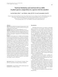
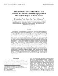
1–7 μm (Figure 1.1).<br />
The morphology of agar-grown yeasts shows great diversity in terms of colour,<br />
texture and geometry (peripheries, contours) of giant colonies. Several yeasts
4 INTRODUCTION TO FUNGAL PHYSIOLOGY<br />
Figure 1.1 Scanning electron micrograph of a typical yeast cell. (×10 000).<br />
BS, bud scar; BirS, birth scar. (Reproduced with kind permission of Professor<br />
Masako Osumi, Japan Women’s University, Tokyo.)<br />
are pigmented, and the following colours may be visualized in surface-grown<br />
colonies: cream (e.g. S. cerevisiae); white (e.g. Geotrichum candidum); black (e.g.<br />
Aureobasidium pullulans); pink (e.g. Phaffia rhodozyma); red (e.g. Rhodotorula<br />
rubra); orange (e.g. Rhodosporidium spp.); and yellow (e.g. Cryptococcus laurentii).<br />
The pigments of some yeasts have biotechnological uses, including astaxanthin<br />
from P. rhodozyma in aquacultural feed supplements for farmed salmon<br />
(that are unable to synthesize these natural pink compounds) (Table 1.1).<br />
1.3 Ultrastructure and Function of Fungal Cells<br />
1.3.1 The Fungal Cell Surface<br />
The cell envelope in yeasts and fungi is the peripheral structure that encases the<br />
cytoplasm and comprises the plasma membrane, the periplasm, the cell wall and<br />
additional extracellular structural components (such as fimbriae and capsules).<br />
The cell wall represents a dynamically forming exoskeleton that protects the<br />
fungal protoplast from the external environment and defines directional growth,<br />
cellular strength, shape and interactive properties. In filamentous fungi, cell-wall<br />
formation and organization is intimately bound to the process of apical growth.<br />
Thus, for example in Neurospora crassa, the wall is thin (approximately 50 nm)<br />
at the apex but becomes thicker (approximately 125 nm) at 250 μm behind the<br />
tip. The plasma membrane component of the fungal cell envelope is a phospholipid<br />
bilayer interspersed with globular proteins that dictates entry of nutrients<br />
and exit of metabolites and represents a selective barrier for their translocation.
ULTRASTRUCTURE AND FUNCTION OF FUNGAL CELLS 5<br />
Table 1.1<br />
Diversity of yeast cell shapes.<br />
Cell shape Description Examples of yeast genera<br />
Ellipsoid Ovoid shaped cells Saccharomyces<br />
Cylindrical<br />
Elongated cells with<br />
hemispherical ends<br />
Schizosaccharomyces<br />
Apiculate Lemon shaped Hanseniaspora, Saccharomycodes<br />
Ogival<br />
Elongated cell rounded at one<br />
end and pointed at other<br />
Dekkera, Brettanomyces<br />
Flask-shaped Cells dividing by bud-fission Pityrosporum<br />
Miscellaneous<br />
shapes<br />
Pseudohyphal<br />
Hyphal<br />
Dimorphic<br />
Triangular<br />
Curved<br />
Spherical<br />
Stalked<br />
Chains of budding yeast cells<br />
which have elongated without<br />
detachment<br />
Branched or unbranched<br />
filamentous cells which form<br />
from germ tubes. Septa may be<br />
laid down by the continuously<br />
extending hyphal tip. Hyphae<br />
may give rise to blastospores<br />
Yeasts that grow vegetatively<br />
in either yeast or filamentous<br />
(hyphal or pseudohyphal) form<br />
Trigonopsis<br />
Cryptococcus (e.g. Cryptococcus cereanus)<br />
Debaryomyces<br />
Sterigmatomyces<br />
Candida (e.g. Candida albicans)<br />
Candida albicans<br />
Candida albicans<br />
Saccharomycopsis fibuligera<br />
Kluyveromyces marxianus<br />
Malassezia furfur<br />
Yarrowia lipolytica<br />
Histoplasma capsulatum<br />
Ergosterol is the major sterol found in the membranes of fungi, in contrast to<br />
the cholesterol found in the membranes of animals and phytosterols in plants.<br />
This distinction is exploited during the use of certain antifungal agents used to<br />
treat some fungal infections, and can be used as an assay tool to quantify fungal<br />
growth. The periplasm, or periplasmic space, is the region external to the plasma<br />
membrane and internal to the cell wall. In yeast cells, it comprises secreted proteins<br />
(mannoproteins) and enzymes (such as invertase and acid phosphatase) that<br />
are unable to traverse the cell wall. In filamentous fungi, the cell membrane and<br />
wall may be intimately bound as hyphae and are often resistant to plasmolysis.
6 INTRODUCTION TO FUNGAL PHYSIOLOGY<br />
Fungal cell surface topological features can be visualized using scanning electron<br />
microscopy (SEM), and nanometre resolution is achieved using atomic<br />
force microscopy (AFM). The latter is beneficial, as it can be employed with unfixed,<br />
living cells and avoids potentially misleading artefacts that may arise when<br />
preparing cells for electron microscopy. Figure 1.1 shows SEM micrographs of<br />
a typical unicellular yeast cell envelope.<br />
Ultrastructural analysis of fungal cell walls reveals a thick, complex fibrillar<br />
network. The cell walls of filamentous fungi are mainly composed of different<br />
polysaccharides according to taxonomic group. For example, they may contain<br />
either chitin, glucans, mannoproteins, chitosan, polyglucuronic acid or cellulose,<br />
together with smaller quantities of proteins and glycoproteins (Table 1.2). Generally,<br />
the semi-crystalline microfibrillar components are organized in a network<br />
mainly in the central cell wall region and are embedded within an amorphous<br />
matrix. Bonding occurs between certain components behind the extending hyphal<br />
tip, thereby strengthening the entire wall structure. There is evidence to<br />
suggest that the cell wall is a dynamic structure where considerable quantitative<br />
and qualitative differences occur not only between different fungal species,<br />
but also between different morphological forms of the same species and even<br />
in response to environmental stress. For example, a class of hydrophobic proteins<br />
called hydrophobins are localized within the aerial growth or appresoria<br />
(terminal swellings involved in infection) of certain fungi, whereas pigmented<br />
melanins are often found within some fungal cell walls to insulate against biotic<br />
and abiotic stresses.<br />
Table 1.2 The major polymers found in different taxonomical groups of<br />
fungi, together with the presence of perforate septa in these groups (adapted<br />
from Deacon (2005) and Carlile et al. (2001)).<br />
Taxonomic<br />
grouping Fibrillar polymers Matrix polymers<br />
Perforate septa<br />
present or absent<br />
Oomycetes<br />
(1,3)-, (1,6)-Glucan<br />
Cellulose<br />
Glucan<br />
Absent<br />
Chytridomycetes Chitin; glucan Glucan Absent<br />
Zygomycetes Chitin; chitosan Polyglucuronic acid;<br />
glucuronomannoproteins<br />
Absent<br />
Basidiomycetes<br />
Chitin; (1,3)-,<br />
(1,6)-glucans<br />
(1,3)-Glucan;<br />
xylomannoproteins<br />
Present (mostly<br />
Dolipore)<br />
Ascomycetes/<br />
Deuteromycetes<br />
Chitin; (1,3)-,<br />
(1,6)-glucans<br />
(1,3)-Glucan;<br />
galactomannoproteins<br />
Present (mostly<br />
simple with large<br />
central pore)
ULTRASTRUCTURE AND FUNCTION OF FUNGAL CELLS 7<br />
The hyphae of higher fungi extend via tip growth followed by cross-wall<br />
formation or septation, whereas the lower fungi remain aseptate (except when<br />
segregating spores or in damaged colony regions). Septa may offer some structural<br />
support to hyphae. Significantly, septa serve to compartmentalize hyphae<br />
but are typically perforated, thereby permitting passage and communication of<br />
cytoplasm or even protoplasm between compartments. However, septal pores<br />
can become blocked by Woronin bodies or other materials. This aids morphological<br />
and biochemical differentiation and serves to seal off stressed or damaged<br />
hyphae from undamaged colony regions. Again, different pore types are representative<br />
of different taxonomic groups and species (Table 1.2).<br />
In yeasts, the cell-wall structure comprises polysaccharides (predominantly<br />
-glucans for rigidity), proteins (mainly mannoproteins on the outermost layer<br />
for determining porosity), together with some lipid, chitin (e.g. in bud scar<br />
tissue) and inorganic phosphate material. Figure 1.2 shows the composition<br />
and structure of the S. cerevisiae cell wall. Hyphal cell walls generally contain<br />
fewer mannans than yeast cell forms, and such changes in composition<br />
are even observed during the transition from unicellular to mycelial growth of<br />
dimorphic fungi.<br />
Chitin is also found in yeast cell walls and is a major constituent of bud scars<br />
(Figure 1.3). These are remnants of previous budding events found on the surface<br />
of mother cells following birth of daughter cells (buds). The chitin-rich bud scars<br />
of yeast cells can be stained with fluorescent dyes (e.g. calcoflour white), and this<br />
can provide useful information regarding cellular age, since the number of scars<br />
represents the number of completed cell division cycles. Outside the cell wall<br />
in fungi, several extramural layers may exist, including fimbriae and capsules.<br />
Figure 1.2 Cell envelope structure of the yeast S. cerevisiae (from Walker<br />
(1998). Permission obtained for First Edition).
8 INTRODUCTION TO FUNGAL PHYSIOLOGY<br />
Figure 1.3 Transmission electron microscopy of ultrathin sections of fungal<br />
cells reveals intracellular fine structure.<br />
Fungal fimbriae are long, protein-containing protrusions appearing from the cell<br />
wall of certain basidiomycetous and ascomycetous fungi that are involved in cellcell<br />
conjugation. Capsules are extracellular polysaccharide-containing structures<br />
found in basidiomycetous fungi that are involved in stress protection. In Cryptococcus<br />
neoformans (the pathogenic yeast state of Filobasidiella neoformans) the<br />
capsule may determine virulence properties and evasion from macrophages. One<br />
extrahyphal substance, the polymer pullulan, is produced commercially from<br />
A. pullulans.<br />
1.3.2 Subcellular Architecture and Organelle Function<br />
Transmission electron microscopy of ultrathin sections of fungal cells reveals<br />
intracellular fine structure (Figures 1.2 and 1.4). Subcellular compartments (organelles)<br />
are bathed in an aqueous cytoplasm containing soluble proteins and<br />
other macromolecules, together with low-molecular weight metabolites. However,<br />
the hyphae of central (and therefore older) colony regions of filamentous<br />
fungi may become devoid of protoplasm and organelles, as protoplasmic<br />
components are driven forward or are recycled, to support the growth
ULTRASTRUCTURE AND FUNCTION OF FUNGAL CELLS 9<br />
Figure 1.4 Electron micrograph of a typical yeast cell. (CW, cell wall; CM, cell<br />
membrane; CMI, cell membrane invagination; BS, bud scar; M, mitochondrion,<br />
N, nucleus; V, vacuole; ER, endoplasmic reticulum. (Reproduced with kind permission<br />
of Professor Masako Osumi, Japan Women’s University, Tokyo.)<br />
of actively growing hyphal tips. Cytoplasmic components additionally comprise<br />
microbodies, ribosomes, proteasomes, lipid particles and a cytoskeletal network.<br />
The latter confers structural stability to the fungal cytoplasm and consists of microtubules<br />
and microfilaments. The following membrane-bound organelles may<br />
be found in a typical fungal cell: nucleus: endoplasmic reticulum (ER), mitochondria,<br />
Golgi apparatus, secretory vesicles and vacuoles. Several of these organelles<br />
form extended membranous systems. For example, the ER is contiguous with<br />
the nuclear membrane and secretion of fungal proteins involves intermembrane<br />
trafficking in which the ER, Golgi apparatus, plasma membrane and vesicles all<br />
participate. The physiological function of the various fungal cell organelles is<br />
summarized in Table 1.3.<br />
The nucleus is the structure that defines the eukaryotic nature of fungal cells.<br />
It is bound by a double membrane and encases the chromosomes in a nucleoplasm.<br />
Most yeast and fungi are haploid, although some (e.g. S. cerevisiae) may<br />
alternate between haploidy and diploidy. Chromosomes comprise DNA–protein<br />
structures that replicate and segregate to newly divided cells or hyphal compartments<br />
at mitosis. This, of course, ensures that genetic material is passed onto<br />
daughter cells or septated compartments at cell division. Yeasts usually contain<br />
a single nucleus per cell. However, the hyphal compartments of filamentous<br />
fungi may contain one or more nuclei. Monokaryotic basidiomycetes possess<br />
one nucleus per compartment, whereas dikaryons or heterokaryons possess two
10 INTRODUCTION TO FUNGAL PHYSIOLOGY<br />
Table 1.3<br />
Organelle or cellular<br />
structure<br />
Cell envelope<br />
Nucleus<br />
Mitochondria<br />
Endoplasmic reticulum<br />
Proteasome<br />
Golgi apparatus and<br />
vesicles<br />
Vacuole<br />
Peroxisome<br />
Functional components of an idealized fungal cell.<br />
Function<br />
Comprising: the plasma membrane, which acts as a selectively<br />
permeable barrier for transport of hydrophilic molecules in and<br />
out of fungal cells; the periplasm, containing proteins and<br />
enzymes unable to permeate the cell wall; the cell wall, which<br />
provides protection, shape and is involved in cell–cell<br />
interactions, signal reception and specialized enzyme activities;<br />
fimbriae involved in sexual conjugation; capsules to protect cells<br />
from dehydration and immune cell attack.<br />
Relatively small. Containing chromosomes (DNA–protein<br />
complexes) that pass genetic information to daughter cells at cell<br />
division and the nucleolus, which is the site of ribosomal RNA<br />
transcription and processing.<br />
Site of respiratory metabolism under aerobic conditions and,<br />
under anaerobic conditions, for fatty acid, sterol and amino acid<br />
metabolism.<br />
Ribosomes on the rough ER are the sites of protein biosynthesis.<br />
Multi-subunit protease complexes involved in regulating protein<br />
turnover.<br />
Secretory system for import (endocytosis) and export (exocytosis)<br />
of proteins.<br />
Intracellular reservoir (amino acids, polyphosphate, metal ions);<br />
proteolysis; protein trafficking; control of cellular pH. In<br />
filamentous fungi, tubular vacuoles transport materials<br />
bidirectionally along hyphae.<br />
Oxidative utilization of specific carbon and nitrogen sources<br />
(contain catalase, oxidases). Glyoxysomes contain enzymes of the<br />
glyoxylate cycle.<br />
or more genetically distinct haploid nuclei. The maintenance of multiple nuclei<br />
within individual hyphal compartments allows fungi to take advantage of both<br />
haploid and diploid lifestyles. This is discussed further in Chapter 2.<br />
In filamentous fungi, a phase-dark near-spherical region, which also stains<br />
with iron haemotoxylin, is evident by light microscopy at the apex during hyphal<br />
tip growth. The region is termed the Spitzenkörper, the apical vesicle cluster<br />
or centre or apical body, and it consists of masses of small membrane-bound vesicles<br />
around a vesicle-free core with emergent microfilaments and microtubules.<br />
The Spitzenkörper contains differently sized vesicles derived from Golgi bodies,
FUNGAL NUTRITION AND CELLULAR BIOSYNTHESES 11<br />
either large vesicles or microvesicles (chitosomes), with varying composition. It<br />
orientates to the side as the direction of tip growth changes, and disappears<br />
when growth ceases. This vesicle supply centre is involved in wall extension and,<br />
hence, tip growth, branching, clamp connection formation (in Basidiomycetes)<br />
and germ tube formation.<br />
1.4 Fungal Nutrition and Cellular Biosyntheses<br />
1.4.1 Chemical Requirements for Growth<br />
Yeasts and fungi have relatively simple nutritional needs and most species would<br />
be able to survive quite well in aerobic conditions if supplied with glucose,<br />
ammonium salts, inorganic ions and a few growth factors. Exceptions to this<br />
would include, for example, obligate symbionts such as the vesicular–arbuscular<br />
mycorrhizal (VAM) fungi which require growth of a plant partner for cultivation.<br />
Macronutrients, supplied at millimolar concentrations, comprise sources<br />
of carbon, nitrogen, oxygen, sulfur, phosphorus, potassium and magnesium;<br />
and micronutrients, supplied at micromolar concentrations, comprising trace elements<br />
like calcium, copper, iron, manganese and zinc, would be required for<br />
fungal cell growth (Table 1.4). Some fungi are oligotrophic, apparently growing<br />
with very limited nutrient supply, surviving by scavenging minute quantities of<br />
volatile organic compounds from the atmosphere.<br />
Being chemoorganotrophs, fungi need fixed forms of organic compounds for<br />
their carbon and energy supply. Sugars are widely utilized for fungal growth and<br />
can range from simple hexoses, like glucose, to polysaccharides, like starch and<br />
cellulose. Some fungi can occasionally utilize aromatic hydrocarbons (e.g. lignin<br />
by the white-rot fungi). Table 1.5 outlines the variety of carbon sources which<br />
can be utilized by yeasts and filamentous fungi for growth.<br />
Fungi are non-diazotrophic (cannot fix nitrogen) and need to be supplied with<br />
nitrogenous compounds, either in inorganic form, such as ammonium salts, or<br />
in organic form, such as amino acids. Ammonium sulfate is a commonly used<br />
nitrogen source in fungal growth media, since it also provides a source of utilizable<br />
sulfur. Many fungi (but not the yeast S. cerevisiae) can also grow on<br />
nitrate and, if able to do so, may also utilize nitrite. Nitrate reductase followed<br />
by nitrite reductase are the enzymes responsible for converting nitrate to ammonia.<br />
Most fungi can assimilate amino acids, amines and amides as nitrogen<br />
sources. Most fungi (but not many yeasts) are also proteolytic and can hydrolyse<br />
proteins (via extracellularly secreted proteases) to liberate utilizable amino acids<br />
for growth. Urea utilization is common in fungi, and some basidiomycetous<br />
yeasts are classed as urease positive (able to utilize urea) whilst most ascomycetous<br />
yeasts are urease negative.<br />
In terms of oxygen requirements, most fungi are aerobes and are often microaerophilic<br />
(preferring an oxygen tension below that of normal atmospheric).
12 INTRODUCTION TO FUNGAL PHYSIOLOGY<br />
Table 1.4<br />
Elemental requirements of fungal cells.<br />
Element Common sources Cellular functions<br />
Carbon Sugars Structural element of fungal cells in combination<br />
with hydrogen, oxygen and nitrogen. Energy<br />
source<br />
Hydrogen<br />
Protons from acidic<br />
environments<br />
Transmembrane proton motive force vital for<br />
fungal nutrition. Intracellular acidic pH (around<br />
5–6) necessary for fungal metabolism<br />
Oxygen Air, O 2 Substrate for respiratory and other<br />
mixed-function oxidative enzymes. Essential for<br />
ergosterol and unsaturated fatty acid synthesis<br />
Nitrogen<br />
NH 4 + salts, urea, amino<br />
acids<br />
Structurally and functionally as organic amino<br />
nitrogen in proteins and enzymes<br />
Phosphorus Phosphates Energy transduction, nucleic acid and membrane<br />
structure<br />
Potassium K + salts Ionic balance, enzyme activity<br />
Magnesium Mg 2+ salts Enzyme activity, cell and organelle structure<br />
Sulfur Sulfates, methionine Sulfydryl amino acids and vitamins<br />
Calcium Ca 2+ salts Possible second messenger in signal transduction<br />
Copper Cupric salts Redox pigments<br />
Iron<br />
Ferric salts. Fe 3+ is<br />
chelated by siderophores<br />
and released as Fe 2+<br />
within the cell<br />
Haem-proteins, cytochromes<br />
Manganese Mn 2+ salts Enzyme activity<br />
Zinc Zn 2+ salts Enzyme activity<br />
Nickel Ni 2+ salts Urease activity<br />
Molybdenum Na 2 MoO 4 Nitrate metabolism, vitamin B 12<br />
Although yeasts like S. cerevisiae are sometimes referred to as facultative anaerobes,<br />
they cannot actually grow in strictly anaerobic conditions unless supplied<br />
with certain fatty acids and sterols (which they cannot synthesize without<br />
molecular oxygen). In fact, there are thought to be very few yeast species that<br />
are obligately anaerobic. For aerobically respiring yeasts and fungi, oxygen is<br />
required as the terminal electron acceptor, where it is finally reduced to water<br />
in the electron-transport chain. Different fungal species respond to oxygen
FUNGAL NUTRITION AND CELLULAR BIOSYNTHESES 13<br />
Table 1.5 Diversity of carbon sources for yeast and filamentous fungal<br />
growth (adapted from Walker (1998)).<br />
Carbon source Typical examples Comments<br />
Hexose sugars<br />
Pentose sugars<br />
Disaccharides<br />
D-Glucose,<br />
D-galactose,<br />
D-Fructose,<br />
D-mannose<br />
L-Arabinose,<br />
D-xylose, D-xyulose,<br />
L-rhamnose<br />
Maltose, sucrose,<br />
lactose, trehalose,<br />
melibiose, cellobiose,<br />
melezitose<br />
Glucose metabolized by majority of yeasts<br />
and filamentous fungi<br />
If a yeast does not ferment glucose, it will not<br />
ferment other sugars. If a yeast ferments<br />
glucose, it will also ferment fructose and<br />
mannose, but not necessarily galactose<br />
Some fungi respire pentoses better than<br />
glucose. S. cerevisiae can utilize xylulose but<br />
not xylose<br />
If a yeast ferments maltose, it does not<br />
generally ferment lactose and vice versa.<br />
Melibiose utilization used to distinguish ale<br />
and lager brewing yeasts. Large number of<br />
yeasts utilize disaccharides. Few filamentous<br />
fungi (e.g. Rhizopus nigricans) cannot utilize<br />
sucrose<br />
Trisaccharides Raffinose, maltotriose Raffinose only partially used by S. cerevisiae,<br />
but completely used by other Saccharomyces<br />
spp. (S. carlsbergensis, S. kluyveri)<br />
Oligosaccharides<br />
Polysaccharides<br />
Lower aliphatic<br />
alcohols<br />
Maltotetraose,<br />
maltodextrins<br />
Starch, inulin,<br />
cellulose,<br />
hemicellulose, chitin,<br />
pectic substances<br />
Methanol, ethanol<br />
Metabolized by amylolytic yeasts, not by<br />
brewing strains<br />
Polysaccharide-fermenting yeasts are rare.<br />
Saccharomycopsis spp. and S. diastaticus can<br />
utilize soluble starch; Kluyveromyces spp.<br />
possess inulinase. Many filamentous fungi can<br />
utilize these depending on extracellular<br />
enzyme activity<br />
Respiratory substrates for many fungi. Several<br />
methylotrophic yeasts (e.g. Pichia pastoris,<br />
Hansenula polymorpha) have industrial<br />
potential<br />
Sugar alcohols Glycerol, glucitol Can be respired by yeasts and a few fungi<br />
Organic acids<br />
Acetate, citrate,<br />
lactate, malate,<br />
pyruvate, succinate<br />
Many yeasts can respire organic acids, but few<br />
can ferment them<br />
(continued)
14 INTRODUCTION TO FUNGAL PHYSIOLOGY<br />
Table 1.5<br />
(Continued)<br />
Carbon source Typical examples Comments<br />
Fatty acids Oleate, palmitate Several species of oleaginous yeasts can assimilate<br />
fatty acids as carbon and energy sources<br />
Hydrocarbons n-Alkanes Many yeast and a few filamentous species grown<br />
well on C 12 -C 18 n-alkanes<br />
Aromatics<br />
Miscellaneous<br />
Phenol, cresol,<br />
quinol, resourcinol,<br />
catechol, benzoate<br />
Adenine, uric acid,<br />
butylamine,<br />
pentylamine,<br />
putrescine<br />
Lignin<br />
‘Hard’ keratin<br />
Few yeasts can utilize these compounds. Several<br />
n-alkane-utilizing yeasts use phenol as carbon<br />
source via the -ketoadipate pathway<br />
Some mycelial fungi and yeasts, for example,<br />
Arxula adeninivorans and A. terestre can utilize<br />
such compounds as sole source of carbon and<br />
nitrogen<br />
Can be decayed only by white-rot fungi<br />
(basidiomycotina). Little net energy gained directly,<br />
but makes available other polysaccharides such as<br />
cellulose and hemicellulose<br />
Keratinophilic fungi<br />
availability in diverse ways, and Table 1.6 categorizes fungi into different groups<br />
on this basis.<br />
Sulfur sources for fungal growth include sulfate, sulfite, thiosulfate, methionine<br />
and glutathione with inorganic sulfate and the sulfur amino acid methionine<br />
being effectively utilized. Virtually all yeasts can synthesize sulfur amino acids<br />
from sulfate, the most oxidized form of inorganic sulfur.<br />
Phosphorus is essential for biosynthesis of fungal nucleic acids, phospholipids,<br />
ATP and glycophosphates. Hence, the phosphate content of fungi is considerable<br />
(e.g. in yeast cells it accounts for around 3–5 % of dry weight; the major part of<br />
this is in the form of orthophosphate (H 2 PO 4 − ), which acts as a substrate and<br />
enzyme effector). The fungal vacuole can serve as a storage site for phosphate<br />
in the form of complexed inorganic polyphosphates (also referred to as volutin<br />
granules). Both nitrogen and phosphorus availability may be growth limiting in<br />
nature. Filamentous fungi have evolved a number of biochemical and morphological<br />
strategies allowing capture of often poorly available phosphorus within<br />
the natural environment. Plants exploit such efficiency during symbioses between<br />
their roots and certain mycorrhizal fungi. The major storage form of phosphorus<br />
in plants is phytic acid (myo-inositol hexa-dihydrogenphosphate), which is<br />
poorly utilized by monogastrics (e.g. humans, pigs, poultry), and fungal (and<br />
yeast) phytases have applications in reducing phytate content of foods and feeds.<br />
Concerning requirements for minerals, potassium, magnesium and several<br />
trace elements are necessary for fungal growth. Potassium and magnesium are
FUNGAL NUTRITION AND CELLULAR BIOSYNTHESES 15<br />
Table 1.6 Yeast and fungal metabolism based on responses to oxygen<br />
availability.<br />
Mode of energy<br />
metabolism Examples Comments<br />
Obligate<br />
fermentative<br />
Yeasts: Candida<br />
pintolopesii<br />
(Saccharomyces telluris)<br />
Fungi: facultative and<br />
obligate anaerobes<br />
Naturally occurring respiratory-deficient<br />
yeasts. Only ferment, even in presence of<br />
oxygen<br />
No oxygen requirement for these fungi.<br />
Two categories exist with respect to the<br />
effects of air: facultative anaerobes (e.g.<br />
Aqualinderella and Blastocladia) and<br />
obligate anaerobes (e.g. Neocallimastix)<br />
Facultatively<br />
fermentative<br />
Crabtree-positive<br />
Saccharomyces<br />
cerevisiae<br />
Such yeasts predominantly ferment high<br />
sugar-containing media in the presence of<br />
oxygen<br />
Crabtree-negative Candida utilis Such yeasts do not form ethanol under<br />
aerobic conditions and cannot grow<br />
anaerobically<br />
Non-fermentative<br />
Yeasts: Rhodotorula<br />
rubra<br />
Fungi: Phycomyces<br />
Such yeasts do not produce ethanol, either<br />
in the presence or absence of oxygen<br />
Oxygen essential for such (obligately<br />
oxidative) fungi<br />
Obligate aerobes<br />
Gaemannomyces<br />
graminis (the take-all<br />
fungus)<br />
The growth of these is markedly reduced<br />
if oxygen partial pressure falls below<br />
normal atmospheric<br />
Adapted from Walker (1998), Deacon (2005) and Carlile et al. (2001).<br />
macroelements required in millimolar concentrations, primarily as enzyme cofactors,<br />
whereas other microelements (trace elements) are generally required in<br />
the micromolar range. These include Mn, Ca Fe, Zn, Cu, Ni, Co and Mo.<br />
Table 1.7 summarizes the main metals required for fungal growth. Toxic minerals<br />
(e.g. Ag, As, Ba, Cs, Cd, Hg, Li, Pb) adversely affect fungal growth generally<br />
at concentrations greater than 100 μM.<br />
Fungal growth factors are organic compounds occasionally needed in very<br />
low concentrations for specific enzymatic or structural roles, but not as energy<br />
sources. These include vitamins (e.g. thiamine, biotin), purines, pyrimidines,<br />
nucleosides, nucleotides, amino acids, fatty acids and sterols. For fungi to have<br />
a growth factor requirement, this indicates that cells cannot synthesize the<br />
particular factor, resulting in the curtailment of growth without its provision
16 INTRODUCTION TO FUNGAL PHYSIOLOGY<br />
Table 1.7 Metals required for fungal growth and metabolic functions<br />
(adapted from Walker (2004)).<br />
Metal ion<br />
Concentration supplied<br />
in growth medium a<br />
Main cellular functions<br />
Macroelements<br />
K 2–4 mM Osmoregulation, enzyme activity<br />
Mg Mg 2–4 mM Enzyme activity, cell division<br />
Microelements<br />
Mn 2–4 μM Enzyme cofactor<br />
Ca μM Second messenger, yeast flocculation<br />
Cu 1.5 μM Redox pigments<br />
Fe 1–3 μM Haem-proteins, cytochromes<br />
Zn 4–8 μM Enzyme activity, protein structure<br />
Ni ∼10 μM Urease activity<br />
Mo 1.5 μM Nitrate metabolism, vitamin B 12<br />
Co 0.1 μM Cobalamin, coenzymes<br />
a Figures relate to yeast (S. cerevisiae) growth stimulation and are dependent on the species/strain and<br />
conditions of growth, but they would be generally applicable for fungal growth.<br />
in culture media. Some fungi (e.g. Aspergillus niger, Penicillium chrysogenum)<br />
have very simple nutritional needs and are able to synthesize their own growth<br />
factors from glucose.<br />
1.4.2 Fungal Cultivation Media<br />
Fungal nutritional requirements are important not only for successful cultivation<br />
in the laboratory, but also for the optimization of industrial fermentation<br />
processes. In the laboratory, it is relatively easy to grow yeasts and fungi on complex<br />
culture media such as malt extract or potato–dextrose agar or broth, which<br />
are both carbon rich and in the acidic pH range. Mushrooms are cultivated on<br />
various solid substrates, depending on provincial availability. Therefore, Agaricus<br />
bisporus (common button mushroom) is grown in the UK, USA and France<br />
on wheat straw; the padi-straw mushroom (Volvariella volvacea) is grown in<br />
South-east Asia on damp rice-straw and in Hong Kong on cotton waste; and<br />
in Japan, the shiitake mushroom (Lentinus edodes) is cultivated on fresh oak<br />
logs. In industry, media for fungal fermentation purposes need to be optimized<br />
with regard to the specific application and production process. For some industrial<br />
processes, growth media may already be relatively complete in a nutritional<br />
sense, such as malt wort or molasses for brewing or baker’s yeast production<br />
respectively (Table 1.8). However, for other processes, supplementation of
FUNGAL NUTRITION AND CELLULAR BIOSYNTHESES 17<br />
Table 1.8 Principal ingredients of selected industrial media for yeasts and fungi.<br />
Components Molasses Malt wort Wine must Cheese whey Corn steep liquor<br />
Carbon<br />
sources<br />
Sucrose Maltose Glucose Lactose Glucose, other<br />
Fructose Sucrose Fructose<br />
sugars<br />
Glucose Fructose Sucrose (trace)<br />
Raffinose Glucose<br />
Maltotriose<br />
Nitrogen<br />
sources<br />
Nitrogen compounds as<br />
unassimilable proteins.<br />
Nitrogen sources need to<br />
be supplemented<br />
Low molecular -amino<br />
nitrogen compounds,<br />
ammonium ions and a<br />
range of amino acids<br />
Variable levels of ammonia<br />
nitrogen, which may be<br />
limiting.<br />
Range of amino acids<br />
Unassimilable globulin<br />
and albumin proteins.<br />
Low levels of ammonium<br />
and urea nitrogen<br />
Amino acids,<br />
protein<br />
Minerals Supply of P, K and S<br />
available. High K + levels<br />
may be inhibitory<br />
Supply of P, K, Mg and<br />
S available<br />
Supply of P, K, Mg and S<br />
available. High levels of<br />
sulfite often present<br />
Supply of P, K, Mg, S Supply of P, K,<br />
Mg, S<br />
Vitamins Small, but generally<br />
adequate supplies. Biotin<br />
is deficient in beet<br />
molasses<br />
Supply of vitamins is<br />
usually adequate. High<br />
adjunct sugar wort may<br />
be deficient in biotin<br />
Vitamin supply generally<br />
sufficient<br />
Wide range of vitamins<br />
present<br />
Biotin,<br />
pyridoxine,<br />
thiamin<br />
Trace<br />
elements<br />
Range of trace metals<br />
present, although Mn 2+<br />
may be limiting<br />
All supplied, although<br />
Zn 2+ may be limiting<br />
Sufficient quantities<br />
available<br />
Fe, Zn, Mn, Ca, Cu<br />
present<br />
Range of trace<br />
elements present<br />
Other<br />
components<br />
Unfermentable sugars<br />
(2–4 %), organic acids,<br />
waxes, pigments, silica,<br />
pesticide residues,<br />
carmelized compounds,<br />
betaine<br />
Unfermentable<br />
maltodextrins,<br />
pyrazines, hop<br />
compounds<br />
Unfermentable pentoses.<br />
Tartaric and malic acids.<br />
Decanoic and octanoic<br />
acids may be inhibitory.<br />
May be deficient in sterols<br />
and unsaturated fatty acids<br />
Lipids, NaCl. Lactic and<br />
citric acids<br />
High levels of<br />
lactic acid<br />
present. Fat and<br />
fibre also present
18 INTRODUCTION TO FUNGAL PHYSIOLOGY<br />
agriculturally derived substrates, like corn steep liquor, molasses or malt broth,<br />
with additional nutrients and growth factors may be necessary. For example,<br />
the following may constitute a suitable fermentation medium for penicillin<br />
production by Penicillium spp.: sucrose (3 g/L), corn steep liquor (100 g/L),<br />
KH 2 PO 4 (1 g/L), (NH 4 ) 2 SO 4 (12 g/L), CaCl 2·2H 2 O (0.06 g/L), phenoxyacetic<br />
acid (5.7 g/L) – information from Jorgensen et al. (1995). However, other industrial<br />
processes, such as the growth of Fusarium graminarium for production<br />
of Quorn TM mycoprotein, require culture on a completely defined medium (see<br />
Chapter 5).<br />
1.4.3 Nutrient Uptake and Assimilation<br />
Fungal cells utilize a diverse range of nutrients and employ equally diverse nutrient<br />
acquisition strategies. Fungi are nonmotile, saprophytic (and sometimes<br />
parasitic), chemo-organotrophic organisms. They exhibit dynamic interactions<br />
with their nutritional environment that may be exemplified by certain morphological<br />
changes depending on nutrient availability. For example, the filamentous<br />
mode of growth observed at the periphery of yeast colonies growing in agar is<br />
akin to a foraging for nutrients as observed in certain eucarpic fungi. Metabolic<br />
dynamism is also evident in yeasts which, although not avid secretors of hydrolytic<br />
enzymes like higher fungi, are nevertheless able to secrete some enzymes<br />
to degrade polymers such as starch (as in amylolytic yeasts like Schwanniomyces<br />
occidentalis).<br />
Several cellular envelope barriers to nutrient uptake by fungal cells exist,<br />
namely: the capsule, the cell wall, the periplasm and the cell membrane. Although<br />
not considered as a freely porous structures, fungal cell walls are<br />
relatively porous to molecules up to an average molecular mass of around<br />
300 Da, and will generally retain molecules greater than around 700 Da. Typically,<br />
fungi absorb only small soluble nutrients, such as monosaccharides and<br />
amino acids.<br />
The plasma membrane is the major selectively permeable barrier which dictates<br />
nutrient entry and metabolite exit from the fungal cell. Membrane transport<br />
mechanisms are important in fungal physiology, since they govern the rates at<br />
which cells metabolize, grow and divide. Fungi possess different modes of passive<br />
and active uptake at the plasma membrane: free diffusion, facilitated diffusion,<br />
diffusion channels and active transport (Table 1.9). Active transport of nutrients,<br />
such as sugars, amino acids, nitrate, ammonium, sulfate and phosphate,<br />
in filamentous fungi involves spatial separation of the ion pumps mostly behind<br />
the apex, whereas the symport proteins are active close to the tip. Thus, nutrient<br />
uptake occurs at the hyphal tip as it continuously drives into fresh resource, and<br />
the mitochondria localized behind the apex supply ATP to support the ion pump<br />
and generate proton motive force.
FUNGAL NUTRITION AND CELLULAR BIOSYNTHESES 19<br />
Mode of nutrient<br />
transport<br />
Table 1.9<br />
Description<br />
Modes of nutrient transport in fungi.<br />
Examples of nutrients<br />
transported<br />
Free diffusion<br />
Facilitated<br />
diffusion<br />
Diffusion<br />
channels<br />
Active transport<br />
Passive penetration of lipid-soluble<br />
solutes through the plasma membrane<br />
following the law of mass action from a<br />
high extracellular concentration to a<br />
lower intracellular concentration<br />
Translocates solutes down a<br />
transmembrane concentration gradient<br />
in an enzyme (permease)-mediated<br />
manner. As with passive diffusion,<br />
nutrient translocation continues until the<br />
intracellular concentration equals that of<br />
the extracellular medium<br />
These operate as voltage-dependent<br />
‘gates’ to transiently move certain<br />
nutrient ions down concentration<br />
gradients. They are normally closed at<br />
the negative membrane potential of<br />
resting yeast cells but are open when the<br />
membrane potential becomes positive<br />
The driving force is the membrane<br />
potential and the transmembrane<br />
electrochemical proton gradient<br />
generated by the plasma membrane<br />
H + -ATPase. The latter extrudes protons<br />
using the free energy of ATP hydrolysis<br />
that enables nutrients to either enter with<br />
influxed protons, as in ‘symport’<br />
mechanisms, or against effluxed protons,<br />
as in ‘antiport’ mechanisms<br />
Organic acids, short-chain<br />
alkanes and long-chain fatty<br />
acids by fungi and the export<br />
of lipophilic metabolites (e.g.<br />
ethanol) and gaseous<br />
compounds<br />
In the yeast S. cerevisiae,<br />
glucose is transported in this<br />
manner<br />
Ions such as potassium may<br />
be transported in this fashion<br />
Many nutrients (sugars,<br />
amino acids, ions)<br />
1.4.4 Overview of Fungal Biosynthetic Pathways<br />
Anabolic pathways are energy-consuming, reductive processes which lead to<br />
the biosynthesis of new cellular material and are mediated by dehydrogenase enzymes<br />
which predominantly use reduced NADP + as the redox cofactor. NADPH<br />
is generated by the hexose monophosphate pathway (or Warburg–Dickens pathway)<br />
which accompanies glycolysis (see Section 1.5.1). In S. cerevisiae, upto
20 INTRODUCTION TO FUNGAL PHYSIOLOGY<br />
20 % of total glucose may be degraded via the hexose monophosphate pathway.<br />
This pathway generates cytosolic NADPH (following the dehydrogenation of<br />
glucose 6-phosphate using glucose 6-phosphate dehydrogenase and NADP + as<br />
hydrogen acceptor) for biosynthetic reactions leading to the production of fatty<br />
acids, amino acids, sugar alcohols, structural and storage polysaccharides and<br />
secondary metabolites. Besides generating NADPH, the hexose monophosphate<br />
pathway also produces ribose sugars for the synthesis of nucleic acids, RNA<br />
and DNA and for nucleotide coenzymes, NAD, NADP, FAD and FMN. This is<br />
summarized as follows:<br />
Glucose 6-phosphate + 2NADP + → Ribulose 5-phosphate + CO 2 + NADPH<br />
+ 2H +<br />
And complete oxidation of glucose 6-phosphate would result in<br />
Glucose 6-phosphate + 12NADP + → 6CO 2 + 12NADPH + 12H + → Pi<br />
Fungal growth on non-carbohydrate substrates as sole carbon sources (e.g.<br />
ethanol, glycerol, succinate and acetate) may lead to gluconeogenesis (conversion<br />
of pyruvate to glucose) and polysaccharide biosynthesis. Gluconeogenesis may<br />
be regarded as a reversal of glycolysis and requires ATP as energy and NADH<br />
as reducing power.<br />
Concerning fungal amino acid biosynthesis, simple nitrogenous compounds<br />
such as ammonium may be assimilated into amino acid families, the carbon<br />
skeletons of which originate from common precursors of intermediary carbon<br />
metabolism.<br />
The two main fungal storage carbohydrates are glycogen and trehalose. Glycogen<br />
is similar to starch with -1,4-glucan linear components and -1,6-branches.<br />
Trehalose (also known as mycose) is a disaccharide of glucose comprising an ,-<br />
1,1-glucoside bond between two -glucose units. Both trehalose and glycogen are<br />
synthesized following the formation of UDP-glucose, catalysed by UDP-glucose<br />
pyrophosphorylase:<br />
UTP + Glucose 1-phosphate → UDP-glucose + Pyrophosphate<br />
Glycogen is synthesized by glycogen synthase. Glycogen may be metabolized by<br />
glycogen phosphorylase when nutrients become limited under starvation conditions,<br />
and this contributes to the maintenance metabolism of cells by furnishing<br />
energy in the form of ATP. In yeast cells, glycogen breakdown is accompanied<br />
by membrane sterol biosynthesis, and this is important for brewing yeast vitality<br />
and successful beer fermentations. The other major storage carbohydrate, trehalose,<br />
is synthesized from glucose 6-phosphate and UDP-glucose by trehalose<br />
6-phosphate synthase and converted to trehalose by a phosphatase. In addition
FUNGAL METABOLISM 21<br />
to a storage role, trehalose is an important translocation material in filamentous<br />
forms and is also involved in stress protection in yeasts and fungi, accumulating<br />
when cells are subject to environmental insults such as heat shock or osmotic<br />
stress, or during plant host–fungal parasite interactions. Polyols, such as mannitol<br />
derived from fructose phosphate, are also translocated by fungi.<br />
1.4.5 Fungal Cell Wall Growth<br />
The structural polysaccharides in fungal cell walls include mannans, glucans<br />
and chitin and are synthesized from sugar nucleotides substrates formed by<br />
pyrophosphorylase enzymes. For example:<br />
Glucose 1-phosphate + UTP → UDP-glucose + PPi<br />
Mannose 1-phosphate + GTP → GDP-mannose + PPi<br />
Glucan synthesis involves plasma membrane-associated glucan synthetases for<br />
assembly of -1,3 linkages and -1,6 branches of cell-wall glucan. Chitin (a<br />
polymer of N-acetylglucosamine) is an important fungal cell-wall structural<br />
component and is involved in the yeast budding process and in dimorphic transitions<br />
from yeast to filamentous forms. Chitin synthetases catalyse the transfer of<br />
N-acetylglucosamine from UDP-N-acetylglucosamine to a growing chitin polymer<br />
within the fungal cell wall. The mannoproteins predominantly of unicellular<br />
forms are pre-assembled within the Golgi and are delivered to the cell wall via<br />
vesicles from the vesicle supply centre. Various vesicles containing cell-wallsynthetic<br />
enzymes, wall-lytic enzymes, enzyme activators and certain preformed<br />
wall components are transported to the tip where they fuse with the plasma<br />
membrane and release their contents, which, together with substrates delivered<br />
from the cytosol, facilitate synthesis of the growing cell wall.<br />
1.5 Fungal Metabolism<br />
1.5.1 Carbon Catabolism<br />
Being chemoorganotrophs, fungi derive their energy from the breakdown of<br />
organic compounds. Generally speaking, fungi, but few yeast species, extracellularly<br />
break down polymeric compounds by secreted enzymes prior to utilization<br />
of monomers as carbon and energy sources. Owing to their relatively large size<br />
(20–60 kDa), enzymes assembled by the Golgi are transported in vesicles to<br />
be secreted from sites of cell growth, essentially from extending hyphal tips.<br />
Enzymes may either become linked to the cell wall as wall-bound enzymes or<br />
may diffuse externally to decay substrates within the local environment.
22 INTRODUCTION TO FUNGAL PHYSIOLOGY<br />
Some examples follow of hydrolytic, oxidative, peroxidative and free-radicalgenerating<br />
enzyme systems produced by fungi for the degradation of polymeric<br />
compounds:<br />
Pectin lyase, polygalactorunase<br />
Pectin −−−−−−−−−−−−−−−−−→ Galacturonic acid<br />
Amylases, glucoamylase<br />
Starch −−−−−−−−−−−−−−→ Glucose<br />
Inulin −−−−−→ Inulinase<br />
Fructose<br />
Cellulases<br />
Cellulose −−−−−−→ Glucose<br />
Hemicellulases, xylanase<br />
Hemicellulose −−−−−−−−−−−−−−→ Xylose, Glucose<br />
Lipases<br />
Lipids −−−−−→ Fatty acids<br />
Proteinases<br />
Proteins −−−−−−−→ Amino acids<br />
Chitin −−−−−−→ Chitinase<br />
N-acetylglucosamine<br />
Ligninase; manganese peroxidase; laccase; glucose oxidase<br />
Lignin −−−−−−−−−−−−−−−−−−−−−−−−−−−−−−−−−→ Variety of largely phenolic<br />
products<br />
Several lipolytic yeasts are known (e.g. Candida rugosa, Yarrowia lipolytica)<br />
which secrete lipases to degrade triacylgycerol substrates to fatty acids<br />
and glycerol.<br />
In wood, the cellulose and hemicellulose components are embedded within a<br />
heteropolymeric three-dimensional lignin matrix, thus forming a complex lignocellulose<br />
material. Only certain filamentous basidiomycete or ascomycete fungi<br />
are able to degrade the recalcitrant lignin component to make available the cellulose<br />
or hemicellulose components. These are known as white-rot fungi due to<br />
resultant coloration of the delignified wood. Such fungi employ a cocktail of oxidative<br />
(including laccases) and peroxidative enzymes, together with hydrogenperoxide-generating<br />
enzyme systems, to attack at least 15 different inter-unit<br />
bond types extant within the lignin polymer. The manganese and lignin peroxidase<br />
enzyme systems operate by releasing highly reactive but transient oxygen<br />
free-radicals, which bombard and react with parts of the lignin molecule,<br />
generating a chain of chemical oxidations and producing a range of mainly<br />
phenolic end products. White-rot fungi have applications in, for example, upgrading<br />
lignocellulose waste for animal feed, paper production and bleaching,<br />
the bioremediation of contaminated land and water and (potentially) for biofuel<br />
production. Brown-rot and soft-rot (in wet wood) fungi are only able to degrade<br />
the cellulose and hemicellulose components of wood. Cellulose decomposition<br />
involves the synergistic activity of endoglucanases (that hydrolyse the internal<br />
bonds of cellulose), exoglucanases (that cleave cellobiose units from the end of<br />
the cellulose chain) and glucosidases (that hydrolyse cellobiose to glucose). Initial<br />
attack of cellulose microfibrills within the cell wall may involve the generation of<br />
hydrogen peroxide.
FUNGAL METABOLISM 23<br />
Catabolic pathways are oxidative processes which remove electrons from intermediate<br />
carbon compounds and use these to generate energy in the form of<br />
ATP. The catabolic sequence of enzyme-catalysed reactions that convert glucose<br />
to pyruvic acid is known as glycolysis, and this pathway provides fungal cells<br />
with energy, together with precursor molecules and reducing power (in the form<br />
of NADH) for biosynthetic pathways. Therefore, in serving both catabolic and<br />
anabolic functions, glycolysis is sometimes referred to as an amphibolic pathway.<br />
Glycolysis may be summarized as follows:<br />
Glucose + 2ADP + 2Pi + 2NAD + → 2Pyruvate + 2ATP + 2NADH + + 2H +<br />
During glycolysis, glucose is phosphorylated using ATP to produce fructose<br />
1,6-biphosphate, which is then split by aldolase to form two triose phosphate<br />
compounds. Further phosphorylation occurs, forming two triose diphosphates<br />
from which four H atoms are accepted by two molecules of NAD + . In the latter<br />
stages of glycolysis, four molecules of ATP are formed (by transfer of phosphate<br />
from the triose diphosphates to ADP), and this results in the formation of two<br />
molecules of pyruvic acid. ATP production (two molecules net) during glycolysis<br />
is referred to as substrate-level phosphorylation.<br />
In yeast cells undergoing alcoholic fermentation of sugars under anaerobic<br />
conditions, NAD + is regenerated in terminal step reactions from pyruvate. In<br />
the first of these, pyruvate is decarboxylated (by pyruvate decarboxylase) before<br />
a final reduction, catalysed by alcohol dehydrogenase (ADH) to ethanol.<br />
Such regeneration of NAD + prevents glycolysis from stalling and maintains the<br />
cell’s oxidation–reduction balance. Additional minor fermentation metabolites<br />
are produced by fermenting yeast cells, including glycerol, fusel alcohols (e.g.<br />
isoamyl alcohol), esters (e.g. ethyl acetate), organic acids (e.g. citrate, succinate,<br />
acetate) and aldehydes (e.g. acetaldehyde). Such compounds are important in<br />
flavour development in alcoholic beverages, such as beer, wine and whisky.<br />
Aerobic dissimilation of glucose by fungi leads to respiration, which is the<br />
major energy-yielding metabolic route and involves glycolysis, the citric acid<br />
cycle, the electron-transport chain and oxidative phosphorylation. In addition<br />
to glucose, many carbon substrates can be respired by fungi, including: pentose<br />
sugars (e.g. xylose), sugar alcohols (e.g. glycerol), organic acids (e.g. acetic<br />
acid), aliphatic alcohols (e.g. methanol, ethanol), hydrocarbons (e.g. n-alkanes)<br />
and aromatic compounds (e.g. phenol). Fatty acids are made available for fungal<br />
catabolism following extracellular lipolysis of fats and are metabolized by<br />
-oxidation in mitochondria.<br />
During glucose respiration under aerobic conditions, pyruvate enters the mitochondria<br />
where it is oxidatively decarboxylated to acetyl CoA by pyruvate<br />
dehydrogenase, which acts as the link between glycolysis and the cyclic series of<br />
enzyme-catalysed reactions known as the citric acid cycle (or Krebs cycle). This<br />
cycle represents the common pathway for the oxidation of sugars and other carbon<br />
sources in yeasts and filamentous fungi and results in the complete oxidation
24 INTRODUCTION TO FUNGAL PHYSIOLOGY<br />
of one pyruvate molecule to 2CO 2 , 3NADH, 1FADH 2 ,4H + and 1GTP. Like glycolysis,<br />
the citric acid cycle is amphibolic, since it performs both catabolic and<br />
anabolic functions, the latter providing intermediate precursors (e.g. oxaloacetate<br />
and -ketoglutarate) for the biosynthesis of amino acids and nucleotides.<br />
The removal of intermediates necessitates their replenishment to ensure continued<br />
operation of the citric acid cycle. The glyoxylate cycle is an example of<br />
such an anaplerotic reaction and involves the actions of the enzymes pyruvate<br />
carboxylase and phosophoenolpyruvate carboxykinase:<br />
Pyruvate + CO 2 + ATP + H 2 O → Oxaloacetate + ADP + Pi<br />
Phosphoenolpyruvate + CO 2 + H 2 O → Oxaloacetate + H 3 PO 4<br />
During the citric acid cycle, dehydrogenase enzymes transfer hydrogen atoms<br />
to the redox carriers NAD + and FAD, which become reduced. On the inner<br />
membrane of mitochondria, these reduced coenzymes are then reoxidized and<br />
oxygen is reduced to water via the electron-transport chain. Energy released<br />
by electron transfer is used to synthesize ATP by a process called oxidative<br />
phosphorylation. The chemiosmotic theory describes proton pumping across<br />
the inner mitochondrial membrane to create a transmembrane proton gradient<br />
( pH) and a membrane potential difference. Together, these comprise the proton<br />
motive force that is the driving force for ATP synthesis. Each pair of electrons<br />
in NADH yields about 2.5 ATP, while residual energy is largely dissipated as<br />
metabolic heat. Since mitochondria are impermeable to NADH, this reduced<br />
coenzyme generated in the cytoplasm during glycolysis is ‘shuttled’ across the<br />
mitochondrial membrane using either the glycerophosphate shuttle (which uses<br />
NADH to reduce dihydroxyacetone phosphate to glycerol 3-phosphate) or the<br />
malate shuttle (which uses NADH to reduce oxaloacetate to malate). These<br />
processes enable molecules to be oxidized within mitochondria to yield reduced<br />
cofactors which, in turn, are oxidized by the electron-transport chain.<br />
Fungi use molecular oxygen as a terminal electron acceptor in aerobic respiration<br />
in different ways (Table 1.10). Some yeasts, including S. cerevisiae, exhibit<br />
alternative respiration characterized by insensitivity to cyanide but sensitivity<br />
to azide.<br />
1.5.2 Nitrogen Metabolism<br />
Fungi assimilate simple nitrogenous sources for the biosynthesis of amino acids<br />
and proteins. For example, ammonium ions are readily utilized and can be<br />
directly assimilated into the amino acids glutamate and glutamine that serve<br />
as precursors for the biosynthesis of other amino acids. Proteins can also be<br />
utilized following release of extracellular protease enzymes. Glutamate is a key<br />
compound in both nitrogen and carbon metabolism, and glutamine synthetase is
FUNGAL METABOLISM 25<br />
Table 1.10 Respiratory chain characteristics of yeasts and fungi (adapted<br />
from Walker (1998)).<br />
Type Typical species Sensitive to Insensitive to<br />
Normal respiration All aerobic fungi Cyanide and<br />
low azide a<br />
SHAM b<br />
Classic alternative<br />
Yarrowia lipolytica (and in<br />
stationary-phase cultures of<br />
several yeast species)<br />
SHAM<br />
Cyanide,<br />
high azide<br />
New alternative<br />
Schizosaccharomyces pombe,<br />
Saccharomyces cerevisiae<br />
Kluyveromyces lactis<br />
Williopsis saturnus<br />
High azide<br />
Cyanide,<br />
low azide,<br />
SHAM<br />
a The azide-sensitive pathway lacks proton transport capability and accepts electrons from NADH but<br />
not from succinate.<br />
b SHAM: salycil hydroxamate. The SHAM-sensitive pathway transports electrons to oxygen also<br />
without proton transport and, therefore, does not phosphorylate ADP.<br />
important as it catalyses the first step in pathways leading to the synthesis of many<br />
important cellular macromolecules. Other important enzymes of fungal nitrogen<br />
metabolism include glutamate dehydrogenase and glutamate synthase (glutamine<br />
amide: 2-oxoglutarate-aminotransferase, or GOGAT), the latter requiring ATP.<br />
When glutamine synthetase is coupled with glutamate synthase, this represents<br />
a highly efficient ‘nitrogen-scavenging’ process for fungi to assimilate ammonia<br />
into amino acids and citric acid cycle intermediates. The particular route(s)<br />
of ammonium assimilation adopted by fungi depends on the concentration of<br />
available ammonium ions and the intracellular amino acid pools.<br />
Some yeasts and fungi can use nitrate as a sole source of nitrogen through the<br />
activities of nitrate reductase<br />
and nitrite reductase<br />
NO 3 − → NO 2<br />
−<br />
NO 2 − → NO 4<br />
+<br />
The resulting ammonium ions can then be assimilated into glutamate and glutamine,<br />
which represent end products of nitrate assimilation by yeasts.<br />
Urea can also be utilized following its conversion to ammonium by urea<br />
aminohydrolase (urea carboxylase plus allophanate hydrolase):<br />
NH 2 CONH 2 + ATP + HCO 3 → NH 2 CONHCOO − → 2NH 4 + + 2HCO 3<br />
−
26 INTRODUCTION TO FUNGAL PHYSIOLOGY<br />
Amino acids can either be assimilated into proteins or dissimilated by decarboxylation,<br />
deamination, transamination and fermentation. Amino acid degradation<br />
by yeasts and fungi yields both ammonium and glutamate. During fermentation,<br />
yeasts may produce higher alcohols or fusel oils, such as isobutanol and isopentanol,<br />
following amino acid deamination and decarboxylation. These represent<br />
important yeast-derived flavour constituents in fermented beverages.<br />
1.6 Fungal Growth and Reproduction<br />
1.6.1 Physical Requirements for Growth<br />
Most yeast and fungal species thrive in warm, sugary, acidic and aerobic conditions.<br />
The temperature range for fungal growth is quite wide; but, generally<br />
speaking, most species grow very well around 25 ◦ C. Low-temperature psychrophilic<br />
fungi and high-temperature thermophilic fungi do, however, exist in<br />
nature. Fungal growth at various temperatures depends not only on the genetic<br />
background of the species, but also on other prevailing physical growth parameters<br />
and nutrient availability. With regard to high-temperature stress (or heat<br />
shock) on fungal cells, thermal damage can disrupt hydrogen bonding and hydrophobic<br />
interactions, leading to general denaturation of proteins and nucleic<br />
acids. Fungi, of course, have no means of regulating their internal temperature,<br />
and so the higher the temperature, the greater the cellular damage, with cell<br />
viability declining when temperatures increase beyond growth optimal levels.<br />
Temperature optima vary greatly in fungi, with those termed ‘thermotolerant’<br />
growing well above 40 ◦ C. Thermotolerance relates to the transient ability of<br />
cells subjected to high temperatures to survive subsequent lethal exposures to<br />
elevated temperatures, such that intrinsic thermotolerance is observed following<br />
a sudden heat shock (e.g. to 50 ◦ C), whereas induced thermotolerance occurs<br />
when cells are preconditioned by exposure to a mild heat shock (e.g. 30 min at<br />
37 ◦ C) prior to a more severe heat shock. Heat-shock responses in fungi occur<br />
when cells are rapidly shifted to elevated temperatures; and if this is sub-lethal,<br />
then induced synthesis of a specific set of proteins, the highly conserved ‘heatshock<br />
proteins’ (Hsps) occurs. Hsps play numerous physiological roles, including<br />
thermo-protection.<br />
High water activity a w is required for growth of most fungi with a minimum<br />
a w of around 0.65. Water is absolutely essential for fungal metabolism, and any<br />
external conditions which result in reduced water availability to cells (i.e. ‘water<br />
stress’) will adversely affect cell physiology. The term ‘water potential’ refers<br />
to the potential energy of water and closely relates to the osmotic pressure of<br />
fungal growth media. Certain fungal species, for example the yeast Zygosaccharomyces<br />
rouxii, andsomeAspergillus species are able to grow in low water<br />
potential conditions (i.e. high sugar or salt concentrations) and are referred to as<br />
osmotolerant or zerotolerant. By comparison, S. cerevisiae is generally regarded
FUNGAL GROWTH AND REPRODUCTION 27<br />
as a non-osmotolerant yeast. Mild water stress, or hypersomotic shock, occurs<br />
in fungi when cells are placed in a medium with low water potential brought<br />
about by increasing the solute (e.g. salt, sugar) concentration. Conversely, cells<br />
experience a hypo-osmotic shock when introduced to a medium of higher<br />
osmotic potential (due to reducing the solute concentration). Fungi are generally<br />
able to survive such short-term shocks by altering their internal osmotic<br />
potential (e.g. by reducing intracellular levels of K + or glycerol). Glycerol is an<br />
example of a compatible solute that is synthesized in order to maintain low<br />
cytosolic water activity when the external solute concentration is high. Glycerol<br />
can effectively replace cellular water, restore cell volume and enable fungal<br />
metabolism to continue. Trehalose, arabitol and mannitol can similarly protect<br />
against osmotic stress. Evidence suggests that the accumulation of compatible<br />
solutes is attributed not only to their synthesis, but also to control of membrane<br />
fluidity, thus preventing their leakage to the external environment.<br />
As for pH, most fungi are acidiophilic and grow well between pH 4 and 6,<br />
but many species are able to grow, albeit to a lesser extent, in more acidic or<br />
alkaline conditions (around pH 3 or pH 8 respectively). Fungal cultivation media<br />
acidified with organic acids (e.g. acetic and lactic acids) are more inhibitory<br />
to yeast growth than those acidified with mineral acids (e.g. hydrochloric and<br />
phosphoric acids) because organic acids can lower intracellular pH (following<br />
their translocation across fungal plasma membranes). This forms the basis of<br />
action of weak acid preservatives in inhibiting growth of food spoilage fungi.<br />
Many filamentous fungi can alter their local external pH by selective uptake and<br />
exchange of ions (NO 3 − or NH 4 + /H + ), or by excretion of organic acids such as<br />
oxalic acid.<br />
Other physical parameters influencing fungal physiology include radiation<br />
(light or ultraviolet may elicit mycelial differentiation and sporulation in some<br />
fungi that produce airborne spores), aeration, pressure, centrifugal force and<br />
mechanical shear stress.<br />
1.6.2 Cellular Reproduction<br />
Fungal growth involves transport and assimilation of nutrients followed by their<br />
integration into cellular components followed by biomass increase and eventual<br />
cell division (as in yeasts) or septation (as in higher fungi). The physiology of<br />
vegetative reproduction and its control in fungi has been most widely studied<br />
in two model eukaryotes: the budding yeast, S. cerevisiae, and the fission yeast,<br />
Schizosaccharomyces pombe.<br />
Budding is the most common mode of vegetative reproduction in yeasts<br />
and multilateral budding is typical in ascomycetous yeasts (Table 1.11). In<br />
S. cerevisiae, buds are initiated when mother cells attain a critical cell size<br />
and this coincides with the onset of DNA synthesis. The budding processes<br />
results from localized weakening of the cell wall, and this, together with tension
28 INTRODUCTION TO FUNGAL PHYSIOLOGY<br />
Table 1.11<br />
(1998)).<br />
Modes of vegetative reproduction in yeasts (adapted from Walker<br />
Mode Description Representative yeast genera<br />
Multilateral budding<br />
Bi-polar budding<br />
Unipolar budding<br />
Monopolar budding<br />
Binary fission<br />
Bud fission<br />
Budding from stalks<br />
Ballistoconidiogenesis<br />
Pseudomycelia<br />
Buds may arise at any point on<br />
the mother cell surface, but never<br />
again at the same site.<br />
Branched chaining may<br />
occasionally follow multilateral<br />
budding when buds fail to<br />
separate<br />
Budding restricted to poles of<br />
elongated cells (apiculate or<br />
lemon shaped) along their<br />
longitudinal axis<br />
Budding repeated at same site on<br />
mother cell surface<br />
Buds originate at only one pole<br />
of the mother cell<br />
A cell septum (cell plate or<br />
cross-wall) is laid down within<br />
cells after lengthwise growth and<br />
which cleaves cells into two<br />
Broad cross-wall at base of bud<br />
forms which separates bud from<br />
mother<br />
Buds formed on short denticles<br />
or long stalks<br />
Ballistoconidia are actively<br />
discharged from tapering<br />
outgrowths on the cell<br />
Cells fail to separate after<br />
budding or fission to produce a<br />
single filament. Pseudomycelial<br />
morphology is quite diverse and<br />
the extent of differentiation<br />
variable depending on yeast<br />
species and growth conditions<br />
Saccharomyces,<br />
Zygosaccharomyces,<br />
Torulaspora, Pichia,<br />
Pachysolen, Kluyveromyces,<br />
Williopsis, Debaryomyces,<br />
Yarrowia, Saccharomycopsis,<br />
Lipomyces<br />
Nadsonia, Saccharomycodes,<br />
Haneniaspora, Wickerhamia,<br />
Kloeckera<br />
Pityrosporum, Trigonopsis<br />
Malassezia<br />
Schizosaccharomyces<br />
Occasionally found in<br />
Saccharomycodes, Nadsonia<br />
and Pityrosporum<br />
Sterigmatomyces<br />
Bullera, Sporobolomyces<br />
Several yeast species may<br />
exhibit ‘dimorphism’, e.g.<br />
Candida albicans,<br />
Saccharomycopsis figuligera.<br />
Even S. cerevisiae exhibits<br />
pseudohyphal growth<br />
depending on conditions
FUNGAL GROWTH AND REPRODUCTION 29<br />
exerted by turgor pressure, allows extrusion of cytoplasm in an area bounded by<br />
a new cell wall. Cell-wall polysaccharides are mainly synthesized by glucan and<br />
chitin synthetases. Chitin is a polymer of N-acetylglucosamine, and this material<br />
forms a ring between the mother cell and the bud that will eventually form the<br />
characteristic bud scar after cell division.<br />
Fission yeasts, typified by Schizosaccharomyces spp., divide exclusively by<br />
forming a cell septum, which constricts the cell into two equal-sized daughters. In<br />
Schiz. pombe, newly divided daughter cells grow in length until mitosis is initiated<br />
when cells reach a constant cell length (about 14 μm). The cell septum in Schiz.<br />
pombe forms by lateral growth of the inner cell wall (the primary septum) and<br />
proceeds inwardly followed by deposition of secondary septa. Cellular fission,<br />
or transverse cleavage, is completed in a manner resembling the closure of an<br />
iris diaphragm.<br />
In certain yeast species, the presence or absence of pseudohyphae and true<br />
hyphae can be used as taxonomic criteria (e.g. the ultrastructure of hyphal septa<br />
may discriminate between certain ascomycetous yeasts). Some yeasts grow with<br />
true hyphae initiated from germ tubes (e.g. Candida albicans), but others (including<br />
S. cerevisiae) may grow in a pseudohyphal fashion when starved of nutrients<br />
or when subjected to environmental stress. Filamentous growth of yeasts by hyphal<br />
or pseudohyphal extension represents a different developmental pathway<br />
that is generally reversible. In other words, cells can revert to yeast unicellular<br />
growth in more conducive growth conditions, indicating that a filamentous<br />
mode of growth represents an adaptation by yeast to foraging when nutrients<br />
are scarce.<br />
What constitutes a cell in filamentous fungi is ambiguous. The apical compartments<br />
of higher filamentous fungi are often multinucleate, and so the process of<br />
nuclear replication and segregation into a newly extended septated hyphal compartment<br />
is known as the duplication cycle. Thus, Aspergillus nidulans apical<br />
compartments contain approximately 50 nuclei per compartment produced during<br />
a 2 h duplication cycle period. Continued septation results in the formation<br />
of sub-apical compartments containing fewer nuclei. Hyphae also commonly<br />
branch, usually at some distance behind the leading growing hyphal tip and often<br />
just behind a septum in higher fungi. The processes that control branching<br />
are not fully elucidated, but branch initiation is associated with the appearance<br />
of a Spitzenkörper at the site of tip emergence and extension. Branching allows<br />
filamentous fungi to fill space in an efficient and appropriate way, according<br />
to local environmental circumstances. Therefore, fungi colonizing nutrient-rich<br />
substrata branch frequently, producing dense mycelia for resource exploitation,<br />
whereas hyphae colonizing nutrient-poor substrata branch less frequently, producing<br />
effuse mycelia appropriate for resource exploration.<br />
Rates of branching and tip growth are related to the cytoplasmic volume. Thus,<br />
the hyphal growth unit is a measure of the average length of hypha required to<br />
support hyphal tip growth. It can be calculated from microscopic preparations<br />
growing on agar media as the ratio between the total length of mycelium and
30 INTRODUCTION TO FUNGAL PHYSIOLOGY<br />
the total number of tips. The ratio becomes constant after the initial stages of<br />
growth, and is characteristic of each fungal species or strain.<br />
1.6.3 Population Growth<br />
When yeast or fungal cells are inoculated into a nutrient medium and incubated<br />
under optimal physical growth conditions, a typical batch growth curve will result<br />
comprising lag, exponential and stationary phases. The lag phase represents<br />
a period of zero population growth and reflects the time required for inoculated<br />
cells to adapt to their new physical and chemical growth environment (by synthesizing<br />
ribosomes and enzymes). The exponential phase is a period of logarithmic<br />
cell (or mycelial biomass in the case of filamentous growth) doublings and constant,<br />
maximum specific growth rate ( max , in dimensions of reciprocal time,<br />
h −1 ), the precise value of which depends on the prevailing growth conditions. If<br />
growth is optimal and cells double logarithmically, then<br />
When integrated, this yields<br />
dx<br />
dt = x max<br />
(where x 0 is the initial cell mass) or<br />
ln x − ln x 0 = t max<br />
x = x 0 exp( t max )<br />
which is the fundamental equation for exponential batch growth. According to<br />
these kinetic expressions, a plot of ln x versus time is linear with the slope being<br />
max . Calculation of the doubling time t d of a yeast or fungal culture can be<br />
achieved from knowledge of max as follows:<br />
t d = ln 2<br />
max<br />
= 0.693<br />
max<br />
During the exponential phase of balanced growth, cells are undergoing primary<br />
metabolism, explicitly those metabolic pathways that are essential for growth of<br />
the cell. Industrial fermentations requiring maximum cell biomass production or<br />
the extraction of primary metabolites or their products, therefore, aim to extend<br />
this phase of growth, often via fed-batch culture (incremental nutrient feeding)<br />
or continuous culture techniques (continuous nutrient input with concomitant<br />
withdrawal of the biomass suspension).
FUNGAL GROWTH AND REPRODUCTION 31<br />
Following the exponential phase, cells enter a period of zero population<br />
growth rate, the stationary phase, in which the accumulated fungal or yeast<br />
biomass remains relatively constant and the specific growth rate returns to zero.<br />
After prolonged periods in stationary phase, individual cells may die and autolyse<br />
(see below). The stationary phase may be defined as cellular survival for<br />
prolonged periods (i.e. months) without added nutrients. In addition to nutrient<br />
deprivation, other physiological causes may promote entry of fungal cells into<br />
stationary phase, including: toxic metabolites (e.g. ethanol in the case of yeasts),<br />
low pH, high CO 2 , variable O 2 and high temperature. During the stationary<br />
phase of unbalanced growth, fungi may undergo secondary metabolism, specifically<br />
initiating metabolic pathways that are not essential for growth of cells<br />
but are involved in the survival of the organism. The industrial production of<br />
fungal secondary metabolic compounds, such as penicillin and the ergot alkaloids,<br />
therefore, involves the controlled maintenance of cell populations within<br />
a stationary phase of growth.<br />
Filamentous fungi tend to grow as floating surface pellicles when cultivated<br />
in static liquid culture. In agitated liquid culture, fungi grow either as dispersed<br />
filamentous forms or as pellets of aggregated mycelia subject to species, inoculum<br />
size, agitation rate and nutrient availability. Different growth forms will<br />
locally experience different micro-environmental conditions that will affect fungal<br />
physiology and, hence, fermentation processes. In fungal biotechnology, cell<br />
morphology may directly influence fermentation progress. For example, the rheological<br />
properties of the growth medium, oxygen transfer and nutrient uptake<br />
may adversely affect bioproduct formation.<br />
Yeast or fungal cell immobilization onto inert carriers has many advantages<br />
over free cell suspension culture in industrial processes. Cells may be successfully<br />
immobilized by entrapment, aggregation, containment, attachment or deposition.<br />
Fungal biofilms represent a natural form of cell immobilization resulting<br />
from cellular attachment to solid support materials. Yeast biofilms have several<br />
practical applications in fermentation biotechnology and are also medically<br />
important with regard to colonization of human tissue. Regarding the former<br />
case, with dimorphic yeasts such as Kluyveromyces marxianus, filamentous cells<br />
with a large surface area may be better suited to immobilization than ellipsoidal<br />
unicellular yeast forms with a low surface area are. In this latter case<br />
of pathogenic yeast biofilms, C. albicans has been shown to adhere to surgical<br />
devices such as heart pacemakers and catheters, human epithelial cells and<br />
dental acrylic.<br />
1.6.4 Fungal Cell Death<br />
An understanding of the death of fungal cells is important from a fundamental<br />
viewpoint because fungi, especially yeasts, represent valuable model systems for<br />
the study of cellular ageing and apoptosis (programmed cell death). From a
32 INTRODUCTION TO FUNGAL PHYSIOLOGY<br />
practical perspective, cell death in fungi is pertinent in relation to the following<br />
situations: industrial fermentation biotechnology (where high culture viabilities<br />
are desired), food preservation (regarding inhibition of spoilage fungal growth),<br />
food production (promotion of cellular autolysis for yeast extracts) and clinical<br />
mycology (where fungal death is the goal in treatment of human mycoses).<br />
Numerous physical, chemical and biological factors influence fungal cell death,<br />
which may be defined as complete and irreversible failure of cells to reproduce.<br />
Fungi will die if confronted with excessive heat, extreme cold, high-voltage<br />
electricity, ionizing radiation, high hydrostatic and osmotic pressures and if<br />
exposed to chemical or biological fungicidal agents. When the cells’ physiological<br />
protection responses are insufficient to counteract the cellular damage caused<br />
by physical stress, cells will die. In industrial situations, physical treatments<br />
can be used to eradicate contaminant fungi. For example, yeasts exposed to<br />
elevated temperatures may lead to their thermal death, and this is exploited in<br />
the pasteurization of foods and beverages to kill spoilage yeasts.<br />
There are numerous chemical factors influencing survival of fungi. Several<br />
external chemical agents act as fungicides, including toxic organic compounds,<br />
oxygen free-radicals and heavy metals. Chemical preservatives are commonly<br />
employed as antifungal agents in foodstuffs, including weak acids such as sorbic,<br />
benzoic and acetic acids. These agents, which are generally fungistatic rather<br />
than fungicidal, act by dissipating plasma membrane proton gradients and depressing<br />
cell pH when they dissociate into ions in the yeast cytoplasm. Similarly,<br />
sulfur dioxide, which has long been used to eliminate undesirable yeasts (and<br />
bacteria) from wine, dissociates within the yeast cell to SO 3 2− and HSO 3 − ,<br />
resulting in a decline in intracellular pH, and this forms the basis of its antizymotic<br />
action. Fungicidal acids include medium-chain fatty acids (e.g. decanoic<br />
acid), which may cause rapid cell death of yeasts and fungi by disruption of<br />
cell membrane integrity. Endogenous chemical factors, such as ethanol and<br />
other toxic metabolites (e.g. acetaldehyde) produced by fermentative activity,<br />
excessive intracellular acidity or alkalinity, inability to protect against oxidative<br />
damage or sequester toxic metals, may also prove lethal to fungi. If fungal<br />
cells are unable to detoxify or counteract detrimental effects of chemicals, they<br />
may die.<br />
Examples of lethal biotic interactions with fungi include direct ingestion<br />
(by insects, protozoa), engulfment and lysis (by mycoparasitizing fungi), direct<br />
predation (by haustoria-mediated processes) and intoxication (by killer toxinproducing<br />
yeasts). Killer yeasts secrete proteinaceous toxins that are lethal to<br />
other yeasts but to which the killers themselves are immune. Several yeast species<br />
have now been identified as possessing killer character, but the best known is<br />
the K1 system in S. cerevisiae. The K1 toxin from this species acts by binding to<br />
cell-wall receptors in sensitive yeast cells, followed by plasma membrane channel<br />
formation. This latter event causes disruption of membrane permeability, which<br />
leads to the death of sensitive cells. Killer cells synthesize a membrane-bound
REVISION QUESTIONS 33<br />
immunity protein that prevents cellular suicide. In recent years, it has been established<br />
that some killer yeasts may also possess antimycotic activity against<br />
filamentous fungi. This has led to the potential use of killer yeasts and their<br />
toxins as novel antifungal biocontrol agents for combating important fungal<br />
pathogens in agriculture. For example, the killer yeast Pichia anomala (Wickerhamomyces<br />
anomalus) has been shown to inhibit the growth of grain-storage<br />
fungi (Penicillium spp.) and fungal spoilage of fruits (caused by Botrytis cinerea).<br />
With regard to endogenous biotic factors influencing fungal cell survival, several<br />
physiological, morphological, genetic and biochemical events may take place<br />
leading to ‘self-inflicted’ death. For example, fungal autolysis may be described<br />
as cellular self-digestion and occurs when endogenous (vacuolar) hydrolytic enzymes,<br />
notably proteases and carbohydrases, cause dissolution of cytoplasmic<br />
proteins and cell wall polysaccharides respectively. Autolytic enzymatic activity<br />
is encouraged during the production of yeast extracts in the food industry by<br />
using high temperatures (e.g. 45 ◦ C), salt (to encourage plasmolysis) and solvents<br />
(to promote lipid dissolution). Exogenous hydrolytic enzymes, such as papain,<br />
can also be used to accelerate cell-wall breakdown.<br />
Genetic factors also influence fungal cell death. For example, cells may commit<br />
suicide following DNA damage, presumably to avoid the risk of producing<br />
genetically altered progeny. Cellular ageing and apoptotic cell death have been<br />
widely studied in yeasts, especially in S. cerevisiae, which is a valuable model<br />
organism for understanding molecular genetic basis of the ageing process in<br />
eukaryotic cells. Beyond a certain finite limit (termed the Hayflick limit) of cell<br />
division cycles (generally around 20 in S. cerevisiae), this yeast can generate no<br />
further progeny and cells enter a senescent physiological state leading to death.<br />
Aged and senescent populations of this yeast can be isolated, together with mutants<br />
displaying age-related phenotypes. In S. cerevisiae, UTH (youth) genes have<br />
now been identified that appear to influence both stress resistance and longevity.<br />
1.7 Conclusions<br />
This chapter has highlighted the physiological diversity of yeasts and fungi in<br />
terms of morphology, growth, metabolism and cell death. Understanding the<br />
ways in which fungi interact with their growth environment is crucial in medical<br />
mycology to control fungal pathogens and also in industry to exploit yeasts and<br />
fungi for production of biotechnological commodities.<br />
Revision Questions<br />
Q1.1 Describe the fine structure of the fungal cell envelope and explain its main<br />
physiological roles.
34 INTRODUCTION TO FUNGAL PHYSIOLOGY<br />
Q1.2 Outline the main nutrients required for the growth of yeasts and fungi and<br />
indicate how such nutrients may be accumulated by fungal cells from their<br />
growth environment.<br />
Q1.3 Explain the metabolic fate of glucose (a) by fungi under aerobic growth conditions<br />
and (b) by yeasts under anaerobic growth conditions.<br />
Q1.4 Describe the major physical factors influencing the growth of fungal cells.<br />
Q1.5 Compare and contrast the modes of cellular reproduction in yeasts and fungi.<br />
Q1.6 For yeast cells, explain what is meant by exponential growth and describe how<br />
the doubling time of a yeast culture population may be determined.<br />
References<br />
Carlile, M.J., Watkinson, S.C. and Gooday, G.W. (2001) The Fungi, 2nd edn, Academic<br />
Press, London.<br />
Deacon, J. (2005) Fungal Biology, 4th edn, Wiley-Blackwell Publishing.<br />
Jørgensen, H., Nielsen, J., Villadsen, J. and Møllgaard, H. (1995) Biotechnology and<br />
Bioengineering, 46, 117–131.<br />
Walker, G.M. (1998) Yeast Physiology and Biotechnology, John Wiley & Sons, Ltd,<br />
Chichester.<br />
Walker, G.M. (2004) Metals in yeast fermentation processes. Advances in Applied<br />
Microbiology, 54, 197–229.<br />
Further Reading<br />
Arora, D. (ed.) (2003) Handbook of Fungal Biotechnology, 2nd edn, Marcel Dekker,<br />
Inc.<br />
Arora, D. (ed.) (2004) Fungal Biotechnology in Agricultural, Food and Environmental<br />
Applications, Marcel Dekker, Inc.<br />
Daniel, H.-M., Passoth, V. and Walker, G.M. (2011) (eds). Antonie Van Leeuwenhoek<br />
International Journal of Microbiology, 99(1) (Pichia anomala special issue).<br />
Deak, T. (2008) Handbook of Food Spoilage Yeasts, 2nd edn, CRC Press/Taylor &<br />
Francis, Boca Raton, FL.<br />
Jennings, D.H. and Lysek, G. (1999) Fungal Biology; Understanding the Fungal Lifestyle,<br />
Bios.<br />
Moore, D. (1998) Fungal Morphogenesis, Cambridge University Press.<br />
Querol, A. and Fleet, G.H. (eds) (2006) Biodiversity and Ecophysiology of Yeasts, The<br />
Yeast Handbook, vol. 1, Springer-Verlag, Berlin.<br />
Rosa, C.A. and Peter, G. (eds) (2006) Yeasts in Food and Beverages, The Yeast Handbook,<br />
vol. 2, Springer-Verlag, Berlin.<br />
Walker, G.M. (1998) Yeast Physiology and Biotechnology, John Wiley & Sons, Ltd,<br />
Chichester.
FURTHER READING 35<br />
Walker, G.M. (2004) Metals in yeast fermentation processes. Advances in Applied<br />
Microbiology, 54, 197–229.<br />
Walker, G.M. (2009) Yeasts, in The Desk Encyclopedia of Microbiology (ed.<br />
M. Schaechter), Elsevier, Oxford, pp. 1174–1187.<br />
Walker, G.M. (2010) Bioethanol: Science and Technology of Fuel Alcohol, Ventus<br />
Publishing ApS, Copenhagen, ISBN 978-87-7681-681-0 [http://bookboon.com/int/<br />
student/chemical/bioethanol-science-and-technology-of-fuel-alcohol].
10<br />
Fungal Infections of Humans<br />
Derek J. Sullivan, Gary P. Moran and David C. Coleman<br />
10.1 Introduction<br />
The ubiquity of fungi in the environment has already been alluded to in earlier<br />
chapters of this book. Fungal species have evolved and adapted to live in a wide<br />
variety of environments and ecological niches and, consequently, constitute a<br />
very diverse group of organisms. There are fewer than 100 000 species of fungi<br />
that have been identified to date; however, this is likely to be just the tip of<br />
the iceberg and it has been conservatively estimated that there are probably at<br />
least 1.5 million fungal species inhabiting our planet. Given this vast number<br />
of species and their prevalence in the environment, it is hardly surprising that<br />
humans unwittingly come into contact with many different types of microscopic<br />
fungus every day and that humans offer a potential source of nutrients for<br />
some of these fungal species. For the most part, transient exposure to fungi or<br />
fungal colonization occurs without the knowledge of the affected individual.<br />
This is primarily due to the inherent low virulence of most fungi, especially<br />
when confronted with the full arsenal of the human immune system. However,<br />
some species of fungi are long-established members of the human microflora<br />
(i.e. commensals in the oral cavity and the gastrointestinal tract) and under<br />
certain conditions some fungi can cause disease and even death. As many as 200<br />
fungal species have been associated with human infections (known as mycoses);<br />
however, only a handful of these species are responsible for the vast majority of<br />
infections. Fortunately for us, fungi are far more important pathogens of plants<br />
(see Chapter 12) and insects.<br />
Some of the most common microbial infections in humans are caused by<br />
microscopic fungi (e.g. thrush, dandruff and ‘athlete’s foot’). These are superficial<br />
Fungi: Biology and Applications, Second Edition. Edited by Kevin Kavanagh.<br />
© 2011 John Wiley & Sons, Ltd. Published 2011 by John Wiley & Sons, Ltd.
258 FUNGAL INFECTIONS OF HUMANS<br />
and relatively innocuous; however, fungi can also cause far more devastating<br />
diseases, such as invasive aspergillosis and systemic candidosis, both of which<br />
have very high associated mortality rates. The incidence of these latter infections<br />
has been increasing in recent decades, and this has fuelled a heightened interest<br />
in mycoses and the fungal species responsible for them amongst the clinical and<br />
scientific communities.<br />
The types of infection caused by fungi can be classified in a number of ways.<br />
One division is based on whether the infection occurs in an otherwise healthy host<br />
(i.e. primary mycoses) or whether the host has an underlying medical condition<br />
causing impaired immune function (i.e. opportunistic mycoses). These groups<br />
of diseases can be further subdivided depending on whether the infection is<br />
confined to the outer layers of the epithelia (i.e. superficial mycoses) or whether<br />
the infecting organisms penetrate through this barrier into the bloodstream and<br />
disseminate throughout the body (i.e. systemic or disseminated mycoses).<br />
10.2 Superficial Mycoses<br />
The human body is covered by skin, hair and nails which, given their location in<br />
the body, are continuously exposed to the environment and, consequently, a wide<br />
variety of environmental microbes. For the most part, the keratinised epithelia<br />
which comprise the outer layers of the skin constitute an effective barrier that<br />
excludes microorganisms from gaining entry to deeper tissues. In addition, the<br />
skin also produces secretions, including sweat, sebum, transferrin and antimicrobial<br />
peptides known as defensins, that have the ability to kill many bacterial and<br />
fungal species. The skin is also equipped with intra-epithelial T and B cells, as<br />
well as a range of phagocytes. However, a small number of fungal species have<br />
evolved mechanisms of overcoming these defensive mechanisms and can actively<br />
colonize the skin surface, becoming established as members of the normal skin<br />
microbial flora. From time to time (mainly for reasons that are still unclear) these<br />
fungi can cause disease (Table 10.1). Two examples of such infections are pityriasis<br />
versicolor and tinea nigra. The former is caused by a yeast-like organism<br />
known as Malassezia furfur. This species thrives on the fatty acids found in sebum<br />
secreted by the skin and affects pigment-producing cells, resulting in a pink<br />
rash on pale skin and hypopigmentation in darker skin. Interestingly, this species<br />
and the related species Malassezia globosa have also been associated with dandruff,<br />
a common ailment characterized by increased shedding of skin cells from<br />
the scalp. Tinea nigra, a rare dermatomycosis characterized by a rash caused<br />
by the mould species Hortaea werneckii, results from the production of melanin<br />
by the fungus that causes the formation of brown macular patches on the palms<br />
and soles of the feet. As well as infecting the skin, fungi can also infect hair and<br />
nails. For example, fungi belonging to the genus Trichosporon cause a disease<br />
in hair known as white piedra (from the Spanish for stone), while Trichophyton<br />
rubrum causes the nail infection onychomycosis, also known as tinea unguium.
OPPORTUNISTIC MYCOSES 259<br />
Table 10.1<br />
Examples of superficial mycoses.<br />
Site of infection Disease Examples of causative species<br />
Skin Pityriasis versicolor Malassezia furfur<br />
Dandruff<br />
Malassezia globosa<br />
Tinea nigra<br />
Hortaea werneckii<br />
Ringworm (e.g. tinea capitis) Trichosporon/Microsporon spp.<br />
Athlete’s foot (e.g. tinea pedis) Trichosporon/Microsporon spp.<br />
Hair White piedra Trichosporon beigleii<br />
Nail Tinea unguium Trichophyton rubrum<br />
Subcutaneous Chromoblastomycosis Fonsecaea spp.<br />
Sporotrichosis<br />
Sporothrix schenkii<br />
Mycetoma<br />
Pseudallescheria boydii<br />
Owing to the confinement of these infections to the extreme outer layers<br />
of the body there generally is no cellular immune response to the pathogens<br />
responsible for the disease. However, if the infecting fungi penetrate deep enough<br />
into the tissues to elicit an immune response, the infections are referred to as<br />
cutaneous mycoses. The most important examples of these infections are known<br />
collectively as tinea (more commonly known as ringworm). These infections can<br />
occur in various locations in the body, ranging from the feet (i.e. tinea pedis,<br />
better known as athlete’s foot) to the head (i.e. tinea capitis) and are caused<br />
by keratin-degrading fungi collectively known as the dermatophytes, a group of<br />
organisms that includes species such as Trichophyton spp. and Microsporum spp.<br />
These infections are usually self-limiting and can be treated relatively easily using<br />
topical antifungal drugs, such as members of the azole family and terbinafine,<br />
although oral drugs may be administered in severe cases.<br />
Rarely, fungi manage to penetrate deeper through the epidermis and cause<br />
infection in the underlying subcutaneous tissues (sometimes penetrating as deep<br />
as underlying muscle and bone). These infections are usually the result of the<br />
fungus gaining access to these tissues following trauma (e.g. wounds, splinters<br />
and bites). Examples of these infections are chromoblastomycosis, sporotrichosis<br />
and mycetoma.<br />
10.3 Opportunistic Mycoses<br />
As mentioned previously, the innate and adaptive human immune systems are<br />
remarkably adept at protecting the human body from infection by fungi. Consequently,<br />
in normal healthy individuals, systemic fungal infections are relatively<br />
uncommon. However, some fungi can capitalize on defects in the host
260 FUNGAL INFECTIONS OF HUMANS<br />
Table 10.2 Examples of risk factors<br />
for opportunistic fungal infection.<br />
HIV infection and AIDS<br />
Solid-organ transplantation<br />
Anti-cancer chemotherapy<br />
Granulocytopenia<br />
Premature birth<br />
Old age<br />
Use of corticosteroids<br />
Use of broad-spectrum antibiotics<br />
Central vascular catheters<br />
Gastrointestinal surgery<br />
Colonization with fungus (e.g. Candida spp.)<br />
(see Table 10.2 for specific risk factors) and overgrow and cause infection. These<br />
infections (Table 10.3) are known collectively as opportunistic mycoses, owing<br />
to the fact that the fungi that cause them are opportunists that exploit the imbalance<br />
between the host and the pathogen that occurs, for instance, when patients’<br />
defence systems are not functioning adequately. The two most important opportunistic<br />
fungal pathogens are yeast species belonging to the genus Candida<br />
(which cause candidosis) and moulds belonging to the genus Aspergillus (which<br />
cause aspergillosis).<br />
Table 10.3<br />
Causative species<br />
Candida spp.<br />
Aspergillus spp.<br />
Cryptococcus neoformans<br />
Pneumocystis jiroveci<br />
Examples of opportunistic mycoses.<br />
Disease<br />
Oropharyngeal candidosis/denture stomatitis (OPC)<br />
Vulvovaginal candidosis (VVC)<br />
Chronic mucocutaneous candidosis (CMC)<br />
Invasive candidosis (IC)<br />
Invasive aspergillosis (IA)<br />
Aspergilloma<br />
Allergic bronchopulmonary aspergillosis (ABPA)<br />
Cryptococcal meningitis<br />
Pneumocystic mneumonia
OPPORTUNISTIC MYCOSES 261<br />
10.3.1 Candidosis<br />
The genus Candida is comprised of approximately 200 yeast species, most of<br />
which have no known teleomorphic (i.e. sexual) reproductive phase. They are<br />
ubiquitous in the environment (often associated with plants and animals), but<br />
little more than a dozen have been associated with human commensalism or<br />
disease (see Table 10.4). Most of these Candida species are carried innocuously<br />
by a large proportion of humans, particularly on the epithelial surfaces of the<br />
mouth, gastrointestinal tract, vaginal tract and skin. They typically grow as ovoid<br />
blastospores; however, under specific conditions most can produce filamentous<br />
cells known as pseudohyphae, while Candida albicans and Candida dubliniensis<br />
can produce true mycelium and refractile spore-like structures known as chlamydospores<br />
(see Figure 10.1). Although these species are usually harmless in healthy<br />
individuals, when the host’s immune defences are compromised in any way they<br />
have the potential to overgrow and cause infection which can, depending on<br />
the circumstances, be severe. Predisposing factors (see Table 10.2) to candidosis<br />
include immunosuppression (e.g. due to HIV infection, anticancer therapy<br />
and treatment with immunosuppressive drugs used in organ transplantation),<br />
Table 10.4 Candida species most<br />
commonly associated with human disease.<br />
Species<br />
Candida albicans<br />
Candida glabrata<br />
Candida parapsilosis<br />
Candida tropicalis<br />
Candida dubliniensis<br />
Candida krusei<br />
Candida guilliermondii<br />
Candida lusitaniae<br />
Candida kefyr<br />
Candida norvegensis<br />
Candida famata<br />
Candida inconspicua<br />
Candida metapsilosis<br />
Candida orthopsilosis<br />
Frequency<br />
Common<br />
Common<br />
Common<br />
Common<br />
Infrequent<br />
Infrequent<br />
Infrequent<br />
Infrequent<br />
Rare<br />
Rare<br />
Rare<br />
Rare<br />
Rare<br />
Rare
262 FUNGAL INFECTIONS OF HUMANS<br />
Figure 10.1 (a) C. albicans yeast cells (blastospores) producing hyphae and<br />
(b) chlamydospores.<br />
catheterization (which allows the direct inoculation of the yeast cells into tissue<br />
and blood vessels), premature birth (immature immune system), extreme old<br />
age (defective immune system), use of broad-spectrum antibiotics (disruption of<br />
the normal bacterial microflora), use of corticosteroids (disruption of local immune<br />
response), gastrointestinal surgery (direct inoculation of yeast cells into the<br />
blood stream) and prior colonization with Candida spp. (most cases of candidosis<br />
are acquired endogenously from the patient’s normal microbial flora). Once<br />
Candida cells have overcome the (usually impaired) immune response they can<br />
cause a wide range of infections. These range from superficial infections of the<br />
skin and the mucous membranes of the oral cavity and the vagina to cases when<br />
the cells penetrate through the epithelia and are disseminated throughout the<br />
body by the blood to infect a wide variety of organs, including the kidney, liver<br />
and brain.<br />
One of the most common fungal infections is vulvovaginal candidosis (VVC),<br />
an infection of the vulva and vaginal area, also known as vaginal thrush. This infection<br />
can occur in apparently otherwise normal healthy women, approximately<br />
75 % of whom will become infected at least once during their reproductive years.<br />
The symptoms of the disease include discomfort, itching, erythema and the production<br />
of a white discharge. While VVC is usually easily treated using topical<br />
antifungal agents, a small proportion of women suffer from recurrent infections<br />
which are particularly recalcitrant to conventional therapies. The predisposing<br />
factors for VVC are not clear; however, an association with pregnancy, diabetes,<br />
antibiotic use and HIV infection have been suggested.<br />
Candida species are also associated with infections of the mucosal tissues of<br />
the mouth and oropharynx. These infections are known as oropharyngeal candidosis<br />
(OPC) and can occur following antibacterial therapy or in individuals<br />
whose immune systems have been compromised, such as those infected with<br />
HIV, neonates with immature immune systems, patients receiving steroid therapy<br />
for asthma and patients receiving head and neck radiotherapy therapy for the<br />
treatment of cancer. In addition, patients wearing dentures who practice poor<br />
oral hygiene can present with overgrowth of Candida spp. and inflammation
OPPORTUNISTIC MYCOSES 263<br />
of oral tissues in contact with the denture (i.e. denture stomatitis). There are<br />
several forms of OPC (Plate 10.1), the most common of which is known as<br />
pseudomembranous candidosis (more commonly known as oral thrush). This<br />
is characterized by the presence of creamy-white patches (comprised of buccal<br />
cells, host protein exudate and candidal yeasts and hyphae) overlying red patches<br />
primarily found on the palate and dorsum of the tongue. Other forms of OPC<br />
include erythematous candidosis and angular cheilitis. OPC causes a high degree<br />
of discomfort, particularly when eating and swallowing. It was originally treated<br />
using topical agents such as nystatin lozenges; however, severe cases are now<br />
treated with systemic oral azole drugs, especially fluconazole. Recurrent infections<br />
(due to acquired or intrinsic resistance to antifungals) can also occur in<br />
specific patient populations, and in these cases amphotericin B is often the most<br />
effective treatment option. OPC is most commonly observed in individuals infected<br />
with HIV. The vast majority of AIDS patients suffer from oral candidosis<br />
at some stage during their disease progression, and in the early stages of the<br />
AIDS epidemic OPC was often used as one of the diagnostic markers for HIV<br />
infection. On occasion, the infection can extend into the oesophagus, resulting in<br />
oesophageal candidosis. Fortunately, the introduction of highly active antiretroviral<br />
therapy (HAART) for the treatment of AIDS in the developed world has led<br />
to a significant reduction in the incidence of OPC in HIV-infected individuals.<br />
Candida spp. can also cause infections of the nail bed and skin (e.g. nappy<br />
rash and intertrigo). In addition to these infections, Candida spp. can also cause<br />
a more severe skin disease that is known as chronic mucocutaneous candidosis<br />
(CMC). This infection can occur in HIV-infected patients, individuals with certain<br />
types of cancer (e.g. thymoma) and patients with endocrine and immune<br />
dysfunction (e.g. autoimmune polyendocrinopathy candidiasis ectodermal dystrophy<br />
(APECED)).<br />
The infections described so far are superficial and confined to the outer layers<br />
of the skin and mucosal surfaces. However, under specific circumstances Candida<br />
cells can sometimes penetrate through these barriers, eventually reaching<br />
the bloodstream, thus causing transient candiaemia and ultimately resulting in<br />
disseminated infections in a wide range of organs (e.g. kidney, spleen liver) and<br />
systems (e.g. urinary tract). Invasive candidosis (IC) usually occurs in patients<br />
with severe neutropaenia (i.e. patients with severely depleted neutrophil levels,<br />
often due to treatment for cancer and haematological malignancy), patients who<br />
have undergone major abdominal surgery, patients receiving prolonged broadspectrum<br />
antibacterial therapy and in patients with catheters (Table 10.2). Consequently,<br />
these infections are usually only encountered in hospitalized individuals<br />
who are already very ill. How the yeast cells gain access to the bloodstream<br />
is not entirely clear. However, antineoplastic chemotherapy can damage the epithelia<br />
of the gastrointestinal tract sufficiently to allow the yeasts to translocate<br />
across the gut wall into the local blood vessels. Similarly, intestinal surgery and<br />
intravenous catheters can allow the yeasts direct access to the bloodstream. Under<br />
most circumstances the human immune system can eradicate low numbers
264 FUNGAL INFECTIONS OF HUMANS<br />
of invading candidal cells (transient candidaemia); however, in patients with<br />
reduced numbers of neutrophils, the yeasts are allowed to overgrow and spread<br />
throughout the body. The symptoms associated with invasive candidosis are<br />
very difficult to discriminate from those of systemic infections caused by other<br />
pathogens (e.g. bacteria). One of the earliest indicators of systemic fungal infection<br />
is persistent fever that does not respond to broad-spectrum antibacterial<br />
therapy; however, in some cases skin lesions can also appear. If left untreated,<br />
candidaemia can result in candidal cells being distributed to organs such as the<br />
kidney, liver and brain, ultimately leading to death. Because of the underlying<br />
illness of most invasive candidiasis patients, it can be difficult to attribute patient<br />
death to Candida infection. However, attributable mortality rates have been estimated<br />
to range between 14.5 and 49 %, far higher than for other common<br />
causes of systemic infection (e.g. methicillin-resistant Staphylococcus aureus). In<br />
addition to the personal costs of candidal infection, in the USA alone in 2002 the<br />
attributable financial costs associated with candidaemia were calculated to be as<br />
high as US$1.7 billion per year. Invasive candidosis is usually treated with azole<br />
and polyene drugs (especially expensive lipid formulations of amphotericin B).<br />
In addition, novel azoles (e.g. voriconazole) and the new class of drugs known as<br />
the echinocandins offer the chance of improved survival rates in infected patients.<br />
10.3.1.1 Diagnosis and Epidemiology of Candida Infections<br />
In order to effectively treat candidal infections it is imperative that the yeast<br />
is detected and identified as rapidly as possible. In the case of superficial infections,<br />
such as VVC and OPC, this is relatively straightforward. Swabs can<br />
be taken from the affected area and inoculated onto routine mycological agar<br />
plates. The chromogenic medium CHROMagar Candida has been shown to<br />
be particularly useful in enumerating and identifying the yeast species present,<br />
with different species of Candida yielding colonies with characteristic colours<br />
(Plate 10.2). However, the diagnosis of candidaemia is more problematic. As<br />
mentioned earlier, the symptoms of candidaemia are nonspecific, and can easily<br />
be confused with other bloodstream infections. When candidaemia is suspected,<br />
a blood sample should be taken and inoculated into culture medium and analysed<br />
using manual or automated culture methods. However, in some cases it can<br />
take up to 48 h for growth to be detected, and it has also been reported that in<br />
many cases of candidaemia the blood culture fails to detect evidence of Candida,<br />
mainly due to the relatively low levels of Candida cells usually present in the<br />
blood of patients with candidaemia. This can result in a delay in the provision of<br />
appropriate therapy and increased mortality. Other tests to detect Candida spp.<br />
in blood include the detection of anti-Candida antibodies and Candida antigens<br />
in blood samples. In order to improve the speed, sensitivity and specificity of<br />
fungal infection diagnosis, molecular methods based on the polymerase chain<br />
reaction (PCR) are currently being assessed. This test relies on the detection and
OPPORTUNISTIC MYCOSES 265<br />
amplification of very small amounts of candidal DNA in blood samples using<br />
the thermostable enzyme Taq polymerase. PCR is exquisitely sensitive (it can<br />
detect as little as 10 Candida cells per millilitre of blood) and results can be obtained<br />
within a few hours. However, the exquisite sensitivity of PCR can lead to<br />
problems, with false-positive results occurring due to problems associated with<br />
contamination and the detection of nonviable Candida cells. For these reasons,<br />
despite the potential offered by this technique, it has still to gain acceptance by<br />
the clinical community.<br />
The epidemiology of candidosis is constantly changing and mirrors developments<br />
in other aspects of human disease. The incidence of superficial forms of the<br />
disease, particularly OPC in the developed world, increased dramatically during<br />
the 1980s and early 1990s. This increase was due primarily to the HIV pandemic,<br />
which began in the 1980s and which resulted in large numbers of individuals<br />
at risk of infection. However, the introduction in the late 1990s of combination<br />
therapies, including novel anti-HIV agents such as protease inhibitors, which lead<br />
to a reconstitution of the T helper cell count and inhibit candidal proteinases, has<br />
resulted in a marked decline in the prevalence of OPC in developed countries.<br />
Despite this, candidal infections can still occur in HIV-infected patients in whom<br />
HAART has failed or in cases where the patient does not follow the full course<br />
of therapy (i.e. noncompliance). Unfortunately, owing to their high cost, these<br />
therapies are rarely provided in developing countries around the world where<br />
HIV infection continues to be a major health problem.<br />
While the incidence of invasive candidosis was observed to increase during the<br />
1980s, recent studies suggest that it has now levelled off and Candida species<br />
are now recognized as the fourth most common cause of nosocomially acquired<br />
(hospital-based) bloodstream infections (the leading causes are Gram-positive<br />
bacteria such as S. aureus and Staphylococcus epidermidis). The most likely<br />
reasons for the persistence in the prevalence of candidal systemic infection are<br />
the development of more aggressive anticancer therapies and more powerful<br />
immunosuppressive drugs associated with organ transplantation. These have<br />
resulted in a greater number of individuals surviving life-threatening diseases;<br />
however, these patients are usually extremely ill and are immunocompromised<br />
for increasingly long periods of time. Consequently, these patients provide an<br />
ever-growing population of at-risk individuals in our hospitals who are very<br />
susceptible to fungal infection. In order to protect patients from these infections<br />
and to decrease the incidence of systemic candidal disease, many hospitals treat<br />
their ‘at-risk’ patients prophylactically with antifungal drugs, although this runs<br />
the risk of causing the development of resistance in colonizing strains or in the<br />
selection of species such as C. glabrata and C. krusei, both of which have reduced<br />
susceptibility to azole drugs.<br />
The Candida species most frequently associated with human infection is<br />
C. albicans. This species is widely regarded as being the most important human<br />
yeast pathogen, and when people refer to Candida it is often assumed that the<br />
species in question is C. albicans. However, in many epidemiological studies it is
266 FUNGAL INFECTIONS OF HUMANS<br />
clear that Candida species other than C. albicans can cause both superficial and<br />
systemic disease (see Table 10.4). The exact proportion of species responsible for<br />
cases of candidiasis can vary from country to country and even between different<br />
hospitals in the same geographical region. In the majority of studies C. albicans<br />
is the most commonly identified species in clinical samples. However, species<br />
such as C. glabrata and C. parapsilosis are often also identified as contributors<br />
to human disease, while in some studies in Asia the most commonly isolated<br />
Candida species in blood samples was found to be C. tropicalis. The reasons for<br />
the apparent increased prevalence of these species are not clear. However, since<br />
C. glabrata can rapidly develop resistance to antifungal drugs, it has been suggested<br />
that prior treatment with these drugs can select for infections caused by<br />
this species. In the case of C. parapsilosis, this species is often found in biofilms<br />
growing on plastic surfaces and has been primarily associated with infections<br />
resulting from procedures requiring the use of intravenous lines and catheters.<br />
The fact that some species are inherently resistant (e.g. C. krusei) or can develop<br />
resistance (e.g. C. glabrata) to many antifungal drugs has significant implications<br />
for the choice of antifungal therapy. Therefore, in order to prescribe<br />
the optimum therapy it is important to be able to identify the species responsible<br />
for a particular infection as rapidly as possible. Unfortunately, it is quite<br />
difficult to readily discriminate between different Candida species. Their cell and<br />
colony morphologies are, for the most part, very similar, with the yeast cells<br />
of most species being similar in size and shape, while colonies cultured on routine<br />
diagnostic agar (such as Sabouraud’s dextrose agar) are often very similar<br />
in size, shape, texture and colour. However, as mentioned earlier, the chromogenic<br />
agar medium CHROMagar Candida has been shown to be a useful aid<br />
in species identification, with the most clinically important Candida species being<br />
distinguishable on the basis of colony colour. In diagnostic medical mycology<br />
laboratories there are two ‘gold standard’ tests that have been used for decades<br />
for the identification of C. albicans. These rely on the fact that C. albicans produces<br />
germ tubes (the first stage of hyphal development as the hyphae emerge<br />
from the cell) when incubated in serum and chlamydospores (thick-walled refractile<br />
spores of unknown function) when cultured on particular nutrient-depleted<br />
media. Until recently, C. albicans was the only species known to produce germ<br />
tubes and chlamydospores; however, in 1995, a novel, closely related species,<br />
C. dubliniensis, was identified in the oral cavities of HIV-infected individuals,<br />
which can also exhibit these two morphological characteristics. Other commercially<br />
available tests used routinely to identify specific Candida species include<br />
the analysis of carbohydrate assimilation profiles and serological tests. Molecular<br />
tests, in particular those based on PCR and microarray technology, have<br />
also been developed to allow the rapid identification of specific species and offer<br />
great potential for future rapid and accurate diagnostic tests.<br />
While it is certainly very important to be able to discriminate between Candida<br />
species, for epidemiological studies it is often also very useful to be able<br />
to distinguish between strains within a species. This is particularly important<br />
when tracking the source of an infection in an outbreak, in determining if a
OPPORTUNISTIC MYCOSES 267<br />
recurrent infection is due to reinfection with the original or a new strain or<br />
whether more than one strain is present in a clinical sample, all of which can<br />
have a significant effect on the course of therapy for an infection. A wide variety<br />
of methods of differentiating between strains have been developed. The earliest<br />
methods used were based on the comparison of phenotypic characteristics (e.g.<br />
sugar assimilation profiles, colony morphology, antifungal drug-resistance profiles);<br />
however, these tests have poor discriminatory power and have largely been<br />
superseded by molecular methods, which are far more discriminatory. Molecular<br />
strain-typing methods are mainly based on the comparisons of the genetic content<br />
of individual strains. Until recently, the commonly used method for strain<br />
discrimination was DNA fingerprinting using species-specific probes homologous<br />
to regions of DNA repeated in the candidal genome. This generates a bar<br />
code-like pattern which is strain specific and, with the aid of computer software,<br />
can be compared quantitatively with the patterns obtained for other strains.<br />
DNA fingerprint analysis allows strains to be tracked epidemiologically and has<br />
revealed that the C. albicans species is comprised of more than 17 clades. Discriminatory<br />
DNA fingerprinting probes have been used in the epidemiological<br />
analysis of C. albicans, C. glabrata, C. parapsilosis and C. dubliniensis, amongst<br />
others. The karyotype (chromosome content) and the electrophoretic mobility<br />
of certain enzymes can also be used as a means of strain comparison. Owing to<br />
problems concerning the subjectivity of interpreting and comparing fingerprint<br />
and karyotype patterns, these methods have largely been superseded by multilocus<br />
sequence typing (MLST) analysis. This technique relies on the comparison<br />
of sequences of ∼500 bp segments of seven housekeeping genes, and the data,<br />
which can be generated rapidly, are reproducible and not subjective. Because<br />
the sequence data can be stored in publicly accessible databases, many hundreds<br />
of strains can be compared. MLST has been shown to be as discriminatory as<br />
DNA fingerprinting and has identified the same clade structure in C. albicans<br />
and C. dubliniensis.<br />
These methods have been applied widely to investigate the origin of Candida<br />
infections. In the case of the majority of superficial and systemic infections it<br />
appears that the infecting strains are mainly acquired endogenously from the<br />
patients’ own colonizing flora, confirming the observation that prior colonization<br />
with Candida is a risk factor for candidosis. However, there are numerous cases<br />
in the literature describing the identification of strains present in a patient and<br />
on environmental surfaces and on the hands of health-care workers, suggesting<br />
that in some cases of disease the source of infection may be acquired from an<br />
exogenous source.<br />
10.3.1.2 Candidal Virulence Factors<br />
The capacity of a pathogenic microorganism to cause disease is usually the result<br />
of a complex interaction between the microbe and its host. Since Candida<br />
species mainly cause disease in immunocompromised individuals, it would be
268 FUNGAL INFECTIONS OF HUMANS<br />
Table 10.5<br />
Virulence factors of C. albicans.<br />
Adhesins (e.g. Hwp1, Als proteins)<br />
Yeast ↔ hyphal dimorphism<br />
Phenotypic switching<br />
Extracellular hydrolases (e.g. Sap proteinases and lipases)<br />
easy to conclude that the most important factors contributing to the establishment<br />
of candidal infections are purely related to the host. While host factors<br />
undoubtedly play a significant role in the development of candidosis, traits associated<br />
with the yeast are also very important. The capacity of a microorganism<br />
to cause disease in a host is known as its virulence. Clearly, certain microbes<br />
are more predisposed to cause disease than others, and this is usually due to<br />
the possession of specific attributes related to the ability to grow in vivo and to<br />
cause damage to host tissue. These attributes are often referred to collectively as<br />
virulence (or virulence-associated) factors. In the case of some bacterial species,<br />
it is very easy to identify virulence factors (e.g. the toxins produced by Vibrio<br />
cholerae and Clostridium botulinum); however, in the case of Candida species<br />
it is quite difficult to establish which of its phenotypic characteristics contribute<br />
to pathogenesis. Since C. albicans remains the most important yeast pathogen,<br />
its virulence mechanisms have been studied far more intensively than any other<br />
fungal pathogens of humans. From these studies a number of traits expressed by<br />
C. albicans have been identified as being putative virulence factors (Table 10.5).<br />
One of the most important requirements for any microbe, whether commensal<br />
or pathogenic, is the ability to adhere to host tissue. This requires that the<br />
microbial cell be able to recognize and to stick to host ligands, such as extracellular<br />
matrix proteins and cell-membrane components. This allows the organisms<br />
to establish a foothold on the host surface, which prevents it from being dislodged<br />
by blood or host secretions, such as saliva or sweat. So far, a number of<br />
such proteins have been identified in C. albicans. Adhesins have been identified<br />
that bind to a range of host proteins, such as fibronectin and components of<br />
complement, as well as to host-cell-surface carbohydrate moieties of membrane<br />
glycolipids and glycoproteins. In addition, a large family of related proteins,<br />
known as the Als (agglutinin-like sequence) family have been shown to be involved<br />
in yeast–host cell interactions. One of these proteins, Als3, which is only<br />
expressed by C. albicans hyphae, has been proposed to act as an invasin and<br />
has been shown to sequester iron, suggesting that this protein is an important<br />
multifunctional virulence factor. The surfaces of hyphal C. albicans cells have<br />
also been shown to express a protein called hyphal wall protein (Hwp1), which<br />
appears to act as a substrate for host enzymes (transglutaminases) that can<br />
covalently link the hyphal Hwp1 proteins directly to proteins on the surfaces of<br />
the epithelial cells.
OPPORTUNISTIC MYCOSES 269<br />
One of the most important phenotypic characteristics of C. albicans (and<br />
C. dubliniensis) is its ability to grow as ovoid blastospores and elongated hyphal<br />
filaments; the transition between yeast and hyphal forms is known as dimorphism.<br />
Both yeast and hyphal cells have been observed in tissue samples, and it<br />
is possible that each form may contribute to different stages or different types<br />
of candidal infection, as mutants locked in either the yeast or filamentous forms<br />
have a reduced capacity to cause disease. A range of environmental conditions<br />
have been shown to induce hypha formation (e.g. incubation in serum, growth<br />
temperature greater than 35 ◦ C, nutrient starvation and pH greater than 6.5);<br />
however, the precise nature of the genetic switch(es) involved is complex and,<br />
as yet, not fully understood. Interestingly, several virulence factors have been<br />
associated with specific morphological forms (e.g. the adhesin Hwp1 and several<br />
aspartyl proteinases (see below)). In reality, C. albicans is in fact polymorphic, as,<br />
in addition to yeast and hyphae, it can also grow as pseudohyphae and chlamydospores;<br />
however, the role of the latter morphological forms in pathogenesis is<br />
not clear.<br />
As in the case of most other pathogens, C. albicans has been shown to produce<br />
a wide range of extracellular enzymes that can digest host proteins. In particular,<br />
C. albicans possesses a family of 10 closely related secreted aspartyl proteinases<br />
(Saps) that have the ability to hydrolyse a wide range of host proteins, ranging<br />
from matrix components to immune-system proteins such as antibodies. In<br />
addition, C. albicans also encodes a family of lipases and phospholipases that<br />
have also been implicated in virulence. Interestingly, genes encoding individual<br />
proteinases and lipases are quite stringently regulated, only being expressed in<br />
specific morphogical or switch phenotypes.<br />
Clearly, candidal pathogenesis is a complicated and multifactorial process,<br />
with a complex array of different virulence factors interacting in specific microenvironments<br />
to contribute to the survival and proliferation of the organism<br />
(see Plate 10.3 for a summary of events). However, it is becoming clear that it is<br />
naïve to think of Candida cells acting in isolation. In nature, it is now believed<br />
that the majority of microbes exist in complex communities of microorganisms<br />
attached to surfaces and covered in extracellular matrix. These communities<br />
are known as biofilms, and it is now believed that the ability to form biofilm<br />
is an integral component of the candidal offensive armoury. Candidal biofilms<br />
are of particular interest because it has been demonstrated that cells in biofilm<br />
are more resistant to the activity of antifungal drugs. In addition to C. albicans,<br />
C. parapsilosis has also been demonstrated to form biofilms, particularly on the<br />
surfaces of plastic catheters, possibly explaining the high level of association<br />
between this species and catheter-related infections.<br />
The recent completion of the genome sequences for a range of pathogenic and<br />
nonpathogenic yeast species offers great potential to further our understanding<br />
of these species and how they cause disease. Genome sequence analysis has,<br />
for instance, allowed the discovery of a parasexual mating cycle in C. albicans<br />
that is related to the ability of this species to undergo phenotypic switching
270 FUNGAL INFECTIONS OF HUMANS<br />
between white and opaque cells. The availability of these genomic sequences<br />
also allows comparisons to be made between species and has confirmed that<br />
virulence is associated with expansion of specific gene families, such as the ALS<br />
and SAP families. Post-genomic studies investigating global transcriptional and<br />
proteomic responses in a range of infection models are also currently being used<br />
to identify the functions of previously uncharacterized genes and novel patterns<br />
of gene expression in disease.<br />
10.3.2 Aspergillosis<br />
In addition to pathogenic yeast species, some filamentous fungi and moulds,<br />
under specific circumstances, have the ability to cause serious infections in humans.<br />
The most important of these opportunist moulds belong to the genus<br />
Aspergillus. Approximately 20 Aspergillus species have been associated with human<br />
infections; however, the vast majority of cases of aspergillosis are caused<br />
by only a handful of species, especially Aspergillus fumigatus, Aspergillus niger,<br />
Aspergillus terreus and Aspergillus flavus,withA. fumigatus being the most commonly<br />
identified cause of infection. A. fumigatus is a saprophytic fungus that is<br />
ubiquitous in the environment, and is particularly associated with soil and decaying<br />
vegetable matter. Although it has been thought that this fungus is asexual, it<br />
has been shown recently to have the capacity to reproduce sexually in the laboratory.<br />
In the wild, A. fumigatus grows as a mass of branching hyphae; however,<br />
it also produces vast numbers of asexual spores, known as conidia, in structures<br />
known as conidiophores (Figure 10.2). Conidia are released into the environment<br />
(especially in construction and demolition sites) and can be carried great<br />
distances by air currents, thus providing the means to contaminate food and<br />
water sources. The concentration of conidia in air can range from 1 to 100 per<br />
cubic metre. Therefore, they are routinely inhaled by humans and their size (approximately<br />
2–3 μm) allows them to penetrate deep into the lower respiratory<br />
tract. In normal healthy individuals these are easily detected and destroyed by<br />
Figure 10.2<br />
A. fumigatus (a) conidiophore and (b) germinating conidia.
OPPORTUNISTIC MYCOSES 271<br />
alveolar macrophages present as part of the innate pulmonary immune system.<br />
However, in certain subsets of the population the spores can settle and cause a<br />
wide range of diseases.<br />
The most important form of aspergillosis is known as invasive aspergillosis<br />
(IA). Patients at risk of acquiring this infection are generally profoundly<br />
neutropaenic for prolonged periods of time. Patients most at risk of acquiring<br />
IA include those receiving immunosuppressive treatment for organ transplantations<br />
(especially bone marrow transplants) and those receiving cytotoxic<br />
anti-neoplastic therapy. Reduced neutrophil counts and impaired alveolar<br />
macrophage function prevent the host from destroying Aspergillus conidia that<br />
are inhaled into the alveoli, allowing the conidia to germinate and the developing<br />
germlings to proliferate. If not detected and treated in time the hyphae produce<br />
hydrolytic enzymes and cause tissue damage, eventually breaching the alveolar<br />
wall, before ultimately reaching and penetrating into the circulatory system.<br />
Fragments of hyphae can then be spread to a wide range of organs throughout<br />
the body, resulting in disseminated aspergillosis.<br />
Clearly, the human body represents a very different environment for aspergilli<br />
in comparison with their normal habitat in compost. However, these species have<br />
the ability to adapt to growth in vivo when the immune response is compromised.<br />
Identification of virulence factors or virulence-associated traits in opportunistic<br />
pathogens, such as A. fumigatus, is notoriously difficult. However, virulenceassociated<br />
traits identified in these fungi include thermal tolerance, production of<br />
proteinases, production of gliotoxin and rapid response to environmental stress.<br />
Completion of its genome sequence in 2005 has facilitated the investigation of the<br />
molecular basis of A. fumigatus pathogenesis and has facilitated the development<br />
of Aspergillus microarrays. Global transcriptional profiling studies using these<br />
arrays have revealed that alterations in temperature are an important trigger for<br />
virulence, and that genes involved in iron and nitrogen assimilation, as well as<br />
alkaline stress response, also play an important role in adaptation to growth<br />
in vivo.<br />
The symptoms of IA are nonspecific, but usually include fever and sometimes<br />
chest discomfort and cough (sometimes with blood). The mortality rate<br />
of IA is very high (50–100 %), especially once the fungi have spread to other<br />
organs, particularly the brain. Clearly, early diagnosis of IA is essential to allow<br />
optimum treatment; but this is very difficult to achieve, as the available diagnostic<br />
methods are not sufficiently effective. The gold standards of diagnosis include<br />
histopathological analysis and culture of biopsy and broncoalveolar lavage<br />
fluid taken from the infected area of the lung. However, improved radiographic<br />
imaging methods (e.g. CT scans) and serological tests to detect fungal galactomannan<br />
have been demonstrated to facilitate accurate and early diagnosis.<br />
Although the potenial usefulness of PCR analysis of blood samples to detect fungal<br />
DNA has been investigated for almost 20 years, this method has not achieved<br />
widespread usage for technical reasons. Despite all of the tests available, definitive<br />
diagnosis of IA is still often only obtained at post-mortem. Because of the
272 FUNGAL INFECTIONS OF HUMANS<br />
problems associated with diagnosis, cases of suspected aspergillosis are often<br />
treated empirically (e.g. in patients with persistent fever that is unresponsive to<br />
antibacterial therapy) with antifungal agents. Liposomal amphotericin B is one<br />
of the most commonly used drugs to treat aspergillosis; however, recently developed<br />
drugs, such as itraconazole, voriconazole and caspofungin, have also been<br />
shown to be effective. Since acquired resistance to these drugs is relatively rare in<br />
A. fumigatus, attempts can be made to minimize the risk of IA by using prophylactic<br />
doses of drug during periods of profound neutropaenia. Other strategies to<br />
reduce infection risk in susceptible patients include pre-emptive antifungal therapy<br />
and the use of high-efficiency air filters to remove conidia from the air in the<br />
patients’ rooms.<br />
Aspergillus spp. can also cause a range of other diseases, often in nonimmunocompromised<br />
individuals. One example is aspergilloma, an infection<br />
that develops due to Aspergillus species colonizing areas in the lung that have<br />
been damaged (e.g. by tuberculosis and sarcoidosis scars and cavities). The fungus<br />
proliferates and forms a large ball-like hyphal mass (the aspergilloma). The<br />
infection is often asymptomatic; however, fever and coughing up blood (haemoptysis)<br />
can also develop. It is usually treatable using conventional antifungal drugs;<br />
however, in extreme cases the fungal ball has to be surgically excised. Another<br />
disease caused by Aspergillus species is allergic bronchopulmonary aspergillosis<br />
(ABPA). This is an allergy to toxic products produced by Aspergillus cells that<br />
chronically colonize the upper respiratory tract, especially in asthmatic and cystic<br />
fibrosis patients.<br />
There are, of course, many other filamentous fungi in the environment that<br />
can cause infection in severely immunocompromised individuals. These include<br />
the dematiaceous fungi (so called because of their dark colour) (e.g. Alternaria<br />
and Pseudallescheria spp.), Fusarium spp. and the zygomycetes, such as Mucor<br />
and Rhizopus spp. While infections caused by these species are still relatively<br />
rare, their incidence is increasing due to the growing numbers of patients with<br />
severely compromised immune systems for longer periods of time. It is particularly<br />
noteworthy that it is often difficult to identify many of these moulds in<br />
clinical samples; and in any case, many of them are not susceptible to most of<br />
the commonly used antifungal drugs. Consequently, these mycoses represent an<br />
increasing challenge for clinicians.<br />
10.3.3 Cryptococcosis<br />
Cryptococcus neoformans is the most pathogenic Cryptococcus species, although<br />
the closely related species Cryptococcus gatii is also an opportunistic human<br />
pathogen. Cryptococci are sexually reproducing basidiomycetous yeast-like fungi<br />
that produce a characteristic carbohydrate capsule (an important virulence factor)<br />
which is required for pathogenesis. The natural environmental reservoir of<br />
C neoformans is believed to be the soil, particularly in areas with high levels of
ENDEMIC SYSTEMIC MYCOSES 273<br />
pigeon guano. Infectious propagules of the fungus are thought to be carried by air<br />
currents and are regularly inhaled by humans. In the majority of cases the fungi<br />
are cleared by the immune system or a transient nonsymptomatic infection can<br />
sometimes occur. However, in susceptible individuals, especially HIV-infected<br />
and AIDS patients, the yeast cells can disseminate from the lungs into the bloodstream.<br />
Yeasts ultimately reach the central nervous system and the meninges,<br />
resulting in cryptococcal meningitis, a very serious infection with high mortality<br />
rates. The most common treatment is with a combination of amphotericin B and<br />
5-fluorocytosine. While the levels of cryptococcosis were very high during the<br />
1980s and 1990s, the introduction of HAART for the treatment of AIDS has<br />
resulted in a marked decrease in the incidence of the disease.<br />
10.3.4 Pneumocystis Pneumonia<br />
Pneumonia caused by the ascomycetous fungal species Pnemocystis jiroveci<br />
(until recently known as Pneumocystis carinii or P. carinii f. sp. hominis) is<br />
one of the most commonly encountered opportunistic infections associated with<br />
AIDS, although a wide range of other immunocompromised individuals can also<br />
acquire the disease, which is commonly referred to as PCP. PCP has also been<br />
associated with outbreaks of infection in malnourished children in crowded institutions.<br />
P. jiroveci was originally considered to be a protozoan; however, in<br />
depth phenotypic and genotypic analyses have confirmed it is more closely related<br />
to fungi. It is believed that this species is prevalent subclinically in the lungs<br />
of a large proportion of the human population (particularly young children) and<br />
is either latent or is continuously encountered by individuals through person-toperson<br />
exposure. In either case, when the human T cell count becomes depleted<br />
(e.g. by HIV infection or immunosuppressive therapy) the infection becomes activated,<br />
resulting in pneumonia-like symptoms, which if untreated can lead to<br />
death due to hypoxia. PCP is difficult to diagnose because P. jiroveci cannot be<br />
cultured in vitro and it is difficult to treat because P. jiroveci cell membranes do<br />
not contain ergosterol (the target of common antifungal drugs such as the azoles<br />
and amphotericin B). Consequently, the disease is treated using trimethoprimsulfamethoxazole,<br />
which can have serious side effects. Fortunately, owing to the<br />
recent success of anti-HIV HAART therapy, the incidence of PCP is decreasing,<br />
although it still remains as the most common AIDS-defining opportunistic<br />
infection in developed countries.<br />
10.4 Endemic Systemic Mycoses<br />
Not all systemic fungal infections require an immunocompromised host. There<br />
are several examples of fungi that are primary pathogens (i.e. naturally virulent<br />
for humans) and which can cause symptoms of disease in individuals who
274 FUNGAL INFECTIONS OF HUMANS<br />
Table 10.6<br />
Examples of endemic systemic mycoses.<br />
Causative species<br />
Histoplasma capsulatum<br />
Blastomyces dermatitidis<br />
Coccidioides immitis<br />
Paracoccidioides brasiliensis<br />
Disease<br />
Histoplasmosis<br />
Blastomycosis<br />
Coccidioidomycosis<br />
Paracoccidioidomycosis<br />
are apparently otherwise healthy, as well as in patients who are immunocompromised<br />
(Table 10.6). These infections are usually most often found in very specific<br />
geographic locations and, hence, are often referred to as endemic mycoses.<br />
10.4.1 Histoplasmosis<br />
Histoplasmosis is caused by the dimorphic fungus Histoplasma capsulatum.This<br />
sexually reproducing fungal species is naturally found in the soil (often associated<br />
with avian and bat guano) and is endemic in tropical areas of the world and<br />
in the Ohio and Mississippi river basins in the USA. In the soil and at 25 ◦ Cinthe<br />
laboratory the fungus exists in a hyphal/mould form; however, in the human host<br />
and at 37 ◦ C in vitro it exists as small yeast-like spherules (conidia). It is interesting<br />
to note that in C. albicans both the yeast and hyphal forms are thought to<br />
be important in virulence, while in H. capsulatum (and other systemic mycoses)<br />
only the conidial form is associated with disease. Inhalation of the infectious<br />
propagules by normal healthy individuals usually results in a self-limiting subclinical<br />
infection, although in a small number of cases the patient may complain<br />
of mild flu-like symptoms. In cases where the patient happens to be immunocompromised<br />
(e.g. infected with HIV), histoplasmosis can be life threatening.<br />
An important virulence factor of H. capsulatum is the ability of this species to<br />
survive and proliferate within macrophages following phagocytosis. In certain<br />
circumstances, the H. capsulatum cells can remain latent and become reactivated<br />
in tissue years following the original exposure to the fungus, appearing once the<br />
immune system begins to deteriorate. Histoplasmosis hit the news headlines in<br />
1997 when the singer/songwriter Bob Dylan was hospitalized due to the illness.<br />
10.4.2 Blastomycosis, Coccidioidomycosis and<br />
Paracoccidioidomycosis<br />
The causative agent of blastomycosis, Blastomyces dermatitidis, is very similar<br />
to H. capsulatum, in that it is also a sexually reproducing dimorphic fungus that<br />
can cause primary human infections. As with histoplasmosis, blastomycosis is
MYCOTOXICOSES 275<br />
acquired by inhaling conidia from contaminated damp soil and is endemic in<br />
large areas of North America. In the majority of cases the infection causes little<br />
or no symptoms; however, progressive pulmonary and/or systemic infection can<br />
rarely occur.<br />
Coccidioides immitis and Coccidoides posadasii are the causative agents of<br />
coccidioidomycosis (also known as Valley fever). These grow in the hyphal form<br />
in their natural habitat (alkaline soils in dry and arid regions of the southwest<br />
USA and northern Mexico). These fungi produce spore-like structures known<br />
as arthroconidia, which when inhaled into the lungs of normal healthy individuals<br />
develop into multinucleate spherical structures, called spherules, that<br />
are filled with endospores. This results in respiratory infection, which is normally<br />
self-limiting, although infected individuals sometimes complain of cough<br />
and fever. However, in approximately 5 % of cases the disease can disseminate<br />
and progress to become a more serious disease. Another disease caused by a<br />
dimorphic fungus is paracoccidioidomycosis, which is caused by the dimorphic<br />
pathogen Parracoccidioides brasiliensis. This infection is endemic in Central and<br />
South America and, as occurs with the other systemic mycoses, results from the<br />
inhalation of spores originating from the soil. The primary disease is usually<br />
subclinical; however, for reasons as yet unknown, symptomatic infections are<br />
primarily diagnosed in males.<br />
10.5 Mycotoxicoses<br />
Like all organisms, fungi produce by-products of metabolism as they grow.<br />
The majority of these low molecular weight secondary metabolites are harmless<br />
to humans; however, some can deleteriously affect human health, and these<br />
are referred to as mycotoxins. It should also be remembered that some fungal<br />
metabolites are beneficial to humans (e.g. antibiotics produced by fungi). While<br />
there are approximately 300–400 recognized mycotoxins, only 10 or so are<br />
commonly observed in disease, which are collectively known as mycotoxicoses.<br />
The best known mycotoxins are the aflatoxin family and the ergot alkaloids.<br />
There are four major aflatoxins (B 1 ,B 2 ,G 1 and G 2 ); however, B 1 is the most<br />
toxic (see Figure 10.3). In biochemical terms the aflatoxins are difuranocoumarin<br />
derivatives and are produced by a range of Aspergillus species, particularly<br />
Figure 10.3 The chemical structure of aflatoxin B 1 .
276 FUNGAL INFECTIONS OF HUMANS<br />
A. flavus. These fungi frequently contaminate and produce toxins while growing<br />
on crops such as corn and peanuts that are subsequently consumed by animals<br />
and humans. Consumption of meat and milk from cows who have been exposed<br />
to the mycotoxins can also result in exposure of humans to the toxins. While<br />
acute aflatoxicosis is a relatively rare phenomenon, ingested aflatoxins are notorious<br />
for their carcinogenicity as a result of chronic exposure. The mutagenic<br />
nature of aflatoxins is believed to be due to the DNA-damaging properties of<br />
aflatoxin metabolic derivatives. The most common form of disease associated<br />
with dietary exposure to aflatoxins is liver cancer; however, it may also be implicated<br />
in other forms of cancer. Because Aspergillus species are ubiquitous<br />
in the environment, it is impossible to prevent foodstuff contamination with<br />
fungi. However, this contamination can be minimized by the use of stringent<br />
production, storage and monitoring procedures.<br />
Ergotism (also known as St Anthony’s fire) is a disease associated with the ingestion<br />
of cereals contaminated with the fungus Claviceps purpurea. This fungus,<br />
which infects the flowers of grasses and cereals, produces a range of alkaloids, of<br />
which ergotamine is the best known. Ergotamine is related to the hallucinogen<br />
LSD, and ingestion of cereals and cereal-derived products such as rye bread that<br />
are contaminated with the fungus can result in serious symptoms of disease, such<br />
as convulsions and gangrene.<br />
It has been suggested that mycotoxins might also exert a damaging effect<br />
through inhalation, rather than by ingestion. Most homes, office buildings and<br />
factories provide many niches suitable for the growth of a myriad of filamentous<br />
fungi, including Aspergillus spp., Claviceps spp. and Stachybotrys spp., and it<br />
has been suggested that mycotoxins produced by these species might contribute<br />
to the phenomenon known as ‘sick building syndrome’. This syndrome has been<br />
associated with an ill-defined group of nonspecific symptoms (usually including<br />
fatigue, minor respiratory problems and headache) that are only experienced<br />
within a particular building. The aetiology of sick building syndrome is not<br />
known; however, poor air quality and ventilation, cleaning chemicals and microbial<br />
contamination have all been suggested as contributory causes. However,<br />
the role of fungi in the syndrome is a source of considerable conjecture.<br />
Spores and volatile by-products from a wide range of filamentous fungal<br />
species can also act as allergens. Since most fungi come into contact with humans<br />
by inhalation into the respiratory tract, the symptoms of allergy usually occur<br />
in the sinuses and lungs. Symptoms are similar to other allergies, such as hay<br />
fever and asthma, and are usually caused by IgE-mediated type I hypersensitivity<br />
reactions.<br />
10.6 Concluding Remarks<br />
Considering the vast number of fungi that most of us come into contact with<br />
everyday, it is surprising that fungal diseases are less prevalent than perhaps
REVISION QUESTIONS 277<br />
might be expected. The fact that fungal infections are relatively rare is testament<br />
to the amazing efficiency of the human immune system; and from the descriptions<br />
of the mycoses described above, it is clear that when the immune system<br />
fails the fallout can be catastrophic for the patient. As great improvements continue<br />
to be made in medical science, especially in organ transplantation and<br />
cancer treatment, there will be an ever-increasing number of severely ill patients<br />
with profound immunosuppression for longer time periods in our hospitals.<br />
Therefore, the number of individuals at risk of acquiring life-threatening fungal<br />
infections is growing all the time. Mycologists are becoming increasingly able to<br />
prevent, diagnose and treat these diseases, thus improving the prognosis for a<br />
wide range of patients.<br />
Over the past 30 years the prevalence of many fungal infections has changed<br />
dramatically for a wide variety of reasons. For instance, in the early 1980s<br />
it would have been impossible to forecast the huge increase (and subsequent<br />
decrease during the late 1990s) in the incidence of oral candidosis associated with<br />
AIDS. Therefore, it is essential that clinicians, epidemiologists and mycologists<br />
remain vigilant and ready for future changes in the epidemiology of fungal<br />
diseases. Future medical advances, viral epidemics and bioterrorism could all<br />
alter the spectrum of fungal pathogens plaguing mankind in the future. The<br />
armamentarium of antifungal agents available to clinicians continues to grow<br />
(e.g. the echinocandins, voriconazole); however, the greatest challenge facing<br />
mycologists is the development of improved, rapid diagnostic techniques to allow<br />
antifungal drugs to be administered in sufficient time to maximize the chances<br />
of survival of infected patients.<br />
Revision Questions<br />
Q 10.1<br />
Q 10.2<br />
Q 10.3<br />
Q 10.4<br />
Q 10.5<br />
Q 10.6<br />
Q 10.7<br />
What are the most common superficial fungal infections and what are the<br />
causative agents?<br />
Name the four Candida species most commonly associated with human infections.<br />
What are the most important risk factors for opportunistic Candida infections?<br />
What are the most important virulence factors associated with the ability of<br />
C. albicans to cause disease?<br />
What types of infection can be caused by Aspergillus species?<br />
By what name is the fungal species formerly known as Pneumocystis carinii<br />
now known?<br />
In histoplasmosis, what is the pathogenic form of the infecting fungus?
278 FUNGAL INFECTIONS OF HUMANS<br />
Q 10.8<br />
What is the most important form of aflatoxin and what are its deleterious<br />
effects on humans?<br />
Further Reading<br />
Journal Articles<br />
Butler, G., Rasmussen, M.D., Lin, M.F. et al. (2009) Evolution of pathogenicity and<br />
sexual reproduction in eight Candida genomes. Nature, 459, 657–662.<br />
Cairns, T., Minuzzi, F. and Bignell, E. (2010) The host-infecting transcriptome. FEMS<br />
Microbiology Letters, 307, 1–11.<br />
Calderone, R.A. and Fonzi, W.A. (2001) Virulence factors of Candida albicans. Trends<br />
in Microbiology, 9, 327–335.<br />
Dagenais, T.R.T. and Keller, N.P. (2009) Pathogenesis of Aspergillus fumigatus in invasive<br />
aspergillosis. Clinical Microbiology Reviews, 22, 447–465.<br />
Niermann, W.C. (2005) Genomic sequence of the pathogenic and allergenic filamentous<br />
fungus Aspergillus fumigatus. Nature, 438, 1151–1156.<br />
Pfaller, M.A. and Diekema, D.J. (2010) Epidemiology of invasive mycoses in North<br />
America. Critical Reviews in Microbiology, 36, 1–53.<br />
Book<br />
Calderone, R.A. (ed.) (2002) Candida and Candidiasis, ASM Press, Washington, DC.<br />
Web Sites<br />
http://www.doctorfungus.org/.<br />
http://www.mycology.adelaide.edu.au/.<br />
http://www.aspergillus.man.ac.uk.
11<br />
Antifungal Agents for Use<br />
in Human Therapy<br />
Khaled H. Abu-Elteen and Mawieh Hamad<br />
11.1 Introduction<br />
Treating human fungal infections by chemicals and medicinal plant extracts is<br />
a century-old tradition. Potassium iodide was first introduced as an antifungal<br />
agent in 1903. This was followed by Whitefield’s ointment in 1907 and undecylenic<br />
acid in the 1940s. Garlic extracts (allicin), seed and leaf oils (tea tree,<br />
orange, olive), probiotic therapy (yogurt), benzoic acid, zinc and selenium were<br />
also used during the early days of antifungal therapy. In fact, some of these agents<br />
are still in use as folk medicines. In the last 60 years, however, systematic antifungal<br />
chemotherapy has advanced significantly through the introduction of drugs<br />
capable of treating most forms of human mycosis. Antifungal chemotherapy, the<br />
mainstay treatment of most fungal infections, relies on the capacity of various<br />
drugs to inhibit the synthesis or disrupt the integrity of cell walls, plasma membranes,<br />
cellular metabolism and mitotic activity (Table 11.1). Antifungal agents<br />
currently available for clinical use belong to four major classes: polyenes, azoles,<br />
5-fluorocytosine and echinocandin. Allylamines, thiocarbamates, nikkomycin,<br />
aureobasidin, sordarin and others are also available to treat specific forms of<br />
mycosis. Antifungals differ in structure, solubility, mode of action, pharmacokinetic<br />
profile, spectrum of activity, therapeutic effect (fungistatic or fungicidal),<br />
degree of toxicity and ability to induce resistance. They are either topical (treat<br />
local infections) or systemic (treat systemic and disseminated infections) and they<br />
can be naturally derived (antibiotic) or chemically synthesized (synthetic).<br />
Fungi: Biology and Applications, Second Edition. Edited by Kevin Kavanagh.<br />
© 2011 John Wiley & Sons, Ltd. Published 2011 by John Wiley & Sons, Ltd.
280 ANTIFUNGAL AGENTS FOR USE IN HUMAN THERAPY<br />
Table 11.1<br />
Commonly used antifungal drugs.<br />
Class and agents General mechanism of action Susceptible fungi<br />
I. Active against plasma membrane integrity or synthesis<br />
Polyenes (AMB and<br />
nystatin)<br />
Azoles (imidazoles and<br />
triazoles)<br />
Echinocandins<br />
(cilofungin)<br />
Allylamines (terbinafine<br />
and naftifine),<br />
Thiocarbamates<br />
(tolnaftate)<br />
Octenidine and<br />
Pirtenidine<br />
Sphingofungin<br />
Floimycin<br />
(concanamycin A)<br />
Hydroxypyridones<br />
Autooxidation of ergosterol and<br />
formation of free radicals which<br />
compromise plasma membrane<br />
integrity and increase permeability<br />
Inhibit ergosterol synthesis by<br />
blocking the activity of the<br />
P450-dependent enzyme<br />
14 -demethylase<br />
Inhibit the activity of squalene<br />
epoxidase, which is responsible for<br />
the cyclization of squalene to<br />
lanosterol<br />
Affect ergosterol biosynthesis by<br />
inhibition of 14 – demethylase<br />
Interrupts sphingolipid synthesis by<br />
inhibiting serine palmitoyltrasferase<br />
activity<br />
Inhibitor of V-type Proton-ATPase<br />
Inhibits ATP-synthesis and cellular<br />
uptake of essential components<br />
Different species of<br />
Candida, Aspergillus,<br />
Histoplasma,<br />
Coccidioides,<br />
Cryptococcus and<br />
Saccharomyces<br />
Different species of<br />
Candida, Aspergillus and<br />
Cryptococcus<br />
Different species of<br />
Aspegillus, Fusarium,<br />
Penicillium,<br />
Trichoderma,<br />
Acremonuim and<br />
Arthrographis<br />
C. albicans, S. cerevisiae<br />
Different fungal species<br />
Different fungal species<br />
II. Active against cell-wall components<br />
Echinocandins<br />
(caspofungin, micafungin<br />
anidulafungin)<br />
Nikkomycin,<br />
Aureobasidin, Polyoxins<br />
Pradimicin A<br />
Benanomycin A<br />
Inhibit cell-wall glucan synthesis by<br />
blocking (1,3)--D glucan synthase<br />
activity<br />
Inhibit chitin synthesis and<br />
assembly<br />
Calcium-dependent complexing<br />
with saccharides of manno protein<br />
and disruption of membrane<br />
causing leakage of intracellular<br />
potassium<br />
Candida spp.<br />
Candida and<br />
Cryptococcus spp.<br />
Candida and Aspergillus<br />
spp. and C. neoformans
DRUGS TARGETING THE PLASMA MEMBRANE 281<br />
Table 11.1<br />
(Continued)<br />
Class and agents General mechanism of action Susceptible fungi<br />
III. Active against cellular anabolism<br />
Sordarin (sordaricin<br />
methyl ester)<br />
5-Fluorocytosine<br />
Disrupt displacement of tRNA from<br />
A site to P site and movement of<br />
ribosomes along the mRNA thread<br />
by blocking the activity of<br />
elongation factor EF2 and the large<br />
ribosomal subunit stalk rpPO<br />
Inhibits pyrimidine metabolism by<br />
disrupting nucleic acid and protein<br />
synthesis<br />
C. albicans and<br />
S. cerevisiae<br />
Different species of<br />
Candida, Aspergillus and<br />
Cryptococcus<br />
11.2 Drugs Targeting the Plasma Membrane<br />
Agents that disrupt plasma membrane integrity and/or synthesis represent the<br />
vast majority of Food and Drug Administration (FDA)-approved drugs prescribed<br />
to treat pre-systemic and systemic fungal infections. They include<br />
polyenes, azoles, allylamines and thiocarbamates, octenidine and pirtenidine,<br />
and morpholines amongst others.<br />
11.2.1 Polyenes<br />
The discovery of nystatin (fungicidin; C 47 H 75 NO 17 ) and amphotericin B (AMB<br />
or fungizone; C 47 H 73 NO 17 ) in the 1950s has led to the isolation and characterization<br />
of numerous antibiotics. AMB, first isolated in the 1957 from<br />
Streptomyces nodosus, was the first systemic antifungal drug to be commercially<br />
available. More than 200 polyenes have been described so far; SPK-843<br />
(N-dimethylaminoacetyl-partricin A 2-dimethylaminoethylamide diascorbate), a<br />
water-soluble polyene with a heptaene structure, is a recent promising addition.<br />
However, concerns regarding stability, solubility, toxicity and absorption profiles<br />
of most such compounds greatly limit the number of polyenes that gain<br />
approval as drugs.<br />
11.2.1.1 General Properties of Polyenes<br />
Polyenes have a large macrolide ring consisting of 12–37 carbon atoms closed<br />
by an internal ester of lactone and 6–14 hydroxyl groups are distributed at<br />
alternate carbon atoms along the ring (Figure 11.1). AMB has a free carboxyl
282 ANTIFUNGAL AGENTS FOR USE IN HUMAN THERAPY<br />
Figure 11.1<br />
Chemical structure of polyene antifungal agents.<br />
group and a primary amine group, which makes it amphoteric and capable<br />
of forming channels across cell membranes. The amine group present in some<br />
polyenes is associated with an amine sugar linked to the macrolide ring through<br />
a glycosidic bond. The carbohydrate moiety in AMB, candicidin, trichomycin,<br />
pimaricin, candidin and nystatin is mycosamine (C 6 H 13 O 4 N). Polyenes show<br />
limited solubility in water and nonpolar organic solvents, but they dissolve easily<br />
in polar solvents like dimethyl sulfoxide and dimethyl formamide.<br />
Although AMB remains the mainstay treatment of systemic mycoses, low<br />
water solubility, high toxicity and ineffectiveness against mould diseases in immunocompromised<br />
hosts limit its therapeutic potential. Three lipid formulations<br />
(AMB lipid complex, AMB cholesteryl sulfate and liposomal AMB) have been<br />
developed and approved for clinical use. These delivery systems offer several<br />
advantages over conventional AMB. Usage of higher doses (up to 10-fold) of the<br />
parent drug, higher tissue concentration of the parent drug in primary reticuloendothelial<br />
organs and reduced toxicity are some of the advantages. All three<br />
formulations are indicated for patients with systemic mycoses who are either intolerant<br />
to conventional AMB or have pre-existing renal dysfunction. There is no<br />
consensus regarding the use of lipid AMB formulations in immunocompromised<br />
patients with invasive mould infections (aspergillosis and zygomycoses).
11.2.1.2 Mechanism(s) of Action<br />
DRUGS TARGETING THE PLASMA MEMBRANE 283<br />
The interaction between AMB (large polyenes) and membrane sterols generates<br />
aqueous pores, each consisting of an annulus of eight AMBs linked hydrophobically<br />
to sterols (Figure 11.2). The hydroxyl residues in AMB face inwards to give<br />
an effective pore diameter of 0.4–1.0 nm. This leads to leakage of cytoplasmic<br />
components, which amongst other consequences results in inhibition of aerobic<br />
and anaerobic respiration, cell lysis and death. Like AMB, nystatin, when available<br />
in sufficient amounts, tends to form pores in the membrane that lead to K +<br />
leakage and cell death.<br />
Pore formation is only part of a complex and multifaceted process that produces<br />
the full extent of polyene activity against target cells. For example, inhibition<br />
of fungal growth depends on the ability of appreciable amounts of polyene<br />
to directly bind target cells. Polyenes also induce oxidative damage to fungal<br />
plasmalemma, which may contribute to their fungicidal activity. Chemotherapeutic<br />
effects can also result due to the ability of AMB to induce oxidationdependent<br />
stimulation of macrophages. The selective mode of action of polyenes<br />
is related to their differential affinity to membrane sterols on target cells:<br />
ergosterol in fungi and cholesterol in mammals. In other words, specificity to<br />
ergosterol rather than cholesterol greatly minimizes potential adverse effects of<br />
(a)<br />
Extracellular<br />
medium<br />
Cytoplasm<br />
(b)<br />
Phospholipid Cholesterol<br />
Amphotericin B<br />
PCHCE<br />
Water<br />
Lipid bilayer<br />
Figure 11.2 Schematic representation of the interaction between amphotericin<br />
B and cholesterol in a phospholipid bilayer.
284 ANTIFUNGAL AGENTS FOR USE IN HUMAN THERAPY<br />
polyenes in mammalian hosts. Resistance of sterol-lacking Mycoplasma laidlawii<br />
to polyenes is evidence of this type of association. In sterol-containing media,<br />
cells tend to gradually show signs of sensitivity to polyenes as sterols start to incorporate<br />
into the plasmalemma. Furthermore, sensitive fungi can be protected<br />
from the inhibitory effects of polyenes by adding sterols into the growth media to<br />
compete with membrane sterols. Polyene toxicity is also dependent on the fatty<br />
acyl composition of membrane phospholipids; changes in the ratio of various<br />
phospholipids may affect the internal viscosity and molecular motion of lipids<br />
within the membrane.<br />
11.2.1.3 Spectrum of Activity<br />
Significant activity of polyenes against most Candida spp., Cryptococcus neoformans,<br />
dimorphic fungi (Sporothrix schenkii) and some dematiaceous fungi is<br />
well established. Compared with AMB, SPK-843 has superior in vitro inhibitory<br />
activity against Candida spp., C. neoformans and Aspergillus spp. Synergistic<br />
effects have been reported in mice infected with C. neoformans, C. albicans,<br />
Aspergillus spp. or Histoplasma capsulatum following treatment with AMB plus<br />
5-fluorocytosine and in mice infected with Coccidioides immitis following treatment<br />
with AMB plus tetracycline or rifampicin. Nystatin is used against cutaneous,<br />
vaginal and oesophageal candidiasis as well as cryptococcosis. It is often<br />
used as a prophylactic agent in patients at risk of developing fungal infections<br />
(e.g. AIDS patients with low CD4 + counts).<br />
11.2.1.4 Pharmacokinetics<br />
Following intravenous (IV) administration, AMB dissociates from deoxycholate;<br />
90 % of freed AMB becomes protein bound and redistributes to tissues. An<br />
IV infusion of 0.6 mg/kg in adults yields a peak serum concentration (PSC) of<br />
1–3 mg/L, which rapidly falls to a prolonged plateau phase of 0.2–0.5 mg/L.<br />
AMB is found primarily in the liver and the spleen and to a lesser extent in the<br />
kidneys and lung. Tissue reservoirs elute the drug back to blood as its plasma<br />
levels fall. The concentration of AMB in peritoneal, pleural, and synovial fluids<br />
is usually less than half that in serum.<br />
AMB follows a biphasic pattern of elimination from serum; an initial half<br />
life t 1/2 of 24–48 h followed by a prolonged elimination t 1/2 of up to 15 days<br />
owing to slow release from tissues. Detectable levels of AMB can stay in bile<br />
for up to 12 days and in urine for up to 35 days following administration. AMB<br />
can remain in liver and kidneys for as long as 12 months following cessation<br />
of therapy. At therapeutic doses, 2–5 % of AMB is excreted in urine and bile.<br />
Elimination rates are unchanged in anephric patients or those on haemodialysis;<br />
in hyperlipidaemic patients on haemodialysis, however, drug concentration
DRUGS TARGETING THE PLASMA MEMBRANE 285<br />
may drop due to AMB–lipoprotein complex binding to dialysis membranes. In<br />
children, the volume of distribution is smaller (4 L/kg) than in adults, rate of<br />
clearance is larger (0.026 L/(h kg)) and PSC is approximately half that reached<br />
in adults receiving an equivalent dose.<br />
The pharmacokinetic profile of liposomal AMB significantly differs from that<br />
of the conventional micellar form. Liposomal AMB is selectively taken up by<br />
the reticulo-endothelial system to be concentrated in the liver, spleen and lungs.<br />
Lipid-rich particles are also ingested by monocytes, which helps to channel the<br />
drug to sites of infection. Lipid formulations of AMB have significantly less<br />
nephrotoxicity than deoxycholate AMB even at higher doses. The pharmacokinetic<br />
profile of SPK-843 seems to be compatible with the drug being used in<br />
clinical settings.<br />
11.2.1.5 Administration and Dosage<br />
The deoxycholate AMB comes as a lyophilized powder containing 50 mg AMB<br />
and 41 mg deoxycholate and an infusion solution of 5 % dextrose in water. An infusion<br />
of ∼1 mg is often used to test for immediate reactions (fever, hypotension,<br />
chills and dyspnoea), especially in patients with cardiac or pulmonary problems.<br />
Depending on illness severity, two to three escalating doses can be given every<br />
8 h or once daily until a full therapeutic dose is reached. Doses can then be given<br />
once daily or once every other day. For most indications, the therapeutic dose is<br />
0.4–0.6 mg/kg daily or 1 mg/kg every other day. Tolerance is assessed by starting<br />
the treatment with a low dose (0.05–0.1 mg), which is then gradually increased<br />
as tolerated. The addition of 10 to 15 mg hydrocortisone hemisuccinate to the<br />
infusion can decrease some of the acute reactions. Injectable formulations of<br />
nystatin have been discontinued owing to high toxicity; however, the drug is<br />
safe to use in oral (0.1–1.0 million units/dose) and topical forms, as absorption<br />
through gut and skin mucocutaneous membranes is minimal.<br />
11.2.1.6 Adverse Effects<br />
Nephrotoxicity is the most significant adverse effect of AMB treatment. Manifestations<br />
may include decreased glomerular filtration rate, decreased renal blood<br />
flow, casts in urine, hypokalaemia, hypomagnesaemia, renal tubular acidosis,<br />
and nephrocalcinosis. The drug can cause changes in tubular cell permeability<br />
to ions; tubuloglomerular feedback (increased delivery and reabsorption of<br />
chloride ions in the distal tubule) may also reduce glomerular filtration rates.<br />
Tubuloglomerular feedback is amplified by sodium deprivation and is suppressed<br />
by previous sodium loading. This may explain renal arteriolar spasm<br />
and calcium deposition during periods of ischaemia and direct tubular or renal
286 ANTIFUNGAL AGENTS FOR USE IN HUMAN THERAPY<br />
cellular toxicity following intake of AMB. Sodium loading is effective in attenuating<br />
nephrotoxicity, especially reduced glomerular filtration rates. Cancer patients<br />
concomitantly receiving AMB with high sodium content antibiotics (e.g.<br />
carbenacilin) experience less severe nephrotoxicity than those receiving AMB<br />
alone or in combination with low-sodium-content antibiotics. However, sodium<br />
loading requires close monitoring to avoid hypernatraemia, hyperchloraemia,<br />
metabolic acidosis and pulmonary oedema. Prostaglandin and tumour necrosis<br />
factor (TNF-) may play a role in AMB-induced azotaemia. Renal azotaemia is<br />
reversible following cessation of therapy; however, return to pretreatment levels<br />
could take months. Anaemia (normocytic normochromic) commonly develops<br />
due to suppressed erythropoietin synthesis and is exacerbated by reduced renal<br />
function and red blood cell production. Anaemia is reversible within several<br />
months after treatment cessation. Renal toxicity caused by aminoglycosides and<br />
cyclosporin is often worsened by AMB therapy.<br />
AMB-induced hypokalaemia may result from increased renal tubular cell<br />
membrane permeability to potassium or from enhanced excretion via activation<br />
of Na + /K + exchange. It requires parenteral administration of 5–15 mmol/L supplemental<br />
potassium per hour. Magnesium wasting may also be associated with<br />
AMB therapy, but it is more profound in cancer patients with divalent cation losing<br />
nephropathy associated with antineoplastic (cisplatin) therapy. Fever, chills<br />
and rigors are frequently associated with AMB therapy and are thought to be<br />
mediated by TNF and interleukin-l (IL-1) produced by AMB-induced peripheral<br />
monocytes. Acute reactions to AMB can be minimized with corticosteroids,<br />
paracetamol (acetaminophen), aspirin or pethidine (meperidine). Aspirin should<br />
be avoided in AMB infusion-associated thrombocytopenia. Slow infusion of the<br />
drug, rotation of infusion site, small dose of heparin in the infusion, hot packs,<br />
inline filters, avoidance of AMB concentrations 0.1 g/L or infusion of AMB<br />
through a central venous line can help to minimize thrombocytopenia. Nausea,<br />
vomiting, anorexia, headaches, myalgia and arthralgia are infusion-related reactions<br />
that associate with the initiation of AMB therapy. Clinical trials with<br />
SPK-843 indicate that it results in lower renal toxicity than AMB or liposomal<br />
AMB.<br />
11.2.1.7 Resistance to Polyenes<br />
Resistance to polyenes remains insignificant despite decades of clinical use. Decreased<br />
binding capacity of polyenes to mutant strains of C. albicans occurs<br />
due to one of several mechanisms: (i) decreased total ergosterol content without<br />
concomitant changes in sterol composition; (ii) presence of low-affinity polyenebinding<br />
sterols (3-hydoxysterol or 3-oxosterol) instead of ergosterol; (iii) reorientation<br />
or masking of existing ergosterol so that binding with polyene is<br />
thermodynamically less favoured. The majority of polyene-resistant Candida
DRUGS TARGETING THE PLASMA MEMBRANE 287<br />
isolates belong to species other than C. albicans (Candida tropicalis, Candida<br />
lusitaniae, Candida glabrata and Candida guilliermondii) and tend to have low<br />
membrane ergosterol content. Aspergillus fennellial mutants resistant to polyenes<br />
have sterols other than ergosterol. Nystatin-resistant strains of Saccharomyces<br />
cerevisiae have 5,6-dihydroergosterol instead of ergosterol as the main membrane<br />
sterol.<br />
11.2.2 Azoles<br />
Inhibition of fungal growth by azoles was first described in the 1940s and the<br />
fungicidal properties of N-substituted imidazoles were described in the 1960s.<br />
Since then, several azoles have been developed to treat various forms of mycosis.<br />
More than 40 -substituted 1-phenethylimidazoles have potent antifungal activity.<br />
Imidazoles in clinical use include clotrimazole, miconazole, econazole and<br />
ketoconazole. Triazole drugs include itraconazole, fluconazole, voriconazole,<br />
lanconazole, ravuconazole, posaconazole and isavuconazole.<br />
11.2.2.1 General Properties of Azoles<br />
Imidazoles are five-membered ring structures that contain two nitrogen atoms<br />
(C 3 H 4 N 2 ) with a complex side chain attached to one of the nitrogen atoms.<br />
Triazoles have a similar structure, but they contain three nitrogen atoms in the<br />
ring (C 2 H 3 N 3 ). The ring structure in both imidazoles and triazoles contains a<br />
short aliphatic chain in which the second carbon is linked to a halogenated<br />
phenyl group (Figure 11.3). The molecular weight of azoles is 279–700 kDa,<br />
depending on the specific compound. The unsubstituted imidazole and the N-C<br />
covalent linkage between the imidazole and the rest of the molecule are two<br />
features required for antifungal activity. Imidazoles come as white crystalline or<br />
microcrystalline powders; they are soluble in most organic solvents; clotrimazole<br />
and itraconazole are insoluble in water.<br />
11.2.2.2 Mechanism of Action<br />
Antifungal activity of azoles is mediated through inhibition of cytochrome P450<br />
(CYP)-dependent membrane ergosterol synthesis. CYPs are integral components<br />
of the smooth endoplasmic reticulum and the inner mitochondrial membrane of<br />
eukaryotic cells. An iron protoporphyrin moiety located at the active site plays<br />
a key role in metabolic and detoxification reactions. The principle molecular<br />
target of azoles is CYP-Erg 11P, which catalyses the oxidative removal of the<br />
14 -methyl group in lanosterol and/or eburicol. The drug binds to iron in protoporphyrin<br />
via a nitrogen atom in the imidazole or triazole ring. Inhibition of
288 ANTIFUNGAL AGENTS FOR USE IN HUMAN THERAPY<br />
IMIDAZOLE<br />
Cl<br />
Cl<br />
Cl<br />
Cl<br />
O<br />
N<br />
Cl<br />
N<br />
O<br />
O<br />
N<br />
O<br />
N<br />
N<br />
N<br />
O<br />
C CH 3<br />
Miconazole<br />
Cl<br />
Cl<br />
Cl<br />
TRIAZOLE<br />
Ketoconazole<br />
O<br />
O<br />
N<br />
O<br />
N N CH 3<br />
O N N N N CH C 2 H 5<br />
N<br />
Itraconazole<br />
CH<br />
OH 3 F<br />
N<br />
N<br />
N F<br />
N<br />
F<br />
N<br />
Voriconazole<br />
N<br />
N<br />
N<br />
N CH 3<br />
S<br />
N<br />
N<br />
OH<br />
C CH 2<br />
F<br />
F<br />
N<br />
N<br />
N<br />
Fluconazole<br />
OH<br />
N<br />
N<br />
F<br />
F<br />
Ravuconazole<br />
Figure 11.3<br />
Chemical structure of imidazole and triazole antifungal agents.<br />
14 -demethylase leads to depletion of ergosterol and accumulation of sterol<br />
precursors (Figure 11.4). Depletion and replacement of ergosterol with unusual<br />
sterols alters membrane permeability and fluidity. The anti-dermatophyte abafungin<br />
(abasol; C 21 H 22 N 4 OS) can also disrupt ergosterol synthesis in much the<br />
same way as azoles. Fluconazole and itraconazole can affect the reduction of<br />
obtusifolione to obtusifoliol, which results in the accumulation of methylated<br />
sterol precursors. Mammalian cholesterol synthesis is also blocked by azoles<br />
at the 14 -demethylation stage (Figure 11.5). However, the dose required to<br />
reach inhibition in mammalian cells is much higher than that in fungi; the IC 50 of<br />
voriconazole against CYP-dependent 14 -demethylase in rat liver cells is 7.4 μM<br />
compared with 0.03 μM in fungi.
DRUGS TARGETING THE PLASMA MEMBRANE 289<br />
HO<br />
H3 C CH 3<br />
CH 3<br />
LANOSTEROL<br />
O<br />
HO<br />
H 3 C<br />
CH 3<br />
CH 3<br />
2,4-METHYLENEDIHYDRO-LANOSTEROL<br />
Demethylation at C4<br />
Demethylation at C14<br />
(inhibited by miconazole)<br />
O<br />
O<br />
HO<br />
CH 3<br />
HO<br />
H 3 C CH 3<br />
14-Methylfecosterol<br />
4,4-dimethylergosta<br />
8,24-dineol<br />
Demethylation at C14<br />
(inhibited by miconazole)<br />
Demethylation at<br />
C4 and reduction<br />
HO<br />
Ergosterol<br />
Figure 11.4<br />
Inhibition of sterol demethylation by miconazole.<br />
Miconazole and ketoconazole can inhibit the membrane ATPase in C. albicans<br />
and other yeasts; hence, the rapid collapse of the electrochemical gradient and<br />
drop of intracellular ATP levels. At growth inhibitory concentrations (ICs),<br />
miconazole and ketoconazole can block the activity of C. albicans membrane<br />
glucan synthase, chitin synthase, adenylcyclase and 5-nucleotidase. Incubation
290 ANTIFUNGAL AGENTS FOR USE IN HUMAN THERAPY<br />
Sequaline<br />
Morpholine<br />
Octenidine<br />
Pirtenidine<br />
Terbinafine<br />
HO<br />
CH3<br />
CH 3<br />
HO<br />
Obtusifolid<br />
Fluconazole<br />
Itraconazole<br />
CH3<br />
14-Methylfecosterol<br />
CH 3<br />
O Sequalene-2,3-eposide<br />
HO<br />
CH 3<br />
HO<br />
CH 3 CH 3 Fluconazole<br />
H 3 C<br />
Itraconazole<br />
Lanosterol<br />
4,14-Dimethylzymosterol<br />
Voriconazole<br />
CH 3<br />
H 3 C CH 3<br />
24-Methylene dihydrolanosterol<br />
Allylamine<br />
Thiocarbamate<br />
Octenidine<br />
HO<br />
HO<br />
HO<br />
Pertinidine<br />
Ergosterpl<br />
Zymosterol<br />
Figure 11.5 Ergosterol biosynthetic pathways and inhibition sites of clinically<br />
important antifungal agents.<br />
of C. albicans and other yeasts at fungistatic concentrations with clotrimazole,<br />
miconazole, econazole, voriconazole, posaconazole or ketoconazole results in<br />
extensive changes in plasma and nuclear membranes. Azoles can also inhibit<br />
cytochrome C oxidative/peroxidative enzymes, alter cell membrane fatty acids<br />
(leakage of proteins and amino acids), inhibit catalase systems and decrease<br />
fungal adherence. Ketoconazole can disrupt the transformation of C. albicans<br />
from budding to pseudomycelial form, the prevailing type in infected hosts. At<br />
fungicidal concentrations, azole treatment disintegrates mitochondrial internal<br />
structures and nuclear membranes.<br />
11.2.2.3 Spectrum of Activity<br />
Azoles exhibit significant activity against candidiasis, cryptococcosis, coccidiodomycosis,<br />
blastomycosis and histoplasmosis. In experimental animals with<br />
aspergillosis or sporotrichosis, a partial therapeutic response can be achieved<br />
with itraconazole. Voriconazole and fluconazole synergize with neutrophils and
DRUGS TARGETING THE PLASMA MEMBRANE 291<br />
monocytes to enhance killing of Aspergillus fumigatus and C. albicans. Azoles<br />
are also active against fusariosis, Scedosporium prolificans infections and clinical<br />
isolates of filamentous species. The new triazole isavuconazole (BAL4815)<br />
is active against several species of Candida, Cryptococcus, Aspergillus, Absidia,<br />
Rhizopus and Rhizomucor, and dimorphic fungi. Its activity against Candida<br />
spp. is superior to that of itraconazole or voriconazole. Dermatophytosis is reasonably<br />
responsive to some benzimidazole derivatives (e.g. chlormidazole).<br />
11.2.2.4 Pharmacokinetics<br />
Azoles in use differ substantially in their pharmacokinetic properties. Clotrimazole,<br />
one of the earliest azoles to be considered for systemic use, is unstable<br />
to sustain adequate blood concentrations. Miconazole, the first azole to be approved<br />
for treatment of systemic fungal infections, is administered IV to establish<br />
adequate blood concentrations. This leads to local and systemic toxicity and restricted<br />
ability to reach the cerebrospinal fluid (CSF), urine and joints.<br />
Ketoconazole produces adequate serum concentration following oral administration,<br />
but levels of absorption are dependent on gastric acidity; antacids<br />
or H 2 -blockers greatly reduce absorption. Administration of 200 mg ketoconazole<br />
in combination with 400 mg cimetidine in an acid solution increases PSC<br />
from baseline levels (1.3 μg/mL) to 5.6 μg/mL. Following the administration of<br />
200 mg, mean PSC in young adults is 3.9 μg/mL, compared with 2.6 μg/mL in<br />
older patients. In bone marrow transplant recipients on prophylactic ketoconazole<br />
therapy, mean serum concentration drops to 0.5 μg/mL after 3 weeks of<br />
daily intake. Absorption in patients on peritoneal dialysis is greatly reduced and<br />
distribution into peritoneal fluid is negligible. About 85 % of ketoconazole binds<br />
plasma proteins and small percentages associate with haemoglobulin. More than<br />
70 % of the normal dose is excreted within 4 days; ∼80 % is excreted in faeces<br />
and 20 % in urine.<br />
Fluconazole is available in oral and IV forms. It is effective in children, but<br />
appropriate milligram/kilogram dosage adjustments are required depending on<br />
the patient’s clinical status. The drug is rapidly and completely absorbed; serum<br />
concentrations using the oral route are comparable to those of the IV route.<br />
Steady-state serum concentration is attained within 5–10 days, but an initial<br />
loading dose is recommended (twice the daily dose). Fluconazole is evenly distributed<br />
in body tissues, crosses the blood–brain barrier and penetrates into the<br />
vitreous and aqueous humours of the eye. It is minimally metabolized in the liver<br />
and excreted largely unchanged in the urine. Terminal elimination t 1/2 is ∼60 h if<br />
glomerular filtration rates are 20–70 mL/min. Three hours of haemodialysis generally<br />
reduces drug serum concentration by 50 %, but peritoneal dialysis clears<br />
the drug.<br />
Itraconazole is highly lipophilic and almost insoluble in water or diluted acids.<br />
It is only ionized at low pH (gastric juice); high concentrations can be reached in
292 ANTIFUNGAL AGENTS FOR USE IN HUMAN THERAPY<br />
polar organic solvents, cyclodextrins and acidified polyethylene glycol. Average<br />
bioavailability after a single oral dose is ∼55 %, depending on whether the drug<br />
is administered in capsule or solution form. Absorption is dependent on stomach<br />
acidity; it is enhanced by the presence of food in the stomach, but reduced in the<br />
presence of antacids. Serum elimination t 1/2 is dose dependent; it is about 15–25 h<br />
after the first dose, which increases to 34–42 h once a steady state is reached<br />
(2 weeks). Acute leukaemia and AIDS patients show reduced drug absorption.<br />
Itraconazole plasma concentration is low, but tissue concentration is two to three<br />
times greater, and adipose tissue concentration may be up to 20 times that of<br />
plasma levels. Drug concentration in the eye fluid, CSF and saliva is negligible, but<br />
it significantly increases when sputum, bronchial exudates and pus are present.<br />
The drug persists in tissues for long periods; it is highly metabolized in the liver,<br />
with 54 % of it excreted in faeces and 35 % in urine. The pharmacokinetics of<br />
itraconazole are not affected by renal impairment and the drug is not removed by<br />
haemodialysis. Drug metabolism is somewhat reduced in patients with hepatic<br />
impairment.<br />
11.2.2.5 Adverse Effects<br />
Gastrointestinal, hepatic, metabolic, haematological and endocrinological toxicities<br />
may associate with azole use (Table 11.2). Carcinogenic side effects also<br />
occur due to drug–drug interactions. At conventional doses, azoles are well tolerated<br />
even if administered for prolonged periods of time. However, nausea and<br />
vomiting may occur in up to 10 % of patients receiving ketoconazole at conventional<br />
doses. At higher doses (≥1600 mg/dL), however, such effects occur in<br />
50 % of patients. Other adverse effects include headache, fever, fatigue, abdominal<br />
pain, diarrhoea, nonfatal uriticaria, exfoliative dermatitis and anaphylaxis.<br />
Hypersensitivity reactions like pruritis, rash and eosinophilia are rare.<br />
Transient nonfatal elevations of hepatic transaminase and alkaline phosphate<br />
enzymes during the first 2 weeks occasionally occur in ∼10 % of patients receiving<br />
ketoconazole. Transient abnormalities of liver function occur in ∼3% of<br />
patients receiving fluconazole. Endocrinological toxicity is associated with ketoconazole<br />
in a dose- and time-dependent manner. Serum testosterone levels are<br />
reduced and may cause decreased libido and potency.<br />
Decreased serum cortisol in response to adrenocorticotropic hormone stimulation<br />
occurs in patients receiving ketoconazole at doses 400 mg/day. At doses<br />
600 mg/day, itraconazole causes endocrinological toxicity due to accumulation<br />
of steroid precursors with aldosterone-like effects. These adverse effects are<br />
manifested as hypokalaemia and hypertension. At doses 1200 mg/day, ketoconazole<br />
reduces mean serum LDL cholesterol levels by 27 % with no effect on<br />
triglycerides; lanosterol concentration increases by ∼45 %. Miconazole causes<br />
normocytic or microcytic anaemia in 45 % of patients and thrombocytosis in
DRUGS TARGETING THE PLASMA MEMBRANE 293<br />
Table 11.2<br />
antifungals.<br />
Tolerability and drug interactions of membrane targeting<br />
Drug Adverse effects Drug–drug interactions<br />
AMB deoxycholate<br />
Lipid AMB<br />
formulations<br />
5-FC<br />
Imidzoles<br />
Miconazole<br />
Nephrotoxicity, fever, chills,<br />
phlebitis, hypokalem, anaemia,<br />
GI disturbance<br />
Reduced azotaemia<br />
Bone marrow suppression,<br />
hepatotoxicity, GI disturbance<br />
Headache, pruritus,<br />
thrombophlebitis,<br />
hepatotoxicity, autoinduction of<br />
hepatic degrading enzymes<br />
Azotaemia with aminoglycosides,<br />
cyclosporin, pulmonary toxicity<br />
with granulocyte transfusion<br />
Minimal<br />
Ketoconazole GI disturbance, hepatotoxicity Drugs that induce hepatic<br />
microsomal enzymes, e.g.<br />
rifampicin, cyclosporine,<br />
antacids, H 2 -receptor blockers<br />
Itraconazole<br />
Fluconazole<br />
Saperconazole<br />
GI disturbance, rare<br />
hepatotoxicity<br />
GI disturbance, rare<br />
hepatotoxicity, rare<br />
Stevens–Johnson syndrome<br />
GI disturbance, rare<br />
hepatotoxicity<br />
Rifampicin, phenytoin,<br />
cyclosporine<br />
Phenytoin, warfarin,<br />
cyclosporine<br />
As itraconazole<br />
30 % of patients; thrombocytopenia due to fluconazole therapy is rare. A daily<br />
dose of 100 mg ketoconazole increases serum concentration of sulfonylurea;<br />
therefore, close monitoring of serum concentration is indicated.<br />
11.2.2.6 Resistance to Azoles<br />
In clinical practice, C. albicans resistance to azoles appears only with protracted<br />
exposure. Resistance may involve reduced permeability to azoles and possible alterations<br />
in the cell membrane rather than perturbations in cytoplasmic enzymes<br />
(Table 11.3). Pathogenic strains resistant to both polyenes and azoles have higher<br />
lipid content and lower polar/neutral membrane lipid ratios than with doubly
294 ANTIFUNGAL AGENTS FOR USE IN HUMAN THERAPY<br />
Table 11.3<br />
1999).<br />
Biochemical basis of azole resistance (Ghannoum and Rice,<br />
Mechanism Cause Comments<br />
Alteration in drug target<br />
(14-demethylase)<br />
Alteration in sterol<br />
biosynthesis<br />
Reduction in the<br />
intercellular concentration<br />
of target enzyme<br />
Overexpression of<br />
antifungal drug target<br />
Mutations which alter drug<br />
binding but not binding of<br />
the endogenous substrate<br />
Lesions in the 5(6) –<br />
desaturase<br />
Change in membrane lipid<br />
and sterols, overexpression<br />
of specific drug efflux pumps<br />
(CDR1, PDR5 and BEN r )<br />
Increased copy number of the<br />
target enzyme<br />
Target is active (i.e. can<br />
catalyse demethylation) but<br />
has a reduced affinity towards<br />
azoles<br />
Results in accumulation of<br />
14-methyl fecosterol instead<br />
of ergosterol<br />
Poor penetration across the<br />
fungal membrane, active drug<br />
efflux<br />
Results in increased ergosterol<br />
synthesis, contributes to<br />
cross-resistance between<br />
fluconazole and itraconazole<br />
Reproduced with permission, Ghannoum, M.A. and Rice, L.B., Clinical Microbiol. Rev., 12(4),<br />
501–517, 1999, © 1999 American Society for Microbiology.<br />
sensitive or only azole-resistant strains. In other words, altered membrane sterols<br />
that result in reduced binding to polyenes and reduced permeability to azoles may<br />
provide a common basis for double resistance.<br />
11.2.3 Allylamines and Thiocarbamates<br />
Drugs available for clinical use in this synthetic class are the allylamines<br />
naftifine (C 21 H 21 N) and terbinafine (C 21 H 25 N) and the thiocarbamate tolnaftate<br />
(C 19 H 17 NOS). Naftifine is applied topically, while terbinafine is taken orally.<br />
The general chemical structure consists of a naphthalene ring substituted at one<br />
position with an aliphatic chain (Figure 11.6). All three agents function as noncompetitive<br />
inhibitors of squalene epoxidase (monooxygenase), an enzyme that<br />
uses NADPH to oxidize squalene to squalene epoxide as a preliminary step in<br />
membrane ergosterol synthesis. Butenafine hydrochloride (C 23 H 27 N), a synthetic<br />
benzylamine derivative that is structurally related to synthetic allylamines, can<br />
disrupt ergosterol synthesis by inhibiting squalene epoxidase as well.<br />
Cell death is dependent on the accumulation of squalene, where high levels<br />
increase membrane permeability and disrupt cellular organization. Terbinafine<br />
inhibits the growth of dermatophytes in vitro at concentrations ≤0.01 μg/mL.<br />
Naftifine hydrochloride cream (1 %) is active against tinea cruris and tinea
DRUGS TARGETING THE PLASMA MEMBRANE 295<br />
CH 3<br />
N<br />
CH 3<br />
CH 3 CH 3<br />
N<br />
CH 3<br />
HCL<br />
HCL<br />
Naftifine<br />
Terbinafine<br />
H 3 C(H 2 C) 7 N<br />
+<br />
N(CH 2 ) 7 CH 3 CL –<br />
H<br />
N<br />
H 3 C(H 2 C) 7<br />
N(H 2 C) 10 N<br />
H<br />
N<br />
(CH 2 ) 7 CH 3<br />
-2CL –<br />
PIRTENIDINE<br />
OCTENIDINE<br />
Cl<br />
NHCHNHCNH(CH 2 ) 6 NHCNHCHNH<br />
NH NH<br />
NH NH<br />
Cl<br />
CHLORHEXIDINE<br />
Figure 11.6 Chemical structures of allylamines (naftifine and terbinafine),<br />
pirtenidine, octenidine and chlorhexidine.<br />
corporis, while 1 % butenafine cream is active against Microsporum canis, Microsporum<br />
gypseum, Trichophyton mentagrophytes, Trichophyton rubrum and<br />
Epidermophyton floccosum. Butenafine has superior fungicidal activity against<br />
many fungi compared with that of terbinafine, naftifine, clotrimazole or tolnaftate.<br />
It also displays superior activity against C. albicans and demonstrates low<br />
minimum inhibitory concentrations (MICs) for C. neoformans and Aspergillus<br />
spp. Although allylamine-resistant strains of S. cerevisiae and Ustilago maydis<br />
(a plant pathogen) are known, resistance to allylamines and/or thiocarbamates<br />
amongst medically important fungi is rare.<br />
11.2.4 Octenidine and Pirtenidine<br />
Octenidine (C 36 H 64 Cl 2 N 4 ) and pirtenidine (C 21 H 38 N 2 ), which are structurally<br />
similar to chlorhexidine, were first developed as mouthwashes (Figure 11.6).<br />
They are both alkylpyridinylidine–octanamine derivatives and cause extensive<br />
leakage of cytoplasmic contents from C. albicans and S. cerevisiae. Such<br />
changes correlate with gross morphological and ultrastructural changes in treated
296 ANTIFUNGAL AGENTS FOR USE IN HUMAN THERAPY<br />
cells. Treatment of C. albicans with sub-ICs of octenidine or pirtenidine alters<br />
membrane lipid and sterol contents. A significant increase in squalene and 4,14-<br />
dimethylzymosterol occurs in pirtenidine-treated cells. Octenidine-treated cells<br />
show an increase in zymosterol and obtusifoliol content, a change that affects<br />
ergosterol biosynthesis.<br />
11.2.5 Morpholines and Other Agents<br />
Morpholines are synthetic phenylmorpholine derivatives with amorolfine<br />
(C 21 H 35 N) as the sole representative in clinical use. The drug, discovered in<br />
the 1980s, shows a broad range of MICs for Candida isolates in vitro. It disrupts<br />
the ergosterol pathway by inhibiting the Erg24P ( 14-reductase) and the Erg2P<br />
( 8- 7 isomerase enzyme) reactions. Amorolfine is used for topical treatment<br />
of superficial mycoses; resistance to the drug is rare.<br />
Pradimicin A (BMY-28567) and pradimicin B (BMY-28864), described in the<br />
late 1980s, and the recently described pradimicin B analogue (BMS-181184)<br />
represent a new class of antifungal compounds with broad spectrum in vitro<br />
activity. BMS-181184, a water-soluble derivative of pradimicin, has an MIC<br />
between 1 and 8 μg/mL against most clinical isolates of C. albicans, C. glabrata,<br />
C. tropicalis, Candida krusei, C. lusitaniae, C. neoformans and A. fumigatus.<br />
The definitive mode of action of pradimicin remains unclear, but appears to<br />
involve initial calcium-dependent complexing of the free carboxyl group with<br />
the saccharide portion of cell-surface mannoproteins. This could presumably<br />
perturb the cell membrane and cause leakage of intracellular K + and cell death.<br />
Quinoline nitroxoline (C 9 H 6 N 2 O 3 ) (Figure 11.7), a urinary antiseptic, has<br />
significant activity against pathogenic Candida spp., with MIC ranging between<br />
0.25 and 2 μg/mL depending on the pathogenic strain. A-39806 is another nonazole<br />
compound (Figure 11.7) with significant activity against several Candida<br />
spp., Cortinarius albidus and Aspergillus niger. These two compounds increase<br />
the 4,14-dimethyl sterols and concurrently decrease desmethyl sterols indicative<br />
of lanosterol 14--demethylase inhibition.<br />
Ciclopirox olamine, ciclopirox and rilopirox (Figure 11.7) are hydroxypyridones<br />
with in vitro activity against several medically important dermatophytes,<br />
yeasts and moulds. Hydroxypyridones do not affect fungal sterol synthesis, but<br />
they inhibit cellular uptake of essential compounds and alter membrane permeability<br />
at high concentrations. Cilofungin (C 49 H 71 N 7 O 17 ), the first clinically<br />
applied echinocandin drug, is an Aspergillus-derived antifungal that disrupt fungal<br />
cell membrane (and possibly cell wall) synthesis by specifically inhibiting the<br />
conversion of lanosterol to ergosterol.<br />
11.3 Drugs Targeting the Cell Wall<br />
The cell wall is the interface between cells and their environment; it protects<br />
against osmotic pressure and controls passage of molecules in and out of cells.
DRUGS TARGETING THE CELL WALL 297<br />
OH<br />
N<br />
O<br />
Cl<br />
O<br />
O<br />
OH<br />
O<br />
CH 3<br />
Ciclopirox<br />
Rilopirox<br />
CH 3<br />
OH<br />
N<br />
N<br />
N<br />
NO 2<br />
F<br />
F<br />
5, nitro-8-hydroxyquinoline (Nitroxoline)<br />
A39806<br />
O<br />
Cl<br />
H<br />
O<br />
O<br />
O<br />
H<br />
O<br />
N<br />
O<br />
H<br />
CO 2 H<br />
CHO<br />
H<br />
CO 2 H<br />
CHO<br />
SORDARINES (GM193663)<br />
SORDARINES (GM531920)<br />
Figure 11.7 Chemical structure of two sordarins (GM 193663 and GM 531920)<br />
in addition to some promising antifungal compounds under development.<br />
Hence, disruption of cell-wall integrity and/or synthesis is detrimental to cell<br />
viability and survival. Compounds that affect fungal cell-wall glucans are the<br />
candins (echinocandin and aminocandin) and those that target cell-wall chitins<br />
are nikkomycin and aureobasidins.<br />
11.3.1 Echinocandins<br />
Echinocandins in clinical use, or pneumocandins, are semisynthetic lipopeptides<br />
consisting of a cyclic hexapeptide core and a fatty acid side chain responsible<br />
for antifungal activity. Caspofungin, anidulafungin and micafungin are FDAapproved<br />
echinocandins indicated in the treatment of candidiasis and some<br />
other forms of mycosis (Figure 11.8). They are specific noncompetitive inhibitors<br />
of -(1,3)-glucan synthase, a 210 kDa integral membrane heterodimeric protein,<br />
hence the name penicillin antifungals. Although Fks1p is the component to<br />
which echinocandins bind, it may not necessarily be the catalytic subunit that<br />
mediates the noncompetitive inhibition of glucan synthesis. Glucan synthesis
298 ANTIFUNGAL AGENTS FOR USE IN HUMAN THERAPY<br />
ECHINOCANDINS<br />
Na<br />
O<br />
OS<br />
O<br />
HO<br />
H 3 C N<br />
H 2 N<br />
O<br />
HO O<br />
HO<br />
O<br />
HO OH O<br />
HN NH<br />
O<br />
O HN OH<br />
NH O<br />
N<br />
NH<br />
OH<br />
O<br />
OH<br />
O<br />
N<br />
Micafungin<br />
O<br />
HO<br />
H 2 N<br />
HO O<br />
N<br />
H 2 N<br />
O<br />
HO<br />
HO<br />
HN<br />
HN<br />
O<br />
NH<br />
OH<br />
NH<br />
OH<br />
O<br />
O<br />
NH<br />
O<br />
HN<br />
N<br />
O<br />
OH<br />
OH<br />
Caspofungin<br />
HO O<br />
H 3 C N<br />
HO<br />
O<br />
HO<br />
HO<br />
HO OH<br />
O<br />
HN<br />
NH<br />
O<br />
O<br />
HN<br />
OH<br />
O<br />
NH<br />
N<br />
NH<br />
OH<br />
OH<br />
O<br />
Anidulafungin<br />
O<br />
Figure 11.8<br />
Chemical structure of three echinocandins.<br />
inhibitors have secondary effects in the form of reduced ergosterol and lanosterol<br />
content and increased cell-wall chitin content. Cytological and ultrastructural<br />
changes characterized by growth of target fungi as pseudohyphae, thickened cell<br />
wall and buds failing to separate from mother cells associate with echinocandin<br />
use. Cells also become osmotically sensitive, with lysis being restricted largely
DRUGS TARGETING THE CELL WALL 299<br />
to growing tips of budding cells. Echinocandins have low oral bioavailability;<br />
therefore, they are only available as IV infusions. Nonetheless, owing to their<br />
broad spectrum of activity, minimal toxicity and negligible resistance, they<br />
are becoming the drugs of choice to treat various forms of mycosis. They<br />
are also becoming a first-line treatment for Candida infections before species<br />
identification and for anti-Candida prophylaxis in haematopoietic stem cell<br />
transplantation (HSCT) recipients.<br />
In addition to echinocandins, genetic analysis of resistant S. cerevisiae mutants<br />
to the pyridobenzimidazole derivative D75-4590 suggests that this agent can<br />
specifically inhibit -1,6 glucan synthesis (KRE6p). Interestingly, as a KRE6p<br />
homologue is present in A. fumigatus, partial silencing of its gene may render<br />
A. fumigatus susceptible to the activity of D 75-4590. D75-4590 can also inhibit<br />
hyphal elongation of C. albicans in a dose-dependent manner.<br />
11.3.1.1 Caspofungin<br />
Caspofungin (Cancidas; C 57 H 88 N 10 O 15 ) was approved for use in 2001 for the<br />
treatment of fungal infections in febrile neutropenic adults, patients with refractory<br />
invasive aspergillosis and those with invasive aspergillosis intolerant to<br />
AMB and/or itraconazole therapy. It is also indicated for the treatment of candidemia<br />
and Candida-related infections (intra-abdominal abscesses, pertussis,<br />
pleural cavity infections and esophagitis). Mean duration of treatment is slightly<br />
greater than 1 month, although the range extends between 1 day and 4 months.<br />
In general terms, duration of treatment is dictated by disease severity, response<br />
to treatment and immunostatus of the patient.<br />
So far, caspofungin seems to be associated with minimal side effects compared<br />
with AMB. Rare cases of symptomatic liver damage, peripheral oedema,<br />
swelling and hypercalcaemia have also been noted. Hypersensitivity and histamine<br />
release-related reactions, such as rashes, facial swelling, pruritus and<br />
sensation of warmth, have been reported and should be closely monitored. Embroyotoxic<br />
effects have been reported following treatment of pregnant animals<br />
with caspofungin; therefore, pregnant women should be given the drug only if<br />
benefits to them outweigh potential risks to the unborn. Adverse interactions<br />
with cyclosporin and tacrolimus are possible and deserve caution. Use of caspofungin<br />
in combination with cyclosporin may increase liver enzyme production<br />
to abnormal levels. Hence, dosage adjustment in patients with moderately impaired<br />
liver function is recommended. Although resistance in C. albicans has<br />
been described, it remains rare.<br />
11.3.1.2 Anidulafungin<br />
Formerly known as LY303366, anidulafungin (Eraxis; C 58 H 73 N 7 O 17 )isan<br />
echinocandin that received FDA approval as an antifungal in 2006. The drug
300 ANTIFUNGAL AGENTS FOR USE IN HUMAN THERAPY<br />
has significant activity against invasive and oesophageal candidiasis and other<br />
infections caused by Candida and Aspergillus spp. The drug significantly differs<br />
from other antifungals, as it undergoes chemical degradation to inactive forms at<br />
body pH and temperature. Because it does not rely on enzymatic degradation and<br />
hepatic or renal excretion, it is safe to use in patients with hepatic or renal impairments.<br />
Levels of distribution are in the range of 30–50 L and half life is ∼27 h.<br />
Close to 30 % of the drug is excreted in faeces and 1 % is excreted in urine.<br />
11.3.1.3 Micafungin<br />
Micafungin (Mycamine; C 56 H 71 N 9 O 23 S), a natural antifungal produced by the<br />
fungus Coleophoma empetri, received FDA and European Medicines Agency<br />
(EMEA) approval in 2005 and 2008 respectively. It is indicated in the treatment<br />
of candidemia and acute disseminated candidiasis as well as Candida-related pertussis,<br />
abscesses and oesophagitis. Recently, micafungin has received approval<br />
for use as prophylaxis in HSCT recipients at risk of developing candidiasis. The<br />
fungicidal effects of micafungin relate to its concentration-dependent inhibition<br />
of 1,3--D-glucan synthase, which significantly reduces 1,3--D-glucan formation<br />
and leads to osmotic instability and cell lysis. The drug is administered<br />
IV and dosage per day varies depending on indication: 100 mg for candidemia,<br />
150 mg for oesophageal candidiasis and 50 mg for prophylaxis in HSCT recipients.<br />
It is metabolized in the liver as a substrate for the enzyme CYP3A4.<br />
Hypersensitivity reactions may associate with micafungin therapy.<br />
11.3.2 Nikkomycin and Chitin Synthesis<br />
Polyoxin (C 17 H 25 N 5 O 13 ) and nikkomycin (C 20 H 25 N 5 O 10 ) are streptomycetederived<br />
nucleoside antibiotics capable of inhibiting chitin synthesis. They specifically<br />
and competitively inhibit chitin synthase by acting as decoys in place<br />
of the substrate uridine diphosphate-N-acetylglucosamine. In vitro testing of<br />
nikkomycins X and Z against various fungi shows moderate susceptibility of<br />
C. albicans and C. neoformans. Activity against C. albicans and C. neoformans<br />
improves significantly, however, when nikkomycin Z is used in combination with<br />
fluconazole and itraconazole. Nikkomycin is fungicidal against the dimorphic<br />
fungi C. immitis and Blastomyces dermatitidis. Aureobasidins alter chitin assembly<br />
and sphingolipid synthesis; the drug is mainly active against Candida spp. and<br />
C. neoformans but has some potency against H. capsulatum and B. dermatitidis.<br />
11.4 Drugs Targeting Nucleic Acid and<br />
Protein Synthesis<br />
Compounds targeting fungal protein and nucleic acid synthesis are sordarin and<br />
azosordarin, as well as 5-fluorocytosine (5-FC). 5-FC, which inhibits pyrimidine
DRUGS TARGETING NUCLEIC ACID AND PROTEIN SYNTHESIS 301<br />
metabolism by interfering with RNA and protein synthesis, is the sole antimitotic<br />
antifungal in clinical use. The drug is active against Candida, Aspergillus<br />
and Cryptococcus spp., as well as dematiaceous fungi that cause chromomycosis<br />
(Phialophora and Cladosporium).<br />
11.4.1 Sordarin<br />
Sordarin inhibits protein synthesis by targeting the polypeptide elongation factor<br />
2 (EF2) and the large ribosomal subunit during RNA translation. It disrupts<br />
the displacement of tRNA from A site to P site and blocks the movement of<br />
ribosomes along the mRNA thread. In the case of candidiasis, azasordarin targets<br />
and disrupts polypeptide chain elongation more effectively than sordarin.<br />
C. albicans EF2 displays more than 85 % amino acid sequence homology to the<br />
human equivalent, making the likelihood of toxicity to host cells relatively high.<br />
However, high specificity for respective fungal targets tends to minimize toxicity.<br />
Icofungin (PLD-118 or BAY-10-8888) is a synthetic derivative of the naturally<br />
occurring -amino acid cispentacin. It targets isoleucyl-tRNA synthase and disrupts<br />
protein synthesis and cell growth. Although it has poor in vitro activity<br />
against C. albicans, it has significant activity against disseminated and invasive<br />
candidiasis in neutropenic animals.<br />
11.4.2 5-Fluorocytosine<br />
The oral antifungal 5-FC (Ancobon; C 4 H 4 FN 3 O) is a synthetic fluorinated<br />
pyrimidine analogue. Although it lacks cytostatic and anti-neoplastic activities,<br />
for which it was first identified in the 1950s, 5-FC has noticeable antifungal activity.<br />
It is commonly used as an adjunct to polyene therapy, as AMB potentiates<br />
the uptake of 5-FC by increasing membrane permeability. The chemical formula<br />
of 5-FC is 4-amino-5-fluoro-2-pyrimidine; the compound has a molecular weight<br />
of 129 kDa (Figure 11.9). It is an odourless white crystalline substance, relatively<br />
stable at normal temperatures, and soluble in water up to 1.2 %. It tends to crystallize<br />
if kept at low temperatures and partially deaminates to 5-fluorouracil<br />
(5-FU) when stored at high temperatures or once taken by cells.<br />
11.4.2.1 Mechanism of Action<br />
5-FC disrupts nucleic acid and protein synthesis and alters the amino acid<br />
pool (Figure 11.9). It first enters susceptible cells by means of cytosine permease,<br />
which is usually responsible for the uptake of cytosine, adenine, guanine<br />
and hypoxanthine. Once internalized, it is converted by cytosine deaminase to<br />
5-FU, which is then converted by uridine monophosphate pyrophosphorylase
302 ANTIFUNGAL AGENTS FOR USE IN HUMAN THERAPY<br />
FLUCYTOSINE<br />
O<br />
HN<br />
NH 2<br />
N<br />
H<br />
(a)<br />
F<br />
NH 2<br />
NH2<br />
NH 2<br />
HN<br />
F<br />
Deaminase<br />
HN<br />
F<br />
Nucleotide HN<br />
pyrophophorylase<br />
F<br />
O<br />
N<br />
H<br />
5-Fluorocytosine<br />
(5FC)<br />
O N<br />
H<br />
5-Fluorouracil<br />
(5-FU)<br />
PRPP<br />
O<br />
PPI<br />
P OCH 2<br />
O<br />
N<br />
5-fluorouridine<br />
monophosphate<br />
(5-FUMP)<br />
OH<br />
OH<br />
Kinase<br />
(Phosphorylation)<br />
NH 2<br />
HN<br />
F<br />
RNA<br />
RNA<br />
Polymerase<br />
P<br />
O N<br />
O P O P OCH 2<br />
O<br />
(b)<br />
OH<br />
OH<br />
5-Fluorouridine triphosphate<br />
(5-FUTP)<br />
Figure 11.9<br />
Metabolic conversions and mode of action of 5-FC.<br />
to 5-fluorouridylic acid (FUMP) that is then phosphorylated and incorporated<br />
into RNA, resulting in disruption of protein synthesis. Extensive replacement<br />
of uracil by 5-FC in fungal RNA leads to alterations in the amino acid pool.<br />
Some 5-FU can be converted to 5-fluorodeoxyuridine monophosphate, which<br />
functions as a potent inhibitor of thymidylate synthase, one of the enzymes
DRUGS TARGETING NUCLEIC ACID AND PROTEIN SYNTHESIS 303<br />
involved in DNA synthesis and nuclear division. Inhibition of DNA synthesis in<br />
C. albicans can take place before 5-FU incorporation into RNA or inhibition of<br />
protein synthesis. Resistant strains of C. neoformans incorporate 5-FC into RNA<br />
at levels comparable to that in sensitive strains. This could mean that resistance<br />
inhibition of DNA synthesis is more important than the production of aberrant<br />
RNA in mediating the effects of 5-FC. The drug incorporates in large quantities<br />
into the 80S ribosomal subunits in C. albicans. The number of ribosomes<br />
synthesized in the presence of high concentrations of 5-FC is greatly reduced.<br />
C. albicans cells growing at sub-inhibitory 5-FC concentrations show increased<br />
cell diameter due to excessive carbohydrate and protein synthesis. Additional<br />
morphologic changes include enlarged and translucent nucleus with filamentous<br />
components, thinner cell wall and increased budding in C. albicans and<br />
C. neoformans. Such changes are attributable to unbalanced growth activities<br />
where DNA synthesis is halted while residual metabolism is retained.<br />
11.4.2.2 Spectrum of Activity<br />
5-FC displays significant activity against Candida, Torulopsis, Cryptococcus and<br />
Geotrichum genera. It also exhibits moderate activity against Aspergillus spp.<br />
and chromomycosis-causing dematiaceous fungi. However, Coccidioides, Histoplasma,<br />
dermatophytes and other medically important fungi do not respond well<br />
to 5-FC treatment. 5-FC exerts both fungicidal and fungistatic activities against<br />
C. albicans, C. tropicalis, C. parapsilosis, C. glabrata and C. neoformans. Fungicidal<br />
effects usually occur at relatively high concentrations and prolonged exposure.<br />
Combining 5-FC with various AMB formulations is useful in clearing the<br />
CSF in non-HIV patients with cryptococcal meningitis, candidiasis, Candida endophthalmitis,<br />
renal and hepatosplenic candidiasis, Candida thrombophlebitis of<br />
the great veins, aspergillosis and central nervous system (CNS) phaeohyphomycosis.<br />
It is also effective against large cryptococcal intra-cerebral masses, minimizing<br />
the need for surgical intervention. 5-FC activity against C. neoformans<br />
and C. albicans is enhanced when used in combination with fluconazole.<br />
11.4.2.3 Pharmacokinetics and Dosage<br />
5-FC is completely absorbed from the intestines following oral intake. The drug<br />
has excellent penetration with a volume of distribution approximating that of<br />
total body water. The drug is available in 250 and 500 mg capsules; US manufacturers<br />
no longer supply it in IV form. Administration of 150 mg/(kg day) in<br />
adults with normal renal function produces a PSC of 50–80 mg/L within 1–2 h.<br />
CSF concentration can reach greater than 70 % that of serum concentration,<br />
making the drug suitable to treat CNS mycoses. Initial dosage in patients with<br />
normal renal function is 37.5 mg/kg every 6 h (150 mg/(kg day)). Elimination t 1/2<br />
in adults with normal renal function is 3–6 h; patients on haemodialysis can
304 ANTIFUNGAL AGENTS FOR USE IN HUMAN THERAPY<br />
be given 37.5 mg/kg after each dialysis session as dosage adjustment. The drug<br />
accumulates in patients with azotaemia, which could be toxic unless dosage is<br />
reduced to keep blood levels within the acceptable range (50–100 μg/mL). About<br />
80 % of 5-FC is excreted unchanged in the urine, while negligible quantities bind<br />
to serum proteins.<br />
11.4.2.4 Adverse Effects<br />
Gastrointestinal-tract-related symptoms like diarrhoea, nausea and vomiting are<br />
amongst the most common but least serious side effects associated with 5-FC<br />
therapy. Hepatitis can occur at concentrations 50 μg/ml or higher; it usually<br />
resolves in days to weeks. Bone marrow suppression in the form of neutropenia,<br />
thrombocytopenia and pancytopenia may develop following treatment with<br />
5-FC. Death occurs from sepsis or intra-cerebral haemorrhage. Although human<br />
cells do not deaminate 5-FC, metabolites of 5-FU can be found in the urine and<br />
5-FU can be found in blood at low concentrations. Colonic bacteria like Escherichia<br />
coli rich with cytosine deaminase could deaminate 5-FC that then gets<br />
reabsorbed. This may explain why 5-FC enteritis is largely confined to the colon.<br />
Additionally, variations in colonic mircoflora amongst patients may explain why<br />
some tolerate 5-FC blood levels of 150 μg/ml for up to 6 weeks while others<br />
show signs of toxicity much earlier.<br />
11.4.2.5 Resistance to 5-FC<br />
5-FC is often administered in low doses in combination with other drugs to limit<br />
secondary drug resistance, which can be profound and accompanied by clinical<br />
deterioration. Many fungi are either inherently resistant to 5-FC or develop resistance<br />
following exposure. Resistance develops by loss of cytosine permease,<br />
loss of cytosine deaminase, deficiency in uridine monophosphate pyrophosphorylase<br />
or increased de novo synthesis of pyrimidine via increased orotidylic acid<br />
pyrophosphorylase and orotidylic decarboxylase. Loss of feedback regulation<br />
of aspartic transcarbamylase by ATP leading to increased de novo synthesis<br />
of pyrimidine may also facilitate development of resistance. The frequency of<br />
C. albicans resistance to 5-FC at concentrations 25 μg/ml is generally greater<br />
amongst strains of serotype B than serotype A. Serotype B, though, comprising<br />
a small minority of clinical isolates, is usually responsible for the majority of<br />
primary resistance cases.<br />
11.5 Novel Therapies<br />
Despite the great benefits of antifungals, the incidence of opportunistic and<br />
invasive fungal infections continues to rise. Although infections caused by
NOVEL THERAPIES 305<br />
C. albicans, A. fumigatus and C. neoformans remain very common, infections<br />
caused by other Candida and Aspergillus spp., opportunistic yeast-like fungi<br />
(Trichosporon spp., Rhodotorula spp., Geotrichum capitatum), zygomycetes,<br />
hyaline moulds (Fusarium, Acremonium, Scedosporium, Paecilomyces, Trichoderma<br />
spp.) and dematiaceous fungi are also on the rise. Increasing numbers<br />
of hosts with compromised immunity due to underlying diseases (cancer, AIDS<br />
and diabetes) and/or immunosuppressive therapy are partly to blame for this<br />
trend. However, toxicity, resistance, narrow spectrum of activity and inability<br />
to fully clear sites of infection, which continue to limit the clinical applicability<br />
and therapeutic efficacy of the dozen or so available antifungals, remain the<br />
chief culprits.<br />
To overcome the limitations of antifungal chemotherapy and make it more<br />
accessible and more effective, the search for drugs with enhanced potency,<br />
broad spectrum of activity, minimal toxicity, flexible mode of administration<br />
and favourable pharmacokinetics (bioavailability and effective tissue penetration)<br />
has become a continuous work in progress. New generations of triazoles<br />
(posaconazole, ravuconazole and voriconazole), lipid formulations of AMB and<br />
echinocandins (anidulafungin, caspofungin and micafungin) go a considerable<br />
way in meeting many of these requirements. However, the emergence of resistance<br />
and the development of toxic side effects following the use of these new<br />
drugs have been reported.<br />
New insights into fungal immunity and pathology along with several technical<br />
advances in immunology and mycology are furnishing the essential tools<br />
to search for safe and effective immune-base antifungals. This new paradigm<br />
shift has literally produced hundreds of experimental antimicrobial peptides,<br />
vaccines, monoclonal antibodies monoclonal antibody (mAbs), cell transfer procedures<br />
and cytokine regimens with potential antifungal activity (Table 11.4).<br />
Some immune-based agents and modalities are at various stages of clinical<br />
evaluation; some have even been submitted to the FDA and/or EMEA<br />
for approval.<br />
Leukine ® (sargramostim) is a granulocyte macrophage colony-stimulating factor<br />
(GM-CSF) preparation that is currently at phase III clinical trial as an antimycotic<br />
agent. A phase II clinical trial to test the safety and efficacy of granulocyte<br />
colony-stimulating factor (G-CSF)-elicited cross-matched granulocyte transfusion<br />
(GTX) therapy in neutropenic children with severe infections has been<br />
successfully concluded. Results from that study suggest that early-onset GTX<br />
therapy is effective (clears infections) and safe (minimal pulmonary reactions). A<br />
phase III clinical trial to evaluate the safety and efficacy of GTX therapy against<br />
bacterial and fungal infections during neutropenia is currently underway. Once<br />
again, high hopes for immune-based antifungal therapies have often been dashed<br />
despite early promising results. For example, a phase I clinical trial conducted<br />
several years ago on the anti-cryptococcal potential of the mAb 18B7 (specific<br />
against C. neoformans capsular polysaccharide) in HIV patients concluded with<br />
encouraging results. However, the agent is yet to gain approval as an agent
306 ANTIFUNGAL AGENTS FOR USE IN HUMAN THERAPY<br />
Table 11.4 A tentative list of promising alternative approaches to prevent<br />
and/or treat fungal infections.<br />
Specific examples Antifungal activity Mechanism(s) of action<br />
Antimicrobial<br />
peptides<br />
Pentraxin 3 (PTX3)<br />
Pulmonary<br />
aspergillosis<br />
Modulate cytokine<br />
production and<br />
complement responses<br />
Cathelicidin and other<br />
cationic peptides<br />
A. fumigatus<br />
infections in animal<br />
models<br />
Production IL-6 and<br />
TNF-, minimize<br />
inflammation<br />
Kininogen-derived<br />
peptides GKH17 and<br />
HKH17<br />
Candidiasis<br />
Fungicidal activity<br />
Synthetic<br />
lactoferrin-derived<br />
peptides Lfpep and<br />
kaliocin-1<br />
Fluconazole- and<br />
AMB-resistant<br />
strains of C. albicans<br />
Fungicidal activity<br />
(membrane<br />
permeabilization)<br />
Synthetic analogues of<br />
cationic polypeptide<br />
CAP37<br />
Fluconazolesensitive<br />
and<br />
-resistant isolates of<br />
many Candida spp.<br />
Fungicidal activity<br />
Cystatin<br />
Azole-resistant<br />
C. albicans isolates<br />
overproducing<br />
multidrug efflux<br />
transporters Cdr1p<br />
and Cdr2p<br />
Fungicidal activity<br />
Histatins 1, 3 and 5<br />
C. albicans,<br />
C. glabrata,<br />
C. krusei,<br />
S. cerevisiae,<br />
C. neoformans and<br />
A. fumigatus<br />
Fungistatic and<br />
fungicidal activity<br />
(target mitochondria<br />
and initiate loss of<br />
transmembrane<br />
potential)<br />
Trappin-2<br />
(endogenous serine<br />
protease inhibitor)<br />
A. fumigatus and<br />
C. albicans<br />
Fungistatic and<br />
fungicidal activities<br />
Monoclonal<br />
antibodies<br />
Anti-CD40 Systemic C.<br />
neoformans<br />
infections<br />
Production IFN- and<br />
TNF-, expression of<br />
MHC-II<br />
Anti--glucan in<br />
C. albicans<br />
Candidiasis<br />
Inhibits synthesis of<br />
-glucan
NOVEL THERAPIES 307<br />
Table 11.4<br />
(Continued)<br />
Specific examples Antifungal activity Mechanism(s) of action<br />
G5 (an anti-<br />
C. albicans IgA)<br />
G15 (IgM<br />
recognizing epitopes<br />
on capsular<br />
polysaccharide GXM<br />
of C. neoformans)<br />
A9 (IgG1 with high<br />
affinity for surface<br />
peptides<br />
4F11 (an anti-<br />
P. carinii IgM)<br />
Candidiasis<br />
Virulent C.<br />
neoformans in<br />
pre-sensitized mice<br />
Invasive<br />
aspergillosis in mice<br />
Protect SCID mice<br />
against the<br />
development of pcp<br />
Fungicidal activity<br />
Fungistatic and<br />
fungicidal activities<br />
Inhibit hyphal<br />
development<br />
Fungistatic/fungicidal<br />
activities<br />
Anti-gp70 P. brasiliensis Abolish lung<br />
granulomas<br />
Anti-KT mAb KT4<br />
nmAb-KT (IgG1<br />
specific to peptide<br />
sequence<br />
41GSTDGK46 of<br />
HM-1 KT in<br />
Williopsis saturnus)<br />
KT-sensitive<br />
C. albicans strains<br />
Aspergillosis in<br />
neutropenic T<br />
cell-depleted bone<br />
marrowtransplanted<br />
mice<br />
Anti-idiotype activity<br />
Neutralizing antibody<br />
(reduces HM-1 killing<br />
and glucan synthase<br />
activity)<br />
Vaccines<br />
P13 decapeptide<br />
mimotope of<br />
cryptococcal capsular<br />
polysaccharides<br />
C. neoformans Upregulate expression<br />
of IFN-<br />
HSP90-DNA vaccine<br />
Systemic candidiasis<br />
in mice<br />
Production of protective<br />
IgGs<br />
A. fumigatus-derived<br />
rAspf3<br />
Invasive<br />
aspergillosis<br />
T cell-dependent<br />
immunity<br />
N-terminus of<br />
Candida adhesin<br />
Als3p<br />
Lethal candidemia<br />
and S. aureus<br />
infections<br />
T cell-dependent<br />
immunity<br />
Cytokines<br />
IFN-, IL-12,<br />
anti-IL-4, G-CSF or<br />
GM-CSF<br />
C. neoformans,<br />
C. immitis,<br />
P. carinii and others<br />
Modulation of<br />
Th1-dependent<br />
immunity<br />
(Continued)
308 ANTIFUNGAL AGENTS FOR USE IN HUMAN THERAPY<br />
Table 11.4<br />
(Continued)<br />
Specific examples Antifungal activity Mechanism(s) of action<br />
GM-CSF and G-CSF<br />
plus conventional<br />
antifungals<br />
Human lymphocytes<br />
cultured with IL-2<br />
Polyene- and<br />
azole-sensitive and<br />
-resistant strains of<br />
C. albicans and<br />
A. fumigatus<br />
C. neoformans<br />
in vitro<br />
Modulation of<br />
Th1-dependent<br />
immunity, synergism<br />
with chemotherapy<br />
Conjugation and direct<br />
inhibition of fungal<br />
growth<br />
Cell transfer<br />
therapy<br />
Human lymphocytes<br />
cultured with<br />
encapsulated<br />
C. neoformans<br />
C. neoformans Fungistatic (involves<br />
NK, CD4 + ,CD8 + ,and<br />
CD16/56 + cell activity<br />
Blood cells stimulated<br />
by Aspf16-pulsed<br />
DCs<br />
Granulocyte<br />
transfusion<br />
G-CSF-activated<br />
granulocyte<br />
transfusion<br />
CD4 + or CD8 +<br />
T cells from<br />
immunized animals<br />
Aspergillus spp.<br />
in vitro<br />
In neutropenic<br />
patients<br />
Clears Zygomycete<br />
infections<br />
Immunizing<br />
pathogen in naïve<br />
mice<br />
Cytotoxic activity and<br />
IFN- production<br />
Innate<br />
immunity-dependent<br />
clearance of infecting<br />
agent<br />
Modulate Th1- or<br />
Th2-dependent<br />
responses<br />
Oligonucleotides<br />
2 ′ -O-methyl<br />
backbone (19-mer<br />
2 ′ -OMe) hairpin<br />
L (TACCTTTC) and<br />
T L CT L AC L GA L<br />
CG L GC L C<br />
30 bp oligonucleotide<br />
targeting calcineurin<br />
A (CNA1)<br />
C. albicans Antisense<br />
oligonucleotide<br />
C. albicans Oligonucleotidedirected<br />
misfolding of<br />
RNA<br />
C. neoformans Antisense repression<br />
Olidos targeting<br />
PI3K/Akt/mTOR<br />
inflammatory<br />
pathway in lung DCs<br />
Aspergillosisrelated<br />
inflammation<br />
Antisense small<br />
interfering RNAs<br />
(siRNA)
NOVEL THERAPIES 309<br />
Table 11.4<br />
(Continued)<br />
Specific examples Antifungal activity Mechanism(s) of action<br />
Drugs and<br />
inhibitors in<br />
use for other<br />
indications<br />
Deferasirox (iron<br />
chelator)<br />
Mucormycosis in<br />
neutropenic mice<br />
Fungistatic especially if<br />
combined with LAMB<br />
Indinavir<br />
(antiretroviral<br />
protease inhibitor)<br />
C. neoformans<br />
infections<br />
Expands splenic CD8 +<br />
DCs, expression of<br />
costimulatory molecules<br />
and proinflammatory<br />
cytokines<br />
Daucosterol (a<br />
-sitosterol glycoside)<br />
Disseminated<br />
candidiasis<br />
Induction of<br />
Th2-dependent<br />
protective immunity<br />
Amiodarone (sodium<br />
channel blocker)<br />
C. albicans,<br />
C. neoformans, and<br />
S. cerevisiae<br />
infections<br />
Depletion of cellular<br />
Ca 2+ stores especially if<br />
used with miconazole or<br />
fluconazole<br />
CPT (Camptothecin,<br />
C 20 H 16 N 2 O 4 )<br />
Eflornithine<br />
(C 6 H 12 F 2 N 2 O 2 )<br />
C. neoformans Topoisomerase I<br />
inhibitor<br />
C. neoformans Inhibitor of ornithine<br />
decarboxylase<br />
FK506 and CsA<br />
(calcineurin<br />
inhibitors)<br />
C. albicans,<br />
C. neoformans,<br />
A. fumigatus,<br />
cryptococcal<br />
meningitis<br />
Suppress T cellmediated<br />
immunity<br />
Probiotic<br />
therapy<br />
Lactobacillus<br />
rhamnosus GR-1 and<br />
L. reuteri RC-14<br />
Mucosal and<br />
vaginal candidiasis<br />
in humans and<br />
animals<br />
L. casei GG Vaginal candidiasis<br />
during human<br />
pregnancy<br />
Restore and/or improve<br />
microbial balance<br />
Modulation immunity<br />
to better respond to<br />
infection<br />
L. acidophilus<br />
(NCFM and LA-1)<br />
Bifidobacteria<br />
animalis<br />
Candidiasis in<br />
immunodeficient<br />
bg/bg-nu/nu mice<br />
Mucosal candidiasis
310 ANTIFUNGAL AGENTS FOR USE IN HUMAN THERAPY<br />
to treat cryptococcal meningitis owing to safety and efficacy concerns. A second<br />
example in this regard pertains to the anti-HSP90 mAb (Efungumab or<br />
Mycograb ® ), which has received a lot of attention in recent years as a candidate<br />
for the treatment of different forms of candidiasis. Although the drug entered<br />
phase III clinical trial, safety concerns prompted the FDA and EMEA to reject the<br />
drug. Novartis (the drug’s developer) has recently announced that is it dropping<br />
mycograb from its list of drugs in development.<br />
11.6 Conclusions<br />
As discussed in the preceding sections, the complexities and intricacies involved<br />
in fungal pathogen–host interactions make the development of safe and effective<br />
antifungals, whether conventional or novel, very challenging. The rising number<br />
of hosts susceptible to invasive and opportunistic mycosis and the ever-evolving<br />
repertoire of human fungal pathogens add to the complexity and urgency of<br />
this issue. Therefore, the focus on combating fungal infections should rely on a<br />
wide array of preventive and therapeutic measures involving pathogen- as well<br />
as host-targeting agents and strategies. Short of that, the incidence of invasive<br />
and opportunistic fungal infections will continue to increase and our ability to<br />
counter them will continue to diminish precipitously.<br />
Revision Questions<br />
Q 11.1<br />
Q 11.2<br />
Q 11.3<br />
Q 11.4<br />
Q 11.5<br />
Q 11.6<br />
Q 11.7<br />
Q 11.8<br />
Q 11.9<br />
Discuss the major molecular targets of antifungal chemotherapy.<br />
Discuss the mechanism of action of polyenes.<br />
Discuss the most common side effects of amphotericin B therapy, paying special<br />
attention to how polyene-induced nephrotoxicity results in anaemia.<br />
What is the general mechanism of action of azoles?<br />
What is the general mechanism of resistance to azoles?<br />
Name three specific agents that inhibit cell-wall glucan synthesis, indicating<br />
their common target enzyme.<br />
Which antifungals tend to inhibit cell-wall chitin synthesis?<br />
Describe the mechanism of action and clinical indications of caspofungin.<br />
Which antifungals target and disrupt cellular metabolism and/or mitotic<br />
activity?
FURTHER READING 311<br />
Reference<br />
Ghannoum, M.A. and Rice, L.B. (1999) Antifungal agents: mode of action, mechanisms<br />
of resistance, and correlation of these mechanisms with bacterial resistance. Clinical<br />
Microbiology Reviews, 12(4), 501–517.<br />
Further Reading<br />
Journal Articles<br />
Almyroudis, N.G. and Segal, B.H. (2009) Prevention and treatment of invasive fungal<br />
diseases in neutropenic patients. Current Opinion in Infectious Diseases, 22, 385–<br />
393.<br />
Fera, M.T., La Camera, E. and De Sarro, A. (2009) New triazoles and echinocandins:<br />
mode of action, in vitro activity and mechanisms of resistance. Expert Review of<br />
Anti-Infective Therapy, 7, 981–998.<br />
Groll, A.H. and Tragiannidis, A. (2009) Recent advances in antifungal prevention and<br />
treatment. Seminars in Hematology, 46, 212–229.<br />
Hamad, M. (2008) Antifungal immunotherapy and immunomodulation: a double-hitter<br />
approach to deal with invasive fungal infections. Scandinavian Journal of Immunology,<br />
67, 533–543.<br />
Hwang, Y.Y. and Liang, R. (2010) Antifungal prophylaxis and treatment in patients<br />
with hematological malignancies. Expert Review of Anti-Infective Therapy, 8, 397–<br />
404.<br />
Kanafani, Z.A. and Perfect, J.R. (2008) Antimicrobial resistance: resistance to antifungal<br />
agents: mechanisms and clinical impact. Clinical Infectious Diseases, 46, 120–<br />
128.<br />
Lai, C.C., Tan, C.K., Huang, Y.T. et al. (2008) Current challenges in the management<br />
of invasive fungal infections. Journal of Infection and Chemotherapy, 14, 77–<br />
85.<br />
Magill, S.S., Chiller, T.M. and Warnock, D.W. (2008) Evolving strategies in the management<br />
of aspergillosis. Expert Opinion on Pharmacotherapy, 9, 193–209.<br />
Marr, K.A. (2010) Fungal infections in oncology patients: update on epidemiology,<br />
prevention, and treatment. Current Opinion in Oncology, 22, 138–142.<br />
Pasqualotto, A.C., Thiele, K.O. and Goldani, L.Z. (2010) Novel triazole antifungal<br />
drugs: focus on isavuconazole, ravuconazole and albaconazole. Current Opinion in<br />
Investigational Drugs, 11, 165–174.<br />
Pound, M.W., Townsend, M.L. and Drew, R.H. (2010) Echinocandin pharmacodynamics:<br />
review and clinical implications. Journal of Antimicrobial Chemotherapy, 65,<br />
1108–1118.<br />
Spellberg, B., Walsh, T.J., Kontoyiannis, D.P. et al. (2009) Recent advances in the management<br />
of mucormycosis: from bench to bedside. Clinical Infectious Diseases, 48,<br />
1743–1751.<br />
Staatz, C.E., Goodman, L.K. and Tett, S.E. (2009) Effect of CYP3A and ABCB1 single<br />
nucleotide polymorphisms on the pharmacokinetics and pharmacodynamics of<br />
calcineurin inhibitors: Part I. Clinical Pharmacokinetics, 49, 141–175.
312 ANTIFUNGAL AGENTS FOR USE IN HUMAN THERAPY<br />
Books<br />
Bryskier, A. (2005) Antimicrobial Agents: Antibacterials and Antifungals, 1st edn, ASM<br />
Press.<br />
Hamad, M. and Abu-Elteen, K.H. (2010) Antifungal immunotherapy: a reality check,<br />
in Immunotherapy: Activation, Suppression and Treatments (ed. B.C. Facinelli), Nova<br />
Science Publishers, Inc.<br />
Wingard, J.W. and Anaissie, E. (2005) Fungal Infections in the Immunocompromised<br />
Patient: Infectious Disease and Therapy, 1st edn, Informa Healthcare.
12<br />
Fungal Pathogens of Plants<br />
Fiona Doohan<br />
12.1 Fungal Pathogens of Plants<br />
A wide range of fungi cause diseases of plants (see Chapter 1 for a description<br />
of fungal taxonomy, morphology and reproduction). To be classified as a fungal<br />
plant pathogen or phytopathogen (phyto = plant), a fungus should, if possible,<br />
satisfy Koch’s postulates or rules. Koch determined that an organism was the<br />
cause of an infectious disease if it can: (a) be isolated from a diseased host, (b)<br />
be cultured in the laboratory, (c) cause the same disease upon reintroduction<br />
into another host plant and (d) be re-isolated from that host. However, some<br />
fungal pathogens cannot be cultured or in some cases it is not easy to carry out<br />
Koch’s tests.<br />
Since early in the nineteenth century, thousands of fungi have been recognized<br />
as parasites of plants and almost all plants are hosts to particular fungal<br />
diseases. Parasitism occurs where one species lives off another, as distinct from<br />
symbiosis, where different species live in harmony with each other and the relationship<br />
is mutually beneficial, or saprophytism, where organisms grow on dead<br />
organic matter.<br />
Plant-pathogenic fungi as classified as:<br />
Biotrophs: only grow and multiply when in contact with their host plants<br />
(and, therefore, cannot be cultured on nutrient media); for example, the fungi<br />
that cause rusts, powdery mildews and downy mildews.<br />
Non-obligate pathogens: grow and multiply on dead organic matter (and<br />
can, therefore, be cultured on nutrient media) as well as on living host<br />
Fungi: Biology and Applications, Second Edition. Edited by Kevin Kavanagh.<br />
© 2011 John Wiley & Sons, Ltd. Published 2011 by John Wiley & Sons, Ltd.
314 FUNGAL PATHOGENS OF PLANTS<br />
tissue. These can be further distinguished as facultative saprophytes or facultative<br />
pathogens. Facultative saprophytes complete most of their life cycle<br />
as parasites, but under certain conditions they grow on dead organic matter.<br />
Conversely, facultative saprophytes complete most of their life cycle on<br />
dead organic matter but under certain conditions they attack and parasitize<br />
living plants.<br />
12.2 Disease Symptoms<br />
Plant disease symptoms caused by fungal parasitism include:<br />
Spots – localized lesions on host leaves.<br />
Wilts – fungal colonization of root or stem vascular tissue and subsequent<br />
inhibition of translocation leading to wilting.<br />
Blight – browning of leaves, branches, twigs or floral organs.<br />
Rots – disintegration of roots, stems, fruits, tubers, fleshy leaves, corms or<br />
bulbs.<br />
Cankers – localized, often sunken, wounds on woody stems.<br />
Dieback – necrosis of twigs initiated at the tip and advancing to the<br />
twig base.<br />
Abnormal growth – enlarged gall-like or wart-like swelling of plant stems,<br />
roots, tubers leaves or twigs, root and shoot proliferation, etc.<br />
Damping off – rapid death and collapse of young seedlings.<br />
Decline – loss of plant vigour, retarded development.<br />
Anthracnose – necrotic (often sunken) ulcer-like blemishes on stem, fruit, leaf<br />
or flower.<br />
Scab – localized lesions of scabby appearance on host fruit, leaves, tubers.<br />
Rusts – many small, often rust-coloured lesions on leaves or stems.<br />
Mildews – chlorotic or necrotic leaves, stems and fruits covered with<br />
mycelium and fruiting bodies of the fungus, often giving a white ‘woolly’<br />
appearance.<br />
Many diseases are associated with more than one of these symptoms.<br />
Table 12.1 lists some of the economically significant fungal pathogens, associated<br />
symptoms and host plants.
DISEASE SYMPTOMS 315<br />
Table 12.1 Examples of economically significant fungal diseases, causal<br />
organisms, hosts and associated symptoms a .<br />
Diseases Fungus Hosts Symptoms<br />
Vascular wilts<br />
Fusarium wilt<br />
Fusarium<br />
oxysporum<br />
Most vegetable and<br />
flower crops, cotton,<br />
tobacco, banana,<br />
plantain, coffee,<br />
turfgrass, ginger,<br />
soybean<br />
Wilting, vein clearing in<br />
younger leaflets, epinasty,<br />
stunting and yellowing of older<br />
leaves<br />
Verticillium<br />
wilt<br />
Verticillium<br />
dahliae and<br />
Verticillium<br />
albo-atrum<br />
Many vegetables,<br />
flowers, field crops,<br />
fruit trees, roses and<br />
forest trees<br />
Similar to Fusarium wilt<br />
Dutch elm<br />
disease<br />
Ophiostoma<br />
ulmi and<br />
Ophiostoma<br />
novo-ulmi<br />
Elm<br />
Wilting, yellowing/browning of<br />
leaves, brown/green streaks in<br />
the infected sapwood<br />
underlying bark of infected<br />
branches<br />
Oak wilt<br />
Ceratocystis<br />
fagacearum<br />
Oaks<br />
Downward wilting and<br />
browning of leaves, defoliation.<br />
Brown discoloration of the<br />
sapwood underlying bark of<br />
infected branches<br />
Blights<br />
Late blight of<br />
potato<br />
Phytophthora<br />
infestans<br />
Potato and tomato<br />
Water-soaked lesions turning to<br />
dead brown blighted areas on<br />
lower leaves, white woolly<br />
growth on underside of infected<br />
leaves, tubers have dark surface<br />
blotches, and internal watery<br />
dark rotted tissue<br />
Downy<br />
mildews<br />
Several genera,<br />
e.g. Erysiphe<br />
Wide range of crops,<br />
ornamentals and<br />
shrubs<br />
Powdery growth covering<br />
shoots, leaf surfaces and<br />
sometimes flowers<br />
Alternaria<br />
blight (early<br />
blight)<br />
Alternaria<br />
solani<br />
Potato, tomato<br />
Dark lesions on stems, leaves,<br />
potato tuber lesions and<br />
internal dry rotting, tomato<br />
blossom blight, fruit rot and<br />
stem lesions<br />
(continued)
316 FUNGAL PATHOGENS OF PLANTS<br />
Table 12.1<br />
(Continued)<br />
Diseases Fungus Hosts Symptoms<br />
Helminthosporium<br />
leaf blight<br />
Cochliobolus<br />
sativus<br />
Cereals and grasses<br />
Dark brown spotting of<br />
leaves (also causes root rot,<br />
seedling blight and head<br />
blight)<br />
Botrytis blossom<br />
blight (and rots)<br />
Botrytis spp.<br />
(e.g. Botrytis<br />
cinerea)<br />
Ornamentals and<br />
fruit trees<br />
Water-soaked and rotting<br />
blossoms, grey/brown<br />
powdery lesions on fruit,<br />
leaves, stems and bulbs,<br />
rotting of fruit<br />
Fusarium head<br />
blight<br />
Fusarium spp. Cereals Premature bleaching of<br />
cereal spikelets and<br />
shrivelled pale/pink grain<br />
Rots<br />
Phytophthora root<br />
rots<br />
Phytophthora<br />
spp.<br />
Fruit, forest,<br />
ornamental trees and<br />
shrubs, annual<br />
vegetables and<br />
ornamentals<br />
Rotting of roots, plant<br />
stunting and wilting, death<br />
in severe cases<br />
Damping off and<br />
associated rots<br />
Several genera,<br />
e.g. Pythium<br />
spp.<br />
Many hosts;<br />
broadleaved weeds<br />
and grasses are very<br />
susceptible<br />
Seedling death, rotting of<br />
seed,roots,andfruitin<br />
contact with soil<br />
Soft rot Rhizopus spp. Fruit and vegetables Softening and rotting of soft<br />
fleshy organs and fruit<br />
Brown rot of stone<br />
fruits<br />
Monilia spp. Stone fruits Brown rotting of stone<br />
fruits<br />
Anthracnose<br />
Several genera,<br />
e.g.<br />
Colletotrichum<br />
spp.<br />
Fruits, fruit and some<br />
forest trees, beans,<br />
cotton, ornamentals,<br />
rye, etc.<br />
Dark sunken lesions on<br />
stems or fruits, may cause a<br />
rot of fruit<br />
Leaf and stem spots<br />
Septoria leaf spot<br />
Mycosphaerella<br />
graminicola<br />
Cereals (primarily<br />
wheat)<br />
Grey to brown<br />
water-soaked leaf lesions<br />
that, when mature, bear<br />
black visible pycnidia
DISEASE SYMPTOMS 317<br />
Table 12.1<br />
(Continued)<br />
Diseases Fungus Hosts Symptoms<br />
Scabs<br />
Apple scab<br />
Venturia<br />
inaequalis<br />
Apples<br />
Dark lesions on fruit, leaves, and<br />
sometimes on stems and bud scales<br />
Rusts<br />
Black stem rust<br />
of cereals<br />
Puccinia<br />
graminis<br />
Cereals<br />
Diamond-shaped raised orange/red<br />
powdery lesions on leaves, stems,<br />
cereal heads, that when mature, bear<br />
black teliospores<br />
Coffee rust<br />
Hemileia<br />
vastatrix<br />
Coffee<br />
Yellow/orange oval lesions on leaves<br />
with powdery orange/yellow lesions<br />
on the leaf undersurface, infected<br />
leaves eventually drop off<br />
Smuts<br />
Covered smut<br />
of oats<br />
Covered smut<br />
(bunt) of wheat<br />
Ustilago hordei Oats Grain appears black due to its<br />
replacement with black fungal spore<br />
masses<br />
Tilletia spp. Wheat As above<br />
Powdery mildews<br />
Powdery<br />
mildew of<br />
cereals<br />
Erysiphae<br />
graminis<br />
Cereals<br />
Chlorotic or necrotic leaves, stems and<br />
heads covered with mycelium and<br />
spores of the fungus, often giving a<br />
white ‘woolly’ appearance<br />
Cankers, galls and malformations<br />
Clubroot of<br />
brassicas<br />
Plasmodiophora<br />
brassicae<br />
Brassicas<br />
Wilting of leaves, swelling and<br />
distortion of roots, stunted growth<br />
and when severe roots rot and plant<br />
dies<br />
a Many fungi cause more than one of the above diseases (e.g. Botrytis spp. can cause blights and rots).
318 FUNGAL PATHOGENS OF PLANTS<br />
12.3 Factors Influencing Disease Development<br />
The relationship between a phytopathogen and plant and the development of<br />
disease is influenced by the nature of the pathogen and host, and the prevailing<br />
environmental conditions; all three interact and form integral parts of the disease<br />
triangle (Plate 12.1). Each component represents one side of the triangle. For example,<br />
if environmental conditions are unfavourable for disease development,<br />
for example wrong temperature, wind or moisture conditions, then the environment<br />
side of the triangle would be shorter and, therefore, the overall disease level<br />
would be low or nonexistent.<br />
12.3.1 The Pathogen<br />
In order for disease to occur, a pathogen must be virulent towards, and compatible<br />
with, its host. The aggressiveness of a pathogen will influence disease<br />
development. Some pathogens have a broad host range, while others attack relatively<br />
few plant species. Some fungal species are comprised of formae specialis<br />
(f.sp.) each of which parasitize and cause disease of one or a small number of host<br />
plants. For example, there are many different f.sp. of Fusarium oxysporum cause<br />
wilt disease of many host species. Also, races may exist within a fungal species<br />
that differ in their pathogenicity towards different genotypes (i.e. genetically<br />
distinct variants) of the same host species.<br />
Infective propagules (e.g. fungal spores) must be present for disease to occur<br />
and, to some extent, the amount of inoculum influences disease development.<br />
The length of time that a pathogen is in contact with a host is also critical for<br />
disease development. Also, the pathogen must be able to compete with other<br />
organisms present on or in the plant.<br />
12.3.2 The Host<br />
Disease development requires that the host plant be at a stage of development<br />
susceptible to infection. For example, damping-off disease only affects<br />
seedlings. Infection and colonization will not occur unless the host is susceptible<br />
or tolerant to disease. Plant species and even genetic variants of the same<br />
species differ in their susceptibility to disease. For example, wheat cultivars<br />
or genotypes differ in their susceptibility to Fusarium head blight (FHB) disease<br />
caused by Fusarium spp. (Plate 12.2); this is a serious disease or cereals<br />
worldwide). Also, resistance to one pathogen does not mean immunity; that<br />
is, plants may be susceptible to other fungal diseases (e.g. the FHB-resistant<br />
wheat cultivar Sumai 3 is susceptible to the potentially serious stem rust disease<br />
caused by Puccinia graminis). Plants may be disease tolerant; that is, even
THE DISEASE CYCLE 319<br />
though infected, they survive and grow and symptoms are not visible or at a<br />
nondestructive level.<br />
12.3.3 Environmental Conditions<br />
There is great degree of uncertainty regarding (a) what plants will be grown and<br />
what diseases will prevail under the future climatic conditions and (b) how climate<br />
change will influence disease development. Temperature, wind, moisture,<br />
sunlight, nutrition and soil quality are environmental factors that have a major<br />
impact on disease development. Fungal pathogens differ in the optimal environmental<br />
conditions required for inoculum production, dispersal and disease<br />
development. Often, disease development by fungal pathogens requires a minimum<br />
exposure time to particular temperature and moisture combinations. For<br />
example, low relative humidity can reduce the development of powdery mildew<br />
disease of tomato caused by Erysiphe lycopersicon.<br />
12.4 The Disease Cycle<br />
The disease cycle describes the events that occur from initiation of disease to<br />
the dispersal of the pathogen to a new host plant (Plate 12.3). This is distinct<br />
from the life cycle of either the pathogen or plant; life cycle describes the stage<br />
or successive stages in the growth and development of an organism that occur<br />
between the appearance and reappearance of the same state (e.g. spore or seed)<br />
of the organism. Pathogenesis describes events that occur in the disease cycle<br />
from infection to final host reaction.<br />
Plate 12.4 depicts a generalized disease cycle. Inoculum is produced and disseminated<br />
and on reaching its target host plant tissue (inoculation) it penetrates<br />
the host. The type and mode of production of inoculum (e.g. sexual and asexual<br />
spores, resting spores, mycelium) and the method of dissemination (e.g. wind,<br />
water, insect) depends on the particular pathogen. For many important plant<br />
pathogens, a sexual stage has not been identified. Penetration is through wounds<br />
or natural plant pores (e.g. stomata), and some fungi produce specialized penetration<br />
structures called appressoria (singular = appressorium) (see Section 12.6.1<br />
and Plate 12.5). Having penetrated its host, the fungus then grows within plant<br />
tissue (infection). Incubation period defines the period between inoculation and<br />
infection. As it grows within the host, it utilizes the plants’ cellular resources<br />
as a nutrient source and the damage inflicted on the plant manifests as disease<br />
symptoms. Latent period defines the period between infection and symptom development.<br />
The pathogen forms survival structures such as spores that are then<br />
disseminated. During the plant pathogenesis, some diseases only involve one<br />
such disease cycle (primary cycle), while others have the potential to do more<br />
damage, as they involve a secondary or multiple cycles of disease.
320 FUNGAL PATHOGENS OF PLANTS<br />
Table 12.2 The gene-for-gene interaction between<br />
fungal pathogens and plants. a<br />
Host → Resistant (R) Susceptible (r)<br />
Pathogen ↓<br />
Avirulent (A) AR (compatible) Ar (incompatible)<br />
Virulent (a) aR (incompatible) ar (incompatible)<br />
a Incompatible interactions result in no disease, i.e. plant is resistant to<br />
that pathogen, while compatible interactions result in disease, i.e. plant<br />
is susceptible (Flor, 1956). Only AR interactions are resistant; in all<br />
other cases, disease will occur.<br />
12.5 Genetics of the Plant–Fungal<br />
Pathogen Interaction<br />
Fungal genes encode proteins that make it specific and virulent to particular<br />
hosts and, in turn, host plants have genes that make it susceptible or resistant<br />
to a particular fungus. The gene-for-gene hypothesis explains the interaction<br />
between cognate sets of pathogen and host genes. The host has dominant genes<br />
for resistance (R) and recessive genes for susceptibility (r). The pathogen has<br />
recessive genes for virulence (a) and dominant genes for avirulence or inability to<br />
infect (A). The interaction between specific sets of pathogen avirulence/virulence<br />
and host resistance/susceptibility genes determines whether disease develops.<br />
Table 12.2 explains the possible interactions between such plants and pathogens.<br />
Of the possible combinations of genes, only AR interactions are resistant and in<br />
all other cases, disease will occur. R genes encode receptors that interact with A<br />
genes. Many avirulence genes (A) act as virulence genes (a) in the absence of the<br />
corresponding host resistance gene (R).<br />
12.6 Mechanisms of Fungal Plant Parasitism<br />
Fungi parasitize plants using physical, chemical and biological means. In doing<br />
so, they adversely affect the photosynthesis, translocation of water and nutrients,<br />
respiration, transcription and translation in host tissue.<br />
12.6.1 Mechanical Means of Parasitism<br />
Mechanical means of plant parasitism include the adherence to host tissue and<br />
forceful penetration; chemical and biological molecules often facilitate such steps.
MECHANISMS OF FUNGAL PLANT PARASITISM 321<br />
Biotrophic plant pathogens have evolved specialized structures called appressoria<br />
(singular = appresorium), which is a highly organized enlarged end of a<br />
hypha (Plate 12.4). Once the hypha senses an appropriate site, it enlarges and<br />
adheres itself to the leaf surface. This adherence is necessary to support the<br />
amount of mechanical force used to penetrate into the plant via a hypha called a<br />
penetration peg.<br />
12.6.2 Pathogen Metabolite-Mediated Parasitism<br />
12.6.2.1 Enzymes<br />
Fungi produce a range of enzymes that facilitate host plant infection and colonization<br />
by degrading the cellular and intercellular constituents of plants (certain<br />
fungal pathogens also produce nonenzymatic proteins that inhibit the activity of<br />
plant enzymes involved in the host defence response). The cuticle forms a continuous<br />
layer over aerial plant parts and is an important barrier against pathogens<br />
and other stresses. Cutin is the major structural component of the cuticle barrier,<br />
and some fungi secrete cutinases that hydrolyse ester linkages between cutin<br />
molecules. In doing so, they, release monomers and oligomers, thus breaking<br />
the integrity of the cuticle barrier and facilitating plant parasitism. Indeed, their<br />
capacity to degrade plant cell walls makes them attractive organisms to exploit<br />
in order to degrade waste plant residue.<br />
The plant cell wall presents a complex and important physical barrier against<br />
invading fungi. Pectinaceous substrates form part of the cell wall and are usually<br />
a major constituent of the middle lamella adhering adjacent plant cells. They are<br />
polysaccharides consisting mostly of galacturonan molecules interspersed with<br />
rhamnose molecules and side chains of galacturonan and other sugars. There are<br />
many pectin-degrading enzymes that attack different parts of the polysaccharide,<br />
including pectin esterases, polygalgacturonases and pectate lyases, resulting in a<br />
general disintegration of host tissue.<br />
Cellulose, the most abundant natural polymer and a component of plant cell<br />
walls, consists of repeating units of glucose molecules and these chains cross-link<br />
to form fibrils embedded in a matrix of other polymers, such as pectin and lignin.<br />
Fungi often produce cellulose-degrading enzymes, or cellulases, in the latter<br />
stages of plant parasitism. Different cellulases attack the cross-linkages, and the<br />
polymeric, or degraded oligomeric or dimeric cellulose chains in a series of steps,<br />
resulting in the degradation of cellulose into small oligomers or the monomer<br />
glucose. Hemicellulose represents a complex mix of polysaccharides that forms<br />
part of the plant cell-wall matrix and is also present in the middle lamella.<br />
The primary polysaccharide component is xyloglucan, and others include glucomannan,<br />
galactomannans and arabinogalactans. Fungi produce an array of<br />
hemicellulose-degrading enzymes necessary for complete hydrolysis of the substrate.<br />
Thus, cellulases and hemicellulases reduce the host cell-wall integrity.
322 FUNGAL PATHOGENS OF PLANTS<br />
Lignin is the second most abundant natural polymer on Earth, being ubiquitous<br />
in monocots and dicots and a major component of woody plants. The basic<br />
lignin polymer consists of chains of substituted phenylpropanoid molecules, and<br />
this polymer is substituted with a variety of side chains. Certain fungi, mostly<br />
basidiomycetes, can degrade ligin. The white-rot fungi produce liginase enzymes<br />
that degrade most of the lignin in the world.<br />
Various other fungal enzymes facilitate plant pathogenesis. These include<br />
protein-degrading proteases, chitinases and lipid and starch-degrading enzymes.<br />
Proteases can interfere with membrane integrity by degrading the protein<br />
component of the plasma membrane. Chitinases (that degrade the chitin component<br />
of fungal cell walls) are recognized as important pathogenicity determinants<br />
in some plant diseases. Some enzymes degrade plant compounds involved<br />
in the host defence response (e.g. -glucosidases cleave and detoxify<br />
glucoside residues).<br />
12.6.2.2 Polysaccharides, Toxins and Growth Regulators<br />
Certain fungi also produce polysaccharides that facilitate host colonization or<br />
deactivation of the host defence response. Some fungi produce toxic compounds<br />
that seriously damage or kill the host cells. Toxins vary in composition from<br />
low molecular weight metabolites to proteins. Examples of fungal toxins, their<br />
producers and the associated disease and hosts are listed in Table 12.3. Fungal<br />
toxins may be host or non-host specific. Host-specific toxins are usually required<br />
for pathogenicity, but are only toxic to the host plants of a pathogen and show<br />
no or low toxicity to other plants. In contrast, non-host-specific toxins affect a<br />
wide range of host plants and may act as virulence factors, increasing the extent<br />
of the disease, but are not essential for disease development. In addition to being<br />
phytotoxic, some non-host-specific toxins are also classified as mycotoxins; that<br />
is, they are harmful to human and animal health (e.g. trichothecenes, fumonisins<br />
and fusaric acid). Fungal toxins may cause visible symptoms, such as wilting,<br />
chlorosis, stunting (Plate 12.5) and so on, but may operate at the biochemical<br />
level and not cause visible effects.<br />
Increased levels of growth regulators or hormones are often associated with<br />
diseases of plants; whether these increases originate from the fungus or the<br />
plant is often unclear. Growth regulators affect plant growth in many ways.<br />
Increased cytokinin production is associated with tumour formation (e.g. clubroot<br />
of brassicas caused by Plasmodiophora brassicae; Plate 12.6) and peach leaf<br />
curl caused by Taphrina deformans. The pathogen Gibberella fujikuroi secretes<br />
gibberellins, causing ‘foolish seedling’ disease of rice characterized by overgrown,<br />
weak spindly plants. Ethylene is a volatile hormone, and increased levels are<br />
sometimes associated with fungal diseases, but its origin is often unclear; that is,<br />
fungus or plant.
Table 12.3 Examples of fungal toxins, their specificity, producer fungi and<br />
associated diseases.<br />
Toxin Producer fungi Associated diseases<br />
Host specific<br />
Victorin Cochliobolus victoriae Victoria blight of oats<br />
HS toxin Bipolaris sacchari Eyespot disease of sugar cane<br />
HC toxin<br />
T-toxin<br />
AK-toxin, AF-toxin,<br />
ACT-toxin, AM-toxin,<br />
AAL-toxin, ACR(L) toxin<br />
Cochliobolus carbonum<br />
race 1<br />
Cochliobolus<br />
heterostrophus race T<br />
F.sp. of Alternaria alternata<br />
Northern leaf spot and ear rot of<br />
maize<br />
Southern corn (maize) leaf blight<br />
Diseases of Japanese pear,<br />
strawberry, tangerine, apple,<br />
tomato, rough lemon respectively<br />
PM-toxin Mycosphaerella zeae-maydis Yellow leaf blight of corn (maize)<br />
Peritoxin (PC-toxin) Periconia circinata Sorghum root rot<br />
Ptr ToxA, Ptr ToxB Pyrenophora tritici-repentis Tan spot of wheat<br />
Non-host-specific<br />
Tentoxin Alternaria spp. Various<br />
Solanopyrones Alternaria solani Early blight of potato<br />
Didmella rabiei (anamorph:<br />
Ascochyta rabiei)<br />
Blight of chickpea<br />
Trichothecenes Fusarium spp. Head blight, seedling blight and<br />
root rot of wheat<br />
Fumonisins<br />
Enniatins<br />
Gibberella fujikuroi<br />
(anamorph: Fusarium<br />
moniliforme)<br />
Anternaria alternata<br />
Fusarium spp.,<br />
e.g. F. avenaceum<br />
Ear rot of maize<br />
Dry rot of potato<br />
Fusicoccin Fusicoccum amygdali Fusicoccum canker of peaches,<br />
nectarine and almonds<br />
Sirodesmin PL,<br />
depsipeptide HST,<br />
phomalide<br />
Oxalic acid<br />
Leptosphaeria maculans<br />
Sclerotium, Sclerotinia<br />
Blackleg of crucifers<br />
Fomannoxin Heterobasidion annosum Root and butt rot of conifers<br />
Cerato-ulmin Ophiostoma novo-ulmi Dutch elm disease<br />
Cercosporin Cercospora spp. Various
324 FUNGAL PATHOGENS OF PLANTS<br />
12.7 Mechanisms of Host Defence<br />
Different host species and genotypes of a host plant species vary in their susceptibility<br />
to fungal diseases and non-hosts are disease resistant. This variation<br />
in disease resistance is due to structural and/or metabolic differences<br />
amongst plants. Table 12.4 highlights some of the preformed and induced structural<br />
and metabolic defence strategies used by different plants or plant cultivars<br />
to combat fungal diseases. The host defence response may be localized<br />
or systemic.<br />
Plant disease resistance (R) genes encode receptors that interact with fungal<br />
avirulence (A) genes. The protein products of R genes have to both perceive and<br />
activate signals; that is, to recognize the pathogen signal (A gene) and activate a<br />
plant defence response. Many R genes encode structurally related proteins with<br />
motifs that target their intracellular or extracellular localization (leucine-rich<br />
repeat or LRR motifs) and nucleotide binding (NB) motifs. Having perceived<br />
the pathogen, R genes’ products in disease-resistant hosts induce an active host<br />
defence response.<br />
Plant defences vary in their time of activation in response to fungal attack (for<br />
a review of rapid and slow plant defence responses, see Strange (2003)). Rapid<br />
defence responses include the oxidative burst, callose synthesis and deposition,<br />
generation of nitric oxide (NO) and cross-linking of cell wall proteins. NO and<br />
oxidative burst-generated O 2 − and H 2 O 2 possess antimicrobial activity. Callose,<br />
a -1,3-linked glucan that is deposited as papillae (localized wall appositions),<br />
together with the cross-linked cell-wall proteins, increases the cellular resistance<br />
to fungal penetration.<br />
Slower responses include the production of phytoalexins, pathogenesis-related<br />
proteins (PRPs) and hydroxyproline-rich glycoproteins (HGRPs), and the induction<br />
of lignification, suberization, the hypersensitive response (HR) and<br />
induced systemic resistance (ISR) or systemic acquired resistance (SAR). Phytoalexins<br />
are low molecular weight compounds that, nonspecifically, inhibit<br />
a wide range of organisms. HGRPs are a constitutive part of cells, but can<br />
also be induced by wounding and infection. Although it is known that they<br />
play a role in cross-linking of cell-wall proteins, much remains to be discovered<br />
as to their role in plant defence. Lignin and suberin are both constitutive<br />
components of healthy plant cell walls; enhanced synthesis and deposition<br />
of these molecules in response to infection is thought to increase the resistance<br />
of cell walls to penetration and degradation. HR involves a genetically<br />
programmed suicide of the cells surrounding the infecting fungus, and both<br />
O 2 − and H 2 O 2 may participate in this response. The HR is particularly debilitating<br />
to biotrophic and hemibiotrophic pathogens that require living plant<br />
tissue to survive; its role in response to necrotrophic pathogen invasion is not<br />
so clear. Also, HR is not an obligatory component of disease resistance and<br />
is absent, or occurs as a late event, in some resistant responses of plants to<br />
pathogens.
MECHANISMS OF HOST DEFENCE 325<br />
Table 12.4 Preformed and induced defence strategies used by plants to<br />
combat fungal diseases.<br />
Type<br />
Time of<br />
formation<br />
Examples<br />
Structural Preformed Thick wax or cuticle layer covering the epidermis<br />
Thickness and/or tough cell walls (e.g. highly lignified walls<br />
impede pathogen advancement)<br />
Size, shape and location of stomata<br />
Induced<br />
Cork cells impeding pathogen advancement<br />
Isolation of infected area by formation of abscission layers<br />
Tylose formation in xylem vessels (blocking pathogen<br />
advancement through the vascular system)<br />
Gums deposited in intra- and inter-cellular spaces forming an<br />
impenetrable barrier to the pathogen<br />
Morphological cell-wall alterations (e.g. lignification)<br />
Hypersensitive response (destroying cells in contact with the<br />
pathogen and thus starving the fungus of nutrients)<br />
Fungitoxic exudates<br />
Metabolic Preformed Lack of elicitor production<br />
Lack of host receptor production<br />
Low content of essential pathogen nutrients<br />
Antifungal phenolics, tannins, saponins or antifungal enzymes<br />
(e.g. glucanases and chitinases)<br />
Plant defence proteins<br />
Induced<br />
Formation of callus and cork cells around infection<br />
Production of phenolics and phytoalexins (that may be<br />
hypersensitive reaction-associated metabolites)<br />
Transformation of non-toxic to toxic compounds, e.g. phenolics,<br />
cyanides<br />
Production of fungal-degrading enzymes<br />
Production of compounds that inhibit pathogen enzymes<br />
Detoxification of pathogen toxins<br />
Complexing of plant substrates to resist fungal enzyme attack<br />
Metabolites involved in localized and systemic induced resistance
326 FUNGAL PATHOGENS OF PLANTS<br />
SAR describes the resistance that develops in plants at a distance from the initial<br />
infection point; that is, a process that is induced by infection and immunizes<br />
the plant against future pathogen attack. A salicyclic acid-dependent process<br />
mediates the SAR response and mitogen-activated protein kinase (MAPK) signal<br />
transduction cascades have been implicated as negative regulators of salicyclic<br />
acid accumulation and inducers of resistance. PRPs are acidic, protease-resistant<br />
proteins, some of which possess antifungal activity (e.g. glucanases and chitinases<br />
that degrade the chitin component of fungal cell walls). Some increase the<br />
permeability of fungal cell membranes (thionins), or affect membrane transport<br />
(defensins) or inactivate fungal ribosomes (ribosome-inactivating proteins). SAR<br />
results in the formation of plant PRPs. In response to infection, PRPs are known<br />
to accumulate in resistant plants in tissue distant from the infection point and<br />
their accumulation is correlated with the development of SAR. ISR differs from<br />
SAR, at least in some systems, in that it is mediated by a jasmonic acid/ethylenedependent<br />
process and is not accompanied by the accumulation of PR proteins.<br />
12.8 Disease Control<br />
12.8.1 Cultural Practices that Aid Disease Control<br />
Removal of crop debris, stubble or alternative host plants such as weeds helps<br />
control fungal diseases, as many causal organisms survive and overwinter on<br />
these materials. The most famous example of a fungus with an alternative host<br />
was that of Puccinia graminis f.sp. tritici, the cause of the devastating black<br />
stem rust disease of wheat. This fungus completes its life cycle on barberry bush.<br />
For this reason, barberry bushes are eradicated in the USA. Plant rotation in<br />
cropping systems will help prevent inoculum build-up provided that all hosts are<br />
not susceptible or do act as alternative hosts for prevalent diseases. Good sanitary<br />
practices will help prevent dissemination of inoculum by mechanical means, such<br />
as contaminated equipment. The date and method of sowing and the quality of<br />
the seedbed can influence disease development. As a general rule (but dependent<br />
on the pathogen, host and environmental conditions), earlier-sown seed tends to<br />
suffer more disease problems than does late-sown seed. Planting seed too deep<br />
will increase the time to emergence and increase the susceptibility to root rot and<br />
damping-off diseases.<br />
12.8.2 Fungicidal Control of Plant Pathogens<br />
For a more complete overview of modern fungicides and their modes of action,<br />
please refer to Kramer and Schirmer (2007). There is a huge array of fungicides on<br />
the market for controlling fungal diseases, many being variants of the same active<br />
ingredient(s). Depending on the disease and the host, fungicides are applied as a
DISEASE CONTROL 327<br />
fumigant, spray, dust, paint, soil treatment and so on. Fungicides are classified in<br />
several different ways according to how they penetrate plant tissue, what effect<br />
they have and according to their mode of action. Fungicides may act as:<br />
Protectant: They do not penetrate the plant, but affect pathogen viability and<br />
germination on the surface of the host plant. Such fungicides must adhere to<br />
the plant surface and resist weathering and are most effective when applied<br />
as a preventative measure; that is, before plant inoculation with the fungus.<br />
Systemic: They can act on the plant surface and be translocated throughout<br />
the plant vascular system to kill the fungus. Systemic fungicides may also<br />
exhibit translaminar movement within the leaves.<br />
Eradacative: They are applied post-infection and act on contact by killing the<br />
organism or by preventing its further growth and reproduction.<br />
Strobilurins represent a relatively new class of broad-spectrum fungicides that<br />
exhibit translaminar properties. Some are systemic and some are mesostemic; that<br />
is, act on plant surface, penetrate plant tissue, show translaminar movement, are<br />
absorbed by the waxy layer and exhibit vapour movement and redistribution<br />
within the crop canopy.<br />
Fungicides are further classified into numerous groups according to their target<br />
sites. For example, systemic sterol biosynthesis inhibiting fungicides (SBIs)<br />
interfere with ergosterol production in fungi and, hence, with the integrity of<br />
the cell membrane. Strobilurins disrupt the production of ATP. Some chemicals<br />
induce systemic disease resistance in the plant (defence activators).<br />
Many fungal pathogens have evolved resistance to fungicides by changing the<br />
target site of the fungicide. For example, many target pathogens have evolved<br />
resistance to the broad-spectrum methyl benzimidazole carbamate (MBC) fungicides.<br />
These fungicides inhibit -tubulin assembly (and hence mitosis) and a<br />
point mutation in the -tubulin gene of some fungi renders them unaffected by<br />
such fungicides. More recently, strains of Mycosphaerella graminicola that cause<br />
Septoria tritici blotch (STB) disease of wheat have developed tolerance to SBI<br />
fungicides and resistance to strobilurin fungicides. These are the major classes<br />
of fungicide used to control this pathogen, and because STB is one of the most<br />
economically important diseases of wheat, this poses a significant threat for the<br />
future security of wheat grain supply.<br />
12.8.3 Host Resistance to Disease<br />
If screening studies identify genotypes of a host that are disease resistant, the<br />
resistance can be introgressed into host cultivars that possess other desirable<br />
characteristics, such as high yield. If the resistance is already in a cultivar of<br />
the required plant species, or in closely related species, this may be achieved by
328 FUNGAL PATHOGENS OF PLANTS<br />
conventional plant breeding techniques. Also, identified gene(s) associated with<br />
resistance can be incorporated into the required plant via genetic engineering.<br />
Ongoing research attempts to identify such resistance genes using a variety of<br />
molecular biology approaches. Candidate genes for enhancing disease resistance<br />
include those that code for proteins that are either antifungal or regulate pathways<br />
involved in the host defence response (see Section 12.7). New molecular<br />
technologies, such as enabling the plant to turn off or silence genes involved<br />
in host susceptibility or pathogen virulence (RNA silencing), will increase the<br />
number of potential target genes for genetic engineering.<br />
The decline in the effectiveness of genes to confer resistance over time to one<br />
or more races of a pathogen in the field led to the quest for durable (long-lasting)<br />
non-race-specific resistance. Vertical resistance describes resistance to particular<br />
races of a fungal pathogen (i.e. race specific) and is usually conferred by one<br />
(monogenic) or a few genes. Horizontal resistance describes intermediate resistance<br />
to all races of the pathogen (i.e. non-race-specific) and is usually conferred<br />
by many genes (polygenic). This is achieved by pyramiding R genes into a single<br />
or multiple isogenic plant lines (genotypes that are genetically identical except for<br />
a single character, e.g. the R genes), or by growing mixes of genetically distinct<br />
genotypes that each contains one or more different R genes.<br />
12.8.4 Biological Control of Fungal Pathogens<br />
Biological control uses organisms (i.e. biocontrol agents (BCAs)) to directly or<br />
indirectly control fungal diseases of plants. Most BCAs used to control fungal<br />
diseases of plants are environmentally acceptable and non-plant-pathogenic bacteria<br />
or fungi. Biological control is not a new phenomenon, as it occurs naturally.<br />
For example, the natural microbial population of certain soils suppresses the<br />
development of wilt diseases caused by vascular wilt fungi (e.g. F. oxysporum).<br />
Also, many organic matter soil amendments (e.g. compost) provide nutrients for,<br />
and therefore increase, the resident microbial population, some of which may<br />
be disease suppressive. Biological control can, therefore, be attempted either by<br />
the introduction of foreign organisms or, less commonly, through the manipulation<br />
of the natural microbial population to enhance the activity of resident<br />
disease-suppressive organisms.<br />
There are three means by which microbes can inhibit the development of a<br />
fungal disease (Plate 12.7):<br />
Direct: The BCA colonizes directly, suppressing the pathogen by one or more<br />
of the following means: hyphal interference, secretion of antifungal compounds<br />
(e.g. antibiotics) or enzymes. For example, the fungus Trichoderma<br />
harzianum is used for the biocontrol of various fungi, which it achieves by<br />
means of hyphal interference and the secretion of hydrolytic enzymes that<br />
attack the causal fungi.
DISEASE DETECTION AND DIAGNOSIS 329<br />
Indirect: The BCA physically excludes the pathogen from contact with its<br />
host or out-competes the pathogen for nutrients in a particular niche. For<br />
example, in the rhizosphere, hypovirulent (non-virulent) strains of F. oxysporum<br />
can out-compete some virulent strains associated with certain vascular<br />
wilt diseases.<br />
Induction of host defence: The BCA induces the host defence mechanisms.<br />
For example, Pseudomonas spp. induce SAR to vascular wilt fungi in<br />
various hosts.<br />
An effective BCA must be produced, formulated and delivered in such a way<br />
that it can reach its target ecological niche in a viable and active form. Although<br />
many potential BCAs are effective under experimental conditions, many prove<br />
erratically effective under field conditions. Many potential BCAs are isolated<br />
from a habitat similar to that in which they will be applied (e.g. Plate 12.8).<br />
Table 12.5 lists some of the commercially available BCAs for the control of<br />
fungal diseases.<br />
12.9 Disease Detection and Diagnosis<br />
Host disease symptoms may enable the assessor to make a preliminary disease diagnosis<br />
and give the first clue as to the causal organism. Isolation and purification<br />
of the pathogen on culture media and subsequent macroscopic and microscopic<br />
analysis of fungal structures (e.g. hyphae, spores, resting structures) may confirm<br />
its identity. For new pathogens, their ability to fulfil Koch’s postulates should<br />
be determined.<br />
These traditional diagnostic techniques, although invaluable, often require a<br />
considerable amount of time and plant pathology expertise. Also, the disease<br />
will have advanced to the stage where symptoms are expressed before detection<br />
and diagnosis can occur, and at this stage the control measures adopted<br />
may be reduced in their efficacy. These disadvantages have led to the development<br />
of more rapid diagnostic techniques for many diseases, which can often<br />
detect as little as a few propagules of the pathogen in asymptomatic plant material.<br />
Such techniques include the commonly used pathogen-specific polymerase<br />
chain reaction (PCR) test that detects DNA sequences specific to the pathogen.<br />
Future developments are likely to include biosensors and microchips for disease<br />
diagnosis. Biosensors convert a biological reaction (for example, between<br />
a pathogen cell-wall component and an enzyme) into a detectable, quantifiable<br />
signal. Microchips contain nucleic acid, protein or antibody probes for a range<br />
of pathogens on a single micro-slide. A reaction between a cellular extract from<br />
a plant suspected to be diseased and an entity on the probe indicates a positive<br />
reaction. A positive reaction can be detectable via several methods (most often<br />
via fluorescence chemistry).
330 FUNGAL PATHOGENS OF PLANTS<br />
Table 12.5 Examples of commercially available biocontrol agents for the<br />
control of fungal diseases.<br />
Biocontrol agent<br />
Commercial product<br />
trademarks<br />
Disease<br />
Bacteria<br />
Bacillus spp. Companion Soil-borne diseases of greenhouse and<br />
nursery crops<br />
Bacillus subtilis<br />
QWT713<br />
Serenade<br />
Various diseases including powdery and<br />
downy mildews of vegetables, grapes, hops<br />
and other crops<br />
Burkholderia cepacia Intercept, Deny Damping off and rot diseases caused by<br />
soil-borne Rhizoctonia, Pythium and<br />
Fusarium spp.<br />
Candida oleophila<br />
I-182<br />
Pseudomonas<br />
aureofaciens<br />
Pseudomonas<br />
chlororaphis<br />
Aspire<br />
BioJect Spot-Less<br />
Cedomon<br />
Diseases of citrus and pome fruit caused by<br />
Botrytis and Penicillium spp.<br />
Anthracnose, damping-off and pink now<br />
mould diseases of turfgrass and other hosts<br />
Seed-borne and foliar diseases of cereals<br />
caused by various pathogens<br />
Fungi<br />
Ampelomyces<br />
quisqualis isolate<br />
M-10<br />
Fusarium oxysporum<br />
(non-pathogenic)<br />
Gliocladium virens<br />
GL-21<br />
Trichoderma spp.<br />
AQ10 Biofungicide<br />
Biofox C, Fusaclean<br />
Soilgard<br />
Various, e.g. Binab,<br />
Supresivit<br />
PlantShield, T-22<br />
Planter box<br />
Powdery mildew of apples, cucurbits,<br />
grapes, ornamentals, strawberries and<br />
tomatoes<br />
Fusarium wilt of basil, carnation, cyclamen,<br />
tomato<br />
Damping off and root rot diseases of<br />
ornamentals and greenhouse and nursery<br />
food crop plants, e.g. caused by<br />
Rhizoctonia solani and Pythium spp.<br />
Soil-borne fungi causing rots, damping off<br />
and wilts of various hosts, including fruit,<br />
ornamentals, turf, and vegetables
12.10 Vascular Wilt Diseases<br />
VASCULAR WILT DISEASES 331<br />
Wilt diseases arise from a water deficiency in plant foliage. This wilting results<br />
because fungus infection and colonization of the host results in a blocking of the<br />
xylem vessels and consequent inhibition of water translocation. The fungus may<br />
directly or indirectly mediate the blocking of the xylem vessels:<br />
Direct: The fungus colonizes the xylem vessels and physically blocks water<br />
movement within the vessel.<br />
Indirect: The host responds to fungal infection by secreting substances that<br />
block its xylem vessels in order to limit the spread of the pathogen.<br />
Fusarium and Verticillium species cause vascular wilt diseases of a range of<br />
host plants; other fungal wilts of economic significance include Dutch elm disease<br />
and oak wilt (Table 12.1).<br />
12.10.1 Fusarium Wilts<br />
Vascular wilt caused by soil-borne Fusarium species, especially F. oxysporum, is<br />
common in many parts of the world. It produces asexual macroconidia (characteristic<br />
of Fusarium species) (Plate 12.9a) and resting spores called chlamydospores<br />
(hyphal swellings) and smaller asexual microconidia characteristic of<br />
select Fusarium species. Mycelia are yellow/red pigmented when grown in culture<br />
on potato dextrose agar (Plate 12.9b). The sexual state of this fungus, if it<br />
exists, has not yet been found.<br />
F. oxysporum attacks and causes serious economic losses of most vegetable<br />
and flower crops, cotton, tobacco, banana, plantain, coffee, turfgrass, ginger<br />
and soybean in warm and temperate climates. More than 120 f.sp. have been<br />
identified (e.g. wilt disease of tomato is caused by F. oxysporum f.sp. lycopersici,<br />
while wilt disease of banana is caused by F. oxysporum f.sp cubense). Some<br />
f.sp. are primarily associated with root rots, foot rots or bulb rots of plants,<br />
rather than with vascular wilt disease. Within many f.sp., F. oxysporum isolates<br />
are further classified into races according to their virulence against different host<br />
genotypes. Non-pathogenic populations of F. oxysporum also exist in soils, some<br />
of which have, or show potential to provide, biological control of wilts and other<br />
fungal diseases of plants.<br />
Plate 12.9b depicts the typical disease symptoms associated with vascular wilt<br />
disease of tomato plants caused by F. oxysporum f.sp. lycopersici. In addition<br />
to wilting, other symptoms associated with this disease include vein clearing in<br />
younger leaflets in the early stages of disease, epinasty, stunting and yellowing<br />
of older leaves.<br />
Plate 12.10 depicts the typical wilt disease cycle for F. oxysporum. Conidia<br />
of the fungus germinate in response to root exudates, producing penetration
332 FUNGAL PATHOGENS OF PLANTS<br />
hyphae that attach to the root surface and penetrate it directly. Mycelium then<br />
advances intercellularly through the root cortex and enters the xylem vessels<br />
through the pits. Within the xylem vessels, the fungus produces microconidia<br />
that are carried upwards in the sap stream. Upon germination, microconidia<br />
penetrate the upper wall of the vessels, producing more microconidia in the next<br />
vessel. The plant is subjected to water stress and subsequently wilts due to both<br />
the accumulation of fungal mycelium and/or toxin production, and the host defence<br />
responses induced as a result of pathogen attack (e.g. production of gels,<br />
gums, tyloses and physical crushing of the vessels due to induced proliferation<br />
of adjacent parenchyma cells). Once the plant is killed, the fungus spreads to<br />
invade the parenchymatous tissue and sporulates profusely on the plant surface.<br />
These conidia can be returned to the soil, where they can survive for extended<br />
periods. Conidia are disseminated to new plants or areas by wind, water<br />
and so on.<br />
Plant infection and colonization by F. oxysporum is facilitated by the<br />
pathogen’s perception of signals present in root exudates, hyphal adherence<br />
to the root surface, secretion of a battery of cell-wall-degrading enzymes and the<br />
pathogen’s possession of multiple mechanisms of overcoming host defence mechanisms.<br />
Root exudates elicit a fungal response and evidence suggests that cellular<br />
signal transduction cascades mediate this response. Host molecules that activate<br />
such cascades and the mechanisms by which the fungus perceives such signals<br />
(i.e. the signal receptors) are not yet known. F. oxysporum secretes an array of<br />
cell-wall-degrading enzymes that may contribute to infection and colonization<br />
(e.g. polygalacturonases, pectate lyases, xylanases and proteases). Pectate lyases<br />
are thought to play an important role in the virulence of Fusarium wilt fungi.<br />
Plants have evolved a number of physical, chemical and biological defence strategies<br />
against Fusarium wilt disease, including the deposition of callose adjacent<br />
to infected cells, the ‘blocking off’ of colonized vessels by gels, gums and tyloses,<br />
the synthesis of fungitoxic compounds and antifungal enzymes.<br />
Control of Fusarium wilt is difficult due to the soil-borne nature of the<br />
pathogen and its ability to persist for extended periods in the absence of a<br />
host. Disease control is usually achieved via an integrated pest management<br />
system; that is, the combined use of one or more of the following: cultural practices,<br />
resistant host genotypes, fungicides and biological control agents. Cultural<br />
practices that help control this disease include using rotating land with nonhosts<br />
such as cereals (avoiding solanaceous crops such as potato and tomato),<br />
removal of crop debris, non-excessive irrigation and the maintenance of vigorous<br />
plants via fertilizer application. Using Fusarium wilt-resistant host cultivars<br />
is an option: many host cultivars differ in their susceptibility to this disease.<br />
Resistance to this disease is thought to be mediated by a gene-for-gene relationship<br />
and Takken and Rep (2010) review the arms race that is ongoing between<br />
F. oxysporum and tomato. Wilt-resistance genes can be introgressed into cultivars<br />
with other desirable characteristics (e.g. carrying resistance genes for other<br />
races, good yield, etc.).
VASCULAR WILT DISEASES 333<br />
Traditionally, chemical control of Fusarium wilt was often attempted by soil<br />
fumigation with methyl bromide to kill the soil-borne conidial inoculum. In more<br />
recent years, environmentally friendly alternatives have been revisited, such as<br />
soil steam sterilization of limited amounts of substrates (e.g. for glasshouse use)<br />
and reduction of seed-borne inoculum using hot water treatment. Fungicide seed<br />
treatment can protect the plant during the early stages of establishment. Certain<br />
systemic fungicides can protect the plant at later developmental stages, but their<br />
use is not encouraged for food plants. Biological control of Fusarium wilt has<br />
received a lot of attention in recent years as an attractive means of disease control.<br />
Currently, potential biocontrol organisms for controlling Fusarium wilt are on<br />
the market (Table 12.5), and others are being developed (e.g. non-pathogenic<br />
strains of F. oxysporum).<br />
12.10.2 Other Wilts of Economic Significance<br />
Other wilts of economic significance include Verticillium wilt, Dutch elm disease<br />
and oak wilt. Verticillium wilt is commonly caused by the soil-borne Verticillium<br />
albo-atrum and Verticillium dahliae. These lack known sexual reproductive<br />
structures (deuteromycetes) and the asexual conidia are produced. V. albo-atrum<br />
grows best at 20–25 ◦ C, while V. dahliae grows better at 25–28 ◦ C. Verticillium<br />
wilt affects a wide array of vegetables, flowers, field crops, fruit trees, roses and<br />
forest trees. Both V. albo-atrum and V. dahliae exist as various strains or races<br />
whose host range, virulence and other characteristics vary considerably. The<br />
symptoms and disease cycle of Verticillium wilt are similar to those of Fusarium<br />
wilt. Symptoms, often not obvious until either dry weather or later in the<br />
growing season, include wilting, stunting, vascular discoloration, leaf epinasty,<br />
and chlorosis and necrosis of leaves. Compared with Fusarium wilt, Verticillium<br />
wilt symptoms develop more slowly, at lower temperatures, often only develop<br />
on one side of a plant and are usually confined to lower plant parts. Both Verticillium<br />
species can be spread in contaminated seed, propagative plant parts,<br />
by wind, water and soil and can overwinter and persist as mycelium in perennial<br />
hosts, plant debris and vegetative propagative parts. As with Fusarium wilt,<br />
control is usually based on an integrated pest management strategy similar to<br />
that described above for Fusarium wilt. Resistant or partially resistant cultivars<br />
of some susceptible plant species are available, and durable resistance has been<br />
incorporated into many commercial tomato cultivars.<br />
Dutch elm disease has devastated elm populations worldwide over the last<br />
100 years; the devastation continues, particularly in the USA. Two species of the<br />
ascomycetous heterothallic Ophiostoma genus are responsible for the disease,<br />
Ophiostoma ulmi and the more aggressive Ophiostoma novo-ulmi (the causal<br />
organism was traditionally called Ceratocystis ulmi). Disease symptoms include<br />
wilting, yellowing/browning of leaves of individual branches or of the entire<br />
tree (Plate 12.11) and the formation of brown or green streaks in the infected
334 FUNGAL PATHOGENS OF PLANTS<br />
sapwood underlying the bark. The disease is most destructive when trees are<br />
infected early in the growing season. The fungus overwinters as a saprophyte in<br />
dying or dead elm trees or logs as mycelium or conidia-bearing coremia. The fungus<br />
is spread by emerging adult elm bark beetles that become coated with sticky<br />
conidia. These beetles wound and colonize other elms, to which they transfer<br />
their conidial loads (the fungus can also spread through grafted root systems).<br />
The conidia germinate and rapidly parasitize and colonize the wood until it<br />
reaches the large xylem vessels of the spring wood, where it may produce more<br />
conidia that are carried up in the sap stream and initiate new infections. Control<br />
of Dutch elm disease spread may be facilitated by proper sanitation, removal<br />
of localized branch infections by pruning, avoidance of contact between healthy<br />
and infected roots and immediate Elms. Root injection with systemic fungicides<br />
may give protective or short-term curative control, but this may be expensive.<br />
Elimination of the disease vector, the elm bark beetles, can be attempted (using<br />
pesticides, pathogenic nematodes or trapping using pheromones), but, in the<br />
past, this has not proved to be very effective in controlling Dutch elm disease.<br />
Control via innate host resistance is unlikely, as most elm species are susceptible<br />
to this disease.<br />
The ascomycetous fungus Ceratocystis fagacearum causes oak wilt disease. Infected<br />
trees rapidly wilt from the top of the canopy downward and trees usually<br />
die within 2 months. Leaf symptoms of oak wilt include wilting, clearly delineated<br />
browning and defoliation. These symptoms are sometimes accompanied<br />
by a brown discoloration of the sapwood underlying the bark of symptomatic<br />
branches. Red oaks are very susceptible to the disease; members of the white<br />
oak family are generally more resistant. The fungus overwinters as mycelium<br />
in still-living, infested trees and as fungus pads on dead trees. Insects such as<br />
sap- and bark-feeding beetles can spread the fungus. Control of oak wilt disease<br />
spread may be facilitated by means similar to those described above for Dutch<br />
elm disease.<br />
12.11 Blights<br />
Fungal blight diseases of plants are characterized by a rapid browning and death<br />
of the plant leaves, branches, twigs and floral organs. Table 12.1 lists some of<br />
the economically important blights caused by plant-pathogenic fungi (many of<br />
these fungi cause additional disease symptoms, such as rots, etc., on these and<br />
other hosts). The most famous example is late blight of potatoes.<br />
12.11.1 Late Blight of Potatoes<br />
Phytophthora comprises a genus of fungi that cause a range of disease symptoms<br />
on different host plants. Of the different species, Phytophthora infestans is best
BLIGHTS 335<br />
known as the causal agent of late blight of potatoes and tomatoes. This is a<br />
ubiquitous disease in most potato-growing regions of the world and is very<br />
destructive in the potato-growing regions of Europe and the USA. This disease<br />
precipitated the Irish potato famine in the nineteenth century.<br />
P. infestans is a heterothallic oomycete that produces motile zoospores (that<br />
have two flagella) in lemon-shaped sporangia that form at the top of sporangiophore<br />
branches (Plate 12.12). At higher temperatures (15 ◦ C), sporangia<br />
can germinate directly by producing a germ tube, while at lower temperatures<br />
(12–15 ◦ C) sporangia germinate almost entirely by means of zoospores. Sexual<br />
reproduction requires the mating of an A1 and A2 type of the fungus whose<br />
hypha, on contact, differentiate to form an antheridium and oogonium. The antheridium<br />
fertilizes the oogonium (a process called karyogamy) that develops into<br />
a thick-walled oospore that germinates by means of a germ tube (Plate 12.12b).<br />
This germ tube then forms a sporangium or, occasionally, directly forms a<br />
mycelium. Until recently, mating type A1 was rarely found outside Central and<br />
South America and, therefore, sexual reproduction did not play an important<br />
part in the disease cycle in many parts of the world. Several races of P. infestans<br />
exist that vary in their pathogenicity towards different host cultivars.<br />
This disease affects stem, leaf and tuber tissue. Leaf symptoms of late blight of<br />
potato include water-soaked spots on lower leafs that, under moist conditions,<br />
subsequently enlarge to form brown areas (Plate 12.12c). On the underside<br />
of these infected leaves a white woolly fungal growth appears, and soon all the<br />
leaflets on the leaf die. Under prolonged periods of wet or damp weather, all aerial<br />
plant parts blight and rot, giving a foul odour. Affected tubers have brown/black<br />
blotches on their surface, and internally they exhibit water-soaked dark-brown<br />
rotted tissue (Plate 12.12d). Such tissue may be colonized by secondary softrotting<br />
invaders (bacteria and fungi), resulting in a foul odour.<br />
The pathogen overwinters as mycelium in infected potato tubers. Overwintering<br />
mycelium infects and spreads though the cortical region of the stems of plants<br />
arising from infected tuber seed, and later colonizes the pith cells of the stem.<br />
Once the mycelium reaches the aerial plant parts it produces sporangiophores<br />
that protrude through the leaf and stem stomata and project into the air. Sporangia<br />
are produced on the sporangiophores and, when mature, they are released<br />
into the air or are dispersed by rain. These sporangia land on potato leaves or<br />
stems where they form appresoria, and under moist conditions they germinate<br />
and the arising penetration peg penetrates the plant and causes new infections.<br />
The penetrating hypha grows intercellularly, and the resulting mycelium penetrates<br />
and forms intracellular haustoria; that is, hyphae that enter and draw<br />
nutrients from the host cells. These colonized cells die and the disease spreads<br />
to fresh tissue. A large number of these asexual generations and new infections<br />
can occur in a short time period. These infections result in the blighting and<br />
premature death of foliage and a reduction in tuber yield. Tuber infection begins<br />
when, under wet conditions, sporangia are washed downwards from infected<br />
foliage into the soil, releasing zoospores that reach the tubers near the surface
336 FUNGAL PATHOGENS OF PLANTS<br />
of the soil. Zoospores germinate and the germ tube penetrates the lenticels or<br />
wounds of tubers. The fungus colonizes the tuber cells by growing intercellularly<br />
and producing haustoria within the cells. Infected and colonized tuber tissue<br />
decays and rots, either in the field or in storage. Also, seemingly healthy tubers<br />
inoculated with sporangia from soil can become infected and rot during storage.<br />
Relatively warm days and early evenings promote infection and sporulation;<br />
lowering of temperature during the night induces dew formation and zoospore<br />
production; gradually increasing temperature during the day promotes zoospore<br />
encystment and penetration. Infection and colonization of potato by P. infestans<br />
is facilitated by adhesion of the pathogen to the host surface, penetration and<br />
infection via physical force, degradative enzymes and other virulence molecules,<br />
and by suppression of host defence responses. Zoospores of other Phytophthora<br />
species exhibit electrotactic swimming towards weak electric fields generated by<br />
plant roots, chemotaxis (i.e. attraction towards plant-derived compounds) and<br />
chemotrophic and contact-induced responses.<br />
Control of late blight of potato has received much attention. It is generally<br />
attempted by an integrated approach involving cultural, chemical and disease<br />
forecasting control strategies. Proper sanitation, including the use of disease-free<br />
seed and removal of plant debris and volunteer potato plants, helps control<br />
late blight disease. Regular fungicide applications are often used as a preventative<br />
measure under late-blight-favourable conditions; that is, wet or humid<br />
weather and cool nights. Disease forecasting systems are widely used to indicate<br />
environmental conditions conducive to blight. Advanced systems take into<br />
account the resistance of the potato cultivar to late blight, the effectiveness of<br />
the fungicide and in some cases also the local disease risk. Most commercial<br />
cultivars are susceptible to late blight disease of potato. However, some do offer<br />
partial resistance to the disease, and research is continually ongoing to develop<br />
new cultivars with enhanced disease resistance via both traditional breeding and<br />
transgenic approaches. R genes (for single-gene resistance and genes for quantitative<br />
resistance) to late blight are known to be present in both wild and cultivated<br />
potato germplasm. The potential of controlling late blight using biological control<br />
methods is being extensively investigated. Isolates of several fungal species<br />
(e.g. Aspergillus, Fusarium, Mucor, Penicillium, Rhizoctonia and Trichoderma)<br />
and bacterial species (e.g. Bacillus subtilus) have shown promise for disease<br />
control and some have been commercialized.<br />
12.12 Rots and Damping-Off Diseases<br />
Many fungi cause rots of a wide variety of plants. These fungi vary greatly in<br />
terms of their taxonomic classification, their host and tissue specifics and mechanisms<br />
of parasitism. Different fungi cause soft rots of fruits, tubers, corms bulbs,<br />
root rots, stem rots and so on, and Table 12.1 lists some of the economically<br />
important rots of plants caused by fungal diseases. Rots can occur in the field,
ROTS AND DAMPING-OFF DISEASES 337<br />
or in stored fruits, tubers and bulbs. In addition to causing a rotting of roots and<br />
stems of young plants, some fungi cause a rapid death and collapse of young<br />
seedlings; that is, damping-off disease.<br />
12.12.1 Pythium Damping-Off Disease<br />
Species of the genus Pythium include Pythium irregulare, Pythium aphanidermatum,<br />
Pythium debarynum and Pythium ultimum. P. ultimum can cause root<br />
rot, seedling damping off and seed rot of many plants and soft rot of fleshy<br />
fruits that are in contact with the soil. Broadleaf and grass plant species are<br />
particularly susceptible to the disease. P. ultimum prevails in cooler to cold soils,<br />
while P. irregulare and P. aphanidermatum are adapted to higher soil temperatures.<br />
Like Phytophthora, Pythium species are soil-borne oomycetous fungi that<br />
produce asexual motile sporangia-derived zoospores (Plate 12.13a) and sexual<br />
oospores (Plate 12.13b), although some pathogenic isolates have no known sexual<br />
reproduction stage, and most species are homothallic.<br />
The symptoms caused by damping-off fungi such as Pythium species depend<br />
on the plant age and developmental stage. Seeds sown into infested soil can<br />
become infected and disintegrate. Post-germination seedling infections manifest<br />
as either a poor rate of emergence above the soil line (i.e. pre-emergence damping<br />
off), or the collapse of emerged seedlings (i.e. post-emergence damping off)<br />
(Plate 12.13c). Pythium species can attack and cause lesions of the stems or root<br />
rot of older plants that, if severe, can cause plant stunting, leaf yellowing, wilting<br />
and death. Pythium soft rots of fleshy vegetable organs manifest as woolly fungus<br />
growth on the surface (often referred to as ‘cottony leak’), accompanied by<br />
internal soft rotting of the organ. These symptoms are also be commonly caused<br />
by species of the genera Rhizoctonia, Fusarium and Phytophthora, other fungi<br />
and some bacteria.<br />
Sporangia and thick-walled oospores constitute important soil-borne survival<br />
structures Pythium species. Seed, root and stem infection (at the soil line) occurs<br />
when, under moist conditions, soil-borne mycelium or released zoospore<br />
germ tubes come into contact with, and penetrate the tissue (either directly or<br />
through cracks, wounds or natural openings). Exudates from seeds and roots<br />
can induce fungal spore germination, hyphal growth and penetration. The fungus<br />
then uses physical force and an enzymatic degradation of middle lamellae,<br />
cell walls and protoplasts to colonize the host, leading to pre-emergence death<br />
or to post-emergence damping off of seedlings. Once the tissue is colonized by<br />
mycelium, sporangia and oospores are produced inside or outside the host tissue<br />
and these return to the soil with decaying plant material, providing a new source<br />
of inoculum for future infections. The disease is spread through soil water and<br />
through the movement of infected plant debris.<br />
Good resistance to Pythium damping off and associated diseases is virtually<br />
unknown amongst cultivars of many hosts. Cultural practices, including good
338 FUNGAL PATHOGENS OF PLANTS<br />
sanitary practice, shallow seed planting, provision of adequate plant nutrition<br />
and maintaining well-drained plots or beds, help prevent or reduce the risk of<br />
Pythium diseases. Because many plants are susceptible to these diseases, crop<br />
rotation may not prevent disease and inoculum build-up in soil. Pre-plant soil<br />
fumigation was commonly used in contained conditions (e.g. glasshouses) to kill<br />
soil-borne inoculum. Soil sterilization, and soil solarization in warmer countries,<br />
may reduce inoculum build-up. Systemic fungicides can be applied as a preventative<br />
seed dressing or as a water-soluble preventative or curative soil drench.<br />
However, as with many diseases, when applied post-infection, the fungicide<br />
efficacy in controlling disease will be reduced. The success of biological control<br />
agents in controlling Pythium diseases has been very variable, especially under<br />
field conditions. Bacteria that have shown potential to control Pythium diseases<br />
include species of Pseudomonas, Burkholderia, Streptomyces and Bacillus; fungal<br />
antagonists include species of Trichoderma, Gliocladium and non-pathogenic<br />
Pythium and F. oxysporum isolates.<br />
12.13 Leaf and Stem Spots, Anthracnose and Scabs<br />
Leaf and stem spots are characterized by the occurrence of localized lesions<br />
on either the host leaves or stems. Several fungi cause such foliar and stem<br />
diseases of cereals that manifest as localized spots (Table 12.1). Septoria leaf<br />
blotch (Plate 12.14) (and glume blotch) are considered amongst the most serious<br />
diseases of cereals, especially in maritime climates. Anthracnoses are diseases<br />
in which spots or sunken lesions with a slightly raised rim occur on the stems,<br />
foliage or fruits of plants; other symptoms of this disease may include dieback of<br />
twigs or branches and infected fruit may drop and rot (Table 12.1). Anthracnose<br />
diseases of curcurbits and common beans caused by Colletotrichum species cause<br />
significant yield losses worldwide; the latter is particularly serious in Africa,<br />
Central and South America and Asia (Plate 12.15). Scabs are characterized by<br />
localized lesions of scabby appearance on host fruit, leaves and/or tubers. Fungal<br />
scab is the most important disease of apples, reducing quality and size of infected<br />
fruit (causal organism = Venturia inaequalis).<br />
12.13.1 Spot and Blotch Diseases Caused by<br />
Mycosphaerella Species<br />
Mycosphaerella comprises an ascomycetous genus of fungi with anamorphs of<br />
various types; for example, Cercospora, Septoria, Ascochyta, Ramularia and<br />
Didymella. Associated disease names often incorporate the anamorph state. Between<br />
them, members of this genus, or their anamorph state, cause serious leaf<br />
spots of cereals, banana, pea and other vegetable crops. Economically important<br />
diseases caused by fungi from this genera include leaf spot diseases of banana
RUSTS, SMUTS AND POWDERY MILDEW DISEASES 339<br />
caused by Mycosphaerella species, Ramularia blotch of barley caused by Ramularia<br />
collo-cygni (telomorph not known) and Septoria blotch of wheat caused by<br />
Mycosphaerella graminicola (anamorph: Septoria tritici).<br />
Septoria blotch of wheat is amongst the most serious diseases of cereals,<br />
particularly in maritime climates. This fungus produces sexual ascospores and<br />
asexual conidia called pycnospores (produced in a spore case called a pycnidia).<br />
Disease symptoms include elongate oval lesions on leaves, running parallel to<br />
the leaf. These become water soaked and brown, and a chlorotic halo may<br />
develop around the lesion. Black pycnidia develop on mature lesions and cirri<br />
may form on the lesions if the weather is dry for prolonged periods. The fungus<br />
overwinters on seed, stubble, debris and overwintering cereal crops. In the spring,<br />
it reproduces sexually to form ascospores that are wind dispersed to wheat<br />
leaves. But asexual pcynospores that are water splashed to host leaves provide<br />
the major source of inoculum for disease development. Spore germ tubes directly<br />
penetrate the plant or enter via stomata where they colonize host tissue and<br />
produce pycnidia. Moisture in the form of rain, dew or irrigation stimulates<br />
pycnospore release from conidia that are water splashed to leaves to cause new<br />
infections. Control of this disease is attempted by good sanitary practices, such<br />
as removal of crop debris, crop rotation, using resistant wheat cultivars and<br />
chemical control. Many isolates of this fungus are resistant to MBC fungicides.<br />
Systemic fungicides are used to control this disease, but recent resistance to the<br />
newer strobilurin fungicides has been reported amongst strains of the fungus.<br />
Some wheat cultivars show more resistance to the disease than others.<br />
12.14 Rusts, Smuts and Powdery Mildew Diseases<br />
Rusts, smuts and powdery mildew diseases are amongst the most common<br />
and devastating fungal diseases of plants. Control of rusts, smuts and powdery<br />
mildews is achieved by good clean cultural practices, using resistant hosts<br />
and fungicide application. Rust diseases have caused devastating losses on coffee<br />
and grain crops, but also of pine, apple, other field crops and ornamentals.<br />
There are thousands of rust fungi that attack different plants. They are basidiomycetes<br />
and most are obligate parasites. The most destructive diseases caused<br />
by rusts include stem rusts of wheat and other cereals caused by Puccinia graminis<br />
(Plate 12.16), coffee rust caused by Hemileia vastatrix and Puccinia rusts of field<br />
crops, vegetables and ornamentals caused by Puccinia species. Rust diseases<br />
appear as yellow to brown, white or black rusty spots; the rusty appearance<br />
results from epidermal rupture by the emerging spore masses that often have a<br />
powdery appearance.<br />
Smut diseases, also caused by basidiomycetes, were a serious problem prior<br />
to the development of contemporary fungicides. These fungi are not obligate<br />
parasites (i.e. they can be cultured on media), but in nature they exist almost<br />
exclusively as parasites. Most smut fungi attack and devastate attack the ovaries
340 FUNGAL PATHOGENS OF PLANTS<br />
of cereal grains and grasses, turning seed to black powdery masses (e.g. covered<br />
smut of oats caused by Ustilago hordei and covered smut or ‘bunt’ of wheat<br />
caused by Tilletia species; Plate 12.17), but some attack leaves, stems or seeds.<br />
Smut diseases either destroy the affected tissue or replace it with black spores.<br />
Powdery mildew diseases are amongst the most conspicuous plant diseases<br />
affecting all kinds of plants. They are caused by obligate parasites that are<br />
members of the fungal family Erysiphaceae. Plate 12.18 depicts the symptoms<br />
of powdery mildew disease of wheat caused by Blumeria (= Erysiphae)<br />
graminis. Grasses and cereals are amongst the plants severely affected by this<br />
disease, as are ornamentals such as roses, field crops and trees. Symptoms of<br />
powdery mildew disease include chlorotic or necrotic leaves, stems and fruits<br />
covered with mycelium and fruiting bodies of the fungus, often giving a white<br />
‘woolly’ appearance.<br />
12.15 Global Repercussions of Fungal<br />
Diseases of Plants<br />
The economic implications of plant disease are a reduction in crop or harvest<br />
value due to decreased quality and/or quantity of produce. Plant disease<br />
epidemics can directly or indirectly impact on the health of humans and animals.<br />
Direct impacts include mycotoxicosis associated with the consumption<br />
of mycotoxin-contaminated foods. Mycotoxins have also been investigated as<br />
potential biological warfare agents; but, conversely, some mycotoxin derivatives<br />
have provided medicinal compounds beneficial to human health. Historic examples<br />
that clearly illustrate the far-reaching and indirect repercussions of fungal<br />
diseases of plants include ergot disease of rye and other cereals and the Irish<br />
potato famine. A more recent example is sudden oak death caused by Phythophthora<br />
ramorum.<br />
Ergot is a disease of cereal heads caused by Claviceps species, including Claviceps<br />
purpurea and Claviceps fusiformis. In infected grain, C. purpurea produces<br />
ergotamine ergocristine alkaloid mycotoxins and C. fusiformis produces clavine<br />
alkaloid mycotoxins; these are responsible for gangrenous and convulsive ergotism<br />
diseases respectively of humans and animals. Gangrenous ergotism is<br />
characterized by a loss of extremities and convulsive ergotism normally manifests<br />
as a nervous dysfunction of the victim. In the Middle Ages these compounds<br />
caused many disease epidemics, and outbreaks of ergotism have been<br />
documented throughout history. They were responsible for many thousands<br />
of deaths and social upheaval in Europe. In France in 800–900 AD, the consumption<br />
of bread contaminated with toxins (and the ergot alkaloid derivative<br />
lysergic acid diethylamide (LSD) produced during the baking of contaminated<br />
wheat) caused what was termed ignis sacer (sacred fire) or St Anthony’s fire<br />
disease of humans. This ergotism epidemic was so called because it was believed<br />
that a pilgrimage to St Anthony’s shrine would alleviate the intense burning
GLOBAL REPERCUSSIONS OF FUNGAL DISEASES OF PLANTS 341<br />
sensation associated with ergotism disease. It is theorized that women accused<br />
of witchcraft and sentenced to death in European and in the Salem trials of 1692<br />
were suffering from ergotism. An outbreak of gangrenous ergotism occurred<br />
in Ethiopia in 1977–1978; convulsive ergotism occurred in areas of India in<br />
1975. But contemporary grain processing and cooking processes employed in<br />
the western world eliminate or detoxify most ergot alkaloids present in grain.<br />
Contrastingly, ergot alkaloids have yielded beneficial medicinal compounds; the<br />
commonly used derivative ergotamine is often prescribed for patients suffering<br />
from vascular headaches.<br />
The Irish potato famine (1845–1850) was the consequence of P. infestans late<br />
blight of potato. Although this epidemic of late blight was not unique to Ireland,<br />
the consequences for Ireland were more severe because, being nutritious, easy to<br />
grow and to store, the potato then constituted the bulk of the diet of the impoverished<br />
Irish. This dependence on potato, particularly on the blight-susceptible<br />
cultivar lumper, together with the blight-favourable weather conditions in Ireland,<br />
rendered the population at the mercy of potato blight disease that arrived<br />
from North America in 1845. More than 1 million died as a consequence of the<br />
famine, and many people tried to escape from hunger by emigrating to the USA,<br />
Canada and Britain (reducing Ireland’s population from 8 million to 5 million in<br />
a matter of years). The famine highlighted the need for cultural change in Ireland<br />
and heralded the end of the traditional practice of dividing family estates in small<br />
plots amongst descendants. It also heralded the beginning of political upheaval<br />
in the then British-ruled Ireland.<br />
The advent and development of fungicides over the last century means that,<br />
today, the repercussions of fungal diseases are not as widespread as those of the<br />
Irish potato famine or ergotism in the Middle Ages. However, such compounds<br />
do not easily control some pathogens, and others evolve resistance to fungicides.<br />
Also, new pathogens and associated disease outbreaks are continuously emerging.<br />
Sudden oak death caused by Phytophthora ramorum is an example of such<br />
a disease. This has emerged over the last decade as a devastating disease of oak<br />
and other forest trees in cooler, wet climatic regions and has reached epidemic<br />
proportions along the Californian coast. It has spread to Europe, and more recently<br />
this pathogen has been identified as the causal agent of disease of other<br />
broad-leaf trees and larch. In 2010 it devastated larch plantations in Britain.<br />
Plant pathogens and their toxic metabolites have been investigated as terrorism<br />
and military weapons and, thus, have raised public and security concerns.<br />
But the usefulness of such organisms as biological weapons is open to debate.<br />
Some suggest that yellow rain caused thousands of deaths in South East Asia<br />
(1974–1981) and that T-2 toxin, which belongs to the trichothecene group of<br />
mycotoxins produced by some Fusarium species, was the agent of disease, but<br />
this is highly controversial. Strains of the non-trichothecene-producing Fusarium<br />
species F. oxysporum and the fungus Pleospora papaveracea that are natural<br />
pathogens of poppies are under investigation for their potential control<br />
of narcotic-yielding poppy fields to aid in the ‘war on drugs’. However, the
342 FUNGAL PATHOGENS OF PLANTS<br />
consequences of the deliberate release of such organisms or derivative compounds<br />
is highly controversial and a subject of world debate. In the future it<br />
must be considered that genetic engineering provides the potential for improved<br />
virulence or altering the metabolite profile of a fungus, thus increasing its usefulness<br />
as a biological weapon.<br />
12.16 Conclusions<br />
This chapter is too concise to cover all fungal plant diseases of economic significance,<br />
and some important diseases (e.g. those that cause cankers and galls)<br />
did not even feature. From this very shallow insight into plant pathology, it<br />
is very obvious that fungal diseases of plants have, and will in the future, put<br />
tremendous stress on world food production. From the wilts to the gall diseases,<br />
these fungi have evolved, and are continually evolving, diverse mechanisms for<br />
infecting and colonizing the world plant population. Plant pathology researchers<br />
have made a tremendous contribution to the science of understanding these diseases.<br />
The ‘global economy’ and changing cultural and breeding practices (e.g.<br />
organic farming and genetic engineering) are now placing further challenges on<br />
plant pathologists to determine how these virulent pathogens are moving around<br />
the world (gene flow), if host or non-host disease resistance exists and how it<br />
can be introgressed into agronomically desirable crops and if new environmentally<br />
friendly and durable chemical and biological disease control methods can<br />
be developed. Both traditional and more novel molecular biology techniques<br />
are being adopted to answer these questions in order to safeguard the world<br />
plant population. But pressures on food and feed supply are ever increasing due<br />
to both a growing population and climate change. There is an urgent need to<br />
test models in order to get a clearer picture as to how climate change will impact<br />
upon agronomic practices, pathogen dynamics and population biology, and<br />
disease epidemiology.<br />
Acknowledgements<br />
Thanks to Seamus Kennedy for help with graphical illustrations and to Brian<br />
Fagan and Gerard Leonard for supplying diseased material.<br />
Revision Questions<br />
Q 12.1<br />
Q 12.2<br />
Define the terms parasitism, symbiosis and saprophytism.<br />
What is the disease triangle?
FURTHER READING 343<br />
Q 12.3<br />
Q 12.4<br />
Q 12.5<br />
Q 12.6<br />
Q 12.7<br />
Q 12.8<br />
Q 12.9<br />
Describe a generalized disease cycle.<br />
Describe how enzymes produced by fungi are critical to pathogenesis.<br />
Define the two types of fungal toxin.<br />
Define protectant, systemic and eradactive in relation to fungicides.<br />
Give an example of the use of direct biological control for the control of fungal<br />
pathogens.<br />
What plants are affected by Verticillium wilts?<br />
List some of the global impacts of fungal diseases of plants.<br />
References<br />
Agrios, G.N. (ed.) (1998) Plant Pathology, 1st edn, Academic Press, California.<br />
Flor, H.H. (1956) The complementary genic systems in flax and flax rust. Advances in<br />
Genetics, 8, 29–54.<br />
Kramer, W. and Schirmer, U. (eds) (2007) Modern Crop Protection Compounds, volII,<br />
Wiley–VCH Verlag GmBH & Co., Weinheim, Germany.<br />
Strange, R.N. (ed.) (2003) Introduction to Plant Pathology, John Wiley and Sons, Inc.,<br />
New York.<br />
Takken, F. and Rep, M. (2010) The arms race between tomato and Fusarium oxysporum.<br />
Molecular Plant Pathology, 11, 309–314.<br />
Further Reading<br />
Journal Articles<br />
Brasier, C. (2010) Plant pathology: sudden larch death. Nature, 466, 824–825.<br />
Di Pietro, A., Madrid, M.P., Caracuel, Z. et al. (2003) Fusarium oxysporum: exploring<br />
the molecular arsenal of a vascular wilt fungus. Molecular Plant Pathology 4(5),<br />
315–325.<br />
McDowell, J.M. and Woffenden, B.J. (2003) Plant disease resistance genes: recent insights<br />
and potential applications. Trends in Biotechnology, 21, 178–183.<br />
Books<br />
Agrios, G.N. (ed.) (1998/2005) Plant Pathology, 1st and 5th edn, Academic Press,<br />
California.<br />
Beckman, C.H. (1987) The Nature of Wilt Diseases of Plants, American Phytopathological<br />
Society Press, St. Paul, MN.
344 FUNGAL PATHOGENS OF PLANTS<br />
Bélanger, R.R., Bushnell, W.R., Dik, A.J. and Carver, T.L.W. (eds) (2002) The Powdery<br />
Mildews: A Comprehensive Treatise, American Phytopathological Society Press,<br />
St. Paul, MN.<br />
Dickinson, M.D. and Beynon, J.L. (eds) (2004) Molecular Plant Pathology, Blackwell<br />
Publishing, Oxford.<br />
Kramer, W. and Schirmer, U. (eds) (2007) Modern Crop Protection Compounds, volII,<br />
Wiley–VCH Verlag GmBH & Co., Weinheim, Germany.<br />
Lucas, J.A., Bowyer, P. and Anderson, H.M. (eds) (1999) Septoria on Cereals: A Study<br />
of Pathosystems, CABI Publishing, Oxford.<br />
Lucas, J.A. and Dickinson, C.H. (eds) (1998) Plant Pathology and Plant Pathogens,<br />
Blackwell Scientific Publishing, Oxford.<br />
Murray, T.D., Parry, D.W. and Cattlin, N.D. (eds) (2008) A Colour Handbook of<br />
Diseases of Small Grain Cereals, Manson Publishing, London.<br />
Parker, J. (ed.) (2008) Molecular Aspects of Plant Disease Resistance. Annual Plant<br />
Reviews, vol.34, Wiley–Blackwell Publishing.<br />
Slusarenko, A.J., Fraser, R.S.S. and van Loon, L.C. (eds) (2000) Mechanism of Resistance<br />
to Plant Diseases, Kluwer Academic Press, Dordrecht.<br />
Strange, R.N. (ed.) (2003) Introduction to Plant Pathology, John Wiley and Sons, Inc.,<br />
New York.
2<br />
Fungal Genetics<br />
Malcolm Whiteway and Catherine Bachewich<br />
2.1 Introduction<br />
2.1.1 Fungi as Pioneer Organisms for Genetic Analysis<br />
Genetic manipulation of organisms implies the ability to direct the formation of<br />
new combinations of traits within an individual. This process has historically<br />
been an important human endeavour, providing us with our breeds of domestic<br />
animals, economically important plants and industrially important fungi. Genetic<br />
manipulation can be as simple as identifying and selecting, within a population,<br />
rare individuals that contain interesting traits, but is made more powerful by the<br />
ability to enhance the rate of individual variation, and more powerful still when<br />
traits identified in different individuals can be combined. Thus, mutagenesis<br />
and genetic recombination underpin the process of genetic manipulation. Such<br />
genetic manipulation can be used directly for practical ends (new varieties of<br />
tomatoes for example), or for more academic aims directed at an understanding<br />
of life.<br />
Fungi were amongst the first organisms to be studied scientifically through<br />
genetics. Although peas and fruit flies provided the initial evidence for genes<br />
and for genetic linkage, some of the earliest fundamental insights into the genetic<br />
structure of organisms came from pioneering studies in fungal systems.<br />
One exceptional insight developed from analysis of fungal systems, in this case<br />
Neurospora crassa, was the recognition that individual enzymatic functions were<br />
encoded by the information from individual genes, a result rewarded by the 1958<br />
Nobel Prize in Physiology or Medicine to G. Beadle and E. Tatum. Other fundamental<br />
advances based on the genetic analysis of fungi included the dissection<br />
Fungi: Biology and Applications, Second Edition. Edited by Kevin Kavanagh.<br />
© 2011 John Wiley & Sons, Ltd. Published 2011 by John Wiley & Sons, Ltd.
38 FUNGAL GENETICS<br />
of the cell cycle of the yeasts Saccharomyces cerevisiae and Schizosaccharomyces<br />
pombe, and the analysis of S. cerevisiae telomeres, work that led to the Nobel<br />
Prize in Medicine in 2001 to L. Hartwell and P. Nurse and in 2009 to J.<br />
Szostak. These awards, more than 40 years after that to Beadle and Tatum,<br />
show that fungal systems have maintained their utility in uncovering important<br />
biological truths.<br />
More recently, large-scale, semi-industrialized international efforts have had<br />
an enormous impact on the field of fungal genetics. These efforts have been<br />
at the forefront of the development of the science of genomics. Many of the<br />
primary successes of genomics have come through studies on fungi, in particular<br />
the baker’s yeast S. cerevisiae. Efforts directed at the analysis of the yeast genome<br />
have provided the first sequence of a eukaryotic chromosome, the first sequence<br />
of an entire eukaryotic genome and the first development of a systematically<br />
created collection of null mutants of all the genes of an organism. Building<br />
on this pioneering work, the sequences of many fungal genomes are now fully<br />
completed or available as unannotated draft sequences.<br />
2.1.2 Significance/Advantages of Fungi<br />
as Model Organisms<br />
Fungi have become amongst the pre-eminent models for the genetic investigation<br />
of basic cellular processes. There are many intrinsic characteristics of fungal<br />
systems that make them ideal model organisms for genetic studies. As unicellular<br />
organisms, they can grow in simple defined media and they are easy to culture.<br />
Because they are often contain a stable, propagatable haploid phase they are<br />
easy to mutate, and in those organisms with a well-characterized sexual cycle<br />
the mutants can be readily combined. Finally, because they are eukaryotic cells<br />
they exhibit many of the properties and functions characteristic of human cells,<br />
and thus served as a better model for many cellular processes than the bacterial<br />
systems that had been investigated in depth previously.<br />
The advent of the molecular biological revolution in the 1970s strengthened<br />
the importance of fungal systems as models for genetic studies of eukaryotic cell<br />
function. Because each individual fungal cell was autonomous, transformation<br />
with external DNA allowed the efficient genetic engineering of an entire organism.<br />
The rapid development of fungal transformation systems after the initial<br />
successes with S. cerevisiae, followed by the construction of efficient vectors for<br />
the transfer of genes and the cloning of the various components of chromosomes,<br />
allowed for effective manipulation of fungal cells. Ultimately, this technology<br />
permits the construction of strains of fungi genetically designed to differ from a<br />
standard strain by as little as a single selected nucleotide.<br />
This chapter provides an overview of various aspects of fungi and their<br />
use as model organisms for genetic analysis. Representative species from the<br />
higher fungi, including ascomycetes and basidiomycetes, comprise the focus for
FUNGAL LIFE CYCLES 39<br />
discussion, and comparisons between the yeasts (S. cerevisiae) and the filamentous<br />
organisms (N. crassa, Aspergillus nidulans, Coprinus cinereus) are emphasized<br />
to introduce both the advantages of particular systems and the diversity<br />
within the kingdom. Examples of dimorphic fungi, which live in both a yeast and<br />
filamentous form, are also discussed to highlight unique features and variations<br />
on themes within the fungi.<br />
2.2 Fungal Life Cycles<br />
2.2.1 Ascomycete Yeast (Saccharomyces cerevisiae)<br />
S. cerevisiae is an extremely well studied organism, with a clearly defined and<br />
experimentally manipulable life cycle. The life cycle of yeast involves mitotically<br />
propagating haploid forms of two distinct mating types, and a diploid form that<br />
can either grow vegetatively or can be induced into a meiotic developmental<br />
pathway through manipulation of the nutrient conditions of the growth media.<br />
The cellular pathways regulating processes such as mitotic proliferation, cell<br />
recognition and mating, meiosis and sporulation have been extensively studied<br />
on a molecular level, and are generally well understood.<br />
Mitotic growth of yeast cells involves budding (Figures 2.1 and 2.6). During<br />
this process, growth of the cell is directed to a specific location on the surface<br />
of the mother cell, and a new cell is formed somewhat like blowing up a balloon<br />
through a hole in the mother cell. This involves highly polarized growth of<br />
the developing daughter cell, implicating both the actin and microtubule-based<br />
cytoskeletal networks, and is tightly coordinated with the cell cycle. This coordination<br />
ensures that the daughter cell receives a complete copy of the genetic<br />
material. Both haploid and diploid cells divide by the budding process, although<br />
there are subtle differences in the choice of the sites of bud emergence between<br />
haploids and diploids. In addition, some diploid cells can also modify the coordination<br />
of the cell cycle and polarized growth to switch to a pseudohyphal<br />
growth mode. In this growth pattern, individual cells are more elongate, and the<br />
budding pattern leads to the formation of chains of cells rather than compact<br />
colonies characteristic of the true budding mode.<br />
Genetic analysis is highly developed in S. cerevisiae. When vegetatively growing<br />
haploid cells of opposite mating types are brought into proximity, they communicate<br />
with each other by diffusible pheromones, synchronize their cell cycles,<br />
conjugate and then fuse their nuclei to create nonmating, meiosis-proficient<br />
diploids. These diploids can be identified visually in their initial zygote form,<br />
and separated from the haploids by micromanipulation, or identified selectively<br />
because they contain a pattern of genetic traits not possessed by either<br />
haploid parent.<br />
Under rich growth conditions, such diploid cells themselves propagate vegetatively;<br />
but under conditions of nitrogen and fermentable carbon limitation,
40 FUNGAL GENETICS<br />
Figure 2.1<br />
Life cycles of Saccharomyces cerevisiae.<br />
the diploid cells are induced to initiate meiosis and sporulation. The ability to<br />
propagate the diploid allows the amplification of the initial mating product,<br />
and provides an essentially unlimited source of potential meiotic events from a<br />
single mating.<br />
2.2.2 Ascomycete Filamentous Fungi (Neurospora crassa<br />
and Aspergillus nidulans)<br />
The filamentous fungi differ from the yeasts in that they grow vegetatively as<br />
hyphae, which are highly polarized filaments that extend indefinitely at their<br />
tips. The hypha initiates new tips in the form of branches from sub-apical regions,<br />
and together the growing mass constitutes the mycelium. Hyphae are<br />
predominantly multinucleated, with cross-walls called septa dividing the hypha<br />
into compartments. The compartments are connected through pores in the<br />
septa and, therefore, display cytoplasmic continuity. The hypha functions primarily<br />
in acquisition of nutrients and exploration of the environment. Enzyme
FUNGAL LIFE CYCLES 41<br />
secretion at the tip assists both processes. In pathogenic fungi, the hyphal growth<br />
form can also be important for virulence. In filamentous fungi, vegetative hyphal<br />
growth initiates from a spore. Spores are products of either sexual (ascospores,<br />
basidiospores) or asexual (conidia) reproduction. Conidia are typically produced<br />
from a differentiated structure called a conidiophore, whereas ascospores and<br />
basidiospores are produced within an ascus or basidium respectively contained<br />
within the fruiting body called an ascocarp or basidiocarp.<br />
During asexual reproduction in the ascomycetes, such as A. nidulans (Figures<br />
2.2 and 2.6), a spore containing a single nucleus (monokaryotic) germinates into<br />
a mutinucleate, homokaryotic hypha. The hypha grows and develops branches<br />
Figure 2.2<br />
Life cycles of Aspergillus nidulans.
42 FUNGAL GENETICS<br />
for a period of time and then initiates a specialized branch called the conidiophore.<br />
Development of the conidiophore involves numerous different cell<br />
types, and is investigated as a model developmental process. The nucleus divides<br />
mitotically within the conidiophore, allowing the ultimate production of asexual,<br />
haploid conidia. Upon release, conidia germinate into vegetatively growing<br />
hyphae, and the cycle continues. The factors that trigger initial conidiophore<br />
development in Aspergillus are not clear, but involve the supply of carbon and<br />
nitrogen. The process can normally only occur in cultures grown on solid media<br />
with an air interphase; conidiation does not occur in liquid.<br />
Sexual reproduction in A. nidulans begins when vegetatively growing hyphae<br />
fuse to create a heterokaryon, or dikaryotic hypha (Figure 2.2). The dikaryotic<br />
hyphae differentiate into a developing fruiting body called a cleistothecia.<br />
The fruiting body is a complex structure composed of many cell types,<br />
including both sterile and fertile hyphae. The dikaryotic fertile hyphae within<br />
the cleistothecium develop into hooked structures called croziers, which then<br />
differentiate into developing asci. Karyogamy or nuclear fusion occurs within<br />
the crozier, creating a diploid. The diploid undergoes meiosis and the four<br />
meiotic products then undergo mitosis, creating eight haploid ascospores. The<br />
ascospores undergo another round of mitosis and are thus binucleate. Thousands<br />
of asci are contained within a cleistothecium and are fragile, hampering<br />
their individual isolation. Upon release, the ascospores germinate into hyphae<br />
as described. Aspergillus is homothallic, or self-fertile, and sexual reproduction<br />
can be initiated within one colony containing genetically identical nuclei.<br />
In the absence of heterokaryon formation with another strain, the individual<br />
strain differentiates a cleistothecium as described, into which the hypha develops<br />
into a crozier and an ascogenous hypha. Unlike S. cerevisiae, A. nidulans<br />
does not undergo any mating-type switching. A. nidulans hyphae can also grow<br />
as heterokaryons and diploids as part of a parasexual cycle, which will be discussed<br />
later in this chapter. In N. crassa (Figure 2.3), asexual reproduction is<br />
triggered by circadian rhythyms, or an internal clock mechanism, and produces<br />
both macro and microconidia. Macroconidia are produced first from<br />
aerial hyphae and are used for subculturing strains, whereas microconidia are<br />
produced later in the growth process and have poor viability. Macroconidia<br />
germinate into vegetatively growing hyphae, but also serve a function during<br />
sexual reproduction.<br />
The sexual cycle is initiated in response to nitrogen strarvation, or changes<br />
in temperature or light. N. crassa is heterothallic and, therefore, requires genetically<br />
different mating partners. Macroconidia or microconidia produced from<br />
hyphae serve as the ‘male’ and produce a pheromone, which is a hydrophobic<br />
peptide. The opposite strain serving as the female develops a fruiting body intermediate<br />
called a protoperithecia. A polarized structure called a trichogyne grows<br />
from the protoperithecium of one mating-type female and fuses with the male<br />
conidia of the opposite mating type. The nucleus from the latter moves through<br />
the trichogyne into the ascogonium within the protoperithecium, which is then
FUNGAL LIFE CYCLES 43<br />
Figure 2.3<br />
Life cycles of Neurospora crassa.<br />
referred to as the perithecium. Nuclei from both mating partners divide within a<br />
developing dikaryotic ascogenous hyphal structure. The ascogenous hypha develops<br />
a crozier, where nuclear fusion or karyogamy takes place, followed quickly<br />
by meiosis within the developing ascus. Mitosis and subsequent ascosporogenesis<br />
results in eight spores within an ascus within the perithecium. Acsi are long<br />
and slender in Neurospora, allowing for individual dissection and separation of<br />
ordered ascospores.
44 FUNGAL GENETICS<br />
2.2.3 Basidiomycete Filamentous Fungi (Coprinus cinereus)<br />
The basidiomycete life cycle is typically similar to the ascomycetes with a few<br />
exceptions. In C. cinereus (Figure 2.4), a typical mushroom fungus, monokaryotic<br />
hyphae produce asexual spores called oidia, which germinate and form<br />
hyphae. To initiate sexual reproduction, monokaryotic hyphae fuse at their tips<br />
(anastomosis) to create a dikaryotic hypha. The dikaryotic hypha grows vegetatively,<br />
and is distinguished from hyphae of ascomycetes by the presence of<br />
hooked cells or clamp connections, which connect septated compartments of<br />
the hypha. Changes in temperature and light can trigger the hypha to undergo<br />
Figure 2.4<br />
Life cycles of Coprinus cinerus.
FUNGAL LIFE CYCLES 45<br />
differentiation into the fruiting structure, or basidiocarp. Within the basidiocarp,<br />
basidium fomation (the equivalent of an ascus) and karyogamy takes<br />
place. Meiosis produces the basidiospores, which hang off the basidium contained<br />
within the gills of the mushroom cap, as opposed to being encased as in<br />
the asci of ascomycetes. Haploid basidiospores are then released and germinate<br />
into monokaryotic hyphae and continue the cycle.<br />
In dimorphic basidiomycetes that exist in both yeast and hyphal forms, such as<br />
the human pathogen Cryptococcus neoformans (Figure 2.5), vegetative growth<br />
occurs via budding yeast. Sexual reproduction involves the differentiation of<br />
yeast cells into hyphae upon exposure to pheromone, resulting in dikaryotic<br />
hyphae. Basidia differentiate from the ends of the hyphae, in which karyogamy<br />
followed by meiosis and mitosis occurs, producing haploid basidiospores. Upon<br />
Figure 2.5<br />
Life cycles of Cryptococcus neoformans.
46 FUNGAL GENETICS<br />
release, the spores grow as budding yeast. The yeast cells can also undergo asexual<br />
sporulation in response to nitrogen limitation or desiccation. Under these<br />
conditions, yeast cells differentiate into hyphae, which differentiate monokaryotic<br />
basidia at their tips. Mitosis occurs within the basidia, producing haploid<br />
spores, which then germinate into yeast cells.<br />
Filamentous fungi exhibit many variations in their lifecycles, and some do<br />
not exhibit a known sexual phase (the Deuteromycetes). Others exploit parts<br />
of the life cycle for pathogenesis, particularly in the pathogenic basidiomycetes.<br />
For example, the dimorphic corn smut fungus Ustilago maydis exists as a nonpathogenic<br />
yeast form, but upon mating the yeast cells differentiate into dikaryotic<br />
hyphae, which are associated with virulence.<br />
2.3 Sexual Analysis: Regulation of Mating<br />
2.3.1 Ascomycete Yeast<br />
The yeast S. cerevisiae has a simple mating system, with cells of two haploid<br />
mating types termed a and . These cells can conjugate to form a diploid cell<br />
containing both a and information. The a/ diploid is not capable of mating but<br />
can initiate meiosis to form four haploid products, two of which are mating-type<br />
a and two of which are mating-type . Laboratory strains typically have stable<br />
mating types and are termed heterothallic. In contrast, most wild strains are<br />
homothallic and do not have stable mating types. Instead, during mitotic growth,<br />
the cells are capable of switching their mating types; thus, a growing spore<br />
colony develops cells of both a and . These cells mate with each other, resulting<br />
in a colony that grows up as an a/ diploid. The regulation of the stability of<br />
the mating type is controlled by a single locus, termed the homothallism locus<br />
(HO). The HO locus contains a functional endonuclease, whereas the stable<br />
mating types of the heterothallic strains result from a defective allele of this<br />
locus, designated ho.<br />
The reason that an endonuclease controls the stability of the mating type<br />
is the result of a sophisticated genetic exchange system involving expressed and<br />
unexpressed cassettes of information. The mating type of the cell is controlled by<br />
a single locus termed MAT close to the centromere of chromosome 3. When this<br />
locus contains the MATa allele the cells are of the a mating type; when the locus<br />
contains the MAT allele the cells are mating-type . If there is no information<br />
at MAT then the cells select a as the default mating type. However, the typical<br />
S. cerevisiae cell also contains an extra copy of both mating-type genes, typically<br />
a information near the right telomere of chromosome 3 at a locus called HMR,<br />
and information at the left telomere of chromosome 3 at locus HML. The<br />
sequences at HML and HMR, although structurally identical to the sequences<br />
for a and information at MAT, are not expressed due to the action of a<br />
series of proteins designated silent information regulators. In the presence of a
SEXUAL ANALYSIS: REGULATION OF MATING 47<br />
functional HO endonuclease the information is exchanged between the HMR or<br />
HML loci and the MAT locus as often as once per cell division. This exchange is<br />
recombinational; the HO endonucleases make a double-strand cut at the MAT<br />
locus and gene conversion then transfers the information from one of the silent<br />
loci to the MAT locus. The same machinery that keeps the information at HML<br />
and HMR unexpressed blocks the cutting of these DNA sequences by HO, so<br />
the informational exchange is typically unidirectional.<br />
The MAT locus defines the mating type of the cell through direct transcriptional<br />
control. Each allele, MATa and MAT, expresses transcription factors,<br />
and these transcription factors control the expression of blocks of genes defining<br />
the two cell types. MAT encodes two transcription factors, 1 and2. The<br />
role of 1 is to stimulate the expression of -specific genes such as STE3, encoding<br />
the receptor for the a-factor mating pheromone, and Mf 1 and Mf 2, the<br />
genes specifying the -factor mating pheromone. The 2 transcription factor is<br />
a repressor, serving to repress a-specific gene expression in the MAT cells and,<br />
together with the a1 factor, to shut off haploid gene expression in MATa/MAT<br />
diploid cells. Amongst the key genes shut off in the diploid cell is RME1, which<br />
encodes a repressor of meiosis; this regulatory circuit ensures that it is the a/<br />
diploid cell that is uniquely capable of meiosis and sporulation.<br />
2.3.2 Filamentous Ascomycetes<br />
N. crassa is heterothallic and requires two mating types, A and a, for sexual<br />
reproduction. The mating-type loci, mat A and mat a, control gene expression<br />
required for the mating process. The DNA sequences at the mating loci of opposite<br />
mating types are very different and, therefore, are regarded as ‘idiomorphs’<br />
as opposed to alleles. The mat a idiomorph is 3.2 kb in length, and encodes<br />
one gene called mat a-1 while the mat A idiomorph is 5.3 kb long and encodes<br />
three genes, including mat A-1, A-2 and A-3. Themat a-1 and mat A-3 genes<br />
encode for HMG box-containing DNA binding proteins, and are the major regulators<br />
of mating in both strains. Homologues of such factors are required for<br />
mating in other organisms, including some filamentous fungi and the fission<br />
yeast S. pombe. Themat A-1 product is also a DNA binding protein and similar<br />
to 1p from S. cerevisiae. mat A-3 encodes a protein with little homology in<br />
other organisms and of unknown function but which, together with mat A-2,<br />
is required for ascosporogenesis. mat A-2 and A-3 are expressed constitutively<br />
during both vegetative growth and sexual development. In contrast to the situation<br />
in S. cerevisiae, the downstream targets of the mating regulatory genes<br />
are unknown. They presumably control expression of the mating pheromone,<br />
but other processes must also be regulated, including nuclear migration, nuclear<br />
compatibility and fruiting body development. Since filamentous fungi utilize multiple<br />
different cell types for sexual reproduction and contain complex fruiting<br />
body structures, including different thalli for male and female mating partners,
48 FUNGAL GENETICS<br />
the regulation and function of the mating type loci must be more complex than<br />
in yeast. In contrast to S cerevisiae, mating type-switching does not occur in<br />
N. crassa.<br />
The mating loci in Neurospora regulate additional processes, including vegetative,<br />
heterokaryon compatibility. Vegetative hyphae can fuse to form a heterokaryon,<br />
but only if they arise from opposite mating-type strains. If hyphal<br />
fusion occurs between incompatible cells, the fused hyphal compartment seals<br />
off from the rest of the hyphae through deposition of cross-walls, and the compartment<br />
undergoes a type of programmed cell death characterized by DNA<br />
fragmentation, organelle and cytoplasmic breakdown and vacuole production.<br />
Mutants of the mat a strain, which lost the incompatibility response, were affected<br />
in the mat a-1 orf, and analysis of the mat a-1 protein identified domains<br />
important for mating versus vegetative incompatibility.<br />
Several filamentous fungi, including A. nidulans, are homothallic or self-fertile,<br />
where a colony derived from a single spore is able to undergo sexual reproduction.<br />
Although the genetic basis of homothallism is not fully understood, recent<br />
completion and analysis of the genome from A. nidulans has uncovered the presence<br />
of many conserved elements of mating from heterothallic species, suggesting<br />
that sexual reproduction may be regulated by similar genes in ‘selfing’ fungi. For<br />
example, MAT-2 and MAT-1 genes encoding an HMG box-containing DNA<br />
binding protein and an 1p domain homologue respectively have now been identified.<br />
In addition, genes encoding homologues to the hydrophilic pheromone<br />
alpha factor in Saccharomyces, the mating pheromone protease KEX2, and<br />
pheromone receptors STE2 and STE3 are present in the genome. However, no<br />
homologue to a factor mating pheromone was detected, despite the fact that its<br />
receptor was present. The MAT genes and factors associated with a pheromone<br />
response MAPK pathway were shown to be important for sexual development,<br />
suggesting that similar pathways underlie self and non-self mating.<br />
2.3.3 Filamentous Ascomycete Dimorphic Fungi<br />
Candida albicans (Figure 2.6) is an important human pathogen and has been<br />
extensively studied for this reason. C. albicans had been classified as an asexual<br />
Deuteromycete, but recent genomic studies have provided convincing evidence<br />
for the potential for a sexual cycle. However, although a well-defined mating<br />
system has been identified that allows the conjugation of mating-type locus<br />
homozygous diploid cells, there is currently no evidence for a functional meiotic<br />
pathway that allows reductional division and a return to the diploid state from<br />
the tetraploid.<br />
The detection of the potential mating ability of C. albicans arose through<br />
analysis of the genome sequence. A region of the genome was detected that<br />
encoded genes similar to those found at the mating type locus of S. cerevisiae.<br />
Further analysis of this region uncovered a more complex locus than that found
SEXUAL ANALYSIS: REGULATION OF MATING 49<br />
Figure 2.6 (a) Developing conidiophore composed of a vesicle giving rise to<br />
phialides and conidia in the filamentous fungus A. nidulans. Released conidia<br />
are indicated by an arrow. (b) Hyphae growing from yeast cells of the dimorphic<br />
fungus C. albicans. (c) Various stages in the life cycle of the yeast S. cerevisiae.<br />
The first panel demonstrates vegetatively growing yeast, the second panel<br />
shows a zygote and the third panel shows demonstrates asci (arrows). Bars:<br />
10 μm.<br />
in S. cerevisiae; in addition to the candidate cell-type regulating transcription factors,<br />
there were other genes whose function implied no obvious link to the mating<br />
process. Such complex loci are found in other fungi, such as C. neoformans,<br />
and in algae such as Chlamydomonas reinhardtii; analysis across the spectrum<br />
of mating proficient lower eukaryotes suggests that many organisms contain<br />
mating-type loci that mix genes with roles in controlling the mating type with<br />
genes involved in other cellular processes. In C. albicans these genes include<br />
oxysterol-binding proteins (OBPa and OBP), poly-A polymerases (PAPa and<br />
PAP) and phosphatidyl inositol kinases (PIKa and PIK).Theaand versions<br />
of each protein are somewhat divergent, but the ability to homozygose each<br />
MTL allele shows that both versions of the proteins are capable of supporting<br />
cell viability.
50 FUNGAL GENETICS<br />
In contrast to S. cerevisiae, where the MATa locus expresses one transcription<br />
factor and the MAT locus encodes two factors, both the MTLa and MTL<br />
loci of C. albicans express two transcription factors. Genetic analysis suggests<br />
that MTLa2, an HMG box-containing protein, is a positive effector of MTLa<br />
functions. This positive function (and gene) is missing from S. cerevisiae cells,<br />
where the MATa phenotype is expressed as the default state in the absence<br />
of any MAT information. This makes the C. albicans MTL loci structurally<br />
more complex than the S. cerevisiae loci, but the mating-type regulatory circuit<br />
is actually more direct. In C. albicans, theMTLa locus expresses MTLa2 that<br />
directs the a mating functions and the MTL locus expresses MTL1 controlling<br />
the mating functions. In the diploid state, the other transcription factors,<br />
Mtla1p and Mtl2p, combine to repress mating functions, as well as whiteopaque<br />
switching. Thus, each locus has both positive and negative roles within<br />
the mating-type circuit.<br />
The link between mating ability and the phenomenon of white-opaque switching<br />
represents another detail of the C. albicans mating process. White-opaque<br />
switching was identified as a high-frequency event occurring in some Candida<br />
cells through which cells changed their morphology, some of their physiological<br />
characteristics and their colony morphology. It is now appreciated that the ability<br />
to undergo this switching was occurring in cells that had homozygosed their<br />
mating type, thus relieving the repression of the switching process caused by the<br />
a1/2 regulatory molecule. Efficient mating in Candida cells requires that the<br />
cells be in the opaque state, rather than the white state. Recent work has clarified<br />
this connection, as the major regulator of the opaque state, the transcription<br />
factor Wor1p, is repressed in the a1/2 expressing cells, but has the potential to<br />
be transcribed in MTL homozygotes. Because WOR1 is auto-regulated, it can set<br />
up a positive loop of high WOR1 expression generating the opaque state, which<br />
is epigentically stable. However, fluctuations in Wor1p levels or function can<br />
break this loop, leading to a stable low-WOR1-expression state that generates<br />
the white form of cell.<br />
2.3.4 Filamentous Basidiomycetes<br />
The basidiomycetes are novel in that they have multi-allelic mating type genes. In<br />
C. cinereus, for example, more than 12 000 mating types exist, demonstrating the<br />
diversity in mating systems and complex levels of control within the filamentous<br />
fungi. Coprinus has two unlinked mating type loci, A and B, which are polymorphic<br />
and contain sub-loci called and . The mating system, therefore, is<br />
described as being tetrapolar. The and loci are redundant, but recombination<br />
occurs between them. Together, these loci contribute hundreds of specificities to<br />
A and B loci, creating thousands of different mating types.<br />
The A locus encodes genes for homeodomain proteins, and regulates nuclear<br />
pairing, clamp connection formation and septation. At A, two classes of
UNIQUE CHARACTERISTICS OF FILAMENTOUS FUNGI 51<br />
homeodomain proteins are produced. The HD1 and HD2 classes contain homologues<br />
to 2p and a1p respectively from S. cerevisiae. HD1 and HD2 proteins<br />
from different specificity loci dimerize and subsequently regulate the dikaryon.<br />
The B loci encode six pheromone genes and three pheromone receptor genes. In<br />
contrast to ascomycetes, where pheromone signalling stimulates the formation of<br />
mating-specific morphological shapes, pheromone signalling in Coprinus stimulates<br />
fusion of the monokaryotic hyphae to initiate the sexual cycle. The B locus<br />
also regulates nuclear migration and attachment between the clamp connection<br />
and the corresponding sub-apical hyphal compartment. As in the filamentous<br />
ascomycete N. crassa, the targets of the mating loci genes are currently not<br />
well characterized.<br />
2.4 Unique Characteristics of Filamentous Fungi<br />
that are Advantageous for Genetic Analysis<br />
2.4.1 Parasexual Analysis<br />
Parasexual genetics involves examination of recombination in the absence of<br />
sexual reproduction, and has been helpful in mapping genes to chromosomes.<br />
The unique feature of heterokaryosis, or maintaining two genetically distinct<br />
nuclei within one thallus in filamentous fungi, allows for this type of analysis.<br />
The parasexual cycle has been extensively utilized in A. nidulans, and involves<br />
heterokaryon formation, followed by karyogamy to produce a diploid that then<br />
undergoes spontaneous mitotic recombination. Thus, genetic recombination can<br />
occur within the vegetatively growing hypha. Diploids can be differentiated from<br />
heterokaryons in parasexual analysis in Aspergillus based on spore colour. Heterokaryons<br />
formed from fusion of strains containing white or yellow spores will<br />
produce conidia of either colour. Sections of the colony that undergo karyogamy<br />
and form a heterozygous diploid, however, can be recognized by the resulting<br />
spore colour green, since the recessive mutations in spore colour leading to white<br />
and yellow spores will complement each other. The diploid is then isolated and<br />
forced to undergo haploidization through treatment of drugs, such as benomyl,<br />
to induce chromosome loss, and the resulting haploid products are analysed<br />
for evidence of mitotic crossing over. In A. nidulans, master strains containing<br />
unique markers per chromosome are used as a reference during ‘crossing’ with<br />
the test strain.<br />
Parasexual analysis has recently been utilized to great advantage in C. albicans,<br />
a diploid pathogen that does not have a known sexual phase involving meiosis<br />
and, therefore, could not be analysed through traditional genetic analysis involving<br />
crossing of strains and sexual reproduction. C. albicans can mate and contains<br />
the necessary mating genes, as described earlier, but the resulting tetraploid<br />
products of mating break down to diploids through spontaneous chromosome<br />
loss, not meiosis. Therefore, C. albicans may naturally use a parasexual cycle to
52 FUNGAL GENETICS<br />
produce recombinant individuals. Parasexual analysis has been used for genetic<br />
linkage and construction of new strains in this organism, and holds promise for<br />
future mutagenic analysis.<br />
2.4.2 Gene Silencing<br />
A unique feature that has greatly facilitated genetic/molecular analysis in the filamentous<br />
fungus N. crassa is the process of repeat induced point (RIP) mutation.<br />
If more than one copy of a gene is introduced in tandem into the haploid prior<br />
to sexual reproduction, the tandem copies are inactivated through GC to AT<br />
mutations when passed through the sexual cycle. Therefore, gene inactivation<br />
can be achieved by simply introducing additional copies, and allowing the strain<br />
to undergo sexual reproduction. N. crassa was one of the first eukaryotic systems<br />
in which a form of RNA interference (RNAi) was investigated. The fungus<br />
demonstrates the process called quelling, where genes that are introduced at heterologous<br />
locations are silenced. This silencing involves degradation of mRNA<br />
through several factors, including homologues of Argonaute and Dicer which<br />
are involved in RNAi in other systems. RIPing and quelling, therefore, are very<br />
useful for investigating gene function in N. crassa, and for understanding the<br />
related mechanisms of gene silencing in other eukaryotes, including plants and<br />
worms. Because of the utility of RNAi, recent efforts have been made to transfer<br />
the molecular machinery to organisms like S. cerevisiae and C. albicans that do<br />
not naturally posses the capacity.<br />
2.5 Genetics as a Tool<br />
2.5.1 Tetrad Analysis<br />
2.5.1.1 Saccharomyces cerevisiae<br />
The classic strategy for genetic analysis of S. cerevisiae meiosis involves tetrad<br />
analysis. Each meiotic event from a typical diploid cell generates four haploid<br />
spores. These spores are arranged in an ascus sac that is degraded by enzymatic<br />
treatment with an endoglucanase. This liberates the spores, which are then placed<br />
in separate locations on rich media plates by micromanipulation and allowed to<br />
divide to form a spore colony. The spore colonies are then analysed to determine<br />
the distribution of the markers introduced into the initial cross. Recombination<br />
occurs when the diploid undergoes meiosis. During meiosis I, chromosomes<br />
from each parent pair up, then duplicate, creating two chromatids that remain<br />
attached. Chiasma formation between paired homologous chromosomes at this<br />
stage results in recombination of genetic material. Homologous chromosomes<br />
then separate to opposite poles of the meiotic spindle, and nuclear division
GENETICS AS A TOOL 53<br />
results in two diploid nuclei. Independent assortment of chromosomes occurs at<br />
this stage. Meiosis II follows, which involves the splitting of the attached chromatids<br />
to opposite poles, and another round of nuclear division, resulting in<br />
four haploid nuclei. A critical advantage of the yeast meiotic process for genetic<br />
analysis is that all four products of the meioses are detected and available for<br />
analysis. This avoids questions of statistics in the analysis of segregation patterns,<br />
and allowed for the identification of ‘non-Mendelian’ segregation patterns<br />
characterized by a 3 : 1 rather than 2 : 2 distribution of heterozygous markers.<br />
The recombinational replacement of information from one allele to the other<br />
that generates this pattern is termed gene conversion.<br />
Because there is no pattern to the position of the spores within the ascal sac,<br />
S. cerevisiae does not provide ordered tetrads as are found in some of the filamentous<br />
fungi. However, the identification of markers tightly linked to centromeres<br />
permits the indirect ordering of the spores relative to the actual meiotic event, because<br />
the centromere, and therefore all markers tightly linked to the centromere,<br />
segregate in the first meiotic division.<br />
A modification of the classic tetrad analysis involves selected tetrads. This<br />
approach has been used during the analysis of meiotic recombination within a<br />
single locus. The frequency of intra-allelic recombinants is low, so selection is<br />
used to identify those infrequent meiotic events where a recombination has taken<br />
place. Typically, such intragenic recombination studies involve heteroalleles of<br />
an auxotrophic marker, so all the nonrecombinant products are auxotrophs.<br />
In contrast to standard tetrad analysis, the glucanase-treated ascal sacs are not<br />
spread on plates containing a rich growth medium, but rather are spread on<br />
plates that lack the nutrient required by the auxotrophic cells. The separated<br />
ascal groups are monitored microscopically to detect sets in which at least one<br />
member of the tetrad begins to germinate. These tetrads are then micromanipulated<br />
to permit subsequent analysis of the genetic structure of all four spores –<br />
this approach enriches for tetrads in which a recombination event is known to<br />
occur within the gene under study. This allows for a fine structure analysis of<br />
the recombination process, and the sophisticated ability to monitor all the consequences<br />
of the meiotic events has been critical to the development of models<br />
of the process of meiotic recombination.<br />
In situations where the pattern of marker segregation in individual asci is not<br />
important, random spore analysis can be applied. In this approach, populations<br />
of asci are digested with glucanase en mass, and the mixture of haploid meiotic<br />
products and unsporulated diploid cells are spread on plates. These plates are<br />
designed to select against the initial diploid cells, typically by containing a recessive<br />
drug resistance marker that is only uncovered in the haploid cells. These<br />
haploid segregants can be rapidly screened to identify a cell containing a desired<br />
combination of markers, or the population can be scored to determine the overall<br />
patterns of segregation. An interesting recent development of the random spore<br />
strategy is found in the synthetic genetic array (SGA) approach pioneered by<br />
C. Boone and collaborators. In this approach, whole-genome wide screens for
54 FUNGAL GENETICS<br />
synthetic lethal interactions are created by robotic replica plating, and sophisticated<br />
use of mating-type-specific gene expression is used to permit the growth of<br />
only a desired haploid cell type.<br />
2.5.1.2 Neurospora crassa<br />
N. crassa is the pioneering organism for genetic analysis in microorganisms, predating<br />
work with bacteria and with the yeast S. cerevisiae. It is an attractive<br />
model genetic system, since it is haploid and asci are large enough to remove<br />
ascospores, allowing the recovery of all products of meiosis and determining recombination<br />
of the parental genes within the progeny. Neurospora is particularly<br />
attractive for tetrad analysis, since the ascospores are ordered within the linear<br />
ascus, allowing genes to be mapped in relation to the centromere. In N. crassa,<br />
meiosis is followed by another mitotic division, producing eight haploid nuclei,<br />
or four pairs of sister nuclei. Sporogenesis then produces eight ascospores within<br />
the ascus (Figure 2.7). As in Saccharomyces, the individual spores are isolated<br />
Figure 2.7 Chromosome assortment in N. crassa (adapted from Davis and De<br />
Serres (1970)).
GENETICS AS A TOOL 55<br />
and tested for traits such as nutrient requirements. Alternatively, spores from<br />
many asci are collected and analysed as a random sample.<br />
Owing to independent assortment of chromosomes at meiosis I, if two strains<br />
that differ at loci A and B mate (A/b × a/B) and A and B are located very<br />
close together, the chances of recombination between these loci is small, and<br />
the resulting meiotic products will be 50 % parental type (A/b and a/B). Such<br />
close genes are described as being linked. If A and B are far apart, crossing over<br />
or chiasma formation can occur during meiosis I, resulting in a proportion of<br />
recombinant progeny. The proportion increases the farther apart the genes, to<br />
a maximum of 50 % (25 % Ab, 25 % aB, 25 % AB, 25 % ab). In addition, if A<br />
and B are on different chromosomes, 50 % recombinant progeny are expected.<br />
The tetrad of Neurospora is ordered, where the products of the first division are<br />
located within one half of the ascus. This feature contributes to the efficiency<br />
and convenience of tetrad analysis in this organism. To set up strains for sexual<br />
reproduction and subsequent genetic analysis, the strain designated as the female<br />
is grown first, and conidia from the male are spread over the female culture.<br />
Perithecia develop within a few days, and acsi are mature and start to discharge<br />
ascospores by 10 days. Spores collect on the side of the incubation tube and can<br />
be taken for random spore analysis. Alternatively, a perithecium is dissected out<br />
and crushed in a water droplet to release asci. An ascus is pressed with a needle<br />
to discharge a spore, break the ascus and subsequently release the remaining<br />
spores. Spores are teased apart in order and separately analysed.<br />
Prior to sequencing of the genomes, tetrad analysis was used for linkage<br />
analysis and mapping of genes. Reference strains with known markers were<br />
crossed with a test strain containing the gene of interest to detect any linkage,<br />
and to map the gene relative to the known position of the marker gene in the<br />
reference strain.<br />
2.5.1.3 Aspergillus nidulans<br />
Analysis of ascospores is different in A. nidulans, since traditional tetrad analysis<br />
is not as feasible as in N. crassa. However, Mendelian segregation for all unlinked<br />
nuclear markers is demonstrated in random spore samples. Sexual reproduction<br />
for genetic analysis involves growing two strains of different auxotrophy and<br />
other genetically distinct markers, such as spore colour, on media that selects<br />
against the individual strains and, thus, forces heterokaryon formation. The plate<br />
containing the resulting heterokaryons is sealed to prevent any aeration, and after<br />
2 to 3 weeks the fruiting bodies or cleistothecia form. Since the asci are extremely<br />
fragile, they are not dissected out from the cleistothecium, so traditional tetrad<br />
analysis is not usually performed. Instead, a cleistothecium is rolled onto a semisolid<br />
water–agar surface to remove other cellular tissue, then crushed and spread<br />
across a plate to observe segregation of markers such as ascospore colour. Some<br />
cleistothecia are derived from self-fertilization, which is evident upon analysis
56 FUNGAL GENETICS<br />
of ascospore colours of the products. A stock of the spores from the crushed<br />
cleitothecium is maintained and tested on different selective media to determine<br />
the genotype of the individual products and extent of recombination.<br />
2.5.2 Molecular Methods for Genetic Screens<br />
2.5.2.1 Transformation<br />
S. cerevisiae was the first fungus to be transformed with exogenous DNA, and<br />
the development and refinement of this technology has led to the ability to effectively<br />
manipulate the genome of this yeast. Initial proof of the transformation<br />
potential of S. cerevisiae involved the construction of a yeast strain that contained<br />
two separate point mutants within a single locus, preventing any reversion of<br />
the gene to functionality, followed by introduction of a bacterial plasmid containing<br />
a cloned copy of the inactivated gene. A technique initially developed for<br />
protoplast (cells digested of their cell walls) fusion was modified to allow uptake<br />
of the DNA into the yeast cells, and proof that the exogenously added DNA<br />
was the source of the restored function of the missing gene was provided by the<br />
detection of the sequence of the bacterial plasmid in the clones that contained the<br />
restored function. There have been many modifications of the initial transformation<br />
protocol; currently, the use of electroporation or treatment of cells with<br />
lithium salts to trigger DNA uptake has essentially replaced the initial protocol<br />
of generating spheroplasts which had to regenerate their walls in an osmotically<br />
stabilized medium.<br />
Transformation of filamentous fungi is performed either in protoplasts or<br />
in asexual conidia. In the former, protoplasts are typically transformed using<br />
polyethylene glycol and calcium to facilitate entry of DNA. Conidia in some<br />
fungi, including Aspergillus fumigatus, can be transformed without degrading<br />
the cell wall, through electroporation. Lithium-acetate-based transformation<br />
has also been used. Transformation efficiencies and optimum methods vary<br />
between fungi.<br />
2.5.2.2 Plasmids, Transforming DNA<br />
Transformation requires that the DNA contains a selectable marker, which is<br />
normally a nutrition or drug-resistance gene. For example, if pryG encoding for<br />
orotidine-5 ′ -phosphate decarboxylase, which is part of the uridine biosynthesis<br />
pathway in A. nidulans, is used as a marker on transforming DNA, the resulting<br />
positive transformants will grow on media lacking uridine and uracil, whereas<br />
untransformed cells will not. The use of such selectable markers is common to<br />
all fungal transformation systems.
GENETICS AS A TOOL 57<br />
The initial transformation of yeast cells was not highly efficient, and involved<br />
sequences that integrated into the genome. The great utility of episomal DNA<br />
sequences for the transformation of bacterial cells led to the search for equivalent<br />
tools for the manipulation of S. cerevisiae. These tools took two forms; episomes<br />
based on the backbone of the endogenous yeast 2μ plasmid, and episomes that<br />
contained origins of replication from the chromosomes. The 2μ plasmids contained<br />
sequences for efficient segregation, and thus were more stable than those<br />
based solely on the autonomous replication sequences (ARS) elements derived<br />
from chromosomes. However, a further introduction of centromeric sequences,<br />
which provide efficient segregation and maintain a low plasmid copy number,<br />
allow ARS-based plasmids to be very stable. Such plasmids can be maintained<br />
for many generations in the absence of selection and now provide the workhorse<br />
tools for the molecular manipulation of yeast cells. The selection of the plasmids<br />
typically involves nutritional markers – the standard markers (URA3, HIS3,<br />
TRP1 and LEU2) represent genes that were initially cloned by complementation<br />
of Escherichia coli mutations, and were available in cloned form prior to the<br />
development of yeast transformation. Dominant drug-resistant markers are also<br />
available for selection of plasmids; resistance to G418 has been a useful marker<br />
in S. cerevisiae. Essentially all commonly used yeast transformation plasmids<br />
include selection markers and replication origins for propagation of the plasmid<br />
in E. coli, so the plasmids can be shuttled between the prokaryotic and<br />
eukaryotic hosts.<br />
Most filamentous fungi lack the ability to maintain extrachromosomal plasmids,<br />
in contrast to the yeast S. cerevisiae, so transformed DNA typically integrates<br />
homologously or heterologously in the genome. The frequency of homologous<br />
recombination increases with increasing length of homologous DNA.<br />
Transforming DNA, therefore, typically involves a vector background containing<br />
an E. coli origin of replication and ampicillin resistance marker to allow replication<br />
and selection in bacteria for plasmid propagation, as well as a fungal-specific<br />
marker and the desired gene sequence.<br />
Transformation is performed to accomplish things such as knocking out a<br />
gene, replacing a gene with a mutated version or modifying gene expression.<br />
A gene knockout construct typically contains a marker gene surrounded by<br />
5 ′ -and3 ′ -flanking DNA of the gene of interest, so that the linear ends can<br />
recombine with the endogenous 3 ′ -and5 ′ -flanking ends of the endogenous<br />
gene, allowing its replacement with the marker gene. Gene expression can be<br />
controlled by recombining a regulatable promotor in front of the endogenous<br />
ORF. In S. cerevisiae, the promotor for a galactose-regulated gene such as GAL1<br />
is often used, where galactose or glucose in the medium regulates overexpression<br />
or repression respectively. In A. nidulans, thealcA promotor is commonly<br />
used; this becomes overexpressed if the cells are grown on media containing<br />
ethanol as a carbon source or repressed when cells are grown on media<br />
containing glucose.
58 FUNGAL GENETICS<br />
2.5.2.3 Genetic Screens<br />
In a post-genomic fungal world where genomes are sequenced and annotated,<br />
and genes no longer need to be mapped to chromosomes through traditional<br />
genetic techniques, genetic analysis is still a very powerful tool, particularly<br />
in construction of mutant screens. Genetic screens in filamentous fungi have<br />
uncovered an enormous amount of information about diverse cellular processes,<br />
and in many cases identified the first examples of conserved genes and their<br />
functions. Pioneering work in N. crassa by Beadle and Tatum demonstrated<br />
that individual genes encoded for individual enzymes, bringing together genetic<br />
and biochemical analyses for the first time. Subsequent screens in this organism<br />
have provided novel information on gene conversion, recombination, circadian<br />
rhythms, gene silencing and DNA methylation. Genetic screens in A. nidulans<br />
and S. cerevisiae have and continue to uncover novel information in diverse<br />
areas, including cell cycle regulation, cellular motors and cytoskeletal dynamics,<br />
signalling and development.<br />
Classical screening involves mutagenizing cells, typically with ultraviolet light,<br />
radiation or chemicals, then allowing growth of survivors. This approach was<br />
used by Beadle and Tatum in 1941 to uncover the metabolic mutants in N. crassa<br />
(Figure 2.8). Single conidia colonies were exposed to X-irradiation on complex<br />
medium and transferred to minimal medium. Growth on complex versus minimal<br />
medium was screened, and any colony that could not grow on minimal<br />
medium was considered to contain a nutritional mutation. The strain was maintained<br />
on complex media, but subsequently tested for the restoration of growth<br />
on minimal media containing defined additives, such as tyrosine, leucine or alanine.<br />
If growth was restored only when tyrosine was added, for example, the<br />
specific mutation could then be identified. From this approach, strains containing<br />
Figure 2.8<br />
Genetic screen identifying metabolic mutants in N. crassa.
GENETICS AS A TOOL 59<br />
mutations in vitamin B 6 , vitamin B 12 and para-aminobenzoic acid were uncovered.<br />
The mutant strains were crossed with wild-type parental strains to ensure<br />
that only a single gene was mutated.<br />
Another classical mutagenesis screen identified many genes that control a<br />
circadian rhythm in N. crassa. Circadian rhythms are present in fungi to humans,<br />
and are biological processes that are sensitive to light and temperature and,<br />
therefore, oscillate every 24 h in the absence of environmental signals. The timing<br />
of conidia formation in N. crassa is regulated by such an internal clock, and<br />
genes involved in responding to the clock were uncovered by using mutagenesis<br />
and race tube assays. When inoculated at one end of a long glass horizontal<br />
tube, called a race tube, Neurospora hyphae grow to the other end, creating a<br />
periodic banding pattern along the tube from the coloured conidia that form<br />
every 24 h. Mutagenized conidia were placed at one end of the tube and changes<br />
in banding pattern, reflecting changes in day length, relative to control strains<br />
were screened. The frequency, period and chrono genes were uncovered, most of<br />
which have homologues involved in clock functions in Drosophila melanogaster<br />
and humans.<br />
In S. cerevisiae, classical screens were applied to identify the cell division cycle<br />
(cdc) genes that uncovered many of the key controlling elements underlying<br />
what is now considered the universal eukaryotic cell cycle. Because defects in<br />
the cell cycle blocked cellular proliferation, it was necessary to identify conditional<br />
mutations, in this case temperature-sensitive mutants that arrested with a<br />
uniform terminal phenotype. In general, the uniform terminal phenotype arose<br />
because a particular cellular function necessary for completion of the cell cycle<br />
was missing at the restrictive temperature. Key proteins identified through this<br />
screening included Cdc28p, the cyclin-dependent protein kinase controlling both<br />
mitosis and the G1 to S transition, Cdc35p, which is adenylyl cyclase, Cdc12p,<br />
which is a septin, and Cdc9p, which is DNA ligase. Novel cellular processes were<br />
uncovered through the analyses of these mutations, and genes identified initially<br />
as cdc mutants form the underpinning of much of the current cell biology of<br />
S. cerevisiae.<br />
Other strategies for gene identification in S. cerevisiae involved modifications<br />
of the classical screen approach that added enrichment protocols to improve the<br />
frequency of mutant identification. For example, screening for mutations that<br />
blocked the process of secretion was made more efficient by treating the mutagenized<br />
cell population to a density enrichment prior to looking for temperaturesensitive<br />
mutants.<br />
The traditional screen for mutants has become more powerful and specific<br />
through introduction of various types of selection. A classic screen designed to<br />
identify mutants of the cell cycle in A. nidulans was designed by Ron Morris in<br />
1975 (Figure 2.9a). Conidia were mutagenized with ultraviolet light and then allowed<br />
to grow at 32 ◦ C. Cells were replica spotted onto media at 42 ◦ C to screen<br />
for temperature sensitivity. Cells that could not grow at the restrictive temperature<br />
were analysed for phenotype and stained to visualize nuclei. Several classes
60 FUNGAL GENETICS<br />
Figure 2.9 Selected genetic screens identifying cell cycle mutants in A. nidulans<br />
(adapted from Casselton and Zolan (2002)).<br />
of mutants were uncovered and characterized as the NIM (never-in-mitosis), BIM<br />
(blocked-in-mitosis) and NUD (nuclear distribution) mutants. These acronyms<br />
have subsequently been used for mutants in other systems, including S. pombe<br />
and S. cerevisiae. The genes responsible for the mutant phenotypes were identified<br />
through complementation analysis. Several of the genes, including NIMA<br />
kinase, which is essential for the G2/M transition, and BIMC, which is a kinesin<br />
motor, were the founding members of families of related proteins subsequently<br />
found in other organisms, from yeast to man. Another gene identified in the<br />
screen, NUDF, has a homologue in humans that, when mutated, is the cause of<br />
the human neuronal disease lissencephaly.<br />
In other landmark genetic screens performed in A. nidulans, resistance to the<br />
anti-microtubule drug benomyl was incorporated to select for mutations in tubulin.<br />
The subsequent benA mutants were confirmed to be 1- and 2-tubulin.<br />
Suppression analysis of the benA mutants was then used to identify mutations<br />
that affected proteins that interacted with -tubulin. Based on work done in<br />
bacteriophage, if mutations of a protein prevented its interaction with another
GENETICS AS A TOOL 61<br />
protein and thereby inactivated its function, a compensatory mutation in the second<br />
factor which restores the ability of the two proteins to interact would also<br />
restore function. The benA mutant, therefore, was subjected to another round of<br />
mutagenesis, and temperature-insensitive revertants were isolated. Through subsequent<br />
analysis, -tubulin was identified, which forms a dimer with -tubulin.<br />
Suppression screening of the benA mutant also identified the mipA mutants,<br />
which, upon mapping and cloning, provided the first example of -tubulin in<br />
any organism.<br />
A classic selection approach was applied to identify mutants defective in the<br />
mating process in S. cerevisiae. This selection was based on the observation that<br />
a functioning mating response pathway resulted in cells arresting the cell cycle<br />
in the presence of the mating pheromone produced by cells of the opposite cell<br />
type. Thus, mutagenized MATa cells were treated with the mating pheromone<br />
-factor, and the only cells that could grow to form colonies would be those that<br />
were insensitive to the pheromone. This selection allowed the identification of<br />
many of the key kinases and regulators of this pathway.<br />
The mutagenesis screen has more recently been adapted to identify synthetic<br />
lethals, which can also uncover potential functionally interacting proteins. In<br />
one example, a strain of A. nidulans in which nudA (dynein) was placed under<br />
control of the alcA regulatable promotor was utilized (Figure 2.9b). On glucose,<br />
dynein expression was off, but dynein was expressed on glycerol. After ultraviolet<br />
mutagenesis, the strain was grown on glycerol at 32 ◦ C and then replica<br />
spotted onto glycerol or glucose at the restrictive temperature of 42 ◦ C. The ability<br />
of temperature-sensitive mutations to grow on glycerol (+dynein) but not<br />
on glucose (−dynein) was screened. The resulting strains were back-crossed to<br />
wild-type and nudA strains to isolate single and double mutants. The mutants,<br />
called sld for synthetic lethal with dynein, were cloned and determined to be<br />
homologues of Bub1p and Bub3p spindle checkpoint factors.<br />
Insertional mutagenesis is highly advanced in S. cerevisiae. Initially, circular<br />
plasmids containing selectable markers were transformed into cells and were<br />
found to integrate at homologous sites quite efficiently. Markers inserted into<br />
repetitive sequences such as the ribosomal DNA allowed the analysis of both mitotic<br />
and meiotic recombination. The recognition that double-stranded breaks<br />
both dramatically enhanced the frequency of insertion and provided for efficient<br />
targeting to the homologous site in the genome greatly improved the technology<br />
of directed gene replacement. Several strategies are currently available for<br />
disrupting gene function by replacing part or all of a gene with foreign DNA,<br />
thus inactivating the function of the gene of interest. The use of such homologydriven<br />
gene inactivation has been extensively applied to investigations of individual<br />
genes. A modification of this approach has been used to provide efficient<br />
insertional mutations for random yeast sequences. In this approach, a library<br />
of random yeast DNA inserts in the 10 kB range is constructed in an E. coli<br />
plasmid vector. This library is transformed into E. coli and the E. coli strain is<br />
then subjected to a transposon mutagenesis with a modified bacterial transposon
62 FUNGAL GENETICS<br />
containing at least a selectable marker for S. cerevisiae. Plasmids are selected<br />
which have picked up a transposon insertion, and many of these insertions have<br />
occurred in the yeast DNA portion of the plasmid. The yeast inserts are liberated<br />
from the vector by restriction digest and the digestion products used to transform<br />
yeast cells. Selection for the yeast marker on the transposon allows detection of<br />
integration events, and these integration events represent transposon insertions<br />
into essentially random regions of the yeast genome – the overall distribution<br />
dependent on the randomness of the initial library of yeast sequences and the<br />
randomness of the transposon insertions into this library. Strategies that made<br />
use of the endogenous yeast transposable element to do the transposon hops<br />
directly in yeast have also been developed, but were limited by the nonrandom<br />
characteristics of the insertion locations.<br />
Insertional mutagenesis has also been applied to genetic screens in filamentous<br />
fungi. In REMI (restriction enzyme mediated insertions), a plasmid that has<br />
been linearized is transformed into protoplasts in the presence of the restriction<br />
enzyme that was used for cutting. The enzyme is thought to cleave the genomic<br />
DNA at restriction sites that are compatible with the ends of the cut plasmid,<br />
allowing integration at multiple sites throughout the genome and creation of<br />
multiple, potentially mutagenized transformants. To determine where the plasmid<br />
integrated, and the identity of the mutagenized gene, genomic DNA from<br />
the strain is isolated and cut with enzymes that would not cut within the plasmid<br />
itself. Fragments of DNA that contain the plasmid plus flanking genomic DNA<br />
are allowed to ligate and then transformed into E. coli to rescue the plasmid<br />
using a plasmid-specific drug resistance marker.<br />
A more efficient system for random mutagenesis incorporates a tag on the<br />
mutagenic DNA, as is the case with the transposon mutagenesis library used<br />
in Saccharomyces. In the system termed TAGKO, a cosmid library containing<br />
the genome of Magneporthe griseae was mutagenized with a transposon. The<br />
individual mutated cosmids were sequenced off of the transposon and annotated.<br />
The cosmids with known mutated genes were then transformed into the fungus to<br />
determine the effect of replacing the endogenous gene with the mutated version.<br />
Transposon mutagenesis is also routinely used with other filamentous fungi,<br />
including A. nidulans.<br />
2.6 Conclusion<br />
Fungi have important interactions with humans. Many of these organisms are<br />
economically significant, such as the baker’s or brewer’s yeast S. cerevisiae generating<br />
our bread and alcohol, or medically important, such as the human<br />
pathogens C. albicans and A. fumigatus. They represent, as well, experimental<br />
systems that have been extensively exploited to investigate the molecular details<br />
of eukaryotic cell function. In particular, the fungi include some of the most<br />
highly developed eukaryotic genetic systems. From early work in Neurospora
REVISION QUESTIONS 63<br />
leading to recognition of the relationship of genes and enzymatic functions, to<br />
more recent work in the yeasts S. cerevisiae and S. pombe that has uncovered<br />
the molecular basis of control of cell proliferation, we can see that the isolation<br />
and characterization of genetic variants of fungal cells has revolutionized our<br />
understanding of cellular function.<br />
A key characteristic of genetic systems is that they can be exploited to create<br />
new combinations of variations. This ability is highly developed in fungal systems.<br />
The capacity to identify all the products of the meiotic event has allowed for<br />
such a sophisticated analysis of meiotic recombination that fungal systems have<br />
played leading roles in the development of molecular models of DNA recombination.<br />
Only through the analysis of complete individual meioses would we be<br />
able to detect the gene conversions and post-meiotic segregations that established<br />
the existence of hetroduplex DNA during the recombination process.<br />
We have only touched the surface of the uses of fungi in uncovering details of<br />
the function of eukaryotic cells. Our current work focuses on a few fungi, but the<br />
ability to sequence whole genomes promises to allow many more organisms to be<br />
investigated through the tools of genetics and genomics. In the future we should<br />
be able to study the characteristics of more and more of the fungal diversity, and<br />
investigate the current model organisms with even greater sophistication. Based<br />
on the success of the investigations of the prior decades, we can expect the fungi<br />
to continue to be in the forefront of research in providing new information on a<br />
diversity of cellular processes in the future.<br />
Acknowledgement<br />
We acknowledge the assistance of Andre Migneault (BRI/NRC).<br />
Revision Questions<br />
Q2.1 What are some of the advantages of using fungi as model systems to investigate<br />
biological questions?<br />
Q2.2 Describe some key structural differences between yeast and filamentous fungi.<br />
Q2.3 What are some differences in ascus structure between S. cerevisiae and<br />
N. crassa, and what are the advantages of some of these features for genetic<br />
analysis?<br />
Q2.4 Describe the first genetic screen used in fungi and the main finding of the study.<br />
Q2.5 Describe different selection criteria used in devising genetic screens in fungi.<br />
Q2.6 Genetic screens have not been extensively used in the analysis of C. albicans.<br />
Why?
64 FUNGAL GENETICS<br />
Q2.7 In S. cerevisiae, the genes NUP60 and TRP1 are located close to centromeres<br />
of chromosomes 1 and 4 respectively. A strain with the mutant TRP1 locus<br />
(trp1) and a functional NUP60 locus (NUP60) has the genotype trp1 NUP60.<br />
This strain was crossed to a strain with a functional TRP1 gene and a mutant<br />
NUP60 gene (TRP1 nup60). The segregation of these two markers was examined<br />
in 20 dissected tetrads. The patterns of spores could be: parental ditypes<br />
(PD), where the four spores consist of two trp1 NUP60 and two TRP1 nup60<br />
spores; nonparental ditypes (NPD), where the four spores consist of two trp1<br />
nup 60 and two TRP1 NUP60 spores; or tetratypes (TT), where all four spore<br />
combinations are present. In the 20 tetrads, would tetratype tetrads be rare or<br />
common? Would this change if the starting parents were the double mutant<br />
trp1 nup60 and the wild-type TRP1 NUP60?<br />
Q2.8 What are some key differences in the mating loci of N. crassa and C. cinereus<br />
compared with that of S. cerevisiae?<br />
Q2.9 What is parasexual genetic analysis, where has it been used and what information<br />
can it provide?<br />
Q2.10 What is a major difference between transformation plasmids used in<br />
S. cerevisiae and those used in most filamentous fungi, and how is this difference<br />
advantageous for genetic analysis?<br />
References<br />
Casselton, L. and Zolan, M. (2002) The art and design of genetic screens: filamentous<br />
fungi. Nature Reviews Genetics, 3(9), 683–697.<br />
Davis, R.H. and De Serres, F.J. (1970) Genetic and microbiological research techniques<br />
for Neurospora crassa. Methods in Enzymology A, 17, 79–143.<br />
Further Reading<br />
Bennett, R.J. and Johnson, A.D. (2003) Completion of a parasexual cycle in Candida<br />
albicans by induced chromosome loss in tetraploid strains. Embo Journal, 22(10),<br />
2505–2515.<br />
Casselton, L. and Zolan, M. (2002) The art and design of genetic screens: filamentous<br />
fungi. Nature Reviews Genetics, 3(9), 683–697.<br />
Davis, R. H. and De Serres, F.J. (1970) Genetic and microbiological research techniques<br />
for Neurospora crassa. Methods in Enzymology A, 17, 79–143.<br />
Davis, R.H. and Perkins, D.D. (2002) Timeline: Neurospora: a model of model microbes.<br />
Nature Reviews Genetics, 3(5), 397–403.<br />
Deacon, J.W. (1997). Modern Mycology, Blackwell Science Ltd, Oxford.<br />
Herskowitz, I. (1989) A regulatory hierarchy for cell specialization in yeast. Nature,<br />
342(6251), 749–757.<br />
Kues, U. (2000) Life history and developmental processes in the basidiomycete Coprinus<br />
cinereus. Microbiology and Molecular Biology Reviews, 64(2), 316–353.
FURTHER READING 65<br />
Morris, N.R. and Enos, A.P. (1992) Mitotic gold in a mold: Aspergillus genetics and the<br />
biology of mitosis. Trends in Genetics, 8(1), 32–37.<br />
Timberlake, W.E. (1990) Molecular genetics of Aspergillus development. Annual Review<br />
of Genetics, 24, 5–36.<br />
Timberlake, W.E. (1991) Temporal and spatial controls of Aspergillus development.<br />
Current Opinion in Genetics & Development, 1(3), 351–357.<br />
Timberlake, W.E. and Marshall, M.A. (1988) Genetic regulation of development in<br />
Aspergillus nidulans. Trends in Genetics, 4(6), 162–169.<br />
Tong, A.H., Evangelista, M. et al. (2001) Systematic genetic analysis with ordered arrays<br />
of yeast deletion mutants. Science, 294(5550), 2364–2368.
3<br />
Fungal Genomics<br />
David Fitzpatrick and Edgar Mauricio Medina Tovar<br />
3.1 Introduction<br />
Genomics is defined as the study of an organism’s complete genome sequence.<br />
The first complete (nonviral) genome to be sequenced was the bacterium<br />
Haemophilus influenzae in 1995. Today, more than 1300 bacterial genomes<br />
have been sequenced. Baker’s yeast (Saccharomyces cerevisiae) was the first eukaryote<br />
to have its genome completely sequenced (released in 1996). Since then,<br />
over 250 eukaryote genomes have been completed, including our own (in 2001).<br />
Because of their relatively small genome size, roles as human/crop pathogens<br />
and importance in the field of biotechnology, 102 fungal genomes (Table 3.1)<br />
have been sequenced to date, accounting for approximately 40 % of all available<br />
eukaryotic genomic data. Some species, such as S. cerevisiae have actually had<br />
multiple strains sequenced. Presently, the majority (78 %) of fungal species that<br />
have been sequenced belong to the Ascomycota phylum; furthermore, there is a<br />
significant bias towards species that are pathogens of humans. Reduced costs and<br />
recent improvements associated with new sequencing technologies (Section 3.2)<br />
should mean that a wider range of evolutionarily, environmentally and biotechnologically<br />
interesting organisms will become available in the coming years.<br />
This abundance of genomic data has moved the fungal kingdom to the forefront<br />
of eukaryotic genomics. While some of the species sequenced are closely<br />
related, others have diverged 1 billion years ago. This enables us to use fungi<br />
to study evolutionary mechanisms associated with eukaryotic genome structure,<br />
organization and content. Furthermore, doing a direct comparison between two<br />
or more closely related pathogenic and nonpathogenic species, a process called<br />
comparative genomics (Section 3.4), permits us to locate metabolic pathways or<br />
genes associated with virulence.<br />
Fungi: Biology and Applications, Second Edition. Edited by Kevin Kavanagh.<br />
© 2011 John Wiley & Sons, Ltd. Published 2011 by John Wiley & Sons, Ltd.
68 FUNGAL GENOMICS<br />
Table 3.1<br />
List of fungal genomes currently sequenced.<br />
Species Lineage No. genes<br />
Allomyces macrogynus Chytridiomycetes 17 600<br />
Batrachochytrium dendrobatidis Chytridiomycetes 8 732<br />
Spizellomyces punctatus Chytridiomycetes 8 804<br />
Mucor circinelloides Zygomycota 10 930<br />
Phycomyces blakesleeanus Zygomycota 16 528<br />
Rhizopus oryzae Zygomycota 17 459<br />
Alternaria brassicicola Ascomycota 10 688<br />
Ashbya gossypii Ascomycota 4 717<br />
Aspergillus carbonarius Ascomycota 11 624<br />
Aspergillus clavatus Ascomycota 9 120<br />
Aspergillus flavus Ascomycota 12 587<br />
Aspergillus fumigatus Ascomycota 9 887<br />
Aspergillus nidulans Ascomycota 10 560<br />
Aspergillus niger Ascomycota 8 592<br />
Aspergillus oryzae Ascomycota 12 063<br />
Aspergillus terreus Ascomycota 10 406<br />
Blastomyces dermatitidis Ascomycota 9 522<br />
Botryotinia cinerea Ascomycota 16 448<br />
Candida albicans Ascomycota 6 205<br />
Candida dubliniensis Ascomycota 5 928<br />
Candida glabrata Ascomycota 5 202<br />
Candida guilliermondii Ascomycota 5 920<br />
Candida lusitaniae Ascomycota 5 941<br />
Candida parapsilosis Ascomycota 5 823<br />
Candida tropicalis Ascomycota 6 258<br />
Chaetomium globosum Ascomycota 11 124<br />
Coccidioides immitis Ascomycota 10 654<br />
Coccidioides posadasii Ascomycota 10 124
INTRODUCTION 69<br />
Table 3.1<br />
(Continued)<br />
Species Lineage No. genes<br />
Cochliobolus heterostrophus Ascomycota 9 633<br />
Cryphonectria parasitica Ascomycota 11 184<br />
Debaryomyces hansenii Ascomycota 6 272<br />
Fusarium graminearum Ascomycota 13 321<br />
Fusarium oxysporum Ascomycota 17 608<br />
Fusarium verticillioides Ascomycota 14 195<br />
Histoplasma capsulatum Ascomycota 9 251<br />
Kluyveromyces lactis Ascomycota 5 076<br />
Kluyveromyces waltii Ascomycota 5 350<br />
Lachancea thermotolerans Ascomycota 5 091<br />
Lodderomyces elongisporus Ascomycota 5 799<br />
Magnaporthe grisea Ascomycota 11 109<br />
Microsporum canis Ascomycota 8 765<br />
Microsporum gypseum Ascomycota 8 876<br />
Mycosphaerella fijiensis Ascomycota 10 313<br />
Mycosphaerella graminicola Ascomycota 10 933<br />
Nectria haematococca Ascomycota 15 707<br />
Neosartorya fischeri Ascomycota 10 403<br />
Neurospora crassa Ascomycota 9 908<br />
Neurospora discreta Ascomycota 9 948<br />
Neurospora tetrasperma Ascomycota 10 640<br />
Paracoccidioides brasiliensis Ascomycota 9 136<br />
Penicillium chrysogenum Ascomycota 12 773<br />
Penicillium marneffei Ascomycota 10 638<br />
Pichia pastoris Ascomycota 5 040<br />
Pichia stipitis Ascomycota 5 807<br />
Podospora anserina Ascomycota 10 601<br />
Pyrenophora triticirepentis Ascomycota 12 169<br />
(continued)
70 FUNGAL GENOMICS<br />
Table 3.1<br />
(Continued)<br />
Species Lineage No. genes<br />
Saccharomyces bayanus Ascomycota 9 417<br />
Saccharomyces castelli Ascomycota 4 677<br />
Saccharomyces cerevisiae Ascomycota 5 885<br />
Saccharomyces kluyveri Ascomycota 5 321<br />
Saccharomyces kudriavzevii Ascomycota 3 768<br />
Saccharomyces mikatae Ascomycota 9 057<br />
Saccharomyces paradoxus Ascomycota 8 955<br />
Schizosaccharomyces cryophilus Ascomycota 5 057<br />
Schizosaccharomyces japonicus Ascomycota 4 814<br />
Schizosaccharomyces octosporus Ascomycota 4 925<br />
Schizosaccharomyces pombe Ascomycota 5 010<br />
Sclerotinia sclerotiorum Ascomycota 14 522<br />
Sporotrichum thermophile Ascomycota 8 806<br />
Stagonospora nodorum Ascomycota 16 597<br />
Talaromyces stipitatus Ascomycota 13 252<br />
Thielavia terrestris Ascomycota 9 815<br />
Trichoderma atroviride Ascomycota 11 100<br />
Trichoderma reesei Ascomycota 9 143<br />
Trichoderma virens Ascomycota 11 643<br />
Trichophyton equinum Ascomycota 8 560<br />
Trichophyton rubrum Ascomycota 8 625<br />
Trichophyton tonsurans Ascomycota 8 230<br />
Uncinocarpus reesii Ascomycota 7 798<br />
Vanderwaltozyma polyspora Ascomycota 5 367<br />
Verticillium alboatrum Ascomycota 10 220<br />
Verticillium dahliae Ascomycota 10 535<br />
Yarrowia lipolytica Ascomycota 6 448<br />
Zygosaccharomyces rouxii Ascomycota 4 991
INTRODUCTION 71<br />
Table 3.1<br />
(Continued)<br />
Species Lineage No. genes<br />
Agaricus bisporus Basidiomycota 10 438<br />
Coprinopsis cinerea Basidiomycota 13 394<br />
Cryptococcus gattii Basidiomycota 6 210<br />
Cryptococcus neoformans Basidiomycota 6 967<br />
Heterobasidion annosum Basidiomycota 12 299<br />
Laccaria bicolour Basidiomycota 19 036<br />
Moniliophthora perniciosa Basidiomycota 13 560<br />
Phanerochaete chrysosporium Basidiomycota 10 048<br />
Pleurotus ostreatus Basidiomycota 11 603<br />
Postia placenta Basidiomycota 9 113<br />
Schizophyllum commune Basidiomycota 13 181<br />
Serpula lacrymans Basidiomycota 14 495<br />
Tremella mesenterica Basidiomycota 8 313<br />
Melampsora laricis-populina Basidiomycota 16 831<br />
Puccinia graminis Basidiomycota 20 566<br />
Sporobolomyces roseus Basidiomycota 5 536<br />
Malassezia globosa Basidiomycota 4 286<br />
Ustilago maydis Basidiomycota 6 522<br />
3.1.1 The Fungal Kingdom<br />
Fungi are eukaryotic organisms (contain a nucleus and membrane-bound<br />
organelles) and form one of the kingdoms of life. They lack chlorophyll and<br />
are saprobic (live on dead organic matter). Traditionally, fungi were thought to<br />
be closely related to plants; however, recent phylogenetic studies have shown<br />
that fungi are more closely related to animals than plants. Fungi are ubiquitous<br />
and can be beneficial (useful in biotechnology), harmful (cause disease) or mutualistic<br />
(symbionts with plants). The exact number of fungal species is unknown,<br />
but it is estimated to be 1.5 million. Phylogenetic analysis (Figure 3.1) has revealed<br />
that there are four distinct phyla within the fungal kingdom; they are the<br />
Chytridiomycota, Zygomycota, Ascomycota and Basidiomycota.
72 FUNGAL GENOMICS<br />
Zygomycota<br />
Basidiomycota<br />
100<br />
100<br />
Hymenomycetes 100<br />
Ascomycota<br />
Pezizomycotina<br />
100<br />
100<br />
100<br />
Sordariomycetes<br />
100<br />
100<br />
100<br />
100<br />
100<br />
100<br />
Leotiomycetes<br />
100<br />
100<br />
100<br />
Botrytis cinerea<br />
100<br />
100 CTG 60<br />
Saccharomycotina<br />
100<br />
100<br />
100<br />
100<br />
100<br />
WGD<br />
100<br />
90<br />
100<br />
0.1<br />
Eurotiomycetes<br />
Rhizopus oryzae<br />
Ustilago maydis<br />
Cryptococcus neoformans<br />
Phanerochaese chrysosporium<br />
Coprinus cinereus<br />
Schizosaccharomyces pombe<br />
100<br />
100<br />
100<br />
100<br />
Histoplasma capsulatum<br />
Uncinocarpus reesii<br />
Coccidiodies immitis<br />
Aspergillus nidulans<br />
Aspergillus fumigatus<br />
Aspergillus oryzae<br />
Aspergillus terreus<br />
Stagonospora nodorum<br />
Magnaporthe grisea<br />
Neurospora crassa<br />
70<br />
70<br />
100<br />
Podospora anserina<br />
Chaetomium globosum<br />
Trichoderma reesei<br />
Fusarium verticillioides<br />
Fusarium graminearum<br />
Sclerotinia sclerotiorum<br />
Yarrowia lipolytica<br />
Candida lusitaniae<br />
Candida guilliermondii<br />
Debaryomyces hansenii<br />
Candida parapsilosis<br />
Candida tropicalis<br />
Candida dubliniensis<br />
Candida albicans<br />
Saccharomyces castellii<br />
100<br />
60<br />
100<br />
100<br />
Candida glabrata<br />
Saccharomyces bayanus<br />
Saccharomyces kudriavzevii<br />
Saccharomyces mikatae<br />
Saccharomyces cerevisiae<br />
Saccharomyces paradoxus<br />
Kluyveromyces waltii<br />
Saccharomyces kluyveri<br />
Ashbya gossypii<br />
Kluyveromyces lactis<br />
Figure 3.1 Fungal phylogeny based on 42 complete genomes. The Ascomycota,<br />
Basidiomycota and Zygomycota make up three of the four fungal phyla<br />
and are present as monophyletic clades.<br />
The Chytridmycota (chytrids) is the only fungal phylum to produce zoospores<br />
and requires water for their dispersal. They are an ancient group of organisms<br />
and are thought to have changed little since fungi first diverged from the last<br />
common ancestor of all eukaryotes. Most chytrids live in soil or freshwater,<br />
although some are found in marine environments, where they have important<br />
roles in the decomposition of organic matter. The chytrid Batrachochytrium<br />
dendrobatidis has been shown to be responsible for a disease in amphibians<br />
called chytridiomycosis, which is responsible for declining frog populations in<br />
tropic regions.
GENOME SEQUENCING 73<br />
The Zygomycota reproduce sexually and form thick-walled sexual spores<br />
called zygospores. Zygomycetes are morphologically diverse and account for<br />
1 % of all described fungal species. They are also the most ecologically diverse<br />
phyla of fungi, living as saprophytes on dung, fruit and soil. They can also found<br />
in the gut of arthropods and some are pathogens of plants, animals and other<br />
fungi. Some well-known members include Mucor and Rhizopus species, which<br />
cause bread mould and fruit rots respectively.<br />
The Ascomycota is the largest fungal phylum, accounting for approximately<br />
65 % of all know fungal species. The distinguishing feature of this phylum is an<br />
ascus. The ascus is the sexual spore-bearing cell where meiosis followed by one<br />
round of mitosis occurs to generate eight (or a multiple of eight) ascospores. Ascospores<br />
have thick walls and, therefore, are resistant to adverse conditions; but<br />
under favourable conditions they will germinate to form a haploid fungus. Three<br />
subphyla have been described in the Ascomycota; they are the subphyla Saccharomycotina,<br />
Pezizomycotina and Taphrinomycotina. The Saccharomycotina<br />
lack an ascoma, resulting in naked asci, and include important species such as<br />
S. cerevisiae (brewer’s yeast) and Candida albicans (human pathogen). Members<br />
of the Pezizomycotina include all filamentous fungi (moulds) and include<br />
species such as Aspergillus fumigatus (human pathogen) and Penicillium chrysogenum<br />
(produces penicillin antibiotic). The Taphrinomycotina phylum includes<br />
many diverse morphologies, including the fission yeast form of Schizosaccharomyces<br />
pombe.<br />
The Basidiomycota accounts for approximately 35 % of the known fungal<br />
species. A number of Basidiomycetes are instantly recognizable, as they produce<br />
elaborate fruit bodies, including puffballs and mushrooms. Well-known edible<br />
Basidiomycota mushrooms include Agaricus bisporus (common mushroom) and<br />
Pleurotus ostreatus (oyster mushroom). The ability to degrade lignin (found in<br />
plant cell wall) by certain members (e.g. Armillaria mellea) of the Basidiomycota<br />
is significant, as few microbes have this ability. Fungi that can degrade lignin are<br />
interesting in a biotechnological sense, as they have the potential to detoxify and<br />
delignify waste products.<br />
3.2 Genome Sequencing<br />
3.2.1 Sanger Sequencing<br />
Fredrick Sanger won the Nobel Prize in 1958 for determining the amino acid<br />
sequence of the protein insulin. After this, he turned his attentions to developing<br />
sequencing methods for RNA and DNA. In 1977 Sanger published a method<br />
for DNA sequencing commonly referred to as dideoxy sequencing (or chain<br />
termination) and won his second Nobel Prize for this work in 1980.<br />
Sanger sequencing relies on DNA polymerase (a replication enzyme) to synthesize<br />
a new strand of DNA, which in turn can reveal the sequence of the
74 FUNGAL GENOMICS<br />
target DNA strand. DNA polymerase replicates a new DNA strand complementary<br />
to a piece of single-stranded DNA, by linking the 5 ′ -hydroxyl end of a<br />
free nucleotide to the 3 ′ -OH group of the nucleotide at the end of a primer. A<br />
primer is a small piece of single-stranded DNA that can hybridize to one strand<br />
of the template DNA and be extended by successive additions of nucleotides.<br />
As well as a supply of nucleotide triphosphates (dNTPs), the Sanger method<br />
also requires 2 ′ ,3 ′ -dideoxynucleotide triphosphates (ddNTPs) in small quantities<br />
relative to dNTPs. ddNTPs contain no reactive 3 ′ -OH and, therefore, terminate<br />
DNA synthesis once they are incorporated into the primer extension.<br />
A typical reaction mixture contains dATP, dTTP, dCTP, dGTP and one ddNTP<br />
(ddGTP, for example). Primer extension continues until an unmatched nucleotide<br />
is paired with a complementary ddNTP. Many fragments, each ending with<br />
a ddNTP of varying length, are produced from such a reaction (Figure 3.2).<br />
(a)<br />
DNA<br />
Primer<br />
DNA polymerase<br />
dATP, dCTP, dGTP, dTTP<br />
(b)<br />
ddATP ddGTP ddCTP ddTTP<br />
(c)<br />
G<br />
T<br />
A<br />
C<br />
G<br />
A<br />
T<br />
G<br />
A<br />
T<br />
G<br />
C<br />
A<br />
T<br />
Figure 3.2 Schematic of the Sanger sequencing method. (a) Four separate DNA<br />
extension reactions are carried out. Materials required include single-stranded<br />
DNA, DNA polymerase, DNA primers and all four dNTPs. One of the dNTPs is radioactively<br />
labelled to enable visualization in (c). (b) Each of the four reactions<br />
contains a different dideoxynucleoside triphosphate (ddNTP). Synthesis continues<br />
until a ddNTP is incorporated, terminating extension reaction. (c) Products<br />
are separated based on size on a polyacrylamide gel and the sequence can be<br />
determined.
GENOME SEQUENCING 75<br />
Radioactive sulfur or phosphorus isotopes are incorporated into the newly synthesized<br />
DNA template via labelled dNTPs, therefore making all fragments detectable<br />
by radiography. Fragments can then be separated based on length with<br />
polyacrylamide gel electrophoresis and the sequence can be determined (Figure<br />
3.2). To determine the relative position of all four nucleotides it is necessary<br />
to run four reactions (each with a different ddNTP) in parallel (Figure 3.2).<br />
3.2.2 Next-Generation Sequencing<br />
When Fred Sanger and co-workers first developed their enzyme-based chain termination<br />
method for DNA sequencing they could not have predicted the massive<br />
advances in sequencing technology that have taken place in recent years. Nextgeneration<br />
sequencing (NGS) refers to novel commercial technologies which<br />
make it possible to generate millions of sequence reads (hundreds of base pairs in<br />
length) in a single sequencing reaction. With respect to fungi, NGS has been used<br />
to resequence targeted strains (such as S. cerevisiae), sequence de novo genomes<br />
(such as the xylose fermenter Pichia stipitis), analyse transcriptomes and characterize<br />
fungi in environmental samples. In the following two sections we will<br />
examine two of the most popular NGS platforms: Roche/454 GS FLX pyrosequencer<br />
and the Illumina genome analyser.<br />
3.2.2.1 Roche/454 GS FLX Pyrosequencer<br />
The first commercial NGS was introduced in 2004 by 454 Life Sciences (now<br />
Roche Diagnostics). It utilizes pyrosequencing, a technique that ultimately emits<br />
light (using the firefly enzyme luciferase) after each incorporation of a nucleotide<br />
by DNA polymerase. With the latest instrument and sequencing kits and reagents<br />
it is possible to generate more than 1 million reads (average length 400 bases) in<br />
a single 10 h run.<br />
The Roche/454 sequencer has three basic steps: single-stranded template DNA<br />
library preparation, emulsion-based clonal amplification of the library and sequencing<br />
by synthesis.<br />
The DNA library preparation stage fragments sample DNA into small singlestranded<br />
DNA fragments (300–800 base pairs). Universal adapters specific for<br />
3 ′ and 5 ′ ends are added to each fragment. Each universal adapter is 44 bases in<br />
length and consists of a 20-base PCR primer, a 20-base sequencing primer and<br />
an initiating 4-base (TCGA) sequence.<br />
For the clonal amplification stage the single-stranded DNA library is mixed<br />
with small DNA capture beads (∼35 μm in size). The beads contain one of<br />
the adapter primers and ligate a single-stranded DNA library fragment. The<br />
ratio of capture beads to library DNA is chosen to ensure that each bead<br />
binds a single DNA fragment. The bead-bound library complexes are emulsified<br />
with amplification reagents, resulting in microreactors containing just one<br />
bead with one unique sample–library fragment. In parallel, each library fragment
76 FUNGAL GENOMICS<br />
is amplified using thermal cycling within its own microreactor. The end result<br />
is several million copies of unique library DNA per bead. At the end of this<br />
phase the emulsion is broken down. DNA-positive beads are enriched and deposited<br />
onto a PicoTiterPlate (PTP) (a solid surface containing wells (∼44 μm)).<br />
The DNA-positive beads are overlaid with packing and enzyme (luciferase and<br />
sulfurylase) beads.<br />
The final step is sequencing by synthesis. Nucleotides are flown across the PTP<br />
sequentially in a fixed order. Nucleotides that are complementary to the template<br />
strand are incorporated by the DNA polymerase, extending the DNA strand. If<br />
the template DNA contains three adjacent guanines (G), three cytosines (C) will<br />
be incorporated into the sequencing strand. As incorporation of nucleotides occurs<br />
at different rates, strands extend at different rates. Nucleotide incorporation<br />
generates free pyrophosphate, which is converted to ATP by the sulfurylase enzyme.<br />
ATP results in the oxidation of luciferin by the enzyme luciferase and light<br />
whose intensity is proportional to the number of bases incorporated is emitted.<br />
Light photons are captured by a charge-coupled-device camera and signal<br />
intensity per nucleotide is used to determine the sequence of template DNA.<br />
3.2.2.2 Illumina Genome Analyser<br />
The Illumina genome analyser (IGA) was released in 2006 and currently can<br />
output 300 gigabases in a single run. Currently, read lengths (up to 150 bp)<br />
are shorter than those of 454 sequencing. The major advantage of the IGA<br />
over other sequencing platforms is the quantity of data produced at low cost.<br />
The sequencing process used by the IGA has three main steps: DNA library<br />
preparation, generation of clonal clusters and sequencing by synthesis.<br />
The DNA library is prepared by fragmenting sample DNA by sonication<br />
(ultrasound) or nebulization (vaporizing) and sequencing adapters are ligated to<br />
the fragments (Figure 3.3a).<br />
Clonal clusters are then generated by immobilizing sequencing templates on a<br />
flow cell. The flow cell is composed of silica and has eight lanes running lengthways.<br />
Separate DNA libraries can be loaded into different lanes, enabling eight<br />
individual sequencing runs per slide. Adapter-ligated template is pumped into<br />
the flow cell and template DNA is captured by forward/reverse ‘lawn’ primers<br />
that are covalently linked to the flow cell. Free ends of DNA template attach to<br />
lawn primers forming U-shaped bridges (Figure 3.3b). Unlabelled nucleotides are<br />
added and solid-phase bridge amplification occurs, resulting in double-stranded<br />
clonal clusters. Reverse strands are removed from double-stranded DNA and<br />
sequencing primers are hybridized to free 3 ′ ends; this step ensures all clusters<br />
are sequenced in the same direction from the same end. The flow cell is then<br />
transferred to the IGA for sequencing.<br />
IGA sequencing by synthesis involves the incorporation of fluorescent terminator<br />
deoxynucleoside triphosphate (dNTP) (Figure 3.3c). During each sequencing
BIOINFORMATICS TOOLS 77<br />
(a)<br />
Adapters<br />
(b)<br />
Bridge amplication<br />
DNA fragment with adapters<br />
DNA<br />
Dense primer lawn<br />
labelled reversible terminators,<br />
primers and polymerase are added<br />
(c)<br />
Double stranded DNS<br />
denatured to yield<br />
clonal clusters<br />
Laser excitation captures emitted<br />
fluorescence and identifies base<br />
Figure 3.3 Schematic of the Illumina genome analyser sequencing technology.<br />
(a) DNA is fragmented and adapters are ligated to both ends of the fragments.<br />
(b) Single-stranded fragments bind randomly to the surface of the flow cell; see<br />
main text. (c) Sequencing by synthesis; see main text.<br />
cycle, DNA polymerase incorporates a single dNTP to each of the growing nucleic<br />
acid chains (Figure 3.3). After each cycle, the IGA images the relevant<br />
fluorescent dye identifying the base and then cleaves the terminator dye so that<br />
addition of the next nucleotide can proceed. The sequence lengths of all clusters<br />
are identical as they are governed by the number of cycles (nucleotide incorporation,<br />
imaging and cleavage) undertaken.<br />
3.3 Bioinformatics Tools<br />
3.3.1 Locating Homologues<br />
Sequence similarity searches are an essential component of genomic studies.<br />
They allow researchers to identify homologues, conserved structural motifs and<br />
help assign putative functions to unannotated genes in de novo genomes. The
78 FUNGAL GENOMICS<br />
Table 3.2<br />
Useful online resources.<br />
Database<br />
FGI<br />
SGD<br />
CGD<br />
AspGD<br />
CADRE<br />
Aspergillus fumigatus<br />
database<br />
CandidaDB<br />
NCBI<br />
Fungal Genomes Central<br />
Wellcome trust Sanger<br />
Institute<br />
EMBL<br />
DDBJ<br />
SWISSPROT<br />
Fungal Tree of Life<br />
CGOB<br />
URL address<br />
http://www.broadinstitute.org/annotation/fungi/fgi/index.html<br />
http://www.yeastgenome.org/<br />
http://www.candidagenome.org/<br />
http://www.aspgd.org/<br />
http://www.cadre-genomes.org.uk/<br />
http://old.genedb.org/genedb/asp/<br />
http://genodb.pasteur.fr/cgi-bin/WebObjects/CandidaDB<br />
http://www.ncbi.nlm.nih.gov/<br />
http://www.ncbi.nlm.nih.gov/projects/genome/guide/fungi/<br />
http://www.sanger.ac.uk<br />
http://www.embl.de/<br />
www.ddbj.nig.ac.jp/<br />
http://expasy.org/sprot/<br />
http://aftol.org/<br />
http://cgob.ucd.ie/<br />
last 15 years has seen an exponential increase in the quantity of genetic data<br />
available in public databases such as NCBI (Table 3.2). To utilize this deluge of<br />
genetic data, it is imperative we have efficient similarity search techniques.<br />
3.3.1.1 Global and Local Alignments<br />
The methods used to infer homology can be categorized into two main types.<br />
A global alignment attempts to align two sequences over their entire length.<br />
Global sequence alignment works best when the sequences being compared are<br />
approximately the same length and highly similar. A local alignment, on the other<br />
hand, attempts to align two sequences at regions where high similarity is observed<br />
instead of trying to align the entire length of the two sequences being compared.<br />
Local alignments are usually more meaningful than their global counterparts, as<br />
they align conserved domains that may be important functionally even though<br />
the matched region is only a small proportion of the entire sequence length.
BIOINFORMATICS TOOLS 79<br />
Needleman and Wunsch first implemented dynamic programming in 1970 to<br />
align sequences globally. Dynamic programming is a computational technique<br />
that determines the highest scoring alignment between two sequences. Smith and<br />
Waterman later adapted the Neeleman and Wunsch method to align sequences<br />
locally. Both methods utilize a scoring matrix where rows and columns correspond<br />
to the bases/amino acids being aligned. The first row and column of the<br />
matrix are filled with zeroes; the remaining cells are filled iteratively with values<br />
dependent on neighbouring cell values. If a matrix cell corresponds to an<br />
identical base/residue, a match score is added to the score from the neighbouring<br />
diagonal square. Alternatively, the maximum score is determined from cells<br />
above by adding a gap penalty. Gap penalties are generally negative numbers.<br />
Local alignments are produced by starting at the highest scoring cell in the matrix<br />
and following a trace path to a cell that scores a zero.<br />
3.3.1.2 BLAST<br />
The basic local alignment search tool (BLAST) is the most commonly used<br />
method for locating homologues in a sequence database. BLAST is both sensitive<br />
and efficient at locating regions of sequence similarity between nucleotide<br />
or protein sequences.<br />
The BLAST algorithm begins by ‘seeding’ the search with a small subset of<br />
letters (query word) from the query sequence (Figure 3.4). The query word<br />
and related words (where conservative substitutions have been introduced) are<br />
located. All words are scored by a scoring matrix and this yields the ‘neighbourhood’<br />
(Figure 3.4). BLAST uses a neighbourhood threshold T to determine<br />
which words are closely related to the original query word. Increasing the value<br />
of T implies that only closely related sequences are considered, while decreasing<br />
it allows for distantly related sequences to be considered.<br />
The original query word is aligned to a word above the neighbourhood threshold<br />
(Figure 3.4). The BLAST algorithm then proceeds to extend the alignment in<br />
both directions, tracking the alignment score by addition of matches, mismatches<br />
and gaps. The maximal length of the alignment is determined by the number of<br />
positions aligned versus the cumulative score of the alignment. The alignment<br />
extension continues until the number of mismatches starts to decrease the cumulative<br />
score of the alignment; if this decrease is large enough (above a predefined<br />
value X, Figure 3.4), the alignment procedure ceases and the resultant alignment<br />
is called the high-scoring segment pair (HSP). A score threshold is defined by the<br />
algorithm, and if the HSP clears this score then the alignment is reported in the<br />
BLAST result file.<br />
Finally, the biological significance of an HSP is determined. BLAST uses the<br />
E-value to calculate the number of HSPs that that would have a score greater<br />
than S by chance alone. Lower values of E imply greater biological significance;<br />
in essence, E can infer whether the HSP is a false positive.
80 FUNGAL GENOMICS<br />
(a)<br />
MYSASVTAPVNIATLKYWGKRDKSLNLPRNSSISVTLSQDDLRTLTTASASESFEKDQL<br />
(b)<br />
A<br />
R<br />
N<br />
D<br />
C<br />
Q<br />
E<br />
G<br />
H<br />
I<br />
L<br />
K<br />
M<br />
F<br />
P<br />
S<br />
T<br />
W<br />
Y<br />
V<br />
A<br />
4<br />
–1<br />
–2<br />
–2<br />
0<br />
–1<br />
–1<br />
0<br />
–2<br />
–1<br />
–1<br />
–1<br />
–1<br />
–2<br />
–1<br />
1<br />
0<br />
–3<br />
–2<br />
0<br />
R<br />
–1<br />
5<br />
0<br />
–2<br />
–3<br />
1<br />
0<br />
–2<br />
0<br />
–3<br />
–2<br />
2<br />
–1<br />
–3<br />
–2<br />
–1<br />
–1<br />
–3<br />
–2<br />
–3<br />
N<br />
–2<br />
0<br />
6<br />
1<br />
–3<br />
0<br />
0<br />
0<br />
1<br />
–3<br />
–3<br />
0<br />
–2<br />
–3<br />
–2<br />
1<br />
0<br />
–4<br />
–2<br />
–3<br />
D<br />
–2<br />
–2<br />
1<br />
6<br />
–3<br />
0<br />
2<br />
–1<br />
–1<br />
–3<br />
–4<br />
–1<br />
–3<br />
–3<br />
–1<br />
0<br />
–1<br />
–4<br />
–3<br />
–3<br />
C<br />
0<br />
–3<br />
–3<br />
–3<br />
9<br />
–3<br />
–4<br />
–3<br />
–3<br />
–1<br />
–1<br />
–3<br />
–1<br />
–2<br />
–3<br />
–1<br />
–1<br />
–2<br />
–2<br />
–1<br />
Q<br />
–1<br />
1<br />
0<br />
0<br />
–3<br />
5<br />
2<br />
–2<br />
0<br />
–3<br />
–2<br />
1<br />
0<br />
–3<br />
–1<br />
0<br />
–1<br />
–2<br />
–1<br />
–2<br />
E<br />
–1<br />
0<br />
0<br />
2<br />
–4<br />
2<br />
5<br />
–2<br />
0<br />
–3<br />
–3<br />
1<br />
–2<br />
–3<br />
–1<br />
0<br />
–1<br />
–3<br />
–2<br />
–2<br />
G<br />
0<br />
–2<br />
0<br />
–1<br />
–3<br />
–2<br />
–2<br />
6<br />
–2<br />
–4<br />
–4<br />
–2<br />
–3<br />
–3<br />
–2<br />
0<br />
–2<br />
–2<br />
–3<br />
–3<br />
H<br />
–2<br />
0<br />
1<br />
–1<br />
–3<br />
0<br />
0<br />
–2<br />
8<br />
–3<br />
–3<br />
–1<br />
–2<br />
–1<br />
–2<br />
–1<br />
–2<br />
–2<br />
2<br />
–3<br />
I<br />
–1<br />
–3<br />
–3<br />
–3<br />
–1<br />
–3<br />
–3<br />
–4<br />
–3<br />
4<br />
2<br />
–3<br />
1<br />
0<br />
–3<br />
–2<br />
–1<br />
–3<br />
–1<br />
3<br />
L<br />
–1<br />
–2<br />
–3<br />
–4<br />
–1<br />
–2<br />
–3<br />
–4<br />
–3<br />
2<br />
4<br />
–2<br />
2<br />
0<br />
–3<br />
–2<br />
–1<br />
–2<br />
–1<br />
1<br />
K<br />
–1<br />
2<br />
0<br />
–1<br />
–3<br />
1<br />
1<br />
–2<br />
–1<br />
–3<br />
–2<br />
5<br />
–1<br />
–3<br />
–1<br />
0<br />
–1<br />
–3<br />
–2<br />
–2<br />
M<br />
–1<br />
–1<br />
–2<br />
–3<br />
–1<br />
0<br />
–2<br />
–3<br />
–2<br />
1<br />
2<br />
–1<br />
5<br />
0<br />
–2<br />
–1<br />
–1<br />
–1<br />
–1<br />
1<br />
F<br />
–2<br />
–3<br />
–3<br />
–3<br />
–2<br />
–3<br />
–3<br />
–1<br />
0<br />
0<br />
–3<br />
0<br />
6<br />
–4<br />
–2<br />
–2<br />
1<br />
3<br />
–1<br />
P<br />
–1<br />
–2<br />
–2<br />
–1<br />
–3<br />
–1<br />
–1<br />
–2<br />
–2<br />
–3<br />
–3<br />
–1<br />
–2<br />
–4<br />
7<br />
–1<br />
–1<br />
–4<br />
–3<br />
–2<br />
S<br />
1<br />
–1<br />
1<br />
0<br />
–1<br />
0<br />
0<br />
0<br />
–1<br />
–2<br />
–2<br />
0<br />
–1<br />
–2<br />
–1<br />
4<br />
1<br />
–3<br />
–2<br />
–2<br />
T<br />
0<br />
–1<br />
0<br />
–1<br />
–1<br />
–1<br />
–1<br />
–2<br />
–2<br />
–1<br />
–1<br />
–1<br />
–1<br />
–2<br />
–1<br />
1<br />
5<br />
–2<br />
–2<br />
0<br />
N<br />
–3<br />
–3<br />
–4<br />
–4<br />
–2<br />
–2<br />
–3<br />
–2<br />
–2<br />
–3<br />
–2<br />
–3<br />
–1<br />
1<br />
–4<br />
–3<br />
–2<br />
11<br />
2<br />
–3<br />
Y<br />
–2<br />
–2<br />
–2<br />
–3<br />
–2<br />
–1<br />
–2<br />
–3<br />
2<br />
–1<br />
–1<br />
–2<br />
–1<br />
3<br />
–3<br />
–2<br />
–2<br />
2<br />
7<br />
–1<br />
V<br />
0<br />
–3<br />
–3<br />
3<br />
–1<br />
–2<br />
–2<br />
–3<br />
–3<br />
3<br />
1<br />
–2<br />
1<br />
–1<br />
–2<br />
–2<br />
0<br />
–3<br />
–1<br />
4<br />
(c)<br />
PRN 18 PQN 14 PRD 13 PRR<br />
PKN 17 PEN 13 PRQ 12 PRK<br />
PHN 15 PRN 13 PRG 12 ARD<br />
12<br />
12<br />
11<br />
(d)<br />
Query: 1 MYSASVTAPVNIATLKYWGKRDKSLNL PRN SSISVTLSQDDL RTLTTASASESFEK-DQL 59<br />
+Y A+ +APVNIA +KYWGKRD L LP_NSS_SVTL QD LR + TT + A SFE D+L<br />
Sbjct : 2 VYEATASAPVNI A CIKYWGKRDTRLIL PQN SSLSVTLDQDHLRSTTTSRA DASFE AGDKL 61<br />
(e)<br />
Alignment score<br />
X<br />
S<br />
T<br />
Alignment Length<br />
Figure 3.4 Steps taken by the BLAST algorithm when searching a database.<br />
The query sequence (a) is compared with a scoring matrix (b) and scores for<br />
query words of a given length (three in this case) are calculated (c), query words<br />
greater than a certain threshold T are used to search the database. (d) The<br />
algorithm attempts to extend the alignment either side of the query word that<br />
has a hit in the target database. (e) Extension continues until the alignment<br />
score falls off more than the allowable significance decay X.
BIOINFORMATICS TOOLS 81<br />
3.3.1.3 FASTA<br />
Like BLAST, FASTA is a program for rapid alignment of pairs of protein or<br />
DNA sequences and was the first widely used algorithm utilized for database<br />
similarity searching.<br />
FASTA begins by locating subsequences above a particular word length from<br />
the database sequence to subsequences of the query sequence. In FASTA, the<br />
word length parameter is termed ktup and it is equivalent to W in BLAST<br />
searches. FASTA generates diagonal lines on a dotplot where residues match<br />
up (Figure 3.5). The FASTA algorithm next locates diagonal regions in the<br />
alignment matrix that contain as many ktup matches as possible with short<br />
distances separating them (Figure 3.5). The top 10 highest scoring diagonal<br />
regions are retained and correspond to high scoring local alignments that do not<br />
contain gaps.<br />
FASTA then determines which of the adjacent diagonals can be joined together<br />
thereby increasing the overall length of the alignment. For each diagonal that<br />
are connected a joining penalty is invoked and the overall score is determined<br />
by addition of the net scores of individual diagonals minus the joining penalties.<br />
The score of the enlarged diagonals is referred to as initn. All enlarged diagonals<br />
are ranked based on score and the highest scoring ones are aligned optimally<br />
using a local alignment strategy. Finally, FASTA assesses the significance of the<br />
alignment by randomly generating sequences of similar length and composition<br />
as the query sequences and calculates the probability that an alignment would<br />
be seen my random chance.<br />
(a) (b) (c)<br />
Figure 3.5 Steps taken by the FASTA algorithm when searching a database.<br />
(a) Words common to the query and target sequence are located. FASTA connects<br />
words close to one another and these are represented by diagonal lines. (b) The<br />
top 10 diagonals are selected for further analysis. (c) Diagonals are aligned<br />
optimally using a local alignment strategy.
82 FUNGAL GENOMICS<br />
3.3.2 Multiple Sequence Alignment<br />
Multiple sequence alignment (MSA) is a method that allows us to infer the<br />
interrelationships between DNA or protein families. While pairwise alignments<br />
are useful for locating homologues in databases and illustrating conservation<br />
between two sequences, they are not as informative as MSA. MSA has the ability<br />
to locate conserved residues/domains amongst thousands of sequences that can<br />
provide insights into important evolutionary and physiochemical processes. MSA<br />
is the first step in phylogenetic analysis and is commonly used when designing<br />
primers for DNA amplification.<br />
Multiple sequence alignment is much more computationally intensive and<br />
difficult than the pair-wise strategy employed by BLAST and FASTA. One of<br />
the most commonly used MSA algorithms is CLUSTAL, and it utilizes progressive<br />
alignment to efficiently align all sequences of interest. CLUSTAL follows<br />
three steps.<br />
1 An initial assessment of how closely related different sequences are to one<br />
another by performing pair-wise alignments.<br />
2 A guide tree is generated based on the pairwise alignment scores.<br />
3 Sequences are aligned progressively guided by the phylogenetic tree. Closely<br />
related sequences are aligned first, and then additional sequences and groups<br />
are aligned.<br />
CLUSTAL refines its progressive alignments by implementing a number of<br />
alignment penalties. For example, gap insertion and extension penalties exist to<br />
reflect that the chances of a gap within a hydrophilic region are more likely, as<br />
these are generally loops or random coil regions where gaps are more common.<br />
Similarly, residue-specific penalties are enforced so that domains that are rich in<br />
glycine are more likely to have an adjacent gap than positions that are rich in<br />
valine, for example.<br />
3.3.3 Gene Ontology<br />
When the first comparison between two complete eukaryotic genomes (yeast<br />
and nematode worm) was performed, researchers were surprised to discover<br />
a high proportion of genes displayed orthology between these two distantly<br />
related organisms (diverged ∼1.6 billion years ago). Orthologues are genes that<br />
are derived from a common ancestor and commonly have the same function.<br />
Following from this, knowledge of the biological role of an orthologue in one<br />
species can be used to illuminate the putative function of the other orthologue.<br />
However, organizing biological data from multiple species databases is a major
COMPARATIVE GENOMICS 83<br />
challenge and is made harder when different databases use different terminologies<br />
to describe the same process.<br />
To overcome these difficulties, the Gene Ontology (GO) Consortium was set<br />
up in 2000 with the goal of producing a structured, precisely defined, common,<br />
controlled vocabulary for describing the roles of genes and gene products<br />
in any organism. Ontology terms provide a framework for storing and<br />
querying different databases using the same search terms. The GO Consortium<br />
provides detailed annotations for 12 important model organisms (Arabidopsis<br />
thaliana, Caenorhabditis elegans, Danio rerio, Dictyostelium discoideum,<br />
Drosophila melanogaster, Escherichia coli, Gallus gallus, Mus musculus, Rattus<br />
norvegicus, Saccharomyces cerevisiae and Schizosaccharomyces pombe). Collectively,<br />
those 12 species are referred to as the GO reference genomes. The GO<br />
consists of over 26 000 terms arranged in three branches.<br />
1 Cellular component: an individual component of a cell, but part of some<br />
larger object, such as an anatomical structure (nuclear membrane, for<br />
example).<br />
2 Biological process: describes broad biological goals, such as mitosis or purine<br />
metabolism.<br />
3 Molecular function: describes the roles carried out by individual gene products,<br />
examples include transcription factors and DNA binding.<br />
The annotation of newly sequenced fungi can be greatly accelerated by comparisons<br />
to the GO reference genomes. De novo genes can be assigned putative<br />
functions based on sequence similarity to existing genes in one of the model<br />
organisms. The fact that two of the model organisms are fungi (S. cerevisiae and<br />
S. pombe) makes the GO resource highly applicable to genome annotation in<br />
newly sequenced fungal genomes.<br />
3.4 Comparative Genomics<br />
3.4.1 Gene Families Associated with Disease<br />
Comparative genomic analyses have shown that certain gene families are important<br />
for virulence in some fungal species. For example, a comparative analysis<br />
of 34 fungal genomes identified gene families that are specific to fungal<br />
plant pathogens (Botryotinia cinerea, Ashbya gossypii, Magnaporthe grisea,<br />
Sclerotinia sclerotiorum, Stagonospora nodorum, Ustilago maydis and Fusarium<br />
graminearum). These families have expanded in terms of number (through<br />
duplication) during the evolution of phytopathogens. The same study also predicted<br />
the set of secreted proteins encoded by each phytopathogen and located<br />
gene families that were significantly enriched in the secretome (proteins secreted
84 FUNGAL GENOMICS<br />
from a cell) of these species. Not surprisingly, many of the protein families identified<br />
are associated with pathogenic processes such as plant cell-wall degradation<br />
and biosynthesis of toxins. Similarly, the complete genome sequence of the corn<br />
smut fungus U. maydis uncovered a large set of secreted proteins, many of which<br />
are arranged in clusters. These genes account for ∼20 % of all proteins secreted<br />
from U. maydis. Furthermore, the deletion of individual clusters seriously affects<br />
virulence, implicating the importance of these extracellular proteins.<br />
In Candida species there are a number of gene families that are particularly enriched<br />
in highly pathogenic species (C. albicans, C. parapsilosis and C. tropicalis)<br />
compared with nonpathogenic species. For example, comparative analysis has<br />
shown that the Hyr/Iff proteins are present in all Candida species, but they are<br />
present in large numbers in the Candida pathogens (11, 17 and 18 copies respectively).<br />
Members of this family are components of the cell wall and, based<br />
on sequence similarity, are known to be evolving rapidly. They most likely play<br />
a role in host/fungal recognition, as rapid evolution of cell-wall proteins is a<br />
common escape mechanism employed by microbial pathogens. Another family<br />
enriched in pathogenic Candida species is the agglutinin-like sequence (Als)<br />
family. ALS genes are well characterized in C. albicans and are important for<br />
adhesion to host cells, plastic surfaces and biofilm formation. The ability to bind<br />
to plastic surfaces is a major problem in a hospital setting, as it allows Candida<br />
species to enter the blood stream via medical devices, such as intravenous drips;<br />
similarly reduced susceptibility to antifungal drugs is observed when Candida<br />
species grow as biofilms.<br />
3.4.2 Synteny<br />
The term synteny was traditionally used by geneticists to indicate the presence of<br />
two or more loci on the same chromosome. In the postgenomic era the concept of<br />
synteny has been expanded to address the relative order of genes on chromosomes<br />
that share a common evolutionary history. Two regions are considered syntenic<br />
if multiple consecutive genes are found in a conserved order between the two<br />
genomes under consideration (Figure 3.6). Synteny between two species may<br />
break down due to genome rearrangements in one or both species since they last<br />
shared a common ancestor.<br />
Comparative fungal genomic analyses have shown that syntenic structure is<br />
generally conserved between very closely related fungal species, but it reduces<br />
as species become more distantly related. For example, the main subdivisions of<br />
Saccharomycotina yeasts share minimal synteny conservation between one another.<br />
However, large syntenic blocks are observed when members of the same<br />
subdivision are compared. For example, an analysis of nine Candida genomes<br />
(using the Candida genome order database (CGOB, Table 3.2)) showed they<br />
shared a high proportion of syntenic blocks. Conservation of gene order between
COMPARATIVE GENOMICS 85<br />
A B C D E F G H<br />
Calb<br />
A B C D E F G H<br />
Cdub<br />
A B C D E F G H<br />
A B C D E F G H<br />
Ctro<br />
Cpar<br />
A B C D E F G H<br />
Lelo<br />
Figure 3.6 Synteny of eight orthologues in five Candida species. Genes A, D<br />
and F are syntenic in all species; this is represented by conservation of colour<br />
between them. Genes B, C, G and H are syntenic in C. albicans, C. dubliniensis<br />
and C. tropicalis. Orthologues of B, C, G and H are present in C. parapsilosis and<br />
L. elongisporus and are syntenic with one another, although they are located<br />
in different genomic locations relative to the first three species. The degree<br />
of synteny between the five species closely matches the phylogeny of these<br />
species.<br />
closely related species correlates with phylogenetic analyses (Figure 3.6). Other<br />
studies have shown that homologous chromosomes between the Saccharomyces<br />
‘sensu stricto’ species are almost collinear, differing only by a small number of<br />
translocations and large inversions (segment of DNA is reversed). A comparison<br />
of two Basidiomycete genomes (Coprinopsis cinerea and Laccaria bicolor)<br />
showed they share extensive regions of synteny. The largest syntenic blocks occur<br />
in regions with low meiotic recombination rates and contain no transposable<br />
elements (cause translocations).<br />
3.4.3 In Silico Metabolic Analysis<br />
The availability of fungal genomes allows us to compare and contrast the<br />
metabolic repertoire of different species in silico. Detailed information from a<br />
metabolic pathway in one organism allows us to locate similarities or differences<br />
in another. Comparative metabolic analysis provides us with insights into potential<br />
disease mechanisms between pathogenic and nonpathogenic fungal species.<br />
Similarly, they also allow us to investigate the metabolic differences that allow<br />
one species to break down a particular substrate while another cannot.<br />
Comparative studies of fungal species have shown that the genomic locations<br />
of certain genes are not random. For example, an analysis of the S. cerevisiae<br />
genome revealed that there is a significant tendency for genes from the same<br />
metabolic pathway to cluster in its genome. Similarly, genes involved in secondary<br />
metabolism are often clustered in the genomes of filamentous fungi (such<br />
as Aspergilli species).
86 FUNGAL GENOMICS<br />
An analysis of synteny between nine Candida genomes showed that approximately<br />
20 % of metabolic pathways analysed display some evidence of clustering<br />
(lie within 10 genes of one another). One of the clustered pathways is involved<br />
in the metabolism of N-acetylglucosamine (Nag) to fructose-6-phosphate. It<br />
had initially been proposed that the ability of pathogenic strains of Candida<br />
to utilize Nag as alternative carbon sources is an important virulence factor.<br />
The three genes involved in the conversion of Nag to fructose-6-phosphate are<br />
hexokinase kinase (HXK1), Nag-6-phosphate deaminase (NAG1) and Nag-6-<br />
phosphate deacetylase (DAC1). These act sequentially on Nag and are present in<br />
C. albicans in a cluster termed the Nag regulon. Synteny analysis showed that the<br />
Nag regulon is conserved in nearly all Candida species. The conservation of the<br />
Nag regulon in pathogens like C. albicans, C. tropicalis and C. parapsilosis and<br />
nonpathogens such as C. dubliniensis, Lodderomyces elongispors and Debaryomyces<br />
hansenii suggests that the ability to utilize Nag is not a virulence factor.<br />
S. cerevisiae is missing the Nag regulon and cannot utilize Nag; however, it has<br />
been shown that expression of C. albicans NAG genes in S. cerevisiae enables it<br />
to utilize Nag.<br />
3.4.4 Horizontal Gene Transfer<br />
Horizontal gene transfer (HGT) is the exchange of genes between different strains<br />
or species. HGT introduces new genes into a recipient genome that are either homologous<br />
to existing genes or belong to entirely new sequence families. Bacterial<br />
genomic sequencing has revealed that HGT is prominent in bacterial evolution<br />
and has been linked to the acquisition of drug resistance and the ability to catabolize<br />
certain amino acids that are important virulence factors. There are numerous<br />
methods to detect genes that have been transferred horizontally into a genome,<br />
including locating genes with an atypical base or codon usage pattern. Another<br />
approach is to perform a similarity search of candidate genes against a database<br />
and locate unexpected top database matches. These approaches have the advantage<br />
of speed and automation, but do not have a high degree of accuracy.<br />
Some notable flaws with the similarity-based approach of detecting HGT were<br />
brought to attention when the initial publication of the human genome reported<br />
that there were 223 genes that have been transferred from bacterial pathogens<br />
to humans. These findings were based on top hits from a BLAST search, but<br />
subsequent phylogenetic analyses showed these genes were not recently transferred<br />
from bacterial species through HGT. Indeed, the most convincing method<br />
to detect HGT is by phylogenetic inference. Topological disagreement (incongruence)<br />
between trees inferred for one gene family and that inferred for another<br />
can often be parsimoniously explained only by invoking HGT.<br />
The process of gene transfer has been assumed to be of limited significance to<br />
fungi. However, the availability of fungal genome data (Table 3.1) and subsequent<br />
comparative genomic analyses are showing the importance of HGT in the
GENOMICS AND THE FUNGAL TREE OF LIFE 87<br />
genome evolution of fungi. For example, S. cerevisiae has acquired 13 genes (from<br />
bacteria) via HGT since it diverged from its close relative A. gossypii (Figure 3.1).<br />
This number corresponds to a small minority of the S. cerevisiae genome (less<br />
than 1 %). However, these 13 genes have contributed to important functional<br />
innovations, including the ability to synthesize biotin, to grow under anaerobic<br />
conditions and to utilise sulfate from several organic sources. Other documented<br />
examples of HGT in fungi include the acquisition of bacterial metabolic genes by<br />
C. parapsilosis and the acquisition of a toxin gene (ToxA) by Pyrenophora triticirepentis<br />
from Stagonospora nodorum resulting in Pyrenophora infestations<br />
of wheat.<br />
Unlike prokaryotes, the mechanisms of gene transfer into fungi are poorly<br />
understood. To date, no DNA uptake mechanism has been identified. Interkingdom<br />
conjugation between bacteria and yeast has been observed, however,<br />
and S. cerevisiae is transformant competent under certain conditions. However,<br />
HGT is probably facilitated by the fact that fungi are saprobes that live in close<br />
proximity with other organisms.<br />
3.5 Genomics and the Fungal Tree of Life<br />
3.5.1 Phylogenetics<br />
The goal of phylogenetics is to arrange a set of populations, species, individuals<br />
or genes into a logical arrangement that infers the evolutionary relationships<br />
amongst them. Evolutionary relationships infer the historical development of<br />
species and are usually presented as an evolutionary tree (Figure 3.1). Traditional<br />
methods of fungal systematics, such as vegetative cell morphology, sexual<br />
states, physiological responses to fermentation and growth tests, can assign fungal<br />
species to particular genera and families. The fungal fossil record is poor,<br />
however, and fungi exhibit few morphological characters; therefore, an alternative<br />
approach is desirable. Fungal sequence data (RNA, DNA and protein) have<br />
been used successfully to infer evolutionary relationships amongst species. In<br />
many cases, aligned sequences (Section 3.3.2) are processed as a distance matrix.<br />
Species that are most closely related will have a small distance, while distantly<br />
related species will have a larger distance measure. Phylogenetic algorithms such<br />
as UPGMA, minimum evolution and neighbour joining are used to represent<br />
distance matrices as phylogenetic trees.<br />
The choice of phylogenetic markers for inferring the fungal tree of life is a<br />
contentious issue. Ideally, a phylogenetic marker should be ubiquitous throughout<br />
the species under consideration, present in single copy, have slowly evolving<br />
sites and be unlikely to undergo HGT. For this reason a significant majority of<br />
accepted relationships between fungal organisms are determined using 18S ribosomal<br />
DNA. However, single-gene analyses are dependent on the phylogenetic<br />
markers having an evolutionary history that reflects that of the entire organism,
88 FUNGAL GENOMICS<br />
an assumption which is frequently violated. Also, individual genes contain a<br />
limited number of sites and, in turn, limited resolution. An alternative approach<br />
to single-gene phylogenies are multigene phylogenies. These attempt to combine<br />
all available phylogenetic markers. There are two commonly used methods to do<br />
this: concatenated multigene phylogeny reconstruction and supertree analysis.<br />
3.5.1.1 Concatenated Multigene Phylogenies<br />
Multigene concatenation essentially appends many aligned genes together to<br />
give a large super alignment. Combining the data increases its informativeness,<br />
helps resolve nodes, basal branching and improve phylogenetic accuracy. Numerous<br />
species phylogenies have been derived by concatenation of universally<br />
distributed genes. Recently, the Fungal Tree of Life consortium (Table 3.2) used<br />
six housekeeping genes (18S rRNA, 28S rRNA, 5.8S rRNA, elongation factor<br />
1-alpha and two RNA polymerase II subunits (RPB1 and RPB2)) from 199 fungal<br />
species to reconstruct the evolutionary history of the fungal kingdom. As well<br />
as showing the evolutionary history of all fungal phyla, this analysis showed that<br />
the loss of spore flagella from early diverging fungi (similar to extant chytrids)<br />
coincided with the development of novel spore dispersal mechanisms leading to<br />
the diversification of terrestrial fungi.<br />
3.5.1.2 Supertrees<br />
Supertree methods take all input trees and generate a single representative species<br />
phylogeny (Figure 3.7). Individual input trees are derived from single genes.<br />
Comparative fungal genomic analyses have shown that less than 1 % of all<br />
fungal genes are universally distributed. This situation implies that when we<br />
reconstruct multigene phylogenies we are ignoring 99 % of the genes found in<br />
fungi. Ideally we would use 100 % of the gene data. Supertree methods enable<br />
us to do this.<br />
Supertree methods generate a phylogeny from a set of input trees that possess<br />
fully or partially overlapping sets of taxa (Figure 3.7). Therefore, supertree<br />
methods take as input a set of phylogenetic trees and return a phylogenetic tree<br />
that represents the input trees. This type of analysis yields a phylogeny that<br />
maximizes the number of genes used and, therefore, is truly representative of<br />
the entire genome. A supertree analysis of 42 complete fungal genomes identified<br />
4805 individual gene families (Figure 3.1). Individual phylogenies for each<br />
gene family were reconstructed and the complete set was summarized by supertree<br />
techniques. This analysis showed that within the Saccharomycotina a<br />
monophyletic (single) clade containing C. albicans and close relatives is evident<br />
(Figure 3.1). Species within this clade translate the codon CTG as serine rather<br />
than leucine. A second monophyletic clade (Figure 3.1) containing genomes that<br />
have undergone a whole-genome duplication (S. cerevisiae and close relatives) is
ONLINE FUNGAL GENOMIC RESOURCES 89<br />
(1)<br />
A. gossypii<br />
F. verticillioides<br />
F. graminearum<br />
A. nidulans<br />
A. niger<br />
(2) (3) (n)<br />
F. verticillioides F. verticillioides<br />
C. albicans<br />
F. graminearum F. graminearum<br />
D. hansenii<br />
A. nidulans<br />
A. gossypii A. gossypii<br />
A. niger<br />
S. cerevisiae<br />
S. cerevisiae<br />
C. albicans<br />
D. hansenii<br />
F. verticillioides<br />
F. graminearum<br />
A. nidulans<br />
A. niger<br />
A. gossypii<br />
S. cerevisiae<br />
C. albicans<br />
D. hansenii<br />
Figure 3.7 Representation of supertree reconstruction. Supertree methods<br />
take individual gene trees and express them as a single representative phylogeny.<br />
Thousands of trees (expressed as n) can be used as input for supertree<br />
techniques.<br />
also evident. Supertree techniques are becoming more popular in phylogenetic<br />
analysis and will be useful in reconstructing the fungal tree of life as additional<br />
fungal genomes become available.<br />
3.6 Online Fungal Genomic Resources<br />
3.6.1 NCBI: Fungal Genomes Central<br />
The National Centre for Biotechnology Information (NCBI; Table 3.2) is a<br />
resource that houses public sequence databases (such as GenBank), supporting<br />
bibliographic/biological annotation, bioinformatics tools and associated<br />
applications.<br />
The NCBI has a dedicated fungal genome section (Fungal Genomes Central<br />
(FGC)). The FGC has direct links to the 309 fungal genome sequencing projects<br />
currently underway or completed, as well as links to raw sequence reads generated<br />
by fungal sequencing centres such as the BROAD and Wellcome Trust<br />
Sanger Institute (Table 3.2). The complete sequences of 80 fungal mitochondria<br />
are also available for download. A fungal BLAST search utility is present enabling<br />
users to search genes of interest against all available fungal genomic data<br />
housed by the NCBI. A system for automated detection of homologues amongst
90 FUNGAL GENOMICS<br />
completely sequenced fungal genomes is available; more than 9000 fungal gene<br />
families are currently catalogued. Finally, the FGC has a taxonomy search engine<br />
containing the names and phylogenetic lineages of all fungal organisms that have<br />
molecular data in NCBI databases.<br />
3.6.1.1 Saccharomyces, Candida and Aspergillus Genome Databases<br />
The Saccharomyces Genome Database (SGD) (Table 3.2) went online in 1997<br />
and is a specialized database dedicated specifically to S. cerevisiae. Itishoused<br />
at the Stanford Human Genome Center and currently receives over 200 000<br />
database hits a week. SGD provides users with access to the complete S. cerevisiae<br />
genome, its genes and their products, mutant phenotypes and the literature supporting<br />
these data. It should be noted that SGD is not a primary sequence<br />
database (contains information of the sequence alone) but instead collects DNA<br />
and protein sequence information from primary providers (such as GenBank,<br />
EMBL, DDBJ and SwissProt (Table 3.2)) and assembles all available information<br />
into datasets that are useful for molecular biologists. Therefore, SGD is<br />
considered a composite database, as it amalgamates a variety of different primary<br />
database sources and cuts out the need to search multiple resources.<br />
SGD is highly annotated, and supporting literature linked to each gene is curated<br />
by dedicated SGD curators. Weekly automated searches of PubMed locate<br />
literature associated with S. cerevisiae genes or products and these are refined<br />
by curators who assign a given publication with appropriate genes. SGD provides<br />
an excellent text-based search interface that allows users to search by gene<br />
name, gene information, protein information, author name or full text. Amongst<br />
other things, SGD also allows users to perform BLAST database searches, view<br />
yeast metabolic pathways, search yeast-specific literature, view gene expression<br />
data from multiple microarray studies and view genes relative positions<br />
on chromosomes.<br />
SGD organizes gene information around locus pages. The gene name and associated<br />
systematic name are shown at the top of each locus page. Information<br />
about the feature of the gene is also given; genes can be ‘verified’, meaning there<br />
is experimental evidence to show they are expressed or ‘uncharacterized’ implying<br />
a lack of experimental evidence. The ‘description’ section details important<br />
information known about the gene and associated products. Each gene product<br />
is assigned gene ontology terms that describe its molecular functions, location<br />
within the cell and putative biological processes in which it participates. A mutant<br />
phenotype section is also visible on the locus page. This section lists the type<br />
of mutation and any corresponding observable phenotype. Links to sequence<br />
information and literature describing the gene of interest are also available from<br />
the locus page.<br />
The Candida Genome Database (CGD) went online in 2005 and is the central<br />
resource for researchers studying Candida pathogenesis and genetics. Before the<br />
launch of CGD, three independent web sites contained information about the
ONLINE FUNGAL GENOMIC RESOURCES 91<br />
Candida genome sequence and associated gene products. The Stanford Genome<br />
Technology Center (Table 3.2) sequenced and distributed the genome; CandidaDB<br />
(Table 3.2) contained annotated genes for early assemblies of C. albicans,<br />
as did the Candida Working Annotation group (Table 3.2). The information<br />
available in these three web sites was initially pooled together and has subsequently<br />
been expanded on. CGD is based on the SGD framework; therefore,<br />
the software, user interfaces and data structure in CGD are identical to those<br />
described for the SGD above.<br />
The Aspergillus Genome Database (AspGD, Table 3.2) went online in 2009<br />
and is an online genomic resource for scientists studying the genetics and molecular<br />
biology of Aspergilli species. Currently, there are a number of databases<br />
containing information for multiple Aspergillus genomes. For example the Central<br />
Aspergillus Data Repository (CADRE) database (Table 3.2) contains clinical<br />
and patient-oriented information, the Aspergillus genome site at the Broad Institute<br />
(Table 3.2), the Aspergillus fumigatus database (Table 3.2) and also other<br />
web sites that focus on sequencing projects of one or several Aspergillus species.<br />
AspGD aims to link the resources of these individual databases and complement<br />
them by implementing in-depth manual curation of the primary scientific<br />
literature associated with the data. As with the CGD, AspGD is based on the<br />
SGD framework described above. AspGD is currently focusing on high-quality<br />
cuaration of A. nidulans, the best-characterized species of the Aspergilli, but will<br />
add information for other Aspergilli species (A. fumigatus, A. flavus, A. oryzae,<br />
A. niger, A. clavatus, A. terreus and Neosartotya fischeri) in the near future.<br />
3.6.2 The Fungal Genome Initiative<br />
The Fungal Genome Initiative (FGI, Table 3.2) is a partnership between the<br />
Broad Institute of Harvard and MIT and the broad fungal research community.<br />
It is directed by a steering committee of fungal geneticists and biologists who<br />
back in 2000 realized the slow pace of fungal genome sequencing projects was a<br />
major barrier to fungal biomedical and evolutionary research. A strategy where<br />
multiple organisms would be sequenced simultaneously as part of a cohesive<br />
strategy instead of individual ad hoc projects was conceived. The primary selection<br />
criteria for sequencing were (a) the importance of the organism in human<br />
health and commercial activities, (b) the value of the organism as a tool for<br />
studies of fungal diversity and comparative genomics and (c) presence of genetic<br />
resources and an established research community.<br />
Initially, 15 fungi were selected for sequencing covering three broad aspects<br />
of fungal diversity:<br />
1 Medical; e.g. Rhizopus oryzae, cause of mucormycosis.<br />
2 Commercial; e.g. Aspergillus flavus, source of aflatoxin in food.<br />
3 Evolutionary; e.g. Neurospora discreta, study of population genetics.
92 FUNGAL GENOMICS<br />
A subsequent 48 genomes have been targeted. In order to utilize the strengths<br />
of a comparative approach, strategic clusters were chosen (Candida, Cryptococcus,<br />
Aspergillus, Fusarium, Histoplasma, Coccidioides, Penicillium, Neurospora,<br />
Schizosaccharomyces, Puccinia and Schizosaccharomycetes).<br />
To date, over 50 fungal genomes have been sequenced by the FGI and all<br />
sequence data is can be accessed publicly via the FGI homepage (Table 3.2). Information<br />
pertaining to genome maps and basic statistics about genome size, gene<br />
density and GC content are available. As well as providing standard database<br />
search tools such as BLAST, the FGI also incorporates text-based searches that<br />
allow researchers to locate genes based on a particular function or protein domain.<br />
Users can also select closely related organisms (from the same cluster) and<br />
view synteny maps.<br />
3.7 Conclusion<br />
The majority of fungi that have been sequenced to date are important biological<br />
pathogens (C. albicans, A. fumigatus and Cryptococcus neoformans, for<br />
example) or helpful species involved in brewing/fermentation (S. cerevisiae and<br />
Aspergillus niger, for example). Because of this, there is an unintentional bias in<br />
terms of the phylogenetic distribution of species sequenced. Owing to falling sequencing<br />
costs and their relatively small genome size, a deluge of fungal genomic<br />
data from all fungal phyla is expected in the years ahead. This data will allow<br />
us to address many new questions about fungal evolution and pathogenicity<br />
and will undoubtedly help uncover novel proteins with medical and biotechnological<br />
potential.<br />
Revision Questions<br />
Q3.1 List the four major phyla of the fungal kingdom.<br />
Q3.2 Outline the steps taken when sequencing DNA using the Sanger method.<br />
Q3.3 What is next-generation sequencing?<br />
Q3.4 What information is contained in online databases such as the Saccharomyces<br />
genome database (SGD), Candida genome database (CGD) and the Aspergillus<br />
database (AspGD)?<br />
Q3.5 List the main difference between a global and local alignment.<br />
Q3.6 What is a phylogenetic supertree?<br />
Q3.7 What is the gene ontology (GO)? List and explain the three main branches<br />
of GO.
FURTHER READING 93<br />
Q3.8 Explain how comparative genomics has the potential to uncover the genetic<br />
basis of disease.<br />
Q3.9 What is gene synteny?<br />
Q3.10 What is HGT?<br />
Further Reading<br />
Journal Articles<br />
Altschul, S.F., Gish, W., Miller, W. et al. (1990) Basic local alignment search tool. Journal<br />
of Molecular Biology, 215, 403–410.<br />
Fitzpatrick, D.A., Logue, M.E., Stajich, J.E. and Butler, G. (2006) A fungal phylogeny<br />
based on 42 complete genomes derived from supertree and combined gene analysis.<br />
BMC Evolutionary Biology, 6(1), 99.<br />
Fitzpatrick, D.A., O’Gaora, P., Byrne, K.P. and Butler, G. (2010) Analysis of gene evolution<br />
and metabolic pathways using the Candida Gene Order Browser. BMC Genomics,<br />
10–11(1), 290.<br />
Hall, C. and Dietrich, F.S. (2007) The reacquisition of biotin prototrophy in Saccharomyces<br />
cerevisiae involved horizontal gene transfer, gene duplication and gene clustering.<br />
Genetics, 177(4), 2293–2307.<br />
The Gene Ontology Consortium (2000) Gene Ontology: tool for the unification of biology.<br />
Nature Genetics, 25(1), 25–29.<br />
Thompson, J.D., Higgins, D.G. and Gibson, T.J. (1994) CLUSTAL W: improving the<br />
sensitivity of progressive multiple sequence alignment through sequence weighting,<br />
position-specific gap penalties and weight matrix choice. Nucleic Acids Research, 22,<br />
4673–4680.<br />
Books<br />
Janitz, M. (2008) Next-Generation Genome Sequencing, Wiley-Blackwell.<br />
Baxevanis, A.D. and Ouellette, B.F. (2005) Bioinformatics: A Practical Guide to the<br />
Analysis of Genes and Proteins, 3rd edn, Wiley-Interscience.
4<br />
Fungal Genetics:<br />
A Post-Genomic Perspective<br />
Brendan Curran and Virginia Bugeja<br />
4.1 Introduction<br />
Ushered in by the exponential accumulation of DNA sequences in databases<br />
throughout the world, the post-genome era is characterized by the application<br />
of computer technology to a deluge of information arising from the large-scale<br />
parallel analysis of biological molecules. With information accumulating from<br />
an ever-increasing network of resources, the challenge now is to reintegrate<br />
these molecular details to reveal the secrets of the dynamic processes they<br />
mediate within the cell, an experimental and theoretical approach referred to as<br />
systems biology.<br />
4.1.1 The Yeast S. cerevisiae: ACornerstoneof<br />
Post-Genomic Research<br />
A forerunner of the much more ambitious project to sequence the human genome,<br />
the yeast Saccharomyces cerevisiae entered the history books in 1996 as the first<br />
eukaryotic organism to have its entire genome sequence deposited in a computer<br />
database. With other eukaryotic genomes entering the databases, and the development<br />
of computational tools for capturing, storing, displaying, distributing<br />
and comparatively analysing the rapidly accumulating information, biologists<br />
were able for the first time to analyse and compare entire eukaryotic genomes –<br />
the post-genomic era had begun. However, the DNA sequence of S. cerevisiae<br />
was just the starting point for large-scale molecular analysis of eukaryotic cells.<br />
Fungi: Biology and Applications, Second Edition. Edited by Kevin Kavanagh.<br />
© 2011 John Wiley & Sons, Ltd. Published 2011 by John Wiley & Sons, Ltd.
96 FUNGAL GENETICS: A POST-GENOMIC PERSPECTIVE<br />
Within a very few years this extremely tractable model organism rapidly yielded<br />
a whole series of molecular secrets on a global scale: each of its genes was systematically<br />
deleted in search of phenotypes; technology to allow its global mRNA<br />
profiles to be identified was developed; all possible protein–protein interactions<br />
were examined; and cellular metabolites exhaustively characterized. In short this<br />
simple eukaryote became the key to post-genomic research.<br />
4.1.2 Of -Omics and Systems Biology<br />
Whereas pre-genomic research was characterized by hypothesis-driven sequential<br />
experiments, post-genomic research is driven by the massively parallel analysis<br />
of biological information, followed by pattern recognition within datasets. Genomics,<br />
the accumulating and analysis of massive amounts of DNA sequence<br />
data, was possible because an appropriate technology platform was put in place.<br />
The components of this ‘platform’ included automated preparation of DNA, automated<br />
sequencing of multiple DNA fragments and the computational tools to<br />
store and then process the data generated. As the science of examining genomes<br />
is referred to as genomics, so to the complementary global analysis of other biological<br />
molecules is given the -omic suffix: transcriptomics concerns the accumulation<br />
of information on RNA sequences and their expressed levels; proteomics<br />
deals with protein sequences, protein structures, protein levels and protein interactions,<br />
both with DNA and other proteins. Each one of these has required<br />
the development of automated and computationally intensive technology platforms<br />
to complement the high-throughput DNA sequence and analysis platform,<br />
which characterizes genomics. These include DNA array technology, which allows<br />
multiple DNA or RNA sequences to be simultaneously identified, and mass<br />
spectrometry for the identification of multiple protein samples. Once again the<br />
yeast S. cerevisiae has led the way in the development of these platform technologies.<br />
Much more than this, however, S. cerevisiae has now become a central<br />
player in the development of an entirely new approach to biological research –<br />
systems biology. This newly emerging field uses a cross-disciplinary approach<br />
involving biology, chemistry, physics, mathematics, computer science and engineering<br />
to develop working models of how these molecules interact to generate<br />
biological phenomena.<br />
4.2 Genomics<br />
4.2.1 Analysing Encoded Information<br />
Genomics, the study of whole genomes, encompasses: (1) searches for patterns<br />
of relatedness within and between genome sequences and (2) attempts to ascribe<br />
specific biological functions to particular DNA sequences within those
GENOMICS 97<br />
genomes. The successful completion of fungal genomes, in particular the annotated<br />
genomes of the three model organisms S. cerevisiae, Schizosaccharomyces<br />
pombe and Neurospora crassa, excited much interest within and beyond the<br />
fungal community.<br />
Three hundred and fifty-two fungal genome sequencing projects can be accessed<br />
at http://www.ncbi.nlm.nih.gov/genome of which 19 have already been<br />
completed (Table 4.1), 149 are in the assembly process, and a further 184 are<br />
‘in progress’. Fungal sequencing projects to date include model organisms, fungi<br />
with relevance to healthcare and fungi of agricultural and commercial importance.<br />
Each genome project is powerful in its own right; however, comparative<br />
analysis of fungal genomes is set to revolutionize our understanding of this<br />
ancient and evolutionarily diverse group of organisms. Moreover, given the underlying<br />
unity of biological information, many of these findings will also be<br />
relevant to our understanding of plant and animal biology.<br />
4.2.2 Pattern Recognition within and between<br />
Genome Sequences<br />
Without ever revealing anything specific about biological function, computer<br />
analysis of a complete genome sequence provides lots of interesting and revealing<br />
facts about its topology and evolutionary history. For instance, computer<br />
programs use pattern recognition to identify tracts of DNA that start with a<br />
methionine codon and run for another 99 codons without hitting a nonsense<br />
one (Figure 4.1). Such sequences, referred to as open reading frames (ORFs), are<br />
then annotated as highly likely to encode proteins. More advanced programs even<br />
allow for the presence of introns by being able to recognize intron–exon boundaries.<br />
Sequence recognition programs provide a detailed map of the genome and<br />
lots of useful information, including: the number of genes, the size of the gene<br />
sequences, the presence or absence of introns, the spacing of genes within the<br />
chromosomes and lots more besides.<br />
Such programs identified the precise position of approximately 6000 genes<br />
in the S. cerevisiae genome – the best labours of yeast geneticists during the<br />
preceding 40 years had revealed the position of less than 1000 genes! Sequence<br />
analysis also revealed that the yeast genome is extremely compact, with 70 %<br />
consisting of protein-encoding sequences – protein encoding sequences that are<br />
almost completely devoid of introns. In addition, the number, and chromosomal<br />
locations, of genes encoding non-coding RNA species, such as ribosomal RNA,<br />
small nuclear RNAs and transfer RNA (tRNA) were also identified. A similar<br />
pattern-recognition analysis revealed that the S. pombe genome carried 600 fewer<br />
protein-encoding genes (approximately 4900 in all), whereas their multicellular<br />
filamentous cousin N. crassa requires twice that number: 10 000 genes. Unlike<br />
S. cerevisiae, the protein-encoding genes in both S. pombe and N. crassa carry<br />
introns. The percentage of the genome consisting of protein-encoding sequences
98 FUNGAL GENETICS: A POST-GENOMIC PERSPECTIVE<br />
Table 4.1 Details of the 19 fungal genomes completed to date. Almost all of the completed fungal genomes are<br />
Ascomycetes, two are basidiomycetes a and two are neither of these b .<br />
Organism<br />
Genome<br />
size (Mb) Release date Centre/consortium and useful URL<br />
Cryptococcus gattii WM276 a 18.3 01/12/2011 Canada’s Michael Smith Genome Sciences Centre<br />
http://www.bcgsc.ca/project/cryptococcus/<br />
Encephalitozoon intestinalis ATCC 2.22 08/16/2010 University of British Columbia, Canada http://www.botany.ubc.ca/keeling/<br />
50506 b<br />
Saccharomyces cerevisiae S288c 12.08 02/03/2010 Saccharomyces Genome Database http://www.yeastgenome.org/<br />
Aspergillus nidulans FGSC A4 29.8 09/24/2009 Eurofungbase (Eurofung) http://eurofung.net/<br />
Lachancea thermotolerans CBS 6340 10.38 06/05/2009 Genolevures http://www.genolevures.org/#consortium<br />
Zygosaccharomyces rouxii CBS 732 9.76 06/05/2009 Genolevures http://www.genolevures.org/#consortium<br />
Pichia pastoris GS115 9.2 05/25/2009 Unit for Molecular Glycobiology, VIB, UGent, Belgium<br />
http://bioinformatics.psb.ugent.be/genomes/view/Pichia-pastoris<br />
Candida dubliniensis CD36 14.61 02/16/2009 Wellcome Trust Sanger Institute http://www.sanger.ac.uk/<br />
Magnaporthe oryzae 70-15 3.99 01/30/2006 North Carolina State University (NCSU) http://www.cifr.ncsu.edu/
GENOMICS 99<br />
Aspergillus oryzae RIB40 37.08 12/20/2005 NITE http://www.bio.nite.go.jp/dogan/project/view/AO<br />
Cryptococcus neoformans var.<br />
19.05 01/07/2005 TIGR http://www.jcvi.org/cms/research/projects/tdb/overview/<br />
neoformans JEC21 a<br />
Kluyveromyces lactis NRRL Y-1140 10.68 07/02/2004 Genolevures http://www.genolevures.org/#consortium<br />
Yarrowia lipolytica CLIB122 20.50 07/02/2004 Genolevures http://www.genolevures.org/#consortium<br />
Debaryomyces hansenii CBS767 12.22 07/02/2004 Genolevures http://www.genolevures.org/#consortium<br />
Candida glabrata CBS 138 CBS138 12.28 07/02/2004 Genolevures http://www.genolevures.org/#consortium<br />
Ashbya gossypii ATCC 10895 8.76 03/06/2004 Zoologisches Institut der Univ. Basel, Switzerland http://evolution.unibas.ch/<br />
Schizosaccharomyces pombe 972h- 12.57 02/21/2002 S. pombe European Sequencing Consortium (EUPOM)<br />
http://www.sanger.ac.uk/Projects/S_pombe/EUseqgrp.shtml<br />
Encephalitozoon cuniculi GB-M1 b 2.49 11/24/2001 Genolevures<br />
http://www.genoscope.cns.fr/spip/Encephalitozoon-cuniculi-whole.html<br />
Saccharomyces cerevisiae S288c 12.08 10/25/1996 Wellcome Trust Sanger Institute<br />
http://www.sanger.ac.uk/Projects/S_cerevisiae/
100 FUNGAL GENETICS: A POST-GENOMIC PERSPECTIVE<br />
GTC<br />
TGT<br />
ATG<br />
5 -ATGTCGAATTCGCCTATAG - 3<br />
3 -TACAGCTTAAGCGGATATC - 5<br />
ATC<br />
TAT<br />
ATA<br />
Figure 4.1 Finding an ORF. Finding the ATG methionine codon requires a computer<br />
search of six different reading frames. An ORF continues for a further 99<br />
codons before hitting a nonsense codon.<br />
is also lower in both than in S. cerevisiae. Finally, the gene density of the latter<br />
two is also significantly lower than that found in their cousin (Table 4.2).<br />
Having identified the majority of the ORFs in a genome, a computer-aided<br />
comparison that aligns each ORF against all other ORFs within the same genome<br />
can reveal much about the evolutionary history of a genome. One such study revealed<br />
that an ancient genome duplication had occurred during the evolutionary<br />
history of S. cerevisiae. ORF comparisons within the genome sequence identified<br />
55 segments of chromosomes carrying three or more pairs of homologous<br />
genes – despite this being a haploid genome This supported a model in which two<br />
ancestral diploid yeast cells, each containing about 5000 genes, fused to form a<br />
tetraploid. Most of the duplicate copies were then subsequently lost by deletion<br />
as this species evolved, leaving it in its haploid phase with approximately 5500<br />
genes. It is estimated that protein pairs derived from this duplication event make<br />
up 13 % of all yeast proteins. The same study also revealed that transposable elements<br />
play an important role within the genome: there are 59 such elements, constituting<br />
2.4 % of the entire genome. On the other hand, intragenomic searches<br />
in the S. pombe genome sequence failed to find evidence of large-scale genome<br />
duplications. However, they did find evidence suggesting that gene duplication<br />
played a key role in the evolution of this yeast. In fact, as many as 10 % of the<br />
ORFs can be defined as having paralogous sequences within this genome (paralogous<br />
genes being homologous genes that arose by gene duplication within<br />
the same species). ORF comparisons also reveal that transposable elements are<br />
Table 4.2<br />
Genome topology of model fungi.<br />
Fungal<br />
species<br />
Organismal<br />
complexity<br />
Genome<br />
size (Mb)<br />
Protein encoding<br />
sequences<br />
Gene density<br />
(excluding introns)<br />
S. cerevisiae Budding yeast 12.07 5 500 1 every 2.09 kB<br />
S. pombe Fission yeast 13.8 4 900 1 every 2.53 kB<br />
N. crassa Filamentous fungus 41 12 000 1 every 3.1 kB
GENOMICS 101<br />
Table 4.3<br />
Evolutionary history of model fungal genomes.<br />
Fungal species<br />
Evidence of former<br />
genome duplication<br />
Evidence of gene<br />
duplication<br />
Evidence of<br />
transposon activity<br />
S. cerevisiae yes yes abundant<br />
S. pombe no yes abundant<br />
N. crassa no no very little<br />
important to this yeast: there are 11 intact transposable elements, which account<br />
for 0.35 % of the genome (Table 4.3).<br />
Within-genome comparison of ORFs in N. crassa, the multicellular distant<br />
cousin to both of these yeasts, fails to find evidence for large-scale genome<br />
duplication, gene duplication or transposon activity. The level of redundancy<br />
amongst ORFs of N. crassa is very low: less than 4 % can be defined as having<br />
a paralogous ORF within the genome. This analysis suggests that the genome<br />
did not undergo duplication, that gene duplication is rare and that transposon<br />
activity is extremely limited (Table 4.3). The paucity of transposable elements<br />
and paralogues in the Neurospora genome can perhaps be explained by the<br />
process of RIP (repeat-induced point mutation), which involves the hypermutation<br />
of duplicated sequences of more than 1 kb in length during sexual development<br />
in this fungus. However, these results pose the interesting biological<br />
question: Can Neurospora currently utilize gene duplication as a means of gene<br />
diversification?<br />
4.2.3 Assigning Biological Functions to Fungal<br />
Genome Sequences<br />
Although analyses of individual genome sequence databases can be interesting<br />
and informative in their own right, biologists are more interested in using the<br />
sequence to understand the biology of the cell. Functional genomics, the name<br />
given to this process of assigning functions to ORF sequences, is multifaceted. Potential<br />
functions can be attributed to some ORFs by identifying similar sequences<br />
of known function in existing databases (in silico analysis). Alternatively, ORFs<br />
of unknown function can be disrupted in order to produce a phenotype (reverse<br />
genetics). Failing this, clues to the potential biological significance of unknown<br />
ORFs can be gleaned by large-scale comparisons of genomes from a variety of<br />
different organisms. This so-called comparative genomics can identify potential<br />
gene functions by virtue of the fact that certain sequences are present in some<br />
genomes but absent from others (identification by association).
102 FUNGAL GENETICS: A POST-GENOMIC PERSPECTIVE<br />
4.2.4 Identification by In Silico Analysis<br />
Even before genome-sequencing projects were initiated, research groups around<br />
the world had already set up databases containing large numbers of annotated<br />
DNA, RNA and protein sequences. As these grew exponentially, computational<br />
techniques were developed to make these resources readily available and searchable.<br />
These databases, which frequently annotate DNA sequences with encoded<br />
protein functions, provided invaluable information on the potential or actual<br />
function of proteins encoded by newly identified ORFs. Basic local alignment<br />
sequence tool (BLAST) searches of these resources were used to annotate about<br />
50 % of the ORFs in the S. cerevisiae genome. This allowed biologists to discover<br />
the percentage of genes that this simple eukaryotic cell dedicated to various<br />
aspects of cellular biology: there are in excess of 600 metabolic proteins, in<br />
excess of 400 proteins involved in intracellular trafficking or protein targeting<br />
and approximately 200 transcription factors. Specific genes whose existence in<br />
S. cerevisiae had hitherto been in doubt were also identified in this way: histone<br />
H1 was found on chromosome XVI (Figure 4.2) and a yeast gamma tubulin<br />
gene, which had previously eluded yeast geneticists despite intensive efforts, was<br />
identified on chromosome XII.<br />
Figure 4.2 The sequence of the H1 gene in the S. cerevisiae database. Using<br />
an appropriate search, the bases encoding the H1 gene (blocked in above) were<br />
found as part of the sequence of chromosome 16.
4.2.5 Identification by Reverse Genetics<br />
GENOMICS 103<br />
Although in silico analysis is useful, it can only annotate ORFs from new genome<br />
sequences using functions that had previously been identified elsewhere. Indeed,<br />
because it frequently identifies protein domains (rather than the entire protein<br />
sequence) the annotation is often no more than a general assignment (e.g. ORF<br />
‘X’ encodes a ‘protein kinase’ or ORF ‘Y’ encodes a ‘transcription factor’). A<br />
specific and unequivocal assignment is possible, however, if a phenotype can<br />
be identified when a particular ORF is deleted or disrupted. It was for this<br />
reason that an international consortium of scientist undertook the daunting<br />
task of using S. cerevisiae’s ability to undergo homologous recombination to<br />
produce yeast strains in which specific ORFs had been deleted. It was hoped<br />
that such specific deletions would provide insight as to the biological function of<br />
the affected ORF. The EUROFAN project deleted and grossly characterized the<br />
mutant phenotypes from 758 ORFs of unknown function. A number of other<br />
European laboratories complemented this by undertaking a more focused range<br />
of deletion mutants. However, a transatlantic consortium undertook the most<br />
ambitious project: they used PCR to generate hybrid DNA molecules consisting<br />
of a selectable marker flanked by the 5 ′ and 3 ′ ends of the ORF of interest. They<br />
then transformed this into wild-type cells and selected for recombinants that<br />
had integrated the PCR fragment into the target ORF (Figure 4.3). By repeating<br />
this with each ORF in turn, they systematically delete all 6000 or so genes. A<br />
number of mutant yeast libraries have also been generated using transposons to<br />
randomly generate mutations by insertional mutagenesis. Many of these strains<br />
have subsequently been characterized for phenotypes. Of the approximately<br />
3000 ORFs of unknown function after the yeast sequence was released in 1996,<br />
5' End ORF1 Kan R Gene 3′ End ORF1<br />
ORF1 Target<br />
Figure 4.3 ORF knockout in the yeast S. cerevisiae. Appropriate PCR primers<br />
are used to generate hybrid DNA molecules consisting of the Kan R -selectable<br />
marker flanked by blocks of DNA consisting of the 5 ′ and 3 ′ ends of the ORF<br />
of interest. Homologous recombination in transformants knocks out the ORF of<br />
interest.
104 FUNGAL GENETICS: A POST-GENOMIC PERSPECTIVE<br />
such approaches have to date identified biochemical or physiological functions<br />
for more than 1000 of them.<br />
Gene knockout technologies using a variety of molecular approaches are<br />
also valuable in the study of other fungi. With appropriate selectable markers<br />
and transformation procedures available for many pathogenic fungi, including<br />
Candida albicans, Candida dubliniensis, Cryptococcus neoformans, Aspergillus<br />
fumigatus, Histoplasma capsulatum and Coccidioides immitis, the application<br />
of gene knockout technology to investigate the basis of their pathogenicity is<br />
awaited with great interest. Such practical application of the basic approach<br />
taken in the investigation of S. cerevisiae will empower the development of antimycotic<br />
drugs in post-genomic research in these organisms. High-frequency<br />
homologous recombination is possible in a number of these, and complete or<br />
partial genome sequences for a number of pathogens are already available. However,<br />
the relationship between gene knockouts and pathogenicity is complicated<br />
by a number of factors, including the fact that high-frequency homologous recombination<br />
is not possible in all cases: different isolates, and the same isolate<br />
under different nutrient conditions, vary in their virulence; and, in the case of<br />
Candida (a diploid), knockouts require two independent deletion events. Nevertheless,<br />
the application of reverse genetics to some of these organisms is already<br />
underway.<br />
However, as it is not feasible to analyse all fungi in this way, alternative strategies<br />
are being devised. One such strategy uses a process of ‘guilt by association’<br />
to link ORFs of unknown function (so-called orphans) with biological function.<br />
4.2.6 Identification by Association<br />
When the fission yeast S. pombe entered the history books as the second completed<br />
fungal genome it became the sixth eukaryotic genome to enter the fully<br />
annotated genome databases. It also presented the first opportunity to undertake<br />
a comparative genomic analysis of fungal organisms, namely S. cerevisiae<br />
and S. pombe – both with one another and also with other genomes. Using the<br />
genome sequence of Caenorhabditis elegans as a simple multicellular eukaryote,<br />
this analysis revealed that 681 ORFs (14 %) were uniquely present in S. pombe,<br />
769 (16 %) were homologous to S. cerevisiae ORFs and about two-thirds of the<br />
S. pombe ORFs (3281) had homologues in common with both S. cerevisiae and<br />
C. elegans (Figure 4.4).<br />
With the availability of the Neurospora genome sequence it became possible<br />
to compare these single celled yeasts with their multicellular cousins. Given that<br />
Neurospora produces at least 28 morphologically distinct cell types, and has a<br />
preponderance of 11 000 overwhelmingly non-paralogous genes, it is perhaps<br />
not surprising that a comparative genomic analysis reveals that a large proportion<br />
of Neurospora genes do not have homologues in the yeasts S. cerevisiae and<br />
S. pombe. Analysis of other genome databases reveals that, in relation to yeasts,
GENOMICS 105<br />
Pombe specific<br />
Yeast specific<br />
Present in yeast and in<br />
C.elegans<br />
Figure 4.4 Using comparative genomics to assign biological significance to<br />
ORFs. BLAST searches of all the S. pombe ORFs revealed that 681 ORFs (14 %)<br />
were uniquely present in S. pombe, 769 (16 %) were homologous to S. cerevisiae<br />
ORFs and approximately 3300 S. pombe ORFs (70 %) had homologues in common<br />
with both S. cerevisiae and C. elegans.<br />
this particular filamentous fungus possesses an expanded group of sugar transporters,<br />
transcription factors, environmental sensing pathways and a diversified<br />
metabolic machinery: Neurospora possess 38 proteins with a cytochrome P450<br />
domain; S. cerevisiae and S. pombe possess fewer than five such proteins.<br />
As more fungal genomes become available it will become possible to identify<br />
subgroups of genes that are associated with specific types of fungal biology.<br />
Already, relatively crude comparisons between very distantly related fungi reveal<br />
much about the subset of fungal-specific genes, and genes that are yeast or<br />
multicellular specific. Evermore accurate predictions become possible as more<br />
genomes and, indeed, more closely related genomes become available. But the<br />
power of comparative fungal genomics does not stop with revealing the biology<br />
of this important and ubiquitous group of organisms. Over 200 predicted Neurospora<br />
proteins exhibit significant similarity to human gene products associated<br />
with disease states in humans. Many of these have counterparts in S. cerevisiae<br />
and S. pombe, including 23 that are cancer-related proteins. Such findings lend<br />
support to the notion that fungi could be good model organisms for studying the<br />
associated human disease pathways – it has even been used to model neurodegeneration.<br />
Annotated genomes reveal much about the topology, evolutionary history<br />
and biological potential of cells, but not much about how this develops into an<br />
integrated biological entity. This requires a global view of how two-dimensional<br />
(2-D) DNA information manifests in four-dimensional (4-D) time and space.<br />
Experimental approaches to characterize global changes in cellular molecules, as<br />
they obey the central dogma ‘DNA makes RNA makes protein’ have, therefore,<br />
been devised.
106 FUNGAL GENETICS: A POST-GENOMIC PERSPECTIVE<br />
4.3 Transcriptomics and Proteomics<br />
4.3.1 Analysing Encoded Information in Time and Space<br />
In the pre-genome era, biologists used northern blot analysis of mRNA transcripts<br />
to explore the expression of the information stored in the DNA in the<br />
cell. Likewise, they relied on pattern changes in one-dimensional and 2-D polyacylamide<br />
gel electrophoresis to monitor changes in cellular protein profiles.<br />
Researchers were limited to analysing, at best, time-lapsed gene expression from<br />
a very limited number of genes, and piecemeal revelations as to precise threedimensional<br />
(3-D) cellular context in which the encoded proteins carried out<br />
their functions. In the post-genomics world, biologists exploit the availability<br />
of genome sequences to examine simultaneous changes in gene expression<br />
profiles for every gene in the genome using array technology. Moreover, highthroughput<br />
protein analysis provides detailed information on cellular location,<br />
and protein–protein interactions, for thousands of characterized and uncharacterized<br />
proteins. The post-genomic era is characterized, therefore, by massively<br />
parallel molecular analyses of information-rich molecules.<br />
4.3.2 Transcriptomics<br />
If genomics deals with the relationship between DNA sequence and its encoded<br />
function, transcriptomics looks at RNA sequences and their expressed levels.<br />
If the pre-genomic technique of northern blot analysis of mRNA acted as an<br />
index of expression for one or two genes, the post-genomic world demands a<br />
more global perspective on gene expression patterns. As was the case with fungal<br />
genome analysis, global fungal transcriptome analysis began with S. cerevisiae.<br />
4.3.3 Dissecting the Diauxic Shift Using a Yeast Microarray<br />
In a classic paper, DeRisi et al. (1997) PCR-amplified each of the 6400 distinct<br />
ORFs described in the first edition of the yeast genome and printed these unique<br />
DNA molecules onto glass slides using a simple robotic printing device. The<br />
resulting DNA microarrays, in which each of the known DNA sequences was<br />
attached at a particular grid reference on an 18 mm square area of a glass slide,<br />
was then a multi-gene probe. This was then used simultaneously to explore<br />
the expression profile of mRNA as yeast cells underwent a diauxic shift from<br />
fermentative growth to aerobic respiration. mRNA samples taken over a 21 h<br />
period as the cellular metabolism changed from glucose fermentation to ethanol<br />
respiration were reverse-transcribed into cDNA molecules labelled with a red<br />
fluorescent dye. Green fluorescent cDNA was also prepared from the mRNA at<br />
the first time point to serve as reference. The expression level of each gene was<br />
measured by hybridizing fluorescently labelled cDNA to the probe, visualizing the
TRANSCRIPTOMICS AND PROTEOMICS 107<br />
fluorescence pattern using a confocal microscope and using a computer to analyse<br />
the image of the relative intensity of the spots. At the initial time point, the green<br />
and red signals were equal and all spots appear yellow. At later time points, red<br />
colour indicates gene expression increased relative to the reference, while green<br />
colour indicates gene expression decreased relative to the reference (Plate 4.1).<br />
The analysis revealed that a staggering 28 % of all yeast genes underwent<br />
a significant alteration in gene expression level as a result of a diauxic shift:<br />
710 genes were induced and 1030 genes repressed by a factor of 2 or more.<br />
Moreover, cluster analysis identified groups of genes whose pattern of expression<br />
changed in association with one another. Such co-ordinated gene regulation<br />
points towards a common promoter element – a co-ordinated regulation that<br />
helps to identify possible cellular roles for ORFs that encode proteins of unknown<br />
function. For example, the sequences upstream of the named genes in (Plate 4.2)<br />
all contain stress response elements (STREs). When the promoter sequences of<br />
the 13 additional (not previously recognized as stress-inducible) genes that shared<br />
this expression profile were examined, nine were found to contain one or more<br />
recognizable STRE sites. This suggests that many of these 13 additional genes,<br />
which were unidentified heretofore, have a role that is linked to stress response.<br />
The same DNA microarrays were also used successfully to identify genes whose<br />
expression was affected by deletion of the transcriptional co-repressor TUP1 or<br />
overexpression of the transcriptional activator YAP1, thereby demonstrating the<br />
feasibility and utility of this approach to the dissection and characterization of<br />
regulatory pathways and networks on a genome-wide basis.<br />
4.3.4 The Vocabulary of Transcriptomics<br />
Encouraged by the successful demonstration of this approach to global gene expression<br />
studies, other yeast studies rapidly followed. Then, as genomes became<br />
available, other fungi were explored in the same way. Fungal researchers found<br />
themselves embracing the new vocabulary of post-genomic analyses as they set<br />
about exploiting this approach in their organism of choice. Copy DNA (cDNA)<br />
libraries, expressed sequence tags (ESTs), serial analysis of gene expression<br />
(SAGE), macro- and micro-arrays, BLAST searches and sequence alignments<br />
(Table 4.4) all entered the fungal literature within 5 years of the DeRisi et al.<br />
(1997) paper. S. cerevisiae has led the way in the microarray-based analysis<br />
of fungal transcriptomes because it was the first available fungal genome and<br />
because its genes lack introns, therefore allowing the ORFs to be PCR-amplified<br />
directly from genomic DNA. A number of other global approaches to mRNA<br />
analyses are also available, including methods based on cDNA, SAGE and EST<br />
analysis.<br />
Next-generation DNA sequencing is poised to replace all of these techniques.<br />
Next-generation sequencing (NGS) refers to a series of new technologies in which<br />
the DNA sequence is recorded as the DNA molecules are synthesized in real time.
108 FUNGAL GENETICS: A POST-GENOMIC PERSPECTIVE<br />
Table 4.4<br />
Post-genomic vocabulary.<br />
BLAST<br />
cDNA<br />
NGS<br />
ESTs<br />
Microarray<br />
ORF<br />
SAGE<br />
Sequence<br />
alignment<br />
An algorithm that searches a sequence database for sequences that are similar<br />
to the query sequence. There are several variations for searching nucleotide<br />
or protein databases using nucleotide or protein query sequences<br />
A DNA molecule synthesized by reverse transcriptase using an mRNA<br />
molecule as template. Hence, cDNA lacks the introns found in genomic DNA<br />
Refers to a series of technologies in which the DNA sequence is recorded as<br />
the DNA molecules are synthesized in real time.<br />
Short cDNA sequences that are derived from sequencing of all the mRNAs<br />
present in a cell. ESTs represent the expression profile of the cell at the time<br />
point of RNA isolation<br />
An ordered grid of DNA sequences fixed at known positions on a solid<br />
substrate, e.g. glass slide<br />
The sequence of codons, in DNA or RNA, that extends from a translation<br />
start codon to a stop codon<br />
Extremely short ESTs that are linked together as DNA chimeras consisting of<br />
15 base pair sequences from 40 different mRNAs. The sequence analysis of<br />
thousands of these 40 X15bp chimeras permits a quantitative estimation of<br />
the mRNAs in the original sample<br />
A linear comparison of nucleotide or protein (amino acid) sequences.<br />
Alignments are the basis of sequence analysis methods and are used to<br />
identify conserved regions (motifs)<br />
Sequence data is collected across millions of simultaneous reactions and powerful<br />
bioinformatics tools used to interpret them. This completely circumvents<br />
the traditional dideoxy-nucleotide (Sanger) sequencing, which required labelled<br />
DNA fragments to be physically resolved by electrophoresis. The commercial<br />
companies involved in this include: 454 Genome Sequencer (Roche), Illumina<br />
(formerly Solexa) genome analyser, the SOLiD system (Applied Biosystems/Life<br />
Technologies) and the Heliscope (Helicos Corporation), and such technology<br />
will revolutionize SAGE and EST analysis. Moreover, because it can use mRNA<br />
as a primary template directly, NGS also has significant advantages over microarrays<br />
because the genome sequence is no longer needed to set up the DNA<br />
probe on the chip.<br />
4.3.5 From Clocks to Candidosis: the ‘When’ of<br />
Inherited Information<br />
DeRisi et al.’s seminal paper revealed the power of transcriptomics in dissecting<br />
out complex differential gene expression when cells are undergoing diauxic<br />
growth and when regulatory gene expression levels are altered. In short, it
TRANSCRIPTOMICS AND PROTEOMICS 109<br />
reveals the information stored in the genome as a dynamic process. This<br />
technology allows scientists simultaneously to analyse the expression levels of<br />
any number of individual genes as they change in time, thereby identifying<br />
co-ordinated gene expression, and most critically understanding what happens<br />
when environmental changes and gene perturbations impact on the flow of<br />
information through the cell.<br />
Knowing the when of a gene’s expression often provides a strong clue as to its<br />
biological role. SAGE and microarray technologies have already been used to:<br />
identify the set of fungal genes most highly expressed during cerebrospinal fluid<br />
infection by C. neoformans; identify the pattern of differential gene expression<br />
that solved the problem of why glucose metabolism by Trichoderma reesei is so<br />
different to that of S. cerevisiae; identify 18 genes of unknown function implicated<br />
in the production of carcinogenic aflatoxins by Aspergillus parasiticus.<br />
Conversely, the pattern of genes expressed in a cell when it has been subjected<br />
to various treatments can identify critical points of great biological significance.<br />
This approach has been used to identify potential anti-mycotic targets to combat<br />
the increasing threat posed by Candida infections. Microarray analysis has been<br />
used: to identify the two transcription factors EFG1 and CPH1 induced when<br />
C. albicans is shifted to 37 ◦ C in the presence of serum; to establish that the final<br />
transcriptional profile found in some clinical isolates resistant to fluconazole<br />
could also be reproduced by allowing resistance to evolve in the laboratory; and<br />
to demonstrate that the antimycotic itraconazole affected the expression of 296<br />
ORFs in this fungus.<br />
Transcriptomics has been used to accelerate research projects which could be<br />
solved without this technology, but in other cases it allows fungal biologists to<br />
explore biological phenomena that are quite simply too complicated to approach<br />
in any other way. Moreover, this applies to fungal biology at both basic and applied<br />
levels. In an example of basic research, S. pombe workers used expression<br />
data for each pombe gene to reveal that many conserved genes are expressed<br />
at high levels, whereas a disproportionate percentage of the poorly expressed<br />
genes are organism specific. This analysis was only possible because the expression<br />
data were available and database searches were possible of all ORFs in<br />
S. pombe, S. cerevisiae and the nematode worm C. elegans. In another example<br />
of basic research that was impossible by pre-genomic methods, microarrayed<br />
cDNA sequences of 1000 different genes revealed that circadian clock control in<br />
N. crassa cover a range of cellular functions rather than preferentially belonging<br />
to specific pathways.<br />
And then sometimes, unexpectedly, what was basic research to begin with<br />
turns out to have applied aspects: microarray analysis in C. albicans of the genes<br />
induced by -factor in mating competent type-a cells revealed 62 genes. Interesting<br />
in their own right as an insight into the cryptic mating pathway of this<br />
fungus, the realization that seven of these genes encoded cell-surface or secreted<br />
proteins that had previously been shown to be required for full virulence of<br />
C. albicans unexpectedly revealed a new potential target for the development of<br />
an antimycotic aimed at the factor(s) regulating the mating pathway.
110 FUNGAL GENETICS: A POST-GENOMIC PERSPECTIVE<br />
An impressive example of purely applied research, on the other hand, is a<br />
microarray produced by the Agilent company. The first of its kind, this array<br />
carries 7137 rice ESTs and all 13 666 predicted ORFs from the major rice fungal<br />
pathogen Magnaporthe grisea. Therefore, it uniquely allows simultaneous analysis<br />
of gene expression profiles in both the host and pathogen. This microarray<br />
has been used in a number of studies since it first appeared in 2005. It has been<br />
responsible for identifying: a serine vacuolar protease involved in pathogenicity;<br />
the suite of genes induced by the Con7 transcription factor (which has a role<br />
in pathogenicity); a comprehensive list of genes thought to be involved in the<br />
formation of the flattened hypha (appressorium) that this parasitic fungus uses<br />
in order to penetrate the host tissues.<br />
4.4 Proteomics<br />
The 2-D and 3-D analyses of biological information are extremely powerful.<br />
However, it is the 4-D manifestation of that information in time and space<br />
that biologists seek to define and understand. Small wonder that proteomics,<br />
which seeks to study biology in that extra dimension, faces challenges that are<br />
orders of magnitude beyond those posed by the global analysis of 2-D and<br />
3-D information in DNA and mRNA respectively. One has only to look at<br />
Table 4.5 to realize that, unlike genomics and transcriptomics, proteomics is<br />
in fact an entire suite of distinct but intricately interrelated technologies. Even<br />
though 2-D gel electrophoresis provided a platform for the global analysis of<br />
cellular proteins as early as 1978, it revealed nothing of the spatial arrangement<br />
of the proteins in the cell, little of post-translational modifications, only<br />
Table 4.5<br />
genomics.<br />
Proteomics is much more challenging than transcriptomics or<br />
DNA (information)<br />
RNA (information in time)<br />
Protein (information in time and<br />
space)<br />
Nucleotide sequence Nucleotide sequence Amino acid sequence<br />
Level in cell<br />
Modifications (splicing)<br />
Level in cell<br />
Modifications (glycosylation,<br />
phosphate groups, etc.)<br />
3-D structure<br />
Function<br />
Location<br />
Interactions
PROTEOMICS 111<br />
visualized moderately abundant proteins and told the researcher nothing of the<br />
protein’s structure and function. Developments such as micro-sequencing of the<br />
gel-separated proteins and the use of sensitive glyco-specific stains addressed<br />
some of these limitations in the pre-genomic era, but global analysis of proteins<br />
in the post-genomic era seeks to build on genomic and transcriptomic projects<br />
to provide databases of information that address all of the parameters listed in<br />
Table 4.5 and more.<br />
4.4.1 Protein Sequence and Abundance<br />
At its most basic level proteomics seeks to identify at a given moment in time<br />
which ORFs in a genome are expressed as proteins in a cell, and what level of<br />
each one is present. Unlike RNA and DNA sequences that can be enzymatically<br />
amplified, large-scale parallel analysis of proteins must deal with unavoidable<br />
problems of limited sample material and abundance variation over six orders<br />
of magnitude. Nevertheless, a combination of tryptic digestion of protein mixtures<br />
coupled with chromatographic separation of the resulting peptides allows<br />
increasingly sophisticated mass spectrometry (MS) to identify the presence and<br />
abundance of ever smaller amounts of protein from increasingly complex protein<br />
mixtures. Peptide fingerprints (the spectrum of ionized molecules generated<br />
by the peptides in an MS system) can then be used to identify the same pattern<br />
already entered into the databases from MS analysis of known proteins. Alternatively,<br />
a further round of MS can be performed on each peptide to identify<br />
its constituent amino acids and these sequences can be compared with entries in<br />
protein and DNA databases using the appropriate database searching tools. In a<br />
less high-throughput system, protein mixtures can first be separated by 2-D gel<br />
electrophoresis, protein spots excised, digested with trypsin and subjected to MS<br />
analysis in the same way.<br />
This type of proteomic analysis, which essentially generates the same type of<br />
information on the protein level as transcript analysis does at the RNA level, has<br />
already been used to probe fungal biology. 2-D electrophoresis followed by MS<br />
analysis was used to assess the effect of concanamycin A, an antibiotic produced<br />
by Streptomyces, on protein levels in the filamentous fungus Aspergillus nidulans.<br />
Twenty spots were identified and five excised for tryptic digestion followed<br />
by estimation of their complete amino acid sequence by MS. The functions of<br />
four of those proteins were identified using the protein sequence to search ORF<br />
databases; a fifth was identified as being homologous to a protein in A. niger,<br />
but of unknown function. In a more extensive analysis, to identify the extracellular<br />
proteins secreted by A. flavus when provided with the flavonoid rutin<br />
as the only source of carbon, approximately 100 spots were identified by 2-D<br />
electrophoresis. Trypsin digestion and MS analysis allowed 22 of these to be identified<br />
by searching peptide fingerprint databases. Over 90 protein spots remained
112 FUNGAL GENETICS: A POST-GENOMIC PERSPECTIVE<br />
unidentified, however, indicating that these proteins are either novel proteins or<br />
proteins whose peptide fingerprints are not yet available.<br />
A recurring theme in protein sequence and abundance analyses is that proteomic<br />
findings differ with respect to transcriptomic findings. In some cases<br />
mRNAs are present but no corresponding protein is found, indicating that the<br />
protein was in very low abundance, was unstable during extraction or that those<br />
transcripts were the subjects of post-transcriptional regulation. On other occasions<br />
proteins are present but no corresponding RNA is found – indicating a<br />
less than representative RNA sample or a protein with an unusually long halflife.<br />
However, despite these differences, large-scale parallel protein analyses are<br />
becoming a central theme in the fungal community. Indeed, progress has been<br />
so rapid that, in the case of S. cerevisiae, global protein analysis projects have<br />
managed to address, with some success, the much more formidable question of<br />
how the proteins present in the cell interact with one another to form complexes,<br />
and how complexes are organized on a cellular scale. Moreover, given the power<br />
of comparative genomics, the findings in this model organism are already being<br />
extended.<br />
4.4.2 From Locations to Interactomes<br />
Once the presence and abundance of a protein has been determined for a given<br />
situation, the next parameter in the 4-D arrangement of biological information<br />
is the cellular location of the protein in question. Thereafter, a more complete<br />
description requires information on (1) the proteins that it interacts with, (2) the<br />
complexes it is involved with and (3) how these complexes interact within the<br />
cell. The yeast S. cerevisiae is presently in the forefront of turning this dream into<br />
reality. This is because it is an extensively characterized model organism with a<br />
well-developed gene expression system, and its extremely accurate homologous<br />
recombination system allows novel gene constructs to be precisely targeted into<br />
the correct chromosomal locus, thereby ensuring that the gene is subjected to<br />
appropriate native gene regulation. Moreover, the vast majority of its ORFs are<br />
known and many have been characterized.<br />
Using these cellular attributes, Huh et al. (2003) used PCR to amplify hybrid<br />
DNA molecules consisting of the coding sequence for GFP fused in-frame with<br />
the 3 ′ end of the coding sequence for each one of 4100 yeast ORFs (Figure 4.5).<br />
They then used homologous recombination to target each hybrid molecule back<br />
into the correct chromosomal locus, thereby creating 4100 yeast strains, each<br />
one carrying a different GFP-tagged gene sequence. Then, using fluorescence<br />
microscopy to find out where they were in the cell, they managed to classify<br />
these proteins into 22 distinct subcellular locations and in doing so provided<br />
localization information for 70 % of previously unlocalized proteins – a number<br />
that constitutes about 30 % of the yeast proteome.<br />
Hybrid DNA molecules encoding fusion proteins were also the basis of the<br />
first systematic search for interacting proteins in a yeast cell. Using a slight
PROTEOMICS 113<br />
PCR product<br />
GFP<br />
HIS3MX6<br />
Chromosome<br />
ORF<br />
ORF<br />
Homologous<br />
recombination<br />
Amino end Protein GFP<br />
Carboxy End<br />
Figure 4.5 Creating a fluorescently labelled protein. PCR was used to create a<br />
hybrid DNA consisting of a selectable marker (His 3) and the coding sequence<br />
for GFP fused in-frame with the two adjacent sequences of DNA from the 3 ′<br />
end of the coding sequence of the target ORF. Homologous recombination to<br />
target each hybrid molecule back into the correct chromosomal locus, thereby<br />
creating a GFP tagged gene sequence.<br />
variation of the two-hybrid system, described in detail elsewhere in this book<br />
(see Chapter 8 on heterologous protein production), Hua et al. (2003) mated 192<br />
cells expressing different ‘prey’ proteins with each of 6000 strains expressing<br />
different ‘bait’ proteins and used reporter gene expression in the resulting<br />
diploid cells as an index of interaction. Although all two-hybrid studies identify<br />
false-positive interactions and fail to identify weakly interacting proteins ones,<br />
they do provide strong clues as to possible protein interactions and functions. In<br />
this study, two proteins of unknown function were found to be intimately linked<br />
to arginine metabolism, a hitherto unknown cell cycle control circuit was discovered<br />
and small networks of interactions traced a series of protein interactions<br />
joining a protein involved in the formation of double-stranded DNA breaks and<br />
one involved in the formation of the synaptonemal complex during meiosis. In a<br />
subsequent paper using these and related data from other studies, Schwikowski<br />
et al. (2001) compiled a list of about 2700 protein interactions in S. cerevisiae<br />
and found that 1548 yeast proteins could be depicted in a single large network<br />
(Figure 4.6). Moreover, within the network it was found that proteins which<br />
could be allocated to specific cellular functions (e.g. DNA synthesis, amino-acid<br />
metabolism, structural proteins, etc.) showed a high level of interactions and,<br />
therefore, clustered together into functional groups. This allowed them to<br />
ascribe potential functions to almost 40 yeast proteins previously of unknown<br />
function. Their global interactome revealed the ‘great interconnectedness’ of the
114 FUNGAL GENETICS: A POST-GENOMIC PERSPECTIVE<br />
Membrane<br />
fusion<br />
Protein<br />
degradation<br />
Amino acid metabolism<br />
Milosis<br />
Milosis<br />
DNA Synthesis<br />
Vesicular transport<br />
Cell<br />
structure<br />
Protein folding<br />
Lipid/lalty-acid<br />
and sterol<br />
metabolism<br />
Cell<br />
prolarity<br />
Cylokinesis<br />
Protein translocation<br />
Mating<br />
response<br />
Differentiation<br />
Cell-cycle<br />
control<br />
Signal transduction<br />
Nuclear-cyloplasmic<br />
transport<br />
Cell stress<br />
Carbohydrate<br />
metabolism<br />
Protein<br />
modification<br />
Chromatin/chromosome<br />
structure<br />
Pol II transcription<br />
RNA<br />
splicing<br />
Pol I transcription<br />
Recombination<br />
DNA repair<br />
Protein<br />
synthesis<br />
RNA<br />
processing<br />
RNA<br />
turnover<br />
Pol I II transcription<br />
TRENUS in Cell Biology<br />
Figure 4.6 A diagram of the meta-network consisting of 32 functions (and<br />
their 70 or so associated interlinking connections) in place of the 1548 individual<br />
proteins and their 2700 links which constitute the yeast interactome.<br />
yeast proteome. It found that many members of protein clusters associated with<br />
central roles (e.g. cell cycle) had interactions with proteins from many other<br />
cluster classes (e.g. DNA synthesis, transcription, polarity, etc.), thereby connecting<br />
biological functions into larger cellular processes. On the other hand, not<br />
many members of functional clusters for more peripheral cellular functions (e.g.<br />
RNA turnover) interacted with proteins from other clusters. This meta-analysis<br />
allowed a meta-network consisting of 32 functions (and their 70 or so associated<br />
interlinking connections) to be described in place of 1548 individual proteins<br />
and their 2700 links. The graphical representations of protein-interaction maps<br />
provide a rough outline of the complexity of protein associations.<br />
This type of analysis only reveals protein–protein interactions arising under the<br />
artificial circumstances of being constitutively expressed at high levels in the same<br />
cellular compartment at the same time. However, a powerful complementary<br />
study provided a much more biologically relevant analysis of protein–protein interactions<br />
in this yeast: Gavin et al. (2002) exploited homologous recombination<br />
to integrate hundreds of ORFs fused to an affinity tag into their normal regulatory<br />
sequences. The transformed cells were then allowed to express the modified<br />
proteins and tandem affinity purification (TAP) was used to isolate the tagged
PROTEOMICS 115<br />
protein and any associated proteins with it. The co-eluting proteins were then<br />
identified using standard MS methods. This approach identified 1440 distinct<br />
proteins within 232 multi-protein complexes in yeast. This study provided new<br />
information on 231 previously uncharacterized yeast proteins, and on a further<br />
113 proteins to which the authors ascribe a previously unknown cellular role.<br />
Other types of global analysis in yeast serve to complement and support these<br />
studies. These include:<br />
Protein microarrays, a technology analogous to DNA microarrays, in which<br />
target proteins are immobilized on a solid support and probed with fluorescently<br />
labelled proteins. Proteins that bind to the target are thereby implicated<br />
as possibly interacting with them in vivo.<br />
Synthetic lethal screens, a genetic screening process which tests pairs of mutations<br />
together. This identifies mutations that alone are not lethal but together<br />
are incompatible with life and, therefore, provide strong evidence that the<br />
two gene products are functionally related.<br />
The identification of co-regulated mRNAs, which provides evidence that the<br />
encoded proteins are involved in related biological processes.<br />
An overview of high-throughput systems designed to capture information on<br />
protein–protein interactions is provided in Figure 4.7.<br />
Together, these global studies provide biological insights that are much deeper<br />
and dynamic than ever before. Moreover, comparisons of such data with similar<br />
information from other model organisms reveal that the basic constituents<br />
of cells, their interactions and processes have been conserved across aeons of<br />
evolutionary time and, therefore, are present in millions of organisms. By characterizing<br />
and annotating the constituent molecules and their interactions in one<br />
organism, it is often possible to extrapolate such information to other organisms.<br />
However, because the naming and description of genes and gene products<br />
varies widely in different organisms in this post-genomic era, it is even<br />
more vitally important for scientists to be able to communicate their findings to<br />
one another. That is why as far back as 1998, when there were still only three<br />
eukaryotic genomes available, genome researchers developed a shared speciesindependent<br />
controlled common vocabulary, and a carefully defined structural<br />
framework for organizing information, to allow communication across collaborating<br />
databases – the Gene Ontology (GO) project had been born. However,<br />
it is doubtful that the even the original consortium foresaw just how<br />
important GO was to become. Today, it is a key element in post-genomic research,<br />
in allowing scientists to produce standard annotations, improve computational<br />
queries, retrieve and analyse data from disparate sources and, even<br />
more critically, computationally extract biological insights from enormous<br />
data sets.
116 FUNGAL GENETICS: A POST-GENOMIC PERSPECTIVE<br />
(a)<br />
Y2H<br />
(b)<br />
MS<br />
x x<br />
x<br />
x<br />
2 +<br />
6 +<br />
4 +<br />
9 +<br />
BD<br />
X<br />
Y<br />
AD<br />
Mediator<br />
2 +<br />
4 + 6 + 9 +<br />
UAS<br />
Reporter gene<br />
transcription<br />
m/z<br />
(c) TAP (d) Gene co-expression<br />
DPB3<br />
CDC2 MCM3 CDC6<br />
c<br />
Affinity<br />
column<br />
DPB3<br />
CDC2<br />
c<br />
x<br />
MCM3<br />
c<br />
CDC6<br />
(e)<br />
Protein Microarray<br />
(f)<br />
Synthetic lethality<br />
x<br />
Label<br />
Alive<br />
Alive Alive Dead<br />
Activity<br />
a + b + a + b – a – b + a – b –<br />
Figure 4.7 An overview of high-throughput systems designed to capture information<br />
on protein–protein interactions. (a) Yeast two-hybrid detects interactions<br />
between proteins X and Y, where X is linked to a DNA binding domain<br />
which binds to upstream activating sequence (UAS) of a promoter. (b) MS identifies<br />
polypeptide sequence. (c) TAP purifies proteins associated with the target
4.4.3 Functional Annotations are GO ...<br />
PROTEOMICS 117<br />
In the field of informatics an ontology is essentially a graphical representation<br />
of knowledge (a knowledge map) consisting of nodes populated by terms from<br />
a strictly controlled vocabulary, joined by arrows, which represent relationships<br />
between the terms based on strictly defined logical statements. The GO actually<br />
consists of three independently constructed ontologies: the molecular function<br />
is constructed with a vocabulary that defines the biochemical activity of a gene<br />
product – that is, what it does (e.g. transporter, ligand, enzyme). The cellular<br />
component is constructed with a vocabulary that defines where in a cell<br />
the gene product is active (e.g. mitochondrion, cytoplasm, mitotic spindle). The<br />
biological process is constructed with a vocabulary that defines cellular activities<br />
accomplished by multiple molecular events (e.g. growth, translation, signal<br />
transduction). A very low resolution map of part of the biological process ontology<br />
is shown in (Figure 4.8) (a higher resolution map would have much more<br />
detail and complexity). The nodes in this example are populated by terms from<br />
the controlled vocabulary of biological processes joined by arrows representing<br />
the logical relationship statements ‘is_a’ or ‘is_part’.<br />
It is important to realize that the GO project is not a nomenclature for genes<br />
or gene products, but a means of querying and retrieving genes and proteins<br />
based on their shared biology. The power of the GO project lies in the fact<br />
that its vocabulary describes biological phenomena; that is, the attributes of biological<br />
objects (functions, processes and locations), not the objects themselves.<br />
Nevertheless, access to the objects is made possible by associating a GO term<br />
from one of the three ontologies with a gene or gene product stored in keyword<br />
databases such as Genbank, EMBL, MIPS and so on. The association must<br />
be further supported with a specific reference in the literature, plus an agreed<br />
evidence code, which is determined by the type of evidence presented in the reference<br />
(e.g. mutant phenotype, microarray expression, etc.), and the date that<br />
the annotation was assigned. The ‘chromatin binding’ node, in a subset of the<br />
← --<br />
Figure 4.7 (Continued) (x). (d) Gene co-expression analysis produces the correlation<br />
matrix, where the dark areas show high correlation between expression<br />
levels of corresponding genes. (e) Protein microarrays (protein chips) can<br />
detect interactions between actual proteins rather than genes; target proteins<br />
immobilized on the solid support are probed with a fluorescently labelled protein.<br />
(f) Synthetic lethality method describes the genetic interaction when two<br />
individual nonlethal mutations result in lethality when administered together<br />
(a − b − ). (Shoemaker et al. (2007). This is an open-access article distributed under<br />
the terms of the Creative Commons Public Domain declaration which stipulates<br />
that, once placed in the public domain, this work may be freely reproduced,<br />
distributed, transmitted, modified, built upon, or otherwise used by anyone for<br />
any lawful purpose).
118 FUNGAL GENETICS: A POST-GENOMIC PERSPECTIVE<br />
biological process<br />
is_a is_a<br />
physiological process<br />
cellular process<br />
is_a<br />
cellular physiological process<br />
is_a<br />
is_a<br />
cell cycle<br />
part_of is_a<br />
M phase meiotic cell cycle<br />
part_of<br />
is_a<br />
M phase of meiotic cell cycle<br />
cell division<br />
is_a<br />
cytokinesis<br />
part_of<br />
is_a<br />
cytokinesis after meiosis I<br />
Figure 4.8 This low-resolution map of the biological process ontology is built<br />
by drawing up a carefully controlled vocabulary of terms about biological processes<br />
and then linking them with logical statements. This ontology is read from<br />
bottom to top. See http://www.geneontology.org/GO.ontology.relations.shtml<br />
for a full description of logical nomenclature for all three ontologies. http://<br />
www.yeastgenome.org/help/GO.html.<br />
molecular function ontology, in Figure 4.9 is a case in point: It has been annotated<br />
with three columns of genes: six from S. cerevisiae, fivefromDrosophila<br />
and five from Mus. Their assigned gene designations reveal how difficult it is to<br />
search for/compare gene functions between these model organisms; the GO term<br />
‘chromatin binding’, however, retrieves all related genes when applied across<br />
these three databases. The converse is also true: New genome sequences can be<br />
matched to GO terms by sequence similarity with sequences in databases that<br />
have already been annotated. The GO project allowed the computational transfer<br />
of biological annotations from the highly characterized and annotated yeast<br />
S. cerevisiae to a whole series of less well characterized fungi, including Ashbya<br />
gossypii and the fungal pathogens Pneumocystis carinii, Sclerotinia sclerotiorum<br />
and C. albicans. This affords biologists (fungal and others) the opportunity of<br />
understanding the biology of their favourite organism without necessarily developing<br />
technologies to undertake saturation analysis of its molecular interactions,<br />
such as those developed to explore the biology of S. cerevisiae.<br />
However, even in the case of this highly characterized organism, identifying<br />
the 4-D arrangement of all the intricate cellular protein interactions, complete<br />
with biochemical kinetics, is still a long way off. Nevertheless, it is now possible<br />
to develop theoretical models of the interactions between collections of elements
SYSTEMS BIOLOGY 119<br />
molecular function<br />
nucleic acid binding<br />
enzyme<br />
DNA binding<br />
helicase<br />
adenosinc<br />
triphosphatase<br />
chromatin binding<br />
MCM2 Mcm2 Mcmd2<br />
MCM3 Mcm3 Mcmd<br />
CDC54/MCM4 Mcmd4<br />
CDC46/MCM5 Mcm5<br />
MCM6 Mcm6 Mcmd6<br />
CDC47/MCM7 Mcm7 Mcmd7<br />
Lamin/chromatin<br />
binding<br />
SACCHAROMYCES<br />
DROSOPHILA<br />
MUS<br />
DNA<br />
helicase<br />
ATP-dependent<br />
helicase<br />
hsy<br />
mcm309<br />
ATP-dependent<br />
DNA helicase<br />
MCM2<br />
MCM3<br />
CDC54/MCM4 Mcmd4<br />
CDC46/MCM5<br />
MCM6 Mcmd6<br />
CDC47/MCM7 Mcmd7<br />
DNA-dependent<br />
adenosinc<br />
triphosphatase<br />
Rad51<br />
Figure 4.9 Using a subset of the molecular function ontology to illustrate the<br />
difference between the annotation and the ontology. The biological terms populating<br />
the nodes, and the arrows between the nodes, constitute the ontology.<br />
The gene designations for the three model organisms, linked to keyword<br />
databases such as Genbank, EMBL and MIPS, are the annotations. In the case of<br />
the single ontological term ‘chromatin binding’, there are 16 separate annotations<br />
arising from three different databases. (Ashburner et al. (2000)).<br />
responsible for carrying out well-defined biological tasks. Such models can provide<br />
a rational framework in which to design a focused range of experiments –<br />
the data from which can, in turn, be used further to refine the model. Such<br />
attempts to reintegrate molecular details to reveal the secrets of the dynamic<br />
processes they mediate within the cell forms an experimental and theoretical<br />
approach referred to as systems biology.<br />
4.5 Systems Biology<br />
4.5.1 Establishing Cause and Effect in Time and Space<br />
The post-genomics era has been dominated to date by gigantic data-accumulating<br />
exercises – data that allow us to analyse encoded information in time and space.<br />
Now the challenge is to reintegrate these data to provide meaningful insight into<br />
biological phenomena.
120 FUNGAL GENETICS: A POST-GENOMIC PERSPECTIVE<br />
4.5.2 Case Study: The Application of Systems Biology to<br />
Modelling Galactose Metabolism – A Basic Fungal<br />
Metabolic Pathway<br />
The process of galactose utilization has been extensively studied for many<br />
decades at both the genetic and biochemical levels in the yeast S. cerevisiae.<br />
Induced by galactose in the absence of glucose, the nine genes, the gene products<br />
(one transporter protein, four enzymes and four transcription factors), metabolic<br />
substrates and key gene regulatory networks have all been worked out during<br />
30 years of pre-genomic experimentation (Figure 4.10).<br />
In step 1 of this systems approach all of this information was used to define<br />
an initial model of galactose utilization. In step 2 each pathway component<br />
was systematically perturbed, yielding 20 separate cellular conditions. These<br />
were wild-type cells and deletion strains for each of the nine genes grown in<br />
the presence and absence of galactose. The global cellular response to each<br />
perturbation was detected and quantified using array technology. Also in the case<br />
of the wild-type cells, large-scale protein expression analysis was performed. In<br />
step 3 all of this new information was integrated with the initially defined model<br />
and also with the information currently available on the global network of<br />
protein–protein and protein–DNA interactions in yeast.<br />
Amazingly, this microarray analysis revealed that mRNA synthesis was significantly<br />
altered in the case of 997 yeast genes (i.e. approximately 20 % of all ORFs)<br />
in one or more of these perturbations. And this despite the fact that only nine gene<br />
GALACTOSE METABOLIC FLOW<br />
Galactose<br />
(out)<br />
GAL4<br />
GAL80<br />
HXTs<br />
Galactose<br />
(in)<br />
GAL2 GAL1<br />
?<br />
GAL3<br />
CORE REGULATORY<br />
MECHANISM<br />
?<br />
GAL6<br />
GLYCOGEN<br />
GAL10<br />
SYNTHESIS<br />
PCL10<br />
UDP- UDP-<br />
Glucose Galactose<br />
Galactose-1-P Glucose-1-P<br />
GAL7<br />
GAL5<br />
Enzymatic reaction<br />
Membrane transport<br />
Transcript activation<br />
Protein activation<br />
Protein inhibition<br />
GLYCOLYSIS<br />
Glucose-6-P<br />
PROTEIN<br />
METABOLISM<br />
RPA49<br />
YPS3<br />
UNKNOWN<br />
FUNCTIONS<br />
MLF3<br />
YMR318C<br />
YEL057C<br />
.<br />
Figure 4.10 System analysis reveals new levels of regulation in a basic<br />
fungal metabolic pathway. Dotted interactions denote model refinements to<br />
galactose metabolism in yeast following a post-genomic global analysis of<br />
the yeast genome sequence, microarray-based mRNA analysis, databases of<br />
protein–protein interactions and rapid MS analysis of proteins.
REVISION QUESTIONS 121<br />
products are needed for galactose metabolism. It would appear, therefore, that<br />
the ‘great interconnectedness’ of the yeast protein interaction network is extensively<br />
perturbed by a handful of perturbations to the galactose metabolic system.<br />
In step 4 the researchers formulated new hypotheses to explain the observations<br />
not predicted by the model, and then designed and executed additional<br />
perturbation experiments to test the new ‘improved’ model. For example, they<br />
detected an unexpected reduction in the expression levels in GAL enzyme genes<br />
(suggesting a regulatory effect) in cells carrying a deleted gal7 gene (encodes<br />
the enzyme responsible for converting galactose-1-P to glucose-1-P). This suggests<br />
the presence of an additional, heretofore unknown, regulatory circuit which<br />
down-regulates GAL gene expression when galactose-1-P accumulates in the cell.<br />
This new ‘improved’ model was tested by additionally deleting gal1 (encodes the<br />
enzyme that converts galactose to galactose-1-P). As predicted, this essentially<br />
reversed the effect.<br />
This seminal systems biology paper was only made possible because the yeast<br />
genome had been sequenced, microarray analysis of yeast mRNA had been<br />
developed, databases of protein–protein interactions were available, rapid MS<br />
analysis of protein presence and abundance was possible and computing facilities<br />
to process all of the information were available. In short, systems biology is only<br />
possible in a post-genomic world.<br />
4.6 Conclusion<br />
From one gene – one enzyme in Neurospora, to cell cycle in yeast, fungal experiments<br />
have been at the forefront of altering the way biologists view their<br />
world. Yet the sum total of all the great discoveries in fungi from Beadle and<br />
Tatum to Hartwell and Nurse are set to pale into insignificance in the face of the<br />
potential offered by fungal systems biology in this, the post-genomic era. Led<br />
by S. cerevisiae, there is a paradigm shift towards massively parallel comparative<br />
biology in all types of organismal research. Even within the context of this<br />
book, such an approach offers rapid progress in every area of this field, including<br />
exploring native and transgenic fungal biology, identifying genomic differences<br />
for strain improvement, understanding the molecular consequences of fungal<br />
infection and identifying targets for the development of new antimycotics. The<br />
opportunities offered by post-genomic research are quite simply limitless.<br />
Revision Questions<br />
Q4.1 Why is yeast a cornerstone of post-genomic research?<br />
Q4.2 What is the fundamental difference between pre- and post-genomics research?<br />
Q4.3 What is systems biology?
122 FUNGAL GENETICS: A POST-GENOMIC PERSPECTIVE<br />
Q4.4 What is an open reading frame (ORF)?<br />
Q4.5 How many ‘new’ ORFs were discovered when the yeast genome was analysed,<br />
how many of the 6000 ORFs were initially annotated with functions and how<br />
many have had functions determined since then?<br />
Q4.6 What does ORF redundancy reveal about the dynamics of genetic information<br />
in S. cerevisiae, S. pombe and N. crassa.<br />
Q4.7 Provide a brief description of the three basic approaches to identifying the<br />
functions of newly discovered ORFs.<br />
Q4.8 Explain how DeRisi et al. (1997) fabricated and used the first yeast microarray?<br />
Q4.9 Explain the meaning of: BLAST searches, complementary DNA (cDNA) libraries,<br />
expressed sequence tags (ESTs), serial analysis of gene expression<br />
(SAGE) and sequence alignments.<br />
Q4.10 Outline the challenges faced by proteomics in defining 4-D biology.<br />
Q4.11 How are fusion proteins made and used in yeast proteomics?<br />
Q4.12 Explain the ‘systems biology’ approach to understanding biological phenomena<br />
in yeast.<br />
References<br />
Ashburner, M., Ball, C.A., Blake, J.A. et al. (2000) Gene Ontology: tool for the unification<br />
of biology. Nature Genetics, 25, 25–29. DOI: 10.1038/75556<br />
DeRisi, J.L., Iyer, V.R. and Brown, P.O. (1997) Exploring the metabolic and genetic<br />
control of gene expression on a genomic scale. Science, 278, 680–686.<br />
Gavin, A.C., Bösche, M., Krause, R. et al. (2002) Functional organization of the yeast<br />
proteome by systematic analysis of protein complexes. Nature, 45, 4–47.<br />
Schwikowski, B., Uetz, P. and Fields, S. (2001) A network of protein–protein interactions<br />
in yeast. Nature Biotechnology, 18, 1257–1261.<br />
Shoemaker, B.A. and Panchenko, A.R. (2007) Deciphering protein–protein interactions.<br />
Part I. Experimental techniques and databases. PLoS Computational Biology, 3,<br />
337–344 (e42). DOI: 10.1371/journal.pcbi.0030042<br />
Further Reading<br />
Ansorge, W.J. (2009) Next-generation DNA sequencing techniques. New Biotechnology,<br />
25, 195–203.<br />
Christie, K.R., Hong, E.L. and Cherry, J.M. (2009) Functional annotations for the Saccharomyces<br />
cerevisiae genome: the knowns and the known unknowns. Trends in Microbiology,<br />
17(7), 286–294.<br />
Dujon, B. (2010) Yeast evolutionary genomics. Nature Reviews Genetics, 11, 512–524.
FURTHER READING 123<br />
Edwards, A.M., Kus, B., Jansen, R. et al. (2002) Bridging structural biology and genomics:<br />
assessing protein interaction data with known complexes. Trends in Genetics, 18,<br />
529–536.<br />
Goffeau, A., Barrell, B.G. and Bussey, H. (1996) Life with 6000 genes. Science, 274,<br />
546–567.<br />
Goffeau, A. (2000) Four years of post-genomic life with 6000 yeast genes. FEBS Letters,<br />
480, 37–41.<br />
Hofmann, G., McIntyre, M. and Nielsenz, J. (2003) Fungal genomics beyond Saccharomyces<br />
cerevisiae? Current Opinion in Biotechnology, 14, 226–231.<br />
Huh, W.K., Falvo, J.V., Gerke, L.C. et al. (2003) Global analysis of protein localization<br />
in budding yeast. Nature, 425, 686–691.<br />
Ideker, T., Thorsson, V., Ranish, J.A. et al. (2001) Integrated genomic and proteomic<br />
analyses of a systematically perturbed metabolic network. Science, 292, 929–934.<br />
Kar-Chun, T., Ipcho, S.V.S. and Trengove, R.D. (2009) Assessing the impact of transcriptomics,<br />
proteomics and metabolomics on fungal phytopathology. Molecular Plant<br />
Pathology, 10, 703–715.<br />
Khurana, V. and Lindquist, S. (2010) Modelling neurodegeneration in Saccharomyces<br />
cerevisiae: why cook with baker’s yeast? Nature Reviews Neuroscience, 11, 436–449.<br />
Kim, D.-U., Hayles, J., Kim, D. et al. (2010) Analysis of a genome-wide set of gene<br />
deletions in the fission yeast Schizosaccharomyces pombe. Nature Biotechnology, 28,<br />
617–628.<br />
Magee, P.T., Gale, C., Berman, J. and Davis, D. (2003) Molecular genetic and genomic<br />
approaches to the study of medically important fungi. Infection and Immunity, 71,<br />
2299–2309.<br />
Mannhaupt, G., Montrone, C., Haase, D. et al. (2003) What’s in the genome of a filamentous<br />
fungus? Analysis of the Neurospora genome sequence. Nucleic Acids Research,<br />
31, 1944–1954.<br />
Skinner, W., Keon, J. and Hargreaves, J. (2001) Gene information for fungal plant<br />
pathogens from expressed sequences. Current Opinion in Microbiology, 4, 381–386.<br />
Tunlid, A. and Talbot, N.J. (2002) Genomics of parasitic and symbiotic fungi. Current<br />
Opinion in Microbiology, 5, 513–519.<br />
Tylres, M. and Mannt, M. (2003). From genomics to proteomics. Nature, 422, 193–197.<br />
Uetz, P., Giot, L., Cagney, G. et al. (2000) A comprehensive analysis of protein–protein<br />
interactions in Saccharomyces cerevisiae. Nature, 403, 623–627.<br />
Winzeler, E.A., Shoemaker, D.D., Astromoff, A. et al. (1999) Functional characterization<br />
of the S. cerevisiae genome by gene deletion and parallel analysis. Science, 285,<br />
901–906.<br />
Wood, V. Gwilliam, R., Rajandream, M.A. et al. (2002) The genome sequence of<br />
Schizosaccharomyces pombe. Nature, 415, 871–880.
5<br />
Fungal Fermentations<br />
Systems and Products<br />
Kevin Kavanagh<br />
5.1 Introduction<br />
Fungi represent one of humanity’s oldest domesticated organisms and are responsible<br />
for the production of some of our most enjoyable (e.g. alcohol), nutritious<br />
(e.g. bread) and medically useful (e.g. penicillin) products. Although not the<br />
subject of the material presented in this chapter, it should also be emphasized<br />
that fungi are extremely important decomposers of organic material, such as leaf<br />
litter. Many fungi engaged in decomposition are also the producers of enzymes<br />
and antibiotics of commercial importance.<br />
The use of yeast to make bread and alcohol has been recorded for thousands<br />
of years. The Babylonians (circa 6000 BC) and Egyptians (circa 5000 BC) have<br />
left written accounts of their production of beers, wines and bread – all of<br />
which warranted the use of yeast. Indeed, the ancient Israelites and Egyptians<br />
recognized the difference between leavened (used yeast to ‘raise’ the dough) and<br />
unleavened bread from as early as 1200 BC. Yeast cells were first observed<br />
microscopically by Van Leeuwenhoek in 1680, and in 1838 the yeast involved in<br />
brewing was called Saccharomyces cerevisiae. Although the biochemical role of<br />
yeast in the fermentation process was not elucidated until 1863 by Louis Pasteur,<br />
the products of the process have been enjoyed for millennia!<br />
Apart from their role in providing humans with food and alcoholic beverages<br />
for thousands of years, fungi have had a significant impact on the course of<br />
human history. The Roman Empire was one of the greatest empires the world<br />
has ever seen, and the reasons for its demise have been a source of much debate.<br />
Fungi: Biology and Applications, Second Edition. Edited by Kevin Kavanagh.<br />
© 2011 John Wiley & Sons, Ltd. Published 2011 by John Wiley & Sons, Ltd.
126 FUNGAL FERMENTATIONS SYSTEMS AND PRODUCTS<br />
Recent evidence suggests that one factor that may have accelerated the decline<br />
of Rome was the reduced yield from cereal crops due to a series of warm, humid<br />
summers facilitating the growth of rusts and smuts. The reduced yield led<br />
to higher prices for bread with associated food riots. A similar scenario may<br />
have contributed to the French Revolution in 1789. The devastating famine of<br />
1845–1848 in Ireland which killed a million people and forced a million to<br />
emigrate was due to potato blight caused by the fungus Phythopthora infestans.<br />
Fungi have also had beneficial effects on human history. The discovery<br />
of penicillin production by Alexander Fleming allowed the treatment of wound<br />
infections and subsequent improvement in patient survival. Prior to the D-day<br />
landings in June 1944, the Allies developed stocks of penicillin to treat wounded<br />
soldiers – without this antibiotic the deaths from the European invasion would<br />
have been far greater and the liberation of Europe might not have been attempted.<br />
5.2 Fungal Fermentation Systems<br />
Fungi are employed to produce a wide range of foods (e.g. bread, mycoprotein),<br />
alcoholic beverages (wine, beer), recombinant proteins, vitamins and antibiotics<br />
(Table 5.1). To achieve this productivity, a variety of fermentation systems are<br />
employed with fungi – the choice of which will depend upon the nature of the<br />
fungus (filamentous or yeast), the type of product that is required and the scale of<br />
the production (Table 5.2). Yeast cells have a typical mean generation time (time<br />
for the population to double) of 1.15–2 h, while filamentous fungi (moulds)<br />
divide every 2–7 h approximately; consequently, fermentation systems that are<br />
used with fungi may be of limited value for animal and plant cells, which have<br />
much longer doubling times. In addition, owing to the difference in growth<br />
morphology between yeast and filamentous fungi, some fermentation systems<br />
are suitable for use with the former cell type but not the latter. While a number<br />
Table 5.1<br />
Product<br />
Biomass<br />
Cell components<br />
Products<br />
Catabolite products<br />
Bioconversion<br />
Industrial applications of fungi.<br />
Example<br />
Production of baker’s yeast<br />
Brewer’s yeast tablets<br />
Single-cell protein (mycoprotein)<br />
Proteins (native or recombinant)<br />
Antibiotics (penicillin)<br />
Vitamins (B 12 )<br />
Ethanol<br />
Breakdown of range of carbohydrates
FUNGAL FERMENTATION SYSTEMS 127<br />
Table 5.2<br />
Fungal fermentation systems and products.<br />
Fermentation type Example Product<br />
1. Solid Cultivation of A. bisporus Mushrooms<br />
2. Batch Brewing, winemaking Beer, wine<br />
3. Fed-batch Cultivation of S. cerevisiae Baker’s yeast<br />
4. Cell recycle batch Wine making (Italy) Summer wines<br />
5. Continuous Cultivation of Penicillium graminerium Mycoprotein<br />
of basic fermentation systems exist, modifications to these are often implemented<br />
to ‘fine tune’ a system to the production of a specific fungal product.<br />
5.2.1 Solid Fermentation<br />
Solid fermentation occurs where the fungus grows on a solid substrate (e.g. grain)<br />
in the absence of free water. In this case the fungus must be able to tolerate low<br />
water activity. This type of fermentation system has been used for the production<br />
of edible mushrooms (Agaricus bisporus) and for the growth of filamentous fungi<br />
for the isolation of antibiotics and enzymes. Solid fermentation is also employed<br />
to produce Soy sauce from soy beans using Rhizopus sp., and cheese may be<br />
produced from milk curd using Penicillium sp. The production of horticultural<br />
compost is also an example of a solid fermentation. A combination of yeast<br />
(Candida lipolytica) and filamentous fungus (Chaetomium celluloyticum) is used<br />
to degrade straw to produce fungal biomass. Solid fermentations have a number<br />
of applications, but they can be difficult to control and it may be expensive to<br />
sterilize the raw material.<br />
5.2.2 Batch Fermentation<br />
The most commonly employed system for the commercial cultivation of fungi is<br />
batch fermentation. This system has been used to produce alcoholic beverages<br />
and also for the production of industrial solvents such as ethanol and acetone. In<br />
batch fermentation the medium is inoculated at a low density with fungal cells.<br />
After a lag phase of minutes or hours, during which there is intense biochemical<br />
activity within the cells, the exponential phase of growth commences, which is<br />
accompanied by maximal utilization of nutrients. As the nutrient level declines,<br />
the rate of population growth slows until, in the stationary phase, there is no<br />
net increase in cell number and the population stabilizes. Prolonged incubation<br />
can result in a decrease in cell viability as cells die and the population declines.
128 FUNGAL FERMENTATIONS SYSTEMS AND PRODUCTS<br />
Ethanol production by brewing yeast strains is maximal as the culture enters<br />
the stationary phase; consequently, brewers monitor the appearance of ethanol<br />
and halt the fermentation when levels have reached a peak. Continuation of<br />
the fermentation will ultimately result in depletion of the ethanol, as this is<br />
consumed by the yeast in the absence of the original carbohydrates. The culture<br />
will consequently display a biphasic growth curve.<br />
Fed batch is very similar to batch fermentation, except that nutrients are added<br />
periodically during the fermentation process. This type of system is optimal<br />
for maximizing biomass and is employed for the production of baker’s yeast.<br />
Another variation of batch fermentation is called the Melle–Boinot process or<br />
cell recycle batch fermentation (CRBF). In this system the cell mass at the end of a<br />
fermentation is used to inoculate the next fermentation, thus ensuring a high cell<br />
density throughout the process. By maintaining a high cell density more energy is<br />
available for ethanol production and the yeast cells become adapted to produce<br />
and tolerate ethanol at a higher level. CRBF typically proceeds for weeks or, in<br />
some cases, months, with each new cycle being inoculated with the cells from the<br />
previous cycle. The Melle–Boinot process is yeast strain- and substrate-specific,<br />
but is used for the production of summer wines in some regions of Italy and<br />
is an efficient system for the production of ethanol from xylose (a five-carbon<br />
sugar) by the yeast Pachysolen tannophilus (Figure 5.1). In this latter case the<br />
elevated ethanol production is accompanied by an enhanced ethanol tolerance.<br />
25<br />
20<br />
Ethanol (g/l)<br />
15<br />
10<br />
5<br />
0<br />
0 100 200 300 400 500 600 700 800<br />
Time (h)<br />
Figure 5.1 Cell recycle fermentation for the production of ethanol from xylose<br />
by the yeast Pachysolen tannophilus. (At the end of each fermentation cycle the<br />
cells are harvested and introduced into fresh fermentation medium.)
FUNGAL FERMENTATION SYSTEMS 129<br />
Interestingly, CRBF has no significant effect on ethanol production from glucose<br />
by P. tannophilus.<br />
5.2.3 Continuous Cycle Fermentation<br />
The other main fermentation system used with fungi is continuous cycle. In<br />
this system cells are grown under steady-state conditions and maintained at a<br />
particular stage of their growth cycle. Nutrients are added at a constant rate and<br />
biomass or spent medium is removed at the same rate in order to maintain a<br />
constant reaction volume. While continuous cycle fermentation has traditionally<br />
been difficult and expensive to establish for fungi, it is now routinely employed<br />
for producing mycoprotein and antibiotics.<br />
5.2.4 Fungal Cell Immobilization<br />
In addition to the different fermentation systems described above, there is also<br />
the possibility of immobilizing fungi in order to optimize productivity. Cell<br />
immobilization ensures that a high cell density is maintained, that cells are not<br />
washed out of the bioreactor, it reduces the opportunity for contamination<br />
and removes growth inhibition due to the production of a toxic metabolite<br />
such as ethanol. The main disadvantage of immobilization is that fungal cell<br />
viability decreases over time and that the immobilization system may degrade<br />
with continual usage.<br />
Four basic immobilization systems are used with fungi. The first system involves<br />
the containment of the fungal cells in a membrane such as dialysis tubing,<br />
microfilters or other porous material (Figure 5.2a). One problem encountered<br />
with this type of system is membrane clogging, which reduces the efficacy of<br />
the system.<br />
Yeast cells may also be attached electrostatically or covanently to solid surfaces<br />
(Figure 5.2b). In one such process, gluteraldehyde is employed to cross-link cells<br />
and attach them to a support which can be immersed into growth medium or<br />
used as a substrate over which medium is passed.<br />
Fungal cells have frequently been immobilized by entrapment in alginate beads<br />
(Figure 5.2c). In this case the cells are resuspended in a 2–4 % (w/v) sodium alginate<br />
solution which is dropped into a calcium chloride solution where insoluble<br />
calcium alginate is produced in the form of spherical beads of varying diameters.<br />
Such beads are biochemically inert, porous and stable and entrap the fungal cells.<br />
Immobilization of yeast in alginate beads has led to an increase in ethanol productivity,<br />
but it has the disadvantage that the build-up of gas (especially carbon<br />
dioxide) within the bead may lead to disruption. Recently, other matrices, such<br />
as agar, gelatine and polyacrylamide, have been used for yeast entrapment.<br />
The final form of immobilization involves inducing yeast cell flocculation or<br />
the formation of clumps. Flocculation is a natural process that normally occurs
130 FUNGAL FERMENTATIONS SYSTEMS AND PRODUCTS<br />
Product<br />
(a)<br />
Medium<br />
Yeast Cells<br />
Gluteraldehyde<br />
(b)<br />
Support<br />
Yeast Cells<br />
Ca Alginate<br />
(c)<br />
Yeast Cells<br />
Gelatin<br />
Figure 5.2<br />
Fungal cell immobilization methods.<br />
at the end of a fermentation. Inducing it at the end of a fermentation ensures a<br />
high cell density (potentially up to 10 11 or 10 12 cells/mL) and removes the need<br />
to centrifuge large volumes of culture medium to harvest cells.<br />
Perhaps the most widely used system for immobilization is that which involves<br />
entrapping cells within membranes. This has a number of advantages, including<br />
the ability to ensure a high cell density, to raise ethanol productivity and to<br />
maintain high dilution rates in continuous cycle systems, since cell runoff is not<br />
a problem. Entrapped systems may present a diffusion barrier to the escape of<br />
metabolites through the surrounding matrix. In addition, there is the possibility<br />
that the metabolism of the yeast may change and affect the nature or quality<br />
of the desired product. Cell viability may be adversely affected in entrapped<br />
systems, since any toxins produced by the cells cannot diffuse away as easily<br />
and, hence, exert a direct impact on cells within the matrix.<br />
5.2.5 Downstream Processing<br />
Fungal fermentations can be employed to yield a range of products, but in many<br />
cases these must be concentrated before use. Typically, antibiotics and lipids are
FUNGAL FERMENTATION SYSTEMS 131<br />
produced at a level of 10–30 g/L, single-cell protein at 30–50 g/L and ethanol at<br />
70–120 g/L. Downstream processing (DSP) ensures that the product of interest is<br />
recovered in a reliable and continuous manner and that there is a large reduction<br />
in volume. The use of DSP will depend upon the nature of the product; for<br />
example, in the case of whisky, distillation is the main form of DSP. In the<br />
case of isolating proteins, the cell must be disrupted by sonication, enzymatic<br />
degradation of the cell wall or compression in a French press. Nucleic acids<br />
from the lysed cell are degraded using a nuclease. The enzymes of interest can<br />
be collected by ammonium sulfate precipitation and purified by running through<br />
a variety of columns containing Sephadex, DEAE-Sephacel or hydroxyapaptite.<br />
Once the product has been recovered it must be prepared for marketing, possibly<br />
by mixing with other agents and by packaging.<br />
5.2.6 Factors Affecting Productivity of<br />
Fungal Fermentation<br />
Irrespective of the nature of the fermentation system chosen or whether cells<br />
are free or immobilized, a number of parameters affect fungal productivity.<br />
Specific process requirements will dictate the choice of a fermentation system.<br />
For example, ethanol production by batch fermentation is well understood, but<br />
would be very poor if film fermentation was attempted. The nature of the fungal<br />
cell is critical, since yeast cells are amenable to growth in a bioreactor which<br />
uses spargers (paddles) to ensure nutrient mixing or oxygen transfer, whereas<br />
spargers would disrupt the mycelium of a filamentous fungus and adversely<br />
affect the fermentation. Agitation of fungal mycelium in bioreactors is usually<br />
achieved by bubbling air or gas (nitrogen) through the medium (Figure 5.3). Some<br />
fermentations require aerobic conditions, while others must be performed under<br />
semi-aerobic or anaerobic conditions. Certain fermentations require a specific<br />
initial inoculum in order to grow at an optimal rate. The nature of the growth<br />
medium must also be optimized for efficient fermentation to occur – many fungi<br />
are subject to catabolite repression; consequently, too high a concentration of<br />
glucose can inhibit the process. The temperature of the fermentation affects<br />
productivity, and a specific temperature can determine, for example, whether an<br />
ale or lager is produced during brewing.<br />
Despite the wide range of fermentation systems available, the majority of<br />
commercially important fermentations are still either batch or fed batch. Part of<br />
the reason for this is that these systems are relatively easy to set up, the batch size<br />
can be varied to meet demand and, historically, most fermented beverages have<br />
been produced by batch fermentation. In addition, in the case of brewing the yeast<br />
cells are discarded at the end of the fermentation and a new inoculum is used,<br />
thus ensuring that strain-specific characteristics associated with a particular beer<br />
are not lost during prolonged fermentation. This is very important in ensuring<br />
the consistency of beers.
132 FUNGAL FERMENTATIONS SYSTEMS AND PRODUCTS<br />
Slirrer<br />
Risor<br />
Stirred-Tank Reactor<br />
Air<br />
Downcorner<br />
Air Lift<br />
Air<br />
Air<br />
Bubble Column<br />
Packed Bed<br />
Figure 5.3<br />
Means of aerating fungal cell cultures.<br />
5.3 Commercial Fungal Products<br />
5.3.1 Brewing<br />
Yeasts are capable of producing ethanol by the semi-anaerobic fermentation of<br />
sugars, as in the case of beer, wine or cider manufacture. In addition to the<br />
production of ethanol, yeast also produces a range of compounds known as<br />
organoleptics, such as isoamyl alcohol, ethyl acetate and butanol, which give<br />
flavour to the final product. During the fermentation process the yeast converts<br />
glucose (C 6 H 12 O 6 ) initially into pyruvate through the process of glycolysis.<br />
Pyruvate is converted to acetaldehyde by the action of the enzyme pyruvate decarboxylase<br />
with the release of carbon dioxide. Acetaldehyde is subsequently<br />
converted to ethanol (2C 2 H 5 OH) by the action of alcohol dehydrogenase. The<br />
overall reaction can be summarized by the following equation:<br />
C 6 H 12 O 6 → 2C 2 H 5 OH + 2CO 2<br />
G ◦ (Fermentation) =−58 kcal/mol<br />
Wine and cider are produced from grapes and apples respectively, where the<br />
fermentable carbohydrates are simple sugars such as glucose or fructose. In<br />
the case of beer production there is an added complication, in that the carbon<br />
source in barley, starch, is nonfermentable by most yeasts, which lack amylases<br />
and glucoamylases. In order to obtain a fermentable matrix, the barley is first
COMMERCIAL FUNGAL PRODUCTS 133<br />
glucoamylase<br />
hydrolyses α-1,4 bonds,<br />
splitting glucose units<br />
from the end of the chain<br />
CH 2 OH CH 2 OH CH 2 OH CH 2 OH<br />
O<br />
O<br />
O<br />
O<br />
CH 2 OH<br />
O<br />
OH<br />
HO<br />
OH OH OH<br />
O<br />
O<br />
O<br />
OH<br />
O<br />
HO<br />
glucose molecule<br />
split from chain<br />
OH<br />
OH<br />
OH<br />
α-1,4-glycosldic<br />
bonds<br />
OH<br />
OH<br />
OH<br />
Figure 5.4 Breakdown of starch by amylases to yield a range of fermentable<br />
and non-fermentable sugars.<br />
germinated in the process of malting. During malting, the endogenous -, -<br />
and gluco-amylases convert the starch into groups of fermentable sugars which<br />
the yeast can subsequently metabolize to ethanol (Figure 5.4). -Amylase acts<br />
internally on the -1,4 bonds in the starch molecule to form oligosaccharides of<br />
7–12 glucose units. -Amylase cleaves maltose units from the nonreducing ends<br />
produced on the oligosaccharide chains. The breakdown of starch to maltose<br />
and dextrins is known as saccharification. Once malting is complete, the barley<br />
seeds are dried and the nature of the drying affects the subsequent type of beer.<br />
A long, cool drying process produces a ‘pale’ malt which retains high enzymatic<br />
activity, whereas a short, hot drying process produces a ‘dark’ malt with low<br />
enzymatic activity. The dried seeds are crushed in the process of milling and the<br />
‘grist’ is sprayed with hot water to extract the sugars and enzymes. At this point,<br />
nonfermentable carbohydrates (e.g. starch) represent the majority of sugars in<br />
the ‘wort’, but over a relatively short time the action of amylases converts such<br />
carbohydrates to fermentable compounds. After 2–3 h incubation, fermentable<br />
sugars represent approximately 75 % of the sugars in a typical wort, with nonfermentable<br />
carbohydrates constituting the remainder of the sugars. A typical<br />
wort at this stage contains the following fermentable sugars: fructose (2.1 g/L),<br />
glucose (9.1 g/L), sucrose (2.3 g/L), maltose (52.4 g/L) and maltotriose (12.8 g/L).<br />
Nonfermentable carbohydrates account for 23.9 g/L. The wort is boiled to sterilize<br />
it and reduce the pH. Hops are also added to the wort during boiling to<br />
impart flavour and tannins to the beer, which can give stability to the ‘head’ on<br />
the beer.<br />
The yeast inoculum is added to the cooled and filtered wort and the brewing<br />
process commences. Traditionally, top-fermenting yeasts were used to make beer.<br />
These had an optimum temperature in the range 15–22 ◦ C and rose to the top<br />
of the beer at the end of the process. Bottom-fermenting yeasts are now employed<br />
to produce the majority of beers and have a lower optimum temperature range<br />
than the top-fermenting yeast and settle to the bottom of the beer at the end of<br />
the fermentation in a process known as flocculation.
134 FUNGAL FERMENTATIONS SYSTEMS AND PRODUCTS<br />
Brewing is a semi-anaerobic process which can be controlled by regulating<br />
temperature and agitation. Controlling the size of the inoculum can also be used<br />
to regulate the ‘speed’ of the fermentation. The flavour of the beer is affected<br />
by the nature of the barley, whether a ‘pale’ or ‘dark’ malt is used, the type<br />
of hops and the ionic composition of the water, in addition to the nature and<br />
amount of organoleptic compounds produced by the yeast during brewing. The<br />
ionic composition has a subtle influence on the nature of the beer. For example,<br />
Guinness produced in Dublin (Ireland) uses water with a low sodium content and<br />
a high carbonate content, whereas the same beer produced in London (UK) uses<br />
water with a high sodium content and a lower carbonate level. These differences<br />
lead to slight variations in the taste of the final products. At the end of the brewing<br />
process the beer is filtered to remove the yeast, pasteurized and packaged prior<br />
to dispatch. Some beers are left to mature prior to this final step (Figure 5.5).<br />
Brewing can be used to give a range of beers depending upon the nature of<br />
the market to be supplied. Ales are typically produced using a fermentation<br />
temperature of 20–25 ◦ C for 36 h, after which the wort is cooled to 17 ◦ Cfor<br />
72 h. In contrast, lager fermentations commence at 7–11 ◦ C and can take up to<br />
12–14 days to complete. Some beers, for example Budweiser, are produced using<br />
an extra source of amylases, such as rice. Human saliva also contains amylase<br />
and this has been used to produce ‘native beers’ from sorghum or millet. In<br />
this case the grain is ground-up and human saliva is added. The amylases in<br />
the saliva break down the starch, releasing fermentable carbohydrates which<br />
are fermented to ethanol by yeast. In recent years, ‘light beers’ have become<br />
commercially available. In these, the nonfermentable carbohydrates (dextrins,<br />
starch) are converted to fermentable carbohydrates by the addition of exogenous<br />
amylases and glucoamylases, which are subsequently fermented to ethanol. In<br />
January 2004 the Neuzeller Kloster brewery in Germany announced the launch<br />
of an anti-ageing beer! This beer is supplemented with vitamins and minerals<br />
‘designed to slow the ageing process’. Time will tell whether this new product<br />
really does delay the ageing process!<br />
5.3.2 Recent Advances in Brewing Technology<br />
Although traditional methods of producing alcohol for human consumption are<br />
still employed, much attention in recent years has focused on improving the<br />
yeast to give a higher ethanol yield, a faster fermentation time or to reduce<br />
the production of unwanted off-flavours (Table 5.3). Genetic engineering has<br />
been employed to generate brewing yeast that converts -acetolactate directly to<br />
acetoin without the involvement of diacetyl – an organoleptic compound that<br />
is responsible for unpleasant flavours in beer. The gene for -acetolactate decarboxylase<br />
(-ALDC) has been introduced into yeast from bacteria and this<br />
allows the conversion of -acetolactate directly to acetoin without the formation<br />
of diacetyl (Figure 5.6). Pilot studies using genetically modified yeast show
COMMERCIAL FUNGAL PRODUCTS 135<br />
Conveyor<br />
Malt<br />
delivery<br />
Malt<br />
silo<br />
Destoner<br />
Malt<br />
bin<br />
Screen<br />
Conveyor<br />
Tip<br />
weight<br />
Hopper<br />
Malt<br />
steep<br />
Cereal<br />
hopper<br />
Masher<br />
Cereal<br />
cooker<br />
Mash<br />
vessel<br />
Syrup<br />
store<br />
Lauter tun<br />
Wort<br />
kettle<br />
Heat recovery<br />
Whirlpool<br />
tank<br />
Wet milt<br />
Spent grains<br />
discharge<br />
Under<br />
back<br />
Spent<br />
grains<br />
silo<br />
Bright<br />
beer<br />
tank<br />
Chiller<br />
Lager<br />
tank<br />
Chiller<br />
Fermenter<br />
Yeast<br />
store<br />
Yeast<br />
propagator<br />
To<br />
packaging<br />
Filter<br />
Centrifuge<br />
Figure 5.5 Flow chart of the stages in the production of beer.<br />
Wort chiller &<br />
heat recovery
136 FUNGAL FERMENTATIONS SYSTEMS AND PRODUCTS<br />
Table 5.3<br />
Reasons for attempting genetic manipulation of brewing yeast.<br />
1. Carbohydrate utilization: utilize all remaining carbohydrate in beers<br />
2. Increased product tolerance: alter tolerance of yeast to ethanol and potentially increase<br />
yield<br />
3. Temperature: alter tolerance of yeast to high or low temperatures<br />
4. Growth yield: curtail growth of yeast cultures but optimize ethanol yield<br />
5. Flocculence: cultures of S. cerevisiae form clumps at the end of the fermentation. May<br />
need to enhance this process<br />
6. Organoleptics: reduce or alter production of organoleptic compounds<br />
7. By-products: introduce the ability to produce vitamins or proteins into brewing strains<br />
8. Novelty beers: design yeast that will produce low-ethanol beers but retain full flavour of<br />
original product<br />
lower levels of diacetyl, but these strains are not yet used commercially. In addition,<br />
brewing yeast has been genetically engineered to incorporate the ability to<br />
produce amylase and glucanase, and to form clumps (flocculate) at a high frequency<br />
at the end of the brewing process. Variations in the brewing process have<br />
also led to enhanced ethanol production and faster fermentation times. Ethanol<br />
fermentations have been demonstrated under continuous culture conditions and<br />
Starch breakdown products (glucose, maltose)<br />
Glycolysis<br />
Pyruvate<br />
Ethanal<br />
Ethanal<br />
Diacety 1<br />
α-acetolactate<br />
α-ALDC<br />
Acetoin<br />
Valine<br />
2,3-Butanediol<br />
α-ALDC: α-acetolactate decarboxylase<br />
Figure 5.6 The introduction of α-acetolactate decarboxylase into brewing<br />
yeast prevents diacetyl formation during ethanol production.
COMMERCIAL FUNGAL PRODUCTS 137<br />
immobilized cells have been employed as the yeast inoculum. In this case a higher<br />
cell density is attainable, there is less production of diacetyl and the fermentation<br />
is faster than when free yeast cells are used. While genetically engineered yeast<br />
has been shown to be superior to conventional yeast in many respects, its use for<br />
the production of commercial products has not yet occurred due in part to the<br />
reluctance of the consumer to purchase genetically engineered produce.<br />
5.3.3 Role of Yeast in Bread Making<br />
Yeast plays a central role in the manufacture of bread. In this case the dough<br />
is infused with S. cerevisiae and aerated. The dough is left to stand for a short<br />
period in a warm environment during which time yeast cell respiration occurs<br />
and carbon dioxide is produced. Respiration can be summarized by the following<br />
equation:<br />
C 6 H 12 O 6 + 6O 2 → 6CO 2 + 6H 2 O<br />
G ◦ (Respiration) =−686 kcal/mol<br />
The production of carbon dioxide gives bread its light, airy quality. While baking<br />
is essentially an aerobic process, some semi-anaerobic conditions develop in the<br />
dough and ethanol can be produced in small amounts. This, together with the<br />
carbon dioxide, is burnt off during the baking process and gives rise to the ‘fruity’<br />
smell often associated with bakeries. In some parts of Japan the bread dough is<br />
left to stand for a few days, during which the oxygen is used up in respiration<br />
and fermentation commences. The dough soon contains appreciable levels of<br />
ethanol and is subsequently eaten!<br />
5.3.4 Single-Cell Protein: A Novel Food Source<br />
A range of fungi can be utilized to yield single-cell protein (SCP) from a variety<br />
of sources (Table 5.4). The filamentous fungus Trichoderma viride has been employed<br />
to obtain SCP from cellulolytic material, and the yeasts Saccharomycopsis<br />
fibuliger and Candida tropicalis are employed to produce SCP from starch in the<br />
‘Symba process’. In this process, starchy effluents from food-processing factories<br />
are inoculated with Sacch. fibuligera, which has the ability to hydrolyse and<br />
ferment soluble starch. The resulting mixture of oligosaccharides and reducing<br />
sugars are used to grow C. tropicalis, which can be used as a dietary supplement.<br />
Although SCP may be produced from a variety of microorganisms, fungi<br />
have a number of advantages. Fungi have been well studied and characterized<br />
and the eating of fungi (e.g. mushrooms) and products containing fungi (e.g.<br />
Roqueforti cheeses) is well accepted. In addition, fungi have a low nucleic acid<br />
content and their filamentous morphology can be manipulated to give a fibrous<br />
appearance similar in appearance and texture to meat. In recent years fungi have<br />
been exploited for the production of SCP which is available commercially as a<br />
human food.
138 FUNGAL FERMENTATIONS SYSTEMS AND PRODUCTS<br />
Table 5.4<br />
Material<br />
Cellulose<br />
Ethanol<br />
Banana peels<br />
Beef fat<br />
Sugars<br />
Starch<br />
Material and fungi used in SCP production.<br />
Fungi<br />
T. viride<br />
Candida utilis<br />
Pichia species<br />
Saccharomycopsis species and C. utilis<br />
Range of yeast species<br />
Saccharomycopsis fibuliger<br />
Fusarium graminearum is employed for the manufacture of mycoprotein and is<br />
marketed as Quorn TM mycoprotein. The search for a good producer of mycoprotein<br />
(also referred to as SCP) commenced in 1968 when Rank Hovis McDougal<br />
Ltd began screening over 3000 soil isolates for a fungus capable of growing on<br />
starch and giving a high yield of protein. Initial work concentrated on a Penicillium<br />
species, but an F. graminearum isolate (labelled A3/5) proved superior.<br />
Isolate A3/5 had an optimum growth temperature of 30 ◦ C and a mean generation<br />
time of 3.5 h. When grown in medium containing 100 g glucose, 54 g of<br />
fungus results, of which 45 % (by weight) is protein.<br />
The fermentation is now conducted in an air-lift continuous fermenter with<br />
cycle times of approximately 3000 h (Figure 5.7). The culture is maintained in the<br />
exponential phase of growth and the feedstock is starch. One problem associated<br />
with the long fermentation time is the appearance of a highly branched variant<br />
that arises spontaneously in the culture which adversely affects the texture of<br />
the final product. The appearance of the variant can be minimized by limiting<br />
nutrients and regulating pH.<br />
Fungal mycelium is harvested on a continual basis, heat treated to reduce the<br />
RNA content from 10 % to 2 % and processed for marketing. The requirement to<br />
reduce the RNA content by thermal treatment is due to the fact that a high RNA<br />
intake can lead to a build up of uric acid in the body, leading to deposition in the<br />
kidneys and joints. Fungal mycoprotein has a net protein utilisation (NPU) of 75<br />
compared with NPU values of 80 for beef and 83 for fish. The fungal mycelium<br />
can be processed to give an appearance and ‘mouth-feel’ of meat.<br />
Quorn mycoprotein was launched on the market in 1985 and in 1999 over<br />
790 t were produced. It is sold in ready-to-eat meals and also in the form of cubes<br />
of SCP which can be flavoured and used in home cooking in much the same way<br />
as chicken. It has the advantage of being suitable for vegetarians and those on<br />
low calorie diets, since it contains no animal ingredients and is low in cholesterol<br />
and fat. It is currently available in a range of European countries and the USA.
COMMERCIAL FUNGAL PRODUCTS 139<br />
CO 2 produced by<br />
fungal respiration is<br />
continuously extracted<br />
The ‘downcorner’ –<br />
as O 2 is consumed<br />
and CO 2 disengaged,<br />
the culture becomes<br />
denser and descends<br />
the fermenter loop<br />
The ‘riser’ –<br />
rising bubbles cause<br />
circulation of the<br />
culture up the<br />
fermenter loop<br />
RNA reduction<br />
vessel<br />
Glucose, biotin and<br />
mineral salts<br />
pumped in at a<br />
constant rate to<br />
give a dilution<br />
rate of 0–19 h –1<br />
Ammonia<br />
and compressed<br />
air<br />
Steam to increase<br />
temperature to 64°<br />
for RNA reduction<br />
Myco-protein<br />
harvested<br />
Heat exchanger –<br />
the culture generates<br />
heat but the exchange<br />
ensures a constant<br />
temperature of 30°<br />
Culture is harvested<br />
at the same rate as<br />
fresh medium is fed<br />
into the fermenter<br />
Figure 5.7 Continuous fermenter used in the production of Quorn TM mycoprotein<br />
(Reproduced with permission from Trinci (1992).)<br />
5.3.5 Antibiotic Production by Fungi<br />
Perhaps one of the most important discoveries regarding the beneficial use of<br />
fungi for humans was the identification in 1928 by Sir Alexander Fleming that<br />
an isolate of Penicillium notatum produced a substance capable of killing Gram+<br />
bacteria. This compound was subsequently identified as penicillin and was the
140 FUNGAL FERMENTATIONS SYSTEMS AND PRODUCTS<br />
first member of the -lactam class of antibiotics to be discovered. These compounds<br />
function by inhibiting peptidoglycan synthesis in bacteria and their use<br />
has reduced the importance of the Gram+ bacteria as a cause of disease. Subsequent<br />
to the identification of penicillin production by P. notatum, a screen<br />
revealed that Penicillium chrysogenum was a superior producer. Following a<br />
series of mutagenic and selection procedures, the strain used in conventional fermentations<br />
is capable of producing penicillin at a rate of 7000 mg/L compared<br />
with the 3 mg/L of Fleming’s P. notatum isolate. The type of fermentation system<br />
that is employed has also changed, so that most systems are submerged fermentations<br />
rather than solid fermentation systems. A typical penicillin fermentation<br />
yields three types of penicillin, namely F, G and V. The latter can be used directly;<br />
however, G is modified by the action of penicillin acylase to give a variety<br />
of semi-synthetic penicillins which show resistance to the action of bacterial<br />
penicillinases which are implicated in conferring anti-bacterial drug resistance.<br />
The majority of antibiotics obtained from fungi are produced by fermentation<br />
and most are secondary metabolites, production of which occurs in the stationary<br />
phase and is linked to sporulation. Catabolite repression can inhibit antibiotic<br />
production, and one way to avoid this is to use low levels of glucose in the fermentation<br />
medium or to obtain a mutant which is not catabolite repressed. The<br />
chemical content of the medium must be monitored, since high levels of nitrogen<br />
or phosphate (PO 4 ) retard antibiotic production. One problem that seriously<br />
affects the productivity of antibiotic fermentations is feedback inhibition, where<br />
the antibiotic builds to high intracellular levels and retards production or kills the<br />
cell. One means of reversing this is to introduce low levels of the antifungal agent<br />
amphotericin B. This increases membrane permeability and leads to a decrease<br />
in intracellular antibiotic levels and a concomitant increase in production.<br />
Antibiotic production can be maximized by optimizing production as a result<br />
of random mutagenesis and selection. Another approach has been to fuse or<br />
mate strains capable of producing high amounts of product with strains that are<br />
good secretors of product. Rational selection is a process where a chelating agent<br />
is introduced into the fermentation to complex all the metal ions present and,<br />
consequently, has a beneficial effect on antibiotic production. More recently,<br />
genetic cloning has been employed to express the genes for antibiotic production<br />
in another species – this has the possibility of producing hybrid antibiotics with<br />
potential novel targets.<br />
5.3.6 Enzyme Production by Fungi<br />
Fungi have been widely exploited as a source of industrially important enzymes<br />
for many years, and the principal fungi used in this regard are members of the<br />
Aspergillus and Penicillium genera (Table 5.5). Fungi have been the organism<br />
of choice for enzyme isolation, since their biology is well characterized and the<br />
ones of interest have been awarded ‘generally regarded as safe’ (GRAS) status.
COMMERCIAL FUNGAL PRODUCTS 141<br />
Table 5.5<br />
Fungal enzymes: sources and applications.<br />
Fungus Enzyme Application<br />
Yeast<br />
S. cerevisiae Alcohol dehydrogenase Ethanol assay<br />
S. cerevisiae Invertase Confectionery<br />
Kluyveromyces lactis Lactase Dairy industry<br />
Mould<br />
A. niger -Glucanase Brewing<br />
A. oryzae Protease Meat tenderizer<br />
A. oryzae -Amylase Food industry<br />
A. niger + A. oryzae Lipase Dairy industry<br />
Aspergillus species Pectinase Fruit juice clarification<br />
T. viride Cellulase Dehydrated foods<br />
Mucor miehei Rennin Cheese manufacture<br />
Catalase (EC 1.11.1.6) is used in cold sterilization and has been isolated from<br />
Aspergillus niger, lipase (EC 31.1.3. glycerol ester hydrolase) is used as a flavour<br />
enhancer and amylase (EC 3.2.1.1. -1,4-glucan 4-glucanohydrolase), used for<br />
malting barley or improving the quality of bread, has been obtained from a number<br />
of Aspergillus species. Glucose oxidase (EC 1.1.3.4. -D-glucose:O 2 oxidoreductase)<br />
is employed in glucose assays and can be isolated from P. notatum.<br />
Other enzymes that are obtained from filamentous fungi or yeast include cellulase<br />
(EC 3.2.1.4, -1,4-glucan glucanohydrolase), which degrades cullulose,<br />
invertase (EC 3.2.1.26 -D-fructofuranoside fructohydrolase), which converts<br />
sucrose to glucose and fructose and is used in jams and confectioneries, and<br />
pectinase (EC 3.2.2.15 polygalacturonide), which is used in the clarification of<br />
wine must and fruit juices.<br />
Traditionally, fungi cultivated for the isolation of enzymes have been grown<br />
under solid fermentation conditions where the fungus is allowed to grow across<br />
a solid substrate in a low water environment. Typically, the substrate has been<br />
bran or other grain-based material, although recently other fermentation systems,<br />
such as fed batch, submerged or continuous cycle, using a wider range of<br />
carbon sources, have been utilized. Fungi are a good source of a number of enzymes<br />
and the isolation of these enzymes from fungi has many advantages over<br />
the use of animal or plant cells as sources of enzyme. Fungi display metabolic flexibility;<br />
that is, there are many variations in their metabolic pathways compared
142 FUNGAL FERMENTATIONS SYSTEMS AND PRODUCTS<br />
with animal cells. They can be grown readily using simple growth media and<br />
are amenable to genetic manipulation. Strain development to enhance enzyme<br />
production or stability can be achieved using mutagenesis or by applying a selection<br />
pressure for a specific enzyme. Fungi can also be transformed using a<br />
variety of vectors to produce recombinant proteins. Fungal enzymes, like cells,<br />
may be immobilized to increase their efficacy or to maintain them in a reactor.<br />
Immobilization of enzymes increases their stability but may also alter the pH at<br />
which they demonstrate optimum activity. One of the earliest examples of enzyme<br />
immobilization was performed by Tate and Lyle, who attached invertase<br />
to charcoal, which was subsequently used to hydrolyse sucrose.<br />
In terms of fungal enzyme fermentation, one of the main problems is the<br />
control of bioreactor temperature, particularly when large volumes are used.<br />
Another problem encountered at the industrial level is the loss of strain-specific<br />
characteristics during prolonged fermentation, which can adversely affect the<br />
production of the desired enzyme. Owing to the nature of the fungal cell wall,<br />
intracellular enzymes must be released from the cell by some form of disruption.<br />
Methods such as mechanical breakage, alkali treatment and enzyme digestion<br />
of the cell wall have all been employed to release the enzyme(s) of interest.<br />
Following release of the enzyme there is a requirement to ‘clean up’ the material<br />
by removing nucleic acids, membranes and solids. The enzyme may be purified<br />
by affinity purification and used in its free state or in an immobilized state.<br />
5.3.7 Mushroom Production<br />
Mushrooms are widely consumed throughout the world and are produced in a<br />
multistage process. China, the USA and the Netherlands are the biggest producers<br />
of mushrooms, producing 442 000 t, 358 000 t and 240 000 t respectively. The<br />
biggest importer of fresh mushrooms is the UK (47 000 t), while the Netherlands<br />
and Poland are the two biggest European exporting countries. In western Europe<br />
the most commonly consumed mushroom is the edible mushroom (A. bisporus),<br />
but species such as Pleurotus, Lentinus edodes and Auricularia are the dominant<br />
cultivated mushrooms in the Far East.<br />
The growth medium for the cultivation of A. bisporus is compost which consists<br />
of stable manure, straw and possibly chicken manure. The compost is<br />
spawned with a culture of A. bisporus (edible mushroom) grown on sterilized<br />
cereal grains and cultured at 25–28 ◦ C and high humidity for 14 days to allow<br />
the mushroom mycelium to colonize the compost (Plate 5.1). The chitin-rich<br />
mycelium consists of bunches of septate hyphae which digest food externally<br />
and absorb digested material as the fungus grows throughout the compost. After<br />
this period the compost is ‘cased’ with a layer of neutralized peat, which has the<br />
effect of stimulating the formation of large primordia which will later develop<br />
into mushrooms. The exact function of casing is unclear, but it may provide biotic,<br />
chemical and/or physical factors which trigger mushroom formation. Under
COMMERCIAL FUNGAL PRODUCTS 143<br />
ideal conditions and in the absence of overt disease, flushes or crops appear at<br />
weekly intervals until the nutrients in the compost are depleted.<br />
5.3.7.1 Fungal Diseases of Mushrooms<br />
The conditions under which mushrooms are produced provide an ideal environment<br />
for the growth of a wide range of potentially damaging fungi. The fungi<br />
that affect mushroom crops can be divided into two broad classes: the ‘weed<br />
moulds’, which typically grow in the compost and compete with the mushroom<br />
mycelium for nutrients, water and space, and the fungal pathogens (or mycopathogens),<br />
which directly attack the mushroom. Weed moulds include such<br />
species as Trichoderma, Chrysporium and Coprinus and may affect the actual<br />
growth of the mushroom, but their impact on crop yield may be variable. Some<br />
species of Trichoderma (often referred to as green mould) (Plate 5.2) grow on<br />
the casing, while others, such as Chrysosporium, grow at the casing–compost<br />
interface and inhibit the growth of the mushroom mycelium.<br />
The principal fungal pathogens of mushrooms are Lecanicillium fungicola,<br />
Mycogone perniciosa and Hypomyces rosellus. Each disease is highly destructive<br />
and the spores of each fungus are dispersed by air currents, water splashes and<br />
human contact. Dry bubble is caused by Lecanicillium fungicola and is characterized<br />
by a swelling of the mushroom stipe and the appearance of blue–grey spots<br />
on the cap from which a grey-coloured mycelium may develop (Plate 5.3). Wet<br />
bubble is caused by Mycogone pernicosa and infects cultivated mushrooms and<br />
probably wild mushrooms and toadstools also. Under humid conditions, infected<br />
mushroom stipes swell and the caps are small and misshapen. Amber-coloured<br />
droplets appear over the surface of the mushroom, which subsequently collapses<br />
into a wet bubble-like decaying mass. Cobweb disease of mushrooms is caused<br />
by Hypomyces rosellus, which colonizes the mushroom and also grows through<br />
and over the surface of the casing soil. Infected mushrooms become engulfed by<br />
the growth of the cobweb-like mycelium and the gills fail to develop.<br />
The above pathogens are responsible for approximately 90 % of all fungal diseases<br />
of mushrooms and cause considerable economic loss to mushroom growers.<br />
Conventional control measures involve the use of fungicides and the adherence<br />
to strict hygiene standards. Under ideal conditions a grower would expect four<br />
crops from a batch of compost, but the presence of the above mycopathogens<br />
reduce the usual harvest to two or three flushes.<br />
5.3.8 Soy Sauce<br />
Fungi play an essential role in the production of Soy sauce, which is a very<br />
popular condiment for use with Asian food. Soy sauce is produced from the solid<br />
fermentation of crushed soy beans and wheat. Initially the soybeans are cooked
144 FUNGAL FERMENTATIONS SYSTEMS AND PRODUCTS<br />
to remove contaminants and then mixed with roasted wheat grains and pressed<br />
into cakes. These are then inoculated with Aspergillus oryzae, which, under<br />
aerobic conditions, grows over the surface of the cakes. The fermentation is<br />
allowed to continue for 24–48 h at 25 ◦ C during which time invertases, amylases<br />
and cellulases produced by the fungus begin to degrade the matrix. After this<br />
period the cakes (now referred to as Koji) are mixed with brine (salt–water) and<br />
the yeast Saccharomyces rouxii and a range of lactobacilli are added. Anaerobic<br />
conditions develop which prevent the continued growth of A. oryzae and this<br />
is allowed to ferment for up to 6 months. After that time a dark, salty liquid<br />
is removed from the fermentation which has a high concentration of simple<br />
sugars, amino acids and vitamins. This can be left to mature for a longer period<br />
if required or alternatively it is pasteurized and bottled for sale.<br />
5.3.9 Role of Fungi in Biofuel Production<br />
There has been great interest recently in the production of fuel from renewable<br />
resources, especially as the reserves of fossil fuels begin to diminish and<br />
the costs of petroleum products escalate. Fungi may have a significant role in<br />
commercial biofuel production, especially since they are known to degrade lignocellulose<br />
material very effectively and many are good producers of solvents<br />
(e.g. S. cerevisiae). The fermentation of hexose sugars (e.g. glucose) by yeast is<br />
well established, but their ability to ferment pentose sugars (e.g. xylose) was<br />
only identified in 1980. Pentose sugars such as xylose are located in the xylan<br />
component of the hemicellulose of wood and plant materials. Approximately<br />
175 million tons of potentially fermentable hemicellulose material are produced<br />
each year in the USA, and the efficient conversion of this to an economically<br />
beneficial fuel would be of enormous benefit. The fermentation of xylose to<br />
ethanol can be achieved using S. cerevisiae, but the xylose has to be converted<br />
to xylulose by the action of xylose isomerase in advance and the conversion<br />
requires a very high temperature (75 ◦ C). In the early 1980s a number of xylosefermenting<br />
yeasts were identified and these included yeasts such as Pachysolen<br />
tannophilus, Candida shehatae and Pichia spp. These achieve the fermentation<br />
of xylose to ethanol by first converting it to the pentitol xylitol by the action<br />
of xylose reductase. This is then converted to xylulose by xylitol dehydrogenase<br />
and this can then enter the pentose phosphate pathway of the cell. In contrast<br />
to glucose fermentation, xylose fermentation requires the presence of oxygen in<br />
determining the efficiency of the conversion to ethanol. It is believed that the<br />
requirement for oxygen lies in the ability of the cell to regenerate NAD + for<br />
xylitol dehydrogenase activity during respiration. Anaerobic conditions lead to<br />
an accumulation of xylitol and little ethanol production.<br />
A number of filamentous fungi have been evaluated for their ability to produce<br />
biofuels. Trichoderma reesei is a cellulose-degrading fungus that is common in<br />
soils and decaying plant material throughout the world. This fungus was used to
REFERENCE 145<br />
rapidly degrade plant biomass to produce easily fermentable sugars that could<br />
be subsequently converted to ethanol. In recent years much attention has been<br />
focused on an endophytic fungus isolated from trees growing in Patagonia. This<br />
fungus, Gliocladium roseum, can degrade cellulose and, interestingly, produce a<br />
wide range of chemicals (e.g. heptane, octane, benzene) that can be used as fuels.<br />
The possibility of utilizing this fungus or other closely related species to produce<br />
a type of diesel is being actively pursued.<br />
While fungi will almost certainly play a very significant role in the production<br />
of biofuels, the continued development of this concept will only proceed if the<br />
economics of the process can be optimized and the adverse effects on food<br />
production can be reduced.<br />
5.4 Conclusion<br />
Fungi have been utilized for thousands of years for the production of various<br />
foods and beverages. While these applications are still important, fungi are now<br />
being used in novel ways for the production of SCP, antibiotics, enzymes and as<br />
expression systems for the production and secretion of foreign proteins. Consequently,<br />
the continued use of fungi on a large scale by humans is guaranteed.<br />
Revision Questions<br />
Q5.1 Outline the main fermentation systems used commercially with fungi. Give<br />
examples of a product generated by each system.<br />
Q5.2 Describe the four methods for immobilizing yeast cells.<br />
Q5.3 What factors affect the productivity of a fermentation?<br />
Q5.4 Describe how recent advances in brewing technology could impact on the type<br />
of beer consumed in the future.<br />
Q5.5 Describe the processes involved in the production of Quorn mycoprotein.<br />
Q5.6 What factors affect the synthesis of antibiotics by fungi?<br />
Q5.7 What fermentation system is used to produce fungal enzymes?<br />
Q5.8 Outline the stages in the cultivation of Agaricus bisporus.<br />
Reference<br />
Trinci, A.P.J. (1992) Myco-protein: a twenty-year overnight success story. Mycological<br />
Research, 96(1), 1–13.
146 FUNGAL FERMENTATIONS SYSTEMS AND PRODUCTS<br />
Further Reading<br />
Books<br />
An, Z. (ed.) (2005) Handbook of Industrial Mycology, Taylor & Francis Group.<br />
Baltz, R.H., Davies, J.E. and Demain, A.L. (eds) (2010) Manual of Industrial Microbiology<br />
and Biotechnology, 3rd edn, American Society for Microbiology.<br />
Boulton, C. and Quain, D. (2006) Brewing Yeast and Fermentation, Blackwell Publishing.<br />
Cheung, P.K. (ed.) (2008) Mushrooms as Functional Foods, John Wiley & Sons, Ltd.<br />
Fratamico, P.M., Annous, B.A. and Gunther, N.W. (eds) (2009) Biofilms in the Food<br />
and Beverage Industries, Woodhead Publishing Ltd.<br />
Montville, T.J. and Matthews, K.R. (2008) Food Microbiology: An Introduction,<br />
2nd edn, American Society for Microbiology.<br />
Robson, G.D., van West, P. and Gadd, G.M. (eds) (2007) Exploitation of Fungi, Cambridge<br />
University Press.<br />
Walker, G.M. (1998) Yeast: Physiology and Biotechnology, Wiley.
6<br />
Pharmaceutical and<br />
Chemical Commodities<br />
from Fungi<br />
Karina A. Horgan and Richard A. Murphy<br />
6.1 Introduction to Pharmaceutical and<br />
Chemical Commodities<br />
The economic significance of fungal biotechnology cannot be understated; indeed,<br />
as this chapter will outline, fungi have been exploited to yield a range of<br />
valuable products, some of which have proved extremely useful to mankind.<br />
Since the time of the pharaohs, fungi have been utilized for simple food processing;<br />
however, the last century has seen the development of fungal biotechnology<br />
for the subsequent production of valuable commodities such as antibiotics, enzymes,<br />
vitamins, pharmaceutical compounds, fungicides, plant growth regulators,<br />
hormones and proteins. As we move forward in the twenty-first century, this<br />
list will surely expand further. However, it is beyond the scope of this chapter to<br />
fully appreciate the enormous benefits and economic impact of fungi in the area<br />
of biotechnology. Instead, we will concentrate on a number of the more economically<br />
significant production processes, which have been developed through the<br />
utilization of fungi. The diverse natures of some of the economically important<br />
products produced by fungi are listed in Table 6.1.<br />
Fungi: Biology and Applications, Second Edition. Edited by Kevin Kavanagh.<br />
© 2011 John Wiley & Sons, Ltd. Published 2011 by John Wiley & Sons, Ltd.
148 PHARMACEUTICAL AND CHEMICAL COMMODITIES FROM FUNGI<br />
Table 6.1<br />
Fungal products of economic importance.<br />
Class of<br />
product<br />
Typical example<br />
Industrial/commercial<br />
application<br />
Common production<br />
organism<br />
Enzymes Amylase Starch processing<br />
Fermentation application<br />
Aspergillus niger<br />
Rhizopus oryzae.<br />
Cellulase<br />
Protease<br />
Animal feed industry<br />
Brewing<br />
Meat/leather industry<br />
Cheese manufacture<br />
Trichoderma<br />
longibrachiatum<br />
Aspergillus oryzae<br />
Rhizopus oligosporus<br />
Organic acid Citric acid Soft drinks industry Aspergillus niger<br />
Itaconic acid Chemical industry Candida/Rhodoturula<br />
Malic acid Beverage/food industry Candida<br />
Fumaric acid Food industry Candida<br />
Vitamins Riboflavin Health industry Candida<br />
Pyridoxine Health industry Pichia<br />
D-Erythroascorbic<br />
acid<br />
Health industry<br />
Candida<br />
Antibiotics Penicillin Human/animal health Penicillium chrysogenum<br />
Cephalosporin Human/animal health Cephalosporium<br />
acremonium<br />
Fatty acids Stearic Food industry Cryptococcus<br />
Dicarboxylic Chemical industry Candida<br />
Alcohol Industrial alcohol Fuel industry Saccharomyces<br />
Beverage alcohol Beverage industry Saccharomyces<br />
Pharmaceuticals Lovastatin Human health Monascus ruber<br />
Cyclosporin Human health Tolypocladium inflatum<br />
Amino acids Lysine Health industry Saccharomyces<br />
Tryptophan Health industry Hansenula<br />
Phenylalanine Health industry Rhodoturula<br />
Recombinant<br />
proteins<br />
Insulin<br />
Phytase<br />
Hepatitis B<br />
surface antigen<br />
Treatment of diabetes<br />
Phosphate liberation<br />
Vaccine preparation<br />
Saccharomyces cerevisiae<br />
Aspergillus niger<br />
Saccharomyces cerevisiae<br />
6.2 Fungal Metabolism<br />
A common link between all fungi is their heterotrophic nature; they cannot manufacture<br />
their own food and depend on the organic material in other organisms<br />
for their survival. In a broad sense, however, it is possible to divide fungi into
FUNGAL METABOLISM 149<br />
two main groups depending on how they obtain and assimilate nutrients. One<br />
group, the parasitic and mutualistic symbionts, obtains their nutrients in an effective<br />
manner from living organisms. The second group, saprotrophs, has the<br />
ability to convert organic matter from dead organisms into the essential nutrients<br />
required to support their growth. It is this second group that we are particularly<br />
interested in, as this collective of organisms gives rise to the production of the<br />
main bulk of the commodities commonly associated with fungi. However, regardless<br />
of this division, within the fungal life cycle one can clearly delineate the<br />
production of certain products or metabolites into two phases, namely primary<br />
and secondary metabolism.<br />
Primary metabolites are those that are essential for growth to occur and include<br />
proteins, carbohydrates, nucleic acids and lipids. Indeed, the precursors of these<br />
primary products must be synthesized if they cannot be obtained from the growth<br />
medium. These primary metabolites have essential and obvious roles to play in<br />
the growth of the fungus. Typically, primary metabolites are associated with the<br />
rapid initial growth phase of the organism and maximal production occurs near<br />
the end of this phase. Once the fungus enters the stationary phase of growth,<br />
however, primary metabolites may be further metabolized. Examples of primary<br />
metabolites produced in abundance include enzymes, fats, alcohol and organic<br />
acids. Economically speaking, primary metabolites are easily exploited as the<br />
biochemical pathways involved in their production are widespread throughout<br />
the fungal kingdom, with common metabolites occurring in a wide range of fungi.<br />
This allows for the rapid screening of classes of fungi for such products and the<br />
easy development of production processes for their utilization. Primary metabolic<br />
processes have also been extensively usurped through the use of recombinant<br />
DNA technologies to the extent that heterologous proteins can be routinely<br />
produced by the host fungus as part of its primary metabolic phase.<br />
In contrast to the primary metabolites, secondary metabolites are not essential<br />
for vegetative growth and, indeed, may have little or no primary function within<br />
the organism. Secondary metabolites are produced when the organism enters<br />
the stationary phase, once the initial phase of rapid growth has declined. The<br />
metabolites produced in this phase are often associated with differentiation and<br />
sporulation and can have profound biological activities, which in some instances<br />
have been exploited economically. A number of distinct differences are apparent<br />
between primary and secondary metabolites. In the first instance they have been<br />
shown to possess an enormous variety of biosynthetic origins and structures<br />
that are not in general found amongst the primary metabolites. Second, their<br />
occurrence tends to be restricted to a small number of organisms and, indeed,<br />
can vary between isolated strains of the same species. Finally, their production is<br />
characterized by the generation of groups of closely related compounds, which<br />
may have very different biological properties.<br />
Important examples of secondary metabolites include medically important<br />
compounds such as antibiotics, statins, cyclosporins and ergot alkaloids. Agriculturally<br />
important secondary metabolites include strobilirubin, an antifungal<br />
compound, and plant hormones such as giberellic acid. Table 6.2 outlines some
150 PHARMACEUTICAL AND CHEMICAL COMMODITIES FROM FUNGI<br />
Table 6.2<br />
Example<br />
Example of primary and secondary metabolites.<br />
Production organism<br />
Primary metabolites<br />
Enzymes<br />
Industrial Alcohol<br />
Organic acids<br />
Fats<br />
Polymers<br />
Aspergillus sp.<br />
Saccharomyces cerevisiae<br />
Aspergillus/Candida<br />
Candida<br />
Yarrowia<br />
Secondary metabolites<br />
Antibiotics<br />
Penicillin<br />
Fusidic acid<br />
Penicillium<br />
Fusidium coccineum<br />
Cholesterol lowering agents<br />
Lovastatin<br />
Mevastatin<br />
Monascus ruber<br />
Penecillium citrinum<br />
Immunosuppressing drugs<br />
Cyclosporin A<br />
Tolypocladium inflatum<br />
Plant hormones<br />
Giberellic acid<br />
Giberella fujikuroi<br />
of the more commonly found primary and secondary metabolites and the organisms<br />
from which they have been commercially exploited.<br />
Fungal biotechnology has developed to allow the utilization of the metabolic<br />
processes inherent to the organisms in a commercially viable manner. In this<br />
chapter we will detail a number of the more important commercial commodities<br />
produced by fungi and outline the production processes for them.<br />
6.3 Antibiotic Production<br />
The most well-known and possibly best-studied secondary metabolites of fungi<br />
are a class of compounds known as antibiotics. These low-molecular mass compounds<br />
are so called because at low concentrations they inhibit the growth<br />
of other microorganisms. While many thousands of antibiotics have been
ANTIBIOTIC PRODUCTION 151<br />
discovered, their use has been limited to perhaps 60 at most due to the toxic<br />
properties they exhibit towards humans. Clinically speaking, the majority of<br />
antibiotics are produced by Actinomycetes, a bacterial order, and will not be<br />
dealt with here. Whilst several fungal genera produce antibiotics, only two do so<br />
to a commercially viable extent, and these include Aspergillus and Penicillium.<br />
The -lactams, of which penicillin is the most infamous, not least because of its<br />
fortuitous discovery by Fleming in 1928, comprise a very large group of antibiotics<br />
and includes both the cephalosporins and penicillins. In 2000, the estimated<br />
world market for antibiotics was $28 billion, which underlies the importance<br />
both medically and economically of these metabolites.<br />
At their core, all cephalosporins and penicillins possess a -lactam (four-atom<br />
cyclic amide) ring on which side-chain substitutions and differences give rise to<br />
a series of antibiotics each with differing antibacterial activity. In addition to the<br />
so-called classical -lactams, semi-synthetic varieties can be manufactured by<br />
the removal of the naturally occurring side chains and the subsequent chemical<br />
derivitization of the core -lactam ring. Figure 6.1 illustrates the core -lactam<br />
ring and the basic structures of penicillin and cephalosporin.<br />
Gram-positive bacteria have on the outside aspect of the cell wall a layer<br />
which is composed of characteristic groupings of proteins and carbohydrates<br />
that comprise the antigenic determinants responsible for generating an immune<br />
response. Inside this outermost layer there is a polymeric structural layer known<br />
as peptidoglycan which is composed of repeating units of N-acetylglucosamine<br />
β-lactam<br />
ring<br />
O<br />
N<br />
S CH 3<br />
R<br />
NH<br />
O<br />
O<br />
N<br />
CH 3<br />
COOH<br />
Basic Penicillin Structure<br />
R<br />
NH<br />
S<br />
O<br />
N<br />
O R 1<br />
OH<br />
O<br />
Basic Cephalosporin Structure<br />
Figure 6.1<br />
Structures of the core β-lactam ring and β-lactam antibiotics.
152 PHARMACEUTICAL AND CHEMICAL COMMODITIES FROM FUNGI<br />
(NAG) and N-acetylmuramic acid (NAM). Associated with this cell-wall structure<br />
are a number of proteins known as penicillin binding proteins (PBPs), some<br />
of whose functions are as yet unclear. During cell-wall biosynthesis, a crosslinking<br />
process occurs whereby peptidoglycan strands become linked, leading<br />
to the structural stability of the wall. It is this cross-linking which is extremely<br />
sensitive to -lactam antibiotics. For instance, various penicillins bind to the<br />
PBPs through their different side chains, leading to a variety of effects. Reaction<br />
with PBP-1 (a transpeptidase) produces cell lysis, while binding to PBP-2 (also a<br />
transpeptidase) leads to the generation of oval cells which are unable to replicate.<br />
Cephalosporins act in a very similar fashion to the penicillins and are also able<br />
to react with the PBPs by forming covalent bonds, thus leading to cellular lysis.<br />
Gram-negative cells have a more complex cell-wall structure and usually contain<br />
an outer membrane and a complex periplasm comprised of lipopolysaccharides.<br />
Whilst the Gram-negative cell wall also contains a peptidoglycan layer, it<br />
is not as extensive as that of Gram-positive bacteria but is sensitive to -lactam<br />
antibiotics owing to the presence of PBPs.<br />
The word penicillin can be regarded as a generic term used to describe a<br />
large group of natural and semi-synthetic antibiotics that differ only by the<br />
structure of the side chains on the core aminopenicillanic acid ring. As a rule,<br />
the basic penicillin molecule consists of a -lactam ring, a five-membered<br />
thiazolidine ring and a side chain. -Lactams with non-polar side chains, such<br />
as phenylacetate and phenoxyacetate, are hydrophobic in nature and include<br />
penicillin G (benzylpenicillin) and penicillin V (methylpenicillin). The nonpolar<br />
penicillins are synthesized only by filamentous fungi.<br />
Penicillins with polar side chains, such as D--aminoadipate, include penicillin<br />
N and possess hydrophilic characteristics. They are more widely synthesized<br />
by a range of microorganisms, including fungi, actinomycetes and unicellular<br />
bacteria.<br />
The production of semi-synthetic penicillins is quiet easy and involves the<br />
removal of the side chain from naturally occurring penicillin and its subsequent<br />
replacement with a different side chain to yield a novel -lactam derivative.<br />
Examples of semi-synthetic varieties include methicillin and ampicillin.<br />
Figure 6.2 illustrates the structure of natural and semi-synthetic penicillin.<br />
One serious drawback with penicillins relates to the highly reactive nature<br />
of the -lactam ring, which can result in their being susceptible to a variety<br />
of degradation processes. Factors which can affect their stability include their<br />
reactivity with hydroxide ions, which can result in the formation of inactive<br />
penicilloic acid, or their acid-sensitive nature, which can lead to their degradation<br />
at low pH. Acid sensitivity can be overcome clinically by use of the compounds<br />
in a buffering solution. A more serious limitation to their use, however, is their<br />
susceptibility to a group of enzymes known as penicillinases, which are produced<br />
by bacteria and can result in the generation of antibiotic resistance. The most<br />
common of these enzymes is -lactamase, which cleaves the -lactam ring and<br />
thus inactivates the antibiotic. A variety of acylases have also been identified,<br />
whose mode of action is to cleave the acylamino side chain of the antibiotic, thus
ANTIBIOTIC PRODUCTION 153<br />
NH<br />
H<br />
S<br />
CH 3<br />
O<br />
O<br />
N<br />
CH 3<br />
OH<br />
O<br />
Penicillin G (Natural)<br />
NH 2<br />
NH<br />
H<br />
S<br />
CH 3<br />
CH 3<br />
OH<br />
O<br />
O<br />
N<br />
O<br />
Ampicillin (Semi-Synthetic)<br />
Figure 6.2<br />
Natural and semi-synthetic penicillins.<br />
rendering them inactive. To combat these enzymes, a number of compounds,<br />
such as clavulinic acid or the sublactams, have been developed that when given in<br />
combination with the susceptible antibiotic result in the permanent inactivation<br />
of the antibiotic-degrading enzymes. Of more importance, however, has been<br />
the development of the semi-synthetic penicillins, many of which are resistant<br />
to -lactamase and other penicillinases. For instance, methicillin is completely<br />
resistant to these enzymes, though it does have the disadvantage that it is less<br />
effective. Almost all -lactamase-resistant penicillins are less potent than the<br />
parent molecules.<br />
Cephalosporins are very closely related to the penicillins and were initially<br />
discovered soon after; however, unlike the penicillins, their use was limited for<br />
a long period until a clinically useful agent was found. Cephalasporin C is regarded<br />
as the prototypical cephalosporin, and following its structural elucidation<br />
it was found to be a -lactam with a six-membered dihydrothiazine ring instead<br />
of the five-membered thiazolidine ring characteristic of the penicillins. Chemical<br />
removal of the side chain of cephalosporin C results in the generation of<br />
7-aminocephalosporonic acid (7-ACA), which can be used as a synthetic starting<br />
point for most of the cephalosporins available today. Indeed, it is more economically<br />
feasible to produce 7-ACA from penicillin G by a series of synthesis<br />
reactions rather than to incur the prohibitive costs of fermentation to produce<br />
the antibiotic.<br />
One notable feature of the structure of the cephalosporins is their reduced<br />
chemical reactivity relative to the penicillins. However, some -lactamases are<br />
more efficient at cleaving cephalosporins than penicillins, and this has led to the
154 PHARMACEUTICAL AND CHEMICAL COMMODITIES FROM FUNGI<br />
N N<br />
N<br />
N<br />
CH 2 CONH<br />
O<br />
N<br />
S<br />
COOH<br />
CH 2 S<br />
N<br />
S<br />
N<br />
CH 3<br />
Cefazolin (1 st generation)<br />
CHCONH<br />
S<br />
NH 2<br />
O<br />
N<br />
CI<br />
COOH<br />
Cefachlor (2 nd generation)<br />
H 2 N<br />
N<br />
S<br />
CCONH<br />
S<br />
N<br />
N<br />
O<br />
CH 2 OCOCH 3<br />
OCH 3<br />
COOH<br />
Cefotaxime (3 rd generation)<br />
N<br />
S<br />
CCONH<br />
S<br />
N<br />
O<br />
N<br />
NH 2<br />
OCH 3<br />
CH 2 NH 2<br />
N +<br />
COO –<br />
Cefpirome (4 th generation)<br />
Figure 6.3<br />
Cephalosporin structures.<br />
development of so-called second-, third- and fourth-generation cephalosporins.<br />
These compounds all differ in their antimicrobial properties, susceptibility to<br />
microbial resistance, absorption, metabolism and side effects. Examples of<br />
first-generation cephalosporins include cephalothin and cephazolin, secondgeneration<br />
cephalosporins include cefamandole and cefachlor, third-generation<br />
cephalosporins include cefotaxime and cefixime and fourth-generation examples<br />
include cefapime (Figure 6.3).<br />
6.3.1 Antibiotic Production Cycles<br />
At a cellular level, the production pathways for the cephalosporins and penicillins<br />
share some similarities; indeed, the first two steps are common to both<br />
classes of antibiotic. Initially, a tri-peptide known as ACV is formed from the
ANTIBIOTIC PRODUCTION 155<br />
amino acids L-cysteine, L--aminoadipic acid and L-valine. This key intermediate<br />
is then converted to isopenicillin N (IPN) by the enzyme IPN synthase. It is this<br />
intermediate which gives rise to both the penicillins and the cephalosporins. In<br />
the case of penicillin formation, IPN is hydrolysed to 6-amino penicillanic acid<br />
(6-APA), which can be used subsequently to give rise to specific penicillins. Alternatively,<br />
in the formation of the cephalosporins, IPN is epimerized to penicillin N,<br />
which is further reacted enzymatically to yield deoxycephalosporin C. This last<br />
molecule can then undergo further modification to give rise to cephalosporin C<br />
and cephamycin C.<br />
As discussed earlier, the generation of semi-synthetic varieties of penicillins<br />
and cephalosporins is a simple process. By reacting the core penicillin compound<br />
6-APA with a variety of organic acids, numerous penicillins can be duly formed.<br />
Indeed, the production of 6-APA is now carried out through the removal of<br />
the side chain from penicillin G and then reacted to yield a range of antibiotics.<br />
Similarly, the removal of the side chain from cephalosporin C to yield<br />
7-amino cephalosporonic acid (7-ACA) can lead to the generation of numerous<br />
cephalosporins through the reaction of this compound with a variety of acids.<br />
6.3.2 Industrial Production of Antibiotics<br />
Industrially speaking, penicillin production is a relatively inefficient process,<br />
where it is estimated that only 10 % of the carbon source utilized in the fermentation<br />
ends up as antibiotic. Production of -lactam antibiotics occurs best<br />
under conditions of carbon, nitrogen and phosphorus limitation and at low<br />
growth rates. Each manufacturer uses a different production process, the details<br />
of which are closely guarded. Overall, though, the basics of the production process<br />
are similar in nature. Production starts with the inoculation of a primary<br />
culture from a preserved culture stock. Typically, the culture stock can be in<br />
the form of lyophilized spores or spores preserved in liquid nitrogen. Numerous<br />
other preservation methods exist and will not be outlined here.<br />
Primary culturing can utilize either agar slants or liquid culture, with agar<br />
slants being the most common. The primary culture is then used to inoculate<br />
a secondary culture, which in the case of antibiotic production is aimed at the<br />
generation of spores. Secondary culturing can take place in agar-coated bottles or<br />
on particulate material, both of which result in the generation of a large quantity<br />
of spores.<br />
A spore suspension prepared from the secondary culture is subsequently used<br />
to inoculate liquid media as part of an inoculum build-up process. It should be<br />
pointed out that stringent aseptic techniques are used throughout the process<br />
to prevent the contamination of the antibiotic-producing culture. Practically<br />
speaking, industrial strains of antibiotic-producing fungi are less robust than<br />
naturally occurring fungi due to the aggressive mutation and selection pressures<br />
placed on them when they were originally isolated.
156 PHARMACEUTICAL AND CHEMICAL COMMODITIES FROM FUNGI<br />
Depending on the size of the process, the scale-up procedure can have as<br />
many as three or four stages. Typically, the initial seed culture produced from<br />
the secondary spore suspension is less than 10 L. Following a defined period<br />
of growth, this can be used to inoculate a culture of less than 20 000 L, which<br />
in the final stages of the production cycle will serve to start a culture of up to<br />
300 000 L. One important point is the nature of the product that the production<br />
cycle is centred on. Antibiotics are secondary metabolites, and in order to obtain<br />
the maximum productivity from the final-stage culture it is necessary to ensure<br />
that growth of the organism is limited and the organism enters its secondary<br />
metabolism phase. This is usually achieved by designing the growth medium<br />
to ensure that a key nutrient becomes limiting at the right time to effect the<br />
change in metabolism necessary for antibiotic production. In the case of penicillin<br />
production this is usually achieved by limiting the supply of glucose.<br />
At the end of the fermentation it is necessary to separate the antibiotic material<br />
from the fungal mycelia, medium constituents and any other metabolites<br />
produced during the process. This is known as downstream processing, and the<br />
types of steps involved will depend on the antibiotic in production and also on<br />
the production process. Typically, it will involve some form of centrifugation<br />
or filtration to remove the fungal biomass and additional steps such as solvent<br />
extraction, ultrafiltration, chromatography and drying to produce a relatively<br />
pure antibiotic which can then be used for the manufacture of pharmaceutical<br />
preparations. It is estimated that over 10 000 t of penicillin G alone are produced<br />
by fermentation each year.<br />
6.3.3 Additional Fungal Antibiotics<br />
Fungi also produce a number of other antibiotics that are structurally unrelated<br />
to the -lactams. Griseofulvin, a natural organic compound containing chlorine,<br />
is produced by Penicillium griseofulvin. This compound is interesting, as it<br />
inhibits the growth of fungi by preventing the assembly of fungal microtubules<br />
and, thus, mitosis. Another unrelated antibiotic is the steroidal compound fusidic<br />
acid, which is produced by Fusidium coccineum. This antibiotic is active against<br />
Gram-positive bacteria and has clinical use against -lactam-resistant strains<br />
of bacteria.<br />
6.4 Pharmacologically Active Products<br />
In addition to antibiotics, fungi, as illustrated in Table 6.2, produce a range<br />
of other secondary metabolites. Some of these compounds are very significant<br />
in terms of their medical importance, including cyclosporin A and a group of<br />
compounds with cholesterol-lowering properties known as the statins. Other<br />
compounds, which will be discussed, include the alkaloids and the gibberellins.
6.4.1 Cyclosporin A<br />
PHARMACOLOGICALLY ACTIVE PRODUCTS 157<br />
Immunosuppressive drugs have transformed modern transplant surgery by vastly<br />
reducing the incidence of organ rejection. The discovery and exploitation of the<br />
powerful immunosuppressant cyclosporin A has relied almost completely on<br />
fungal biotechnology. Attempts at chemically synthesizing the drug have served<br />
to illustrate the complexity of fungal secondary metabolism. Cyclosporin A is<br />
produced by the fungus Tolypocladium inflatum and was initially isolated from<br />
a Norwegian soil sample. The compound inhibits the production of interleukin-<br />
2 by T-lymphocytes and in so doing inhibits any potential immune response<br />
stimulated by antigens produced against transplanted organs. Cyclosporin A<br />
has also found use in the treatment of medical conditions such as psoriasis and<br />
eczema, owing to the role of interleukin-2 in mediating inflammatory responses.<br />
The structure of cyclosporine A has shown it to be a heavily methylated cyclic<br />
peptide. In a similar fashion to other secondary metabolites, a range of over 25<br />
cyclosporin analogues is produced by T. inflatum, and while 17 have antifungal<br />
activity, only two are immunosuppressants. Following a series of strain improvements<br />
using mutagenesis and culture optimizations, gram-quantity yields per litre<br />
have been achieved under optimized fermentation conditions. Despite the best<br />
efforts at chemical synthesis of the drug, production of cyclosporine A is still<br />
only economically feasible by natural means.<br />
6.4.2 Statins<br />
The so-called statins are a group of compounds which act as potent competitive<br />
inhibitors of 3-HMG-CoA-reductase, a key enzyme in the biosynthesis of<br />
cholesterol. These organic acids interact with the enzyme through their acidic<br />
side groups and in doing so effect a reduction in plasma cholesterol levels. The<br />
most important statins commercially are the mevinic acids, with the most notable<br />
being lovastatin from Monascus ruber and mevastatin from Penicillium citrinum.<br />
Both mevastatin and lovastatin can be converted into the compounds ML-236A<br />
and monacolin J respectively by chemical means or by microbial transformation.<br />
Each of these compounds differs in their affinity for 3-HMG-CoA-reductase and,<br />
thus, in their effectiveness. Research and development over the last two decades<br />
has shown that a number of fungi produce a range of similar compounds with<br />
cholesterol-lowering effects. One interesting aspect of their commercial production<br />
lies in the ability to produce these compounds by liquid fermentation (e.g.<br />
lovastatin from Aspergillus terreus) or by using solid-state fermentation, the<br />
so-called Koji process (e.g. lovastatin from Monascus rubber).<br />
6.4.3 Alkaloids<br />
Members of the genus Claviceps, a parasitic fungus which grows on a wide variety<br />
of grains, synthesize numerous secondary metabolites known as alkaloids.
158 PHARMACEUTICAL AND CHEMICAL COMMODITIES FROM FUNGI<br />
These compounds are produced in the sclerotia of the fungus, the resting structure<br />
with which the fungus ensures its survival over winter. Many of these alkaloids<br />
are pharmaceutically important and can act in a variety of ways, but they particularly<br />
affect the central nervous system, causing hallucinations or convulsions.<br />
The common core of these compounds is the tetracyclic alkaloid ring structure<br />
(ergoline nucleus) in which the nitrogen atom at position 6 is usually methylated.<br />
This core structure is derived from tryptophan and mevalonic acid and<br />
can be modified with varying degrees of complexity to give rise to a multitude<br />
of alkaloids, each differing in their potency and toxicity. These derivatives can<br />
then be used in the chemical synthesis of additional pharmacologically active<br />
compounds. For instance, lysergic acid diethylamide (LSD) is synthetically derived<br />
from lysergic acid, an alkaloid produced by Claviceps paspali. Medically<br />
speaking, many of these alkaloids are important owing to their negative effects;<br />
indeed, alkaloid-contaminated bread caused numerous outbreaks of ergotism<br />
until the eighteenth century. There is also evidence to suggest that the Salem<br />
Witch trials were brought about following outbreaks of ergotism.<br />
Medically useful alkaloids have, however, been isolated, and the most useful<br />
of these are the alkaloids ergometrine and its methylated derivative methyl ergometrine.<br />
Both of these compounds stimulate contractions of uterine smooth<br />
muscle and can also be used as vasoconstrictors to control excessive bleeding<br />
after childbirth.<br />
6.4.4 Gibberellins<br />
The gibberellins are a group of diterpenoid compounds containing 19 or 20 carbon<br />
atoms and are capable of promoting numerous developmental processes in<br />
plants. Examples of effects that may be mediated by these compounds include<br />
the induction of bolting, production of hydrolytic enzymes and stimulation of<br />
both cell elongation and cell division. These secondary metabolites are biosynthesized<br />
from mevalonic acid by Gibberella fujikora, though they have also been<br />
isolated from Sphaceloma manihoticola, Neurospora crassa, Rhizobium phaseoli<br />
and Azospirillium lipoferrum. They have numerous agricultural applications,<br />
including their use in malting, fruit ripening and improving fruit set and size. On<br />
an industrial scale, the most important production organism is G. fujikora,from<br />
which mutated strains secrete gibberellins at gram per litre quantities.<br />
6.4.5 Endophytic Compounds<br />
The development of drug resistance in infectious microorganisms (e.g. species of<br />
Staphylococcus, Mycobacterium and Streptococcus) to existing antibiotic compounds<br />
has led to a requirement for new chemotherapeutic agents that are highly<br />
effective, possess low toxicity and have a minor environmental impact. In the
PHARMACOLOGICALLY ACTIVE PRODUCTS 159<br />
continual search by both pharmaceutical and agricultural industries for new<br />
products, natural selection has been found to be superior to synthetic chemistry<br />
for discovering novel substances that have the potential to be developed into<br />
new industrial products. Since natural products are adapted to a specific function<br />
in nature, the search for novel secondary metabolites should concentrate on<br />
organisms that inhabit novel ecosystems.<br />
In the past few decades, plant scientists have begun to realize that plants may<br />
serve as a reservoir of untold numbers of organisms known as endophytes. Endophytic<br />
fungi, a polyphyletic group of highly diverse, primarily ascomycetous<br />
fungi that are defined functionally by their occurrence within tissues of plants<br />
without causing any immediate negative effects, are found in liverworts, hornworts,<br />
mosses, lycophytes, equisetopsids, ferns and seed plants from the arctic<br />
tundra to the tropics. Once inside their host plant, endophytes usually assume a<br />
dormant state either for the whole lifetime of the infected plant tissue or for an<br />
extended period of time – that is, until environmental conditions are favourable<br />
for the fungus or until the ontogenetic state of the host changes to the advantage<br />
of the fungus, which may then turn pathogenic. Colonization of host plants by<br />
endophytic fungi is believed to contribute to the host plant an adaptation to<br />
biotic and abiotic stress factors. It is of special interest that in many cases host<br />
plant tolerance to biotic stress has been correlated with fungal natural products.<br />
Although work on the utilization of this vast resource of poorly understood<br />
microorganisms has been initiated, the enormous potential for the discovery of<br />
a wide range of extremely beneficial products holds exciting promise. This is<br />
witnessed by the discovery of a wide range of products and microorganisms<br />
(Table 6.3). There are other characteristics of endophytic fungi that also render<br />
them desirable for manipulation in an industrial screening programme. Most<br />
screening has focused on soil-dwelling fungi; little attention has been directed<br />
toward endophytes. As a consequence, they have not been subjected to intensive<br />
screening programs, which suggests thst the vast majority remain largely<br />
undiscovered.<br />
An array of natural products has been characterized from endophytes, which<br />
include anti-cancerous, anti-fungal, anti-bacterial, anti-viral, insecticidal and immunosuppressants<br />
agents. The discovery of the paclitaxel (Taxol)-producing endophytic<br />
fungus Taxomyces andreanae from the yew plant Taxus brevifolia has<br />
led to a more comprehensive examination of other Taxus species and plants<br />
for the presence of paclitaxel-producing endophytes. Paclitaxel, a multi-billion<br />
dollar anti-cancer compound produced by the yew plant, has activity against a<br />
broad band of tumour types, including breast, ovarian, lung, head and neck cancer.<br />
Taxol, a tetracyclic diterpene lactam (shown in Figure 6.3), was first isolated<br />
from the bark, roots and branches of western yew, Taxus brevifolia, in the late<br />
1960s (Figure 6.4). The main natural source of Taxol is found in the bark of yew<br />
(Taxus), where it exists in low concentrations of 0.01–0.05 %; however, Taxus<br />
species are endangered and grow very slowly. Traditional methods of extracting<br />
taxol from the bark of Taxus species are inefficient and environmentally costly.
160 PHARMACEUTICAL AND CHEMICAL COMMODITIES FROM FUNGI<br />
Table 6.3 Isolated endophytic fungi and their bioactive products.<br />
Endophyte Host plant Metabolite Bioactivity<br />
Colleotrichum gloeosporiodes Artemisia mongolica Colletotric acid Antimicrobial<br />
Muscodor roseus Erythophelum chlorosachys Volatile antibiotic Antimicrobial<br />
Muscodor albus Cinnamomum zeylanicum 1-Butanol, 3-methyl acetate Antimicrobial<br />
Phoma sp. Taxus wallachina 2-Hydroxy-6-methyl benzoic acid Antibacterial<br />
Fusarium sp. Selaginella pallescens CR377 Antifungal<br />
Cryptosporiopsis quercina Tripterigeum wilfordii Cryptocandin Antifungal<br />
Nodulisporium sp. Bontia daphnoides L Nodulisporic acids Anti-insecticidal<br />
Semiatoantlerium tepuiense Maguireothamnus speiosus Taxol ® Anti-cancer<br />
Taxomyces andreanae Taxus brevifolia Nutt Taxol Anti-cancer<br />
Tubercularia sp. Taxus mairie Taxol Anti-cancer<br />
Pestalotiopsis microspora Taxus wallachina Taxol Anti-cancer<br />
Sporomia minima Taxus wallachina Paclitaxel Anti-cancer<br />
Rhinocladiella sp. Tripterygium wilfordii 22-Oxa-[12]-cytochalasin Anti-tumor<br />
Pestalotiopsis guepinii Wollemia nobilis Taxol Anti-tumor<br />
Fusarium subglutinans Tripterygium wilfordii Subglutinols A & B Immunosupressive<br />
Adapted from Tejesvi et al. (2007).
PHARMACOLOGICALLY ACTIVE PRODUCTS 161<br />
O<br />
O<br />
O<br />
OH<br />
O<br />
NH<br />
OH<br />
O<br />
O<br />
OH<br />
H<br />
O<br />
O<br />
O<br />
O O<br />
Figure 6.4<br />
Chemical structure of Taxol.<br />
Microbial fermentation has demonstrated that the isolation and identification of<br />
Taxol-producing endophytic fungi is a new and feasible approach to the production<br />
of Taxol. Presently, the development and utilization of Taxol-producing<br />
fungi have made significant progress worldwide. For industrial production purposes,<br />
optimization of fermentation conditions is necessary to increase the yields<br />
of Taxol-producing fungi. In addition to reducing costs and increasing yields,<br />
fungal fermentation as a way of producing Taxol is also beneficial to protecting<br />
natural Taxus tree resources. Taxol produced by fungi on large-scale industrial<br />
fermentation has attractive development prospects, and it will have enormous<br />
market and social benefits.<br />
Natural products from endophytic microbes have been observed to inhibit<br />
or kill a wide variety of harmful disease-causing agents, including, but not limited<br />
to, phytopathogens, as well as bacteria, fungi, viruses and protozoans that<br />
affect humans and animals. Cryptosporiopsis quercina, a fungus commonly associated<br />
with hardwood species in Europe, has been isolated as an endophyte<br />
from Tripterigeum wilfordii, a medicinal plant native to Eurasia. On Petri plates,<br />
C. quercina demonstrates excellent antifungal activity against some important<br />
human fungal pathogens – Candida albicans and Trichophyton sp. Since infections<br />
caused by fungi are a growing health problem, especially amongst<br />
AIDS patients, and those who are otherwise immunocompromised, new antimycotics<br />
are needed to combat this problem. A unique peptide antimycotic, termed<br />
cryptocandin, has been isolated and characterized from C. quercina. Thiscompound<br />
contains a number of hydroxylated amino acids and a novel amino acid<br />
(3-hydroxy-4-hydroxy methyl proline). The bioactive compound is related to<br />
known antimycotics, such as the echinocandins and the pneumocandins. Cryptocandin<br />
is also active against a number of plant pathogenic fungi, including<br />
Sclerotinia sclerotiorum and Botrytis cinerea, and is currently being tested and<br />
developed by several companies for use against a number of fungi causing diseases<br />
of skin and nails.
162 PHARMACEUTICAL AND CHEMICAL COMMODITIES FROM FUNGI<br />
Endophytes produce substances that can influence the immune system of animals.<br />
Subglutinols A and B are immunosuppressive compounds produced by<br />
Fusarium subglutinans, an endophyte of T. wilfordii. The compounds both have<br />
IC50 values of 0.1 μM in the mixed lymphocyte reaction assay. In the same assay,<br />
cyclosporin is roughly as potent as the subglutinols. These compounds are<br />
being examined more thoroughly as immunosuppressive agents. Their role in the<br />
endophyte and its relationship to the plant are unknown.<br />
In the continuous search for novel drug sources, endophytic fungi have proven<br />
to be a largely untapped reservoir of bioactive products, with great chemical<br />
diversity. These compounds have been optimized by evolutionary, ecological<br />
and environmental factors. The development of drugs from endophytes with<br />
high potency will offer much needed new remedies for acute and chronic human<br />
diseases. As so many bioactive compounds have been isolated from endophytes<br />
which only occupy a small portion of total endophyte species, it is obvious<br />
that there is a great opportunity to find reliable and novel bioactive products in<br />
endophytes which may be used as clinically effective compounds in the future.<br />
6.5 Chemical Commodities<br />
Several industrially important chemicals are produced via biological processes<br />
using moulds and yeasts. In terms of world production volume, the most important<br />
of these is citric acid. From 1978 to 1984, the average rate of increase<br />
of total consumption of citric acid in western countries was about 3.5–6 % per<br />
year; in 1995 the world market for citric acid was at 500 000 t.<br />
6.5.1 Citric Acid<br />
Citric acid is the principal organic acid found in citrus fruit. To meet with<br />
increasing demands, it is produced from carbohydrate feedstock by fermentation<br />
with the fungus Aspergillus niger and yeast of the genus Candida. The initial<br />
commercial production of citric acid was achieved using A. niger in a surface<br />
fermentation process. The development of the process of submerged fermentation<br />
in the 1950s was a major turning point in citric acid production. Citric acid’s<br />
main use is as an acidulant in soft drinks and confectionary. A more recent<br />
application of citric acid is as a metal complexing agent, to reduce oxidative<br />
metal deterioration and for metal cleaning. With the increasing requirement for<br />
citric acid, its production by fermentation is increasing continually with about<br />
500 000 t of it being produced annually.<br />
A number of fungi and yeasts have been used over the years for the production<br />
of citric acid, but A. niger remains the preferred fermentation organism for<br />
commercial production. The main advantages for using this organism are its
CHEMICAL COMMODITIES 163<br />
ease of handling, its ability to ferment a wide variety of cheap raw materials and<br />
its high yields.<br />
A variety of raw materials, such as molasses, starchy materials and hydrocarbons,<br />
have been employed as substrate for the production of citric acid. Sucrose,<br />
cane molasses or purified glucose syrup from maize are sometimes used according<br />
to availability and price. Molasses has been acclaimed as a low-cost raw<br />
material and it contains 40–55 % of sugars in the form of sucrose, glucose and<br />
fructose. There are considerable variations in the culture conditions reported in<br />
the literature for citric acid production by A. niger. To ensure high productivity,<br />
it is essential that the media contain major nutrients like carbon, nitrogen, phosphorous<br />
and also trace elements. The fermentation process is also influenced by<br />
aeration temperature and pH.<br />
The use of different carbon sources has been shown to have a marked effect on<br />
yields of citric acid by A. niger. A. niger can rapidly take up simple sugars such<br />
as glucose and fructose. Sucrose is usually the sugar of choice; at industrial scale,<br />
the fungus possesses an extracellular mycelial-bound invertase which, under the<br />
acidic conditions of citric acid fermentation, hydrolyses sucrose to its monomers.<br />
A sugar concentration of 14–22 % is considered the optimal level for maximum<br />
production yields. Lower sugar concentrations lead to lower yields of citric acid,<br />
as well as the accumulation of oxalic acid.<br />
The nitrogen sources for citric acid production by A. niger are generally ammonium<br />
sulfate, ammonium nitrate, sodium nitrate, potassium nitrate and urea.<br />
The presence of phosphorus in the fermentation medium has a profound effect<br />
on the production of citric acid. Too high a level of phosphorus promotes more<br />
growth and less acid production. Potassium dihydrogen phosphate (0.1 %) has<br />
been reported to be the most suitable phosphorus source. Maintenance of a low<br />
pH is essential for production; generally, a pH below 2 is required for optimal<br />
fermentation. Citric acid fermentation is an aerobic process and increased aeration<br />
rates have resulted in enhanced yields and reduced fermentation times.<br />
Trace elements are also a major factor in the yields obtained in citric acid fermentation.<br />
When trace elements are growth limiting, citric acid accumulates in<br />
larger quantities.<br />
6.5.2 Production by Filamentous Fungi<br />
A number of different fermentation processes exist for the production of<br />
citric acid.<br />
6.5.2.1 Surface Fermentation<br />
Surface culturing was the first process employed for the large-scale production<br />
of microbial citric acid. Despite the fact that more sophisticated fermentation
164 PHARMACEUTICAL AND CHEMICAL COMMODITIES FROM FUNGI<br />
methods (submerged process) have been developed, surface culturing techniques<br />
are still employed, as they are simple to operate and install. Another advantage<br />
of this culturing method is energy costs for surface fermentation are lower than<br />
those of submerged fermentation. The mycelium is grown as a surface mat in<br />
shallow 50–100 L stainless steel or aluminium trays. The trays are stacked in<br />
stable racks in an almost aseptic fermentation chamber.<br />
The carbohydrate sources (usually molasses) for the fermentation medium is<br />
diluted to 15 % sugars, the pH is adjusted to 5–7 and any required pretreatment<br />
is carried out. After the addition of the nutrients, the medium is sterilized, cooled<br />
and pumped into the trays. Inoculation is performed by introducing spores, either<br />
by generating a spore suspension or by blowing spores over the surface of the<br />
trays along with air. Spores subsequently germinate and form a mycelial mat.<br />
The temperature is maintained at 28–30 ◦ C and the relative humidity between<br />
40 and 60 %.<br />
During fermentation, considerable heat is generated, necessitating high aeration<br />
rates. Air provides oxygen to the organism and also controls the fermentation<br />
temperature and the relative humidity. As the fermentation progresses, the<br />
pH decreases to below 2.0. If the fermentation pH rises to 3.0, oxalic acid and<br />
gluconic acid may be formed in considerable amounts. Fermentation progresses<br />
for 8–12 days, after which time the fermented liquid is poured out of the pans<br />
and separated from the mycelium for further processing. Fermentation yields are<br />
in the range of 70–75 %.<br />
6.5.2.2 Submerged Fermentation<br />
This process is now more popular for the commercial production of citric acid.<br />
It requires less space, is less labour intensive and higher production rates are obtained.<br />
With submerged fermentation, a stirred tank reactor or a tower fermentor<br />
may be used (Figure 6.5).<br />
In view of the low pH level which develops during fermentation and the fact<br />
that citric acid is corrosive, the use of acid-resistant bioreactors is desirable. An<br />
important consideration with bioreactors designed for citric acid production is<br />
the provision of an aeration system, which can maintain a high dissolved oxygen<br />
level. With both types of bioreactor, sterile air is sparged from the base, although<br />
additional inputs are often used in tower fermentors.<br />
The medium preparation in submerged fermentations involves appropriate<br />
dilution of the carbon source, pretreatment addition of the appropriate nutrients<br />
and sterilization in line or in the bioreactor. Inoculation is performed either by<br />
the addition of a suspension of spores or precultivated mycelia. When spores are<br />
used, they need to be dispersed in the medium; therefore, addition of a surfactant<br />
is usually necessary. With precultivated mycelia the inoculum size is usually about<br />
10 % of the fresh medium. Air is sparged through the medium at a rate of 0.5–1.5<br />
VVM (volume of air per volume of culture medium per minute) throughout the
CHEMICAL COMMODITIES 165<br />
Motor<br />
Inlet<br />
Drive shaft<br />
Ventilation valve<br />
Foam jets<br />
Foam breaker<br />
Outlet<br />
Stirred tank reactor<br />
Baffles<br />
Stirrer<br />
Temperatured<br />
US-bath<br />
Tower fermentor<br />
Cooling water<br />
Foam tube<br />
Thermostat<br />
Cooling coil<br />
Aeration disc<br />
Sampling tube<br />
Air/Oxygen<br />
Inlet<br />
Figure 6.5<br />
Types of reactor for citric acid production.<br />
fermentation. Under optimal conditions, fermentation is completed in 5–10 days.<br />
Submerged fermentation can be performed by continuous and fed-batch modes,<br />
but generally it is carried out by the batch feed mode.<br />
6.5.2.3 Solid-State Fermentation<br />
The Koji process or solid-state fermentation, developed in Japan, is the simplest<br />
process for production of citric acid. This process is the solid-state equivalent of<br />
surface fermentation. The raw materials used are sweet potato fibrous residues,<br />
rice or wheat bran and fruit wastes. The carbohydrate source is moistened with<br />
water to about 70 % moisture. The moist carbohydrate is then steamed for<br />
sterilization, placed in trays and inoculated using conidia of A. niger. The pH at<br />
the start of fermentation is 5.5. The starch is hydrolysed by amylase produced<br />
by the fungus and subsequently converted to citric acid. The fermentation is<br />
complete in 4–5 days. The main problem with this process is the presence of<br />
trace elements, which cannot be removed by standard methods.<br />
6.5.3 Production by Yeast<br />
Yeasts are also commercially employed in the commercial production of citric<br />
acid from various carbon sources. Yeast strains that are used in the production<br />
of citric acid include Saccharomyces lipolytica, Candida tropicalis, Candida<br />
olephila, Candida guilliermondii, Candida citroformans and Hansenula<br />
anomala. There are a number of advantages when using yeast in comparison with
166 PHARMACEUTICAL AND CHEMICAL COMMODITIES FROM FUNGI<br />
filamentous fungi. Yeasts can tolerate high initial sugar concentration, they are<br />
insensitive to trace metals and can thus ferment crude carbon sources without<br />
any treatment, they also have a great potential for being used in continuous<br />
culture and have a high fermentation rate. For commercial production of citric<br />
acid by yeast, tower fermentors with efficient cooling systems are employed. To<br />
inoculate the fermentation, an inoculum is prepared in a smaller fermentor and<br />
is subsequently transferred into the production fermentor. The temperature of<br />
the fermentation is maintained between 25 and 37 ◦ C, depending on the type of<br />
strain employed. The pH is generally 5.5, but can fall during fermentation. A<br />
continuous process for citric acid production using Candida cultured on cane<br />
molasses has been developed. Processes employing n-paraffin as carbon source<br />
with strains of Candida as the fermenting organisms have been fully developed,<br />
but they have become uneconomic with the rise in the prices of petroleum products<br />
and have never been run on a large scale.<br />
6.5.3.1 Citric Acid Metabolic Pathways<br />
The exact mechanism for citric acid production is not clearly understood, but<br />
involves an incomplete version of the tricarboxylic acid cycle. Possibly during the<br />
initial metabolism of glucose there is an increase in cellular oxaloacetate levels,<br />
which decrease the catabolism of citrate by -ketoglutarate dehydrogenase and<br />
simultaneously increase the rate of citrate synthetase. The condensing enzyme<br />
citrate synthetase brings about the biosynthesis of citric acid by the condensation<br />
of acetyl-CoA and oxaloacetic acid. This condensation of C2 and C4 compounds<br />
is the major route of citrate synthesis (Figure 6.6). Citrate synthetase has been<br />
shown to have allosteric regulation. Oxaloacetic acid for citric acid formation is<br />
achieved by way of the citric acid cycle and by the anaplerotic reaction for the<br />
high yield of citric acid. Once the concentration of citric acid in the cells is high<br />
enough, the acid has to be excreted.<br />
6.5.3.2 Citric Acid Recovery<br />
Following the 5–10 days’ fermentation the microbial cells are separated from<br />
the fermented liquor by centrifugation or filtration. Yeast-based fermentation<br />
liquors kept neutral with CaCO 3 or lime need to be acidified at this point with<br />
mineral acid before this step. Citric acid is then precipitated from the filtrate or<br />
supernatant as insoluble calcium citrate tetrahydrate by the addition of lime. The<br />
filtered, washed calcium salt is then treated with sulfuric acid in an acidulator.<br />
A precipitate of calcium sulfate is formed and filtered off. The remaining citric<br />
acid solution is treated with active carbon, passed through cation exchangers<br />
and concentrated by evaporation before it crystallizes. Alternatively, citric acid<br />
may be extracted from the filtered broth using either tributyl phosphate or longchain<br />
secondary or tertiary amines. The acid is extracted into solvent at low
CHEMICAL COMMODITIES 167<br />
Glucose<br />
Pyruvate<br />
CO 2<br />
CO 2<br />
CO 2<br />
Acetyl-CoA<br />
Extracellular<br />
excretion<br />
Oxaloacetate<br />
Citric acid<br />
Succinate<br />
α-Ketoglutarate<br />
Figure 6.6<br />
Citric acid biosynthetic pathway.<br />
temperatures and re-extracted into water at a higher temperature. The purified<br />
solution is concentrated and crystallized.<br />
6.5.4 Itaconic Acid Production<br />
Itaconic acid is used to alter the dyeing characteristics of vinyl polymers and also<br />
in the manufacture of polymers used in emulsion paints. Itaconic acid accumulation<br />
was originally observed in Aspergillus itaconicus; A. terreus and mutants<br />
of this strain are now more widely used. The main carbon sources used in the<br />
commercial production of itaconic acid include glucose together with inorganic<br />
salts, purified molasses or media containing a portion of beet molasses. Calcium<br />
and zinc are also essential in the growth medium.<br />
6.5.4.1 Fermentation Process<br />
Once the medium is prepared and sterilized, inoculation is performed either<br />
by the addition of a suspension of spores or pre-cultivated mycelia. Though
168 PHARMACEUTICAL AND CHEMICAL COMMODITIES FROM FUNGI<br />
surface fermentation in trays has been used, the submerged method is more<br />
widely preferred. Aerated and agitated stainless steel tanks are employed and<br />
provision for cooling is necessary. Fermentation temperatures for itaconic acid<br />
production are quite high: approximately 40 ◦ C. The pH of the media must be<br />
reduced to 2 to initiate production; once the accumulation of itaconic acid is well<br />
under way, higher yields are obtained if the medium is partially neutralized. The<br />
fermentation is highly aerobic and continuous aeration is required to decrease<br />
production losses. Following 72 h of fermentation, yields of 60 % can be obtained<br />
based on carbohydrate source supplied.<br />
Carbohydrate is metabolized, in A. itaconicus cells, by glycolysis to pyruvate,<br />
which is further converted through the citric acid cycle to aconitic acid. Aconitic<br />
acid is then converted to itaconic acid by the enzyme aconitic acid decarboxylase,<br />
an enzyme that is extremely oxygen dependent.<br />
6.5.4.2 Itaconic Acid Recovery<br />
The mycelium is separated from the fermentation medium by filtration and the<br />
resultant liquor clarified. The itaconic acid is then recovered by evaporation and<br />
crystallization, by ion exchange or solvent extraction.<br />
6.5.5 Vitamin Production<br />
Vitamins, essential nutrients required in small quantities, have a well documented<br />
and accepted value to the health of humans and animals. There is a<br />
large need for extra vitamins, other than those derived from plant and animal<br />
food sources, due to unbalanced food habits or processing, food shortage or<br />
disease. Added vitamins are either prepared chemically or biotechnologically<br />
via fermentation or bioconversion processes (Table 6.4). Several vitamins are at<br />
the moment only produced via organic chemical synthesis. However, for many<br />
of these compounds, microbiological processes for their production are rapidly<br />
emerging and some are already taking over. Compounds such as riboflavin (B 2 ),<br />
ergosterol (provitamin D 2 ), cyanocobalamin (B 12 ), orotic acid (B 13 ), vitamin F<br />
group and vitamin C are now produced exclusively via fermentation.<br />
6.5.5.1 Vitamin B 2 (Riboflavin)<br />
Riboflavin is commonly used in animal feed and human nutrition. It is produced<br />
by both synthetic and fermentation processes, with the latter recently increasing<br />
in application. The first fermentations were initially carried out in 1965, but were<br />
shut down after 3 years as they proved more expensive than chemical processes.
CHEMICAL COMMODITIES 169<br />
Table 6.4<br />
Industrial production of vitamins.<br />
Vitamin<br />
Organic<br />
extraction<br />
Chemical<br />
synthesis<br />
Microbial synthesis<br />
Bacterial Fungal Algal<br />
World<br />
production<br />
(tons year −1 )<br />
Vitamin B 1 + 2 000<br />
Vitamin B 2 + + + 2 000<br />
Niacin (B 3 ,PP) + + 8 500<br />
Pantothenic acid (B 5 ) + 4 000<br />
Vitamin B 6 + 1 600<br />
Biotin (B 8 ) + + 3<br />
Folic acid (B 9 ) + 300<br />
Vitamin B 12 + 10<br />
Vitamin B 13 + 100<br />
Vitamin C + + 70 000<br />
Vitamin A + 2 500<br />
Provitamin D 2 +<br />
Provitamin D 3 + + 25<br />
Vitamin E + + + + 6 800<br />
Vitamin F + + + 1 000<br />
Vitamin K2 + 2<br />
With improvement in producer organisms, the microbial process was revived in<br />
1975 and is now increasingly used for riboflavin production. Although bacteria<br />
(Clostridium sp.) and yeasts (Candida sp.) are also good producers, currently the<br />
two closely related ascomycete fungi Ashbya gossypii and Eremothecium ashbyi<br />
are considered the best riboflavin producers. A. gossypii is the preferred strain<br />
for production, as E. ashbyi is genetically unstable.<br />
Soya bean oil and soya bean meal are the substrates most commonly used in<br />
A. gossypii fermentations. Riboflavin production occurs during the late phase of<br />
growth when all the glucose in the medium is exhausted. While glucose remains<br />
in the medium, during the early phase of growth it is converted to lipid droplets,<br />
which are used later in riboflavin production. Supplementation of the culture
170 PHARMACEUTICAL AND CHEMICAL COMMODITIES FROM FUNGI<br />
medium with glycine or ribitol stimulates riboflavin formation. Both compounds<br />
are precursors in the riboflavin synthetic pathway and their effects suggest a<br />
limitation of central metabolites.<br />
An alternative biotechnological process for the commercial production of riboflavin<br />
is through the fermentation of yeast; that is, Candida famata. Mutants<br />
of this strain also exist which can produce up to 200 g of riboflavin per litre<br />
following 8 days fermentation.<br />
6.5.5.2 Vitamin D<br />
Vitamins D 2 and D 3 are used as antirachitic treatments and large amounts of<br />
these vitamins are also used for fortification of food and feed. Vitamin D 2 (ergocalciferol)<br />
is obtained by the UV irradiation of yeast ergosterol (provitamin D 2 )<br />
(Figure 6.7). Efficient fermentation processes for ergosterol accumulation have<br />
been established. Saccharomyces cerevisiae is known to accumulate high levels<br />
of sterols. Of about 20 sterols encountered in S. cerevisiae, ergosterol, ergosta-<br />
5,7,22,24(28)-tetraen-3-ol, zymosterol and lanosterol are considered to be the<br />
major sterols, of which ergosterol makes up over 90 %.<br />
Yeast cells consume carbohydrate as energy and carbon sources by aerobic<br />
and anaerobic metabolism. The concentration of carbohydrate and the supply of<br />
oxygen determine which metabolic pathway yeast cells utilize. To overcome the<br />
repression caused by insufficient nutrients or an oversupply of carbohydrate, fedbatch<br />
methods have been used in the process of ergosterol fermentation. In the<br />
yeast culture process, glucose is preferred, and when the glucose concentration<br />
reaches a low level the cell growth is confined. Then a short period of adaptation<br />
occurs as cells continue to grow by consuming the ethanol, produced in the<br />
first phase, as the carbon source. The whole process appears to be a two-phase<br />
process, with the ergosterol content increasing when the specific growth rate<br />
is decreased.<br />
6.5.6 Fungal Pigments<br />
Fungi have the potential for use in production processes that are themselves less<br />
polluting than traditional chemical processes. For instance, many fungi produce<br />
pigments which have application in both the textile and food industries, as<br />
evidenced by the way in which mildew growth can lead to permanent staining of<br />
textiles and plastics. They could, therefore, be used for the direct production of<br />
textile dyes or dye intermediates, replacing chemically synthesized forms which<br />
have inherent environmental effects during their production and waste disposal.<br />
The recent approval of fungal carotenoids as food colourants by the European<br />
Union has served to strengthen the prospects and global market for use of noncarotenoid<br />
fungal polyketide pigments. Fungal production of colourants not only
CHEMICAL COMMODITIES 171<br />
CH 3<br />
OH<br />
Ergosterol<br />
UV<br />
CH 3<br />
OH<br />
Ergocalciferol (D 2 )<br />
Figure 6.7<br />
Vitamin D biosynthesis.<br />
has environmental and safety benefits, but also confers on the manufacturer the<br />
benefit of making the production process independent of the seasonal supply<br />
of raw materials and also minimizes batch-to-batch variations. Fungal pigments<br />
are known to exhibit unique structural and chemical diversities and have an<br />
extraordinary range of colours.<br />
Carotenoids such as -carotene are produced by a wide range of Mucorales<br />
fungi and are suitable for addition to a variety of foods. The yeast Phaffia<br />
rhodozyma has become the most important microbial source for the production<br />
of the carotenoid pigment astaxanthin and is responsible for the orange–pink<br />
colour of salmonid flesh and the reddish colour of boiled crustacean shells.
172 PHARMACEUTICAL AND CHEMICAL COMMODITIES FROM FUNGI<br />
Feeding farmed salmonids with a diet containing this yeast induces pigmentation<br />
of the white muscle and imparts the red colour normally associated with wild<br />
fish. Economically, its importance is very high.<br />
Polyketide pigments are produced in abundance by filamentous fungi, and<br />
include quinones such as anthraquinones and naphthaquinones, dihydroxy<br />
naphthalene melanin, and flavin compounds such as riboflavin. Yellow-coloured<br />
anthraquinone pigments are produced by many fungi, including Eurotium spp.,<br />
Fusarium spp., Curvularia lunata and Drechslera spp. Anthraquinone is an important<br />
member of the quinone family and is a building block of many dyes, examples<br />
of which include catenarin, chrysophanol, cynodontin, helminthosporin,<br />
tritisporin and erythroglaucin. Emericella spp. have been shown to produce alternative<br />
yellow-coloured pigments such as the epurpurins, falconensins and<br />
falconensones.<br />
Monascus species produce orange, water-insoluble pigments such as<br />
monascorubrin and rubropunctatin. These well-characterized compounds can<br />
be converted to high-purity red, water-soluble pigments by reaction with amino<br />
acids, yielding monascorubramine and rubropunctamine. These pigments are<br />
suitable as colourants for a broad variety of foodstuffs and often serve as suitable<br />
replacements for the food dyes FD&C Red No. 2 and Red No. 4. Interestingly,<br />
pigment derivatives with improved functional properties in the colour range of<br />
orange–red to violet–red can be produced by Monascus fermentations through<br />
the inclusion of different amino acids in the growth medium. One concern with<br />
the use of Monascus for the purpose of dye production, however, is that the<br />
fermented rice substrate used in the process has been found on occasion to contain<br />
the mycotoxin citrinin. For the present, this limits the use of Monascus as a<br />
producer of natural food colourants.<br />
Clearly, there is significant scope for the identification and development of<br />
food colourants and dyes from fungi.<br />
6.6 Yeast Extracts<br />
Maintenance of an adequate food supply challenged humans during much of<br />
their early existence. Since ancient times, both western and oriental cultures<br />
have used microorganisms to transform or produce food. Fungi have a crucial<br />
role to play in the processing of many foods, improving the texture digestibility,<br />
nutritional value, flavour or appearance of the raw material used. The first<br />
industrial production of micoorganisms for nutritional purposes took place in<br />
Germany, when Torula yeast was produced and incorporated into soup and<br />
sausages. Yeast cells may be solubilized either partially or completely by autolysis<br />
and several other techniques. On further processing, the slurries can be<br />
converted into a variety of preparations and products which are useful in the<br />
laboratory and as ingredients in food, feeds and fermentation media. Amongst
YEAST EXTRACTS 173<br />
the principal products are concentrates of yeast invertase and -galactosidase,<br />
soluble yeast components in liquid, paste, powder or granular form, and isolated<br />
fractions of yeast cell constituents, such as protein and cell walls (glucan,<br />
mannooligosaccharide, emulsifiers mannoprotein) liberated by cells fractured<br />
mechanically. The major commercial products are clear water-soluble extracts<br />
known generally as yeast extract, autolysed yeast extract and yeast hydrolysate.<br />
During the early part of the last century, studies on spent yeast from breweries<br />
led to the development of yeast extracts for the food industry. At present, this<br />
technology has been extended to several other types of yeast, thus providing a<br />
much wider range of yeast extract to the food industry. As natural flavourings<br />
approved by the FDA, yeast extracts are used as condiments in the preparation of<br />
meat products, sauces, soups, gravies, cheese spreads, bakery products, seasonings,<br />
vegetable products and seafoods. A reliable economical source of peptides,<br />
amino acids, trace minerals and vitamins of the B complex group, yeast extracts<br />
are nutritional additives in health-food formulations, baby food feed supplements<br />
and for enrichment of growth and production media for microorganisms<br />
and other biological culture systems.<br />
6.6.1 Yeast Extract Production<br />
There are three distinct manufacturing practices for yeast extract production:<br />
autolysis, plasmolysis and hydrolysis. Autolysis is a process by which the cell<br />
components within the cells are solubilized by activation of enzymes, which are<br />
inherently present in the cell. This is achieved by carefully controlling temperature,<br />
pH and time, with the careful addition of enzymes or reagents to stimulate<br />
degradation and release of the cell contents into the medium. The amount of<br />
free amino acid present in the extract can serve as a rough guide to indicate the<br />
degree of hydrolysis. Free amino acids are known to directly or indirectly exert<br />
major influences on food flavour.<br />
A yeast extract manufacturing process, outlined in Figure 6.8, that has gained<br />
more acceptance in Europe than in the USA is plasmolysis. In this process, yeast<br />
cells are treated with salt and begin to lose water; the cytoplasm separates from<br />
the cell wall. When this happens, cells die and the degradative process begins. The<br />
advantages of this process are the fact that no specialized equipment is required<br />
and salt is relatively cheap to purchase and readily available. One drawback of<br />
this process is the high salt content of the extract. The hydrolytic process utilizes<br />
the action of hydrochloric acid on yeast at specific temperatures and pressure.<br />
Hydrolysis is carried out until the required concentration of free nitrogen is<br />
achieved; this usually takes 6–12 h, and shorter, more efficient hydrolysis can be<br />
achieved at higher temperature and pressure. The hydrolysate is neutralized with<br />
sodium hydroxide; the extract is then filtered and concentrated.
174 PHARMACEUTICAL AND CHEMICAL COMMODITIES FROM FUNGI<br />
Yeast<br />
Yeast cream Pressed yeast Dried yeast<br />
NaCl (optional)<br />
Autolysis<br />
Hydrolysis (HCl)<br />
Cell wall material<br />
Centrifugation<br />
Dried for feed<br />
use<br />
Moisture removal<br />
Flavour enhancement<br />
Filteration &<br />
Evaporation<br />
Liquid (50% solids)<br />
Paste (80% solids)<br />
Drying<br />
Product (96% solids)<br />
Figure 6.8<br />
Yeast extract manufacturing process.<br />
6.7 Enriched Yeast<br />
With increasing demand for organic and non-genetically modified additives in<br />
human and animal feeds, interest in the production of ‘organic vitamins and<br />
minerals’ has increased in recent years. One of the major concerns with the<br />
production of these organic products is economic viability. Thus, to overcome<br />
this concern, the incorporation into or enrichment of yeast (S. cerevisiae) with<br />
minerals has developed as a common industrial process. One of the most commonly<br />
available of these yeast-type products is selenium-enriched yeast, such as<br />
Sel-Plex ® .
6.7.1 Selenium Yeast Production<br />
ENRICHED YEAST 175<br />
Since selenium and sulfur exist in the same group within the periodic table and<br />
have many similar chemical characteristics, microbes such as the bakers yeast,<br />
S. cerevisiae, have been shown to be unable to distinguish between either atom.<br />
Indeed, S. cerevisiae has the ability to metabolize selenium and incorporate it into<br />
molecules where sulfur would normally exists as the native atom. When propagated<br />
in a nutrient-enriched medium containing reduced levels of sulfur but<br />
enriched with selenium as inorganic selenite or selenate salts, S. cerevisiae can<br />
utilize selenium as it does sulfur, resulting in the biosynthesis of various organic<br />
selenium compounds. The majority of the selenium in selenium yeast exists as<br />
analogues of organo-sulfur compounds, such as selenomethionine, selenocysteine<br />
and selenocystine. Small amounts are also thought to exist as selenohomocystenine,<br />
selenocystathione, methylselenocysteine, S-adenosyl-selenomethionine, the<br />
selenotrisulfide, selenoglutathione and various seleno-thiols, some of which are<br />
represented in Figure 6.9. The biosynthesis of such organo-selenium compounds<br />
is thought to be achieved through the biochemical pathways of organo-sulfur<br />
biosynthesis, which have been well characterized. These selenoamino acids can<br />
then be utilized by the protein synthesis machinery of the yeast cell and incorporated<br />
into protein molecules.<br />
Crude protein, shown to account for approximately one half of dry weight of<br />
yeast, was characterized and was shown to be comprised of approximately 80 %<br />
amino acids, 12 % nucleic acids and 8 % ammonia. Examination of these results<br />
shows the total sulfur-containing amino acid content of S. cerevisiae to be 1.99 %<br />
(w/w) with 1.21 % as methionine and 0.78 % as cysteine. This represents 2600<br />
and 2080 ppm of organically bound sulfur in these forms. Studies have shown<br />
that up to 50 % of the methionine moieties in proteins can be replaced with selenomethionine<br />
while retaining biological activity. These findings would indicate<br />
that well in excess of 2000 ppm of yeast sulfur could be replaced by selenium<br />
with possibly no adverse affects on protein synthesis or growth characteristics<br />
of the yeast cells. Therefore, if propagation conditions are carefully controlled,<br />
a growth pattern can be induced which allows the incorporation of selenium in<br />
the yeast to levels in excess of 2000 ppm, over 50 000 times the normal level of<br />
0.04 ppm; the production process for selenium yeast is outlined in Figure 6.10.<br />
Selenium yeast has many advantages over traditional inorganic selenium.<br />
These include a lessening of environmental concerns pertaining to the toxicity<br />
of selenium, as selenium yeast displays reduced toxicity over inorganic selenium<br />
forms. Another advantage associated with increased bioavailability is a reduction<br />
in the level of unabsorbed selenium excreted by the faecal route. This would<br />
prevent toxic selenium build-up by concentration in the faeces and is of significance<br />
where intensive farming techniques are employed. Economically, too,<br />
benefits are to be seen, with increased bioavailability, lower quantities of selenium<br />
would be required to supplement selenium-deficient diets, resulting in less<br />
expensive feeds.
176 PHARMACEUTICAL AND CHEMICAL COMMODITIES FROM FUNGI<br />
O<br />
O<br />
H 2 N CH C<br />
H 2 N CH C OH<br />
CH 3 SeH<br />
CH 2<br />
CH 2<br />
Se<br />
CH 2<br />
OH<br />
Methyl selenocysteine<br />
Selenohomocysteine<br />
CH 3<br />
H 2 N CH C<br />
Se<br />
CH 2<br />
O<br />
CH 2<br />
OH<br />
H 2 N CH C OH<br />
O<br />
Selenomethionine<br />
CH 2<br />
SeH<br />
Selenocysteine<br />
O<br />
H 2 N CH C<br />
OH<br />
CH 2<br />
CH 2<br />
Se<br />
H 2 C<br />
HO<br />
C<br />
CH<br />
NH 2<br />
O<br />
Selenocystathionine<br />
Figure 6.9<br />
Selenium compounds in selenium yeast.
REVISION QUESTIONS 177<br />
Figure 6.10<br />
Selenium yeast production.<br />
6.8 Conclusions<br />
Within the fungal life cycle one can clearly delineate the production of certain<br />
products or metabolites into two phases, namely primary and secondary<br />
metabolism. Fungal biotechnology has developed to allow the utilization of these<br />
metabolic processes in a commercially viable manner. The economic significance<br />
of this cannot be understated when one considers the use of fungi in the production<br />
of valuable commodities such as antibiotics, enzymes, vitamins, pharmaceutical<br />
compounds, fungicides, plant growth regulators, hormones and proteins.<br />
Revision Questions<br />
Q6.1 Give three examples of both primary and secondary metabolites.<br />
Q6.2 Name the main cell-wall component on which penicillins act.<br />
Q6.3 How does cyclosporine A function as an immunosuppressant?<br />
Q6.4 How do the so-called statins act to prevent cholesterol biosynthesis?
178 PHARMACEUTICAL AND CHEMICAL COMMODITIES FROM FUNGI<br />
Q6.5 What fungus is the main producer of cephalosporins?<br />
Q6.6 How does Saccharomyces cerevisiae utilize selenium?<br />
Q6.7 Name the yeast responsible for the production of astaxanthin.<br />
Q6.8 Name the preferred fungal source of vitamin B 2 (riboflavin).<br />
Q6.9 What is the medically useful alkaloid ergometrine used for?<br />
Q6.10 Name three medically important roles that endophytes can play.<br />
References<br />
Tejesvi, M.V., Nalini, M.S., Mahesh, B. et al. (2007) New hopes from endophytic fungal<br />
secondary metabolites. Boletín de la Sociedad Química de México, 1(1), 19–26.<br />
Further Reading<br />
Books<br />
Anke, T. (ed.) (1991). Fungal Biotechnology, Chapman and Hall, London.<br />
Arora, K.D. (ed.) (2005) Fungal Biotechnology in Agricultural, Food and Environmental<br />
Applications (Mycology), Dekker, New York.<br />
Bu’lock, J. and Kristiansen, B. (eds) (1987) Basic Biotechnology, Academic Press, London.<br />
Carlile, M.J., Watkinson, S.C. and Gooday, G.W. (2001) The Fungi, 2nd edn, Academic<br />
Press, London.<br />
Tkacz, J.S. and Lange, L. (eds) (2004) Advances in Fungal Biotechnology for Industry,<br />
Agriculture, and Medicine, Kluwer Academic/Plenum Publishers, New York.<br />
Walsh, G. (1998) Biopharmaceuticals: Biochemistry and Biotechnology, John Wiley and<br />
Sons, Inc., New York.<br />
Walsh, G. and Headon, D. (1994) Protein Biotechnology, John Wiley and Sons, Inc.,<br />
New York.
7<br />
Biotechnological Use of<br />
Fungal Enzymes<br />
Shauna M. McKelvey and Richard A. Murphy<br />
7.1 Introduction to Enzymes<br />
The roots of modern enzymology may be traced back to the nineteenth century,<br />
when it was shown that an alcohol precipitate of malt extract contained a thermolabile<br />
substance which converted starch into fermentable sugars. The enzyme<br />
responsible was termed diastase because of its ability to yield soluble dextrins<br />
from insoluble starch granules. By the mid-ninteenth century the existence of several<br />
additional enzymes, including polyphenol oxidase, peroxidase and invertase,<br />
were recognized. An amylolytic preparation, termed Taka-diastase, was the first<br />
enzyme preparation to be patented for industrial use. It was produced by the filamentous<br />
fungus Aspergillus oryzae when grown on rice. The patent was lodged<br />
in 1884 by Dr Jokichi Takamine, a Japanese immigrant to the USA. Since then,<br />
the technology to identify, extract and produce enzymes on a commercial scale<br />
has progressed dramatically, and they are now used in many industrial processes.<br />
Of all the enzymes discovered, approximately half are of fungal origin. Numerous<br />
factors favour microbially sourced enzymes over those derived from plants<br />
or animals, including the wide variety of catalytic activities, higher yields, ease<br />
of genetic manipulation and rapid production in inexpensive media. Other appealing<br />
factors include the fact that they are biodegradable and are active under<br />
mild conditions with respect to temperature and pH. Enzymes have long played<br />
a central role in mankind’s attempts to utilize biological systems for a variety of<br />
purposes. Examples of harnessing their power include cheese making, brewing,<br />
baking, the production of antibiotics and in the manufacture of commodities<br />
Fungi: Biology and Applications, Second Edition. Edited by Kevin Kavanagh.<br />
© 2011 John Wiley & Sons, Ltd. Published 2011 by John Wiley & Sons, Ltd.
180 BIOTECHNOLOGICAL USE OF FUNGAL ENZYMES<br />
such as leather, indigo and linen. They also find applications in areas such as<br />
detergent and paper production, the textile industry and in the food and drinks<br />
industry in products ranging from fruit juice to coffee, tea and wine.<br />
7.2 Enzymes in Industry<br />
The first serious attempt at the turn of the twentieth century to use enzymes<br />
for industrial purposes met with limited success. Disappointing results were due<br />
largely to a lack of understanding of enzyme activity. Only in the last 30–50 years<br />
have enzymes been characterized and their kinetics understood. This increased<br />
knowledge has in turn facilitated the application of enzymes in a variety of<br />
industrial processes.<br />
The majority of enzymes currently used in industry may be described as hydrolytic<br />
depolymerases (i.e. pectinases, lipases, carbohydrases, etc.) and the single<br />
most significant industrial application of enzymes is the inclusion of proteases<br />
and amylases in detergent preparations. Carbohydrases (in particular, amylases<br />
and pectinases) account for a significant portion of the remaining market. The<br />
glucose isomerase enzyme, which is utilized industrially in the production of<br />
high-fructose syrup from glucose syrups, is one of the few non-depolymerases to<br />
command significant industrial volume.<br />
Rapid developments have occurred in the enzyme supply market over the past<br />
few years due to the evolution of the biotechnology industry. Volume growth for<br />
this thriving sector is currently between 4 and 5 % of the annual growth rate and<br />
annual sales figures have crossed the US$2 billion mark. Current advancements<br />
in protein engineering and recombinant gene technology have revolutionized<br />
enzyme production and commercialization by extending the list of enzymes now<br />
available (Table 7.1).<br />
Three main segments dominate the industrial enzyme market: technical enzymes,<br />
food enzymes and animal feed enzymes. Technical enzymes, including<br />
protease, amylase and cellulose, account for the highest percentage of sales.<br />
These enzymes are extensively used in the detergent, starch, textile, leather, paper<br />
and pulp, and personal care industries. The second largest segment in the<br />
market is for food enzymes. Included in this group are enzymes employed in the<br />
dairy, brewing, wine and juice, fats, oils and baking industries, such as lipase<br />
and pectinase. The final area is that of feed enzymes, comprising enzymes such<br />
as phytase, xylanase and -glucanase.<br />
7.3 Current Enzyme Applications<br />
Given the wide and varied applications for industrial enzymes, discussion will<br />
be limited to the most important in financial terms.
CURRENT ENZYME APPLICATIONS 181<br />
Table 7.1<br />
Sources and applications of industrial enzymes.<br />
Enzyme Organism Main application areas<br />
-Amylase<br />
Aspergillus niger,<br />
Aspergillus oryzae<br />
Starch processing and food industry<br />
Chymosin A. niger Food industry<br />
Cellulase<br />
Trichoderma viride,<br />
Trichoderma reesei<br />
Textile, pulp and paper industry<br />
Cellobiohydrolase T. viride, T. reesei Textile, pulp and paper industry<br />
Glucoamylase<br />
Aspergillus phoenicis,<br />
Rhizopus delemar<br />
Starch processing industry<br />
Glucose oxidase A. niger, A. oryzae Textile industry, biosensor<br />
Laccase Trametes versicolor Textile, pulp and paper industry<br />
Lipases A. niger, A. oryzae Food and detergent industry<br />
Pectin lyase T. reesei Food industry<br />
Proteases A. niger, A. oryzae, R. delemar Food and detergent industry<br />
Phytase A. niger, A. oryzae Food industry<br />
Rennin Mucor miehei Food industry<br />
Xylanases<br />
T. reesei, Trichoderma<br />
konignii, A. niger<br />
Textile, pulp, paper and bakery<br />
industry<br />
Amyloglucosidase A. niger Starch syrups, dextrose, foods<br />
Invertase Yeast species Confectionary industry<br />
Adapted from Meyer (2008).<br />
7.3.1 Detergent Industry<br />
The use of enzymes originates from 1913, when pancreatic enzymes were used<br />
in a pre-soaking product. Shortly after this the first enzymatic detergent, Brunus,<br />
was marketed. Since then the application of enzymes in detergents has become<br />
widespread, with different enzymatic formulations continuously emerging, such<br />
as mannanase, which aids in the removal of various food stains containing<br />
guar gum. Detergent additives still represent the largest application of industrial<br />
enzymes, and protease is the most dominant enzyme, accounting for 30 % of<br />
the market. Cellulases have also been employed in household washing powders;<br />
however, they are considered a more expensive enzyme source. They function by
182 BIOTECHNOLOGICAL USE OF FUNGAL ENZYMES<br />
allowing the removal of small, fuzzy fibrils from fabric surfaces such as cotton<br />
and improve the appearance and colour brightness.<br />
The increased usage of these enzymes as detergent additives is mainly due<br />
to their cleaning capabilities in environmentally acceptable, non-phosphate<br />
detergents.<br />
Important factors to be considered when including enzymes in detergents<br />
include:<br />
they should have a high activity and stability over a wide pH and temperature<br />
range;<br />
they should be effective at low levels (0.4–0.8 %);<br />
their compatibility with various detergent components;<br />
having a long shelf life.<br />
7.3.2 Bioethanol and Biodiesel<br />
Owing to recent increased environmental concerns, rising oil prices and fastdiminishing<br />
resources of fossil fuels, there has been a surge of interest in the<br />
production of biofuels such as bioethanol and biodiesel. Ethanol is the most<br />
widely used biofuel today, and intense efforts are currently underway to develop<br />
a technically viable process for conversion of cellulose and lignocellulose into<br />
bioethanol. As an incentive to reduce the price of cellulase enzymes used in<br />
processing cellulosic materials, the US Department of Energy awarded a research<br />
grant of US$32 000 000 to Genencor and Novozyme in an attempt to make<br />
bioethanol production more economically feasible.<br />
Biodiesel is an ester derived from oils such as soybean, rapeseed, and vegetable<br />
and animal fats. Vegetable-derived biodiesel is much cleaner than that<br />
of petroleum, as it does not produce sulfur dioxide and the soot particulate is<br />
minimized by one-third. Through trans-esterification with lipase enzymes, organically<br />
derived oils are combined with ethanol or methanol to form fatty esters<br />
and these esters can then be blended with conventional diesel fuel or used as<br />
100 % biodiesel.<br />
7.3.3 Tanning Industry<br />
Traditionally, lime and sodium sulfide mixtures have been used in the leather<br />
industry for the de-hairing of skins and hides. This method causes pollution<br />
and is unpleasant. However, through the advent of biotechnology, safer, more<br />
environmentally friendly means of treating animal skins have been developed.<br />
The use of a cocktail of proteases and lipases extracted from fungal strains such
CURRENT ENZYME APPLICATIONS 183<br />
as Aspergillus have now become common practice. The enzyme mixture causes<br />
the swelling of hair roots, which allows the hair to be removed easily. Elastin<br />
and keratin are then degraded and hair residues removed in a process called<br />
bating. The end product is of a higher quality than leather manufactured using<br />
traditional methods.<br />
7.3.4 Effluent and Waste Treatment<br />
With respect to the waste treatment industry, enzymes are now playing a significant<br />
role. For instance, lipases are used in activated sludge and other aerobic<br />
waste processes, as they aid in the breaking down of solids and prevention of<br />
fat blockages. Proteases are employed in the processing of waste feathers from<br />
poultry slaughterhouses. Approximately 5 % of the body weight of poultry is<br />
feathers, which can be considered a high protein source of feed provided their<br />
keratin structure is completely degraded. Total solubilization of feathers can be<br />
achieved through enzymatic hydrolysis along with sodium hydroxide addition<br />
and mechanical disintegration.<br />
Vast amounts of wastewater are produced each year in the textile industry,<br />
with China producing in the region of 0.65 billion tonnes per annum, and disposal<br />
of this effluent causes environmental pollution. Currently, textile effluent<br />
is mainly treated using biological methods or a combination of biological and<br />
chemical methods. However, enhancements of biological treatment techniques,<br />
such as bio-augmentation, immobilized microorganisms and microorganism activity<br />
enhancements, are currently being studied with a view to improving the<br />
efficiency of this system.<br />
7.3.5 Food Processing<br />
The food industry is one of the most significant utilizers of enzymatic activities,<br />
ranging from catalases and lipases in cheese making and cheese ripening, to<br />
pectinases in wine and fruit juice clarification, -amylases in dextran liquefaction<br />
and proteases in meat tenderization. The main reasons for the increased interest<br />
in the use of enzymes in food-related processes include:<br />
They are very specific and controllable chemical catalysts; as such, the production<br />
of undesirable by-products may be avoided.<br />
Catalysis takes place under mild conditions of temperature, pH and pressure<br />
compared with chemical methods of conversion.<br />
Enzymes are biodegradable and chemical toxicity problems are greatly<br />
reduced.
184 BIOTECHNOLOGICAL USE OF FUNGAL ENZYMES<br />
Their immobilization on solid supports can provide technological advantages<br />
in processing and also avoid the presence of the enzyme in the final product,<br />
thus reducing the possibility of allergenicity.<br />
Lipases find application in the production of leaner fish, refining rice flavour<br />
and modifying soybean milk. Other applications include the flavour enhancement<br />
of cheeses and the production of cheese products such as soft cheeses. Proteases<br />
play a vital role in meat tenderization, as they possess the ability to hydrolyse<br />
connective tissue proteins as well as muscle fibre proteins. Enzymes are applied<br />
directly to the meat or injected directly into the bloodstream. Bread making is<br />
a technique that has been around for thousands of years. Currently, amylase,<br />
protease and cellulase are being used as dough improvers. Their application<br />
results in improved texture, volume, flavour and freshness of the bread, as well as<br />
improved machinability of the dough. In addition, current efforts are being made<br />
to further understand bread staling and the mechanisms behind the enzymatic<br />
prevention of staling when using -amylases and xylanases.<br />
7.3.6 Fruit Juice Maceration<br />
A combination of pectinases, cellulases and hemicellulases (collectively called<br />
macerating enzymes) are used in the extraction and clarification of fruit and<br />
vegetable juices. In addition, -amylases, amyloglucosidase and laccase have<br />
been used to prevent haze formation in starch-containing fruits such as apples.<br />
Treatment of fruit pulps with pectinase also showed an increase in fruit juice<br />
volume from bananas, grapes and apples. The demand for enzymes such as these<br />
is bound to increase as the range of applicable fruits and vegetables rises.<br />
A process called vacuum infusion has been developed using pectinase to ease<br />
the peeling of citrus fruits. This process was developed from the observation that<br />
the infusion of certain impure naringinase preparations into grapefruit peel in an<br />
attempt to reduce bitterness also softened the albedo (the white portion of citrus<br />
peel). This application may also apply to the pickling process, where excessive<br />
softening may occur during fermentation and storage. Thus, enzyme infusion to<br />
alter the sensory attributes of fruits, vegetables and other foods has enormous<br />
potential in food biotechnology.<br />
7.3.7 Animal Feed<br />
The animal feed industry is an extremely important part of the world’s agroindustrial<br />
activities, with an annual production of 624 million tonnes of feed,<br />
worth in excess of US$50 000 000. It is an industry that has gone through many<br />
changes in the past few years, as consumers and industry have looked into how<br />
compound animal feeds are produced, how the animals are reared and how the
CURRENT ENZYME APPLICATIONS 185<br />
systems of animal husbandry in use today affect the environment. The incorporation<br />
of enzymes, including -glucanase, cellulase, amylase and protease, into<br />
feedstuffs has been demonstrated to contribute to enhanced production performance.<br />
Perhaps the best example is the addition of -glucanase and xylanase to<br />
barley-based diets in the poultry industry. Noted positive effects included improvements<br />
in digestion and absorption of feed components, as well as weight<br />
gain by broiler chicks and egg-laying hens.<br />
Other positive effects of these enzymes are evident in the digestion of nonstarch<br />
polysaccharides (NSPs) by barley-fed piglets. Table 7.2 lists enzyme activities<br />
which may contribute to enhanced animal performance when incorporated<br />
into animal feedstuffs. The target substrates of the enzymes are also listed, as are<br />
the recorded beneficial effects.<br />
Supplementation of the diet with selected enzyme activities may also promote<br />
a decrease in the overall pollutive effect of animal excreta. This is particularly<br />
true in the case of dietary phosphorus, a large proportion of which remains<br />
unassimilated by monogastrics. The inclusion of suitable, microbially derived<br />
phytases in the diet can improve the efficiency of nutrient utilization and reduce<br />
waste. Current research has identified new fungal enzymes with 4–50-fold higher<br />
specific activities than previously reported.<br />
Phytic acid is an organic compound of phosphate and is the main storage<br />
form of phosphorus in plants (60–65 % of phosphorus present in cereal grains).<br />
Phytate-bound phosphorus is poorly hydrolysed, as broilers have a low capacity<br />
Table 7.2<br />
Enzyme activities and associated supplementational effects.<br />
Enzyme(s) Substrate Beneficial effects<br />
-Glucanases<br />
Mixed linked<br />
glucans<br />
Reduction in viscosity of digesta<br />
Reduction in incidence of pasted vents<br />
Improvements in litter characteristics<br />
Reduction in dirty egg problems<br />
Xylanases Pentosans Reduction in viscosity of digesta<br />
Cellulases Cellulose Promotes a more comprehensive digestion of<br />
vegetative matter<br />
Phytases Phytic acid Removal of anti-nutritional effects of phytic acid<br />
Proteases<br />
Amylases<br />
Proteins<br />
Starch<br />
Supplementation of endogenous proteolytic and<br />
amylolytic capacity to benefit young or sick<br />
animals whose digestive function may not be<br />
operating maximally.<br />
Adapted from Walsh (1998).
186 BIOTECHNOLOGICAL USE OF FUNGAL ENZYMES<br />
for its hydrolysis. Phytic acid also hinders the assimilation of other nutritionally<br />
important proteins and metals, such as calcium, zinc and magnesium, as it binds<br />
tightly to these. The majority of this form of phosphorus, therefore, is excreted<br />
in the manure, causing environmental problems in areas of intensive livestock<br />
production. Inclusion of phytate-degrading enzymes such as phytase has yielded<br />
many dramatic beneficial results on an environmental basis and the addition of<br />
such enzymes to animal diets is well documented. Use of phytase has a threefold<br />
beneficial effect: the anti-nutritional properties of phytic acid are destroyed, a<br />
lesser requirement for feed supplementation with inorganic phosphorus results<br />
and reduced phosphate levels are present in the faeces, which leads to less<br />
loading of the environment with phosphorus. Currently, phytase utilization<br />
is restricted to locations where there is a considerable pollution load, but if<br />
increases in efficiency can be achieved, then the application of this enzyme will<br />
become more widespread.<br />
7.4 Future Direction of Industrial Enzymes<br />
There is little doubt that in the near future the use of industrial enzymes will<br />
expand dramatically in areas such as biopulping, food processing, carbohydrate<br />
conversions, chemical conversions, food and animal feed additives, cleaning and<br />
detoxification of environmental toxins. Prior to the advent of the tools of molecular<br />
genetics, the use of enzymes as industrial catalysts was limited to perhaps<br />
20 enzymes that could be produced inexpensively in large amounts. The use of<br />
genetic engineering to clone the genes of non-abundant enzymes for subsequent<br />
overexpression in heterologous hosts promises to expand the opportunities for<br />
using enzymes in industrial applications greatly.<br />
7.5 Specific Enzymes<br />
In a commercial sense the main enzymes include protease, cellulase, xylanase,<br />
lipase, amylase and phytase and can be produced by many different genera of microorganism,<br />
including fungal strains of Aspergillus, Rhizopus and Penicillium.<br />
The following deals specifically with these enzymes.<br />
7.5.1 Protease<br />
Proteases have been widely described as a diverse class of enzymes that are known<br />
to hydrolyse the peptide bond (CO-NH) in a protein molecule. They vary with<br />
respect to their pH optima and, in general, alkaline proteases are produced by<br />
bacteria and acid proteases by fungi. Proteolytic enzymes occur naturally in all<br />
living organisms and represent 2 % of all the proteins present. They have been<br />
applied in numerous industrial processes and the great diversity of proteases
SPECIFIC ENZYMES 187<br />
has attracted worldwide attention in attempts to exploit their physiological and<br />
biotechnological applications.<br />
Proteases are physiologically necessary for living organisms and are found in a<br />
wide diversity of sources, such as plants, animals and microorganisms. However,<br />
microbes possess certain qualities that see them as a preferred choice, including<br />
their broad biochemical diversity and susceptibility to genetic manipulation.<br />
Microbial proteases, therefore, have been extensively studied and their molecular<br />
properties are understood in detail.<br />
Filamentous fungal strains such as Aspergillus have been widely used for<br />
industrial production of protease. Compared with bacteria, fungi produce a<br />
wider variety of protease with broad pH activity ranges.<br />
7.5.1.1 Types of Protease<br />
Proteases are subdivided into two major groups, depending on their site of action:<br />
exopeptidases and endopeptidases. Exopeptidases, also known as peptidases,<br />
are classified according to whether they split off single amino acids from the N-<br />
terminus or C-terminus of peptide chains and are specific for dipeptide substrates.<br />
Additional exo-acting peptidases cleave dipeptide units from the N-terminus or<br />
the C-terminus of proteins. Endopeptidases, also known as proteinases, cleave<br />
peptide bonds internally within a polypeptide. Based on the functional group<br />
present at the active site, endoproteinases are further classified into four prominent<br />
groups, namely serine, cysteine, aspartic and metalloproteases (Table 7.3).<br />
Aminopeptidases are exo-acting peptidases and catalyse the cleavage of amino<br />
acids from the N-terminus of protein or peptide substrates. They are widely<br />
distributed throughout the animal and plant kingdoms and are found in a variety<br />
of microbial species, including bacteria and fungi.<br />
As the name implies, carboxypeptidases cleave amino acids from the<br />
C-terminal of the polypeptide chain and liberate a single amino acid or a dipeptide.<br />
They can be divided into three groups based on the nature of the amino<br />
acid residue at the active site of the enzyme: serine carboxypeptidases, metallocarboxypeptidases<br />
and cysteine carboxypeptidases.<br />
Serine proteases are characterized by the presence of a serine group in their<br />
active site and are characterized as endopeptidases with a strong proteolytic<br />
activity coupled with low specificity. The basic mechanism of action of serine<br />
proteases involves transfer of the acyl portion of a substrate to a functional group<br />
of the enzyme. They are widespread amongst bacteria, viruses and eukaryotes,<br />
suggesting that they are vital to all organisms. Serine proteases have also been<br />
found in the exopeptidase, oligopeptidase and omega peptidase groups. Based on<br />
their structural similarities, they have been further divided into 20–30 families.<br />
Proteases with low pH optima are abundant in filamentous fungi, and a large<br />
proportion of these have been shown to have properties consistent with aspartic<br />
proteinases. These molecules contain an aspartic residue at the active site and
188 BIOTECHNOLOGICAL USE OF FUNGAL ENZYMES<br />
Table 7.3<br />
Classification of proteases.<br />
Protease Mode of action a EC no.<br />
Exopeptidases<br />
Aminopeptidases •↓-o-o-o-o— 3.4.11<br />
Dipeptidyl peptidases •-•↓-o-o-o— 3.4.14<br />
Tripeptidyl peptidase •-•-•↓-o-o— 3.4.14<br />
Carboxpeptidase —o-o-o-o-o↓-• 3.4.16–3.4.18<br />
Serine-type protease 3.4.16<br />
Metalloprotease 3.4.17<br />
Cysteine-type protease 3.4.18<br />
Peptidyl dipeptidases —o-o-o-o↓-•-• 3.4.15<br />
Dipeptidases •↓-• 3.4.13<br />
Omega peptidases ∗ -•↓-o-o— 3.4.19<br />
—o-o-o↓-•- ∗ 3.4.19<br />
Endopeptidases —o-o-o↓-o-o-o— 3.4.21–3.4.34<br />
Serine protease 3.4.21<br />
Cysteine protease 3.4.22<br />
Aspartic protease 3.4.23<br />
Metalloprotease 3.4.24<br />
Endopeptidases of unknown<br />
catalytic mechanism<br />
3.4.99<br />
Adapted from Rao et al. (1998).<br />
a Open circles represent the amino acid residues in the polypeptide chain. Solid circles indicate the<br />
terminal amino acids and stars signify the blocked termini. Arrows show the sites of action of the<br />
enzyme.<br />
are unaffected by chelating agents, thiol-group reagents or by serine protease<br />
inhibitors. They share similarities with the animal digestive enzymes, pepsin and<br />
rennin and, therefore, are employed as replacements for animal proteases and in<br />
cheese manufacture.<br />
Cysteine proteases occur in both prokaryotes and eukaryotes and about 20<br />
families have been recognized; however, reports of their occurrence in fungi are<br />
very limited. The activity of all cysteine proteases depends on a catalytic dyad<br />
consisting of cysteine and histidine, and the order of Cys and His residues differs
SPECIFIC ENZYMES 189<br />
amongst the families. Cysteine proteases have a highly similar mechanism of<br />
action to that of serine proteases.<br />
Metalloproteases are the most diverse of the catalytic types of protease and<br />
about 25 families have been recognized. The metalloproteases typically contain<br />
an essential metal atom and show optimal activity at neutral pH. Ca 2+ is essential<br />
for stability and the molecules, therefore, are inactivated and destabilized by<br />
chelating agents.<br />
7.5.1.2 Application of Proteases<br />
The use of proteases in the food industry dates back to antiquity; for example,<br />
the ancient Greeks utilized enzymes from microorganisms for various purposes<br />
such as baking, alcohol production and meat tenderization. The importance of<br />
proteases in industry has been widely investigated and applications have been<br />
found in sectors such as detergent, feed, pharmaceutical, animal feed, diagnostics<br />
and fine chemical industries. Of these industries, the detergent and feed sectors<br />
are the highest exploiters of proteolytic activity.<br />
There is a wide application for proteolytic enzymes in the detergent industry<br />
due to a number of attractive characteristics, including stability over a<br />
broad temperature range and optimal activity in the alkaline pH range. Proteases<br />
added to laundry detergents enable the release of proteinaceous stains<br />
such as keratin, blood, milk and gravy. In response to the current energy crisis<br />
and awareness for energy conservation, researchers are continually screening<br />
for new proteases. Attempts have been made to produce protease enzyme, using<br />
various substrates such as shrimp and crab shell powder, soybean meal and<br />
fish waste.<br />
With respect to the dairy industry, the main application for proteases is in<br />
the manufacture of cheese, as they have high milk clotting properties. Calf rennet,<br />
extracted from the fourth stomach of calves, has traditionally been used in<br />
the manufacturing of cheese; however, increased demand resulted in a shortage<br />
of rennet and the search began for alternative sources. Recombinant chymosin<br />
has successfully replaced calf rennet and is now produced from fungal strains,<br />
including Mucor miehei and Mucor pusillus. In addition, fungal protease from<br />
Penicillium roqueforti and Penicillium caseicolum play central roles in the cheese<br />
ripening process. A. oryzae proteinases are widely employed in the baking industry<br />
to help control break texture and gain dough uniformity.<br />
The wide diversity and specificity of proteases are used to great advantage<br />
in developing effective therapeutic agents. Collagenases are increasingly used<br />
for therapeutic applications in the preparation of slow-release dosage forms.<br />
Proteolytic enzymes from A. oryzae have been used as digestive aids to correct<br />
lytic enzyme deficiency. Overall, the wide diversity of proteases allows for further<br />
exploitation of these microbial powerhouses.
190 BIOTECHNOLOGICAL USE OF FUNGAL ENZYMES<br />
7.5.2 Cellulase<br />
Cellulose is the principal component of plant cell walls and is the most abundant,<br />
renewable polymer on Earth. Its structure consists of a linear polymer of<br />
1,4--linked glucose residues. Individual cellulose molecules are linked together<br />
by hydrogen bonds to produce larger, crystalline structures. Owing to the complexity<br />
of this structure, crystalline cellulose is not amenable to attack by single<br />
enzymes. As a consequence, cellulolytic microorganisms have been utilized to<br />
secrete mixtures of cellulolytic activities which degrade cellulose.<br />
It is widely documented that complete degradation of cellulose requires the coordinate<br />
action of three main enzymes, including endoglucanases, exoglucanases<br />
and -glucosidases. While cellulase is an endoglucanase that hydrolyses cellulose<br />
randomly, producing numerous oligosaccharides, cellobiose and glucose,<br />
exoglucanases hydrolyse -1,4-D-glucosidic linkages in cellulose, releasing cellobiose<br />
from the non-reducing end. On the other hand, -glucosidases hydrolyse<br />
cellobiose to glucose (Table 7.4). As it is a major waste by-product both in nature<br />
and from man’s activities, cellulose holds tremendous potential as a renewable<br />
energy source.<br />
Table 7.4 Components of aerobic fungal cellulases and their mode of action<br />
on the cellulose chain.<br />
Enzyme type EC code Synonym Mode of action<br />
endo-(1,4)--D-<br />
Glucanase<br />
EC 3.2.1.4<br />
Endoglucanase or<br />
endocellulase<br />
−G—G—G—G—<br />
↑ ↑<br />
Cleaves linkages randomly<br />
exo-(1,4)--D-<br />
Glucanase<br />
EC 3.2.1.91<br />
Cellobiohydrolase<br />
or exocellulase<br />
G—G—G—G—G—<br />
↑<br />
Releases cellobiose either from<br />
reducing or non-reducing end<br />
exo-(1,4)--D-<br />
Glucanase<br />
EC 3.2.1.74<br />
Exoglucanase or<br />
glucohydrolyase<br />
G—G—G—G—G—<br />
↑<br />
Releases glucose from<br />
non-reducing end<br />
-Glucosidase EC 3.2.1.21 Cellobiase G—G, G—G—G—G<br />
↑ ↑<br />
Releases glucose from<br />
cellobiose and short-chain<br />
xylooligosaccharides<br />
Adapted from Bhat and Bhat (1997).
SPECIFIC ENZYMES 191<br />
7.5.2.1 Sources of Cellulase<br />
Cellulolytic enzymes are produced by a wide variety of bacteria and fungi, aerobes<br />
and anaerobes, mesophiles and thermophiles. The most detailed studies<br />
have been on the cellulolytic enzyme systems of the aerobic fungi Trichoderma<br />
reesei, Trichoderma viride, Penicillium pinophilium, Sporotrichum pulverulentum<br />
and Fusarium solani.<br />
7.5.2.2 Application of Cellulase<br />
Active research on cellulases and related polysaccharides began in the early<br />
1950s owing to their enormous potential to convert lignocellulose to glucose<br />
and soluble sugars.<br />
Biotechnology of cellulases and hemicellulases began in early 1980s, first in<br />
animal feed, followed by food applications. Subsequently, these enzymes were<br />
used in the textile, laundry and the pulp and paper industries.<br />
Cellulose is one of the most abundant polymers on Earth with exceeding<br />
good potential as a renewable energy source. During World War 2 the US army<br />
was alarmed at the rate of deterioration of cellulosic materials in the South<br />
Pacific, including clothing, tents and sand bags. Several organizations within the<br />
army set up laboratories to find an immediate solution to this problem. As a<br />
result, a parent strain QM6A was isolated and identified as T. viride and later<br />
recognized as T. reesei. The immediate benefit of the army’s programme led to<br />
further research on selection and characterization of hyper-cellulolytic T. reesei<br />
strains. These projects not only improved the production of cellulase by T. reesei,<br />
but also aroused worldwide activity.<br />
Fungal enzyme systems involved in cellulose degradation have been extensively<br />
studied owing to their potential value in biotechnology. Applications are<br />
found in food sectors such as juicing, in which cellulases, along with numerous<br />
other enzymes, are used in juice extraction and clarification. In the farming industry,<br />
cellulolytic activities are used as supplements for livestock, which in turn<br />
increases yield and performance. In the textile industry, cellulases have achieved<br />
success because of their ability to modify cellulosic fibres in a controlled and desired<br />
manner. Cellulases have only been applied to the textile industry in the past<br />
10 years, but are now the third largest group of enzymes used. Combinations of<br />
cellulase and hemicellulase have been used in the pulp and paper industry in<br />
areas such as biomechanical pulping and biobleaching.<br />
One of the main applications for cellulolytic enzymes is in the conversion of<br />
biomass to ethanol for fuel production. Unlike fossil fuels, ethanol is a renewable<br />
energy source produced through fermentation of sugars. Ethanol is widely used<br />
in the USA as a partial gas replacement, with approximately 1 billion gallons of<br />
fuel ethanol manufactured from cornstarch using a traditional yeast-based process.<br />
This process, using a high-value grain, is made possible by the production
192 BIOTECHNOLOGICAL USE OF FUNGAL ENZYMES<br />
of animal feed and other co-products. Fuel ethanol that is produced from corn<br />
has been used in gasohol or oxygenated fuels since the 1980s; however, the cost<br />
of ethanol as an energy source still remains relatively high compared with fossil<br />
fuels. As this industry is quite mature, there is limited opportunity for major<br />
process improvements. A potential source for low-cost ethanol production is<br />
to utilize lignocellulosic materials such as crop residues (straws, hulls, stems,<br />
stalks) grasses, sawdust, wood chips and solid animal waste. Development<br />
and implementation of such technologies would provide employment, reduce<br />
oil imports, improve air quality and provide a natural solution to the disposal<br />
of solid wastes. An improvement in the organisms and processes for the<br />
bioconversion of lignocellulose to ethanol offers the potential to increase<br />
efficiency and reduce the production cost of fuel ethanol to that of petroleum.<br />
Cellulase costs are a critical factor with respect to improving the economics<br />
of ethanol production. One approach to increase the effectiveness of cellulase<br />
utilization is to develop recombinant microorganisms, which provide some of<br />
the enzymes necessary for cellulose solubilization, minimizing the accumulation<br />
of soluble inhibitory products and reducing the requirement for fungal cellulase.<br />
7.5.3 Xylanase<br />
Xylan is a major structural polysaccharide in plant cells and one of the most<br />
abundant organic substances in nature with a high potential for degradation to<br />
useful end products. It is a heteroglycan composed of a linear chain of xylopyranose<br />
residues bound by (1 → 4) linkages, with a variety of substituents linked<br />
to the main chain by glycosidic or ester linkages. Owing to the structural heterogeneity<br />
of xylan, complete degradation requires the synergistic action of different<br />
xylanolytic enzymes such as endoxylanase, -xylosidase, -glucuronidase,<br />
-arabinofuranosidase and esterase. endo-1-4--Xylanase is the most important,<br />
as it initiates the degradation of xylan into xylooligosaccharides and xylose. Xylan<br />
is found in large quantities in hardwoods from angiosperms (15–30 % of the<br />
cell-wall content) and softwoods from gymnosperms (7–10 %), as well as in annual<br />
plants (30 %), and is typically located in the secondary cell wall of plants.<br />
7.5.3.1 Sources of Xylanase<br />
Xylanases are widespread amongst a wide array of organisms, including bacteria,<br />
algae, fungi, protozoa, gastropods and anthropods. Some of the most important<br />
fungal sources include strains of Aspergillus and Trichoderma. The habitats of<br />
these microorganisms are diverse and typically include environments where plant<br />
materials accumulate and deteriorate, as well as in the stomach of ruminants.<br />
Although numerous organisms produce this class of enzyme, filamentous<br />
fungi are the preferred choice for commercial production owing to their high
SPECIFIC ENZYMES 193<br />
specificity, mild reaction conditions and high level of enzyme production compared<br />
with other sources. Fungal xylanases are generally active at mesophilic temperature<br />
(40–60 ◦ C) with a slightly acidic pH; however, xylanases have also been<br />
reported to be active in extreme environments. The majority of these have been<br />
found to be members of families 10 and 11. Psychrophilic fungi such as Penicillium<br />
sp., Alternaria alternata and Phome sp. have been isolated from the Antarctic<br />
environment. Common features of these xylanases are a low temperature optimum,<br />
high catalytic activity at low and moderate temperatures and poor stability.<br />
7.5.3.2 Application of Xylanase<br />
Xylanases are produced by a large number of different fungal strains; however,<br />
commercial production is more or less restricted to Trichoderma sp. and<br />
Aspergillus sp. Advancements such as increased activity, thermostability and<br />
stability under acidic and alkaline conditions may enhance the repertoire of<br />
fungal xylanases utilized in industry. The potential biotechnological applications<br />
of xylan and xylanases have been of major interest to researchers over the<br />
past 10 years. The major end products of xylan, namely furfural, derived from<br />
agricultural residues, and xylitol, obtained from wood residues, are of major<br />
importance.<br />
Inclusion of xylanolytic activities in industry has been widely documented and<br />
they have found application in the food and beverage industries; for example, to<br />
improve the properties of dough and the quality of baked products, as well as<br />
in the clarification of juice and wines. In addition, they are used in the poultry<br />
industry to increase feed efficiency and improve the nutritional value of silage<br />
and greenfeed. Other less well documented applications include in brewing, to<br />
increase wort filterability in addition to reducing haze in the final product, and<br />
in coffee extraction, in the preparation of soluble coffee.<br />
The widest application for xylanase is in the paper and pulp industry, where<br />
environmental regulations have put a restriction on the usage of chlorine in<br />
the bleaching process. The by-products formed during chemical processing are<br />
toxic, mutagenic, bioaccumulating and cause numerous harmful disturbances<br />
in biological systems. Biobleaching has now replaced traditional methods and<br />
involves using microorganisms and enzymes to bleach pulp. Microbial xylanase<br />
cleaves the xylan backbone and enhances the accessibility of lignin in wood fibres<br />
to bleaching chemicals, such as chlorine dioxide (ClO 2 ), and can reduce the<br />
amount of bleaching chemicals required to produce pulps of desired brightness<br />
value. Besides bleaching, the use of xylanase helps increase pulp fibrillation,<br />
reduces the beating times, hence reducing energy consumption, and increases the<br />
freeness in recycled fibres.<br />
The number of xylanase enzymes in production is on the increase; for instance,<br />
the United States Patent and Trademark Office has listed 468 patents since 2001.<br />
Stringent environmental regulations and awareness to reduce the emission of
194 BIOTECHNOLOGICAL USE OF FUNGAL ENZYMES<br />
greenhouse gases have added an incentive for future research developments in<br />
the study of xylanases.<br />
7.5.4 Amylase<br />
Amylases, including -amylase, -amylase and glucoamylase, are perhaps the<br />
most important enzymes in present-day biotechnology owing to their wideranging<br />
application in numerous industrial processes, including the food, fermentation,<br />
textiles and paper industries. Such is their success that microbial<br />
amylases have successfully replaced the chemical hydrolysis of starch in starchprocessing<br />
industries.<br />
The starch biopolymer consists of -D-glucose joined together in two polymeric<br />
units, amylose and amylopectin.<br />
Amylose can be considered as a linear molecule with glucose units linked<br />
through -1,4 bonds with a double helical crystalline structure containing six<br />
D-glucose molecules per turn. In contrast, amylopectin is a highly branched<br />
structure with an average branch chain of 20–25 glucose units in length and<br />
with 4–6 % -1,6 bonds at branch points.<br />
Amylopectin may have a molecular weight in excess of 10 8 , making it one of<br />
the largest molecules in nature. Starch granules are round or irregular in shape<br />
and in the raw or unprocessed state are between 1 and 100 μm long, being held<br />
together with internal hydrogen bonds.<br />
Amylolytic enzymes occur widely and are produced by many species of fungi.<br />
While many different microorganisms produce amylases, obtaining a strain capable<br />
of producing commercially acceptable yields remains challenging. Commercial<br />
production of amylolytic enzymes is carried out by two main methods:<br />
submerged fermentation and solid-state fermentation. In recent times, solid-state<br />
fermentation has gained in popularity for the production of starch-saccharifying<br />
enzymes.<br />
Thermostability of amylase enzymes has become a feature of most of the<br />
enzymes with industrial application. As a consequence, thermophilic microorganisms<br />
are of special interest for the production of thermostable amylases.<br />
7.5.4.1 -Amylase<br />
-Amylases (also known as endo-1,4--D-glucan glucohydrolyase, 1,4--Dglucan<br />
glucanohydrolase or glycogenase) are extracellular enzymes which randomly<br />
cleave 1,4--D-glucosidic linkages that occur between adjacent glucose<br />
units in linear amylase chains. These endo-acting enzymes hydrolyse amylase<br />
chains in the interior of the molecule and are classified according to their action<br />
and end products. Those that produce free sugars are termed ‘saccharogenic’<br />
and those which liquefy starch without producing free sugars are known as
SPECIFIC ENZYMES 195<br />
‘starch-liquefying’. By hydrolysing randomly along the starch chain, -amylase<br />
breaks down long-chain carbohydrates such as starch, yielding maltotriose and<br />
maltose from amylose, or maltose, glucose and limit dextrin from amylopectin.<br />
Owing to its ability to cleave starch indiscriminately, -amylase tends to be faster<br />
acting than -amylase.<br />
-Amylase is produced commercially from bacterial and fungal sources, including<br />
strains of Aspergillus and Bacillus. In general, bacterial -amylase is<br />
preferred over fungal amylase owing to the characteristic thermostability that<br />
the enzyme has. Typically, Bacillus amyloliquefaciens and Bacillus licheniformis<br />
are employed for commercial applications. The use of thermostable -amylase<br />
can minimize contamination risk in addition to reducing reaction time and also<br />
minimizing the polymerization of D-glucose to iso-maltose. Filamentous fungi<br />
have also been assessed for their ability to produce -amylase. The thermophilic<br />
fungus Thermomyces lanuginosis has been shown to produce high levels of<br />
-amylase with interesting thermostability properties. A strain of Aspergillus<br />
kawachii has been used to produce high levels of acid-stable -amylase using<br />
solid-state fermentation. Pycnoporus sanguineus has been identified as a source<br />
of -amylase and cultivation in the solid state resulted higher enzyme production<br />
than in submerged fermentation. Finally, a strain of Rhizopus has been found<br />
which produces a thermostable -amylase under solid-state conditions.<br />
7.5.4.2 -Amylase<br />
-Amylases (also known as 1,4--D-glucan maltohydrolase, glycogenase or saccharogen<br />
amylase) are typically of plant origin, but a few microbial strains<br />
which produce this enzyme have also been identified. Being an exo-acting enzyme,<br />
it cleaves non-reducing chain ends of amylose, amylopectin and glycogen<br />
molecules through hydrolysis of alternate glycosidic linkages, yielding maltose.<br />
As this enzyme is unable to cleave -1,6-glycosidic linkages in amylopectin, it<br />
results in incomplete degradation of the molecule, producing only 50–60 % maltose<br />
and a -limit dextrin. Unlike other amylase enzymes, only a small number<br />
of -amylases of microbial origin are produced commercially. The enzyme has<br />
primarily been characterized from bacterial strains, although fungal strains such<br />
as Rhizopus have been reported to synthesize -amylase.<br />
7.5.4.3 Glucoamylase<br />
Glucoamylase (also known as glucan 1,4--glucosidase, amyloglucosidase, exo-<br />
1,4--glucosidase, lysosomal -glucosidase or 1,4--D-glucan glucohydrolase)<br />
hydrolyses single glucose monomers from non-reducing ends of amylose and<br />
amylopectin. Unlike -amylase, however, most glucoamylases are also able to<br />
cleave the 1,6--linkages at the branching points of amylopectin, albeit at a
196 BIOTECHNOLOGICAL USE OF FUNGAL ENZYMES<br />
reduced rate. Glucose, maltose and -limit dextrins are the end products of<br />
glucoamylase hydrolysis. The majority of glucoamylases are multidomain enzymes<br />
consisting of a catalytic domain connected to a starch-binding domain.<br />
Glucoamylase enzymes have been isolated from numerous microbial sources.<br />
Filamentous fungi, however, constitute the major source of glucoamylase, which<br />
is widely used in the manufacture of glucose and fructose syrups. Commercially<br />
speaking, glucoamylase is typically produced from solid-state cultures of<br />
Aspergillus niger using numerous agro-industrial waste residues, such as wheat<br />
bran, rice bran, rice husk, gram, wheat and corn flour, tea waste, copra waste and<br />
so on. In addition, strains of Aspergillus awamori have been frequently utilized<br />
for glucoamylase production, as have numerous Rhizopus isolates. Interestingly,<br />
Rhizopus strains can be classified into four groups based on their soluble-starchdigestive<br />
glucoamylase (SSGA) and raw-starch-digestive glucoamylase (RSGA)<br />
activities. Glucoamylase is also produced by yeast cultures such as Saccharomyces<br />
cerevisiae, Saccharomycopsis fibuligera, Candida fennica, Candida famata and<br />
Endomycopsis fibuligera.<br />
7.5.4.4 Application of Amylolytic Enzymes<br />
Starch-containing agricultural biomass can be used as a potential substrate for<br />
the production of gaseous or liquid fuels, feed proteins and chemicals through<br />
microbial conversion. These substrates include corn (maize), wheat, oats, rice,<br />
potato and cassava, which on a dry basis can contain around 60–75 % (wt/wt)<br />
of starch.<br />
Starch liquefaction is achieved by dispersion of insoluble starch granules in<br />
aqueous solution followed by a partial hydrolysis using thermostable amylases.<br />
Industrially speaking, the starch suspension for liquefaction is generally in excess<br />
of 35 % (w/v) and the viscosity is extremely high following gelatinization.<br />
Thermostable -amylase is used to effect a reduction in viscosity and the partial<br />
hydrolysis of starch. Before the introduction of thermostable amylases, starch liquefaction<br />
was achieved by acid hydrolysis (hydrochloric or oxalic acids, pH 2 and<br />
140–150 ◦ C for 5 min). The introduction of thermostable amylases has brought<br />
about the use of milder, more environmentally friendly processing conditions.<br />
The formation of by-products is reduced and refining and recovery costs are<br />
lowered.<br />
Starch represents a high-yielding ethanol resource and production has been<br />
reported from numerous materials, such as corn, wheat, potatoes, cassava and<br />
corn stover. Ethanol production is reliant on the use of fungal saccharifying<br />
enzymes, as the carbohydrates in the raw materials are not directly fermentable<br />
by most yeast. Starch is first hydrated and gelatinized by milling and cooking,<br />
and then broken down by amylolytic enzymes which pretreat the starch and<br />
hydrolyse it into simple sugars. These sugars can then be converted by yeast into<br />
ethanol for use in many different applications.
SPECIFIC ENZYMES 197<br />
Starch conversion into high-fructose corn sweeteners or syrups is of major<br />
industrial importance. Owing to their high sweetening property they can be used<br />
to replace sucrose syrups in foods and beverages. High-fructose corn syrup is<br />
produced by first milling corn to produce corn starch. This is then treated with<br />
-amylase to produce shorter oligosaccharide sugars. Glucoamylase from strains<br />
of Aspergillus breaks the sugar chains down even further to yield glucose. This is<br />
subsequently converted by xylose isomerase into a fructose-rich syrup containing<br />
approximately 42 % fructose. A final purification step yields a high-fructose corn<br />
syrup with a fructose content of 90 %.<br />
Amylase enzymes also find use in bread making. The main component of<br />
wheat flour is starch, from which amylases can produce smaller weight dextrins<br />
for the yeast to utilize, imparting flavour and causing the bread to rise. Amylase<br />
enzymes are added into the bread improver, thus making the overall process<br />
faster and more practical for commercial use.<br />
7.5.5 Lipase<br />
Lipases (triacylglycerol acylhydrolases) are a class of hydrolytic enzyme which are<br />
widely distributed amongst microorganisms, plants and animals. Their principal<br />
function is to hydrolyse triglycerides into diglycerides, monoglycerides, fatty<br />
acids and glycerol, but they can also catalyse esterification, interesterification and<br />
transesterification reactions in nonaqueous media. Approximately 35 different<br />
lipases are commercially available, and one of the main advantages of lipases is<br />
the fact that they can be used not only in water, but also in water–organic solvent<br />
mixtures or even in pure anhydrous organic solvents. This versatility enables<br />
lipases to have potential applications in the food, detergent, pharmaceutical,<br />
leather, textile, cosmetic and paper industries.<br />
7.5.5.1 Sources of Lipase<br />
Lipases from a large number of fungal sources have been characterized and<br />
shown to have a wide range of properties, depending on their source, with respect<br />
to positional specificity, fatty acid specificity, thermostability and activity optima.<br />
Strains of fungi producing lipase of commercial interest include Rhizopus<br />
sp., Rhizomucor miehei, Geotrichum candidum, Pichia burtonii and Candida<br />
cylidracae. Rhizomucor miehei lipase is probably the most used lipase obtained<br />
from fungi, even being used as a model for the determination of the structure of<br />
some other lipases.<br />
7.5.5.2 Application of Lipase<br />
Microbial lipases are an important group of biotechnologically valuable enzymes<br />
and it is anticipated that the market for lipase will continue to grow, mainly in
198 BIOTECHNOLOGICAL USE OF FUNGAL ENZYMES<br />
detergent and cosmetics markets. At present, lipases are considered to be one<br />
of the largest groups based on their commercial use in a billion-dollar business<br />
that comprises a wide variety of different applications. Lipases are mainly used<br />
in two different ways: either as biological catalysts to manufacture food ingredients<br />
or in the manufacture of fine chemicals. Novel biotechnological applications<br />
for lipases have been demonstrated, such as in the synthesis of biopolymers and<br />
biodiesel and in the production of enantiopure pharmaceuticals, agrochemicals<br />
and flavour compounds. The potential for the manufacture of industrially important<br />
chemicals which are typically manufactured from fats and oils by chemical<br />
processes and that could be generated by lipases with greater rapidity and better<br />
specificity under mild conditions also exists.<br />
Their value as biocatalysts lies in their ability to act under mild conditions,<br />
their high stability in organic solvents, broad substrate specificity and a high<br />
degree of regio- and/or stereo-selectivity in catalysis. These enzymes are active<br />
under ambient conditions and this reduces the impact of reaction conditions on<br />
sensitive reactants and products.<br />
Lipases have found extensive application in the dairy industry, where they are<br />
used for the hydrolysis of milk fat. Current uses include enhancement of flavour<br />
in cheese, acceleration of cheese ripening and lipolysis of butterfat and cream.<br />
The use of lipase in milk fat hydrolysis endows many dairy products, such as soft<br />
cheese, with specific flavour characteristics. For example, the addition of lipases<br />
that primarily release short-chain (mainly C4 and C6) fatty acids promotes a<br />
sharp, tangy flavour, while the release of medium-chain (C12, C14) fatty acids<br />
imparts a soapy taste to the product.<br />
The promotion of free fatty acid release by lipolytic enzymes enables their<br />
participation in simple chemical reactions and can initiate, for instance, the<br />
synthesis of flavour ingredients such as acetoacetate, beta-keto acids, methyl<br />
ketones, flavour esters and lactones. A whole range of fungal lipase preparations<br />
have been developed for the cheese manufacturing industry, including enzymes<br />
from M. miehei, A. niger and A. oryzae.<br />
In the food industry, some fats have greater value because of their structure.<br />
However, lipase-catalysed trans-esterification of cheaper oils can enable cheaper<br />
fats to be used, a good example of which is the production of cocoa butter<br />
from palm mid-fraction. Lipase-catalysed trans-esterification in organic solvents<br />
is an emerging industrial application and has been used for the production of<br />
cocoa butter equivalent, human milk fat substitute, pharmaceutically important<br />
polyunsaturated fatty acids and in the production of biodiesel from vegetable oils.<br />
M. miehei and Candida antarctica lipase have been successfully employed in<br />
the esterification of free fatty acids in the absence of organic solvent and in the<br />
trans-esterification of fatty acid methyl esters in hexane with isopropylidene<br />
glycerols. Inter-esterification using an immobilized M. miehei lipase has been<br />
utilized for the production of vegetable oils such as corn oil, sunflower oil,<br />
peanut oil, olive oil and soybean oil containing omega-3 polyunsaturated<br />
fatty acids.
SPECIFIC ENZYMES 199<br />
The use of lipases to carry out industrial hydrolysis of tallow has a number<br />
of advantages; significantly, the heat requirement is reduced, thereby decreasing<br />
the consumption of fossil fuels and obviously the environmental impact. Additionally,<br />
there is less degradation of unsaturated fatty acids due to the lowered<br />
reaction temperature and, as a consequence, pure, natural fatty acids can be<br />
obtained from highly unsaturated oils. With regard to their nutritional value,<br />
un-degraded polyunsaturated fatty acids may be important to preserve in the<br />
production of food additives such as mono- and di-glycerides.<br />
Lipases play a number of roles in the textile industry. They have been used,<br />
for instance, to assist in the removal of size lubricants, and commercial preparations<br />
used for the desizing of cotton fabrics contain both -amylase and lipases.<br />
The most commercially significant application of lipases is in their addition to<br />
detergents, both in household and industrial products. Nowadays, detergents<br />
typically contain one or more enzymes, such as protease, amylase, cellulase and<br />
lipase. These can reduce the environmental impact of detergent products, since<br />
they not only save energy by enabling a lower wash temperature to be used,<br />
but they also allow the content of other chemicals in detergents to be reduced.<br />
Additionally, lipase and other enzymatic components are biodegradable, leave<br />
no harmful residues, have no negative impact on sewage treatment processes and<br />
do not present a risk to aquatic life.<br />
The first commercial lipase, Lipolase, originated from the fungus Thermomyces<br />
lanuginosus. Owing to the low yield of enzyme, the gene encoding<br />
the protein was cloned and subsequently expressed in A. oryzae. This enzyme<br />
has been extensively used in the detergent and textile industries. One of the most<br />
commercially successful lipase preparations is a recombinant Humicola enzyme<br />
which has been heterologously expressed in A. oryzae and which has application<br />
in the detergent sector.<br />
7.5.6 Phytase<br />
Phytase is an enzyme that has found widespread use as a feed additive in the<br />
animal industry owing to its absence in the gastrointestinal tracts of monogastric<br />
species such as pigs and poultry. The enzyme hydrolyses an anti-nutritional factor<br />
known as phytate or phytic acid to liberate inositol and inorganic phosphorus.<br />
As an essential element, phosphorus is essential for the growth and development<br />
of all organisms, playing key roles in skeletal structure and in vital metabolic<br />
pathways too numerous to mention. The negative effects of phosphorus-deficient<br />
diets on livestock performance are manifold and are well documented, including<br />
reduced appetite, bone malformation and lowered fertility. To counteract this,<br />
an external source of phosphorus must be supplied in sufficient quantity to meet<br />
the daily requirements of the animal. This can result in the environment becoming<br />
overloaded with phosphorus in areas of intensive livestock production. The<br />
development of enzyme technology based on supplementing diets with sources of
200 BIOTECHNOLOGICAL USE OF FUNGAL ENZYMES<br />
microbial phytase has proven to be a practical and effective method of improving<br />
phytate digestibility in monogastric animal diets.<br />
The principal storage form of phosphorus in feedstuffs of plant origin is the<br />
hexaphosphate ester of myoinositol, more commonly known as phytic acid, and<br />
it accounts for up to 80 % of the phosphorus in grains and seeds. Phytic acid<br />
and its salts and esters are considered to be anti-nutritional factors owing to<br />
their ability to bind essential minerals such as calcium, zinc, magnesium and<br />
iron. They may also react with proteins, thereby decreasing the bioavailability<br />
of protein and other nutritionally important factors. Considerable interest has<br />
been focused on the supplementation of animal feeds with exogenous (mainly<br />
microbial) sources of phytases with the specific aim of liberating phytate-bound<br />
phosphorus in the gastrointestinal tract.<br />
7.5.6.1 Sources of Phytase<br />
A broad range of microorganisms produce phospohydrolases or phytases capable<br />
of catalysing the hydrolysis of phytate. These include yeast, such as S. cerevisiae.<br />
and fungi such as Aspergillus ficuum, which produces a highly thermostable,<br />
highly active phytase enzyme. Phytases have also been found in bacteria; however,<br />
they tend to display pH optima which are in the neutral to alkaline range<br />
and which, therefore, are relatively inactive at monogastric stomach pH values.<br />
Over 200 fungal isolates belonging to the genera Aspergillus, Mucor, Penicillium<br />
and Rhizopus have been tested for phytase production. An additional<br />
survey of 84 fungi from 25 species for phytase production indicated that the<br />
incidence of phytase production is highest in aspergilli. Of all the organisms<br />
surveyed, A. niger NRRL 3135 produces the most active extracellular phytase.<br />
This fungus produces two different enzymes: one with pH optima at 5.5 and<br />
2.5 and one with a pH optimum of 2.0, which are designated PhyA and PhyB<br />
respectively. In light of these considerations, the favoured microbial source of<br />
exogenous phytase is filamentous fungi such as A. niger.<br />
7.5.6.2 Application of Phytase<br />
The addition of microbial phytase to the feedstuffs of monogastric animals was<br />
described over 35 years ago. Published research shows that enzymatic treatment<br />
of feed using microbial phytase sources increases the bioavailability of essential<br />
minerals and proteins and provides levels of growth performance as good as or<br />
better than those with phosphate supplementation.<br />
Obviously, there is significant potential for the use of fungal phytase in view of<br />
the pollution caused by inorganic phosphorus supplementation and the legislative<br />
regulations to reduce this by at least 50 % in the EU alone. In Europe, livestock<br />
production, and in particular pig production, has received much of the blame
ENZYME PRODUCTION STRATEGIES 201<br />
for phosphorus pollution and has been targeted in an attempt to relieve the<br />
phosphorus burden of the land. Legislation against phosphorus pollution has<br />
been adopted in many countries; farmers must operate within a legal limit per unit<br />
of land. For instance, in the Vendée region in France, the amount of phosphorus<br />
may not exceed 44 kg/ha (100 kg P 2 O 5 /ha).<br />
Current global estimates are that animal feed producers with combined annual<br />
production of 550 million tonnes of animal feed presently use (or will soon use)<br />
phytase as a supplementary enzyme in diet formulations. The commercial potential<br />
for supplementation with phytase preparations, therefore, is quite obvious.<br />
However, a major drawback to the widespread use of phytases in animal feed<br />
is the constraint of thermal stability (65–95 ◦ C) required for these enzymes to<br />
withstand inactivation during the feed-pelleting or expansion processes. As such,<br />
a commercially successful phytase must be able to withstand brief heating at elevated<br />
temperatures prior to being administered to monogastric target species.<br />
In addition, for industrial applications in animal feed, a phytase of interest must<br />
be optimally active in the pH range prevalent in the digestive tract.<br />
7.6 Enzyme Production Strategies<br />
Fungal enzyme production is mainly through the use of submerged fermentation<br />
strategies, although a second method known as solid-state fermentation or the<br />
Koji process is used extensively in Asia. We will concentrate on submerged<br />
fermentation strategies, of which a typical flow diagram of the process is depicted<br />
in Figure 7.1.<br />
Broadly speaking, the fermentation stages involved in the production of fungal<br />
enzymes are relatively similar. Large batch fermentations using inexpensive<br />
culture media are scaled up in a similar manner to antibiotic production. The<br />
downstream processing steps, though, can vary widely, and this is dependent on<br />
the ultimate process in which the enzyme preparation will be used. Similarly,<br />
those that are produced intracellularly, such as lactase, will require additional<br />
processing steps to those that are produced on an extracellular basis. Generally,<br />
though, most industrial enzymes are produced extracellularly in large batch<br />
fermentations and require little in the way of downstream processing. In fact,<br />
fewer processing steps are preferable, as this will lead to a reduction in enzyme<br />
losses. Only enzymes and material likely to interfere with the catalytic process<br />
for which the enzyme is required will be removed. Unnecessary purification is to<br />
be avoided, as each step is costly in terms of equipment, manpower and enzyme<br />
loss. As a consequence, some industrial enzyme preparations are a simple mix<br />
of concentrated fermentation broth and additives to stabilize the preparation. If<br />
required, though, a powder preparation can be prepared from a fermentation<br />
broth. Treatments such as salt or ethanol precipitation will result in the generation<br />
of a protein suspension from the spent culture media, which can then be<br />
filtered and dried to recover an extremely concentrated enzyme preparation.
202 BIOTECHNOLOGICAL USE OF FUNGAL ENZYMES<br />
Batch Fermentation.<br />
Intracellular production.<br />
Extracellular production.<br />
Biomass separation.<br />
Cell disruption.<br />
Liquid/Solid separation.<br />
Solids<br />
Liquids<br />
Waste<br />
Nucleic acid<br />
removal.<br />
Purification<br />
Concentration<br />
Stabilisation<br />
PRODUCT<br />
Figure 7.1<br />
Flow diagram of fungal enzyme production.<br />
7.7 Conclusions<br />
Enzymes have long played a central role in mankind’s attempts to utilize<br />
biological systems for a variety of purposes. Examples of harnessing their power<br />
include cheese making, brewing, baking, the production of antibiotics and in<br />
the manufacture of commodities such as leather, indigo and linen. They also<br />
find applications in areas such as detergent and paper production, the textile
FURTHER READING 203<br />
industry and in the food and drinks industry in products ranging from fruit<br />
juice, to coffee, tea and wine.<br />
Revision Questions<br />
Q7.1 Name five uses for enzymes.<br />
Q7.2 Name three fungal genera which produce enzymes.<br />
Q7.3 What do protease enzymes do?<br />
Q7.4 Name the two main classes of protease.<br />
Q7.5 Name the three main enzymes involved in cellulose breakdown.<br />
Q7.6 Name the two fungal organisms which are mainly responsible for xylanase<br />
production.<br />
Q7.7 What does xylanase do?<br />
Q7.8 Name the three major types of amylase enzyme.<br />
Q7.9 What is the principal function of lipase?<br />
Q7.10 What is the principal benefit of phytase inclusion into monogastric diets?<br />
References<br />
AQ1<br />
Bhat, M.K. and Bhat, S. (1997) Cellulose degrading enzymes and their potential industrial<br />
applications. Biotechnology Advances, 15, 583–620.<br />
Meyer, V. (2008) Genetic engineering of filamentous fungi – progress, obstacles and<br />
future trends. Biotechnology Advances, 26, 177–185.<br />
Rao, M.B., Tanksale, A.M., Ghatge, M.S. and Desphande, V.V. (1998) Molecular and<br />
biotechnological aspects of microbial proteases. Microbiology and Molecular Biology<br />
Reviews, 62, 597–635.<br />
Walsh, G. (1998) Biopharmaceuticals: Biochemistry and Biotechnology, John Wiley and<br />
Sons, Inc., New York.<br />
Further Reading<br />
Journal Articles<br />
Bhat, M.K. (2000) Cellulases and related enzymes in biotechnology. Biotechnology Advances,<br />
18(5), 355–383.
204 BIOTECHNOLOGICAL USE OF FUNGAL ENZYMES<br />
Collins, T., Gerday, C. and Feller, G. (2005) Xylanases, xylanase families and extremophilic<br />
xylanases. FEMS Microbiology Reviews, 29(1), 3–23.<br />
Haltrich, D., Nidetzky, B., Kulbe, K.D. et al. (1996) Production of fungal xylanases.<br />
Bioresource Technology, 58(2), 137–161.<br />
Hasan, F., Shah, A.A. and Hameed, A. (2006) Industrial applications of microbial lipases.<br />
Enzyme and Microbial Technology, 39, 235–251.<br />
Liu, B.L., Rafiq, A., Tzeng, Y. and Rob, A. (1998) The induction and characterization of<br />
phytase and beyond. Enzyme and Microbial Technology, 22(5), 415–424.<br />
Nigam, P. and Singh, D. (1995) Enzyme and microbial systems involved in starch processing.<br />
Enzyme and Microbial Technology, 17(9) 770–778.<br />
Books<br />
Anke, T. (ed.) (1991) Fungal Biotechnology, Chapman and Hall, London.<br />
Beynon, R.J. and Bond, J.S. (eds) (1989) Proteolytic Enzymes, A Practical Approach,<br />
IRL Press, Oxford.<br />
Bu’lock, J. and Kristiansen, B. (eds) (1987) Basic Biotechnology, Academic, London.<br />
Coughlan, M.P. and Hazlewood, G.P. (eds) (1993) Hemicellulose and Hemicellulases,<br />
Portland Press, Colchester.<br />
Fogarty, W. (ed.) (1983) Microbial Enzymes and Biotechnology, Applied Science.<br />
Price, N.C. and Stevens, L. (1982) Fundamentals of Enzymology, Oxford University<br />
Press.<br />
Walsh, G. and Headon, D. (1994) Protein Biotechnology, John Wiley and Sons, Inc.,<br />
New York.
8<br />
The Biotechnological<br />
Exploitation of Heterologous<br />
Protein Production in Fungi<br />
Brendan Curran and Virginia Bugeja<br />
8.1 Introduction<br />
Heterologous, or recombinant, proteins are produced by using recombinant<br />
DNA technology to express a gene product in an organism in which it would not<br />
normally be made. Unlike the synthesis of recombinant DNA, a relatively simple<br />
procedure involving the cutting and joining together of DNA sequences from<br />
different organisms, recombinant protein production is fraught with difficulty<br />
because of the need to transcribe and then translate heterologous DNA into a<br />
correctly folded protein with the appropriate biological activity<br />
Initially developed in the simple prokaryote Escherichia coli, heterologous<br />
protein expression technology is now possible in scores of prokaryotic and eukaryotic<br />
host systems, including Gram-positive and -negative bacteria, yeasts,<br />
filamentous fungi, insect cells, plants, mammalian cells and transgenic animals.<br />
Heterologous gene expression systems are available for an extremely wide<br />
range of fungi, including yeasts such as Saccharomyces cerevisiae and Pichia pastoris,<br />
and many of their filamentous cousins, including Neurospora, Aspergillus<br />
and Penicillium species. Some heterologous proteins have been produced for use<br />
in basic research; others for commercial exploitation. Here, we restrict ourselves<br />
to a very specific brief: the production of biotechnologically relevant heterologous<br />
proteins in fungi.<br />
Fungi: Biology and Applications, Second Edition. Edited by Kevin Kavanagh.<br />
© 2011 John Wiley & Sons, Ltd. Published 2011 by John Wiley & Sons, Ltd.
206 HETEROLOGOUS PROTEIN PRODUCTION<br />
8.2 Heterologous Protein Expression in Fungi<br />
Regardless of their intended use, heterologous protein production requires the<br />
following steps: the insertion of the desired heterologous DNA coding sequence<br />
into appropriate regulatory sequences in specialized expression vectors; a transformation<br />
procedure for the introduction of the construct into the desired host<br />
species. This has then to be followed by the transcription and translation of this<br />
sequence into biologically active protein molecules – a process which can require<br />
post-translational modifications, such as glycosylation.<br />
8.2.1 Heterologous DNA, Vectors, Transformation and<br />
Host Systems<br />
8.2.1.1 Heterologous DNA<br />
The DNA sequence due to be expressed into heterologous protein can be genomic<br />
in origin, thereby possessing introns and/or regulatory sequences from<br />
the original organism from which it has been cloned. Alternatively, it can be a<br />
cDNA sequence derived by reverse transcription of the heterologous mRNA, in<br />
which case it will lack introns and regulatory sequences. Filamentous fungi and<br />
many yeast species are capable of excising introns accurately from the mRNA<br />
transcripts of heterologous genes; indeed, some filamentous species can recognize<br />
heterologous regulatory signals from human DNA. However, efficient expression<br />
almost always requires the heterologous mRNA to be driven from the promoter<br />
of a strongly expressed host cell gene. Therefore, although heterologous genomic<br />
DNA has been successfully expressed in a small number of fungi, commercially<br />
important heterologous protein expression is normally initiated in these organisms<br />
by inserting the appropriate cDNA sequence into an expression vector that<br />
already encodes appropriate promoter and terminator sequences.<br />
8.2.1.2 Vectors<br />
In addition to the backbone of bacterial plasmid DNA, which is common to<br />
expression vectors in all systems and facilitates DNA manipulation/large-scale<br />
plasmid purification in E. coli, fungal expression vectors carry:<br />
a selectable marker for the intended fungal host;<br />
a strong promoter to drive the production of the heterologous mRNA;<br />
appropriate DNA sequences to ensure efficient termination of transcription<br />
and polyadenylation of the mRNA;<br />
appropriate sequences to ensure the correct initiation and termination of<br />
translation.
HETEROLOGOUS PROTEIN EXPRESSION IN FUNGI 207<br />
ATG<br />
C.D.N.A<br />
Choice of<br />
Promoter<br />
Choice of<br />
Terminator<br />
MCS<br />
Choice of four<br />
auxotrophic markers eg<br />
HIS3<br />
Amp R<br />
Ori<br />
2µ<br />
Figure 8.1<br />
A generalized expression vector for use in S. cerevisiae.<br />
The most commonly used shuttle vectors for biotechnological applications in the<br />
yeast S. cerevisiae are autonomously replicating because they carry appropriate<br />
sequences (Figure 8.1) from yeast 2 DNA – a native yeast plasmid. On the other<br />
hand, many of the vectors designed for use in the methylotrophic yeast P. pastoris<br />
(see below), and in filamentous fungi, for example, Penicillium and Aspergillus<br />
species, are integrative vectors (Figure 8.2a) that require the heterologous DNA<br />
to be incorporated into the host cell chromosomal DNA. (Figure 8.2b).<br />
8.2.1.3 Transformation and Selection<br />
Although the DNA manipulation can be achieved easily in E. coli, for protein<br />
expression to occur the heterologous DNA construct has to be transferred into<br />
the fungal host. The constructs, therefore, are transformed into the fungal host<br />
and colonies of transformants identified using appropriate selective agar plates.<br />
Although there are a number of dominant selectable markers conferring antibiotic<br />
resistance on the transformed cells (hygromycin B resistance is a particularly<br />
versatile one), many yeast and filamentous systems exploit auxotrophic marker<br />
complementation for selection. In these cases the plasmids carry the appropriate<br />
wild-type information to complement auxotrophic alleles (e.g. Leu2 − , His3 − ,<br />
Trp1 − , etc.) in the host cells. The types of transformation process used include:<br />
enzymatically removing the cell walls and exposing the resulting sphaeroplasts<br />
to the DNA in the presence of calcium ions and polyethylene glycol; electroporation<br />
of yeast cells and fungal sphaeroplasts; and transformation of yeast cells<br />
by treating them with alkali cations (usually lithium) in a procedure analogous<br />
to E. coli transformation.
208 HETEROLOGOUS PROTEIN PRODUCTION<br />
(a)<br />
3′ AOX1<br />
ORI<br />
AMP R<br />
HIS4<br />
X<br />
5′ AOX1<br />
Promoter<br />
cDNA<br />
3′ AOX1<br />
Terminator<br />
(b)<br />
Transform his4 – yeast with restriction fragment<br />
5′ AOX1<br />
Promoter<br />
AOX1<br />
cDNA Terminator HIS4 AOX1 3′<br />
Homologous<br />
Recombination<br />
Chromosome<br />
AOX1<br />
Chromosome<br />
5′ AOX1<br />
Promoter<br />
cDNA<br />
AOX1<br />
Terminator<br />
HIS4<br />
AOX1 3′<br />
Select HIS4 transformants<br />
Figure 8.2<br />
vector.<br />
The structure (a) and integration (b) of a P. pastoris expression
8.2.1.4 Host Systems<br />
HETEROLOGOUS PROTEIN EXPRESSION IN FUNGI 209<br />
The type of protein product required determines the fungal host that is used.<br />
There are two basic types of biotechnologically relevant protein: enzymes, used<br />
in a wide variety of industrial applications, which have a low unit value and are<br />
required in bulk; and proteins of therapeutic value, which have a high unit value<br />
and are required in more limited amounts.<br />
Filamentous fungi (especially Aspergillus species) are the host systems of choice<br />
for the high-level production of a wide variety of enzymes for industrial applications).<br />
The enzymes produced in this way include amylases (used to convert<br />
corn starch for ethanol fermentation), proteases and lipases in detergents, cellulases<br />
used in paper production and many others. This invariably involves the<br />
expression of heterologous enzymes from other fungal species, and these products<br />
can be recovered at levels up to grams/litre (Table 8.1). However, for a<br />
variety of reasons, including protease contamination and inefficient secretion of<br />
these products (shortcomings which are currently being addressed), filamentous<br />
fungi are relatively inefficient as hosts for the expression of heterologous proteins<br />
of animal/human origin.<br />
Therefore, yeasts, and in particular S. cerevisiae, are preferred as hosts for the<br />
production of therapeutically important proteins (Table 8.2). Here, we focus<br />
on the biotechnological development of the two most important members of<br />
this group: S. cerevisiae, the oldest and most studied star, and P. pastoris, ‘the<br />
new kid on the block’, but increasingly the host of choice. These yeast hosts are<br />
much more tractable in terms of genetic manipulation and product recovery than<br />
Table 8.1 A representative selection of heterologous proteins produced in<br />
filamentous fungi.<br />
Host Product Origin Industrial use<br />
A. niger Glucoamylase Aspergillus sp. Clarifying fruit juices<br />
A. oryzae Lipase Candida sp.,<br />
Fusarium sp.,<br />
Rhizomucor sp.,<br />
Thermomyces sp.<br />
Food, textile, detergent,<br />
leather, pulp and paper<br />
A. oryzae, T. reesei,<br />
T. longibrachiatum<br />
Cellulase Trichoderma sp. Textiles and fruit processing<br />
A. niger, A. oryzae Protease Aspergillus sp.,<br />
Rhizomucor sp.<br />
or calf stomach,<br />
Food and leather<br />
A. niger Glucose oxidase Aspergillus sp. Stabilizing food and beverages
210 HETEROLOGOUS PROTEIN PRODUCTION<br />
Table 8.2<br />
yeast.<br />
A representative selection of heterologous proteins produced in<br />
Host Product Origin Therapeutic Use<br />
S. cerevisiae Human papilomavirus<br />
vaccine<br />
Human<br />
papilomavirus<br />
Vaccine to protect against<br />
papilomavirus infection<br />
S. cerevisiae Glucagon Human cDNA Hormone involved in blood<br />
glucose level regulation<br />
S. cerevisiae Hepatitis B vaccine Hepatitis virus Vaccine to protect against<br />
hepatitis B infection<br />
P. pastoris Hepatitis B vaccine Hepatitis virus Vaccine to protect against<br />
hepatitis B infection<br />
P. pastoris Ecallantide Human cDNA A 60 amino-acid protein<br />
inhibitor of plasma<br />
kallikrein<br />
their filamentous cousins; therefore, most of the high-value heterologous protein<br />
production occurs in these fungi.<br />
8.2.2 Saccharomyces cerevisiae<br />
S. cerevisiae was the first eukaryotic cell engineered to express heterologous<br />
proteins because it shared with E. coli many of the characteristics that make<br />
the latter such a useful host for recombinant DNA technology. S. cerevisiae<br />
grows rapidly by cell division, has its own autonomously replicating plasmid,<br />
can be transformed as intact cells, and forms discrete colonies on simple defined<br />
media. In addition, S. cerevisiae can carry out post-translational modifications<br />
of expressed proteins – essential features of many heterologous proteins that<br />
E. coli is unable to provide. Furthermore, it secretes a small number of proteins<br />
into the growth medium, which, as we shall see, can be exploited to simplify<br />
the purification of heterologous proteins. Finally, it has got a long, safe history<br />
of use in commercial fermentation processes and, unlike E. coli, S. cerevisiae<br />
does not produce pyrogens or endotoxins. These parameters taken together<br />
make it particularly suitable for approval by regulatory bodies charged with<br />
the responsibility of ensuring the safe production of medically important<br />
heterologous proteins.<br />
Despite the versatility of yeast expression systems, the production of high<br />
levels of biologically active heterologous proteins is still largely a matter of trial<br />
and error. The recovery of satisfactory levels of authentic heterologous protein
HETEROLOGOUS PROTEIN EXPRESSION IN FUNGI 211<br />
depends on a number of factors, including the type of expression vector used,<br />
the site of protein expression and the type of protein being expressed.<br />
8.2.2.1 Expression Vectors<br />
There is a wide range of yeast cloning vectors available for use in S. cerevisiae,<br />
but here we restrict ourselves to the self-replicating variety most commonly<br />
used for biotechnological applications (Figure 8.1). These YEp (yeast episomal<br />
plasmid) vectors are based on the autonomously replicating sequence from the<br />
endogenous yeast 2 (so called because of its unique length) plasmid. They<br />
are present at 20–200 copies per cell and under selective conditions are found<br />
in 60–95 % of the cell population. Integrative vectors similar to those used in<br />
Pichia (Figure 8.2a and b) can also be used in S. cerevisiae; indeed, one variant<br />
of this uses homologous recombination to target multiple copies of the gene<br />
construct into middle-repetitive DNA sequences generated by the activity of<br />
the Ty transposable element. However, in the vast majority of cases, plasmidborne<br />
expression vectors are used commercially.<br />
8.2.2.2 Regulating the Level of Heterologous mRNA in Host Cells<br />
The overall level of heterologous mRNA in the cell is determined by the copy<br />
number of the expression vector, the strength of the promoter used to drive<br />
transcription and the stability of the specific mRNA sequence.<br />
Expression vectors based on YEp technology have a high copy number but<br />
require selective conditions to ensure their stable inheritance. High-level mRNA<br />
production is also dependent upon the type of promoter chosen to drive expression<br />
(Table 8.3). The most frequently encountered are based on promoters<br />
from the highly expressed genes that encode glycolytic enzymes. These include<br />
phosphoglycerate kinase (PGK), alcohol dehydrogenase 1 (ADH1) and<br />
glyceraldehyde-3-phosphate dehydrogenase (GAPDH), all of which facilitate<br />
high-level constitutive mRNA production. Constitutive expression can be disadvantageous<br />
when the foreign protein has a toxic effect on the cells. This can<br />
be circumvented by using a regulatable promoter to induce heterologous gene<br />
expression after cells have grown to maximum biomass. There are a number of<br />
regulatable promoters available. One of the most useful ones is based on the<br />
promoter of the galactokinase gene (GAL1), which undergoes a 1000-fold induction<br />
when glucose is replaced by galactose in the medium. Regardless of the<br />
choice of promoter, it is important that transcription of the heterologous mRNA<br />
is terminated properly, otherwise abnormally long mRNA molecules, which are<br />
often unstable, can be generated by read through along the plasmid DNA. It<br />
is for this reason that expression vectors frequently contain the 3 ′ terminator
212 HETEROLOGOUS PROTEIN PRODUCTION<br />
Table 8.3 Promoters used to direct heterologous gene expression in<br />
S. cerevisiae.<br />
Promoter Strength a Regulation<br />
Example<br />
heterologous gene<br />
expressed using<br />
promoter<br />
PGK, 3-phosphoglycerate kinase ++++ Constitutive Human -interferon<br />
ADH1, alcohol dehydrogenase 1 +++ Constitutive Human -interferon<br />
GAPDH, glyceraldehyde-3-phosphate<br />
dehydrogenase<br />
++++ Constitutive Human epidermal<br />
growth factor<br />
GAL1, galactokinase +++ 1000× induction Calf chymosin by<br />
galactose<br />
PHO5, alkaline phosphatase ++ 500× repression Hepatits B surface<br />
antigen by<br />
phosphate<br />
CUP1, copperthionein + 20× induction<br />
by copper<br />
Mouse IG Kappa<br />
chain<br />
a Relative levels of mRNA expression when the promoter is active.<br />
region from a yeast gene (e.g. CYC1, PGK or ADH1) to ensure efficient mRNA<br />
termination (Figure 8.1).<br />
8.2.2.3 Ensuring High-Level Protein Production<br />
The level of heterologous protein produced by the host depends upon the efficiency<br />
with which the mRNA is translated, and the stability of the protein after<br />
it has been produced.<br />
It is vitally important to address control of protein translation and subsequent<br />
translocation when choosing the expression vector. The sophisticated translation<br />
initiation mechanism found in mammalian cells is absent from yeast. Therefore,<br />
in order to ensure efficient initiation of translation, it is important to genetically<br />
engineer mammalian cDNAs to remove regions of dyad symmetry and noncoding<br />
AUG triplets in the leader sequence of heterologous mRNAs upstream of<br />
the AUG encoding the first methionine in the protein.<br />
Once expressed, some proteins form insoluble complexes in S. cerevisiae but<br />
many others do not. Other proteins can be produced as denatured, intracellular<br />
complexes which can be disaggregated and renatured after harvesting. The
HETEROLOGOUS PROTEIN EXPRESSION IN FUNGI 213<br />
first recombinant DNA product to reach the market was a hepatitis B vaccine<br />
produced in this way. Some proteins are rapidly turned over by the ubiquitin<br />
degradative pathway in the cell, while others are degraded by vacuolar proteases.<br />
This can be especially true during cell breakage and subsequent purification. The<br />
powerful tools provided by a detailed knowledge of yeast genetics and biochemistry<br />
can be used to minimize this problem in S. cerevisiae.<br />
The use of protease-deficient host strains can improve both the yield and the<br />
quality of heterologous proteins. One mutant (PEP4-3) is widely used because<br />
it is responsible for the activation of inactive vacuolar zymogen proteases; in its<br />
absence, a wide range of proteinase activities is therefore prevented. Even more<br />
impressively, our detailed knowledge of the yeast secretory pathway can be exploited<br />
to genetically engineer the heterologous protein so that it is smuggled out<br />
of the cell before it can be degraded by either vacuolar proteases or the ubiquitin<br />
degradative pathway. Indeed, secretion not only minimizes the exposure of heterologous<br />
proteins to protease activity, but, because S. cerevisiae only secretes a<br />
handful of proteins, it also facilitates the recovery and purification of heterologous<br />
proteins. Two of these (invertase and acid phosphatase) are targeted to<br />
the periplasmic space, which lies between the cell membrane and cell wall, the<br />
other two ( factor and killer toxin) are secreted out beyond the wall into the<br />
culture medium (Table 8.4). Entry into the secretory pathway is determined by<br />
the presence of short hydrophobic ‘signal’ sequences on the N-terminal end of<br />
secreted proteins. The DNA sequence for these signal peptides can be genetically<br />
engineered onto the DNA sequence for the heterologous protein of choice –<br />
thereby ensuring that it is targeted for export after being synthesized. The ‘signal’<br />
sequences from all four of S. cerevisiae’s secretion proteins have been used<br />
in this way with varying degrees of success. A secretion vector that encodes the<br />
-factor signal peptide is shown in Figure 8.3. A number of medically important<br />
proteins, including insulin, interferon and interleukin 2, have been successfully<br />
secreted using this type of signal peptide.<br />
Table 8.4 Signal sequences used to direct secretion of heterologous proteins<br />
from S. cerevisiae.<br />
Signal sequence<br />
Cellular location of<br />
gene product<br />
Example secreted<br />
heterologous protein<br />
Invertase Periplasm -1-Antitrypsin<br />
Acid phosphatase Periplasm -Interferon<br />
-Factor mating pheromone Culture medium Epidermal growth factor<br />
Killer toxin Culture medium Cellulase
214 HETEROLOGOUS PROTEIN PRODUCTION<br />
URA3<br />
2µ<br />
GAPDH promoter<br />
α-factor secretion leader<br />
Unique cloning site<br />
α-factor terminator<br />
pGAP<br />
LEU2 -d<br />
pBR322<br />
Figure 8.3<br />
A secretion vector for use in S. cerevisiae.<br />
8.2.2.4 Ensuring Authentic Protein Structure and Function<br />
The objective of heterologous gene expression for commercial purposes is not just<br />
the high-level transcription and translation of the appropriate DNA. Proteins are<br />
produced because of their structure; therefore, what are required are appropriately<br />
folded, biologically active, authentic protein molecules. Many proteins of<br />
therapeutic importance undergo sophisticated post-translational modifications in<br />
mammalian cells. These vary from the removal and/or addition of small chemical<br />
moieties such as the removal of the N-terminal methionine, or the addition of<br />
an acetyl group to the N-terminal amino acid (acetylation), through the addition<br />
of large lipid molecules to generate lipo-proteins, to the complex addition of<br />
countless sugar moieties to proteins as they are synthesized and passed through<br />
the cell’s endoplasmic reticulum (ER) and Golgi apparatus to produce glycosylated<br />
proteins. Simple prokaryotic expression systems like E. coli are unable to<br />
carry out many of these processes, and it was for that reason that eukaryotic expression<br />
systems were developed. Although not ideal in all respects, S. cerevisiae<br />
offers solutions to at least some of these problems. When E. coli failed to produce<br />
properly acetylated human superoxide dismutase, S. cerevisiae obliged by intracellularly<br />
expressing a soluble active protein identical to that found in human<br />
tissue – complete with acetylated N-terminal alanine.<br />
Moreover, the yeast secretion system affords options with respect to<br />
ensuring that proteins are folded correctly, that N-terminal methionines are<br />
removed and sugar residues are added to glycoproteins. A direct comparison<br />
between the intracellular production and extracellular secretion of prochymosin<br />
and human serum albumin resulted in the recovery of small quantities<br />
of mostly insoluble, inactive protein when they were produced intracellularly,<br />
but the recovery of soluble, correctly folded, fully active protein when they<br />
were secreted.
HETEROLOGOUS PROTEIN EXPRESSION IN FUNGI 215<br />
Signal sequence<br />
Heterologous protein<br />
MET<br />
N-terminus<br />
Processing<br />
Secretion<br />
Mature protein<br />
Figure 8.4<br />
protein.<br />
Cleavage of a secretory signal sequence from a heterologous<br />
Secretion can also be used to produce proteins that have an amino acid other<br />
than methionine at their N-terminus. If a secretory signal is spliced onto the<br />
heterologous gene at the appropriate amino acid (normally the penultimate one),<br />
then the N-terminal methionine, which is obligatory for translation initiation,<br />
will be on the secretory signal. Proteolytic cleavage of this signal from the heterologous<br />
protein in the ER will generate an authentic N-terminal amino acid<br />
(Figure 8.4). Glycosylation in yeast is of both the N-linked (via an asparagine<br />
amide) and O-linked (via a serine or threonine hydroxyl) types, occurring at the<br />
sequences Asn-X-Ser/Thr and Thr/Ser respectively. However, it is important to<br />
note that the number and type of outer core carbohydrates attached to glycosylated<br />
proteins in yeast are different to those found on mammalian proteins.<br />
Therefore, if the protein is being produced for therapeutic purposes they may<br />
cause unacceptable immunogenicity problems. One approach to overcoming this<br />
problem is to prevent glycosylation of the protein by using site-directed mutagenesis<br />
of the DNA in E. coli to alter one amino acid, thereby removing the<br />
glycosylation recognition site before expressing the protein in S. cerevisiae. This<br />
strategy was successfully used to produce urokinase-type plasminogen activator.<br />
8.2.2.5 Limitations<br />
Despite its ability to express a wide variety of proteins, S. cerevisiae has limitations.<br />
Its very primitive glycosylation system frequently hyperglycosylates heterologous<br />
proteins. Also, the production of alcohol during glucose metabolism<br />
limits the generation of biomass and, therefore, heterologous protein product.<br />
However, another yeast species, P. pastoris, which can grow to much higher cell<br />
densities and does not hyperglycosylate its proteins, has been developed as an<br />
alternative host system for the production of heterologous proteins. It has yet to<br />
outscore S. cerevisiae in terms of the number of therapeutic proteins produced
216 HETEROLOGOUS PROTEIN PRODUCTION<br />
Table 8.5<br />
Heterologous proteins of therapeutic use produced in yeasts.<br />
Period<br />
Fungal hosts<br />
Number of<br />
products<br />
Types of product<br />
1985–1990 S. cerevisiae 1 (1986) First recombinant Hepatitis B vaccine<br />
1991–1995 S. cerevisiae 1 Growth factor for stimulating bone marrow<br />
1996–2000 S. cerevisiae 10 5 vaccines, an anticoagulant, a tissue growth<br />
factor, 3 hormones<br />
P. pastoris 1 Hepatitis B vaccine<br />
2001–2006 S. cerevisiae 8 5 vaccines, 2 hormones, 1 therapeutic enzyme<br />
P. pastoris 2 Insulin and interferon<br />
2006–2010 S. cerevisiae 4 3 hormones, 1 vaccine<br />
P. pastoris 1 Ecallantide, a protein inhibitor of plasma<br />
kallikrein<br />
(see Table 8.5), but new technological developments in glycobiology, which are<br />
explained below, mean that it is set to eclipse its cousin in the very near future.<br />
8.2.3 Pichia pastoris<br />
P. pastoris is one of the methylotrophs, a small number of yeast species belonging<br />
to the genera Candida, Torulopsis, Pichia and Hansenula which share a specific<br />
biochemical pathway that allows them to utilize methanol as a sole carbon<br />
source. The promoters of the genes that encode the enzymes for this pathway are<br />
extremely strong and exquisitely sensitive to the presence or absence of methanol,<br />
making them ideal for the regulation of heterologous gene expression. Offering<br />
the ease of genetic manipulation associated with S. cerevisiae, these species have<br />
a number of advantages over their ethanol-producing cousin:<br />
They grow to much higher cell densities in fermenters owing to the absence<br />
of toxic levels of ethanol.<br />
They use integrative vectors, which removes the need for selective media to<br />
be used in fermenters whilst at the same time offering greater mitotic stability<br />
of recombinant strains.<br />
They have a more authentic type of glycosylation pattern for heterologous<br />
products.<br />
Of these species, P. pastoris is by far the most popular choice when it comes to<br />
producing high-value heterologous proteins of therapeutic value. With a popular
HETEROLOGOUS PROTEIN EXPRESSION IN FUNGI 217<br />
commercial kit (Invitrogen, San Diego) widely availability, P. pastoris has been<br />
used to express over 100 heterologous proteins for both research and commercial<br />
purposes. Selection of transformants for heterologous gene expression commonly<br />
relies on complementation of an auxotrophic his4 marker in the host cells,<br />
although a number of dominant selectable markers are also currently available.<br />
Unlike S. cerevisiae, integrative vectors (as opposed to autonomously replicating<br />
plasmids) are normally used in this yeast species. The gene of interest is spliced<br />
in between the promoter and terminator sequences of the AOX1 gene in an<br />
E. coli vector, which also carries the His4+ gene and further downstream of<br />
this the 3 ′ end of the AOX1 gene (Figure 8.2). A linear fragment bounded by<br />
AOX1 sequences is then transformed into a His4 − host. This DNA construct<br />
can then undergo homologous recombination targeting the gene of interest into<br />
the chromosomal locus of the AOX1 gene (Figure 8.2b). Such cells can grow<br />
either on methanol using an alternative alcohol oxidase locus, in which case<br />
the heterologous protein is continuously expressed, or on glucose, in which<br />
case the heterologous gene is repressed until induced by methanol. The tight<br />
level of regulation allows for extremely precise control of the expression of the<br />
heterologous gene. Integrative vectors are also available that target the constructs<br />
to the His4 locus. Quite apart from the fact that this easily regulated promoter has<br />
practical advantages over the more cumbersome galactose-inducible ones used<br />
to regulate heterologous expression in S. cerevisiae, P. pastoris is also regarded<br />
as a more efficient and more faithful glycosylator of secreted proteins. The most<br />
widely used secretion signal sequences include the S. cerevisiae -factor pre–pro<br />
sequence and the signal sequence from Pichia’s own acid phosphatase gene.<br />
A number of proteins of therapeutic importance (Table 8.5) have been successfully<br />
made in Pichia. However, this modest number is set to increase sharply<br />
because Pichia has just recently been subjected to extensive genetic manipulation<br />
to engineer strains of yeast that can produce human glycoproteins which<br />
are identical in every detail to those found in the human body. This genetic<br />
manipulation entailed knocking out four genes (to prevent yeast-specific glycosylation)<br />
and introducing 14 additional glycosylation genes to generate strains<br />
of P. pastoris capable of producing uniformly glycosylated, sialic acid-capped<br />
proteins. Many human proteins of therapeutic value are glycoproteins that must<br />
possess the correct sugar structures in order to have authentic biological activity<br />
and to prevent unwanted immunological complications. Heretofore, yeast cells<br />
have not been able to reproduce the required pattern of sugar moieties on heterologous<br />
proteins, but now P. pastoris can do so, and indeed their ability to<br />
produce essentially uniform N-glycosylation is superior to that even of mammalian<br />
cells, which are the usual hosts of choice when glycosylation is a key<br />
issue of concern in heterologous protein production.<br />
Heterologous protein production by yeast cells has come a long way since<br />
the tentative steps that culminated in the production of the first recombinant<br />
hepatitis B vaccine in 1986 – the heterologous product that is the subject of the<br />
following case study.
218 HETEROLOGOUS PROTEIN PRODUCTION<br />
8.3 Case Study: Hepatitis B Vaccine: A Billion Dollar<br />
Heterologous Protein from Yeast<br />
With sales figures in excess of US$2 billion per year, recombinant hepatitis B<br />
sub-unit vaccines produced in yeast are one of the major success stories of molecular<br />
biotechnology. Produced as a heterologous protein originally in the yeast<br />
S. cerevisiae and more recently in both P. pastoris and Hansenula polymorpha,<br />
the phenomenal success of the hepatitis B vaccine makes it an ideal candidate<br />
with which to illustrate the research and development of a biotechnologically<br />
important heterologous protein.<br />
8.3.1 Hepatitis: A Killer Disease and a Huge<br />
Market Opportunity<br />
Hepatitis B, a double-shelled virus in the class Hepadnaviridae (Figure 8.5), is<br />
responsible for the death of more than 250 000 people worldwide every year.<br />
The liver infections caused by this organism can manifest in one of two different<br />
modes: either acute or chronic viral hepatitis. In acute hepatitis, the virus is<br />
completely cleared from the body when the symptoms disappear. In chronic<br />
hepatitis, the virus persists in the liver after infection and the patient becomes a<br />
carrier of the disease. The chronic mode of the infection is regularly associated<br />
with progressive cirrhosis and primary hepatocellular carcinoma. Worldwide, a<br />
staggering 200 million men, women and children, carry the disease.<br />
DNA<br />
Core antigen HBcAg<br />
Figure 8.5<br />
The hepatitis B virus.<br />
Surface antigen HBsAg
CASE STUDY: HEPATITIS B VACCINE 219<br />
In the vast majority of cases antiviral interferon therapy is either unavailable<br />
or, in the cases where symptoms have already developed, ineffective. Therefore,<br />
vaccination offers the only useful medical intervention with respect to this disease.<br />
In brutal biotechnological terms: the production of an effective hepatitis B<br />
vaccine constitutes a huge market opportunity. That is why so many companies<br />
develop and sell these products.<br />
8.3.2 A Vaccine from Infected Carriers of the Disease<br />
The first commercially available hepatitis B vaccine was prepared from protein<br />
particles of viral origin isolated from the plasma of chronic carriers of the<br />
disease. These extremely immunogenic 22 nm sub-viral particles, noninfective<br />
by-products of viral replication, consist of multiple molecules of a glycoprotein,<br />
called the hepatitis B surface antigen (HBsAg), embedded in a phospholipid<br />
membrane of cellular origin. These noninfective particles appear as if they are<br />
complete 42 nm viral particles to the immune system. Therefore, they elicit a<br />
strong immune response against the coat of the virus and confer resistance to<br />
subsequent viral infection.<br />
First licensed in 1981 and used for over 10 years in the USA, this vaccine<br />
was safe, effective and well tolerated. It was less than ideal, however, because it<br />
depended upon a continuous supply of plasma and required extensive processing<br />
and safety testing. Although expensive to produce, the vaccine would have been<br />
more widely acceptable except for unbased fears that, despite elaborate safety<br />
precautions to prevent contamination, it had the potential to contain infective<br />
hepatitis B viruses or other blood-borne diseases (such as HIV), originating from<br />
the donor plasma.<br />
Biotechnologists, therefore, turned to recombinant DNA technology to circumvent<br />
the problems associated with the human-derived product. However,<br />
given that genetic manipulation was in its infancy in the early 1980s and that, at<br />
that time, heroic efforts were needed to express even a simple human protein in<br />
E. coli, the expression of an effective heterologous hepatitis B vaccine was a tall<br />
order indeed. Such a challenging objective required genetic engineers to separate<br />
the DNA sequence encoding the HbsAg protein from the rest of the viral genome<br />
and then to arrange to have the information transcribed and translated into an<br />
immunogenic protein in a suitable host cell.<br />
8.3.3 Genetically Engineering a Recombinant Vaccine<br />
Using the previously sequenced 3200-base hepatitis B genome, the short DNA<br />
sequence encoding the 226 amino acids that comprise the major surface protein<br />
was isolated, spliced in frame with a strong promoter in an E. coli expression vector<br />
and transformed into the appropriate cells. Although the viral protein was
220 HETEROLOGOUS PROTEIN PRODUCTION<br />
expressed, it was not glycosylated. Furthermore, the host cells failed to produce<br />
the 22 nm phospholipid–protein particles. The unassembled human HBsAg<br />
protein was known to be 1000 times less immunogenic than the 22 nm plasmaderived<br />
particles; scientists were not surprised, therefore, when the E. coli recombinant<br />
protein failed to elicit an appropriate immune response in animals.<br />
However, undaunted by this failure, scientists then attempted to exploit recombinant<br />
DNA technology that had just been developed to facilitate basic gene<br />
cloning in the simple eukaryotic organism S. cerevisiae. It was hoped that this<br />
yeast, a eukaryote capable of glycosylating and secreting proteins, would be<br />
able to produce immunogenic particles of glycosylated proteins where E. coli, a<br />
prokaryote, had failed. In a proof of principle experiment, the DNA encoding<br />
the HBsAg was spliced downstream of the yeast alcohol dehydrogenase (ADH1)<br />
promoter in an E. coli-based shuttle vector carrying a 2 replication origin and<br />
the TRP1 gene (Figure 8.6a). After transformation and selection in a trp1- host,<br />
the resulting transformants were found not only to express substantial levels<br />
of HBsAg protein, but the proteins also aggregated into phospholipid particles<br />
similar to those found in the plasma from human carriers of the disease. Unlike<br />
the majority of HBsAg proteins synthesized in humans, however, the yeastexpressed<br />
protein lacked glycosylation and, rather than being secreted, it accumulated<br />
inside the cells. Despite the lack of appropriate glycosylation, these<br />
particles elicited the appropriate immunological response when tested in animals,<br />
indicating that glycosylation was not needed for assembly of the particles or for<br />
immunogenicity.<br />
8.3.4 From Proof of Principle to Industrial Scale-Up<br />
Having used a basic expression vector to demonstrate that yeast could produce<br />
immunogenic 22 nm phospholipid–protein particles, thereby circumventing the<br />
problems associated with the blood-borne source of the vaccine, an improved<br />
expression vector (Figure 8.6b) was developed as a prelude to industrial scale-up<br />
of vaccine production. A comparative analysis of the industrial vector and the<br />
vector used in the proof of principle experiments illustrates many of the molecular<br />
subtleties associated with ensuring high-level heterologous protein production<br />
in yeast.<br />
As can be seen in Figure 8.6, both plasmids are shuttle vectors carrying segments<br />
of plasmid DNA from E. coli and both are based on the yeast 2 plasmid.<br />
However, whereas the proof of principle vector has a TRP1 marker, the industrial<br />
one has a LEU-2d gene. This gene has a truncated promoter and, as it is<br />
ineffectively transcribed, the cell requires a higher copy number of the plasmid<br />
encoding it in order to be able to grow in the absence of leucine. On average,<br />
each cell has 150–300 copies of a LEU-2d-carrying plasmid per cell, as against<br />
approximately 30 copies of the TRP1-carrying plasmid. The industrial vector<br />
also has a terminator sequence to ensure efficient termination, whereas the proof
CASE STUDY: HEPATITIS B VACCINE 221<br />
(a)<br />
ORI<br />
2µ DNA<br />
AMP R<br />
X<br />
TRP1<br />
ADH1<br />
Promoter<br />
Hep B Surface Ag.<br />
(b)<br />
ORI<br />
AMP R<br />
2µ DNA<br />
X<br />
LEU2-d<br />
GPDH<br />
Promoter<br />
Hep B<br />
Surface Ag.<br />
ADH1<br />
Terminator<br />
Figure 8.6<br />
Proof of principle (a) and scale-up (b) plasmids.<br />
of principle vector lacks such a sequence. Finally, the reasonably strong ADH1<br />
promoter of the proof of principle vector has been replaced in the industrial one<br />
by an extensively characterized, highly efficient GPDH promoter.<br />
In short, in addition to the basic expression modules found in the proof<br />
of principle vector, the industrial one has DNA sequences to ensure a higher<br />
plasmid copy number, more efficient transcription initiation and more efficient<br />
termination of transcription. All of these lead to high-level mRNA production,<br />
thereby ensuring that the level of protein expressed is significantly higher in the<br />
industrial strain than in the proof of principle host.
222 HETEROLOGOUS PROTEIN PRODUCTION<br />
8.3.5 Much More than a Development in<br />
Expression Technology<br />
The development of high-level hepatitis B vaccine production in S. cerevisiae<br />
illustrated some of the subtleties of gene regulation and heterologous protein<br />
expression – aspects of which can still confound biotechnologists today. It also<br />
marked the first commercial exploitation of gene expression in a eukaryotic host,<br />
and a new era in vaccine development. However, above and beyond expression<br />
technology, the greatest impact of this billion-dollar protein has been the protection<br />
it affords millions of individuals against a debilitating, often deadly, disease.<br />
8.4 Further Biotechnological Applications of<br />
Expression Technology<br />
Quite apart from producing proteins of commercial value, protein expression<br />
can be manipulated in S. cerevisiae to provide in vivo tools with which to probe<br />
molecular interactions. These sophisticated heterologous protein-expression systems<br />
re-engineer promoter elements, transcription factors and signal cascade<br />
proteins to transduce heterologous molecular interactions into easily scorable<br />
phenotypes. Such assays enable biotechnologists to screen for molecules that<br />
interfere with/enhance these interactions – so-called lead molecules in drug development.<br />
Reporter genes and growth on selective media have been used to<br />
examine the molecular biology of expressed heterologous steroid and peptide<br />
receptor proteins respectively, whereas the elegant two-hybrid technique, which<br />
exploits the modular nature of transcription factors, provides a window into<br />
intracellular interactions between proteins.<br />
8.4.1 Expression and Analysis of Heterologous<br />
Receptor Proteins<br />
Oestrogen is an important human hormone that has been linked to breast cancer.<br />
As with many steroid hormones, oestrogen affects gene expression by binding<br />
to a cytoplasmically sequestered receptor protein and this complex then enters<br />
the cell’s nucleus where it binds to promoters containing a specific receptor<br />
recognition sequence referred to as the oestrogen receptor element (ERE). Normal<br />
yeast cells do not contain either the receptor or ERE sequences. However,<br />
yeast cells have been re-engineered so that a -galactosidase gene fused to a disabled<br />
CYC1 promoter carrying the ERE resides at the URA3 chromosomal locus<br />
(Figure 8.7a). The same cells also carry a plasmid constitutively expressing the<br />
receptor protein intracellularly (Figure 8.7b). When these cells are now treated<br />
with oestrogen, the hormone binds to the receptor and enters the nucleus, where<br />
it then binds to the ERE cloned into the promoter in front of the -galactosidase
FURTHER BIOTECHNOLOGICAL APPLICATIONS OF EXPRESSION TECHNOLOGY 223<br />
2µ ori<br />
GPD promoter<br />
YEpER<br />
hER cDNA<br />
(800bp)<br />
PGK terminator<br />
amp r<br />
TRP1<br />
(a)<br />
URA3 ERE ERE CYC1 LACZ<br />
(b)<br />
Figure 8.7<br />
Analysis of oestrogen receptor proteins.<br />
gene, thereby inducing expression. This enzyme can be easily assayed. The level<br />
of enzyme detected in the cells is then an index of oestrogen-induced receptor activity,<br />
allowing antagonists to be tested on the cells to identify which ones are best<br />
at inhibiting the hormones activity. This reporter system was sufficiently sensitive<br />
to analyse the effect of site-specific mutations on hormone binding efficiency<br />
and to measure the effectiveness of agonists and antagonists on hormone action.<br />
Unlike steroid hormone receptors, which when activated by hormone bind<br />
directly to promoter elements in the DNA, peptide hormones bind to receptor<br />
proteins that are embedded in the cell membrane. These molecular interactions<br />
are then communicated to the nucleus by kinase cascades (enzymes that add<br />
phosphate groups to proteins), ultimately resulting in the phosphorylation of<br />
a transcription factor which induces gene expression via specific promoter elements.<br />
This type of signal transduction is controlled by so-called G-proteins<br />
(a heterotrimeric protein complex activated when a constitutively bound GDP<br />
molecule is replaced by a GTP molecule) that reside in the membrane next to the<br />
receptors. These protein complexes consist of three sub-units: , which is the<br />
sub-unit in contact with the receptor protein and; and , which initiate<br />
the kinase cascade. When the receptor is activated by binding of the appropriate<br />
peptide, the sub-unit, which has a GDP molecule bound to it, undergoes<br />
a conformational change during which the bound GDP molecule is replaced by<br />
a GTP molecule with concomitant dissociation of the and sub-units. The<br />
latter translocate to yet another membrane protein that then initiates the kinase<br />
cascade – ending in the phosphorylation of a specific transcription factor and
224 HETEROLOGOUS PROTEIN PRODUCTION<br />
activation of gene expression. The precise structure of the receptors and G proteins,<br />
and the transcription factor/promoter elements that they influence, varies<br />
from one organism to the next. Nevertheless, this overall cellular strategy for<br />
the transduction of a membrane signal into altered gene expression is highly<br />
conserved. It is for this reason that the extremely well characterized matingsignal-transduction<br />
pathway of S. cerevisiae can be re-engineered to analyse the<br />
interactions of human peptides and their target membrane receptors (Figure 8.8).<br />
(a)<br />
α factor<br />
Ste2<br />
Gpa1<br />
Ste4<br />
Ste18<br />
MAP kinase pathway<br />
GDP<br />
Far1<br />
Ste12<br />
GTP<br />
Gpa1<br />
Sst2<br />
Cell cycle arrest<br />
FUS1<br />
(b)<br />
Somatostatin<br />
SST 2<br />
Ste4<br />
G α12 -Gpa1 MAP kinase pathway<br />
Ste18<br />
GDP<br />
Ste12<br />
GTP<br />
G α12 -Gpa1<br />
FUS1 – HIS3<br />
Growth on histidine deficient medium<br />
Figure 8.8 Heterologous receptor analysis using a re-engineered yeast<br />
pathway.
FURTHER BIOTECHNOLOGICAL APPLICATIONS OF EXPRESSION TECHNOLOGY 225<br />
Haploid cells secrete small peptides, which bind to receptors in the membranes<br />
of cells of the opposite mating type in order to synchronize their cell cycles for<br />
mating. The binding of -factor from cells to the receptor protein of a normal<br />
haploid cell of the ‘a’ mating type causes the activation of a G-protein in close<br />
contact with the receptor. The resulting kinase cascade causes a number of<br />
alterations in the cell’s gene expression pattern. These include the activation of<br />
genes to arrest the cell in G1 of the cell cycle and the expression of gene products<br />
ready for cell and then nuclear fusion as the synchronized cells mate together.<br />
With a view to using yeast cells in a high-throughput screening format, yeast<br />
cells have been manipulated to provide a ‘readout’ of cell growth in a selective<br />
medium. The success of this extremely elegant approach to developing tools for<br />
screening drugs hinges on the fact that the mating pathway of yeast cells of the<br />
‘a’ mating type could be re-engineered such that they:<br />
expressed a human receptor protein instead of the -factor receptor;<br />
expressed an -sub-unit of the G-protein re-engineered so it could interact<br />
with the human receptor protein whilst retaining the segment that interacted<br />
with the yeast and sub-units;<br />
carried an HIS3 wild-type gene driven by a promoter sensitive to activation<br />
by the transcription factor normally activated by the mating pathway kinase<br />
cascade;<br />
a deleted FAR1 gene to prevent cell-cycle arrest when the kinase cascade is<br />
activated.<br />
These cells no longer respond to the addition of -factor because they lack the<br />
-factor receptor protein. However, when the appropriate human peptide is<br />
added to the cells, it binds to the heterologous human receptor protein in the<br />
membrane (Figure 8.8). The conformational change this causes is detected by<br />
the ‘humanized’ sub-unit of the G-protein. When GTP replaces GDP in this<br />
sub-unit the and sub-units dissociate, thereby initiating the kinase signal<br />
cascade to the nucleus. The activated transcription factor binds to the promoters<br />
of the genes normally induced by -factor and to the promoter driving the HIS3<br />
gene product, thereby conferring an HIS+ phenotype on the cell. Far1 activation<br />
would normally ensure that the cells arrest in G1 of the cell cycle, but as it has<br />
been deleted in this strain and so the cells progress through G1, allowing them<br />
to divide in His-selective medium.<br />
In short, this re-engineered yeast cell transduces the heterologous human<br />
receptor–agonist interaction into a scoreable HIS+ phenotype. As G-proteincoupled<br />
receptors represent the targets for the majority of presently prescribed<br />
pharmaceutical drugs, this system has exciting potential for the development of<br />
high-throughput screening technology.
226 HETEROLOGOUS PROTEIN PRODUCTION<br />
X<br />
AD<br />
Y<br />
DBD<br />
UAS<br />
P<br />
REPORTER GENE<br />
Transcription<br />
Figure 8.9 The two-hybrid system – only when ‘X’ interacts with ‘Y’ can the<br />
reporter gene get activated.<br />
8.4.2 2-Hybrid Analysis: An Expression System that<br />
Turns Protein–Protein Interactions into a<br />
Scorable Phenotype<br />
This extremely clever expression system hinges on the fact that transcription factors<br />
consist of two separate domains: the DNA binding and activation domains<br />
(DBD and AD). When expressed separately from different expression vectors,<br />
the two domains cannot interact and, therefore, a functional transcription factor<br />
cannot be constructed. However, if the domains are expressed as fusion proteins<br />
with two proteins (X and Y) and the two proteins interact with one another in<br />
the cell, then the two transcription factor domains are brought into contact with<br />
one another and a functional transcription factor is regenerated, which can then<br />
drive gene expression (Figure 8.9).<br />
Yeast vectors are available in which the DNA encoding the DBD of the yeast<br />
GAL4 transcription factor and the DNA sequences encoding the AD of the<br />
same protein are on separate 2-based expression plasmids carrying different<br />
selectable markers. The genes for the proteins under analysis for potential interaction<br />
with one another are inserted as gene fusions with the sequence for<br />
the DBD and AD respectively in these vectors. The plasmids are transformed<br />
into separate haploid cells carrying complementary genetic markers, defective<br />
in HIS4, and one of which has integrated into its chromosomal DNA a gene<br />
construct consisting of a GAL4-inducible promoter fused to the coding sequence<br />
for HIS4+. The two strains are then mated. If the two proteins interact, then<br />
this will bring the DBD and AD together in the cell, thereby reassembling the<br />
dissociated transcription factor. Such cells will express the His4 gene product<br />
and be able to grow on selective His-medium. In cases where the expressed fusion<br />
proteins do not interact, the two transcription factor domains remain apart and,<br />
although the DBD can bind to the His4 promoter, it cannot activate transcription<br />
because it lacks the AD (Figure 8.9). Such a diploid strain will be unable to<br />
express HIS4 and will, therefore, not grow on the selective medium. Not only has<br />
this so-called yeast two-hybrid system been successfully applied to study a broad<br />
spectrum of protein-–protein interactions from many different species, it can also
REVISION QUESTIONS 227<br />
be used to detect small molecules that interfere with the targeted protein–protein<br />
interaction, thereby providing yet another tool for drug discovery.<br />
8.5 Conclusions<br />
The production of heterologous proteins in fungi includes the production by<br />
filamentous fungi of heterologous proteins (mainly of fungal) origin for industrial<br />
applications, the production of high-value therapeutic proteins mainly by<br />
yeast species and the use of yeast cells to probe the protein–protein interactions<br />
of heterologous proteins from a wide range of organisms. Although heterologous<br />
proteins production was initially an empirical science, with hosts being<br />
tested to identify which one(s) provided the most authentic and highest yielding<br />
protein production, the next-generation technologies promise to be much more<br />
considered in their approach. The most impressive example of this to date is<br />
the successful re-engineering of the yeast P. pastoris to produce human proteins<br />
with glycosylation patterns that are more reproducible than those produced by<br />
mammalian host cells. However, this is just the beginning: global transcriptomic<br />
analysis of fungal cells is identifying the rate-limiting factors in the protein folding<br />
and secretion pathways, which are two rate-limiting aspects of heterologous<br />
expression. Guided by this much more rational approach, fungi in general, and<br />
yeast in particular, key players in this technology over the past two decades,<br />
are set to assume an even more central role in the production of heterologous<br />
proteins in the years to come.<br />
Revision Questions<br />
Q8.1 Why are recombinant proteins much more difficult to genetically engineer than<br />
recombinant DNA molecules?<br />
Q8.2 Why is heterologous cDNA, rather than genomic DNA, normally used in<br />
expression vectors?<br />
Q8.3 List the important features of an expression vector.<br />
Q8.4 List three different types of DNA transformation used in fungi.<br />
Q8.5 Why was S. cerevisiae the first eukaryotic heterologous expression system?<br />
Q8.6 Compare and contrast heterologous protein expression vector systems in<br />
P. pastoris and S. cerevisiae.<br />
Q8.7 Outline the important parameters that impact on successful heterologous protein<br />
production.
228 HETEROLOGOUS PROTEIN PRODUCTION<br />
Q8.8 Why was it not possible to use E. coli as the expression system for the hepatitis B<br />
vaccine?<br />
Q8.9 Compare and contrast the plasmids used as proof of principle and scale-up of<br />
the hepatitis B vaccine.<br />
Q8.10 Outline the cloning strategy used to use S. cerevisiae as a surrogate oestrogen<br />
receptor protein testing system.<br />
Q8.11 Outline the cloning strategy used to use S. cerevisiae as a surrogate G-protein<br />
signal transduction testing system.<br />
Q8.12 Explain the basis of the yeast two2-hybrid system.<br />
Further Reading<br />
Journal Articles<br />
Bonander, N., Darby, R.A.J., Grgic, L. et al. (2009) Altering the ribosomal subunit<br />
ratio in yeast maximizes recombinant protein yield. Microbial Cell Factories, 8, 10.<br />
Available from http://www.microbialcellfactories.com/content/8/1/10.<br />
Gasser, B., Saloheimo, M., Rinas, U. et al. (2008) Protein folding and conformational<br />
stress in microbial cells producing recombinant proteins: a host comparative overview.<br />
Microbial Cell Factories, 7, 11. Available from http://www.microbialcellfactories.<br />
com/content/7/1/11.<br />
Hamilton, S.R., Davidson, R.C., Sethuraman, N. et al. (2006) Humanization of yeast to<br />
produce complex terminally sialylated glycoproteins. Science, 313, 1441–1443.<br />
Lubertozzi, D. and Keasling, J.D. (2009) Developing Aspergillus as a host for heterologous<br />
expression. Biotechnology Advances, 27, 53–75.<br />
Nevalainen, K.M.H., Te’o, V.S.J. and Bergquist, P.L. (2005) Heterologous protein expression<br />
in filamentous fungi. Trends in Biotechnology, 23, 468–474.<br />
Pausch, M.H. (1997) G-protein-coupled receptors in Saccharomyces cerevisiae: highthroughput<br />
screening assays for drug discovery. Trends in Biotechnology, 15, 487–494.<br />
Porro, D. and Branduardi, P. (2009). Yeast cell factory: fishing for the best one<br />
or engineering it? Microbial Cell Factories, 8, 51. Aavailable from http://www.<br />
microbialcellfactories.com/content/8/1/51.<br />
Rai, M. and Padh, H. (2001). Expression systems for production of heterologous proteins.<br />
Current Science, 80, 1121–1128.<br />
Uetz, P., Giot, L., Cagney, G. et al. (2000) A comprehensive analysis of protein–protein<br />
interactions in Saccharomyces cerevisiae. Nature, 403, 623–627.<br />
Valenzuela, P., Medina, A., Rutter, W.J. et al. (1982) Synthesis and assembly of hepatitis<br />
B virus surface antigen particles in yeast. Nature, 298, 347–350.<br />
Wrenn, C.K. and Katzenellenbogen, B.S. (1993) Structure–function analysis of the<br />
hormone binding domain of the human estrogen receptor by region-specific mutagenesis<br />
and phenotypic screening in yeast. Journal of Biological Chemistry, 268,<br />
24089–24098.
FURTHER READING 229<br />
Books<br />
Curran, B.P.G. and Bugeja, V. (2009). The biotechnology and molecular biology of<br />
yeast, in Molecular Biology and Biotechnology (ed. J.M. Walker and R. Rapley),<br />
Royal Society of Chemistry, Cambridge, pp. 159–191.<br />
Gamelkoorn, G.J. (1991). Case study: hepatitis B vaccines a product of rDNA techniques,<br />
in Biotechnological Innovations in Health Care (ed. G. Turnock), Butterworth-<br />
Heinemann, Oxford, pp. 141–164.<br />
Glick, B.R. and Paternak, J.J. (2003) Molecular biotechnology principles and applications<br />
of recombinant DNA, in Heterologous Protein Production in Eukaryotic Cells, 3rd<br />
edn, ASM Press, Washington, DC, pp. 163–189.<br />
Price, V.L., Taylor, W.E., Clevenger, W. et al. (1991) Expression of heterologous proteins<br />
in Saccharomyces cerevisiae using the ADH2 promoter, in Gene Expression Technology<br />
(ed. D.V. Goeddel), Methods in Enzymology, vol. 185, Academic Press, San Diego,<br />
CA, pp. 308–318.
9<br />
Fungal Proteomics<br />
Sean Doyle<br />
9.1 Introduction<br />
The term ‘fungal proteomics’ can be defined as the study of the intracellular and<br />
extracellular protein complement of fungi. Historically, individual proteins were<br />
isolated by a combination of chromatographic techniques, enzymatically characterized<br />
and subjected to N-terminal or partial amino acid sequence analysis<br />
for identification. However, the advent of genome sequencing and protein mass<br />
spectrometry (MS), allied to high-resolution separation techniques for proteins<br />
(e.g. molecular mass-based separation by electrophoresis) has meant that hundreds,<br />
if not thousands, of proteins can be simultaneously isolated, separated<br />
and identified from an individual fungus. Although this large-scale approach has<br />
drawbacks, such as the generation of huge amounts of data, it represents the<br />
current situation with respect to the study of fungal proteomics.<br />
The strategies for undertaking fungal proteomic investigations are continually<br />
evolving; however ,all share the following themes:<br />
1 Availability of a full or partial genome sequence (or cDNA) for the fungus<br />
of interest, or establishment of the extent of fungal genomic information<br />
on related fungi in publicly available databases (e.g. Pubmed;<br />
http://www.ncbi.nlm.nih.gov/pubmed).<br />
2 Optimization of protocols for both fungal culture and intracellular and/or<br />
extracellular protein isolation.<br />
3 Separation of proteins by techniques such as chromatography, or preferably<br />
electrophoresis (e.g. sodium dodecyl sulfate-polyacrylamide gel electrophoresis<br />
(SDS-PAGE), Figure 9.1).<br />
Fungi: Biology and Applications, Second Edition. Edited by Kevin Kavanagh.<br />
© 2011 John Wiley & Sons, Ltd. Published 2011 by John Wiley & Sons, Ltd.
232 FUNGAL PROTEOMICS<br />
(a)<br />
kDa<br />
80<br />
High Mr<br />
60<br />
45<br />
20<br />
16<br />
1 2 3<br />
Anode (+)<br />
Low Mr<br />
(b)<br />
pl 3<br />
10<br />
Step 1<br />
Isoelectric Focusing<br />
pl 3<br />
10<br />
High Mr<br />
Low Mr<br />
Step 2<br />
Polyacrylamide Gel Electrophoresis<br />
pl 3<br />
10<br />
High Mr<br />
Low Mr
INTRODUCTION 233<br />
Figure 9.1 Schematic representation of SDS-PAGE and two-dimensional polyacrylamide<br />
gel electrophoresis (2D-PAGE). (a) SDS-PAGE separation of proteins<br />
by molecular mass. Lane 1 illustrates proteins of known molecular mass<br />
(‘marker proteins’), lane 2 contains a protein extract containing proteins of different<br />
molecular mass and lane 3 contains a purified protein preparation (single<br />
band). (b) 2D-PAGE consists of two distinct steps, isoelectric focusing (IEF) followed<br />
by PAGE. In step 1, native proteins are separated according to charge on<br />
an IEF strip. This strip is then placed on top of a PAGE gel and step 2 involves<br />
the additional separation of proteins by molecular mass to yield protein ‘spots’.<br />
These can be excised and digested with trypsin for mass spectrometric analysis.<br />
4 Fragmentation of individual proteins into peptides by digestion with the<br />
proteolytic enzyme trypsin.<br />
5 Analysis of each peptide mixture by liquid chromatography–mass spectrometry<br />
(LC–MS). This technique separates the peptides from one another, determines<br />
the mass and sequence of each and then compares these data to DNA<br />
or cDNA databases.<br />
6 Subsequent identification of a peptide or set of peptides coded by a specific<br />
gene – following in silico translation, which allows one to conclude that these<br />
peptides must have originated from the protein encoded by this gene.<br />
This chapter will focus primarily on proteomics of filamentous fungi such as<br />
Aspergillus spp., primarily because these organisms are reservoirs of valuable<br />
protein products and are effectively ‘black boxes’ with respect to our knowledge<br />
of the detailed biochemical mechanisms used by these fungi to survive in the<br />
environment. Although the biotechnological potential of filamentous fungi in<br />
such areas as antibiotic and hydrolytic enzyme production has been exploited<br />
for a long time, research into fungal proteomics has lagged significantly behind<br />
that of bacteria and viruses for a number of reasons:<br />
1 No filamentous fungal genome sequences were available until the 2000s,<br />
which meant that protein MS was of limited use in facilitating protein identification.<br />
2 Filamentous fungal genes were known to contain multiple introns, unlike<br />
bacterial, yeast and viral genes. This meant that interrogation of available<br />
genomic DNA databases (following in silico translation) with peptide mass<br />
data was often of limited value for protein identification, since inadequate<br />
bioinformatic tools were available for intron–exon splice site identification<br />
in genes.<br />
3 Intracellular protein isolation from fungi is particularly difficult owing to the<br />
rigid nature of the fungal cell wall, allied to the presence of large amounts of<br />
interfering carbohydrate polymers.
234 FUNGAL PROTEOMICS<br />
4 The level of proteins secreted by fungi is low in many wild-type organisms<br />
and is dependent upon the culture medium and conditions used for growth.<br />
Consequently, fungal proteomics is an emerging and important area, where<br />
modern proteomic techniques allow rapid identification of fungal proteins<br />
of biomedical or biotechnological importance. In addition, proteomics can<br />
be further defined in terms of either the study of protein modifications or<br />
protein–protein interactions. This chapter will describe methods used for protein<br />
extraction and isolation, electrophoretic techniques used for protein purification<br />
prior to identification, protein MS and present selected examples of the uses of<br />
proteomics in the study of fungal virulence and commercial potential.<br />
9.2 Protein Isolation and Purification<br />
9.2.1 Cell Lysis Strategies<br />
The extraction or isolation of proteins from fungi is a prerequisite for subsequent<br />
analysis. Unlike animal cells, which do not possess a cell wall and where<br />
the cell membrane can be readily lysed using detergents, the rigid cell wall represents<br />
a significant barrier to the efficient extraction of intracellular proteins from<br />
fungi. Consequently, a number of vigorous techniques have been devised to enable<br />
the release of proteins from fungal mycelia, including hyphal maceration<br />
in liquid N 2 using mortar and pestle, bead-beating, sonication and rapid pressure<br />
changes (French Press technology). One or a combination of these physical<br />
techniques must be used to disrupt fungal mycelia, which when allied to strict<br />
temperature control (2–8 ◦ C), use of protease inhibitors, pH control and optimal<br />
mycelia : extraction buffer ratio will yield efficient release of intracellular contents.<br />
Mycelial lysis is generally followed by high-speed centrifugation to remove<br />
insoluble material such as intact mycelia, cell-wall fragments or cell debris, to<br />
yield a clarified, protein-rich supernatant. This supernatant can then be further<br />
processed by ammonium sulfate fractionation, dialysis or volume reduction by<br />
ultrafiltration, prior to protein purification by chromatographic techniques.<br />
9.2.2 Chromatography<br />
A detailed description of chromatographic techniques is beyond the scope of this<br />
chapter; however, ion-exchange chromatography (cation- or anion-exchange)<br />
facilitates protein separation by charge, gel filtration enables protein separation<br />
based on molecular mass and affinity chromatography allows protein isolation<br />
by virtue of the specific affinity between the protein of interest and an immobilized<br />
ligand. Table 9.1 provides information of the relative merits and demerits<br />
of each chromatographic approach. Once the protein of interest has been
PROTEIN ISOLATION AND PURIFICATION 235<br />
Table 9.1 Advantages and disadvantages of alternative chromatographic<br />
techniques for fungal protein isolation.<br />
Chromatographic<br />
technique Advantages Disadvantages<br />
Ion exchange<br />
Gel filtration<br />
Affinity<br />
Immunoaffinity<br />
High resolution, speed, high<br />
protein-capacity resins, crude<br />
protein preparations suitable<br />
for use<br />
Separation based on molecular<br />
mass, high purity achieveable,<br />
no salt interference<br />
Excellent protein purity<br />
achievable, rapid, no<br />
pre-fractionation required<br />
Excellent protein purity<br />
achieveable, rapid a<br />
Salt interference, total protein<br />
purity rarely achieved<br />
Low resolution, pre-fractionation<br />
required, time consuming<br />
Requires affinity ligand, harsh<br />
protein release conditions<br />
Requires protein-specific purified<br />
IgG, immunoaffinity-purified<br />
protein must be IgG free.<br />
a Chromatography is rapid; however, antibody generation may be time consuming.<br />
isolated, it can then be characterized in terms of activity (if an enzyme), sequence,<br />
immunologically (i.e., antibody reactivity) or protein–protein interaction. Chromatography<br />
is generally used for the preparative isolation of proteins for further<br />
use, rather than as an analytical technique which facilitates global protein isolation<br />
for sequence characterization and identification.<br />
9.2.3 Protein Extraction Prior to 2D-PAGE<br />
Protein separation by 2D-PAGE is an important separation technique that exploits<br />
both protein charge and molecular mass to rapidly (1–2 days) yield individual,<br />
purified proteins, in sufficient amount, which can be analysed and identified<br />
by MS following enzymatic digestion by trypsin. This represents a significant<br />
advantage over chromatographic purification, which can take days or weeks<br />
to complete and often will yield only one or a few pure proteins. Fortunately,<br />
mycelial proteins can be extracted directly into reagents which are compatible<br />
with subsequent separation by 2D-PAGE. Here, mycelia are ground in liquid<br />
N 2 and sonicated in ‘solubilization buffers’ that contain high concentrations of<br />
urea, thiourea and detergents, along with ampholytes. Protein extracts prepared<br />
in this way can be applied directly to isoelectric focusing strips (pH range 4–7 or<br />
3–10) to facilitate separation by isoelectric point prior to subsequent separation<br />
by molecular mass via protein electrophoresis (Figure 9.1).
236 FUNGAL PROTEOMICS<br />
9.2.4 Protein Extraction and Enzymatic Fragmentation<br />
without Prior Purification<br />
Protein fractionation in not always required prior to analysis, and the term ‘shotgun<br />
proteomics’ has been introduced to describe the trypsin-mediated digestion<br />
of a complex mixture of proteins, followed by LC–tandem mass spectrometry<br />
(MS/MS) identification of most constituent proteins following comparison of individual<br />
peptide sequences with in silico databases. Such analyses require significant<br />
computing power and represent the limit of current proteomic approaches<br />
for global protein identification. In practice, this technique is carried out as follows.<br />
Prior to enzyme digestion, total protein can be extracted from mycelia<br />
ground in liquid N 2 in the presence of high concentrations of protein denaturing<br />
agents (e.g. 6 M guanidine-HCl) and reducing agents (-mercaptoethanol<br />
or dithiothreitol (DTT)). This serves to solubilize and release the vast majority<br />
of cellular proteins and, if carried out in the presence of protease inhibitors,<br />
minimizes most nonspecific protein degradation. Solubilized protein can then<br />
be chemically modified with alkylation agents (e.g. iodoacetamide) to prevent<br />
disulfide bridge re-formation and stabilize C residues in the carboxyamidomethyl<br />
form. This step is followed by dialysis into ammonium bicarbonate, which results<br />
in removal of the denaturing, reducing and alkylation agents, and is almost<br />
always accompanied by protein precipitation. The protein precipitate is then<br />
treated with trypsin, which enzymatically fragments all proteins present into<br />
constituent peptides. This peptide mixture can then be subjected to LC–MS/MS<br />
analysis to facilitate global protein identification.<br />
9.2.5 Protein Recovery from Culture Supernatants<br />
In addition to the production and presence of mycelial (intracellular) proteins,<br />
fungi also secrete a wide range of enzymes into the extracellular environment;<br />
indeed, the pattern of secreted enzymes is often dependent on the available carbon<br />
source. Fungal culture can take place on either solid matrices (solid-state<br />
fermentation (SSF)) or in liquid culture, sometimes referred to as submerged fermentation.<br />
In both scenarios, the concentration of secreted enzymes is generally<br />
low (nanograms/milliliter to micrograms/millilitre), and so a concentration step<br />
is often required prior to subsequent analysis and characterization. Initial enzyme<br />
recovery from SSF is generally via resuspension in aqueous buffered solutions (occasionally<br />
containing low concentrations of detergents). Once resuspended SSF<br />
material or submerged culture supernatants are available, enzyme concentration<br />
is effected by (i) ammonium sulfate precipitation, (ii) protein ultrafiltration, (iii)<br />
lyophilization or (iv) trichloroacetic acid (TCA) precipitation. Once one or more<br />
of these concentration steps has been performed, and the volume has been reduced<br />
by up to a factor of 50–100, analytical tests such as protein estimation<br />
or protein electrophoresis are performed to estimate and visualize all proteins
ELECTROPHORETIC TECHNIQUES 237<br />
present. It should be noted that many fungi, especially basidiomycete species,<br />
produce large amounts of extracellular carbohydrate polymers that can interfere<br />
with the isolation of secreted enzymes. Consequently, a high-speed centrifugation<br />
step is often used to remove this material prior to protein concentration, when<br />
investigating the extracellular proteome (secretome) of many fungal species.<br />
9.3 Electrophoretic Techniques<br />
9.3.1 SDS-PAGE<br />
SDS-PAGE is one of the most useful and straightforward techniques for protein<br />
fractionation or separation. SDS is a powerful anionic detergent which binds to,<br />
and denatures, proteins following co-incubation at 95 ◦ C for 3–5 min. Moreover,<br />
complete protein denaturation is enabled by inclusion of reducing agents during<br />
this denaturation step, which serve to cleave all intra- and inter-molecular disulfide<br />
bridges. SDS-PAGE is based on the principle that if all protein molecules<br />
present in a porous polyacrylamide matrix are fully denatured and reduced,<br />
and possess equivalent (negative) charge, then they will migrate towards the<br />
anode (+) when subjected to an electric field. Proteins of low molecular mass<br />
will migrate farthest, while those with the highest molecular mass will migrate<br />
least. In this way, proteins are separated from one another by size to yield a<br />
‘ladder’ of individual proteins in the polyacrylamide gel (Figure 9.1). Following<br />
electrophoresis it is necessary to fix and stain the proteins in the gel using<br />
specific staining reagents, such as Coomassie brilliant blue (CBB), silver staining<br />
or Amido Black. Staining is essential to allow protein visualization in the<br />
gel; fortunately, the most commonly used stain, CBB, does not interfere with<br />
subsequent trypsin digestion of excised protein bands and mass spectrometric<br />
identification of fractionated proteins. Silver stains can interfere with trypsinization<br />
and subsequent LC–MS identification, but good results can occasionally be<br />
achieved.<br />
The immunological identification of proteins separated by SDS-PAGE, using<br />
polyclonal or monoclonal antibodies, is known as western blotting. Here,<br />
an SDS-PAGE gel containing separated but unstained proteins is sandwiched<br />
against either nitrocellulose or polyvinylidene fluoride (PVDF) membrane and<br />
subjected to electrotransfer to move the proteins from the gel onto the membrane.<br />
Once all proteins are replica-transferred to the membrane, a ‘blocking’ step is<br />
carried out to minimize nonspecific antibody binding to the membrane following<br />
by visualization of immunoreactive proteins by a combination of antibody,<br />
antibody–enzyme conjugate and substrate addition (Figure 9.2). Immunoproteomics<br />
is a term used to describe the combined used of immunological and<br />
proteomic techniques for protein identification. It will be appreciated that duplicate<br />
PAGE analysis, where one gel is CBB stained (gel-1) and the other subjected<br />
to western (immuno) blot analysis (gel-2) will allow mass spectrometric
238 FUNGAL PROTEOMICS<br />
(a)<br />
POI<br />
HRP<br />
Precipitate<br />
Soluble substrate<br />
1° Antibody<br />
Conjugate<br />
(b)<br />
2D-PAGE Gel<br />
(gel-1)<br />
2D-Immunoblot<br />
(from gel-2)<br />
Figure 9.2 Immunoproteomics. (a) The protein of interest (POI) is recognized<br />
by an antibody present in serum or by purified IgG. An anti-species IgG-enzyme<br />
(e.g. horseradish peroxidase, HRP) conjugate reveals this interaction, with the<br />
aid of a precipitating substrate. (b) Duplicate 2D-PAGE analysis is performed<br />
and gel-1 is stained with CBB dye while gel-2 is subjected to electrotransfer<br />
onto a suitable membrane. This membrane is subsequently probed with antisera<br />
using an immunoblot procedure as shown in (a) to detect immunoreactive<br />
proteins (2D-immunoblot). The corresponding protein (circled) in gel-1 can then<br />
be excised and identified by protein MS. Note: SDS-PAGE can also be used for immunoproteomic<br />
investigations; however, the resolution is lower than 2D-PAGE.<br />
identification of a protein from gel-1 and co-analysis of the immunoreactivity of<br />
the same protein in gel-2. This approach is widely used to identify immunoreactive<br />
fungal antigens associated with disease states, such as allergy, in humans.<br />
Although SDS-PAGE is an extremely powerful and widely used technique in<br />
fungal proteomics, it does not possess sufficient resolving power to completely<br />
separate every protein in a complex mixture prior to further mass spectrometric<br />
analysis. In addition, because MS detection is such a high-sensitivity technique,<br />
it can detect protein presence even if a protein band is not visualized by CBB<br />
staining! Consequently, 2D-PAGE is the method of choice for fungal protein<br />
separation prior to analytical MS.<br />
9.3.2 2D-PAGE<br />
2D-PAGE is actually two techniques, IEF and SDS-PAGE, combined into a single<br />
process. It is also referred to as 2-DE (two-dimensional electrophoresis). Here,<br />
either total mycelial or secretome protein extracts are obtained from fungi under
ELECTROPHORETIC TECHNIQUES 239<br />
4 ← -------------------------- p/Range--------------------------- → 7<br />
High Mr<br />
Low Mr<br />
Figure 9.3 2D-PAGE of a mycelial protein extract from an ascomycete species<br />
stained with CBB protein stain. IEF was carried out over a pH range 4–7 and 10 %<br />
PAGE was employed for molecular mass separation. Each ‘spot’ corresponds to<br />
an individual protein monomer from the organism. In general, different protein<br />
spot intensity corresponds to different relative amounts.<br />
native conditions and without significant salt contamination. Following addition<br />
of IEF sample buffer, protein extracts (250–400 μg) are applied to isoelectric focusing<br />
strips followed by high-voltage conditions (up to 8000 V) for 24 h. Under<br />
these conditions, the charged proteins migrate in the electric field until they reach<br />
their isoelectric point pI, at which point they cease movement as a result of loss<br />
of charge – the proteins become focused. Once the IEF stage is completed, the<br />
entire strip is removed and placed adjacent to an SDS-PAGE gel, electrophoresis<br />
is commenced and the proteins leave the IEF strip, enter the SDS-PAGE gel and<br />
migrate according to molecular size. After electrophoresis, the gel is stained using<br />
CBB, or the more sensitive colloidal Coomassie dye (detection limit: 0.1 μg protein/spot),<br />
to detect protein spots (Figure 9.3); fluorescent stains including Sypro<br />
Ruby or Deep Purple TM dyes also allow protein visualization; however, these require<br />
fluorescent scanners for detection. Once protein visualization is complete,<br />
individual protein spots can be either manually or automatically excised from
240 FUNGAL PROTEOMICS<br />
2D-PAGE gels for trypsin digestion and mass spectrometric analysis. It should be<br />
noted that proteins present in mycelial or secretome extracts can also be labelled<br />
with fluorescent dyes (termed Cy2, Cy3 and Cy5) prior to IEF and SDS-PAGE<br />
separation; this approach not only removes the requirement for post-2D-PAGE<br />
gel staining, but also facilitates the quantitative assessment of differential protein<br />
expression under different experimental conditions on a single PAGE gel.<br />
This process is known as 2D-DIGE (2D-difference gel electrophoresis) and requires<br />
an extensive instrumentation environment, including fluorescent scanners<br />
and automatic protein ‘spot-picking’ devices. In fungal proteomics, 2D-DIGE<br />
has been primarily employed for the quantitative assessment of differential protein<br />
expression either when the organism has been exposed to environmental<br />
stress (e.g. oxidative stress) to identify protective proteins, or to different carbon<br />
sources to identify hydrolytic enzyme expression.<br />
Like SDS-PAGE, 2D-PAGE gels can be electrotransferred and subjected to immunological<br />
interrogation using antisera (Figure 9.2). Thus, immunoproteomic<br />
approaches are possible for proteins fractionated by both SDS-PAGE and 2D-<br />
PAGE, although the resolution and sensitivity of 2D-PAGE far exceeds that of<br />
SDS-PAGE.<br />
9.3.3 Offgel Electrophoresis<br />
A solution-based fractionation technique, known as ‘offgel electrophoresis’, can<br />
also be used for protein or peptide separation prior to mass spectrometric analysis.<br />
Indeed, peptide fractions obtained following offgel fractionation can be directly<br />
analysed by LC–MS to aid protein identification. However, this technique<br />
has not yet gained widespread application for fungal proteomic investigation<br />
and will not be discussed further.<br />
9.4 Protein Mass Spectrometry<br />
9.4.1 Genome Databases<br />
Before detailing protein MS techniques, and in particular the further consideration<br />
of fungal proteomics, it is necessary to be aware of the availability of<br />
computer databases which store large amounts of DNA and cDNA sequences<br />
from fungi, and indeed other organisms. These databases contain huge amounts<br />
of publicly available nucleic acid sequence data, including that from approximately<br />
100 sequenced fungal genomes, which has been deposited by researchers<br />
from all over the world. The internet addresses of, and portals to, many of these<br />
sites are given in Table 9.2. The pace at which fungal genomic and cDNA data<br />
are deposited at these locations is rapidly accelerating owing to the advent of
PROTEIN MASS SPECTROMETRY 241<br />
Table 9.2<br />
Aspergillus genomes<br />
Genomic, proteomic and general fungal web sites.<br />
http://www.cadre-genomes.org.uk/<br />
Multi-fungal genome database<br />
Aspergillus nidulans genome<br />
Aspergillus niger<br />
US Government Genome<br />
Sequencing Initiative<br />
http://mips.gsf.de/genre/proj/fungi/fungal_overview.html<br />
http://www.broad.mit.edu/annotation/genome/aspergillus<br />
_nidulans/Home.html<br />
http://genome.jgi-psf.org/Aspni5/Aspni5.home.html<br />
http://www.jgi.doe.gov/<br />
http://genome.jgi-psf.org/programs/fungi/index.jsf<br />
Fungal portal<br />
Fungal portal<br />
Swissprot<br />
Expasy<br />
MASCOT<br />
http://fungalgenomes.org/blog/<br />
http://www.fgsc.net/<br />
http://www.ebi.ac.uk/uniprot/<br />
http://expasy.org/<br />
http://www.matrixscience.com/<br />
high-throughput next-generation DNA sequencing and the increased interest in<br />
fungi as a source of enzymes for carbohydrate degradation with a view to biofuel<br />
production. Most importantly, these databases represent the source of raw data,<br />
often referred to as in silico information, which is interrogated by, or compared<br />
against, fungal peptide information (e.g. peptide masses or sequences) to yield<br />
definitive fungal protein identification. Thus, at the heart of fungal proteomics<br />
and protein identification lies the ability to compare the mass or sequence of<br />
tryptic peptides isolated from a particular protein against the theoretical tryptic<br />
peptides present in a predicted protein sequence encoded by a gene in the<br />
aforementioned in silico databases (Figure 9.4).<br />
The data present in these databases can either be downloaded to local servers<br />
or accessed or interrogated over the internet. In addition, it is important to note<br />
that, although extensive DNA sequence information is available, there can often<br />
be minimal information with respect to gene structure (intron/exon presence or<br />
splicing), whether or not a particular gene is expressed and what, if any, posttranslational<br />
protein modifications occur to the encoded proteins. A specific<br />
issue with respect to fungal genomic data is that many predicted genes are<br />
only classified as encoding ‘hypothetical’ or ‘predicted’ proteins, as either the<br />
proteins have never been identified or, even if they have previously been detected,<br />
no function has been assigned to them. This ‘functional proteomics’ challenge<br />
represents one of the major issues in fungal biology today.
242 FUNGAL PROTEOMICS<br />
Fungal Culture<br />
Mycelia<br />
Supernatant<br />
2D-PAGE<br />
Whole Cell Lysate<br />
SDS-PAGE or 2D-PAGE<br />
Excise Proteins<br />
Trypsin Digest<br />
Excise Proteins<br />
MALDI-MS<br />
Genome Sequencing<br />
Mass Spectra<br />
Database Interrogation<br />
LC-MS/MS<br />
Protein Identification<br />
Figure 9.4 Schematic diagram of the overall process involved in fungal proteomics.<br />
Fungal cultures are initially separated into mycelia and culture supernatants.<br />
Protein fractionation can then be carried out using electrophoresis<br />
(SDS-PAGE or 2D-PAGE) or chromatography (not shown). Alternatively, wholecell<br />
lysates can be trypsin digested without prior fractionation. Ultimately,<br />
peptide mixtures derived either from individual proteins (following purification)<br />
or protein mixtures (shotgun proteomics) are analysed by LC–MS/MS, and<br />
to a lesser extent by MALDI-ToF MS, to generate mass or sequence information.<br />
These data are then used to interrogate publicly or local genomic/cDNA<br />
databases to facilitate protein identification.<br />
9.4.2 Protein Fragmentation<br />
Prior to MS analysis of either a gel-purified protein or protein mixture, it must be<br />
digested into constituent peptides using trypsin. Trypsin is the enzyme of choice<br />
for protein fragmentation, as it cleaves specifically at the C-terminal side of either<br />
lysine or arginine to yield a population of peptides, each terminating in a<br />
positively charged amino acid residue. The presence of this positive charge is key<br />
to peptide flight during matrix-assisted laser desorption ionization–time-of-flight<br />
mass spectrometry (MALDI-ToF MS or MALDI MS). Moreover, many of the<br />
peptides generated fall within the mass range 500–2500 Da, which is compatible<br />
with the optimal detection range of most MS instrumentation. High-grade<br />
trypsin is almost always used, which means that no contaminating protease activities<br />
will be present and trypsin is chemically treated to prevent auto-digestion<br />
and inhibit endogenous chymotryptic activity. Any of these interferences could
PROTEIN MASS SPECTROMETRY 243<br />
reduce the efficiency and sensitivity of subsequent peptide detection by MS. A detailed<br />
description of the methodology of protein digestion will not be addressed<br />
here; however, target protein amounts can range from nanogram/microgram to<br />
milligram amounts and the substrate protein : trypsin is generally 20 : 1. It should<br />
be noted that, in theory, digestion of a protein should result in release and detection<br />
of all tryptic peptides. However, in practice, this rarely if ever occurs. From<br />
a chemical perspective, this inefficient detection of all constituent tryptic peptides<br />
occurs for the following reasons: (1) inhibition of peptide bond cleavage, (2) loss<br />
of large hydrophobic peptide by adherence to plastic or insolubility, (3) release<br />
of multiple short peptides of low molecular mass (300 Da), (4) nonspecific<br />
binding of peptides to filters used for sample preparation prior to MS analysis<br />
and (5) modified lysine or arginine residues. From an instrumentation viewpoint,<br />
peptides may not efficiently ionize during MALDI-ToF MS analysis or can be<br />
irreversibly bound to LC columns during LC–MS, thereby resulting in absence<br />
of detection.<br />
9.4.3 Mass Spectrometry<br />
It should be clear at this point that when reference is made to protein MS one is<br />
generally inferring identification of proteins by comparison of peptide data with<br />
genomic or cDNA databases. The fidelity of protein identification, therefore,<br />
is significantly dependent upon the accuracy of peptide mass, and sequence,<br />
determination by MS. Modern mass spectrometers can accurately determine<br />
the masses of either peptides or peptide fragments for subsequent interrogation<br />
against the corresponding theoretical tryptic peptide masses or sequences present<br />
in predicted protein sequences encoded by genes or cDNA in in silico databases<br />
(Figure 9.4). Peptides must generally be ionized to facilitate detection in a mass<br />
spectrometer, and this is achieved either by laser or electrospray techniques.<br />
9.4.4 MALDI-ToF MS<br />
In this type of protein MS, a peptide digest, derived from a purified protein,<br />
is mixed with an energy-absorbing matrix material (e.g. -cyano-4-<br />
hydroxycinnamic acid; HCCA) and spotted onto a metal target plate. After<br />
drying, this sample is placed into the vacuum chamber of a MALDI-ToF mass<br />
spectrometer where it is subjected to ionization by laser light (337 nm), in a<br />
vacuum and at high voltage. The HCCA facilitates laser energy transfer to constituent<br />
peptides which vaporize and, because they are positively charged, electrostatically<br />
‘fly’ in the time-of-flight detector. Peptide ions are thereby separated<br />
according to mass : charge, or m/z, ratio to yield a peptide mass fingerprint or MS<br />
spectrum (Figure 9.5). It is worthwhile recalling that not all peptides present in<br />
the protein will be detected, and that MALDI-ToF MS is suitable for analysing
244 FUNGAL PROTEOMICS<br />
(a)<br />
Intensity<br />
1,650<br />
1,600<br />
1,550<br />
1,500<br />
1,450<br />
1,400<br />
1,350<br />
1,300<br />
1,250<br />
1,200<br />
1,150<br />
1,100<br />
1,050<br />
1,000<br />
950<br />
900<br />
850<br />
800<br />
750<br />
700<br />
650<br />
600<br />
550<br />
500<br />
450<br />
400<br />
350<br />
300<br />
250<br />
200<br />
150<br />
100<br />
50<br />
(b)<br />
×10 7 6<br />
200 400 600 800 1,000 1,200 1,400 1,600<br />
m/z<br />
2_25_180Min.D: TIC +All M Sn<br />
Intensity<br />
4<br />
2<br />
0<br />
20 40 60 80 100 120 140 160<br />
Time (min)<br />
Figure 9.5 MALDI-ToF MS spectrum and LC–MS/MS total-ion chromatogram.<br />
(a) The MALDI-ToF MS spectrum is a plot of signal intensity versus m/z value<br />
of the positively charged peptide ions, and optimal data acquisition is in the<br />
m/z range 500–2500. The m/z values correspond to the precise mass of the<br />
peptide ions derived from the protein of interest and are automatically used<br />
to interrogate in silico databases which contain all theoretical tryptic peptide
PROTEIN MASS SPECTROMETRY 245<br />
other biomolecules (e.g. nucleic acids and carbohydrates) and intact proteins<br />
also. However, alternative matrix materials, such as 2 ′ ,5 ′ -dihydroxybenzoic acid<br />
or sinapinic acid, and target deposition strategies need to be employed to enable<br />
such analyses. While MALDI-ToF MS is a rapid and automated technique,<br />
whereby multiple specimens can be analysed sequentially and individual specimen<br />
analysis can take as little as 5 min, it tends to be less sensitive than other<br />
types of protein MS and requires relatively pure protein samples to identify<br />
proteins with high confidence.<br />
9.4.5 Electrospray Mass Spectrometry<br />
Unlike MALDI-ToF MS, electrospray or nanoelectrospray ionization (ESI or<br />
nanoESI) techniques generate gaseous ionized molecular species (e.g. peptides)<br />
directly from a liquid phase. This type of MS for analysis of peptide mass and sequence<br />
has grown in popularity over the past 10 years owing to the ability to link<br />
the initial LC separation of peptides, via ESI, to high-mass-accuracy analysers. In<br />
practice, a mixture of peptides from a pure or relatively pure protein, or a peptide<br />
mixture from a relatively complex mixed-protein digest is initially fractionated<br />
by reversed phase high-performance liquid chromatography. As peptides elute<br />
from the LC column, they are ionized by nanoESI or ESI and subject to highly<br />
accurate mass determination prior to interrogation against in silico databases<br />
to enable protein identification. This so-called ‘LC–MS approach’ is superior<br />
to MALDI-ToF MS insofar as highly confident protein identification is possible<br />
using significantly smaller amounts of starting material. Moreover, since peptide<br />
purification occurs during the LC step, sample purity is less of an issue than<br />
when using MALDI. Optimal LC run times are, on average, 30 min duration,<br />
and so throughput is less than with MALDI-ToF MS.<br />
Peptides detected by both MALDI-ToF and LC–MS can also be subjected to<br />
repeat ionization to yield smaller peptide fragments. This is sometimes referred<br />
← --<br />
Figure 9.5 (Continued) masses predicted from DNA or cDNA sequences. Protein<br />
hits are generally expressed as (i) percentage sequence coverage or (ii)<br />
peptide number (observed)/peptide number (theoretical). Higher scores imply<br />
greater confidence in the identification. (b) Shotgun proteomics. This total-ion<br />
chromatogram (ion intensity versus time (min)) shows all peptides detected by<br />
LC–MS/MS (via ESI) following trypsin digestion of a total fungal protein lysate<br />
(Figure 9.4). Sample amount used for analysis is generally 1.5 μg total protein<br />
and an LC run time of 180 min is necessary to enable optimal peptide fractionation.<br />
Instrument software (primarily) and operator expertise then match peptide<br />
mass/sequence data to translated DNA/cDNA sequences in in silico databases<br />
to enable multiple protein identification. Hundreds of individual proteins in a<br />
single sample can be confidently identified using this approach.
246 FUNGAL PROTEOMICS<br />
to as either MS/MS or MS n . Here, the observed sub-fragmentation pattern for<br />
any peptide can be compared with the theoretical sub-fragmentation pattern to<br />
yield an improved identification of the peptide sequence, and thereby in silico<br />
protein ‘hit’, with even higher confidence or specificity. This type of analysis is<br />
not performed manually, but carried out using commercially available software<br />
that comes as standard with most commercial instrumentation.<br />
9.4.6 Shotgun Proteomics<br />
So far in this chapter the emphasis has been placed on the requirement to trypsindigest<br />
a purified or semi-purified protein prior to mass spectrometric analysis.<br />
However, owing to improvements in LC fractionation for the separation of peptides<br />
and, more importantly, in the algorithms used to assign detected peptides<br />
to a corresponding protein in a database, it is now possible to simultaneously<br />
identify many of the proteins present in a complex mixture by protein MS. This<br />
approach is known as ‘shotgun proteomics’ and bypasses the need to purify the<br />
protein of interest prior to trypsinization and MS analysis. Figure 9.4 describes<br />
how shotgun proteomic analyses can be carried out. The resultant mixture, after<br />
trypsin digestion, contains peptides derived from all proteins present in the<br />
initial sample, where hundreds of proteins may yield many thousands of distinct<br />
peptides. Following fractionation and detection of this mixture by LC–MS/MS,<br />
a list of proteins is obtained, which have been identified by comparison against<br />
the appropriate in silico database. Some proteins may be identified even from as<br />
little as a single peptide, while in most cases the detection of two peptides per<br />
protein, or sequence coverage of 5–10 %, is necessary for confident identification<br />
of constituent proteins. Although shotgun proteomics yields large amounts of<br />
data from a single experiment, it can be time consuming both in terms of LC run<br />
time (2–3 h) and subsequent data analysis (Figure 9.5).<br />
9.4.7 Quantitative Proteomics<br />
Protein identification using the proteomics strategies described so far is primarily<br />
a qualitative event. In others words, we can detect the presence of a protein but<br />
generally have no quantitative estimate as to how much is actually present, or<br />
if the amount of a particular protein changes under different conditions. Quantitative<br />
proteomics is in part enabled by comparative 2D-PAGE analysis of two<br />
experimental conditions followed by image analysis to detect differential protein<br />
expression; however, gel-free strategies are also available. These generally<br />
involve chemically labelling proteins using either cysteine-specific (isotope-coded<br />
affinity tag (ICAT)) or lysine-specific, commercially available reagents (isobaric<br />
tag for relative and absolute quantification (iTRAQ)), followed by trypsin digestion<br />
and LC–MS/MS analysis. Protein quantification can then be achieved
FUNGAL PROTEOMICS 247<br />
by computation of ion signal intensities of differentially labelled peptide pairs in<br />
comparative mass spectra. The importance of this technique in fungal proteomics<br />
cannot be overestimated, as it provides a means to (i) investigate the effect of<br />
culture conditions on the extent of protein expression, (ii) study altered global<br />
or specific protein expression following gene deletion and (iii) explore altered<br />
virulence factor expression during infection.<br />
9.5 Fungal Proteomics<br />
9.5.1 Trichoderma Proteomics<br />
Trichoderma harzianum is a mycoparasitic fungus and can protect plants against<br />
the deleterious effects of plant pathogenic fungi like Botrytis cinerea and Rhizoctonia<br />
solani. This means that T. harzianum can be considered as a biocontrol<br />
agent. Moreover, since Trichoderma spp., in general, secrete valuable<br />
carbohydrate-degrading enzymes, they have attracted much interest from the<br />
fungal proteomic community. Prior to 2004, little information was available on<br />
T. harzianum proteomics. Thus, initial proteomic investigation of T. harzianum<br />
involved 2D-PAGE fractionation and MALDI and LC–MS/MS identification<br />
of 25 intracellular proteins, including heat-shock proteins and glycolytic enzymes.<br />
These findings were followed by those which examined the types of<br />
protein produced by Trichoderma atroviride (formerly T. harzianum P1) in response<br />
to exposure to cell-wall extracts of the plant pathogenic fungus R. solani.<br />
Here, increased expression of eight T. atroviride enzymes was detected by protein<br />
mass spectrometric techniques and the proteins identified included N-acetyl--<br />
D-glucosaminidase, endochitinase, vacuolar protease A, superoxide dismutase,<br />
trypsin-like protease, a serine protease and a hypothetical protein. It will be immediately<br />
apparent that the majority of proteins identified represent hydrolytic<br />
or protective enzymes which were most likely produced to degrade R. solani<br />
biomolecules, including cell-wall material and constituent proteins. Thus, it is<br />
clear that cell-wall extracts from one fungus can generate a biological response in<br />
another fungal species and that proteomics can reveal these types of interaction.<br />
Interestingly, many of the T. atroviride enzymes were present in multiple ‘spots’<br />
on 2D-PAGE gels. This is a characteristic of this technique and arises because<br />
of the high resolution associated with IEF, which enables separation of different<br />
charged forms of the same protein. It will be noted that a single ‘hypothetical<br />
protein’ was also detected, which means a protein with no known function or homology<br />
to a previously identified protein. Detection of this type of protein during<br />
fungal proteomic studies is common, and, as mentioned previously, represents a<br />
significant functional genomics/proteomics challenge for the future. Proteomics<br />
can also provide an insight into the complex pattern of protein expression in<br />
mixed cultures involving T. atroviride, B. cinerea and plant leaves. These types<br />
of mixed-proteomic study use the high specificity of detection of protein MS
248 FUNGAL PROTEOMICS<br />
(mainly LC–MS/MS) to identify the types of protein produced by each of three<br />
species under conditions which mimic infection and biocontrol conditions. For<br />
instance, expression of cyclophilin A, a protein involved in protein folding, and<br />
superoxide dismutase, a free radical scavenger, was up-regulated in B. cinerea<br />
upon co-incubation with T. atroviride and bean plant leaves.<br />
Subcellular organelles (e.g. mitochondria, nuclei or microsomes) or specific<br />
protein fractions (e.g. high-molecular-mass proteins) can be specifically enriched<br />
from cells or cell lysates prior to proteomic analysis. This pre-enrichment strategy<br />
is often termed ‘sub-proteomics’. Sub-proteomic investigations of Trichoderma<br />
spp. have also been enabled by protein MS. In particular, the mitochondrial<br />
proteome of T. harzianum and that of the 26S proteosome of the industrially<br />
important species Trichoderma reesei have been dissected by combined electrophoretic<br />
and MS analyses. The relevance of these studies lies in the fact that<br />
organelle purification preceded fractionation and identification, and thus served<br />
as an enrichment step to improve the detection of low-abundance and highly<br />
localized proteins within the cell. These studies identified a number of unique<br />
proteins, many of which appear to serve regulatory, as opposed to catalytic,<br />
functions in Trichoderma spp. Importantly, since T. reesei is used for the commercial<br />
production of enzymes, it is imperative that the protein degradation<br />
system (which includes the 26S proteosome) in this microorganism is intensively<br />
and comprehensively studied. This will ultimately ensure that any barriers to<br />
protein stability and/or release can be circumvented, and protein production<br />
optimized to improve yields of secreted enzymes.<br />
9.5.2 Aspergillus Proteomics<br />
Aspergillus fumigatus is an opportunistic human pathogen and causes significant<br />
mortality in immunocompromised patients. Aspergillus niger and Aspergillus<br />
oryzae respectively produce enzymes and metabolites which have applications in<br />
the food industry. Finally, many genome sequences from Aspergillus spp. are now<br />
available (13 genomes from seven species; genome size range: 28.6–36.7 Mb)<br />
and it is clear that many genes encode proteins of unknown function, thus<br />
highlighting the requirement for both functional proteomic and genomic studies<br />
of these species to illuminate the biological roles of these proteins. The difficulty<br />
associated with investigating Aspergillus spp. proteomics is exemplified by the<br />
fact that only about 10 reports of 2D-PAGE and protein MS in these species<br />
were published between 2002 and 2007. The increased interest in these important<br />
species for both biomedical (need for improved diagnostics and new drug targets)<br />
and industrial reasons (valuable source of enzymes) has intensified the research<br />
and, consequently, the number of publications now emerging.<br />
Proteomic and immunoproteomic studies of A fumigatus have investigated<br />
the intracellular proteome, the types of protein secreted by the organism<br />
under different culture conditions, conidial- and biofilm-associated proteins
FUNGAL PROTEOMICS 249<br />
and immunoreactive or antigenic proteins present in the organism. Moreover,<br />
method development for optimal protein extraction from A. fumigatus and<br />
investigation of the intracellular proteome of A. fumigatus have received much<br />
attention. Combined, the work of many groups has resulted in the identification<br />
of hundreds of intracellular proteins in A. fumigatus, many of which are of unknown<br />
function. Predominant amongst the proteins that have been identified are<br />
heat-shock proteins, glycolytic enzymes, mitochondrial enzymes and catalases.<br />
Sub-proteomic pre-enrichment of proteins present in A. fumigatus cell lysates,<br />
by affinity chromatography, has revealed the unexpected identification of glutathione<br />
S-transferase activity in a putative translation elongation factor protein;<br />
gel-permeation chromatographic enrichment, prior to 2D-PAGE and MALDI-<br />
ToF MS, led to the detection of low-abundance, but high-molecular-mass,<br />
siderophore synthetases produced in response to iron-depleted conditions.<br />
In an effort to identify mechanisms by which A. fumigatus may become tolerant<br />
or resistant to antifungal drugs such as voriconazole, caspofungin or amphotericin<br />
B, a number of research groups have investigated the fungal proteomic<br />
response subsequent to drug exposure. The rationale here is that alteration (elevation<br />
or decrease) of intracellular or extracellular protein expression will improve<br />
our understanding of fungal drug resistance and, more importantly, enable researchers<br />
to develop strategies to combat resistance mediated by these proteins,<br />
and thereby enhance the potency of the aforementioned antifungal drugs. In<br />
amphotericin B exposure studies, the differential expression (at least a twofold<br />
difference in expression) of 85 proteins (76 up-regulated and 9 down-regulated)<br />
was detected, compared with normal growth conditions. These included cellstress<br />
proteins, transport proteins and enzymes involved in ergosterol biosynthesis<br />
(which is targeted by amphotericin B). Moreover, the unexpected alteration in<br />
the levels of enzymes involved in protein secretion was evident; the significance<br />
of this finding remains unclear.<br />
Although possible, ethanol (i.e. biofuel) production from lignocellulose<br />
biomass via enzymatic hydrolysis and fermentation is quite constrained owing<br />
to the relatively high cost, and associated minimal efficiency, of the enzymes<br />
required to break down cellulose to fermentable sugars. Proteomic studies on Aspergillus<br />
spp. have focused on identification of carbohydrate-degrading enzymes<br />
for use in substrate generation with potential for biofuel production. One approach<br />
has led to the identification of two thermostable -glucosidases, secreted<br />
by A. fumigatus, which could degrade cellulose in a superior fashion to preexisting<br />
enzymes from other Aspergillus spp. In general, fungi represent a rich<br />
source of commercially relevant enzymes for use in a range of biotechnological<br />
processes – as described in detail elsewhere in this book.<br />
2D-PAGE and MALDI-ToF MS enabled identification of 57 proteins in<br />
A. fumigatus conidia that were overrepresented compared with those present<br />
in fungal mycelia. In particular, enzymes associated with anaerobic fermentation<br />
(e.g. alcohol dehydrogenase and pyruvate decarboxylase) were evident, suggesting<br />
a nonaerobic-type metabolism in resting conidia. Also, stress-resistance
250 FUNGAL PROTEOMICS<br />
enzymes, such as those responsible for pigment biosynthesis, inactivation of reactive<br />
oxygen species (catalase A, thioredoxin reductase and peroxiredoxins) and<br />
conidial surface formation, were found, and it is thought that these may contribute<br />
to the protection of A. fumigatus conidia against the innate or adaptive<br />
animal immune system. Conidial germination in Aspergillus nidulans has also<br />
been studied using proteomics and expression of 241 proteins was altered (P <br />
0.05) at the early phase of germination. In fact, 40 of 57 proteins which were<br />
identified by MALDI-ToF MS were associated with detoxification of reactive<br />
oxygen species (as also noted for A. fumigatus above), energy metabolism, protein<br />
synthesis and protein folding process. Simultaneous analysis of gene expression<br />
using molecular techniques such as northern blot and reverse transcriptase<br />
(RT)-PCR analyses confirmed the altered protein levels found in A. nidulans<br />
conidia. However, it should be noted that coincidence of gene and protein<br />
expression is not seen on all occasions during combined fungal genomic and<br />
proteomic analyses.<br />
Proteomics has a significant role to play in confirming the accuracy of gene<br />
prediction by high-throughput bioinformatics tools used for gene identification<br />
(sometimes called ‘gene or gene model calling’), because tryptic peptide detection<br />
can only result from transcription and translation of actual genes. As noted<br />
earlier, fungal genes contain multiple introns, which can make the absolute delineation<br />
of the start and stop codon of a gene difficult to define, even using the most<br />
sophisticated bioinformatics tools. Moreover, although highly reliable software<br />
tools are used to predict intron : exon splice sites (95 % confidence), they may<br />
not always correctly predict (in silico) the corresponding cDNA sequence of a<br />
fungal gene. In this regard, it will be recalled that a protein sequence corresponds<br />
to the cDNA sequence; thus, the protein sequence will contain tryptic peptides<br />
which derive from both the corresponding internal regions of an exon and from<br />
exon : exon splice regions (Figure 9.6). Moreover, tryptic peptide sequences are<br />
often identified that do not correspond to the internal region of an exon and,<br />
therefore, must result from an exon : exon fusion, thereby confirming the existence<br />
of an intron : exon splice site, and the accuracy of the splice-site prediction<br />
software (Figure 9.6). These important concepts have been clearly elucidated<br />
using LC–MS/MS analysis of tryptic peptides derived from A. niger proteins separated<br />
only by SDS-PAGE. In this study, tryptic peptides were mapped against<br />
two A. niger genome sequences, one of which (ATCC1015) was sequenced by<br />
the Joint Genomes Initiative (Table 9.2) and the other (CBS 513.88) by the Dutch<br />
biotechnology company DSM. Data from 19 628 mass spectra yielded 405 peptide<br />
sequences which were mapped to 214 different A. niger genomic locations,<br />
of which 6 % were not found to be the best predicted gene model. Consequently,<br />
the peptide data were used to modify, or correct, the in silico genome annotation<br />
process and, in addition, confirm the prediction of 54 intron–exon splice<br />
sites. This experimental approach shows how proteomic data can be used to<br />
validate fungal genome annotation, intron : exon splice site identification and<br />
mRNA translation.
FUNGAL PROTEOMICS 251<br />
Geonomic DNA<br />
E1<br />
I1<br />
E2<br />
I2<br />
E3<br />
*<br />
mRNA<br />
E1<br />
E2<br />
E3<br />
*<br />
Protein<br />
Peptides<br />
(LC-MS/MS) A B C D<br />
Figure 9.6 The role of protein MS in confirming in silico splice site identification<br />
in fungal genes. Following fungal genome sequencing, in silico analysis<br />
had predicted the presence of two introns (I1 and I2) and a stop codon in exon<br />
3 (E3) in the gene of interest. LC–MS/MS analysis of tryptic peptides from the<br />
protein revealed the presence of four peptides (A–D). Peptide A was originally<br />
encoded by within exon 1. Peptides B and C could only have been detected if I1<br />
and I2 respectively had been present in DNA and subsequently excised during<br />
mRNA synthesis. Peptide D did not contain a C-terminal K or R residue, and so<br />
its detection by LC–MS/MS strongly indicates correct stop codon ( ∗ ) annotation<br />
in genomic DNA.<br />
The secretome of A. niger, following culture in different carbon sources,<br />
has also been explored using protein fractionation by SDS-PAGE, subsequent<br />
LC–MS/MS shotgun proteomics and interrogation of fungal genomic databases.<br />
This work resulted in the identification of 200 secreted proteins which were<br />
encoded by genes containing in silico predicted signal peptides. Secretome composition<br />
was also observed to respond to changes in culture conditions, where<br />
protease secretion was up-regulated under carbon starvation conditions and<br />
pectinolytic enzyme secretion increased when galacturonic acid was used as<br />
the carbon source. Interestingly, one protein which was quite similar to Asphemolysin<br />
from A. fumigatus, and lacked a signal peptide, was detected in the<br />
secretome.<br />
A. niger is used by the biotechnology industry because of its extensive protein<br />
secretion ability, via a process involving intracellular secretory organelles,<br />
which greatly facilitates downstream processing and enzyme recovery. Indeed,<br />
both transcriptomic and proteomic investigation of the factors affecting protein<br />
secretion by this filamentous fungus have been undertaken; however, only<br />
discussion of proteomic approaches will be detailed here. Following D-xylose<br />
induction of cellulase and hemicellulase secretion, the protein composition of<br />
secretory organelles in A. niger, compared with sorbitol presence only (i.e. no<br />
induction), was investigated. Microsomal membrane fractions, enriched in ER
252 FUNGAL PROTEOMICS<br />
and Golgi component, were isolated by subcellular fractionation followed by<br />
shotgun proteomics involving LC–MS/MS. This led to the identification of over<br />
1000 proteins which were predicted to partake in protein secretion. Some of the<br />
most abundant types of protein identified included protein disulfide isomerases,<br />
mannosyltransferases, chaperones and GTPases. Interestingly, recruitment of the<br />
20S proteosomal complex to the microsomal fraction was noted only under conditions<br />
of D-xylose induction of protein secretion, which suggests involvement of<br />
this complex under conditions of induced protein secretion. Since no altered gene<br />
expression of 20S proteosomal components was observed, quantitative PCR or<br />
microarray analysis could not have been used to reveal this interaction. Thus,<br />
this work not only demonstrates a role for the 20S complex in protein secretion,<br />
but clearly shows the essential, and complementary, role of proteomics for<br />
elucidating protein–protein interactions, in vivo.<br />
Many hypothetical proteins, more correctly called proteins of unknown function,<br />
were also detected by LC–MS/MS. Detection of such unknown-function<br />
proteins not only confirms expression of cognate genes, but also leads to hypotheses<br />
that they may play a role in protein secretion. This type of supposition<br />
can be further investigated by gene knockout technologies, where genes of interest<br />
can be specifically deleted and mutant phenotypes, in this case with respect<br />
to protein secretion, assessed by comparison with wild-type A. niger. Any alteration<br />
in mutant ability to secrete proteins would contribute to assignment of<br />
gene function. Specifically, the quantitative proteomic investigation of protein<br />
secretion from an A niger wild-type and mutant strain has also been achieved<br />
using iTRAQ allied to LC–MS/MS. Although full details of the mutant A. niger<br />
strain used were unavailable, there was a statistically significant increase (P <br />
0.001) in secretion of a range of glycosidases, cellulases, hemicellulases and a<br />
pectinase compared with wild-type cultures. This approach clearly highlights<br />
the potential of gene knockout strategies to positively influence global protein<br />
secretion, allied to highly sensitive quantitative proteomic analysis, to maximize<br />
evaluation of the effect of gene loss on protein secretion.<br />
9.6 Specialized Proteomics Applications in<br />
Fungal Research<br />
Three specific proteomics applications, (i) detection of post-translational modifications<br />
(PTMs), (ii) whole-protein or ‘top-down’ proteomics and (iii) intact<br />
conidial proteomics, merit some additional commentary.<br />
PTMs of fungal proteins include, but are not limited to, glycosylation, phosphorylation,<br />
acetylation, phosphopantetheinylation, ubiquitination and methylation.<br />
While DNA sequence analysis may predict PTM sites or motifs within<br />
proteins, direct biochemical analysis of the protein is necessary to confirm that<br />
the modification actually occurs in vivo. Glycosylation aids protein stability or<br />
helps target localization, while phosphorylation is generally a regulatory switch
CONCLUSION 253<br />
to activate protein–protein interaction and ubiquitination directs proteins for<br />
degradation within the cell. Methodologies for detection of these PTMs are well<br />
described in the scientific literature.<br />
However, protein acetylation and phosphopantetheinylation have emerged as<br />
significant PTMs of fungal proteins, as the former appears to regulate chromatin<br />
activation and, hence, expression of fungal gene clusters, while the latter is essential<br />
for natural product or secondary metabolite biosynthesis (e.g. antibiotics,<br />
siderophores or toxins) via non-ribosomal peptide synthesis. Modern mass spectrometers<br />
can readily detect acetyl mass (42 Da) within peptides and thereby<br />
confirm protein acetylation. Non-ribosomal peptide synthetases must be posttranslationally<br />
activated by a 4 ′ -phosphopantetheinyl transferase (4 ′ -PPTase) activity,<br />
by attachment of 4 ′ -phosphopantetheine (from coenzyme A) to specific S<br />
residues within the synthetase. Non-ribosomal peptide-synthetase-derived peptides,<br />
containing 4 ′ -phosphopantetheine (358.09 Da), can also be detected by<br />
MS, and this approach has been successfully used in A. fumigatus proteomics to<br />
both confirm synthetase activation and 4 ′ -PPTase activity.<br />
Protein PTMs can also be detected using whole-protein MS. This technique<br />
is known as Fourier-transform ion cyclotron resonance (FT-ICR) MS and is the<br />
optimal approach for the analysis of intact proteins because it can reveal subtle<br />
differences in mass, associated with PTM, without prior trypsin fragmentation.<br />
FT-ICR MS has been used in fungal proteomics to determine the extent of histone<br />
acetylation and deacetylation, and the nature of the amino acids bound to the 4 ′ -<br />
phosphopantetheine arms of non-ribosomal peptide synthetases for subsequent<br />
non-ribosomal peptide formation.<br />
Finally, MALDI-ToF MS strategies have been developed using intact conidial<br />
proteomic analysis for strain typing, based on analysis of the species- or strainspecific<br />
mass spectrum patterns observed. Here, fungal conidia are mixed with<br />
MALDI-compatible matrices and spotted onto target plates for ionization. The<br />
technique, also called MALDI-ToF intact cell mass spectrometry (MALDI-ToF<br />
ICMS) uses matrix material such as 2 ′ ,5 ′ -dihydroxybenzoic acid and sinapinic<br />
acid to enable specimen ionization. The spectra obtained appear to be specific for<br />
particular fungal species (e.g. Trichoderma, Fusarium, Penicillium or Aspergillus<br />
spp.) and may also facilitate strain identification (e.g. Fusarium spp.). Although<br />
many species-specific ions, in the m/z range 1500–15 000, can be detected by<br />
MALDI-ToF ICMS, the nature of the actual compounds is often unknown.<br />
9.7 Conclusion<br />
Fungal proteomics has come of age. Advances in proteomic technologies for<br />
individual and large-scale protein identification, allied to the plethora of fungal<br />
genome sequences becoming available, have created ideal conditions for the<br />
exploration of fungal proteomes. It is clear that fungal proteomes are highly<br />
dynamic and demonstrate exquisite responsiveness to the environment in which
254 FUNGAL PROTEOMICS<br />
the organism finds itself. Proteomics provides the tools to explore these changes<br />
in detail. Thus, fungal proteomics research will accelerate our understanding of<br />
both fungal biology and disease-causing mechanisms, and yield identification of<br />
many new enzymes for biotechnological exploitation.<br />
Revision Questions<br />
Q9.1 Give an overview of the strategies employed to investigate fungal proteomes.<br />
Q9.2 What factors have delayed the investigation of fungal proteomes?<br />
Q9.3 Why are proteins treated with iodoacetamide, or other alkylation agents, prior<br />
to MS analysis?<br />
Q9.4 What are the advantages and disadvantages of 2D-PAGE for protein isolation<br />
prior to MS analysis?<br />
Q9.5 What is ‘shotgun proteomics’? Describe, in detail, this approach to protein<br />
identification.<br />
Q9.6 Describe the key applications of proteomics for the study of fungi.<br />
Q9.7 Why is proteomics a complementary, possibly even a superior, technology<br />
to gene expression for assessing fungal responses to altered environmental<br />
conditions?<br />
Q9.8 List the types of PTM which may be associated with fungal proteins. How can<br />
proteomics reveal the nature of selected modifications?<br />
Q9.9 What type of MS is used to study whole-conidia proteomics? Why?<br />
Q9.10 What matrix materials are used for peptide or protein ionization during<br />
MALDI ToF MS?<br />
Further Reading<br />
Journal Articles<br />
Bhadauria, V., Zhao, W.S., Wang, L.X. et al. (2007) Advances in fungal proteomics.<br />
Microbiological Research, 162, 193–200.<br />
Bouws, H., Wattenberg, A. and Zorn, H. (2008) Fungal secretomes – nature’s toolbox<br />
for white biotechnology. Applied Microbiology and Biotechnology, 80, 381–388.<br />
Braaksma, M., Martens-Uzunova, E.S., Punt, P.J. and Schaap, P.J. (2010) An inventory<br />
of the Aspergillus niger secretome by combining in silico predictions with shotgun<br />
proteomics data. BMC Genomics, 11, 584.
FURTHER READING 255<br />
Carberry, S., Neville, C.M., Kavanagh, K.A. and Doyle, S. (2006) Analysis of major intracellular<br />
proteins of Aspergillus fumigatus by MALDI mass spectrometry: identification<br />
and characterisation of an elongation factor 1B protein with glutathione transferase<br />
activity. Biochemical and Biophysical Research Communications, 314, 1096–1104.<br />
Kniemeyer, O., Lessing, F. and Brakhage, A.A. (2009) Proteome analysis for pathogenicity<br />
and new diagnostic markers for Aspergillus fumigatus. Medical Mycology,<br />
47(Suppl 1), S248–S254.<br />
Marra, R., Ambrosino, P., Carbone, V. et al. (2006) Study of the three-way interaction<br />
between Trichoderma atroviride, plant and fungal pathogens by using a proteomic<br />
approach. Current Genetics, 50, 307–321.<br />
Stack, D., Frizzell, A., Tomkins, K. and Doyle, S. (2009) Solid phase 4 ′ -<br />
phosphopantetheinylation: fungal thiolation domains are targets for chemoenzymatic<br />
modification. Bioconjugate Chemistry, 20, 1514–1522.<br />
Suárez, M.B., Sanz, L., Chamorro, M.I. et al. (2005) Proteomic analysis of secreted<br />
proteins from Trichoderma harzianum. Identification of a fungal cell wall-induced<br />
aspartic protease. Fungal Genetics and Biology, 42, 924–934.<br />
Tan, K.C., Ipcho, S.V., Trengove, R.D. et al. (2009) Assessing the impact of transcriptomics,<br />
proteomics and metabolomics on fungal phytopathology. Molecular Plant<br />
Pathology, 10, 703–715.<br />
Wiese, S., Reidegeld, K.A., Meyer, H.E. and Warscheid, B. (2007) Protein labeling by<br />
iTRAQ: a new tool for quantitative mass spectrometry in proteome research. Proteomics,<br />
7, 340–350.<br />
Book<br />
Siuzdak, G. (2003) The Expanding role of Mass Spectrometry in Biotechnology, MCC<br />
Press, San Diego, CA.
(a)<br />
(h)<br />
(i)<br />
(b)<br />
(j)<br />
(j)<br />
(c)<br />
(g)<br />
(k)<br />
(d)<br />
(h)<br />
(e)<br />
(f)<br />
Plate 1.1 Radiating circular pattern of (a) Chlorophyllum molybdites fruitbodies<br />
forming a fairy ring in grassland and (b) Tinea corporis infection of<br />
the skin. (c) Fruiting bodies of Agaricus bitorquis, (d) scanning electron micrograph<br />
of Agaricus bisporus gill surface (Carlie et al. (2001). Permission obtained<br />
for First Edition), (e) light micrograph of basidium bearing two spores (Carlie<br />
et al. (2001). Permission obtained for First Edition), (f) spore print from underside<br />
of fruit-body (Carlie et al. (2001). Permission obtained for First Edition),<br />
(g) germ-tubes emerging from spores, (h) branching hyphae growing on an agar<br />
plate (Carlie et al. (2001). Permission obtained for First Edition); (all from Carlile<br />
et al. (2001), Academic Press). The dry-rot fungus Serpula lacrymans (i) colony<br />
decaying timbers in a wall void, and forming a red–brown fruiting structure,<br />
(j) close-up of underside of a basidiocarp, (k) exploratory fan-shaped mycelium<br />
with connected rhizomorphs, (l) fast- and (m) slow-growing colonies growing<br />
on agar media.<br />
Plate 4.1 Two-colour microarray technology; 6400 distinct ORFs are arrayed<br />
on the slide (only 36 are shown here). Two separate cDNA samples, one from<br />
yeast growing in glucose at the beginning of the fermentation labelled with<br />
a green fluorescent dye and the other from later in the fermentation labelled<br />
with the red dye. Red colour indicates gene expression increased relative to<br />
the reference, green colour indicates gene expression decreased relative to the<br />
reference and yellow colour indicates no change in expression level. Three ORFs<br />
are shown in detail here.
Fold increase in mRNA level<br />
14<br />
12<br />
10<br />
8<br />
6<br />
4<br />
2<br />
0<br />
0<br />
9 11 13 15 17 19 21<br />
Hours of Growth<br />
20<br />
18<br />
16<br />
14<br />
12<br />
10<br />
8<br />
6<br />
4<br />
2<br />
Glucose (grms/L)<br />
HSP42<br />
CTT1<br />
YGR043C<br />
HSP26<br />
HSP12<br />
YKLO26C<br />
Glucose<br />
Plate 4.2 Pattern interpretation of microarray (after DeRisi et al. (1997)). This<br />
shows how the expression levels of genes known to be stress-induced via STREs<br />
in their promoters were identical to one another. (The promoter sequences<br />
of other genes with the same profile but not previously identified as stressinducible<br />
were than examined - many contain one or more recognisable STRE<br />
sites).<br />
(a)<br />
(b)<br />
Plate 5.1 (a) Cultivated mushrooms. (b) A mushroom tunnel with shelves containing<br />
compost and casing for growth of mushrooms.<br />
Plate 5.2<br />
Trichoderma inhibiting the growth of mushrooms.<br />
Plate 5.3<br />
Mushroom showing evidence of Lecanicillium dry bubble infection.
(a)<br />
(b)<br />
(c)<br />
(d)<br />
Plate 10.1 Photographs showing different forms of oral candidosis. Pseudomembranous<br />
candidosis of the palate (a) and oral mucosa (b), erythematous<br />
candidosis of the palate (c) and angular cheilitis (d).<br />
Plate 10.2 Colonies of Candida species grown from a clinical sample taken<br />
from the oral cavity of a patient with oral candidosis and grown on a CHRO-<br />
Magar Candida TM plate. Most Candida species can be identified on the basis of<br />
the colour of the colonies produced (e.g. C. albicans colonies are green–blue,<br />
C. glabrata colonies are pink and C. parapsilosis are pale cream).<br />
1 2 3<br />
4 5 6<br />
Plate 10.3 A schematic diagram indicating the stages of candidal infection. (1)<br />
Candida cells (black) come into contact with host epithelium (blue). (2) Candida<br />
cells, particularly germ tubes and hyphae, adhere to the epithelial cell surface<br />
via specific interactions between candidal adhesins (e.g. Hwp1) and host<br />
ligands. (3) Once they have bound to the tissue the Candida cells begin to proliferate<br />
and to produce hyphae. (4) Growth continues by budding and production<br />
of extensive levels of hyphae (growth is often associated with formation of a<br />
biofilm). (5) Fungal hyphae and yeast cells eventually penetrate through the epithelial<br />
layer to the tissues below, ultimately reaching the bloodstream (red),<br />
via which they disseminate throughout the body to cause disseminated candidosis.<br />
(6) A photograph of reconstituted human epithelial tissue infected with<br />
C. albicans.
Plate 12.1<br />
The disease triangle.<br />
Plate 12.2 Variation in the head blight susceptibility of two barley cultivars<br />
to Fusarium head blight disease. Both heads were inoculated with Fusarium<br />
culmorum, but only the cultivar on the left shows disease symptoms (premature<br />
bleaching of head spikelets).<br />
Plate 12.3<br />
A generalized fungal disease cycle.<br />
Plate 12.4 Illustration of fungal penetration of a host plants by means of a<br />
specialized appresorium and penetration peg.
(a)<br />
(b)<br />
Plate 12.5 Effect of deoxynivalenol, a non-host-specific fungal trichothecene<br />
toxin, on the germination of wheat seedlings. Seeds germinated on toxin solution<br />
(a) show reduced root and coleoptile growth compared with those germinated<br />
on water (b).<br />
Plate 12.6 Clubroot of brassicas caused by Plasmodiophora brassicae. Roots<br />
swell and develop tumours due to increased hormone (e.g. cytokinin) production.<br />
Plate 12.7 Means by which biological control agents can inhibit the development<br />
of a fungal disease.
(a)<br />
(b)<br />
Plate 12.8 Biological control of net blotch disease of barley (caused by<br />
Perenophora teres) bybacteria(Bacillus spp.) originating from a cereal field.<br />
The blotch disease symptom severity caused by this pathogen on inoculated<br />
leaves in the absence (a) and presence (b) of the biocontrol agent.<br />
(a)<br />
(b)<br />
(c)<br />
Plate 12.9 Fusarium wilt. Microscopic analysis of typical asexual macroconidia<br />
(a) and mycelial growth on potato dextrose agar (b) of Fusarium species<br />
(Fusarium oxysporum also produces microconidia and chlamydospores). (c)<br />
Fusarium oxysporum f.sp. lycopersici wilt of tomato: symptoms range from<br />
healthy (left) to severe wilting and stunting (right) (courtesy Dr Antonio Di<br />
Pietro, Cordoba, Spain; Di Petro et al. (2003)).<br />
Plate 12.10 Simplified Fusarium wilt disease cycle (adapted from Agrios<br />
(1988)).
Plate 12.11 Yellowing/browning of leaves of an elm branch due to Dutch elm<br />
disease. The entire tree subsequently wilted and died.<br />
(a)<br />
(c)<br />
(b)<br />
(d)<br />
Plate 12.12 Phytophthora infestans late blight of potatoes. Microscopic analysis<br />
of asexual sporangia containing zoospores (a) and sexual oospore (b). Disease<br />
symptoms: water-soaked dark lesions on leaves (c) and diseased tubers<br />
exhibit brown/black blotches on their surface, and internally they exhibit watersoaked<br />
dark-brown rotted tissue (d).<br />
(a)<br />
(c)<br />
(b)<br />
Plate 12.13 Pythium damping-off disease. (a) Asexual sporangia containing<br />
zoospores; (b) sexually produced oospore formed on the surface of a tomato<br />
seedling; (c) seedling of cress that are healthy (left) and other exhibiting<br />
damping-off symptoms (right).<br />
(a)<br />
(b)<br />
Plate 12.14 Septoria leaf blotch of wheat caused by Mycosphaerella graminicola.<br />
(a) Oval disease lesions (surrounded by a chlorotic halo) on an infected<br />
leaf, running parallel to the leaf blade. (b) Black pycnidia develop on mature<br />
lesions and cirri may form on the lesions if the weather is dry for prolonged<br />
periods.
Plate 12.15 Sunken lesions on broad bean pod due to anthracnose disease<br />
caused by Colletotrichum lindemuthianum.<br />
Plate 12.16 Black stem rust of mature wheat stems caused by Puccinia graminis<br />
f.sp. tritici. The three stems possess black pustules of teliospores; these<br />
pustules arise from red urediospore pustules that erupt through the stem epidermis<br />
earlier in the growing season.<br />
(a)<br />
(b)<br />
Plate 12.17 Smut diseases of cereals. Covered smut of oats caused by Ustilago<br />
hordei (a) and covered smut or ‘bunt’ of wheat caused by Tilletia species (b).<br />
Grain are replaced with black spore masses and hence destroyed.<br />
Plate 12.18 Powdery mildew disease of wheat caused by Blumeria<br />
(= Erysiphae) graminis. Symptoms of powdery mildew disease include chlorotic<br />
or necrotic leaves, stems and fruits covered with mycelium and fruiting bodies<br />
of the fungus, often giving a white ‘woolly’ appearance.