Contingency Plan for Hawaiian Monk Seal Unusual Mortality Events
Contingency Plan for Hawaiian Monk Seal Unusual Mortality Events
Contingency Plan for Hawaiian Monk Seal Unusual Mortality Events

You also want an ePaper? Increase the reach of your titles
YUMPU automatically turns print PDFs into web optimized ePapers that Google loves.
NOAA Technical Memorandum NMFS-PIFSC-2<br />
May 2004<br />
<strong>Contingency</strong> <strong>Plan</strong> <strong>for</strong> <strong>Hawaiian</strong> <strong>Monk</strong> <strong>Seal</strong><br />
<strong>Unusual</strong> <strong>Mortality</strong> <strong>Events</strong><br />
Pamela K. Yochem, Robert C. Braun, Bradley Ryon,<br />
Jason D. Baker, and George A. Antonelis<br />
Pacific Islands Fisheries Science Center<br />
National Marine Fisheries Service<br />
National Oceanic and Atmospheric Administration<br />
U.S. Department of Commerce
About this document<br />
The mission of the National Oceanic and Atmospheric Administration (NOAA) is to<br />
understand and predict changes in the Earth=s environment and to conserve and manage<br />
coastal and oceanic marine resources and habitats to help meet our Nation=s economic,<br />
social, and environmental needs. As a branch of NOAA, the National Marine Fisheries<br />
Service (NMFS) conducts or sponsors research and monitoring programs to improve the<br />
scientific basis <strong>for</strong> conservation and management decisions. NMFS strives to make<br />
in<strong>for</strong>mation about the purpose, methods, and results of its scientific studies widely<br />
available.<br />
NMFS= Pacific Islands Fisheries Science Center (PIFSC) uses the NOAA Technical<br />
Memorandum NMFS series to achieve timely dissemination of scientific and technical<br />
in<strong>for</strong>mation that is of high quality but inappropriate <strong>for</strong> publication in the <strong>for</strong>mal peerreviewed<br />
literature. The contents are of broad scope, including technical workshop<br />
proceedings, large data compilations, status reports and reviews, lengthy scientific or<br />
statistical monographs, and more. NOAA Technical Memoranda published by the PIFSC,<br />
although in<strong>for</strong>mal, are subjected to extensive review and editing and reflect sound<br />
professional work. Accordingly, they may be referenced in the <strong>for</strong>mal scientific and<br />
technical literature.<br />
A NOAA Technical Memorandum NMFS issued by the PIFSC may be cited using the<br />
following <strong>for</strong>mat:<br />
Author. Date. Title. U.S. Dep. Commer., NOAA Tech. Memo., NOAA-TM-<br />
NMFS-PIFSC-XX, xx p.<br />
__________________________<br />
For further in<strong>for</strong>mation direct inquiries to<br />
Chief, Scientific In<strong>for</strong>mation Services<br />
Pacific Islands Fisheries Science Center<br />
National Marine Fisheries Service<br />
National Oceanic and Atmospheric Administration<br />
U.S. Department of Commerce<br />
2570 Dole Street<br />
Honolulu, Hawaii 96822-2396<br />
Phone: 808-983-5386<br />
Fax: 808-983-2902<br />
___________________________________________________________<br />
Cover photo courtesy of Brenda L. Becker, Pacific Islands Fisheries Science Center
Pacific Islands Fisheries Science Center<br />
National Marine Fisheries Service<br />
National Oceanic and Atmospheric Administration<br />
U.S. Department of Commerce<br />
<strong>Contingency</strong> <strong>Plan</strong> <strong>for</strong> <strong>Hawaiian</strong> <strong>Monk</strong> <strong>Seal</strong><br />
<strong>Unusual</strong> <strong>Mortality</strong> <strong>Events</strong><br />
Pamela K. Yochem, 1 Robert C. Braun, 2 Bradley Ryon, 3<br />
Jason D. Baker, 2 and George A. Antonelis 2<br />
1 Hubbs-Sea World Research Institute<br />
2595 Ingraham Street<br />
San Diego, Cali<strong>for</strong>nia 92109<br />
2 Pacific Islands Fisheries Science Center<br />
2570 Dole Street<br />
Honolulu, Hawaii 96822-2396<br />
3 Pacific Islands Regional Office<br />
1601 Kapiolani Boulevard, Suite 1110<br />
Honolulu, Hawaii 96814-4700<br />
NOAA Technical Memorandum NMFS-PIFSC-2<br />
May 2004
iii<br />
CONTENTS<br />
Contents<br />
Page<br />
1. Background 1<br />
1.1 Distribution and Habitat Use 1<br />
1.2 Population Biology 1<br />
1.3 <strong>Mortality</strong> Patterns 3<br />
1.4 <strong>Unusual</strong> <strong>Mortality</strong>: Past <strong>Events</strong> 5<br />
1.4.1 <strong>Hawaiian</strong> <strong>Monk</strong> <strong>Seal</strong> <strong>Unusual</strong> <strong>Mortality</strong> Event,<br />
Laysan Island, 1978 5<br />
1.4.2 <strong>Hawaiian</strong> <strong>Monk</strong> <strong>Seal</strong> <strong>Unusual</strong> <strong>Mortality</strong> Event,<br />
Laysan Island, 2000-2001 5<br />
1.5 Background of this Document 6<br />
2. Federal/State Authority and Jurisdiction 6<br />
3. The On-Site Coordinator 7<br />
3.1 The Appointment of the On-Site Coordinator 7<br />
3.2 Responsibilities of the Federal On-Site Coordinator 8<br />
4. Pre-Event <strong>Plan</strong>ning <strong>for</strong> Rapid Response 8<br />
4.1 Building a Response Team 8<br />
4.2 The Administrative Support Team 9<br />
4.3 Scientific Advisors 9<br />
4.4 Training and Readiness 9<br />
4.5 Establishing MOUs with Cooperators 10<br />
4.6 Pre-<strong>Plan</strong>ning <strong>for</strong> Sample Analyses 10<br />
5. Recognizing an <strong>Unusual</strong> Event 10<br />
5.1 Guidelines <strong>for</strong> a Graduated Response 11<br />
5.2 Protocol <strong>for</strong> Initiating <strong>Unusual</strong> Event Designation 12<br />
6. Notification of Personnel and Agencies 12<br />
7. A Tailored Response 12<br />
7.1 Objectives and General Guidelines 12<br />
7.2 Responding to an Event of Known Cause 13<br />
7.3 Investigating an Event of Unknown Cause 14<br />
7.4 Determining “The End” 14<br />
7.5 Post-Event Assessment and Monitoring 15<br />
8. Communication and Reporting 15<br />
9. Public Health Concerns 15
iv<br />
(CONTENTS, continued)<br />
Page<br />
10. Resources: Location and Utilization 16<br />
10.1 Introduction 16<br />
10.2 Equipment and Logistics 16<br />
10.3 In<strong>for</strong>mation and Data Resources 16<br />
10.4 Development of Temporary Animal Housing and<br />
Emergency Facilities 17<br />
10.5 Funding 17<br />
11. Response and Investigation Protocols 17<br />
11.1 Rescue and Rehabilitation 17<br />
11.2 Release of Rehabilitated <strong>Monk</strong> <strong>Seal</strong>s after a MMUME 17<br />
11.3 Procedures <strong>for</strong> Carcasses 17<br />
11.4 Tissue Sample Collection 17<br />
11.5 Environmental Assessment and Sampling 18<br />
11.6 Packaging and Shipping Samples 18<br />
11.7 Keeping Records and Maintaining Chain of Custody 18<br />
11.8 Surveys 18<br />
12. Acknowledgments 18<br />
13. References 19<br />
14. List of Appendices 23<br />
A: <strong>Hawaiian</strong> <strong>Monk</strong> <strong>Seal</strong> Specimen Collection A-1<br />
B: <strong>Hawaiian</strong> <strong>Monk</strong> <strong>Seal</strong> Necropsy Protocol B-1<br />
C: Equipment and Supplies <strong>for</strong> a Single Necropsy C-1<br />
D: <strong>Hawaiian</strong> <strong>Monk</strong> <strong>Seal</strong> Rescue and Rehabilitation Guidelines D-1<br />
E-1: Additional Samples Requested during the Laysan 2001 MMUME E-1<br />
E-2: Specimen Collection Protocols <strong>for</strong> Specific Biotoxins E-3<br />
E-3: Specimen Collection Protocols <strong>for</strong> Chemical Pollutants E-7<br />
F-1: Epidemiology Sampling Supply List F-1<br />
F-2: Veterinary Medical Kit F-5<br />
G-1: <strong>Monk</strong> <strong>Seal</strong> Specimen Collection Summary Form G-1<br />
G-2: <strong>Hawaiian</strong> <strong>Monk</strong> <strong>Seal</strong> Tagging & Chemical Immobilization Form G-3<br />
G-3: <strong>Hawaiian</strong> <strong>Monk</strong> <strong>Seal</strong> Clinical Examination Form G-7<br />
G-4: <strong>Hawaiian</strong> <strong>Monk</strong> <strong>Seal</strong> Field Hematology Report G-11<br />
G-5: NMFS Human Interaction Forms G-15<br />
G-6: Marine Forensics Chain of Custody Form G-19<br />
H-1: Federal, State and Local Authorities H-1<br />
H-2: Potential Response Team Members H-3<br />
H-3: Diagnostic Laboratories H-7<br />
H-4: Cooperating Experts H-11
v<br />
(CONTENTS, continued)<br />
Page<br />
I-1: Flowchart and timing of response to MMUME in the United States I-1<br />
I-2: Flow Diagram <strong>for</strong> Suspected Marine Biotoxin Incidents I-3<br />
I-3: Flowchart <strong>for</strong> response to a marine mammal mortality event with<br />
known vs. unknown cause I-7<br />
J-1: MOU with participants in UME response J-1<br />
J-2: MOU with laboratories/individuals per<strong>for</strong>ming tests/analyses J-3<br />
K-1: Excerpts, 28 August 2001 Update of the Hawaii Area<br />
<strong>Contingency</strong> <strong>Plan</strong> K-1<br />
K-2: Excerpts, Oil Spill Field Operations Guide K-5
1. Background<br />
1.1 Distribution and Habitat Use<br />
<strong>Hawaiian</strong> monk seals (Monachus schauinslandi) were designated as depleted under<br />
the Marine Mammal Protection Act and endangered under the Endangered Species Act in<br />
1976, following declines of 50% from the late 1950s. In 1988, critical habitat <strong>for</strong> monk<br />
seals was designated as the emergent land, lagoon waters, and ocean waters out to a depth<br />
of 20 fathoms (37 m) around breeding islands (excluding the main <strong>Hawaiian</strong> Islands) and<br />
Maro Reef. In 1991, a Protected Species Zone was established out to 50 nautical miles<br />
from the islands and the corridors between islands.<br />
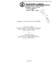
There are six main reproductive populations of <strong>Hawaiian</strong> monk seals in the<br />
Northwestern <strong>Hawaiian</strong> Islands (NWHI): French Frigate Shoals (FFS), Laysan Island,<br />
Lisianski Island, Pearl and Hermes Reef, Midway Atoll, and Kure Atoll (Fig. 1). Smaller<br />
numbers are present on Necker Island and Nihoa Island and on the main <strong>Hawaiian</strong><br />
Islands. <strong>Monk</strong> seals are rarely observed at Johnston Atoll where one female pup was born<br />
in 1969.<br />
Approximately 90% of <strong>Hawaiian</strong> monk seals remain at their natal site <strong>for</strong> life; the<br />
remaining 10% move between or among major population centers. Studies conducted at<br />
FFS in 1996 and 1997 documented the use of <strong>for</strong>aging habitats by monk seals which<br />
were much farther from breeding locations than was previously known or anticipated.<br />
Similar studies in 1997-98 at Pearl and Hermes Reef documented very little extra-atoll<br />
movement. Most feeding appears to occur at depths less than 75-90 m, though seals<br />
occasionally dive to depths exceeding 500 m. Known prey items include reef fishes,<br />
benthic fishes, cephalopods, and crustaceans.<br />
1.2 Population Biology<br />
Since 1985, the average rate of decline was approximately 3% yr -1 , although the<br />
beach counts 1 have been stable from 1993 to 2000 and declined again in 2001 and 2002<br />
(NMFS, unpublished data). Further decline is likely due to high juvenile mortality and an<br />
inverted age structure at FFS, the largest colony (Carretta et al., 2001). The annual<br />
number of births has varied substantially over the past decade and is expected to decline<br />
in the near future due to poor recruitment at FFS. At present the species numbers<br />
approximately 1400 and is the most endangered species of marine mammal that lives<br />
entirely within U.S. jurisdiction (Carretta et al., 2002).<br />
1 Direct enumeration data cannot be used <strong>for</strong> characterizing long-term trends because sufficient<br />
field investigation in the NWHI has not been consistently undertaken at all sites and years.<br />
Instead, a measure of long-term trend is derived from the mean of all the beach counts that have<br />
been conducted with varying frequency since the late 1950s. Beach counts provide a useful index<br />
of population trends and do not account <strong>for</strong> seals at sea. Approximately 1/3 of each seal<br />
population is on land during a survey, but the exact proportion varies (e.g., by site and season).
2<br />
30N<br />
Kure Atoll<br />
Midway Atoll<br />
Pearl and Hermes Reef<br />
Lisianski I.<br />
Laysan I.<br />
25N<br />
French Frigate Shoals<br />
Necker I.<br />
Nihoa I.<br />
20N<br />
Northwestern <strong>Hawaiian</strong> Islands<br />
Main <strong>Hawaiian</strong> Islands<br />
Johnston Atoll<br />
15N<br />
180° 175W 170W 165W 160W 155W<br />
Fig. 1. The <strong>Hawaiian</strong> Archipelago, with the six main monk seal breeding and pupping<br />
sites identified (French Frigate Shoals, Laysan I., Lisianski I., Pearl and Hermes Reef,<br />
Midway Atoll, Kure Atoll).<br />
Females give birth on beaches with adjoining shallow waters, which provide<br />
protection from sharks. Most pups are born between February and August, with a peak<br />
from late March to May (Johnson and Johnson, 1980; Johanos et al., 1994). Females give<br />
birth <strong>for</strong> the first time at ages 5 to 10, and typically 50%-70% of all adult-size females<br />
give birth each year (Johanos et al., 1994). Gestation is approximately 1 year, with a<br />
mean interval between births (<strong>for</strong> those females that pup in consecutive years) of 381<br />
days (Johanos et al., 1994). Females fast during the 5 or 6-week nursing period, and<br />
nursing pups are sometimes exchanged between females (Kenyon and Rice, 1959; Alcorn<br />
and Henderson, 1984; Boness, 1990; Gerrodette et al., 1992). Mating occurs 3 or 4 weeks<br />
after pups are weaned. Although mating takes place in the water and is rarely observed, a<br />
mating peak in May and June is inferred from the occurrence of fresh mounting injuries<br />
and association patterns of males and females (Johanos et al., 1994). Unlike other<br />
gregarious pinnipeds, monk seals are typically solitary and do not <strong>for</strong>m large, seasonal<br />
breeding aggregations. They have a serially monogamous social system with males<br />
<strong>for</strong>ming a dominance hierarchy.<br />
Pups are born with a black coat, which is shed gradually at the end of the nursing<br />
period and replaced by pelage that is silver gray on the dorsum and sides and beige on the<br />
venter. Juveniles and adults undergo a “catastrophic” annual molt, in which the epidermis
3<br />
is shed with the hair in large patches (Kenyon and Rice, 1959; Yochem and Stewart,<br />
2001). The freshly molted pelage is silver gray above and beige below except in adult<br />
males, where it is dark brown. Molting peaks in July, and females with pups molt 5 or 6<br />
weeks after mating (Johanos et al., 1994).<br />
1.3 <strong>Mortality</strong> Patterns<br />
Compared to other marine mammals, very good in<strong>for</strong>mation exists on mortality rates<br />
of <strong>Hawaiian</strong> monk seals. These rates are obtained annually by resighting known-age<br />
animals during 2-5-month field seasons at all six main subpopulations in the NWHI (Fig.<br />
1). Current tag and resight ef<strong>for</strong>ts are sufficient to obtain cohort-and age-specific survival<br />
(the complement of mortality) rates using Jolly-Seber analysis. These data are analyzed<br />
annually, and estimated rates are back-corrected when animals not seen <strong>for</strong> one or more<br />
years are subsequently resighted. Thus, long-term trends and variability in monk seal<br />
mortality rates are well characterized to age 15 years or more. These rates have reflected<br />
trends that greatly influence monk seal population trends. For example, juvenile mortality<br />
rates were low at most sites during the mid-to-late 1980s. Subsequently, juvenile<br />
mortality increased throughout the archipelago to varying degrees and on different time<br />
frames. Most notably, very high juvenile mortality has led to a severe and enduring<br />
population decline at FFS. Recent decline in juvenile survival has also been reported at<br />
Pearl and Hermes Reef and Midway and Kure Atolls in 2001 and 2002 (NMFS<br />
unpublished data).<br />
Although annual mortality rates are well characterized, the timing and relative<br />
importance of various causes of mortality are not well known. Jolly-Seber rates are<br />
available only at the end of a field season, so these data cannot be used to evaluate<br />
mortality processes in real time. Moreover, as noted above, these rates are annually<br />
updated and back-corrected with each new year’s data. The exact timing of mortalities is<br />
rarely known, as field ef<strong>for</strong>t encompasses less than half of any given calendar year. Even<br />
within a field season it is rarely possible to determine when an animal has died because<br />
so few carcasses are observed on the beach. In the few cases where carcasses are<br />
available <strong>for</strong> examination, unequivocal cause of death is often not discernible. For<br />
animals that are no longer sighted during regular beach walks, one can only infer that<br />
they died some time after the final sighting. For the Jolly-Seber analysis, any animal<br />
sighted within the calendar year, whether the first day or the last, is scored as having<br />
survived to that year. This is necessary <strong>for</strong> calculation of annual rates but does not<br />
provide within-season in<strong>for</strong>mation on time of death.<br />
To address the in<strong>for</strong>mation gaps noted above, field teams from the <strong>Hawaiian</strong> <strong>Monk</strong><br />
<strong>Seal</strong> Assessment Program provide a weekly report of the cumulative number of 1- to 3-<br />
year-old seals sighted to date, as well as the total number of confirmed and probable<br />
deaths within the season. In this way, minimum juvenile survival is regularly assessed so<br />
that unusually high mortality within a field season is likely to be detected. Post-mortem<br />
exams are per<strong>for</strong>med, and samples are collected <strong>for</strong> evaluation by a team of marine<br />
mammal health and disease experts to determine healthy body condition and cause of<br />
death where possible. Some laboratory analyses (e.g., erythrogram and leukogram
4<br />
evaluations) are per<strong>for</strong>med in the field; these preliminary findings, as well as descriptions<br />
of gross lesions or clinical signs observed, are reported to the <strong>Monk</strong> <strong>Seal</strong> Assessment<br />
Program and <strong>Monk</strong> <strong>Seal</strong> Health and Disease Program. These measures increase the<br />
likelihood of timely detection of an unusual mortality event, allowing a response to be<br />
mounted within the field season if deemed appropriate.<br />
Known causes of mortality in <strong>Hawaiian</strong> monk seals include emaciation of juveniles,<br />
tiger and Galapagos shark attacks, male aggression (individual and multiple), deleterious<br />
fisheries interactions, and entanglement in marine debris (Balazs and Whittow, 1979;<br />
Kenyon, 1981; Henderson, 1985, 1990; Alcorn and Kam, 1986; Banish and Gilmartin,<br />
1992; Hiruki et al., 1993a, 1993b; Nitta and Henderson, 1993; Starfield et al., 1995).<br />
Disturbance of pregnant or nursing females likely causes them to desert preferred<br />
pupping beaches, resulting in decreased pup survival (Kenyon, 1972; Kenyon, 1981).<br />
Maximum age is believed to be 25-30 years, but few seals live this long (NMFS,<br />
unpublished data).<br />
The influence of disease on monk seal populations is not well understood, but is an<br />
active area of research (Aguirre et al., 1999; Aguirre, 2000; Appendix A, <strong>Hawaiian</strong> <strong>Monk</strong><br />
<strong>Seal</strong> Specimen Collection Protocol and Appendix B, <strong>Hawaiian</strong> <strong>Monk</strong> <strong>Seal</strong> Necropsy<br />
Protocol). Infectious and noninfectious diseases have been reported in <strong>Hawaiian</strong> monk<br />
seals but do not appear to be impeding population recovery: all wild seals carry parasites;<br />
dental and skeletal abscesses and pathology were the apparent causes of death in one<br />
aged seal; and biotoxins may pose a serious risk (Golvan, 1959; Rausch, 1969; Kenyon<br />
and Rauzon, 1977 [cited in Kenyon 1981]; DeLong, 1978; DeLong and Gilmartin, 1979;<br />
Gilmartin et al., 1980; Whittow et al., 1980; Dailey et al., 1988). Of the 15 Marine<br />
Mammal <strong>Unusual</strong> <strong>Mortality</strong> <strong>Events</strong> (UMEs) occurring in other marine mammals species<br />
since 1992 (Dierauf and Gulland, 2001: 72-73, Table 1), infectious diseases and biotoxins<br />
were the most common diagnoses (five cases each). Other factors implicated in UMEs<br />
included fisheries interactions, ship strikes, and large-scale decadal changes in oceanic<br />
productivity (Polovina et al., 1995).<br />
In a recent review of emerging and resurging diseases in marine mammals, Miller et<br />
al. (2001) describes three ways in which wildlife species may be exposed to emerging<br />
diseases (newly evolved or newly identified as pathogens), all three of which are<br />
applicable to <strong>Hawaiian</strong> monk seals. The first is exposure via spillover from domestic<br />
species or humans. Many recent reports of transmissible diseases in marine mammals<br />
have included organisms traditionally associated with domestic animals and humans<br />
(e.g.; Brucella sp., Ewalte et al., 1994; Ross et al., 1994; Jepson et al., 1994;<br />
Mycobacterium bovis and M. tuberculosis, Forshaw and Phelps, 1991; Cousins et al.,<br />
1993; Woods et al.; 1995; Bernardelli et al., 1996). The second method of exposure is via<br />
rehabilitation or translocation of animals (e.g., Stamper et al., 1998, reports the possible<br />
spread of leptospirosis from skunks to rehabilitating harbor seals). The third way that<br />
wildlife species may become exposed to emerging diseases is through the effects of<br />
environmental phenomena such as El Niño and large-scale climate change on the<br />
proliferation or spread of infectious organisms (Fauquier et al. 1998; Harvell et al., 1999;<br />
Reddy et al. 2001). Another source of exposure to infectious diseases in <strong>Hawaiian</strong> monk
5<br />
seals is other wildlife species. Morbillivirus has been detected in the North Pacific Ocean<br />
(in common dolphins; Reidarson et al, 1998), and cross-species infection has been<br />
described <strong>for</strong> this group of viruses (e.g., canine distemper in Baikal and Caspian seals;<br />
Mamaev et al., 1995; Forsyth et al., 1998). A virus closely related to dolphin<br />
morbillivirus was implicated in an UME involving Mediterranean monk seals (Osterhaus<br />
et al., 1998), although saxitoxins were also detected in these seals.<br />
1.4 <strong>Unusual</strong> <strong>Mortality</strong>: Past <strong>Events</strong><br />
1.4.1 <strong>Hawaiian</strong> <strong>Monk</strong> <strong>Seal</strong> UME, Laysan Island, 1978.<br />
At least 50 monk seals died at Laysan Island in 1978, primarily in juvenile-young<br />
adult (1-5 years) and older adult (18-30 years) age classes (Johnson and Johnson, 1981;<br />
Gilmartin et al., 1980). Clinical signs noted be<strong>for</strong>e death included gradual weight loss<br />
(over a period of weeks to months), lethargy, change in behavior patterns, poor quality<br />
pelage (particularly around the face and neck), and presence of fresh lesions (primarily on<br />
the rear flippers and lower back). Affected seals were emaciated and gastric ulceration<br />
was observed in all 15 dead seals <strong>for</strong> which complete tissue sets were collected; this was<br />
accompanied by frank hemorrhage secondary to heavy gastric nematode infestations in<br />
many seals. High levels of ciguatoxin and maitotoxin, neurotoxins produced by benthic<br />
dinoflagellates in association with tropical reef environments, were estimated by bioassay<br />
analyses of the livers of two seals examined; levels were 30-50 times higher than those<br />
found in the liver of a monk seal that had been maintained in captivity <strong>for</strong> 15 years.<br />
However, radioimmunoassays <strong>for</strong> ciguatoxin were not consistent with the bioassay<br />
results: one seal had liver ciguatoxin levels 25% above those in the captive seal, and the<br />
second had levels 9% below the captive seal. Two of 18 live seals sampled had elevated<br />
white blood cell counts. One of these seals disappeared within 2 weeks of sampling, but<br />
the other remained healthy at least through 1979.<br />
1.4.2 <strong>Hawaiian</strong> <strong>Monk</strong> <strong>Seal</strong> UME, Laysan Island 2000-2001.<br />
An <strong>Unusual</strong> <strong>Mortality</strong> Event was declared in the spring of 2001 following the death of<br />
four juvenile monk seals in a 9-day period at Laysan Island. Criteria used by the Working<br />
Group on <strong>Unusual</strong> Marine Mammal <strong>Mortality</strong> <strong>Events</strong> (WGUMMME) included increased<br />
mortality compared to prior records, mortalities occurring in a localized area, mortalities<br />
associated with abnormal behavior patterns, and the mortality of a critically endangered<br />
species (Wilkinson, 1996; Antonelis et al., 2001). An investigation was launched at<br />
Laysan Island and other subpopulations in the NWHI to assess possible rangewide<br />
effects. Survival from weaning to 1 year in 2001 was lower than most previous years at<br />
all but one site. Investigation into causes included infectious diseases, parasitism,<br />
environmental conditions, naturally occurring biotoxins, and anthropogenic<br />
contaminants. Necropsy results from 11 seals revealed emaciation but no signs of<br />
infectious disease or toxicosis linking the UME deaths. The unusually high mortality in<br />
yearling seals was not correlated with their size at weaning, but appeared to be a result of<br />
their inability to <strong>for</strong>age successfully during the post-weaning transition to nutritional
6<br />
independence (Antonelis et al., 2001). High juvenile mortality was also observed in 2002<br />
(NMFS unpublished data).<br />
1.5 Background of this Document<br />
The first attempt to develop a response plan <strong>for</strong> UMEs in <strong>Hawaiian</strong> monk seals is<br />
presented by Gilmartin (1987). The National Marine Fisheries Service (NMFS) has<br />
published a National <strong>Contingency</strong> <strong>Plan</strong> <strong>for</strong> responding to unusual marine mammal<br />
mortality events (Wilkinson, 1996). The WGUMMME recommended that a separate<br />
plan, consistent with the national plan and other relevant documents such as the Hawaii<br />
Area Oil Spill <strong>Contingency</strong> <strong>Plan</strong>, be prepared <strong>for</strong> <strong>Hawaiian</strong> monk seals because of their<br />
endangered status and the logistic difficulties associated with the remoteness of most of<br />
their colonies. A separate plan was prepared <strong>for</strong> manatees by Geraci and Lounsbury<br />
(1997), and the <strong>Hawaiian</strong> monk seal contingency plan follows the manatee <strong>for</strong>mat.<br />
2. Federal/State Authority and Jurisdiction<br />
The Marine Mammal Health and Stranding Response Act was passed by the U.S.<br />
Congress in 1992 (P.L. 102-587, 16 U.S.C. 1421 [c-d]), and it became Title IV of the<br />
Marine Mammal Protection Act (MMPA). Section 404 of MMPA Title 4 (P.L. 103-238,<br />
§24(b)(1)) established a framework <strong>for</strong> responding to Marine Mammal UMEs, described<br />
in detail in the National <strong>Contingency</strong> <strong>Plan</strong> <strong>for</strong> Response to <strong>Unusual</strong> Marine Mammal<br />
<strong>Mortality</strong> <strong>Events</strong> (Wilkinson, 1996). The response sequence is outlined below and is<br />
presented as a flowchart in Appendix I-1.<br />
• An increase in stranding rates or unusual findings is detected through one of three<br />
likely mechanisms:<br />
o NMFS Regional Stranding Coordinator (Pacific Islands Regional Office,<br />
PIRO) detects increased strandings or unusual findings upon reviewing<br />
Level A data and consulting with the Stranding Network;<br />
o Stranding Network directly notifies the NMFS Regional Stranding<br />
Coordinator (PIRO) of an increase in strandings or unusual findings;<br />
o <strong>Monk</strong> <strong>Seal</strong> Research Leader observes an increase in strandings or unusual<br />
finding and notifies the Leader of the Pacific Island Fisheries Science<br />
Center’s Marine Mammal Research Program (MMRP).<br />
• The NMFS Regional Stranding Coordinator (PIRO) or the Leader of MMRP will<br />
contact the Executive Secretary <strong>for</strong> the WGUMMME (NMFS Headquarters), pass<br />
on the in<strong>for</strong>mation, and request a consultation with the WGUMMME.<br />
• The Executive Secretary <strong>for</strong> the WGUMMME will <strong>for</strong>mally request a<br />
consultation and <strong>for</strong>ward a complete summary of the event in question and a<br />
historical record of stranding data to the WGUMMME.
7<br />
• Over a 24-hour period, the WGUMMME will review the data and compare it<br />
against the seven criteria <strong>for</strong> determination of an UME (Wilkinson, 1996:18):<br />
o A marked increase in the magnitude of strandings is occurring when<br />
compared with prior records. There is no set <strong>for</strong>mula <strong>for</strong> determining what<br />
magnitude would trigger a response. The NMFS Southeast Region has<br />
used a <strong>for</strong>mula of the historic mean plus two times the standard deviation<br />
to determine a threshold level. The WGUMMME stated that the<br />
magnitude must be weighed against other knowledge. As a pragmatic<br />
method, it was suggested that if a pulse in strandings is spread over an<br />
area or timeframe that strains the capacity of the Stranding Networks to<br />
respond, it should be cause <strong>for</strong> concern.<br />
o Animals are stranding at a time of the year when strandings are unusual.<br />
o An increase in strandings is occurring in a very localized area (possibly<br />
suggesting a localized problem), is occurring throughout the geographical<br />
range of the species/population, or spreads geographically with time.<br />
o The species, age, or sex composition of the stranded animals is different<br />
than those of animals that normally strand in the area at that time of year.<br />
o Stranded animals exhibit similar or unusual pathologic findings, or the<br />
general physical condition (e.g., blubber thickness) of stranded animals is<br />
different from what is normally seen.<br />
o <strong>Mortality</strong> is accompanied by behavior patterns observed among living<br />
individuals in the wild that are unusual, such as occurrence in habitats<br />
normally avoided or abnormal patterns of swimming and diving.<br />
o Critically endangered species are stranding. Stranding of three or four<br />
right whales, <strong>for</strong> example, may be cause <strong>for</strong> great concern whereas<br />
stranding of a similar number of fin whales may not.<br />
• The WGUMMME will return its decision to the Executive Secretary, and an<br />
official determination will be made if a quorum has been reached (i.e., majority of<br />
2/3 of the voting body).<br />
• The Executive Secretary officially requests concurrence from the NMFS Assistant<br />
Administrator. With this concurrence, the NMFS Assistant Administrator is also<br />
requested to appoint an On-Site Coordinator (see below).<br />
3. The On-Site Coordinator<br />
3.1 The Appointment of the On-Site Coordinator.
8<br />
Following the sequence of events described above, the NMFS Assistant Administrator<br />
will appoint an On-Site Coordinator <strong>for</strong> the UME. The On-Site Coordinator is the<br />
appropriate NMFS Regional Administrator or his or her designee (Wilkinson, 1996). The<br />
selection of On-Site Coordinator is based upon recommendations from the WGUMMME<br />
and the Leader of the MMRP, PIFSC, NOAA Fisheries.<br />
3.2. Responsibilities of the Federal On-Site Coordinator.<br />
The On-Site Coordinator should have strong management and leadership abilities,<br />
strong communication skills, the ability to make decisions with minimal use of<br />
intermediaries, the ability to access in<strong>for</strong>mation and expertise (including interagency<br />
expertise), familiarity with the National <strong>Contingency</strong> <strong>Plan</strong> and associated protocols<br />
(including this monk seal contingency plan), and familiarity with and the ability to work<br />
with the Marine Mammal Stranding Networks (Wilkinson, 1996:19). The On-Site<br />
Coordinator must hold or be included on a federal Enhancement and Scientific Research<br />
Permit (e.g., the permit held by the Leader of MMRP).<br />
The On-Site Coordinator will work closely with the Executive Secretary, NMFS<br />
national and regional offices (e.g., PIRO) and the Leader of MMRP. The On-Site<br />
Coordinator also will notify other federal agencies (e.g., U.S. Coast Guard, U.S. Fish and<br />
Wildlife Service), State of Hawaii wildlife resource agencies (e.g., Hawaii Department of<br />
Land and Natural Resources, Division of Aquatic Resources, and Division of Forestry<br />
and Wildlife,), Hawaii regional stranding network members, and the <strong>Hawaiian</strong> <strong>Monk</strong><br />
<strong>Seal</strong> Recovery Team. A list of federal and state agencies and individuals to be notified is<br />
included in Appendix H-1. Contact lists are updated regularly (at least annually) by<br />
MMRP. Additional notifications will be made as needed; <strong>for</strong> example, the Hawaii<br />
Department of Public Health would be notified if a serious human health hazard (e.g., if a<br />
serious zoonotic disease is identified or suspected) existed on one of the main <strong>Hawaiian</strong><br />
Islands, and USDA/APHIS would be notified if monk seal transport were being<br />
considered. The On-Site Coordinator will also assemble a response team as described<br />
below.<br />
4. Pre-Event <strong>Plan</strong>ning <strong>for</strong> Rapid Response<br />
4.1 Building a Response Team<br />
The response team assembled by the On-Site Coordinator will include one or more<br />
wildlife biologists with knowledge of normal <strong>Hawaiian</strong> monk seal behavior and natural<br />
history and one or more veterinarians familiar with pinniped diseases and monk seal<br />
sampling protocols (including specimen collection and necropsy protocols). Additional<br />
team members will be selected depending on the size and nature of the event and may<br />
include a veterinary technician to assist with sample collection and processing, a rescue<br />
and salvage logistics coordinator, a parasitologist, and an environmental sampling<br />
coordinator. A list of potential team members is included in H-2; this list is updated at<br />
least annually by MMRP to ensure that a qualified team can be assembled immediately.
9<br />
4.2 The Administrative Support Team<br />
Three important administrative areas to be addressed by the On-Site Coordinator in<br />
the event of a <strong>Hawaiian</strong> monk seal UME are financial administration, public in<strong>for</strong>mation<br />
coordination, and tracking of animals and samples. As stipulated in the National<br />
<strong>Contingency</strong> <strong>Plan</strong> (Wilkinson, 1996), a certified Contracting Officer’s Technical<br />
Representative will be appointed as financial officer by the Regional Office and will<br />
work with the On-Site Coordinator and a certified Contracting Officer to negotiate and<br />
enter into agreements <strong>for</strong> services per<strong>for</strong>med in the course of a MMUME response, to<br />
locate and order supplies and equipment, and to be responsible <strong>for</strong> prompt payment <strong>for</strong><br />
services, purchase orders, and expense reimbursements. The roles of On-Site Coordinator<br />
and Contracting Officer’s Technical Representative can be filled by the same individual<br />
or by two separate individuals. The On-Site Coordinator will appoint a local media<br />
contact (either an agency public affairs officer or an individual involved in the response)<br />
to address public concerns and inquiries and to keep the NMFS Office of Public Affairs<br />
(301-713-2370) in<strong>for</strong>med. Procedures <strong>for</strong> tracking data and samples will follow wellestablished<br />
protocols (<strong>Monk</strong> <strong>Seal</strong> Specimen Collection and Necropsy protocols,<br />
Appendices A and B). In some cases (e.g., potential litigation or prosecution related to<br />
the MMUME) chain of custody will be maintained <strong>for</strong> all samples and data collected<br />
during the response (Appendix G-6).<br />
4.3 Scientific Advisors<br />
In addition to the WGUMMME, the On-Site Coordinator may call on scientific<br />
advisors from the <strong>Hawaiian</strong> <strong>Monk</strong> <strong>Seal</strong> Recovery Team, the Marine Mammal<br />
Commission, or the list of attendees to the 11-12 September 2000 <strong>Hawaiian</strong> <strong>Monk</strong> <strong>Seal</strong><br />
Health Studies Workshop. Additional advisors may be consulted depending on the size<br />
and nature of the event (e.g., the NOAA Marine Biotoxins Program, 843-762-8500, will<br />
be contacted if a Harmful Algal Bloom is suspected).<br />
4.4 Training and Readiness<br />
MMRP conducts extensive annual training of field personnel <strong>for</strong> the monk seal<br />
assessment program. Many of the topics addressed in this training program are relevant to<br />
MMUME response, such as the <strong>Monk</strong> <strong>Seal</strong> Health and Disease Program (including<br />
procedures and supply lists <strong>for</strong> specimen collection, necropsies, and field laboratory<br />
assays (Appendices A through H), field camp logistics and set-up, animal care and use<br />
policies, field communications and safety, and first aid training. In addition to this predeployment<br />
training, field camp personnel have practical experience in <strong>Hawaiian</strong> monk<br />
seal data collection and processing obtained during deployments of 2-5 months in the<br />
NWHI.<br />
<strong>Monk</strong> seal rescue and rehabilitation are addressed in the <strong>Monk</strong> <strong>Seal</strong> Rehabilitation<br />
Manual (Appendix D). Lavigne (1999) reviewed past attempts to enhance monk seal<br />
survival and reproduction via the “headstart program,” the rescue, rehabilitation and<br />
transport of undersized females and the translocation or removal of adult males to reduce
10<br />
mobbing. Such ef<strong>for</strong>ts have had varying levels of success in enhancing monk seal<br />
survival.<br />
The Leader of MMRP (Dr. George Antonelis, 808-983-5710) has designated<br />
individuals <strong>for</strong> key roles such as monk seal population assessment (Jason Baker, 808-<br />
983-5711) and health studies (Dr. Robert Braun, 808-254-8181 or 808-254-3530 or 808-<br />
783-6565; Lizabeth Kashinsky, 808-592-8306) with well-defined lines of authority and<br />
responsibilities. Members of the Hawaii Marine Mammal Stranding Network have<br />
responded to events such as monk seal entanglements in fishing gear, vessel grounding,<br />
and the presence of females and pups on public beaches in the main <strong>Hawaiian</strong> Islands.<br />
The <strong>Hawaiian</strong> Islands Response Group V.P. (Marlee Breese, 808-259-5268 or 808-291-<br />
6434) is a key contact in the stranding network of Hawaii.<br />
4.5 Establishing Memoranda of Understanding (MOUs) with Cooperators<br />
The U.S. Navy and the U.S. Coast Guard have provided logistical support to the<br />
Marine Mammal Research Program in past years on a not-to-interfere basis. This support<br />
has included transportation (of personnel, equipment, and supplies) to and from the<br />
NWHI and medical evacuation of injured field personnel. MOU with these agencies are<br />
being explored to establish <strong>for</strong>mal mechanisms <strong>for</strong> support in the event of a monk seal<br />
UME. Additionally, an Oil Spill Response <strong>Plan</strong> is in the final stages of review and will be<br />
available soon.<br />
4.6 Pre-<strong>Plan</strong>ning <strong>for</strong> Sample Analyses<br />
The Marine Mammal Research Program has designated several laboratories<br />
(Appendix H-3) <strong>for</strong> analyses of samples collected from <strong>Hawaiian</strong> monk seals. In addition,<br />
an epidemiology sampling program provides useful baseline data <strong>for</strong> comparison in the<br />
event of a MMUME (Aguirre et al., 1999; Aguirre, 2000). The epidemiology sampling<br />
program is reviewed periodically (e.g., September 11-12, 2000 <strong>Hawaiian</strong> <strong>Monk</strong> <strong>Seal</strong><br />
Health Studies Workshop; November 2001 conference call coordinated by Dr. Robert<br />
Braun, monk seal health studies coordinator) and changes are implemented as needed (cf.<br />
Appendix E-1; following the 2000-2001 abortion investigation at Laysan Island,<br />
sampling and necropsy protocols were modified to improve the efficiency of UME<br />
investigations).<br />
5. Recognizing an <strong>Unusual</strong> Event<br />
The following criteria <strong>for</strong> defining a MMUME are given in the Marine Mammal<br />
Health and Stranding Response Act (Wilkinson, 1996; Dierauf and Gulland, 2001):<br />
• It is unexpected;<br />
• It involves a significant die-off of a marine mammal population; or<br />
• A small number of a severely endangered marine mammal species appear to be<br />
affected;<br />
• Demands an immediate response.
11<br />
During the 2000 abortion investigation at Laysan Island, the following additional<br />
criteria were used: increased mortality compared to prior records, mortalities occurring<br />
in a localized area, and mortalities associated with abnormal behavior patterns (Antonelis<br />
et al., 2001).<br />
5.1 Guidelines <strong>for</strong> a Graduated Response<br />
Current <strong>Hawaiian</strong> monk seal research activities include population monitoring,<br />
epidemiology program sampling (live and dead seals), documentation of injuries (e.g.,<br />
wounds caused by conspecifics or predators) that might affect an individual seal’s<br />
survival, monk seal feeding habits studies, and documentation of the use of marine<br />
habitats by monk seals using satellite-linked telemetry and underwater cameras<br />
(“CRITTERCAM”). Research conducted as part of the <strong>Hawaiian</strong> <strong>Monk</strong> <strong>Seal</strong> Assessment<br />
has generated baseline data on expected numbers and timing of births and deaths at monk<br />
seal colonies; long-term tagging and monitoring programs have generated individual life<br />
history data <strong>for</strong> approximately 90% of the population. The <strong>Hawaiian</strong> <strong>Monk</strong> <strong>Seal</strong> Health<br />
and Disease Program maintains a frozen serum bank and a biomedical database indexed<br />
by island and by individual seal. NMFS field camp personnel and others (e.g., USFWS<br />
refuge biologists) are trained to recognize and report live monk seals exhibiting abnormal<br />
behavior or appearance, as well as any dead seals encountered to the Marine Mammal<br />
Research Program Leader (Dr. George Antonelis) and the program’s designated<br />
veterinarian and coordinator of the monk seal health studies (Dr. Robert Braun). Early<br />
recognition and rapid reporting of a potential MMUME provide time to alert key<br />
individuals and cooperators (including the NMFS Regional Office, NMFS National<br />
MMUME Coordinator, and the NMFS Office of Protected Resources), conduct<br />
preliminary studies and gather additional data, assess capability <strong>for</strong> response, open lines<br />
of communication, and identify and refine (as necessary) protocols <strong>for</strong> response (Geraci<br />
and Lounsbury, 1997).<br />
Baseline data collection and population monitoring of other species occurs seasonally<br />
(in some cases, year round) in the NWHI and may assist in early recognition and<br />
reporting of an UME. Fish censuses are conducted annually (and in some cases, monthly)<br />
in many locations in monk seal habitat as part of ongoing coral reef research programs.<br />
The U.S. Fish and Wildlife Service conducts surveys at least annually of sea turtle<br />
nesting activity and abundance and distribution of resident and migratory birds. Yearround<br />
camps are maintained at some locations in the NWHI (e.g., Midway Atoll, Laysan<br />
Island, FFS), staffed by federal and state wildlife biologists. An ongoing photoidentification<br />
project is documenting occurrence and behavior of Stenella longirostris at<br />
Midway Atoll, Kure Atoll, and Pearl and Hermes. The NMFS Southwest Fisheries<br />
Science Center is surveying cetacean populations along transects near the <strong>Hawaiian</strong><br />
Archipelago. An increase in the number of stranded marine mammals or sea turtles would<br />
be detected by the NMFS Regional Stranding Coordinator.
12<br />
5.2 Protocol <strong>for</strong> Initiating <strong>Unusual</strong> Event Designation<br />
As specified in Title IV of the Marine Mammal Health and Stranding Act, the NMFS<br />
Pacific Area Protected Species Program Coordinator/Regional Stranding Coordinator (or<br />
his or her designee; e.g., the Leader of MMRP) will contact the NMFS National<br />
MMUME Coordinator if dead or moribund monk seals are observed unexpectedly. This<br />
contact initiates a chain of events illustrated in a flow chart by Dierauf and Gulland<br />
(2001, Fig. 1, p. 75), reprinted as Appendix I-1 and briefly summarized here. The NMFS<br />
National MMUME Coordinator will alert the members of the WGUMMME, who will<br />
require in<strong>for</strong>mation on the background levels of morbidity and mortality to decide<br />
whether or not a MMUME is taking place (data to be obtained from prior records;<br />
Wilkinson, 1996; p. 18). For example, is the age and sex composition, behavior or body<br />
condition of the stranded seals different from what occurs normally in that geographic<br />
location or at that time of year? If the Working Group decides that a MMUME is<br />
occurring, the MMUME National Coordinator in<strong>for</strong>ms the Regional Stranding<br />
Coordinator, designates an On-Site Coordinator (though the Secretary of Commerce), and<br />
transfers responsibility <strong>for</strong> action to the On-Site Coordinator.<br />
6. Notification of Personnel and Agencies<br />
The On-Site Coordinator will notify and mobilize federal, state, and other authorized<br />
<strong>Hawaiian</strong> monk seal rescue and recovery program personnel, possible Response Team<br />
members, and pre-identified laboratories and MOU cooperators promptly. The On-Site<br />
Coordinator also will notify other federal agencies, state wildlife resource agencies, local<br />
government agencies, stranding network personnel and the Leader of the <strong>Monk</strong> <strong>Seal</strong><br />
Recovery Team (see Section 3.2 above). Contact in<strong>for</strong>mation <strong>for</strong> agencies and<br />
individuals to be notified are included in Appendices H-1 through H-4; these lists are<br />
updated at least annually by MMRP. Additional notifications of other stakeholders will<br />
be made as needed; <strong>for</strong> example, the Hawaii Department of Public Health will be notified<br />
if a serious human health hazard exists on one of the MHI (e.g., if a serious zoonotic<br />
disease is identified or suspected).<br />
7. A Tailored Response<br />
7.1 Objectives and General Guidelines<br />
The On-Site Coordinator will work with the WGUMMME to determine and initiate a<br />
course of action to protect the public health and welfare, identify the cause(s) of the event<br />
(including possible contributing factors such as malnutrition or contaminants), minimize<br />
deaths and provide <strong>for</strong> the rehabilitation of individual animals, and determine the impact<br />
of the MMUME on the population (Wilkinson, 1996; Geraci and Lounsbury, 1997;<br />
Dierauf and Gulland, 2001).
13<br />
7.2 Responding to an Event of Known Cause<br />
Wilkinson (1996, Table 1; Appendix I-3) and Geraci and Lounsbury (1997) describe<br />
the steps to be taken (including continuing consultation with the WGUMMME) in<br />
response to a MMUME of known cause. Chain of custody procedures will be<br />
implemented <strong>for</strong> all data and samples collected in response to a MMUME (Wilkinson<br />
1996; Appendix G-6), even if the cause is already known or suspected. Pre-identified<br />
risks to <strong>Hawaiian</strong> monk seals include biotoxins such as ciguatera, anthropogenic<br />
chemical spills, abortion “storms,” viral epizootics (e.g., morbillivirus), and humanrelated<br />
trauma (e.g., entanglement).<br />
• Naturally occurring toxins (e.g., harmful algal bloom, ciguatera). The NOAA<br />
Marine Biotoxins Program has posted a Flow Diagram <strong>for</strong> Suspected Marine<br />
Biotoxin Incidents<br />
(www.chbr.noaa.gov/CoastalResearch/Pictures/AnalyticalResponseTeam/FlowDi<br />
agram.gif); this will be used to guide response to a suspected harmful algal<br />
bloom. Specimen collection protocols <strong>for</strong> specific biotoxins are presented in<br />
Rowles et al. (2001:468, Table 5). See Appendix E-2.<br />
• Anthropogenic chemicals (e.g., oil or other chemical spill). Procedures will be<br />
consistent with the contingency plans developed by the U. S. Coast Guard (28<br />
August 2001 update of the Hawaii Area <strong>Contingency</strong> <strong>Plan</strong>,<br />
www.uscg.mil/d14/units/msohono/HACP1/index.htm; Oil Spill Field Operations<br />
Guide, www.uscg.mil/d14/units/msohono/ics/fog/index.htm). Specimen<br />
collection protocols <strong>for</strong> chemical pollutants are presented in Rowles et al.<br />
(2001:466; Table 4). See Appendix E-3.<br />
• Abortion. Four aborted fetuses were observed over a 4-week period at Laysan<br />
Island in 2000. It is not known if these carcasses represented a true increase in<br />
mortality or simply an increase in survey ef<strong>for</strong>t. Baseline data on morbidity and<br />
mortality are sparse <strong>for</strong> this time of year because NMFS field camps at Laysan<br />
Island typically are established several weeks later. Results of the investigation<br />
into this event were inconclusive. Sixteen seals were sampled, including one adult<br />
female thought to have aborted a fetus. Laboratory results (including tests <strong>for</strong><br />
Chlamydia sp., Brucella sp., Leptospira sp., morbillivirus and herpesvirus) were<br />
consistent with baseline serologic test results obtained during previous<br />
epidemiology studies on <strong>Hawaiian</strong> monk seals. Five placentas (from surviving<br />
pups) were examined and considered to be normal. No gross lesions were noted<br />
during one post-mortem exam on an adult female; histopathology revealed only<br />
changes consistent with advanced age. However, because abortion-causing<br />
organisms are a recognized threat to monk seals, genital swabs were added to the<br />
standard epidemiology and necropsy sampling protocols in 2001 (cf. Appendix E-<br />
1).<br />
• Infectious agents (e.g., morbillivirus). Live capture and isolation of affected<br />
individuals (e.g., while laboratory analyses are pending) may be necessary if an<br />
epizootic is suspected. Osterhaus et al. (1998) proposed that vaccination be<br />
considered in the management and conservation of Mediterranean monk seals.<br />
The Marine Mammal Research Program is reviewing the potential risks and
14<br />
benefits of morbillivirus vaccination in the face of an outbreak in <strong>Hawaiian</strong> monk<br />
seals.<br />
• Human-related trauma (e.g., entanglement). Chain of custody procedures<br />
(Appendix G-6) will be implemented <strong>for</strong> all data and samples collected in<br />
response to an MMUME involving human-related trauma. In addition, NMFS<br />
Evaluation of Human Interaction <strong>for</strong>ms (Appendix G-5) will be completed <strong>for</strong><br />
affected animals.<br />
7.3 Investigating an Event of Unknown Cause<br />
Wilkinson (1996, Table 1; Appendix I-3) and Geraci and Lounsbury (1997) describe<br />
the steps to be taken (including continuing consultation with the WGUMMME) in<br />
response to a MMUME of unknown cause. Initial data collection will follow existing<br />
monk seal protocols (e.g., Appendices A through D, G and H) to ensure that in<strong>for</strong>mation<br />
collected during the MMUME are comparable to baseline data. As the event progresses<br />
and at the recommendation of the WGUMMME, additional sampling protocols may be<br />
implemented (cf. Appendix E).<br />
If no cause is readily apparent, the investigation may include analyses of other species<br />
in the area of the monk seal UME in order to identify or validate possible causes (see<br />
Section 5.1 <strong>for</strong> a description of monitoring programs <strong>for</strong> other species). For example,<br />
harmful algal blooms often affect a number of plantivorous and piscivorous species (e.g.,<br />
crustaceans, fish, seabirds, sea turtles). The Marine Mammal Research Program is<br />
preparing cooperative agreements with agencies that oversee these species and resources<br />
(e.g., USFWS Refuge Managers, NOS-NWHI Coral Reef Reserve, State of Hawaii<br />
Department of Land and Natural Resources, NOAA National Marine Sanctuary Program<br />
personnel) to expedite such investigations in the event of a monk seal UME.<br />
Under certain conditions (Wilkinson, 1996; p. 41), and if it does not inhibit the<br />
ongoing investigation, NMFS may accommodate requests from independent researchers<br />
<strong>for</strong> tissue samples and data to address other scientific questions that may or may not be<br />
related to the MMUME. Chain of custody procedures will be implemented <strong>for</strong> all data<br />
and samples collected in response to a MMUME (Wilkinson, 1996; Rowles et al.,<br />
2001:452 [Fig. 1]).<br />
7.4 Determining "The End"<br />
The WGUMMME, in consultation with the On-Site Coordinator, will determine when<br />
a MMUME has ended. The end point of a MMUME may be as difficult to pinpoint as the<br />
onset, particularly if the decline in morbidity or mortality is gradual. If there is a seasonal<br />
component to the event (e.g., an abortion ‘storm’), investigators may not be convinced<br />
that the MMUME is over until the following year, when it is possible to assess whether<br />
or not values (e.g., number of premature pups observed) have returned to historic or<br />
baseline levels (when compared to prior records, cf. Wilkinson, 1996; p. 18).
15<br />
7.5 Post-Event Assessment and Monitoring<br />
Once the end point of a MMUME has been determined, the On-Site Coordinator will<br />
prepare a report <strong>for</strong> submission to the WGUMMME containing the results of all analyses<br />
conducted during the investigation, an assessment of the impact of the MMUME on the<br />
<strong>Hawaiian</strong> monk seal population, and recommendations <strong>for</strong> future monitoring (Wilkinson,<br />
1996). In some cases, post-event monitoring will return to pre-event levels (i.e., the <strong>Monk</strong><br />
<strong>Seal</strong> Assessment and Health and Disease programs of MMRP). In other cases, protocols<br />
may be modified (cf. Appendix E) and additional recovery or population monitoring<br />
activities may be incorporated into existing research and management programs.<br />
8. Communication and Reporting<br />
The On-Site Coordinator must maintain a regular communication and reporting<br />
schedule with the Response Team Leader and the WGUMMME. Telephone or email<br />
briefings every 1 to 2 days will enable the On-Site Coordinator to recognize and address<br />
personnel, equipment, supply, and data needs of the response team and to ensure that<br />
critical data and samples are being collected. The Response Team Leader should submit a<br />
brief written (e-mail) report to the On-Site Coordinator once a week, summarizing the<br />
status of the investigations and noting unresolved problems. The On-Site Coordinator<br />
should <strong>for</strong>ward this report with whatever additional calculations, maps, histograms, or<br />
laboratory analysis results are available to the WGUMMME. The On-Site Coordinator<br />
and WGUMMME may invite additional experts to review and discuss these reports and<br />
will determine what additional ef<strong>for</strong>ts are needed to respond to and investigate the<br />
MMUME.<br />
The On-Site Coordinator will also provide regular official reports on the progress of<br />
the investigation to appropriate federal, state, and local agencies and to the <strong>Hawaiian</strong><br />
<strong>Monk</strong> <strong>Seal</strong> Recovery Team. The media contact appointed by the On-Site Coordinator<br />
will keep the NMFS Office of Public Affairs in<strong>for</strong>med, prepare press releases as needed,<br />
respond to media requests <strong>for</strong> interviews, and address public concerns and inquiries.<br />
9. Public Health Concerns<br />
Public health and welfare are the first priorities in responding to a MMUME<br />
(Wilkinson, 1996). The On-Site Coordinator will ensure compliance with safety<br />
guidelines (e.g., § 300.150 of the National Oil and Hazardous Substances Pollution<br />
<strong>Contingency</strong> <strong>Plan</strong> [40 CFR Part 300] specifies the type of training required <strong>for</strong> personnel<br />
responding to an event involving oil discharges or hazardous chemical releases).<br />
Only properly trained and equipped personnel will participate in a MMUME response.<br />
Safety training programs conducted annually <strong>for</strong> field personnel working with <strong>Hawaiian</strong><br />
monk seals include animal handling skills, first aid, and hygienic precautions such as the<br />
<strong>Monk</strong> <strong>Seal</strong> Clean Protocol and hazardous waste procedures. Additional training (e.g.,<br />
boat handling, aviation safety) will be provided as needed.
16<br />
Carcasses may be left on the beach to decompose naturally unless a transmissible<br />
pathogen or serious toxin is suspected. In this case, the carcass may be buried, taken to a<br />
sanitary landfill, or incinerated.<br />
If it is necessary to isolate or quarantine live animals during a MMUME, existing<br />
protocols developed <strong>for</strong> <strong>Hawaiian</strong> monk seals will be implemented (Appendix D).<br />
10. Resources: Location and Utilization<br />
10.1 Introduction<br />
Although <strong>Hawaiian</strong> monk seals do occur on the MHI, the majority are found on or<br />
around the NWHI, a chain of islands and atolls extending hundreds of kilometers. Some<br />
of these sites are accessible only by boat, and weather and sea conditions often prevent<br />
landing. Time to mount a response to a monk seal UME is likely to range from a few<br />
hours (e.g., Kauai) to a week or more (e.g., Laysan Island).<br />
The presence of other protected species (e.g., nesting seabirds) may affect the<br />
activities of UME response personnel. For example, U.S. Fish and Wildlife Service<br />
quarantine protocols are in effect at some islands requiring special care and treatment of<br />
clothing, footwear, equipment, and supplies. Access to island interiors may be restricted<br />
at some islands to prevent disturbance of sensitive animal or plant species.<br />
10.2 Equipment and Logistics<br />
All supplies and equipment needed <strong>for</strong> a limited MMUME response are<br />
maintained at the Kewalo Research Facility in Honolulu, including capture and<br />
handling gear (including supplies and equipment <strong>for</strong> chemical immobilization and<br />
emergency cardiopulmonary resuscitation), biomedical sampling kits <strong>for</strong> up to 50<br />
monk seals, and a portable laboratory. Equipment lists are included in Appendix<br />
F.<br />
Charter flights can be arranged to Midway Atoll, Tern Island, or FFS. Vessel charters<br />
can also be arranged <strong>for</strong> logistical support. A list of charter companies is maintained by<br />
MMRP. The U.S. Coast Guard (Office of Aids to Navigation <strong>for</strong> ship transport, CGAS<br />
Barbers Point <strong>for</strong> air transport) and U.S. Navy have provided emergency transport and<br />
equipment drops on occasion. MOU with these agencies are being explored to establish<br />
<strong>for</strong>mal mechanisms <strong>for</strong> support in the event of a monk seal UME. The NOAA ship Oscar<br />
Elton Sette and other NOAA research vessels could be diverted from other activities near<br />
the <strong>Hawaiian</strong> Archipelago to support a monk seal UME investigation if approved by the<br />
Director of PIFSC or the Sanctuary Office.<br />
10.3 In<strong>for</strong>mation and Data Resources<br />
Contact in<strong>for</strong>mation <strong>for</strong> key personnel (e.g., potential Response Team members) is<br />
included in Appendix H. MMRP maintains current contact lists and protocols, and the
17<br />
monk seal assessment program maintains a database of relevant historical in<strong>for</strong>mation<br />
such as population counts (beach counts, aerial surveys) and pup mortality rates. Up to<br />
about 90% of all seals in the NWHI are individually identified each field season.<br />
Reproductive histories are known <strong>for</strong> a majority of females at Laysan and <strong>for</strong> many seals<br />
at Lisianski and FFS. Age-specific survival is well known <strong>for</strong> nearly all animals born in<br />
the past 20 years or so. The <strong>Monk</strong> <strong>Seal</strong> Health and Disease Program maintains a baseline<br />
biomedical database <strong>for</strong> <strong>Hawaiian</strong> monk seals, including hematology and serum<br />
biochemistry reference ranges, seroprevalence of potential pathogens, gross necropsy and<br />
histopathology reports, and parasite surveys (cf. Aguirre, 2000). Individual biochemical<br />
serum test results are available <strong>for</strong> almost 25% of the population.<br />
10.4 Development of Temporary Animal Housing and Emergency Facilities<br />
Rehabilitation facilities <strong>for</strong> <strong>Hawaiian</strong> monk seals are maintained at the NMFS<br />
Kewalo Basin site (pools sufficient to hold 3-5 monk seals), although the ability to isolate<br />
seals is limited. The Marine Mammal Research Program has construction plans,<br />
equipment and supply lists, and approximate cost estimates <strong>for</strong> temporary holding<br />
facilities that could be erected in remote locations in the NWHI. Gilmartin et al. (in<br />
review) describe holding pens (35-60 square meters), incorporating sandy haul-out areas<br />
and shallow seawater access.<br />
10.5 Funding<br />
The MMUME Fund, established in Title IV of the Marine Mammal Protection Act, is<br />
an interest-bearing account in the Federal Treasury to be used exclusively <strong>for</strong> costs<br />
associated with preparing <strong>for</strong> and responding to MMUMEs (Dierauf and Gulland, 2001).<br />
In the event of a MMUME, funds are made available through the National <strong>Contingency</strong><br />
<strong>Plan</strong> (Wilkinson, 1996; Dierauf and Gulland, 2001). Post-UME monitoring is not<br />
supported by the MMUME Fund, but other federal sources (e.g., Prescott Grant) are<br />
available <strong>for</strong> possible funding.<br />
11. Response and Investigation Protocols<br />
11.1: Rescue and rehabilitation (Appendix D)<br />
11.2: Release of rehabilitated monk seals after a MMUME<br />
Section 402(b) of the Marine Mammal Protection Act contains guidelines <strong>for</strong> release<br />
of rehabilitated animals. The WGUMMME will be consulted to determine whether<br />
additional event-specific release criteria are needed. The health of the wild population<br />
will take precedence over the health or fate of an individual monk seal (Wilkinson, 1996).<br />
11.3: Procedures <strong>for</strong> Carcasses (Appendix B)<br />
11.4: Tissue Sample Collection (Appendices A, B, G)
18<br />
11.5: Environmental Assessment and Sampling<br />
Environmental data <strong>for</strong> the <strong>Hawaiian</strong> Islands (e.g., sea surface temperature, wind<br />
speed, wave height, and primary productivity) are collected by a variety of national and<br />
international scientific agencies. Wind data used <strong>for</strong> estimates of oceanic (Ekman)<br />
transport and ocean mixing (Reynolds) <strong>for</strong> the period of August 1991 through January<br />
2001 come from the Advanced Microwave Instruments on ERS-1 and ERS-2 spacecraft.<br />
These data are provided courtesy of the European Space Agency. The source of wind<br />
data since January 2000 is the Seawinds sensor on the QuikSCAT spacecraft, provided<br />
courtesy of the NASA Jet Propulsion Laboratory (an element of the Cali<strong>for</strong>nia Institute of<br />
Technology). The multichannel sea surface temperature (MCSST) data set was developed<br />
by the University of Miami and is distributed by the NASA Jet Propulsion Laboratory<br />
PODAAC (JPL PODAAC) administered by the Cali<strong>for</strong>nia Institute of Technology.<br />
Altimetry data also are available from JPL PODAAC. Near real-time data were provided<br />
by the NOAA/NESDIS Office of Research Applications until 2001; these data are now<br />
available from the U.S. Navy Fleet Numerical Meteorological and Oceanographic Center<br />
in Monterey, Cali<strong>for</strong>nia. Chlorophyll a data will be obtained through the SeaWIFS and<br />
MODIS, NASA Active Archive Center at the Goddard Space Flight Center.<br />
11.6: Packaging and Shipping Samples (Appendices A and H)<br />
11.7: Keeping Records and Maintaining Chain of Custody (Appendix G-6)<br />
11.8: Surveys<br />
<strong>Hawaiian</strong> monk seal population assessment field research is conducted at the six main<br />
breeding subpopulations each year. During this field ef<strong>for</strong>t, researchers conduct regular<br />
surveys of the monk seal’s terrestrial habitat, noting in<strong>for</strong>mation on individual animal<br />
identity, location, size class, sex, coarse body condition, and any other notable<br />
in<strong>for</strong>mation such as reproductive condition, nursing pair associations, molt status,<br />
wounding, apparent illness, mortalities, etc. Field research is typically conducted on the<br />
following schedule:<br />
Laysan I., Lisianski I., Midway Atoll: mid-March to mid-July<br />
Kure Atoll, Pearl and Hermes Reef: mid-May to mid-July<br />
French Frigate Shoals: mid-May to mid-August<br />
At Laysan and Lisianski Islands, researchers conduct daily surveys of the island<br />
perimeter. At the remaining multiple-islet atolls, surveys are typically conducted at least<br />
weekly, weather permitting.
19<br />
12. Acknowledgments<br />
We thank the Working Group on <strong>Unusual</strong> Marine Mammal <strong>Mortality</strong> <strong>Events</strong> <strong>for</strong><br />
guidance in preparation of this report. We are particularly grateful to Drs. F.M.D.<br />
Gulland, A. Hall, O. Nielsen, T.J. Ragen, B.S. Stewart, and J. Whaley <strong>for</strong> providing<br />
detailed comments on various drafts of this report. We thank J. Kamiya, J. Kendig, F.<br />
Fiust, and D. Yamaguchi of PIFSC, NOAA Fisheries <strong>for</strong> assistance with manuscript<br />
preparation and editing.<br />
13. References<br />
Aguirre, A.A. 2000. Health assessment and disease status studies of the <strong>Hawaiian</strong> monk<br />
seal (Monachus schauinslandi). Honolulu Lab., Southwest Fish. Sci. Cent., Natl. Mar.<br />
Fish. Serv., NOAA, Honolulu, HI 96822-2396. Southwest Fish. Sci. Cent. Admin. Rep.<br />
H-00-01, 44p.<br />
__________, J.S. Reif and G.A. Antonelis. 1999. <strong>Hawaiian</strong> monk seal epidemiology<br />
plan: Health assessment and disease status studies. U.S. Dep. Commer. NOAA Tech.<br />
Memo. NOAA-TM-NMFS-SWFSC-280, 63 p.<br />
Alcorn, D.J. and J.R. Henderson. 1984. Resumption of nursing in “weaned” <strong>Hawaiian</strong><br />
monk seal pups. ‘Elepaio 45:11-12.<br />
Alcorn, D.J. and A.K.H. Kam. 1986. Fatal shark attack on a <strong>Hawaiian</strong> monk seal<br />
(Monachus schauinslandi). Mar. Mamm. Sci. 3:313-315.<br />
Antonelis, G., B. Ryon, R. Braun, T. Spraker, J. Baker and T. Rowles. 2001. Juvenile<br />
<strong>Hawaiian</strong> monk seal (Monachus schauinslandi) unusual mortality event in the<br />
Northwestern <strong>Hawaiian</strong> Islands. Pp. 7-8 (abstract) In: Proceedings of the 14 th Biennial<br />
Conference on the Biology of Marine Mammals, Vancouver, Canada.<br />
Balazs, G.H. and G.C. Whittow. 1979. First record of a tiger shark observed feeding on a<br />
<strong>Hawaiian</strong> monk seal. ‘Elepaio 39:107-109.<br />
Banish, L.D. and W.G. Gilmartin. 1992. Hematology and serum chemistry of the young<br />
<strong>Hawaiian</strong> monk seal (Monachus schauinslandi). J. Wildl. Dis. 24:225-230.<br />
Bernardelli, A., R. Bastida, J. Loureiro, H. Michelis, M.I. Romano, A. Cataldi and E.<br />
Costa. 1996. Tuberculosis in sea lions and fur seals from the south western Atlantic<br />
coast. Rev. Sci. Tech. Int. Off. Epizootics 15:985-2005.<br />
Boness, D.J. 1990. Fostering behavior in <strong>Hawaiian</strong> monk seals: Is there a reproductive<br />
cost? Behav. Ecol. Sociobiol. 27:113-122.
20<br />
Caretta, J.V., M.M. Muto, J. Barlow, J. Baker, K.A. Forney, and M. Lowry. 2002. U.S.<br />
Pacific Marine Mammal Stock Assessments: 2002. U.S. Dep. Commer. NOAA Tech.<br />
Memo. NOAA-TM-NMFS-SWFSC-3346, 286p.<br />
Cousins, D.V., S.N. Williams, R. Reuter, D. Forshaw, D. Chadwick, D. Coughran, P.<br />
Collins and N. Gales. 1993. Tuberculosis in wild seals and characterization of the seal<br />
bacillus. Aust. Vet. J. 70:92-97.<br />
Dailey, M.D., R.V. Santangelo and W.G. Gilmartin. 1988. A coprological survey of<br />
helminth parasites of the <strong>Hawaiian</strong> monk seal from the Northwestern <strong>Hawaiian</strong> Islands.<br />
Mar. Mamm. Sci. 4:125-131.<br />
DeLong, R.L. 1978. Investigations of <strong>Hawaiian</strong> monk seal mortality at Laysan,<br />
Lisianski, French Frigate Shoals, and Necker Island, May 1978. NOAA/NMFS,<br />
Northwest Alaska Fish. Center, Seattle WA, Interim Rept. (Cited in Kenyon 1981.)<br />
DeLong, R.L. and W.G. Gilmartin. 1979. Ciguatoxin feeding experiment with a model<br />
phocid. <strong>Monk</strong> <strong>Seal</strong> Workshop Report, U.S. Marine Mammal Commission, 30 Aug.<br />
1979. Proc. Rept. Nov. 1979. (Cited in Kenyon 1981.)<br />
Dierauf, L.A. and F.M.D. Gulland. 2001. Marine mammal unusual mortality events. Pp.<br />
69-81 In: L.A. Dierauf. and F.M.D. Gulland (eds.), CRC Handbook of Marine Mammal<br />
Medicine, Second Edition. CRC Press, Boca Raton.<br />
Ewalte, D.R., J.B. Payeur, B.M. Martin, D.R. Cummins and W.G. Miller. 1994.<br />
Characteristics of a Brucella species from a bottlenose dolphin (Tursiops truncatus). J.<br />
Vet. Diag. Invest. 6:448-452.<br />
Fauquier, D., G. Gulland, M. Haulena, L.J. Lowenstine and M. Dailey. 1998. Northern<br />
fur seal (Callorhinus ursinus) strandings along the central Cali<strong>for</strong>nia coast over twentytwo<br />
years, 1975-1977. In: Proceedings of the 29 th Annual Conference, International<br />
Association <strong>for</strong> Aquatic Animal Medicine, San Diego, CA.<br />
Forshaw, D. and G.R. Phelps. 1991. Tuberculosis in a captive colony of pinnipeds. J.<br />
Wildl. Dis. 27:288-295.<br />
Forsyth, M.A., S. Kennedy, S. Wilson, T. Eybatov and T. Barrett. 1998. Canine<br />
distemper virus in a Caspian seal. Vet. Rec. 138:583-586.<br />
Geraci, J.R. and V.J. Lounsbury. 1997. DRAFT <strong>Contingency</strong> <strong>Plan</strong> <strong>for</strong> Manatee Die-offs.<br />
Prepared <strong>for</strong> Florida Department of Environmental Protection, Florida Marine Research<br />
Institute, Division of Marine Resources, St. Petersburg, FL, 4 September 1997.<br />
Gerrodette, T.M., M.P. Craig and T.C. Johanos. 1992. Human-assisted fostering of<br />
<strong>Hawaiian</strong> monk seal pups. ‘Elepaio 52:43-46.
21<br />
Gilmartin, W.G. 1987. <strong>Hawaiian</strong> monk seal die-off response plan, a workshop report.<br />
Honolulu Lab., Southwest Fish. Sci. Cent., Natl. Mar. Fish. Serv., NOAA, Honolulu, HI<br />
96822-2896. Southwest Fish. Sci. Cent. Admin. Rep. H-87-17, 30 p.<br />
__________, R.L. DeLong, A.W. Smith, L.A. Griner and M.D. Dailey. 1980. An<br />
investigation into unusual mortality in the <strong>Hawaiian</strong> monk seal, Monachus schauinslandi.<br />
Pp. 32-41 In: R.W. Grigg and R.T. Pfund (eds.), Proceedings of the Symposium on the<br />
Status of Resource Investigation in the Northwestern <strong>Hawaiian</strong> Islands. University of<br />
Hawaii, Honolulu. UNIHI-SEAGRANT-MR-80-04.<br />
__________, T.C. Johanos, D.P. DeMaster and J.R. Henderson. In review. Some life<br />
history effects following ef<strong>for</strong>ts to enhance survival of <strong>Hawaiian</strong> monk seal pups at Kure<br />
Atoll.<br />
Golvan, Y.J. 1959. Acanthocéphales du genre Corynosoma Lühi 1904. Parasites de<br />
mammifères d’Alaska et de Midway. Annales Parasitologie Humaine et Compare<br />
34:288-321.<br />
Harvell, C.D. and 12 others. 1999. Emerging marine diseases – climate links and<br />
anthropogenic factors. Science 285:1505-1510.<br />
Henderson, J.R. 1985. A review of <strong>Hawaiian</strong> monk seal entanglements in marine debris.<br />
Pp. 326-335 In: R.S. Shomura and H.O. Yoshida (eds.), Proceedings of a Workshop on<br />
the Fate and Impact of Marine Debris. U.S. Dep. Commer. NOAA Tech. Memo. NOAA-<br />
TM-NMFS-SWFSC-54, 580 p.<br />
__________. 1990. Recent entanglements of <strong>Hawaiian</strong> monk seals in marine debris. Pp.<br />
540-553 In: R.S. Shomura and M.L. Godfrey (eds.). Proceedings of the Second<br />
International Conference on Marine Debris, 2-7 April 1989, Honolulu, HI.<br />
NOAA/NMFS Tech. Memo NMFS-SWFSC-154, 1274 p.<br />
Hiruki, L.M., W.G. Gilmartin, B.L. Becker and I. Stirling. 1993. Wounding in <strong>Hawaiian</strong><br />
monk seals (Monachus schauinslandi). Can. J. Zool. 71:458-468.<br />
Hiruki, L.M., I. Stirling, W.G. Gilmartin, T.C. Johanos and B.L. Becker. 1993.<br />
Significance of wounding to female reproductive success in <strong>Hawaiian</strong> monk seals<br />
(Monachus schauinslandi) at Laysan Island. Can. J. Zool. 71:469-474.<br />
Jepson, P.D., S. Brew, A.P. MacMillan, J.R. Baker, J. Barnett, J.K. Kirkwood, T. Kuiken,<br />
I.R. Robinson and V.R. Simpson. 1994. Antibodies to Brucella in marine mammals<br />
around the coast of England and Wales. Vet. Rec. 141:513-515.<br />
Johanos, T.C., B.L. Becker and T.J. Ragen. 1994. Annual reproductive cycle of the<br />
female <strong>Hawaiian</strong> monk seal (Monachus schauinslandi). Mar. Mamm. Sci. 10:13-30.
22<br />
Johnson, B.W. and P.A. Johnson. 1980. The <strong>Hawaiian</strong> monk seal on Laysan Island,<br />
1978. Draft report to the Marine Mammal Commission, Washington, D.C. 20006.<br />
Johnson, B.W. and P.A. Johnson. 1981. The <strong>Hawaiian</strong> monk seal on Laysan Island:<br />
1978. Final Rept. <strong>for</strong> the Marine Mammal Commission, Contract No. MM8AC008.<br />
Report No. MMC-78/15.<br />
Kenyon, K.W. 1972. Man versus the monk seal. J. Mammal. 53:687-696.<br />
Kenyon, K.W. 1981. <strong>Monk</strong> seals Monachus Fleming, 1822. Pp. 195-220 In: S.H.<br />
Ridgway and R.J. Harrison (eds.), Handbook of Marine Mammals, Volume 2 <strong>Seal</strong>s.<br />
Academic Press, London.<br />
Kenyon, K.W. and M.J. Rauzon. 1977. <strong>Hawaiian</strong> monk seal studies, French Frigate<br />
Shoals, Leeward <strong>Hawaiian</strong> Islands National Wildlife Refuge, 15 Feb. to 5 Apr. 1977.<br />
Proc. Rept. Div. Coop. Res., U.S. Fish and Wildlife Service. (Cited in Kenyon 1981.)<br />
Kenyon, K.W. and D.W. Rice. 1959. Life history of the <strong>Hawaiian</strong> monk seal. Pacific<br />
Science 31:215-252.<br />
Lavigne, D.M. 1999. The <strong>Hawaiian</strong> monk seal; Management of an endangered species.<br />
Pp. 246-266 In: J.R. Twiss, Jr. and R.R. Reeves (eds.). Conservation and management<br />
of marine mammals. Smithsonian Institution Press, Washington.<br />
Mamaev, L.V., N.N. Denikina, S.I. Belikov, V.E. Volchkov, I.K. Visser, M. Fleming, C.<br />
Kai, T.C. Harder, B. Liess and A.D. Osterhaus. 1995. Characterization of<br />
morbilliviruses isolated from Lake Baikal seals (Phoca sibirica). Vet. Microbiol. 44:2-4,<br />
251-259.<br />
Miller, D.L., R.Y. Ewing and G.D. Bossart. 2001. Emerging and resurging diseases. Pp.<br />
15-30 In: L.A. Dierauf. and F.M.D. Gulland (eds.), CRC Handbook of Marine Mammal<br />
Medicine, Second Edition. CRC Press, Boca Raton.<br />
Nitta, E.T. and J.R. Henderson. 1993. A review of interactions between Hawaii’s<br />
fisheries and protected species. Mar. Fish. Rev. 55:83-92.<br />
Osterhaus, A. and 14 others. 1998. <strong>Monk</strong> seal mortality: Virus or toxin? Vaccine<br />
16:979-981.<br />
Polovina, J.J., G.T. Mitchum, and G.T. Evans. 1995. Decadal and basin-scale variation in<br />
mixed layer depth and the impact on biological production in the Central and North<br />
Pacific, 1960-88. Deep-Sea Res. 42(10): 1701-1716.<br />
Rausch, R.L. 1969. Diphyllobothriid cestodes from the <strong>Hawaiian</strong> monk seal, Monachus<br />
schauinslandi Matschie, from Midway Atoll. J. Fish. Res. Bd. Canada 26:947-956.
23<br />
Reddy, M.L., L.A. Dierauf and F.M.D. Gulland. 2001. Marine mammals as sentinels of<br />
ocean health. Pp. 3-13 In: L.A. Dierauf. and F.M.D. Gulland (eds.), CRC Handbook of<br />
Marine Mammal Medicine, Second Edition. CRC Press, Boca Raton.<br />
Reidarson, T.H., J. McBain, C. House, D.P. King, J.L. Stott, A. Krafft, J.K.<br />
Taubenberger, J. Heyning and T.P. Lipscomb. 1998. Morbillivirus infection in stranded<br />
common dolphins from the Pacific Ocean. J. Wildl. Dis. 34:771-776.<br />
Ross, H.M., G. Foster, R.J. Reid, K.L. Jahans and A.P. MacMillan. 1994. Brucella<br />
species infection in sea-mammals. Vet. Record 134:359.<br />
Rowles, T.K., F.M. Van Dolah and A.A. Hohn. 2001. Gross necropsy and specimen<br />
collection protocols. Pp. 449-470 In L.A. Dierauf and F.M.D. Gulland (eds.), CRC<br />
Handbook of Marine Mammal Medicine, Second Edition. CRC Press, Boca Raton.<br />
Stamper, M.A., F.M.D. Gulland and T. Spraker. 1998. Leptospirosis in rehabilitated<br />
Pacific harbor seals from Cali<strong>for</strong>nia. J. Wildl. Dis. 34:407-410.<br />
Starfield, A.M., J.D. Roth and K. Ralls. 1995. “Mobbing” in <strong>Hawaiian</strong> monk seals<br />
(Monachus schauinslandi): The value of simulation modeling in the absence of<br />
apparently crucial data. Cons. Biol. 9:166-174.<br />
Whittow, G.C., G.H. Balazs and G.D. Schmidt. 1980. Parasitic ulceration of the stomach<br />
in a <strong>Hawaiian</strong> monk seal (Monachus schauinslandi). ‘Elepaio 39:83-84.<br />
Wilkinson, D.M. 1996. National <strong>Contingency</strong> <strong>Plan</strong> <strong>for</strong> Response to <strong>Unusual</strong> Marine<br />
Mammal <strong>Mortality</strong> <strong>Events</strong>. NOAA/NMFS Tech. Memo NMFS-OPR-9.<br />
Woods, R., D.V. Cousins, R. Kirkwood and D.L. Obendorf. 1995. Tuberculosis in a<br />
wild Australian fur seal (Arctocephalus pusillus doriferus) from Tasmania. J. Wildl. Dis.<br />
31: 83-86.<br />
Yochem, P.K. and B.S. Stewart. 2001. Hair and fur. Pp. 548-549 In: W.F. Perrin, B.<br />
Wursig and J.G.M. Thewissen (eds.), Encyclopedia of Marine Mammals. Academic<br />
Press, San Diego, CA.<br />
14. List of Appendices<br />
A. HAWAIIAN MONK SEAL SPECIMEN COLLECTION<br />
B. HAWAIIAN MONK SEAL NECROPSY PROTOCOL<br />
C. EQUIPMENT AND SUPPLIES FOR A SINGLE NECROPSY<br />
D. HAWAIIAN MONK SEAL RESCUE AND REHABILITATION GUIDELINES
24<br />
E. ADDITIONAL SAMPLE COLLECTION PROTOCOLS<br />
1. Additional Samples Requested during the Laysan 2001<br />
MMUME<br />
2. Specimen Collection Protocols <strong>for</strong> Specific Biotoxins<br />
(Rowles et al., 2001: 468)<br />
3. Specimen Collection Protocols <strong>for</strong> Chemical Pollutants<br />
(Rowles et al., 2001: 466)<br />
F. EQUIPMENT/SUPPLIES CHECKLISTS<br />
G. SAMPLE FORMS<br />
H. IMPORTANT CONTACTS<br />
I. FLOW CHARTS<br />
1. Epidemiology Sampling Supply List<br />
2. Veterinary Medical Kit<br />
1. <strong>Monk</strong> <strong>Seal</strong> Specimen Collection Summary Form<br />
2. <strong>Hawaiian</strong> <strong>Monk</strong> <strong>Seal</strong> Tagging & Chemical Immobilization<br />
Form<br />
3. <strong>Hawaiian</strong> <strong>Monk</strong> <strong>Seal</strong> Clinical Examination Form<br />
4. <strong>Hawaiian</strong> <strong>Monk</strong> <strong>Seal</strong> Field Hematology Report<br />
5. NMFS Human Interaction Forms<br />
6. Marine Forensics Chain of Custody Form (Rowles et al.,<br />
2001. Gross Necropsy and Specimen Collection Protocols.<br />
Pp. 449-470 in Dierauf, L.A. and F.M.D. Gulland [eds]<br />
CRC Handbook of Marine Mammal Medicine, CRC Press,<br />
Boca Raton; Figure 1, p.452).<br />
1. Federal, State and Local authorities<br />
2. Potential Response Team members<br />
3. Diagnostic Laboratories<br />
4. Cooperating Experts<br />
1. Flowchart and timing of response to MMUME in the<br />
United States<br />
2. Flow Diagram <strong>for</strong> Suspected Marine Biotoxin Incidents<br />
(NOAA Marine Biotoxins Program)<br />
3. Flow chart <strong>for</strong> response to a marine mammal mortality<br />
event with known vs. unknown cause (Table 1 in<br />
Wilkinson, D.M. 1996. National <strong>Contingency</strong> <strong>Plan</strong> <strong>for</strong><br />
Response to <strong>Unusual</strong> Marine Mammal <strong>Mortality</strong> <strong>Events</strong>.<br />
NOAA Tech. Memo. NMFS-OPR-9, 118 p.)
25<br />
J. SAMPLE MOUs FROM CONTINGENCY PLAN FOR MANATEE DIE-OFFS<br />
1. MOU with participants in UME response<br />
2. MOU with laboratories/individuals per<strong>for</strong>ming<br />
tests/analyses<br />
K. HAWAII AREA OIL SPILL CONTINGENCY PLAN<br />
1. Excerpts from 28 August 2001 Update of the Hawaii Area<br />
<strong>Contingency</strong> <strong>Plan</strong><br />
(www.uscg.mil/d14/units/msohono/HACP1/index.htm)<br />
2. Excerpts from Oil Spill Field Operations Guide<br />
(www.uscg.mil/d14/units/msohono/ics/fog/index.htm)
27<br />
APPENDICES
29<br />
APPENDIX A:<br />
<strong>Hawaiian</strong> <strong>Monk</strong> <strong>Seal</strong> Specimen Collection Protocol
A-1<br />
Introduction<br />
HAWAIIAN MONK SEAL SPECIMEN COLLECTION PROTOCOL<br />
Jan 29 2003/RCB<br />
The following protocol describes the collection, processing, preservation, packaging, and shipment<br />
techniques <strong>for</strong> specimens collected from <strong>Hawaiian</strong> monk seals. Strict adherence to this protocol is<br />
necessary to ensure that each specimen is handled and preserved in an appropriate and standardized<br />
manner. There is an optimal chance to determine the cause and nature of disease and to assess its<br />
significance, and the resulting analyses are valid and useful to the management of this endangered species.<br />
Additional references are provided in Section IV.<br />
SPECIMEN COLLECTION: POPULATION ASSESSMENT FIELD CAMPS<br />
Specimens to be collected during population assessment work include:<br />
Scats<br />
Spews<br />
Endoparasites<br />
Skin tissue plugs<br />
Molt samples<br />
Placentas<br />
Ticks<br />
Skulls<br />
Additional specimens to be collected during assessment work include specimens collected from necropsies<br />
(see <strong>Hawaiian</strong> <strong>Monk</strong> <strong>Seal</strong> Necropsy Protocol). Strict adherence to the necropsy protocol is necessary in<br />
order to obtain the appropriate specimens from dead monk seals.<br />
I. Specimen Labeling<br />
All specimen labels must include the following in<strong>for</strong>mation:<br />
<strong>Seal</strong> ID<br />
Date collected (YYMMDD <strong>for</strong>mat)<br />
Specimen Type/ Sub-type<br />
Specimen #/sub #, Island/Atoll<br />
Include the following if there is room (container larger than cryovial):<br />
<strong>Seal</strong> size and sex (if known)<br />
Preservation method<br />
Collector initials<br />
When filling out labels, please write legibly and distinguish between "zero" and the letter "0" by drawing a<br />
line through the zero. Unless noted otherwise, all specimens should have both an inner and outer label.<br />
Inner labels<br />
1. Specimens in jars, glass vials, whirlpaks, and ziplocs should have preprinted waterproof labels<br />
inside containers in case the outer labels fall off or become illegible. Fill out the labels with pencil<br />
and be sure to position them inside containers so that labels can be read from the outside<br />
without having to remove the labels to read them.<br />
2. Specimens in cryovials (blood, serum, plasma, certain blubber samples, etc) and teflon vials<br />
should not have inner labels.
A-2<br />
Outer labels<br />
1. Jars and glass vials: use preprinted stick-on specimen labels. After filling out the label, seal with<br />
clear tape.<br />
2. Whirlpaks and ziplocks: write directly on bags with a sharpie.<br />
3. Cryovials: write directly on vial using cryoware markers (provided in necropsy kits). Use the<br />
following markers <strong>for</strong> all epidemiology samples:<br />
black marker=serum<br />
red marker=plasma LTT<br />
green marker=plasma GNTT<br />
blue marker=buffy coats<br />
4. Teflon vials: use pencil to write on the special labels provided with the vials, then put in the<br />
vials in whirlpaks with labels both in and out of whirlpaks.<br />
II. Specimen Preservation<br />
Refer to material safety data sheets (MSDS) <strong>for</strong> health effects, first aid, and toxicity of chemicals.<br />
Common Preservation Methods<br />
PRESERVATION CODE SPECIMEN FORMULA<br />
Dry DR scats, molts n/a<br />
95-100% ethyl alcohol AA tissue plugs, molts n/a<br />
AFA AF parasitology 50 ml 95% ethyl alcohol (not<br />
denatured), 5 ml of acetic acid, and<br />
45 ml of 10% <strong>for</strong>malin (or 10 ml<br />
<strong>for</strong>maldehyde and 35 ml of water<br />
Alcohol/glycerol solution<br />
(70 + 5)<br />
AG parasitology 95 ml of 70% ethyl alcohol (not<br />
denatured) and 5 ml glycerin<br />
70% ethyl alcohol AL parasitology 265 ml fresh water to 735 ml of<br />
95% ethyl alcohol<br />
Polyvinyl alcohol PV parasitology supplied in pre-filled vials<br />
10% buffered <strong>for</strong>malin FM placentas, necropsy<br />
tissues<br />
Liquid nitrogen LN necropsy tissues,<br />
spew, molts, ticks,<br />
100 ml 37% <strong>for</strong>maldehyde, 800 ml<br />
fresh water, and 100 ml seawater<br />
Note: when placing items in liquid nitrogen dewar, BE SURE items are small enough to be removed from<br />
dewar!<br />
NEVER STORE SPECIMENS IN REFRIGERATOR/FREEZER USED FOR STORING FOOD<br />
FOR HUMAN CONSUMPTION<br />
III. SPECIMEN COLLECTION METHODS<br />
A. SCATS<br />
Scats are collected <strong>for</strong> prey species determination and parasite studies. Collect a minimum of 100 scats at<br />
each island or atoll. Always wear disposable latex gloves and avoid direct contact with feces. Select<br />
fresh scats (not hard or chalky white) and eliminate as much extraneous material as possible (e.g., sand,<br />
coral, leaf litter, etc.) Emphasis should be on quality, not quantity. In addition, collect a minimum of 10 and<br />
a maximum of 30 PVA fixed scat sub-samples from fresh scats from known animals. Collection ef<strong>for</strong>ts<br />
should be focused on samples from juvenile animals and collection of repeat samples from the same<br />
individuals. For collection methods, see parasitology section below.
A-3<br />
1. If endoparasites are observed, collect immediately. Rinse, relax, fix, and store as described in<br />
the parasitology section below. Be sure parasites are assigned the same specimen number as the<br />
scat from which it is derived, but give each specimen a unique specimen sub-number.<br />
2. Collect entire scat (or what's remaining if sub-sample(s) <strong>for</strong> parasitology are taken) in whirlpaks<br />
or ziplocs. Be sure to include legible specimen labels both inside (using waterproof labels) and<br />
outside the bags. Take care to ensure that the inside labels are visible from the outside.<br />
3. Once any parasitology sub-samples have been collected, add 20% liquid detergent, 80% sea<br />
water solution to scat sample to begin the degreasing process and to kill some of the pathogens.<br />
Add just enough of this solution to keep samples moist. Remove as much air as possible from bags<br />
be<strong>for</strong>e storing them in buckets. Be sure the samples are stored double bagged and are kept upright<br />
in buckets to prevent leaks.<br />
B. SPEWS<br />
Spews (vomitus) are collected <strong>for</strong> prey species determination and parasite studies. They may also be used<br />
to detect ciguatera (a naturally occurring biotoxin found in dinoflagellates) in tissues of certain prey<br />
species. Spews are often found near seal haul out tracks and may contain partially digested eels, fish,<br />
lobster carapaces, octopus, or whole prey items. Note: seabirds also spew squid, flying fish, and other prey<br />
species, so be sure to closely observe seal tracks in the area the spew is found and collected.<br />
1. If endoparasites are observed in the spew, collect them immediately and preserve as described in<br />
the parasitology section below. Be sure parasites are assigned the same specimen number as the<br />
spew from which it was derived, but give each specimen a unique specimen sub-number.<br />
2. Next, collect a representative sub-sample of spew that is about 300-400 gm (100 gm is<br />
approximately hamburger sized) <strong>for</strong> ciguatera analysis. Once the representative sample is<br />
collected, split this sub-sample up into ~50 gm units and place in whirlpaks. Assign this spew subsample<br />
the same specimen number as the spew from which it was derived, but give only ONE<br />
unique specimen SUB-number to the representative sample, even though it is being split into<br />
multiple bags. In addition to recording specimen number and sub-number on inner and outer<br />
labels, note bag number and total number of bags <strong>for</strong> one sub-sample on each individual bag (1 of<br />
5, 2 of 5, etc.) Be sure to also record the total number of bags <strong>for</strong> one spew sub-sample in the<br />
"notes" field of the specimen collection summary <strong>for</strong>m. NOTE: the splitting of the sample is<br />
necessary to store the samples in liquid nitrogen. BE SURE the split samples will fit easily into<br />
and out of the liquid nitrogen dewar. It can be very difficult, if not impossible, to remove large<br />
items from the dewars due to their internal design.<br />
3. Once any parasites sub-samples and the ciguatera sub-sample have been collected, store<br />
remaining spew DRY in a whirlpak or ziplock, double bagged. Clearly label the outside of the<br />
bag. Be sure to also include a legible specimen label inside (using waterproof labels) the bag.<br />
Arrange the inside label so that it is visible from the outside. No preservation is necessary. Be<br />
sure that the specimen is packed so that it is stored upright in buckets <strong>for</strong> storage and<br />
shipment.<br />
C. PARASITES<br />
Polyvinyl Alcohol (PVA) fixed fecal specimens: Collect a minimum of 10 and a maximum of 30 PVA<br />
fixed scat samples from fresh scats from known animals. Collection ef<strong>for</strong>ts should be focused on samples<br />
from juvenile animals and collection of repeat samples from the same individuals.
A-4<br />
1. After collecting a fresh scat sample, take a fecal sub sample using the scoop of plastic lid of<br />
prefilled vials and/or wooden applicator to collect feces from the center of the scat. Avoid<br />
collecting sand and other non-fecal debris in sample.<br />
2. Add sufficient fecal material to PVA vials (prefilled with 5 ml of PVA) to bring the liquid<br />
volume of the vial to 7 ml. Using scoop and/or applicator sticks, thoroughly mix fecal material<br />
with fixative breaking up as much of the fecal material as possible. Tightly screw cap on, and mix<br />
thoroughly by shaking 10+ cycles with rapid twisting motions of the wrist. If you do not have<br />
prefilled vials, the proper ratio is one part feces to two parts fixative.<br />
Cestodes (tapeworms) and Trematodes (flukes): Trematodes are flatworms with unsegmented, leafshaped<br />
bodies. Cestodes are also flatworms, but differ in that they have ribbon-like bodies that are divided<br />
into segments called proglottids. These proglottids can be observed with the naked eye in scat samples. To<br />
preserve both types, remove all grossly visible parasites using wooden applicator sticks and rinse in a<br />
container with sea water or .85% saline solution. Refrigerate or chill on blue ice <strong>for</strong> at least 4 hours to allow<br />
the parasites to relax. After they have become flattened and extended they can be fixed in pre-warmed or<br />
hot AFA (alcohol-<strong>for</strong>malin-acetic acid) <strong>for</strong> at least 30 minutes (fixatives can be prewarmed by setting out<br />
in sun about 1 hour). If parasites are very long, swirl into fixative using applicator stick. After fixing, rinse<br />
well with fresh water or 70% ethyl alcohol to eliminate acetic acid that can destroy the specimen. Preserve<br />
and store the parasites in AG.<br />
Nematodes (roundworms): Nematodes are round in cross section. To preserve, rinse clean in sea water or<br />
.85% saline and fix directly in warm or hot AG.<br />
D. TISSUE PLUGS<br />
After tagging a seal (see "Tagging/Handling Protocol") , remove tissue plugs from the hole punch<br />
using Q-tips or clean <strong>for</strong>ceps and place both plugs directly into a vial (>3 ml) that has been prefilled<br />
with 95% ethyl alcohol. Label the vial with seal ID, etc. Upon returning to camp, fill out any other<br />
necessary in<strong>for</strong>mation on the label and add a waterproof label to the vial (be sure it is small enough<br />
to be easily read from the outside should the external label fall off or become illegible). If necessary,<br />
top the vial off with additional alcohol so the vial is filled to the top with ethyl alcohol. <strong>Seal</strong> the vial<br />
with parafilm. If alcohol is not available, cover the tissue with salt and seal it up. Change the salt the<br />
following day, when most of the water has been driven out.<br />
E. MOLTS, VIBRISSAE<br />
1. Molt samples from known individuals should be collected <strong>for</strong> DNA analysis in the following order<br />
of priority:<br />
a. Tear a sample (2cm x 2cm) and place in a 5 ml cryovial. Store in liquid nitrogen (LN).<br />
b. Place an additional 2cm x 2cm sample in 95% ethyl alcohol in a 5 ml cryovial.<br />
c. The remainder of the sample can then be stored in a whirlpak at room temperature.<br />
2. Vibrissae can be stored in whirlpaks at room temperature.<br />
F. PLACENTAS<br />
Caution-there is potential <strong>for</strong> zoonotic disease transmission from seals to humans (see "Tagging and<br />
Handling" Protocol). TAKE PRECAUTIONS when handling all samples. Wearing disposable latex<br />
gloves, collect placentas found with or from an "aborted fetus" or perinatal pup death. Examine and collect<br />
these placenta according to the <strong>Hawaiian</strong> <strong>Monk</strong> <strong>Seal</strong> Necropsy Protocol. Also collect any fresh placentas<br />
observed in the following order (if not sure whether it's fresh, collect it):
A-5<br />
1. Collect four 5 cm x 1 cm full thickness strips (extending through to include both the fetal and<br />
maternal sides) representative of normal and any abnormal portions of the placenta. Put 2 of each<br />
sample in 2 separate containers and fix in FM.<br />
G. TICKS<br />
2. In addition to preserving sections in FM, freeze four 5 cm x 1 cm full thickness strips<br />
(extending through to include both fetal and maternal sides) representative of any abnormal portions<br />
of the placenta. CAUTION: BE SURE samples are cut small enough to be removed from the LN<br />
dewar.<br />
To collect ticks, look under objects (rocks, boards, buckets) near camp or bird colonies. The ticks like moist<br />
areas and can be found under the surface layer of the soil. Attempt to collect a minimum of 100 ticks<br />
(25/cryovial), label, and preserve in liquid nitrogen. If you see ticks on any animal that is being restrained<br />
(e.g., tagging a weaner), collect all ticks on the animal. Label ticks with seal ID of animal from which the<br />
ticks are collected. Collect ticks as soon as possible-do not wait until the end of season!<br />
H. SKULLS<br />
Collect all skulls found, even if seal ID and history are unknown. Preserve dry (see <strong>Hawaiian</strong> <strong>Monk</strong> <strong>Seal</strong><br />
Necropsy Protocol)<br />
I. NECROPSY SPECIMENS-see <strong>Hawaiian</strong> <strong>Monk</strong> <strong>Seal</strong> Necropsy Protocol<br />
SPECIMEN COLLECTION-EPIDEMIOLOGY STUDIES ONLY<br />
Specimens to be collected <strong>for</strong> epidemiology studies include:<br />
Whole blood (in LTT, SST, GNNT, toxicology vials)<br />
swabs (nasal, oral, eye, rectal, vaginal, prepucial)<br />
blubber biopsies (<strong>for</strong> fatty acid analysis and toxicology)<br />
fecal samples<br />
1. BLOOD COLLECTION/PROCESSING: Code 1 ideal; Codes 2, 3, 4, 5 useless<br />
BLOOD COLLECTION<br />
Blood will be<br />
the attending<br />
collected from all seals receiving a dose of diazepam and must be authorized by<br />
veterinarian.<br />
The following supplies are needed <strong>for</strong> blood collection:<br />
betadine/ 70% ethyl alcohol squeeze bottles<br />
scrub brush<br />
1.5" and 18 x 3.5" spinal needles (3 per seal)<br />
20 or 35 cc syringes (3 per seal)<br />
extension set with "T" (2 per seal)<br />
serum separator tubes- 6 minimum<br />
3.0 mL Lavendar (LTT) - 1<br />
9.0 mL GNTT-2<br />
sharpie<br />
cooler with blue ice pack.
A-6<br />
4' x 4" gauze pads or something to pad blood from contact with ice<br />
styrofoam test tube holde (to keep SST upright in cooler)<br />
All persons coming into direct contact with the seal or collecting specimens must wear gloves and other<br />
protective attire (see Clean protocol <strong>for</strong> handling seals in “Tagging and Handling” section of HMS<br />
manual).<br />
Once the seal is restrained and positioned symmetrically in ventral recumbency (lying on its ventrum (or<br />
belly) with <strong>for</strong>eflippers tucked to the seal’s sides), locate the vertebral column (spine) by using the thumb<br />
and another finger to straddle it; palpate the hips and move <strong>for</strong>ward from the front most aspect of the hips<br />
approx. 2 vertebrae. Thoroughly clean this sampling site with betadine and scrub brush, then rinse with<br />
70% ethyl alcohol. If the seal rolls on the sampling site, reclean it.<br />
Be<strong>for</strong>e insertion, remove the needle from the plastic case, and if using a 3.5" needle, remove the stylet and<br />
plastic piece around hub. Hold needle from hub only (i.e., do not contaminate the needle by touching the<br />
shaft). Protect the needle with your hand in a position so that the needle does not come into contact with<br />
anything during restraint.<br />
Verbally in<strong>for</strong>m restrainers that you are inserting the needle (“sticking”) so they can be prepared in case the<br />
seal jumps.<br />
Use the thumb or index finger as a guide to insert the needle between or just to the side of the dorsal<br />
spinous processes of the lumbar vertebrae (1) and into the bilaterally divided extradural vein (2) that<br />
overlies the spinal cord (Fig. 1). As the needle is inserted, feel it moving through skin, blubber, and muscle<br />
until you feel it pop through the membrane of the extradural sinus. You should now see blood rising to the<br />
hub of the needle. To move the needle once it is inserted, pull it up part way (until it is almost, but not<br />
completely, out) and then push it back in again. Do not use a slicing motion to move the needle around<br />
because it may cause tissue and nerve damage.<br />
Initially, diazapam will be injected. Slow IV administration over a few seconds is preferred over a bolus<br />
injection. If the animal rolls or otherwise endangers itself with the needle, remove it as soon as possible. If<br />
the animal does not move significantly and then becomes sedate, leave the needle in place and this needle<br />
can be used to collect blood from the animal. If the needle is contaminated or removed <strong>for</strong> any reason,<br />
insert a new needle be<strong>for</strong>e collecting blood.<br />
If you need to reinsert the needle do it at a different spot than the original location. The angle of the needle<br />
may vary from a 45 to 90 degree angle <strong>for</strong>wards or backwards from the dorsal surface of the animal. If the<br />
needle hits bone you know you are in the right area, so just reposition it. It is best to feel your way through<br />
the layers and stop be<strong>for</strong>e hitting bone, as this can cause the animal pain.<br />
When the blood vessel is located (you will see blood coming out the top of the needle), attach the extension<br />
set with syringe to the needle while holding the needle in place with the opposite hand. Make sure to hold
A-7<br />
the needle still and attach the extension set firmly. An assistant should connect the extension set to the<br />
syringe.<br />
Collect 35 - 105 cc of whole blood by slowly drawing upward on syringe. Bubbles in syringe indicate your<br />
needle/extension set or extension set/syringe connections are not secure. This should be corrected as it may<br />
lead to hemolysis and poor filling. When finished collecting blood, disconnect syringe from the extension<br />
set, and immediately remove needle from the seal.<br />
When filling tubes, remove caps (take care to keep inside top of cap from making contact with anything),<br />
tilt tubes and slowly push blood out of syringe and down side of tube to prevent lysis. First fill LTT and<br />
GNTT 3/4 full to allow <strong>for</strong> proper ratio of blood to anti coagulant, immediately replace cap, and mix the<br />
specimen by gently inverting the tube several times to thoroughly mix additive and blood. Fill scintillation<br />
or teflon vial (<strong>for</strong> toxicology) with 5-6 mL of whole blood. Then fill SST with remainder of blood. If<br />
special tubes <strong>for</strong> other studies are required, fill them accordingly.<br />
During restraint, closely monitor respiratory rate, cardiac rate, eyes, and body movements and temperature<br />
of the seal being handled. Changes in temperature, breathing, and heart rate, unresponsive eyes, seizures,<br />
muscle weakness or spasms, and rigid/flacid flippers may indicate severe stress or shock. If this happens,<br />
ABORT the procedure immediately. THE VETERINARIAN ON SITE WILL DIRECT<br />
EMERGENCY PROCEDURES AS NECESSARY. Emergency procedures may include:<br />
a) moving away from the animal to avoid any added stress<br />
b) using emergency equipment to establish an airway<br />
c) cooling the seal by drenching with water<br />
d) loading emergency drugs (see below)<br />
e) taking careful notes<br />
f) treating <strong>for</strong> emergency conditions as necessary, i.e., capture myopathy, dehydration, cardiac arrest,<br />
vomiting,<br />
wounds, seizures<br />
Notify Marine Mammal Research Program Chief of the situation as soon as possible.<br />
Emergency Drug Doses<br />
DRUG SEAL SIZE DRAW ADMINISTRATION<br />
DOPRAM (Doxapram<br />
hydrochloride; 20<br />
mg/ml) Pup or juvenile 10 ml<br />
DOPRAM (Doxapram<br />
hydrochloride; 20<br />
mg/ml) Subadult or adult 20 ml<br />
Give 5 ml, slow IV<br />
Repeat if needed<br />
Give 10 ml, slow IV<br />
Repeat if needed<br />
ATROPINE sulfate<br />
(1/120 gr) Pup or juvenile 1.5 - 3 ml Give 1.5 ml IV or 3 ml IM<br />
ATROPINE sulfate<br />
(1/120 gr) Adult 2.5 - 4 ml Give 1.5 ml IV or 3 ml IM<br />
EPINEPHRINE<br />
(1:1000) All sizes 5 ml<br />
Give 1 ml/100-200 kg IM or IV,<br />
repeat prn<br />
DEXAMETHASONE<br />
4 mg/ml All Sizes Give 10 ml /50-100 kg IM or IV<br />
FLUMAZENIL<br />
(Romazicon, 0.5<br />
mg/5/ml) Pup or juvenile 1 bottle (5 ml)<br />
FLUMAZENIL<br />
(Romazicon, 0.5<br />
mg/5/ml) Subadult or adult 1 bottle (5 ml)<br />
Give 2.5 ml slow IV (over 60 sec)<br />
Repeat in 2-3 min if needed<br />
Give 2.5 ml slow IV (over 60-90 sec)<br />
Repeat in 2-3 min if needed
A-8<br />
BLOOD PROCESSING<br />
LTT, GNTT, and scintillation or teflon vials should be immediately chilled in cooler (avoid contact with<br />
ice to prevent cell lysis) or refrigerator. Blood in LTT is used <strong>for</strong> WBC counts. Blood in GNTT is used <strong>for</strong><br />
collection of buffy coats and plasma. The GNTT and LTT (after hematology is completed) should be spun<br />
in the centrifuge and the plasma pipetted into cryovials. Buffy coats should be collected from GNTT and<br />
placed in cryovials. If sample collection is done in a remote area, total solids and PCV should be measured<br />
from plasma collected in LTT.<br />
The SST tube(s) <strong>for</strong> collecting serum should sit in an upright position at room temperature or in the shade<br />
to clot <strong>for</strong> 15 - 20 minutes, not to exceed 1 hour. Blood should be processed within 4 hours of collection.<br />
Once the clot in the SST is retracted (i.e., visible separation), centrifuge tubes <strong>for</strong> 10 minutes at 2500 RPM<br />
or higher (using the appropriate speed and time according to your centrifuge). The blood will separate into<br />
3 layers: serum on top (clear, yellowish); middle gel; bottom layer of red blood cells (dark red). Remove<br />
the serum using a clean pipet and place the serum in a 1.2 mL cryovials, label, and freeze in liquid nitrogen<br />
or ultrafreeze immediately. The first four 1.2 ml vials of serum (spec sub-#s A-D)will be kept <strong>for</strong> banking<br />
so be certain that they are full vials. Avoid using hemolyzed blood <strong>for</strong> these samples if possible. If any<br />
vials of serum or plasma that are collected are not full, be sure to record the amount in the vial in the<br />
comments section of the “<strong>Monk</strong> <strong>Seal</strong> Specimen Collection Summary.”<br />
Dispose of blood collection supplies. Place needles in sharps container and biohazardous waste in<br />
appropriately labeled bags.<br />
2. SWAB COLLECTION<br />
Use sterile dacron polyester fiber tipped swabs with plastic applicators. Swabs may be collected from<br />
sedated seals in the following order: rectum, nares, medial canthus of eye, lateral commisure of the mouth<br />
and genital orifice. Collect 2 swabs from each location, place in prelabeled cryovials, break off tips of<br />
swabs in sterile manner, and preserve in liquid nitrogen.<br />
If swabs <strong>for</strong> bacteriology are indicated, they may also be collected at this time.<br />
3. BLUBBER BIOPSY PROTOCOL: Code 1 ideal; Codes 2, 3, 4, 5 useless<br />
Supplies needed:<br />
betadine/alcohol in scrub bottles, scrub brush<br />
Sterile surgical gloves<br />
teflon or amber vial <strong>for</strong> tox biopsy<br />
5.0 ml cryovial <strong>for</strong> Fatty acid biopsy<br />
disposable scalpel #11<br />
2 Accupunch biopsy punches<br />
rat toothed <strong>for</strong>ceps<br />
scissors<br />
This technique is to be per<strong>for</strong>med by a veterinarian. Blubber samples will be taken from <strong>Hawaiian</strong> monk<br />
seals using a routine procedure requiring sedation (diazapam). Local anesthesia is an option the veterinarian<br />
may wish to consider, particularly in lightly sedated seals. The blubber biopsy is taken from the lateral<br />
aspect of the seals pelvic girdle, approximately 5-15 centimeters cranial to the wing of the ileum. The<br />
incisions should be made to allow proper fluid drainage. Be<strong>for</strong>e incisions are made, the area should be<br />
thoroughly cleaned with *povidone iodine solution and 70% ethanol (take precautions in windy situations<br />
to avoid getting ethanol in eyes of personnel). Wearing sterile surgical gloves, make two full thickness skin<br />
incisions using disposable, sterile #11 scalpel blade, through to the level of the blubber, 1-2 cm in length,<br />
and parallel to the long axis of the animal. introduction of the biopsy punch. Through each incision, insert a<br />
sterile 6mm biopsy punch. Take a blubber sample through the full depth of the blubber layer (2-5 cm). Use<br />
sterile or sanitized thumb <strong>for</strong>ceps and/or scissors as necessary, without damaging or contaminating blubber
A-9<br />
tissue. Do not allow gloves to contact tissue! The blubber sample from the first incision should be stored in<br />
a 5.0 ml cryovial and frozen in liquid nitrogen <strong>for</strong> fatty acid radioactive isotope analysis. A second blubber<br />
sample will be collected through the second incision <strong>for</strong> toxicology analysis. Follow the same technique but<br />
place samples in glass amber vials and frozen at -20° C as soon as possible. Blubber samples <strong>for</strong> toxicology<br />
are to be paired with 5-6mls of whole blood collected during the same restraint period that the blubber<br />
biopsies are collected. The blood sample should go directly from the withdrawing syringe into scintillation<br />
vials. The blood should be kept cold and frozen at -20° C as soon as possible. After samples have been<br />
collected, clean the incisions with *povidone iodine solution. Allow the wound to heal by second intention.<br />
Suturing is not desirable. When the biopsy is taken in this manner from harbor and grey seals, the incision<br />
has been found to heal almost completely within a week (S. J. Iverson & W. D. Bowen, pers. Comm.,<br />
1998). Over 200 blubber biopsies have been collected from LEvIS using this method, and no ill effect has<br />
been reported.<br />
4. PVA-FIXED FECAL LOOP (PVA-FL) PROTOCOL: Code 1 ideal; Codes 2, 3, 4, 5 useless<br />
Supplies needed:<br />
Vials pre-filled with polyvinyl alcohol<br />
fecal loop<br />
The fecal loop should be inserted into the anus as far as possible. Using 12-15 ml screw cap vials prefilled<br />
with 5 ml of PVA fixative and premarked at the 7 ml fill level, add sufficient feces (~2 ml) from the loop to<br />
bring the liquid volume level to the fill mark. Screw on the cap tightly and mix thoroughly by shaking <strong>for</strong><br />
10+ cycles. Avoid collecting any extraneous materials such as sand, hair, or undigested food items.<br />
GENERAL GUIDELINES FOR COLLECTION, PRESERVATION, STORAGE, AND HANDLING of<br />
specimens <strong>for</strong> diagnostics. Consultation with the laboratory on preferred procedures is required in all<br />
situations to obtain usable specimens. Tissues and specimens may be also preserved <strong>for</strong> other diagnostic<br />
activities, including cytology, immunologic, and nutritional studies (e.g., vitamin A, thiamine,<br />
radioactive isotopes) as required.<br />
Diagnostic<br />
Activity<br />
Histopathology<br />
Type of<br />
Specimen<br />
All tissues and<br />
lesions<br />
Preservation<br />
Method<br />
10% buffered<br />
<strong>for</strong>malin<br />
Type of<br />
Container<br />
Leak-proof glass<br />
or plastic<br />
General<br />
Observations<br />
Formalin ratio<br />
10:1; room<br />
temperature<br />
Toxicology<br />
Organs, blood,<br />
blubber, ingesta<br />
Refrigeration or<br />
freezing<br />
Sterile glass or<br />
plastic, metal foil,<br />
teflon<br />
Procedures<br />
change; need good<br />
records<br />
Microbiology -<br />
virus, bacteria,<br />
fungi, rickettsia<br />
Organs, tissues,<br />
swabs, lesions,<br />
body fluids, buffy<br />
coat<br />
Refrigeration,<br />
freezing or<br />
ultrafreezing<br />
Sterile cryovials,<br />
plastic or glass,<br />
special vials, and<br />
transport media<br />
Avoid<br />
contamination;<br />
sampling varies<br />
with disease<br />
Parasitology<br />
Endoparasites*<br />
AFA or AG<br />
solution<br />
Glass or plastic<br />
Room temperature<br />
Ectoparasites<br />
Hemoparasites<br />
AG solution or<br />
10% buffered<br />
<strong>for</strong>malin<br />
Smears or whole<br />
Glass or plastic<br />
Glass slides or<br />
EDTA tubes<br />
Room temperature<br />
Room temperature<br />
Refrigerated
A-10<br />
Blood<br />
Hematology and<br />
Plasma Analysis<br />
Whole blood in<br />
lavender, green,<br />
and gray top<br />
vacutainers<br />
Refrigeration until<br />
spinning, Liq. N 2<br />
after pipetting<br />
Pipetting of<br />
plasma into<br />
cryovials<br />
Short storage,<br />
immediate<br />
processing<br />
Serology Blood Refrigeration/<br />
freeze (-20C) or<br />
ultrafreeze serum<br />
Sterile cryovials<br />
Handle gently to<br />
avoid red blood<br />
cell rupture<br />
Genetics<br />
Skin punch;<br />
buffy coat and red<br />
blood cells<br />
Ethyl alcohol<br />
or salt; freezing<br />
or ultrafreezing<br />
Vials; cryovials,<br />
Long-term storage<br />
PACKAGING AND SHIPMENT<br />
IMPORTANT: Include original copy of completed Necropsy Report Form when sending necropsy<br />
specimens back to Honolulu. When sending ANY specimens, include a disk with the latest back-up of<br />
the specimen data base.<br />
Notify Honolulu several days in advance be<strong>for</strong>e sending specimens back so that arrangements can be<br />
made to pick them up.<br />
Update the transport in<strong>for</strong>mation in the “<strong>Monk</strong> <strong>Seal</strong> Specimen Collection Summary” prior to sending any<br />
samples back to Honolulu (so that the data includes the shipping in<strong>for</strong>mation).<br />
All buckets and lids used <strong>for</strong> storing/transporting specimens should be labeled “SPECIMENS, NO FOOD”<br />
using a permanent marker.<br />
Bucket lids should be labeled with specimen types (e.g., “SCATS,” “NECROPSY TISSUES,” “TISSUE<br />
PLUGS”)<br />
1. Pack all necropsy tissues and placentas together. Tissue plugs, molts, and PVA vials may be packed<br />
together. Scats and spews may be packed together, but do not pack any other specimen types with them.<br />
Be sure all specimens are double bagged, placed upright to avoid leaking, and be sure there is enough<br />
absorbent material packed around containers in case of leakage.<br />
2. Be sure to secure nitrogen dewar lid with zip ties or duct tape. Ensure that dewar is secured during<br />
transport so it cannot tip over.<br />
3. If sending specimens in a cooler, all individually packaged specimens may be placed together in one<br />
outer bag and then surrounded by packing materials and the proper coolant (i.e., blue ice, dry ice, etc.).<br />
Line the shipment container (e.g., bucket or cooler <strong>for</strong> frozen items) with a plastic bag and place absorbent<br />
packing material such as newspaper, paper towels, or gauze on the bottom and in between layers of sample<br />
containers; also fill any remaining space with packing material to secure samples. When shipping frozen<br />
items via plane or ship, put an ice pack in the cooler; if via ship freeze immediately once aboard the vessel.<br />
4. Make sure lids on sample containers are secure (use Parafilm or tape if necessary), bags are well sealed,<br />
and containers are in an upright position.
A-11<br />
5. Tissues that have been properly fixed may be transported without FM. For those specimens which are<br />
too large to be transported in secondary containers (e.g., GIT specimens in 5 gallon buckets), FM MUST<br />
be drained be<strong>for</strong>e transport. The used FM can be drained into carboys which should be clearly labeled with<br />
both the specimen number and bucket number from which it was derived. Field camp leaders are<br />
responsible <strong>for</strong> ensuring that the specimens are refilled with the used FM upon return to Honolulu.<br />
All buckets containing specimens should be clearly labeled with the following:<br />
Keep Upright<br />
Sample type (i.e., scats, spews, necropsy tissue)<br />
Preservation Method (e.g. 10% buffered <strong>for</strong>malin, polyvinyl alcohol)<br />
Island/Year<br />
Appropriate HazMat stickers<br />
Caution - hazardous/infectious material (if applicable)<br />
IV. REFERENCES<br />
Dierauf, L.A. 1994. Pinniped <strong>for</strong>ensic, necropsy, and tissue collection guide. U.S. Dept. of Commer.,<br />
NOAA Tech Memo. NMFS-OPR-94-3, 80p.<br />
Geraci, J.R., and V. J. Lounsbury. 1993. Marine Mammals Ashore: A field guide <strong>for</strong> strandings. Texas<br />
A&M University Sea Grant College Program, Galveston, Texas, 305 p.<br />
Winchell, J.M. 1990. Field manual <strong>for</strong> phocid necropsies (specifically Monachus schauinslandi). U.S.<br />
Dept. of Commer., NOAA Tech. Memo. NMFS-SWFC-146, 55 p.
A-12<br />
MONK SEAL SPECIMEN COLLECTION SUMMARY INSTRUCTIONS<br />
For each specimen collected, log in<strong>for</strong>mation in the “<strong>Monk</strong> <strong>Seal</strong> Specimen Collection Summary” found in<br />
Master Field Log. Enter data into the SPECIMEN database.<br />
SPECIMEN<br />
NUM<br />
Consecutive 4-digit numbers (0001 - 9999) of samples as they are collected, regardless of<br />
sample type. These numbers are unique <strong>for</strong> a given atoll/year. Each sample container<br />
should have its own specimen number and label.<br />
SUB-NUM<br />
TYPE<br />
Complete only if a specimen is split. If the specimen is split, assign consecutive<br />
sub-numbers (aa - zz). A specimen can be split in the field or back in the lab. If<br />
you have a single specimen 0043, and you split it into two parts, then make one<br />
part 0043a and the other part 0043b. If 0043a is later split into two parts, make<br />
one part 0043aa and the other 0043ab. If 0043a has been split so that you have<br />
0043aa, 0043ab, and 0043ac, and you want to split 0043aa, then leave one of the<br />
new split parts as 0043aa and make the other part the next available sub-number<br />
(i.e., 0043ad). Each container must be labeled, and must have a unique<br />
number/sub-number.<br />
Type of specimen collected.<br />
sample)<br />
BB = blubber biopsy, collected from live animal<br />
BC = blood cells (remainder in tube after removing plasma or serum)<br />
BL = blood (as <strong>for</strong> virology scint. vials)<br />
BU = buffy coat<br />
BS = blood smear<br />
ES = eye swab<br />
FL = fecal loop<br />
GI = gastrointestinal tract section taken anywhere from pharynx to anus<br />
MO = molt<br />
NS = nasal swab<br />
NT = necropsy tissue (tissues from all organs, includes necropsy blubber<br />
PC = placenta<br />
PL = the 'pluck': heart, trachea, lungs and associated glands<br />
PS = plasma<br />
RS = rectal swab<br />
RT = whole reproductive tract of female or male<br />
SC = scats<br />
SE = serum (collected in serum separator tube <strong>for</strong> serology)<br />
SK = skeletal (bones or teeth)<br />
SP = spew<br />
ST = stomach contents<br />
TP = tissue plug<br />
VS = vaginal swab<br />
WB = whole blood<br />
WC = whole carcass<br />
WH = whole heart<br />
ON = other necropsy (non-tissue samples, e.g., bile, urine, aqueous humor)<br />
OS = other swab<br />
OT = other (indicate type in notes section on back of <strong>for</strong>m)<br />
NO = none
A-13<br />
SUBTYPE<br />
tissue<br />
Subtype of specimen collected (i.e., parasite from a spew). Distinguish necropsy<br />
subtypes (*) <strong>for</strong> frozen toxicological samples.<br />
AD = adrenal (*)<br />
BC = blubber chunk (necropsy tissue)<br />
BM = bacteriology medium<br />
BP = blubber biopsy plug (necropsy tissue)<br />
BR = brain (*)<br />
BT = blue topped tube (BTT)<br />
DN = DNA analysis<br />
GI = sample from gastrointestinal tract (*)<br />
GN = green topped tube (GNTT)<br />
GT = grey topped tube (GTT)<br />
KI = kidney (*)<br />
LI = liver (*)<br />
LT = lavender topped tube (LTT)<br />
LU = lung (*)<br />
NP = not processed/intact (scats and spews)<br />
OL = otolith<br />
PA = parasites<br />
RT = red topped tube (RTT)<br />
SP = spleen (*)<br />
SS = serum separator tube (red/grey topped; SST)<br />
VM = virology medium<br />
SEAL ID Record the 4-digit permanent ID number of seal if known.<br />
TEMP ID Record the temporary ID number (or bleach number) of seal if known.<br />
SIZE<br />
SEX<br />
P0 = Fetus (aborted, clearly pre-term pup)<br />
P = Nursing pup<br />
P1 = Nursing pup, wrinkles<br />
P2 = Nursing pup, no wrinkles<br />
P3 = Nursing pup, blimp, black<br />
P4 = Nursing pup, molting<br />
PS = Nursing pup, molted<br />
PW = Prematurely weaned/undersized weaned pup (weaned ≤ 2 wks ago and < 90cm girth)<br />
W =Weaned pup<br />
I = Immature<br />
J = Juvenile<br />
J1 = Juvenile I<br />
J2 = Juvenile II<br />
S = Subadult<br />
S3 = Subadult III<br />
S4 = Subadult IV<br />
A = Adult<br />
U = seal of unknown size<br />
M = Male, F = Female, U = Unknown<br />
SURVIVAL FACTOR NUM<br />
Survival factor number.
A-14<br />
COLLECT DATE Date sample is collected (in MMDD <strong>for</strong>mat).<br />
ISLET<br />
Two-digit numerical code <strong>for</strong> the islet from which the sample is collected.<br />
PRESERVATION METHOD at COLLECTION, during TRANSPORT, and on ARRIVAL in<br />
Honolulu<br />
AA = absolute ethyl alcohol (98-100%)<br />
AF = AFA: 50m1 of 95% ethyl alcohol (not denatured), 5ml of acetic acid, and<br />
45m1 of 10% <strong>for</strong>malin (or 10 ml <strong>for</strong>maldehyde and 35m1 of water)<br />
AG = 95mm of 70% ethyl alcohol (not denatured) and 5ml glycerin<br />
AL = 70% ethyl alcohol<br />
CB = chloro<strong>for</strong>m, BHT mix <strong>for</strong> blubber biopsies<br />
DM = DMSO<br />
DR = dry<br />
FR = frozen<br />
FM = 10% buffered <strong>for</strong>malin (neutral buffered <strong>for</strong>malin=NBF)<br />
GA = 20% glycerin and 80% ethyl alcohol<br />
LN = liquid nitrogen<br />
PV = polyvinyl alcohol<br />
RE = refrigerate<br />
RT = room temperature<br />
UF = ultrafreeze<br />
OT = other (indicate method in notes section on back of <strong>for</strong>m, use <strong>for</strong> whole scat<br />
w/detergent)<br />
NO = none (sample was discarded and not preserved)<br />
TRANSPORT MEANS Method of transportation used to ship sample to Honolulu.<br />
CV = charter vessel (ship)<br />
CP = charter plane<br />
MP = military plane<br />
OS = Oscar Sette<br />
OT = other (indicate method in notes section)<br />
DATE SENT Date sample sent from the field.<br />
PROJECT:<br />
Code what project the specimens were collected <strong>for</strong>.<br />
UM = UME<br />
EP = epidemiology<br />
PA = population assessment<br />
PS = pup survival<br />
ST = satellite tagging<br />
CC = critter cam<br />
OT = other<br />
NOTES<br />
Enter<br />
Blank = no handwritten notes <strong>for</strong> a sample.<br />
1 = handwritten notes <strong>for</strong> a sample are on the back of <strong>for</strong>m, labeled by sample number.<br />
these into the Notes field of the database (do not exceed 250 characters).<br />
2 = sample collected, analyzed, and discarded.
A-15<br />
INIT<br />
Three initials of collector. If no middle initial, use the first and last block.<br />
The following fields will be filled out later in Honolulu:<br />
DATE ARRIVED Date sample arrived in Honolulu.<br />
DIAGNOSTICS<br />
LAB Location sample sent <strong>for</strong> analysis. Code as follows:<br />
AF = Armed Forces Institute of Pathology (AFIP)<br />
CA = University of Cali<strong>for</strong>nia-Davis<br />
CO = Colorado State University-Fort Collins<br />
CV = CVD/IDDEXX<br />
GL = Gwen Lowe<br />
MK = Michael Kliks<br />
NW = NWHRC-Honolulu Station<br />
NY = Cornell University-Ithaca, New York<br />
OA = Oklahoma Animal Disease Diagnostic Laboratory<br />
OS = Oregon State University<br />
SC = NMFS- Charleston, South Carolina<br />
UH = University of Hawaii<br />
US = USDA<br />
WA = NMFS- Seattle, Washington<br />
WD = Washington Animal Disease Diagnostic Lab<br />
YL = Ron Lu (UH)<br />
OT = other<br />
DATE SENT<br />
Date sample sent <strong>for</strong> evaluation.<br />
EVALUATION Code <strong>for</strong> the diagnostic tests per<strong>for</strong>med.<br />
BA = Bacteriology<br />
BP = Blood profile<br />
CP = Chemistry profile<br />
CY = Cytology<br />
EM = Electron microscopy<br />
EP = Endocrinology profile<br />
GE = Genetics<br />
HP = Histopathology<br />
IM = Immunology<br />
MY = Mycology<br />
NP = Nutritional profile<br />
PA = Parasitology<br />
PR = Prey analysis<br />
SE = Serology<br />
SM = Blood smears<br />
TX = Toxicology<br />
UA = Urine analysis<br />
VI = Virology<br />
NE = Necropsy<br />
OT = Other<br />
RESULTS Results of diagnostic tests. Codes not yet available.<br />
DATE TESTED Date of laboratory procedure.
SPECIMEN STORAGE:<br />
FACILITY<br />
HL= Honolulu Lab (Dole Street)<br />
KR= Kewalo Research Facility<br />
LOCATION<br />
A2= Annex 2<br />
DH= Diamond Head Lab<br />
RE= Regular Freezer<br />
UF=Ultra freezer<br />
SHELF<br />
BIN/BOX<br />
CELL<br />
A-16
APPENDIX B:<br />
<strong>Hawaiian</strong> <strong>Monk</strong> <strong>Seal</strong> Necropsy Protocol
B-1<br />
28 Jan 2003/RCB<br />
HAWAIIAN MONK SEAL NECROPSY PROTOCOL<br />
NOTE: Read the "Field Manual <strong>for</strong> Phocid Necropsies (FMPN) (specifically Monachus<br />
schauinslandi)" and this Necropsy Protocol carefully and several times be<strong>for</strong>e per<strong>for</strong>ming necropsies.<br />
Necropsy or post-mortem examination of monk seals is a basic tool <strong>for</strong> the investigation of disease and<br />
monitoring the health of wild populations. It can be defined as the systematic examination of the whole<br />
body, organs, and tissues. Whenever possible, necropsies should be per<strong>for</strong>med by a trained veterinary<br />
pathologist experienced in recognizing and interpreting lesions and abnormalities.<br />
General Considerations<br />
A necropsy has the most scientific value when it is carefully documented. The use of this Necropsy<br />
Protocol will assist in the documentation and standardization of in<strong>for</strong>mation which may be valuable in<br />
determining morbidity and mortality factors within individual seals as well as the population.<br />
Preventing Disease Transmission<br />
Avoid direct contact with dead seals to prevent transmission of infectious diseases that are pathogenic to<br />
humans. Persons per<strong>for</strong>ming the necropsy must:<br />
1. Wear protective gear, including latex or plastic (untorn) gloves, mask, coveralls/apron and<br />
rubber boots.<br />
2. Cover your surface wounds with protective dressings be<strong>for</strong>e putting on gloves.<br />
3. Collect frozen specimens only at locations where freezing is possible. DO NOT STORE<br />
SPECIMENS IN FREEZERS/REFRIGERATORS USED FOR HUMAN FOOD.<br />
4. When the necropsy is completed, wash skin thoroughly with antibacterial soap and wash/sanitize<br />
or dispose of all contaminated clothing in a container clearly marked "Biohazardous waste."<br />
5. If possible, pull the carcass to high ground (so it won't wash into the water) and bury to avoid<br />
attracting predators and disease transmission.<br />
6. Scalpel blades must be disposed of in sharps containers. Disinfect instruments with a 50%<br />
chlorine bleach solution and thoroughly rinse with fresh water; or wash with soap and fresh<br />
water and then rinse with 70% ethyl alcohol. Keep all necropsy supplies clean and dry.<br />
7. Seek medical attention <strong>for</strong> bites, cuts and other injuries and notify physician of their source.<br />
General considerations in per<strong>for</strong>ming a necropsy include:<br />
1. A complete and careful record must be kept of what is observed during the necropsy.<br />
2. First obtain morphometric data, then external samples.<br />
3. The order of the Necropsy Report Form follows the sequence of general dissection and<br />
examination. See sample collection and preservation sheet.<br />
4. Tissues and organs must be examined in a systematic manner. The precise method used <strong>for</strong><br />
a necropsy is less important than establishing a routine in which each body system is<br />
examined fully.<br />
5. Once the carcass is opened, take tissue specimens <strong>for</strong> virology, bacteriology, and<br />
toxicology (only if a freezer and/or liquid nitrogen is available), then sample <strong>for</strong><br />
histopathology and parasitology.<br />
6. The objective is to recognize abnormalities; this requires the ability to distinguish between<br />
normal and abnormal tissues.<br />
7. Samples of normal and abnormal tissues should be collected <strong>for</strong> laboratory analyses.<br />
8. With an "aborted fetus," perinatal death, or newborn, per<strong>for</strong>m necropsy according to<br />
“Fetus” section of necropsy <strong>for</strong>m.
B-2<br />
The ability to obtain reliable data from necropsies depends on the following:<br />
1. Condition and location of the specimens.<br />
2. Adherence to detailed protocols.<br />
3. Number of seals to be necropsied throughout a field season.<br />
4. Amount of YOUR time available to per<strong>for</strong>m a thorough necropsy.<br />
5. Care in sample preservation and labeling of specimens.<br />
6. Care in shipping and storing specimens.<br />
Decomposed carcasses are unsuitable <strong>for</strong> histopathology but are useful <strong>for</strong> describing gross lesions. Brain,<br />
intestines, liver and other enzyme-rich organs are the first to deteriorate. To date, the etiology of the eye<br />
condition of the captive seals remains unknown, there<strong>for</strong>e you should collect eyes first. Also, if the carcass<br />
is a code 1 (see page 8 <strong>for</strong> carcass condition codes) and the necropsy is being per<strong>for</strong>med less than 1 hour<br />
after death has occurred, the brain should be collected second. Take samples from all organs listed, even<br />
those that appear normal. Tissue specimens must be sufficiently thin (less than 1 cm thick) to allow fixation<br />
with <strong>for</strong>malin except <strong>for</strong> brain and lung. Make parallel cuts ( ½ cm in thickness) in <strong>for</strong>malin fixed samples<br />
to allow preservation. Samples preserved in <strong>for</strong>malin should be fixed in 10% buffered <strong>for</strong>malin with 10<br />
units volume of <strong>for</strong>malin to 1 unit tissue volume. Throughout the necropsy, refer to the FMPN.<br />
Specimens <strong>for</strong> toxicology from live animals may be limited to blood and biopsies of skin and blubber (see<br />
Blubber Biopsy Technique). When sampling <strong>for</strong> toxicology, it is important to use standardized sampling<br />
procedures so that even when low levels of contaminants are present, differences may be attributed to<br />
biological processes and contaminant exposure and not to variation in the collection process. Tissues from<br />
dead monk seals must be collected less than 24 hours from time of death. As soon as possible, freeze at<br />
lowest temperature available.<br />
1. Toxicological analyses may be per<strong>for</strong>med <strong>for</strong> heavy metals, organochlorides, selenium, and dioxin.<br />
Tissue samples will be taken in duplicate (2 of each, except when noted).<br />
2. See FMPN <strong>for</strong> collection techniques. Use a new, sterile blade <strong>for</strong> each organ collected. Any stainless<br />
steel instruments used in contact with tissues should be cleaned with distilled water and rinsed with<br />
isopropyl alcohol be<strong>for</strong>e using if possible. Each specimen should be rinsed with distilled water (if<br />
possible), wrapped in aluminum foil (with dull side touching specimen), placed in whirlpaks or<br />
ziplocks, then frozen.<br />
3. Collect the following samples by special request only: Biotoxin (ciguatera poisoning) and/or<br />
hydrocarbons (oil spills) specimens may be collected at selected sites. The specimens, also collected<br />
in duplicate, include:<br />
a. Blubber: One, 100 gram sample, rinse with distilled water, wrap in Teflon sheet, place in<br />
whirlpak or ziplock.<br />
b. Liver: One, 100 gram samples, rinse with distilled water, wrap in Teflon sheet, place in<br />
whirlpak or ziplock.<br />
4. Because analyses may involve detecting extremely small amounts of contaminants, care must be<br />
taken in the collection and processing of these tissues. Avoid salt water, tobacco smoke, bug<br />
sprays, and other aerosolized <strong>for</strong>eign materials during collection.<br />
5. Tissues should be collected as rapidly as possible after opening body cavity to prevent<br />
contamination and deterioration.
B-3<br />
TISSUES FOR MICROBIOLOGY: Code 1 ideal; Codes 2, 3 limited; Codes 4, 5 useless<br />
Collect the following by special request only: For special studies, you may be required to collect<br />
additional specimens from monk seals.<br />
Specimen collection <strong>for</strong> bacteriology and virology is determined primarily by the nature of gross pathologic<br />
lesions. Samples should be taken aseptically, from external surfaces, body cavities,and internal organs as<br />
soon as they are exposed. Place swabs in respective transport media and refrigerate at 4° C or place on blue<br />
ice immediately and freeze upon arrival to laboratory or field camp. If cryovials are available, ultrafreeze<br />
the swabs with tissue samples in liquid nitrogen. Samples <strong>for</strong> microbiology are worth the time and ef<strong>for</strong>t<br />
only when tissues are in suitable condition. With an "aborted fetus," perinatal death, or newborn, collect<br />
specimens according to "Fetus" section of Necropsy Form <strong>for</strong> freezing and later microbial analysis.<br />
INSTRUCTIONS<br />
A. Be<strong>for</strong>e conducting a field necropsy, become familiar with the "Field Manual <strong>for</strong> Phocid Necropsies<br />
(specifically Monachus schauinslandi)" <strong>for</strong> explanations of external and internal examination. Refer to<br />
this manual during the necropsy. Also become familiar with the protocol BEFORE per<strong>for</strong>ming necropsies.<br />
B. Read the "Specimen Collection Protocol" <strong>for</strong> instructions on collection, preservation, packaging, and<br />
shipment techniques of samples collected<br />
C. Use the <strong>Hawaiian</strong> <strong>Monk</strong> <strong>Seal</strong> Necropsy Report Form <strong>for</strong> all seals found dead during the field season,<br />
whether or not a necropsy was per<strong>for</strong>med. Record "N/A" <strong>for</strong> any sections that are not applicable, and state<br />
what organs/tissues were not examined. At a minimum, identify the appropriate descriptors <strong>for</strong> each organ<br />
examined and sample all organs.<br />
D. If the seal has flipper tags, note the condition of the tags on the census and tag condition <strong>for</strong>ms. Recover<br />
flipper tags, place in a whirlpak bag and label the bag with animal ID, island, date, and survival factor<br />
number. Also check <strong>for</strong> PIT tags with the PIT tag reader. Even if PIT tags were not found during a scan,<br />
please indicate that a scan was completed. Also note where on the body the scan was per<strong>for</strong>med. Record<br />
this in<strong>for</strong>mation on the census sheet as well. If the ID of the seal is unknown or questionable, document any<br />
specific external lesions, abnormalities, or scar patterns. For seals that appear to have been mobbed,<br />
examine, describe, and photograph the mobbing injuries, anogenital area, scars, and other distinguishing<br />
characteristics such as injuries and external lesions. Record all photographs on census <strong>for</strong>m and on the<br />
Necropsy Report Form as requested.<br />
TAKE INTERNAL PHOTOGRAPHS ONLY WHEN UNUSUAL CONDITIONS ARE<br />
NOTED OR IF YOU ARE UNSURE IF IT IS UNUSUAL. If unusual conditions are noted,<br />
include a size reference (e.g., ruler) and label with seal ID, survival factor number, date, and<br />
island. Take two photographs, one with the organ in situ (in its anatomical position/location) in<br />
the body and one with the organ removed from the body and placed on a solid white or light blue<br />
surface.<br />
E. Measure axillary girth, dorsal length, and ventral length. Refer to the Necropsy Report Form <strong>for</strong> seal<br />
carcass measurements requested. Record measurements on both the TAGGING/HANDLING CARD<br />
and the Necropsy Report Form.
B-4<br />
F. Carcass evaluation. A carcass condition code system has been developed based on the state of<br />
decomposition of a monk seal carcass (see page 8). Carcass condition is influenced by many factors<br />
including disease, body temperature, and environmental temperature. Rigor mortis (stiffening of the body<br />
following death) may serve as an indicator of carcass evaluation. It can occur within hours in warm<br />
weather, but is extremely variable. Rigor mortis indicates that a carcass may be in good condition (see<br />
Code 2 in Necropsy Report Form).<br />
G. Assume more is better when describing and recording in<strong>for</strong>mation. The rule here is if in doubt, write it<br />
down. If unsure whether something is abnormal, state this and succinctly describe. Descriptions should be<br />
clear, concise, and without personal interpretation. Appropriate tissue preservation along with YOUR<br />
precise description of findings may allow the identification of causes of death in the population.<br />
H. Identify the appropriate descriptors <strong>for</strong> each organ examined. The descriptions provided herein are NOT<br />
an exhaustive list of terms, but rather a list <strong>for</strong> your reference. Describe surface, consistency, color, and cut<br />
surface of both normal tissues and abnormalities or lesions.<br />
DESCRIPTORS of ORGANS AND LESIONS<br />
Surface:<br />
Consistency:<br />
Color:<br />
Cut surface:<br />
Size:<br />
Shape:<br />
Distribution:<br />
Location:<br />
Color:<br />
Fluid:<br />
Consistency:<br />
Texture on cut<br />
surface:<br />
Odor:<br />
Smooth, rough, shiny, dull, thickened, wrinkled.<br />
Firm, soft, flabby, dry, wet, fluid-filled, sharp-edged, friable (easily pulverized<br />
or crumbled).<br />
Translucent, opaque, green, yellow, brown, pink, red, nutmeg (normal pattern of<br />
liver), etc. Also comment how color is spread through tissue, if it is<br />
homogenous or speckled (i.e., pink with specks of red).<br />
Slice organ several times appropriately and spread apart to look at internal<br />
surface. Be sure to describe color of the cut surface. Descriptors include<br />
swollen, bulging, shiny, dull, eroded, glistening, scaly, pitted, oozing.<br />
Record in metric system (mm, cm), measure length, width and depth, or<br />
diameter of the lesion. Enlarged (hypertrophied), small (atrophied), normal size.<br />
Square, rectangular, triangular, oval, round, cuboidal, spherical, discoid,<br />
rhomboid, tear- shaped, wedge-shaped, spindle-shaped, irregular, long, slender,<br />
indented, narrow, lace-like, tortuous, branching, speckled (miliary), flat, raised,<br />
depressed, shrunken, papillary, cauliflower-like.<br />
Single discrete lesion (focal), multiple lesions in one location (multifocal), or<br />
multiple lesions scattered diffusely throughout the organ or body cavity<br />
(diffuse); locally extensive, random, even.<br />
Surface, capsule, wall, dorsal or ventral, caudal or cranial, anterior or posterior,<br />
medial or lateral, proximal or distal, internal or external, full or partial thickness<br />
of a wall of an organ.<br />
Additional descriptors may include bright, pale, streaked, blotchy, dark,<br />
Blanched, mottled yellow with brown specks, brick red, Christmas red, yellow<br />
with white streaks yellowish white, blotchy red brown, creamy, off-white,<br />
transparent, opaque. Use simple colors, do not get complicated.<br />
Clear, cloudy, turbid, thick, thin, bloody, muciod, exudate, dark, tarry.<br />
Spongy, granular, gel-like, firm, soft, hard, rock-hard, dense, creamy, buttery,<br />
brittle lumpy, velvety, warty, tenacious, gritty.<br />
Lesion<br />
Bulging, engorged, granular, nodular, pitted, oozing<br />
None, sweet, sour, rancid, ammonia-like, putrid, fruity,<br />
petroleum-like<br />
I. COLLECT ALL LESIONS. Describe and sample areas that<br />
appear to stand out in marked contrast to the main body of tissue. Include<br />
a description of the margins between the normal and abnormal tissue<br />
Cut<br />
Norma<br />
tissue
B-5<br />
i.e., sharp line versus vague and gradual, circumscribed, encapsulated.<br />
When collecting abnormal tissues <strong>for</strong> histopathology, be sure to include the margin between<br />
the abnormal and adjacent normal tissue and accurately describe abnormalities (including<br />
precise location).<br />
J. Review the completed Necropsy Report Form and file in the<br />
Master Field Log. Review census <strong>for</strong>m <strong>for</strong> sighting of animal as<br />
dead, and include survival factor number, photos taken, tag<br />
condition, and tags recovered. Make sure that the tagging/handling<br />
card, scar card, and tag condition drawing <strong>for</strong>m are complete.<br />
K. Record specimens collected on the Specimen Collection Summary and assign specimen numbers<br />
as outlined in the Specimen Collection Protocol. The 2 jars of tissue sets <strong>for</strong> histopathology<br />
(HP) should have the same specimen number but a different sub-number. Make sure to check<br />
the boxes next to the appropriate specimens as collected on the necropsy report <strong>for</strong>m.<br />
L. Necropsy Report Forms, scar cards, tagging/handling cards, survival factor <strong>for</strong>ms, and photos<br />
should be returned to the NMFS Honolulu Laboratory.<br />
M. Clean necropsy tools (you may also need to spray them with WD-40 or LPS) and restock necropsy<br />
kit so that it is ready <strong>for</strong> the next necropsy.<br />
EQUIPMENT AND SUPPLIES FOR A SINGLE NECROPSY<br />
1. Clipboard, metal<br />
2. Necropsy Table (Checklist)<br />
3. Necropsy Report Form(s)<br />
4. Necropsy Manual<br />
5. Scarcard<br />
6. Census Form<br />
7. Tag/handling card<br />
8. Survival factor <strong>for</strong>m<br />
9. Self-sticking pre-printed specimen labels<br />
(3 sheets)<br />
10. Waterproof labels ("rite-in-rain" paper,<br />
pre-cut) (3 sheets)<br />
11. Pencils (2)<br />
12. Ball-point pens (optional) (2)<br />
13. Permanent markers (sharpies) (2)<br />
14. Ruler (metric)<br />
15. Metal tape measure (metric)<br />
16. Tape measure, flexible (metric)<br />
17. Surgical scissors (2: straight and curved)<br />
18. Rubber gloves<br />
chemical resistant - 2 pr. latex<br />
heavy duty - 6 pr. latex exam<br />
gloves - 15 pr<br />
19. Scalpel handles (4: 2 #8, 2 #6)<br />
20. Scalpel blades (30)<br />
21. Blubber biopsy punch, 6 mm (2)<br />
22. Forceps (2 pr.)<br />
23. Hemostats (1)<br />
24. Dacron Swabs (5)<br />
25. Microscope slides<br />
26. Slide holder <strong>for</strong> microscope slides<br />
27. Small vials (—2 x ½") - 6 ea.<br />
28. Med. vials (—3' x 2) - 6 ca.<br />
29. Whirlpak bags (20 ea.)<br />
30. Ziploc bags (large)<br />
31. Paper towels<br />
32. Antibacterial soap/Hand sanitizer<br />
33. Sharps container<br />
34. Needles, 1 ½', 18 g (5)<br />
35. Syringes, 20 cc or 35 cc (5)<br />
36. Blood tubes, PT, TT (4)<br />
37. Biohazard bag (2)<br />
38. Camera with colored slide film<br />
and/or Digital camera<br />
39. Shovels<br />
40. Jars and buckets partially filled<br />
with 10% NB Formalin<br />
a. 2 large jars <strong>for</strong> duplicate<br />
sets of histopathology<br />
tissue samples<br />
b. 1 bucket <strong>for</strong> entire female<br />
reproductive tract<br />
c. 1 bucket <strong>for</strong> esophagus<br />
d. 1 5gal. bucket <strong>for</strong> entire<br />
gastrointestinal tract from<br />
beginning of stomach to<br />
anus<br />
e. 2 jars <strong>for</strong> eyes<br />
f. 2 jars <strong>for</strong> brain<br />
41. Hacksaw
B-6<br />
42. Axe<br />
43. Cryovials (20)<br />
44. String to tie off beginning of stomach, etc.<br />
(1 roll)<br />
45. Cutting board (not on spc. coil. list)<br />
46. Disposable aprons (3)<br />
47. Bone shears<br />
48. Archival tag labels <strong>for</strong> boxes<br />
49. Dissecting tray<br />
50. White or light blue plastic sheet <strong>for</strong><br />
photographic background<br />
51. 85% saline <strong>for</strong> relaxing parasites<br />
52. 10% NB Formalin<br />
53. Ethyl Alcohol 95%<br />
54. Isopropyl Alcohol 70%<br />
55. Glycerol<br />
56. Teflon container (3)<br />
57. Aluminum foil (1 roll)<br />
58. Knives<br />
59. Blank notecard<br />
17 Jan/RBC<br />
HAWAIIAN MONK SEAL NECROPSY REPORT FORM<br />
NECROPSY #_________ (Assign Necropsy # in order of necropsies per<strong>for</strong>med (only assign # if necropsy<br />
is actually per<strong>for</strong>med). For example: if this was the second necropsy per<strong>for</strong>med<br />
during the 2003 season, the Necropsy # would be 2).<br />
BACKGROUND INFORMATION:<br />
Dorsal Standard Length<br />
<strong>Seal</strong> ID No ___________________<br />
Survival Factor Number _____________<br />
Temporary ID Nos______________<br />
Island/atoll _________ Islet in<br />
atoll ______<br />
Size/sex _________Age (if known)<br />
Sector ________ Beach<br />
Position _________<br />
Girth<br />
Collect/record tag condition<br />
Temple tags ____________________<br />
Date/time found dead<br />
_________________<br />
PIT tags read: Left _______________<br />
Date/time last seen alive<br />
_______________<br />
Right_______________<br />
Ventral Standard Length<br />
Body measurements (also record on Tag/Hand Card):<br />
Axillary girth_______________ cm<br />
Dorsal standard length _______ cm<br />
Ventral standard length _______ cm<br />
Total body mass ____________ kg<br />
Measurer ________________________<br />
Died/euthanized/other (explain) _________<br />
___________________________________<br />
___________________________________<br />
___________________________________
B-7<br />
Photographs taken? (if yes, add roll #s to<br />
Census Form) ________________________<br />
____________________________________<br />
DATE/TIME OF NECROPSY________________RECORDER: ________________<br />
ATTENDING DVM/ PERSON(S) PERFORMING NECROPSY:_______________<br />
CLINICAL HISTORY:<br />
Reproductive status ___________________ Prior injuries/marks ________________<br />
Last event seen, etc.<br />
____________________________________________________________________<br />
____________________________________________________________________<br />
____________________________________________________________________<br />
PROBABLE CAUSE OF DEATH:<br />
Based on gross findings (field diagnosis) ___________________________________<br />
Based on histopathology: _______________________________________________<br />
ENVIRONMENTAL CONDITIONS (around the approximate time of death):<br />
Environmental factors (extreme weather conditions, i.e., dry/hot, storms, precipitation,<br />
temperature changes or other changes that may contribute to stress)__________________<br />
________________________________________________________________________<br />
Ambient Temperature ________<br />
Cloud Cover _______________<br />
Water Temperature __________<br />
Rainfall ___________________<br />
Wind Speed ________________<br />
Wind Direction _____________<br />
GENERAL CONDITION:<br />
Carcass Condition Code<br />
Code 1: just died<br />
Code 2: fresh/carcass in good condition (rigor mortis, fresh smell, normal<br />
appearance, minimal drying of skin and mucous membranes, eyes clear,<br />
carcass not bloated, muscles and blubber firm, viscera intact and welldefined,<br />
guts with no gas)<br />
Code 3: fair/decomposed (carcass and organs intact, bloating, skin sloughing, mild<br />
odor, eyes sunken, dried mucous membranes, friable viscera, blubber oily,<br />
muscles soft but still intact, gut dilated with gas)<br />
Code 4: poor/advanced decomposition (carcass may be intact but collapsed, skin<br />
sloughing, strong odor, blubber soft with pockets of gas, liquified organs,<br />
blood thin and black, viscera friable difficult to dissect and easily torn, gut<br />
filled with gas)<br />
Code 5: mummified/skeletal remains (skin draped around bones, remaining<br />
tissues desiccated)<br />
Gas <strong>for</strong>mation (run hands firmly over body to feel <strong>for</strong> gas): Y/ N<br />
Location:__________________<br />
Maggots: Y/ N Maggot location:_____________________________________________
B-8<br />
Examine all outside surfaces and note all anomalies. Photograph scars and other distinguishing<br />
Characteristics (see Necropsy Instructions section D):<br />
Nutritional state (fat/overweight; normal/average; thin/poor; starving/emaciated): ______<br />
________________________________________________________________________<br />
Hydration state (good; slightly dehydrated; moderately dehydrated; severely dehydrated;<br />
unknown): ______________________________________________________________<br />
Pelage/fur coat (normal; fur loss; oil; molting; scruffy; parasites): __________________<br />
Skin (normal; cracking; bleeding; dried; moist; smooth; rough; wounds; infection;<br />
masses; parasites; scars):___________________________________________________<br />
Nails (normal; present; absent; torn; bleeding; cracked; crushed): __________________<br />
Vibrissae/whiskers (normal; present; absent; torn):______________________________<br />
Peripheral lymph nodes (palpable; obvious; unnoticeable; other): __________________
B-9<br />
Abnormalities and markings (include bruises, wounds, contusions, old scars, condition of and tears of skin,<br />
external parasites). Copy scar card or make drawings as appropriate.<br />
DORSAL<br />
FEMALE<br />
VENTRAL<br />
MALE
B-10<br />
DESCRIBE Conditions of All Organs and Tissues and Any Lesions<br />
(Use The Descriptors Provided)!<br />
EXTERNAL EXAMINATION<br />
Eyes (Describe):<br />
Eyes should be collected first. Remove both eyes with eyelids and lacrimal gland.<br />
Eye, right - fix in <strong>for</strong>malin. Make a 2-3 cm cut in the globe along the interface of the sclera (white part) and<br />
the part normally in the eye socket.<br />
Eye, left - ultrafreeze if liquid nitrogen available; otherwise fix in <strong>for</strong>malin<br />
Attach sterile 18 g x 1.5" needle to sterile syringe and insert needle to collect aqueous humour from both<br />
eyes.<br />
Sp #____________ A Right Eye with Eyelid G (FM) Sp #________ Aqueous Humour<br />
B Left Eye with Eyelid<br />
G (LN OR FM)<br />
G (LN)<br />
Brain:<br />
Brain should be collected second only if the carcass condition is code 1 and it has been less than 1 hour<br />
since death has occurred. Expose the brain on fresh specimens only (Codes 1,2). Remove the top of the<br />
skull and cut away the soft tissues over the cranial vault. The procedure requires a good set of bone cutters<br />
and cleaver and/or ax/hatchet. Clip around the sides and cut dura mater (thin fibrous covering) attached to<br />
bone and then cut the tentorium cerebellum (thick connective tissue roof over cerebellum) with bone<br />
<strong>for</strong>ceps and remove the skull cap.<br />
Remove INTACT. Handle gently and use sufficient <strong>for</strong>malin. Split right and left halves of brain using a<br />
new scalpel blade. Slice right half into 3 sections from front to back and fix <strong>for</strong> HP; slice left half into 1 cm<br />
wide sections from side to side (medial to lateral) and fix <strong>for</strong> HIP. Collect four 25 gm (lx2x2cm) sections<br />
from the left and right lobes and wrap in foil <strong>for</strong> toxicology analysis.<br />
Sp # ________ A Left Brain G (FM) C Left Brain 25 gm G (LN) G Right Brain 25gm G (LN)<br />
B Right Brain G (FM) D Left Brain 25 gm G (LN) H Right Brain 25 gm G (LN)<br />
E Left Brain 25 gm G (LN) I Right Brain 25 gm G (LN)<br />
F Left Brain 25 gm G (LN) J Right Brain 25 gm G (LN)<br />
Skull:<br />
If the brain is not removed, save entire skull including mandible (flense and let sit outdoors in a container<br />
to be reduced by bugs). If the brain is removed, remove mandible (lower jaw) and all teeth within it and<br />
preserve as above.<br />
Sp #________ Entire Skull G OR Sp #________ Mandible G<br />
Mouth/Teeth (Describe conditions of mouth and teeth):
B-11<br />
Dental <strong>for</strong>mula (Incisors (front teeth be<strong>for</strong>e canines) 2/2, Canines 1/1, Post-Canines (all teeth behind<br />
canines) 5/5 each side):<br />
I ______, C______, PC ____ each side<br />
Describe nares:<br />
Insert sterile swab into nares, then place in cryovial.<br />
Sp # Nasal Swab G (LN)<br />
Look <strong>for</strong> nasal mites, if observed, collect with sterile swab and place in vial filled with AG/FM.<br />
Sp #_______ Nasal Mites G (AG/FM)<br />
Describe penile opening/ vulva (circle appropriate orifice):<br />
Sp #<br />
Genital G (LN)<br />
Describe anus:<br />
Blubber Biopsy:<br />
The blubber biopsy is taken from the lateral aspect of the seal’s pelvic girdle, approximately 5-15<br />
centimeters cranial to the wing of the ileum. Gloves, scalpel/knife and sterile biopsy punch are to be used.<br />
One full thickness skin incision, through to the level of the blubber, 1-2 cm in length, and parallel to the<br />
long axis of the animal is made be<strong>for</strong>e the introduction of the biopsy punch.<br />
Through this incision, a sterile 6 mm biopsy punch is inserted, and a blubber sample is taken through the<br />
full depth of the blubber layer (2-5 cm). Use sterile or sanitized thumb <strong>for</strong>ceps and/or scissors as necessary<br />
without damaging or contaminating blubber tissue. No glove contact! This blubber sample will be stored in<br />
a cryovial and frozen in liquid nitrogen <strong>for</strong> fatty acid radioactive isotope analysis.<br />
Sp#________ A Blubber Biopsy G (LN)<br />
Sp# ________ B Blubber Biopsy G (LN)<br />
INTERNAL EXAMINATION<br />
Do the internal examine in 4 phases: integument, head, thorax, and abdomen. Open each part separately to<br />
help prevent contamination and drying out of tissues.<br />
Blubber (measure at sternum or chest between the front flippers).<br />
Sternal blubber thickness: ______________ (mm)<br />
1. INTEGUMENT<br />
<strong>Hawaiian</strong> monk seal dermis and epidermis are fairly thick and dense. The deeper layers are firm and white.<br />
Immediately beneath this the blubber layer begins and <strong>for</strong> all practical purposes continues until the muscle<br />
begins.
B-12<br />
If a carcass is code 1 or 2, two sets of histopathology samples from the organs listed in the Necropsy<br />
Protocol should be collected. Both sets of HP samples will have the same specimen number, but two<br />
different sub-numbers as follows:<br />
Tissue Set Jar #1 = Sp #________ A<br />
Tissue Set Jar #2 = Sp #________ B<br />
Vibrissae:<br />
When collecting vibrissae, include a 2 cm x 2 cm x l cm thick piece of tissue attached to vibrissae.<br />
Tissue Set Jar #1<br />
Left Vibrissae G (FM)<br />
Tissue Set Jar #2<br />
Right Vibrissae G (FM)<br />
Flippers:<br />
Take small pieces of each flipper with nail and fix in <strong>for</strong>malin, as 2 sets. Also take a tissue plug from rear<br />
flipper and fix in 95% ethyl alcohol <strong>for</strong> genetics.<br />
Tissue Set Jar #1<br />
Front Flipper G (FM)<br />
Rear Flipper G (FM)<br />
Tissue Set Jar #2<br />
Front Flipper<br />
Rear Flipper<br />
G (FM)<br />
G (FM)<br />
Sp #_______ Flipper (Tissue Plug <strong>for</strong> genetics) G (AA)<br />
2. HEAD<br />
Remove head from body and examine mouth, tongue, and collect lymphoid tissues and salivary glands.<br />
Lymph Nodes<br />
Submandibular with salivary glands (Describe):<br />
Tonsil (Describe):<br />
Tongue (Describe):<br />
Tissue Set Jar #1<br />
Submandibular/salivary gland G (FM)<br />
Tonsil G (FM)<br />
Tongue<br />
G (FM)<br />
Tissue Set Jar #2<br />
Submandibular/salivary gland G (FM)<br />
Tonsil G (FM)<br />
Tongue<br />
G (FM)<br />
Unless carcass is a code 1 and the brain has already been removed, place head aside in a cool area and<br />
remove the brain last.
B-13<br />
3. THORAX<br />
Take 2 specimens including skin, subcutaneous tissue, and muscle <strong>for</strong> HP. Also collect 8 specimens of<br />
blubber without skin <strong>for</strong> toxicology (see FMPN p. 5) and 2 additional specimens of muscle only if animal<br />
was euthanized.<br />
Skin (Describe):<br />
Tissue Set Jar #1<br />
Skin G (FM)<br />
Tissue Set Jar #2<br />
Skin G (FM)<br />
Blubber (Describe):<br />
Tissue Set Jar #1<br />
Blubber G (FM)<br />
Tissue Set Jar #2<br />
Blubber G (FM)<br />
Sp# __________ A Blubber 25 gm G<br />
(LN)<br />
B Blubber 25 gm<br />
(LN)<br />
(LN)<br />
Muscle (Describe):<br />
C Blubber 25 gm<br />
D Blubber 25 gm<br />
G<br />
G<br />
G<br />
(LN)<br />
E Blubber 25 gm<br />
F Blubber 25 gm<br />
G Blubber 25 gm<br />
H Blubber 25 gm<br />
G (LN)<br />
G (LN)<br />
G (LN)<br />
G (LN)<br />
Tissue Set Jar #1<br />
Muscle G (FM)<br />
Tissue Set Jar #2<br />
Muscle G (FM)<br />
Sp#________ A Muscle 1-100gm G (LN)<br />
Collect additional muscle sample if euthanasia:<br />
B Muscle 25 gm G (LN)<br />
C Muscle 25 gm G (LN)<br />
D Muscle 25 gm G (LN)<br />
E Muscle 25 gm<br />
Brachial plexus (Describe):<br />
G (LN)<br />
Prescapular/Axillary/Brachial plexus (nerve junction-line from thymus to shoulder, vessels and nerve tissue<br />
may be collected as one). Separate integument from rib cage and take nerve bundle.<br />
Tissue Set Jar #1<br />
Brachial plexus G (FM)<br />
Tissue Set Jar #2<br />
Brachial plexus G (FM)
B-14<br />
Thyroid glands (Describe):<br />
Tissue Set Jar #1<br />
Left Thyroid G (FM)<br />
Tissue Set Jar #2<br />
Right Thyroid G (FM)<br />
Open the chest and per<strong>for</strong>m all collections within the chest. Do not open the abdomen yet. Examine and<br />
describe external surfaces of lungs and heart.<br />
After first opening the chest, use sterile syringe to extract chest fluid. Chest fluid will be present around the<br />
lungs or<br />
pooled along the diaphragm.<br />
Sp #_______ Chest Fluid G (LN)<br />
Thymus (Describe):<br />
Tissue Set Jar #1<br />
Thymus G (FM)<br />
Tissue Set Jar #2<br />
Thymus G (FM)<br />
Heart (Describe):<br />
Attach sterile 18 g x 1.5" needle to sterile syringe and insert needle to collect pericardial fluid if present.<br />
Use same procedure with new sterile needle set up <strong>for</strong> whole blood collection from the heart.<br />
Sp #________ Pericardial Fluid G (LN)<br />
Sp #________ Whole Blood<br />
G (LN)<br />
Tissue Set Jar #1<br />
Right Atrium<br />
Left Atrium<br />
Right Ventricle<br />
Left Ventricle<br />
Intraventricular Septum<br />
Pulmonary Arteries<br />
Aorta<br />
G (FM)<br />
G (FM)<br />
G (FM)<br />
G (FM)<br />
G (FM)<br />
G (FM)<br />
G (FM)<br />
Tissue Set Jar #2<br />
Right Atrium<br />
Left Atrium<br />
Right Ventricle<br />
Left Ventricle<br />
Intraventricular Septum<br />
Pulmonary Arteries<br />
Aorta<br />
G (FM)<br />
G (FM)<br />
G (FM)<br />
G (FM)<br />
G (FM)<br />
G (FM)<br />
G (FM)<br />
Sp #________ A Heart<br />
B Heart<br />
C Heart<br />
G (LN)<br />
G (LN)<br />
G (LN)<br />
Lungs (Describe):
B-15<br />
Tissue Set Jar #1 Tissue Set Jar #2<br />
Trachea G (FM)<br />
Trachea<br />
Bronchi G (FM)<br />
Bronchi<br />
Right Lung<br />
Right Lung<br />
Left Lung<br />
G (FM)<br />
G (FM)<br />
Sp #_______ A Lung G (LN)<br />
B Lung<br />
C Lung<br />
G (LN)<br />
G (LN)<br />
Left Lung<br />
G (FM)<br />
G (FM)<br />
G (FM)<br />
G (FM)<br />
Mediastinal Lymph Nodes (Describe):<br />
Tissue Set Jar #1<br />
Mediastinal lymph node G (FM)<br />
Tissue Set Jar #2<br />
Mediastinal lymph node G (FM)<br />
Esophagus (Describe any abnormalities):<br />
Tie off both ends of esophagus (from the mouth-pharynx to within 1-2 cm of stomach) and place in bucket<br />
with <strong>for</strong>malin. When tying off the esophagus, first tie off both the distal end of one section and the<br />
proximal end of the next section about ½-1 inch apart then make the cut between the ties.<br />
Sp #________ Esophagus G (FM)<br />
4. ABDOMEN<br />
Liver (Describe):<br />
Collect liver tissues <strong>for</strong> histopathology (2 sets), toxicology samples (eight, 25 gm samples), biotoxin<br />
analysis (four, 25 gm samples), and <strong>for</strong> microbiology (two lxlxl cm samples). Collect bile from freshly<br />
dead seals only.<br />
Tissue Set Jar #1<br />
Liver G (FM)<br />
Tissue Set Jar #2<br />
Liver G (FM)<br />
Sp #________ A Liver 25 gm G (LN) I Liver 25 gm G (LN) M Liver 1x1x1cm<br />
G (LN)
B-16<br />
G (LN)<br />
B Liver 25 gm G (LN) J Liver 25 gm G (LN) N Liver 1x1x1cm<br />
C Liver 25 gm G (LN)<br />
D Liver 25 gm G (LN)<br />
E Liver 25 gm G (LN)<br />
F Liver 25 gm G (LN)<br />
G Liver 25 gm G (LN)<br />
H Liver 25 gm G (LN)<br />
K Liver 25 gm G (LN)<br />
L Liver 25 gm G (LN)<br />
Sp #________ Bile<br />
G (LN)<br />
Pancreas (describe):<br />
Tissue Set Jar #1<br />
Pancreas G (FM)<br />
Tissue Set Jar #2<br />
Pancreas G (FM)<br />
Spleen (Describe):<br />
Collect tissues <strong>for</strong> HP (2 sets) and <strong>for</strong> microbiology (two lxlxlcm samples) if liquid nitrogen available.<br />
Tissue Set Jar #1<br />
Spleen G (FM)<br />
Tissue Set Jar #2<br />
Spleen G (FM)<br />
Sp# __________ A Spleen C (lxlxl cm)<br />
B Spleen D (lxlxl cm)<br />
G (LN)<br />
G (LN)<br />
Gastro-intestinal Tract/Alimentary Canal:<br />
Having already tied off the distal end of the esophagus in part 3, examine serosa of tract from ties to anus.<br />
When removing GI tract dissect intestines from mesentery, check mesentery <strong>for</strong> parasites, and surface of<br />
intestines <strong>for</strong> nodules, vesicles, segmented discoloration, or adhesions. Color can be normal, green,<br />
congested, purple, black, red, yellow, white, <strong>for</strong> example; contents can be described as empty, bile, digesta,<br />
<strong>for</strong> example. Unless indicated by severe decomposition or suspect condition, GI tract may be removed in its<br />
entirety.<br />
If specimen is too large <strong>for</strong> a 5-gallon bucket, it can be subdivided as follows:<br />
1. Stomach: ligate (tie-oft) at both ends.<br />
2. Intestine-rectum-anus: ligate at both ends.<br />
For ALL necropsies, measure the length of the various components of the GI tract, including esophagus,<br />
stomach, and intestines to rectum. This can be done on fixed tissues later or in the lab. This does not need<br />
to be done on fresh tissues in the field unless you have lots of free time.<br />
Measurements:<br />
Mouth (Pharynx) to anus ___________________<br />
m<br />
Greater curvature of stomach
B-17<br />
________________ cm<br />
Lesser curvature of<br />
stomach_________________ cm<br />
Distal sphincter (end) of stomach to anus<br />
________m<br />
Cecum to anus _________________________<br />
cm<br />
(Cecum is blind pouch at junction of sm and lg<br />
intestine)<br />
GIT (Describe):<br />
Sp #<br />
GIT G (FM)<br />
Mesenteric Lymph Nodes (Describe):<br />
Tissue Set Jar #1 Tissue Set Jar #2<br />
Mesenteric Lymph Node G (FM)<br />
Mesenteric Lymph Node G (FM)<br />
Peritoneal Fluid (Describe):<br />
Collect approximately 10 ml of peritoneal fluid with sterile syringe and store in cryovial.<br />
Sp #<br />
Peritoneal Fluid G (LN)<br />
Urogenital System:<br />
Examine and describe kidney, adrenal glands, and reproductive tract.<br />
Kidney (Describe):<br />
Include cortex when collecting kidney. Collect tissues <strong>for</strong> HP (2 sets), eight, 25 gm samples <strong>for</strong> toxicology,<br />
and two lxlxl cm samples <strong>for</strong> microbiology if liquid nitrogen available.<br />
Tissue Set Jar #1<br />
Left Kidney G (FM)<br />
Tissue Set Jar #2<br />
Right Kidney G (FM)<br />
Sp #_______ A Kidney 25 gm G (LN) E Kidney 25 gm G (LN) I Kidney 1x1x1cm<br />
G (LN)<br />
B Kidney 25 gm G (LN) F Kidney 25 gm G (LN) J Kidney 1x1x1cm<br />
G (LN)<br />
C Kidney 25 gm G (LN) G Kidney 25 gm G (LN)<br />
D Kidney 25 gm G (LN) H Kidney 25 gm G (LN)<br />
Adrenal (Describe):<br />
Tissue Set Jar #1 Left Adrenal G (FM)
B-18<br />
Tissue Set Jar #2<br />
Urinary bladder (Describe):<br />
Right Adrenal G (FM)<br />
The urinary bladder will normally be found attached to the ventral wall of the abdomen. The urinary<br />
bladder can be empty, dilated, or thickened. Examine <strong>for</strong> tumors.<br />
Attach a sterile 18 g x l.5" needle to a sterile syringe, insert the needle into bladder, and draw on<br />
syringe to collect urine.<br />
Urine:<br />
amount: _______ cc color: ________ consistency:____________<br />
Sp #_________ Urine<br />
G (LN)<br />
Tissue Set Jar #1<br />
Urinary Bladder G (FM)<br />
Tissue Set Jar #2<br />
Urinary Bladder G (FM)<br />
Reproductive Tract:<br />
MALES (For all males, examine, describe, and sample reproductive tract):<br />
If both testes appear grossly normal, collect a 1 cm thick cross section of both testes <strong>for</strong> each HP jar and a 1<br />
cm thick cross section of both testes <strong>for</strong> toxicology.<br />
Tissue Set Jar #1<br />
Left Testes<br />
Right Testes<br />
G (FM)<br />
G (FM)<br />
Tissue Set Jar #2<br />
Left Testes<br />
Right Testes<br />
G (FM)<br />
G (FM)<br />
Sp #__________ A Left Testes G (LN)<br />
B Right Testes G (LN)<br />
FEMALES (Externally examine the vestibule and surrounding area and describe<br />
observations):<br />
If lactating, collect as much milk as possible and freeze. Collect double HP specimens from each of the<br />
four teats and fix in <strong>for</strong>malin.<br />
Milk (Describe):<br />
Sp<br />
Milk G (LN)
B-19<br />
Mammary Glands (Describe):<br />
Tissue Set Jar #1<br />
Mammary Gland 1<br />
Mammary Gland 2<br />
Mammary Gland 3<br />
Mammary Gland 4<br />
G (FM)<br />
G (FM)<br />
G (FM)<br />
G (FM)<br />
Tissue Set Jar #2<br />
Mammary Gland 1 G (FM)<br />
Mammary Gland 2 G (FM)<br />
Mammary Gland 3 G (FM)<br />
Mammary Gland 4<br />
G (FM)<br />
Reproductive Tract:<br />
Collect whole reproductive tracts from all females, including immature seals and pups. Include the<br />
external genitalia, ovaries, uterus and cervix; measure ovaries; and fix in <strong>for</strong>malin. Do not slice open<br />
the ovaries (they are small enough that they will fix completely), but note any discolorations, ruptured<br />
follicles, etc. Cut several cuts<br />
into the body of the uterus to allow <strong>for</strong>malin to flow into the lumen <strong>for</strong> better fixation of endometrium.<br />
Note presence of fetus, abscesses, tumors, unusual amounts of fluid, etc. Examine ovaries <strong>for</strong> symmetry,<br />
scars, cysts, or other structures "normal" or "abnormal." Measure ovarian structures.<br />
Sp #<br />
Female Reproductive Tract G (FM)<br />
Ovaries (Describe):<br />
Measurements:<br />
Left Ovary_____ cm x ______cm<br />
Right Ovary _______cm x _______cm<br />
For pregnant females, aborted fetuses, or perinatal pup deaths,<br />
examine and sample (or collect) umbilicus, placenta and<br />
fetus.<br />
Umbilicus (Describe):<br />
Tissue Set Jar #1<br />
Umbilicus G (FM)<br />
Tissue Set Jar #2<br />
Umbilicus G (FM)
B-20<br />
Placenta (Describe):<br />
Handle tissues in a sterile manner. Fix samples <strong>for</strong> histopathology as follows: remove four 5 cm x 1cm full<br />
thickness strips (extending through to include both the fetal and maternal side) representative of normal<br />
and any abnormal portions of the placenta. If freezing is an option, in addition to preserving sections <strong>for</strong><br />
histopathology, freeze four 5 cm x 1 cm full thickness strips (extending through to include both maternal<br />
side) representative of normal and any abnormal portions of the placenta. If freezing in liquid nitrogen,<br />
BE SURE samples are small enough to be removed from the dewar. If this is not possible, but<br />
immediate transportation is available, send to Honolulu on ice. DO NOT STORE TISSUES WITH FOOD<br />
FOR HUMAN CONSUMPTION!<br />
Tissue Set Jar #1<br />
Placenta<br />
Placenta<br />
G (FM)<br />
G (FM)<br />
Tissue Set Jar #2<br />
Placenta<br />
Placenta<br />
G (FM)<br />
G (FM)<br />
Sp # _________ A Placenta G (LN)<br />
B Placenta<br />
C Placenta<br />
D Placenta<br />
G (LN)<br />
G (LN)<br />
G (LN)<br />
Fetus (Describe):<br />
If fetus is 25 cm in length, per<strong>for</strong>m complete necropsy.<br />
If complete necropsy is per<strong>for</strong>med, use separate Necropsy Report Form. Handle tissues in a<br />
sterile manner.<br />
Take one swab from both the throat and rectum be<strong>for</strong>e beginning necropsy.<br />
If fetus is fresh, collect several cc’s of fluid from the stomach and freeze in liquid nitrogen.<br />
Ventral Length: ________ cm
B-21<br />
Axillary Girth: ________ cm<br />
Mass: ________ kg<br />
Sex: M or F<br />
Condition (Describe):<br />
_____________________________________________________________<br />
Fetus Necropsied: Y or N (if Y, see Necropsy # ________)<br />
If Fetus is < 25 cm: OR If Fetus is > 25 cm:<br />
Sp #__________ A Fetus G (FM) Sp #__________ Throat Swab G (LN)<br />
B Fetus G (FM) Sp #__________ Rectal Swab G (LN)<br />
Sp #__________ Stomach Fluid<br />
G (LN)<br />
25 Feb 2003/RCB
B-22<br />
NECROPSY SPECIMEN REFERENCE TABLE (Listed by order of collection):<br />
Tissue Diagnostic Activity Size of Sample<br />
No.<br />
Samples<br />
Preservation<br />
Notes<br />
Eye (right), with<br />
eyelid and<br />
lacrimal duct<br />
Eye (left), with<br />
eyelid and<br />
lacrimal duct<br />
Histopathology Whole eye 1 Make a 2-3 cm cut in the<br />
glove along the interface of<br />
the sclera (white part) and the<br />
part normally in the eye<br />
socket<br />
PCR/Microscopy Whole eye 1 Whirlpak in liquid nitrogen<br />
Fix both eyes in<br />
FM if freezing not<br />
possible<br />
Aqueous humour PCR/Micro As much as<br />
possible<br />
Brain, left half Histopathology Slice into 1 cm<br />
wide sections<br />
Brain, right half Histopathology Slice into 3<br />
sections<br />
Both eyes Cryovial, in liquid nitrogen Collect fluid with<br />
sterile needle/<br />
syringe<br />
1 10% <strong>for</strong>malin Slice from side to<br />
side (medial to<br />
lateral)<br />
1 10% <strong>for</strong>malin Slice from front to<br />
back<br />
Brain Toxicology 25 g 8 Wrap in foil, shiny side out,<br />
in liquid nitrogen<br />
Skull Aging Whole 1 Dry Flense outdoors<br />
Mandible Aging Lower jaw 1 Dry Flense outdoors<br />
Nasal swab Virology 1 Cryovial, in liquid nitrogen<br />
Nasal mites Parasitology 1 AG or 10% <strong>for</strong>malin If nasal mites<br />
present, collect<br />
with swab<br />
Genital swab Virology 1 Cryovial, in liquid nitrogen<br />
Blubber Biopsy Fatty Acid Analysis 6 mm 1 5 ml cryovial, in liquid<br />
nitrogen<br />
Blubber Biopsy Toxicology 6 mm 3-4 plugs Teflon container, in liquid<br />
nitrogen<br />
See blubber<br />
biopsy protocol<br />
See blubber<br />
biopsy protocol<br />
Vibrissae Histopathology 5 cm X 5 cm X<br />
1 cm<br />
2 Include 2x 2 x 1 cm thick<br />
tissue attached to vibrissae<br />
Flipper, front, w/<br />
nail<br />
Histopathology 2 10% <strong>for</strong>malin<br />
Flipper, rear Histopathology 2 10% <strong>for</strong>malin<br />
Flipper, rear Genetics 1 95% ethyl alcohol<br />
Lymph nodes,<br />
submandibular<br />
with salivary<br />
gland<br />
Histopathology<br />
3 cm X 3 cm X<br />
1 cm<br />
2 10% <strong>for</strong>malin
B-23<br />
Tissue Diagnostic Activity Size of Sample<br />
No.<br />
Samples<br />
Preservation<br />
Notes<br />
Tonsil Histopathology 5 cm X 5 cm X<br />
1 cm<br />
Tongue Histopathology 5 cm X 5 cm X<br />
1 cm<br />
Skin Histopathology 5 cm X 5 cm X<br />
1 cm<br />
Blubber Histopathology 1 cm X 3 cm X<br />
3 cm<br />
2 10% <strong>for</strong>malin<br />
2 10% <strong>for</strong>malin<br />
2 10% <strong>for</strong>malin<br />
2 10% <strong>for</strong>malin Sample includes<br />
skin, subcutaneous<br />
tissue<br />
and muscle<br />
Blubber Toxicology 25 gm 8 Wrap in foil, shiny side out,<br />
in liquid nitrogen<br />
Sample from<br />
sternal region,<br />
without skin<br />
Muscle Histopathology 5 cm X 5 cm X<br />
1 cm<br />
2 10% <strong>for</strong>malin<br />
Muscle, skeletal Biotoxins 1-100 g 1 Whirlpak, in liquid nitrogen<br />
Muscle, skeletal Toxicology 25 gm 4 Wrap in foil, shiny side out,<br />
in liquid nitrogen<br />
Collect if<br />
euthanasia only<br />
Brachial plexus Histopathology All one side 2 10% <strong>for</strong>malin<br />
Thyroid Histopathology 5 cm X 5 cm X<br />
1 cm<br />
2 10% <strong>for</strong>malin<br />
Chest Fluid Pathology 10 ml 1 Cryovial, in liquid nitrogen<br />
Thymus Histopathology 5 cm X 5 cm X<br />
1 cm<br />
Pericardial fluid Pathology As much as<br />
possible<br />
2 10% <strong>for</strong>malin<br />
1 Cryovial, in liquid nitrogen<br />
Whole blood Biotoxin 1-100 ml 1 Teflon container, in liquid<br />
nitrogen<br />
Heart, aorta Histopathology 3 cm section 2 10% <strong>for</strong>malin<br />
Heart,<br />
intraventricular<br />
septum<br />
Histopathology<br />
3 cm X 3 cm X<br />
1 cm<br />
2 10% <strong>for</strong>malin<br />
Heart, right atrium Histopathology 3 cm X 3 cm X<br />
1 cm<br />
Heart, left atrium Histopathology 3 cm X 3 cm<br />
X 1 cm<br />
2 10% <strong>for</strong>malin<br />
2 10% <strong>for</strong>malin<br />
Heart, left<br />
ventricle<br />
Histopathology<br />
3 cm X 3 cm X<br />
1 cm<br />
2 10% <strong>for</strong>malin<br />
Pulmonary<br />
arteries<br />
Histopathology<br />
3 cm X 3 cm X<br />
1 cm<br />
2 10% <strong>for</strong>malin<br />
Heart Biotoxins 1-100 g 3 Whirlpak in liquid nitrogen
B-24<br />
Tissue Diagnostic Activity Size of Sample<br />
No.<br />
Samples<br />
Preservation<br />
Notes<br />
Trachea Histopathology 5cm X 5 cm X<br />
1 cm<br />
Bronchi Histopathology 3 cm long<br />
section<br />
2 10% <strong>for</strong>malin<br />
2 10% <strong>for</strong>malin<br />
Lung, right Histopathology 5 x 5 x 2.5 cm 2 10% <strong>for</strong>malin<br />
Lung, left Histopathology 5 x 5 x 2.5 cm 2 10% <strong>for</strong>malin<br />
Lung PCR/Micro 1 x 1 x 1 cm 3 Whirlpak in liquid nitrogen<br />
Lymph nodes,<br />
mediastinal<br />
Histopathology<br />
3 cm X 3 cm X<br />
1 cm<br />
2 10% <strong>for</strong>malin<br />
Esophagus Histopathology 1 10% <strong>for</strong>malin<br />
Liver Histopathology 5 cm X 5 cm X<br />
1 cm<br />
2 10% <strong>for</strong>malin<br />
Liver Toxicology 25 g 8 Wrap in foil, shiny side out,<br />
in liquid nitrogen<br />
Slice from caudal<br />
end of left anterior<br />
lobe; collect 8<br />
samples, 12 if<br />
euthanasia;<br />
Liver Biotoxins 25 g 4 Whirlpak in liquid nitrogen<br />
Liver PCR/Micro 1x1x1 cm 2 Whirlpak in liquid nitrogen<br />
Bile Toxicology No more than<br />
12 ml<br />
Pancreas Histopathology 5 cm X 5 cm X<br />
1 cm<br />
Spleen Histopathology 5 cm X 5 cm X<br />
1 cm<br />
1 Cryovial, in liquid nitrogen Fresh dead only<br />
(codes 1,2)<br />
2 10% <strong>for</strong>malin<br />
2 10% <strong>for</strong>malin<br />
Spleen PCR/Micro 1x1x1 cm 2 Whirlpak in liquid nitrogen<br />
GIT Histopathology Whole GIT 1 10% <strong>for</strong>malin<br />
Lymph nodes,<br />
mesenteric<br />
Histopathology<br />
3 cm X 3 cm X<br />
1 cm<br />
2 10% <strong>for</strong>malin<br />
Peritoneal fluid Microbiology No more than<br />
12 ml<br />
Kidney Histopathology 5 cm X 5 cm X<br />
1 cm<br />
1 Cryovial, in liquid nitrogen<br />
2 10% <strong>for</strong>malin Include cortex<br />
Kidney Toxicology 25 g 8 Wrap in foil, shiny side out,<br />
in liquid nitrogen<br />
Take sample from<br />
caudal end of each<br />
kidney; include<br />
cortex
B-25<br />
Tissue Diagnostic Activity Size of Sample<br />
No.<br />
Samples<br />
Preservation<br />
Notes<br />
Kidney PCR/Micro 1x1x1 cm 2 Whirlpak in liquid nitrogen<br />
Adrenal Histopathology Both 2 10% <strong>for</strong>malin<br />
Urine Toxicology No more than<br />
12 ml<br />
Urinary bladder Histopathology 5 cm X 5 cm X<br />
1 cm<br />
Testes Histopathology 1cm thick cross<br />
section of both<br />
Testes Toxicology 1cm thick cross<br />
section of both<br />
1 Cryovial, in liquid nitrogen<br />
2 10% <strong>for</strong>malin<br />
2 10% <strong>for</strong>malin<br />
2 Wrap in foil, shiny side out,<br />
in liquid nitrogen<br />
Reproductive<br />
tract, female<br />
Histopathology<br />
Entire tract<br />
whole<br />
1 10% <strong>for</strong>malin<br />
Milk Nutritional analysis As much as<br />
possible<br />
Mammary glands Histopathology 5 cm X 5 cm X<br />
1 cm<br />
Umbilicus Histopathology 5 cm X 5 cm X<br />
1 cm<br />
Placenta Histopathology 5 cm long x 1<br />
cm wide<br />
Placenta Microbiology 5 cm long x 1<br />
cm wide<br />
1 Cryovial, in liquid nitrogen<br />
8 10% <strong>for</strong>malin 2 sets of 1 each<br />
2 10% <strong>for</strong>malin<br />
4 10% <strong>for</strong>malin Include both fetal<br />
and maternal sides<br />
4 Whirlpak in liquid nitrogen Include both fetal<br />
and maternal sides<br />
Fetus See notes See notes See notes See notes Complete<br />
necropsy or split<br />
in 2 and preserve<br />
in <strong>for</strong>malin<br />
Fetal Throat Swab Bacteriology 1 Cryovial, in liquid nitrogen<br />
Fetal Rectal Swab Bacteriology 1 Cryovial, in liquid nitrogen<br />
Fetal Stomach<br />
Fluid<br />
Microbiology<br />
Several<br />
ccs<br />
Cryovial, in liquid nitrogen<br />
Lesions Cytology Slides 2 1 fixed in 95% ethyl alcohol,<br />
1 air dried<br />
Take 2 smears<br />
from any lesion<br />
Lesions Histology Include the<br />
margin<br />
between the<br />
abnormal and<br />
adjacent<br />
normal tissue<br />
2 10% Formalin
APPENDIX C:<br />
Equipment and Supplies <strong>for</strong> a Single Necropsy
C- 1<br />
28 Jan2003/RCB<br />
EQUIPMENT AND SUPPLIES FOR A SINGLE NECROPSY<br />
1. Clipboard, metal<br />
2. Necropsy Table (Checklist)<br />
3. Necropsy Report Form(s)<br />
4. Necropsy Manual<br />
5. Scarcard<br />
6. Census Form<br />
7. Tag/handling card<br />
8. Survival factor <strong>for</strong>m<br />
9. Self-sticking pre-printed specimen labels<br />
(3 sheets)<br />
10. Waterproof labels ("rite-in-rain" paper,<br />
pre-cut) (3 sheets)<br />
11. Pencils (2)<br />
12. Ball-point pens (optional) (2)<br />
13. Permanent markers (sharpies) (2)<br />
14. Ruler (metric)<br />
15. Metal tape measure (metric)<br />
16. Tape measure, flexible (metric)<br />
17. Surgical scissors (2: straight and curved)<br />
18. Rubber gloves<br />
chemical resistant - 2 pr. latex<br />
heavy duty - 6 pr. latex exam<br />
gloves - 15 pr<br />
19. Scalpel handles (4: 2 #8, 2 #6)<br />
20. Scalpel blades (30)<br />
21. Blubber biopsy punch, 6 mm (2)<br />
22. Forceps (2 pr.)<br />
23. Hemostats (1)<br />
24. Dacron Swabs (5)<br />
25. Microscope slides<br />
26. Slide holder <strong>for</strong> microscope slides<br />
27. Small vials (—2 x ½") - 6 ea.<br />
28. Med. vials (—3' x 2) - 6 ca.<br />
29. Whirlpak bags (20 ea.)<br />
30. Ziploc bags (large)<br />
31. Paper towels<br />
32. Antibacterial soap/Hand sanitizer<br />
33. Sharps container<br />
34. Needles, 1 ½', 18g (5)<br />
35. Syringes, 20cc or 35cc (5)<br />
36. Blood tubes, PT, TT (4)<br />
37. Biohazard bag (2)<br />
38. Camera with colored slide film and/or<br />
Digital camera<br />
39. Shovels<br />
40. Jars and buckets partially filled with 10%<br />
NB Formalin<br />
a. 2 large jars <strong>for</strong> duplicate<br />
sets of histopathology<br />
tissue samples<br />
b. 1 bucket <strong>for</strong> entire female<br />
reproductive tract<br />
c. 1 bucket <strong>for</strong> esophagus<br />
d. 1 5gal. bucket <strong>for</strong> entire<br />
gastrointestinal tract from<br />
beginning of stomach to<br />
anus<br />
e. 2 jars <strong>for</strong> eyes<br />
f. 2 jars <strong>for</strong> brain<br />
41. Hacksaw<br />
42. Axe<br />
43. Cryovials (20)<br />
44. String to tie off beginning of<br />
stomach, etc. (1 roll)<br />
45. Cutting board (not on spc. coil.<br />
list)<br />
46. Disposable aprons (3)<br />
47. Bone shears<br />
48. Archival tag labels <strong>for</strong> boxes<br />
49. Dissecting tray<br />
50. White or light blue plastic sheet <strong>for</strong><br />
photographic background<br />
51. 85% saline <strong>for</strong> relaxing parasites<br />
52. 10% NB Formalin<br />
53. Ethyl Alcohol 95%<br />
54. Isopropyl Alcohol 70%<br />
55. Glycerol<br />
56. Teflon container (3)<br />
57. Aluminum foil (1 roll)<br />
58. Knives<br />
59. Blank notecard
APPENDIX D:<br />
<strong>Hawaiian</strong> <strong>Monk</strong> <strong>Seal</strong> Rescue and Rehabilitation Guidelines
D - 1<br />
HAWAIIAN MONK SEAL RESCUE AND REHABILITATION GUIDELINES<br />
I. QUARANTINE<br />
A. QUARANTINE DEFINITION AND OBJECTIVES<br />
1. Quarantine refers to isolation or restriction on travel or passage imposed to<br />
keep contagious diseases, insect pests, etc. from spreading. Since 1989, NMFS<br />
has maintained quarantine policies within the rehabilitation program.<br />
2. <strong>Hawaiian</strong> monk seals held at Kewalo Research Facility (KRF) must be<br />
maintained under strict quarantine at all times to a) minimize transmission of<br />
disease from outside sources, b) minimize the spread of disease from these seals<br />
to susceptible animals on Oahu, including humans, and c) minimize transmission<br />
of disease among the three holding tanks at KRF.<br />
3. All personnel involved in the feeding, handling, and care of these seals must be<br />
properly trained in quarantine procedures by an experienced NMFS or the Joint<br />
Institute <strong>for</strong> Marine and Atmospheric Research (JIMAR) staff. (Quarantine<br />
procedures should always be posted in the food preparation area.)<br />
B. NMFS QUARANTINE POLICY<br />
1. All equipment used in the quarantine facility, including feeding, handling, and<br />
medical supplies MUST be A) labeled with “MONK SEAL QUARANTINE”<br />
and its specific use B) used exclusively <strong>for</strong> quarantined seals C) kept separate<br />
from equipment used <strong>for</strong> other animals (wild monk seals, dolphins, etc) and D)<br />
properly disinfected be<strong>for</strong>e and after entering the quarantine enclosures as<br />
dictated by Dr. Bob Braun.<br />
2. Due to the unknown risk of infection from these seals, feeding, handling, and<br />
medical procedures involving non-quarantined animals should precede contact<br />
with the KRF seals whenever possible.<br />
3. Avoid direct contact with domestic or other captive or wild animals be<strong>for</strong>e and<br />
after entering quarantine enclosure.<br />
4. Shower and change clothes be<strong>for</strong>e and/or after going to another animal care<br />
facility if entering the KRF seal enclosures on the same day.<br />
5. Wear rubber boots designated <strong>for</strong> monk seal quarantine use at KRF in the<br />
enclosures at all times. Minimize wearing quarantined boots around KRF<br />
premises. Separate quarantined boots are provided <strong>for</strong> tank 3. No street shoes are<br />
to be worn into the enclosures, including the walkway leading to tanks 1 and 2.
D-2<br />
6. Dip soles of boots in disinfectant footbaths bath upon entering AND leaving<br />
all the enclosures.<br />
7. Immediately upon entering the enclosure to tanks 1 and 2, wash hands with<br />
antibacterial soap. Also wash hands be<strong>for</strong>e and after fish preparation, feeding, or<br />
handling seals. Always wash hands immediately after leaving a separate tank<br />
enclosure. Be<strong>for</strong>e and after entering tank 3, wash hands at the yellow sink next to<br />
the enclosure.<br />
8. Any person who will potentially come into direct contact with quarantined<br />
seals <strong>for</strong> any procedure must wear sanitary protective clothing (i.e., coveralls)<br />
designated <strong>for</strong> quarantine monk seal use only. This clothing should be kept clean<br />
and in a designated area (in the monk seal lab) away from potential sources of<br />
contamination.<br />
9. Restrainers and medical staff must shower after animal handling procedures.<br />
Protective clothing worn during procedures should be immediately washed in the<br />
KRF washer with soap and dilute bleach solution. Do not set this clothing on any<br />
surface outside of quarantine enclosures prior to washing.<br />
II. OBSERVATIONS AND CONDUCT AROUND SEALS<br />
A. INITIAL INSPECTION OF SEALS<br />
1. Prior to feeding, conduct a brief inspection of the seals and tank enclosures.<br />
2. Keys to seal enclosures are kept inside first shelf in MMRP cage (combination<br />
#0723).<br />
3. Try to avoid alerting the seals to your presence. Observe condition of seals,<br />
water levels, feces (scat), urine, regurgitation (spew), and harmful debris on haulout<br />
areas and on bottom of tanks. When possible, note ID of seal that produced<br />
scat, etc.<br />
4. Note anything unusual in a seal’s normal appearance (eyes, nasal discharge,<br />
bite wounds, etc.) and behavior (lethargic, unresponsive, etc.). Notify attending<br />
veterinarian and head caretaker immediately of any abnormal changes in a seal's<br />
health.<br />
5. Succinctly record any observations on health record <strong>for</strong>ms (refer to<br />
"Instructions <strong>for</strong> Completing the <strong>Hawaiian</strong> <strong>Monk</strong> <strong>Seal</strong> Feeding and Health Record<br />
Form").
D-3<br />
B. CONDUCT AROUND SEALS AT ALL TIMES<br />
1. When in enclosures, DO NOT MAKE PHYSICAL CONTACT WITH<br />
SEALS unless necessary <strong>for</strong> procedures requiring handling.<br />
2. If seals are resting or sleeping, do not make loud noises or startling gestures,<br />
and move slowly when in close proximity to them to minimize stress.<br />
3. Whenever possible, observers should remain as inconspicuous and unobtrusive<br />
as possible to observe seals’ normal behaviors in captivity and minimize their<br />
stress in captivity.<br />
4. <strong>Seal</strong> aggression displayed during feedings should be controlled through the<br />
application of behavior modification techniques. Specific aggressive and<br />
undesirable behaviors that are subject to behavior modification include: a)<br />
approaching too closely or lunging at the feeder during feedings; b) splashing,<br />
sharp movements, open mouth displays, or loud vocalizations which appear as<br />
demands <strong>for</strong> fish; c) approaches toward or contact with the fish bucket; and d)<br />
displays of inter-seal aggression. Immediately after the occurrence of any of<br />
these behaviors feeders should avoid administering a fish. If the behavior persists<br />
or escalates impose a "time out" by temporarily leaving the enclosure with the<br />
fish bucket. As a general rule do not administer fish immediately after the<br />
occurrence of any undesirable behavior, and attempt to present fish when the seals<br />
are orderly, well stationed, and nonaggressive.<br />
5. Outside of feeding sessions seals may display undesirable behaviors which<br />
include: a) approaching too closely or too rapidly; b) mouthing hoses, brooms, or<br />
boots; and c) stereotypic behaviors which include repetitive splashing or slapping<br />
at the walls of the enclosure. If seals approach too closely or too rapidly, use the<br />
broom quickly to push the seal away but avoid getting confrontational with seals<br />
or allowing the seal to mouth the broom. The mouthing of brooms, hoses, and<br />
boots should be discouraged by preventing opportunities <strong>for</strong> seals to bite at these<br />
objects in the first place. Stereotypical behaviors are a sign of boredom and may<br />
be reduced by providing seals with their approved environmental enrichment<br />
devices (buoys and PVC pipe toys).<br />
III. SEAL CARE<br />
A. FISH PREPARATION<br />
1. Review posted food amounts be<strong>for</strong>e preparing fish to note any changes in<br />
feeding amounts.<br />
2. Wash hands thoroughly with antibacterial soap be<strong>for</strong>e handling buckets and<br />
fish.
D-4<br />
3. Use buckets hanging in fish house labeled <strong>for</strong> each tank enclosure. Do not<br />
allow unsanitized objects to come in contact with insides of buckets, and<br />
minimize such contact with their bottoms. (Dip bottoms of buckets in footbath<br />
be<strong>for</strong>e feeding if necessary.)<br />
4. Weigh fish from the hanging, digital metric scale. Turn scale “ON,” and allow<br />
it to zero. Hang bucket, let scale equilibrate, then push the “ZERO” button. After<br />
scale has equilibrated to zero again, begin weighing fish. Zero scale <strong>for</strong> each<br />
bucket.<br />
5. Herring is stored in boxes and plastic bags in the freezers marked "MONK<br />
SEAL FOOD ONLY." ALL FISH FOOD MUST HAVE A DOUBLE-LAYER<br />
OF PROTECTION AND BE WELL-SEALED TO PREVENT FREEZER<br />
BURN AND DRYING. Always tie plastic bags of herring after weighing fish.<br />
6. FREEZERS AND REFRIGERATORS MUST REMAIN CLEAN AND<br />
NEAT AT ALL TIMES. All feeders are responsible <strong>for</strong> maintaining cleanliness<br />
of the food preparation area on a daily basis.<br />
7. While weighing fish, inspect and remove any damaged fish (e.g., no head,<br />
bruised). Store these fish in a plastic bag in freezer <strong>for</strong> use by MTRP. Do not<br />
place any trash with damaged fish.<br />
8. Thaw fish in refrigerator.<br />
9. As fish are thawing, inspect again <strong>for</strong> bruises, cuts, or other abnormalities not<br />
detectable when frozen. Replace any unacceptable fish.<br />
10. Promptly feed first tank while fish are cold. Place any other thawed fish in<br />
refrigerator until ready to feed.<br />
11. Prepare vitamins <strong>for</strong> each seal in the morning (type and amount per the<br />
attending veterinarian’s directions). Place vitamins in one fish per seal, inserted<br />
behind gills and into body cavity. Pull off a pectoral fin or cut off a small portion<br />
of the tail to identify this fish and ensure it is ingested. If a seal normally guts<br />
fish, insert vitamins into cut tail, one per fish. If morning vitamins are missed,<br />
leave a note that they should be given in the evening.<br />
B. FREE FEEDING<br />
1. WASH HANDS WITH ANTIBACTERIAL SOAP IMMEDIATELY PRIOR<br />
TO AND AFTER FEEDING.<br />
2. DIP BOOTS IN FOOTBATH UPON ENTERING AND LEAVING SEAL<br />
ENCLOSURES. If bottoms of buckets have touched unsanitized areas, dip them<br />
in footbath be<strong>for</strong>e entering.
D-5<br />
3. Maintain a safe distance from seals at all times. If seals approach gate too<br />
closely to allow entrance, use a quarantine broom to keep them away; or throw a<br />
fish into water be<strong>for</strong>e entering enclosure.<br />
4. ALWAYS KEEP DOOR SECURELY CLOSED WHILE IN SEAL<br />
ENCLOSURES.<br />
5. Give pilled fish after observing that fish are being eaten whole (third or fourth<br />
fish). Closely observe if pills are ingested or sink to bottom. If a seal does not<br />
receive morning vitamins, record this on the feeding record and leave a note <strong>for</strong><br />
the evening feeder. Note if each seal appears satiated or is still hungry after<br />
feeding, and if any unusual behavior or clinical symptoms arise.<br />
6. While feeding, keep seals as far away from feeder and from each other as<br />
possible.<br />
7. Feed at the same pace without letting one seal steal from another. If this<br />
happens, replace stolen fish with fish from stealer. Try not to throw another fish<br />
until all seals have completely ingested their fish.<br />
8. Never allow seals to take fish directly from buckets.<br />
9. When finished feeding, set buckets outside of enclosure and thoroughly clean<br />
deck. While cleaning deck, observe seals <strong>for</strong> any regurgitation, urination,<br />
defecation, and presence or absence of worms in feces.<br />
10. If any uneaten fish remain in bucket, weigh and store in trash bag in freezer<br />
<strong>for</strong> discard (not <strong>for</strong> MTRP). Record how much each seal actually ate, estimating<br />
weight of additional fish ingested or lost fish when necessary.<br />
C. DAILY CLEANING<br />
1. Clean scat, urine, fish parts, and wind-blown debris from the decks and walls<br />
of each enclosure prior to feeding seals. Use the brooms and fresh water hoses<br />
that are specifically designated <strong>for</strong> the cleaning of each seal enclosure.<br />
2. Do not allow seals to mouth or bite brooms or fresh water hoses. If the hose<br />
enters the tank remove it immediately. Bolts have been installed on the railings of<br />
each enclosure to hang hoses out of the seals’ reach during cleaning.<br />
3. Never allow the broom, hose, or any equipment to remain unattended in a seal<br />
enclosure. Return all equipment to its storage area (i.e., coil and hang hose, place<br />
broom in Nolvasan bath) area after use.
D-6<br />
4. When cleaning, take the opportunity to inspect urine <strong>for</strong> color, and feces <strong>for</strong><br />
consistency and parasites. Always record feces and urine in the feeding log and<br />
make special note of any unusual findings.<br />
5. As an aid to cleaning enclosure surfaces the decks may be flooded with sea<br />
water by temporarily closing the outlet valves. If the outlets are closed <strong>for</strong><br />
cleaning, be sure to re-open them immediately after cleaning is complete and<br />
be<strong>for</strong>e feeding begins. Avoid feeding the seals when the enclosure is flooded<br />
because this will distribute scales and fish debris over the surface of the decks.<br />
6. Always keep the enclosure doors securely bolted because the seals are very<br />
adept at exiting the enclosures through a door left ajar.<br />
7. After feeding, thoroughly rinse all fish scales, blood, and debris from the decks<br />
and walls of the enclosure. Special care should be taken to clean scales from<br />
doors, door handles, and bolts.<br />
8. The buckets may be rinsed in the sea water inflow but do not allow the seals to<br />
touch, mouth, or bite at the buckets. Rinse buckets thoroughly in the fish house<br />
sink, then scrub them with scouring powder and a scrubby (inside bottom first,<br />
then inside walls, then outside). Rinse the buckets well and hang to dry with lip<br />
facing down.<br />
9. The sink and any feeding-related equipment (rubber gloves, cutting board,<br />
plastic containers, scrubbies, sink screens etc.) should be cleaned with chlorinated<br />
scouring powder after each feeding and rinsed well.<br />
10. Clean any fish scales, blood, or debris from the fish house after each feeding.<br />
Mop the fish house floor with a 20% bleach solution after the afternoon feeding.<br />
11. The common walkway to enclosures 1 and 2 should be scrubbed and rinsed<br />
after each feeding.<br />
12. Footbaths are changed every 2 days or as needed using 6 tablespoons (3 oz.)<br />
of Nolvasan per gallon of water or designated measuring cup attached to each<br />
bottle.<br />
13. Check seals one last time be<strong>for</strong>e leaving. Ensure gate to enclosure is locked,<br />
water hose is turned off, and drain valve is at appropriate setting. If water to tank<br />
was turned off/on, recheck main source. NEVER LEAVE IN A HURRY;<br />
always double check that all tasks are completed.<br />
14. Return key to seal enclosure to cage and securely lock. If no one else is on<br />
premises, make sure all doors and gates are securely locked.
D-7<br />
D. WEEKLY CLEANING<br />
1. The monk seal tanks and enclosures are drained and cleaned once a week. A<br />
minimum of three people should conduct the weekly cleaning.<br />
2. At the start of cleaning, change all foot and broom baths to a 20% bleach<br />
solution. Thoroughly rinse all Nolvasan solution from the brooms, broom baths,<br />
and footbaths to avoid Nolvasan/bleach reactivity and staining.<br />
3. Any personnel who will be responsible <strong>for</strong> draining the tanks must receive<br />
specific training. The tanks drain in approximately 30 minutes and during this<br />
time the seals must be monitored to ensure that they remain in their tank as it is<br />
lowered. Care must be exercised to assure that a seal is not stranded on the deck<br />
of a drained pool.<br />
4. While the tank is draining, scrub the entire upper deck and walls of the<br />
enclosure with a 20% bleach solution. Let the bleach stand <strong>for</strong> 10 minutes, then<br />
thoroughly rinse with fresh water. When using bleach solutions always direct the<br />
rinse water away from seals because the bleach solution is a skin and eye irritant.<br />
5. Sanitize the fiberglass partitions and place them inside the tank to isolate seals<br />
from the portion of the tank being cleaned. Broom the walls and floor of the tank<br />
with 20% bleach solution. Allow the solution to stand <strong>for</strong> 10 minutes and than<br />
thoroughly rinse with fresh water. Goggles and masks are available and should<br />
be used when cleaning the tank with bleach.<br />
6. Do not clean more than one half of the tank at a time and always avoid seal<br />
exposure to the bleach solution. Keep the seals cool by spraying with fresh water<br />
and monitor the seals’ behavior and status regularly.<br />
7. After the entire tank has been cleaned and rinsed, remove all cleaning<br />
equipment and turn on the sea water inflow.<br />
8. Scrub the common walkway to tanks 1 and 2 with a 20% bleach solution.<br />
9. After all the tanks and walkways have been cleaned, thoroughly rinse the<br />
bleach solution from the brooms, broom baths, and footbaths. Replace the<br />
Nolvasan solution (3 oz/1 gal) to all broom and footbaths.<br />
IV. PROCURING SEAL SUPPLIES<br />
A. HERRING & CAPELIN SUPPLY<br />
1. Each week, assess the amount of herring (in cases) that can be stored in the<br />
freezers at KRF.
D-8<br />
2. Call "Unicold Corporation" (Phone 836-2931) one day in advance and arrange<br />
a time <strong>for</strong> the pickup of a specific amount of herring (in cases).<br />
3. Drive to "Unicold" (address: 3140 Ualena Street, Honolulu, HI 96819) and<br />
pick up the herring<br />
4. Store the herring cases in the freezers at KRF making sure that any old stock is<br />
moved to the fish house freezer first. Always store new cases on the bottom of<br />
the freezer and old cases on top.<br />
5. After delivering the herring to KRF, turn in the invoice from “Unicold” along<br />
with a pinksheet to Administrative Support Assistant Bonnie Oshiro in the<br />
Administrative Office. PIFSC has a blanket purchase order (BPA) with<br />
“Unicold” so be sure to notate that it is a BPA charge on the pinksheet be<strong>for</strong>e<br />
submitting it to Bonnie. If the invoice cannot be taken to Bonnie on the day of<br />
pick-up, e-mail her a message indicating the date and the amount of herring<br />
picked up.<br />
6. Empty herring boxes are folded and thrown in the dumpster.<br />
B. VITAMINS<br />
1. Vendor varies. Refer to attending veterinarian <strong>for</strong> type and amount.<br />
V. WATER QUALITY<br />
1. Water sampling should occur on the same day (Tuesday) and time (1000, no<br />
later than 1430) each week, the day be<strong>for</strong>e tank cleaning. Sample between 270 0<br />
and 360 0 clockwise with water inlet at 360 0 .<br />
2. Do not open lid to bottle until immediately be<strong>for</strong>e collection; do not<br />
contaminate inside of lid or bottle.<br />
3. Place bottle countercurrent and dip underwater approximately 1 foot. Move<br />
bottle underwater countercurrent <strong>for</strong> a few feet and make sure it is at "fill" line,<br />
replace lid, and remove from water.<br />
4. Label bottle with the following in<strong>for</strong>mation:<br />
a. Source of sample: Kewalo monk seal tank<br />
b. Collected by: NMFS/initials of collector<br />
c. Date and time (Tuesday mornings)<br />
d. Tests required: Fecal coli<strong>for</strong>m<br />
5. Fill additional water bottle <strong>for</strong> temperature control only.
D-9<br />
6. Record water sample collection on log sheet.<br />
7. Immediately place samples in small cooler with frozen ice pack (mandatory)<br />
<strong>for</strong> transport to the Food Quality Lab, 3375 Koapaka Street, Suite 314. If<br />
transport is not immediate, place samples in refrigerator. Samples must be<br />
received no later than 1530 on day of sampling (Monday - Thursday only). Sign<br />
initials in log book at Food Quality Lab and pick up a sterile sample bottle with<br />
label and temperature control bottle. Store sample bottles in cooler and ice pack<br />
in freezer until next sampling.<br />
8. Results are FAXED within 2-3 days and mailed within a week. These counts<br />
should not exceed 1000 MF/100 ml. If fecal coli<strong>for</strong>m counts exceed 1000<br />
MF/100 ml, results are reported to Robert Dollar by phone; sampling must be<br />
repeated within 24 hours. Promptly notify Dr. Bob Braun if counts are above<br />
1000 MF/100 ml.<br />
9. Enter date, time, coli<strong>for</strong>m count, and any pertinent comments in Quattro Pro<br />
file H:\REHAB\COLIFORM.WQ1.<br />
DIRECTIONS TO FOOD QUALITY LAB<br />
The Food Quality Lab is located at 3375 Koapaka Street, Suite 314. On<br />
Nimitz highway, turn left on Paea Street and drive past the shopping center<br />
(Airport Mini Mart). Turn right on Ualena Street. Lonestar should be on your<br />
right and a ramp should be on your left. Drive up the ramp to get to the Food<br />
Quality Lab.<br />
VI. VETERINARY CARE/EMERGENCY CARE<br />
1. In case of an emergency or suspected illness, refer to the phone list and call Dr.<br />
Bob Braun immediately to discuss symptoms or circumstances of emergency or<br />
illness. If Dr. Braun does not return the call within 5-10 minutes, contact Rob<br />
Dollar.<br />
2. A veterinarian or trained NMFS staff member will per<strong>for</strong>m any needed<br />
biomedical sampling. Collect one lavender top tube (LTT; <strong>for</strong> CBC) and two<br />
serum separator tubes (SST; <strong>for</strong> serum chemistry and serum banking). Fill LTT<br />
halfway and immediately invert to mix additive with blood (insure blood is not<br />
clotted). Do not invert SST's. Let SST tubes sit at room temperature <strong>for</strong> at least<br />
15-30 minutes (but not more than 1 hour); then spin tubes in centrifuge <strong>for</strong> 10<br />
minutes. Draw off serum using pasteur pipet into 2 portions (at least 2 ml to send<br />
to CVD and remaining sample to freeze in cryovials <strong>for</strong> monk seal serum bank).<br />
Label tubes with animal ID, date, NMFS, and sample type.<br />
3. Medical supplies <strong>for</strong> blood sampling, fluid and antibiotic administration, crash<br />
kit, and emergency drugs are in the monk seal lab at KRF.
D-10<br />
4. All monk seal medications and additional medical supplies are kept at the<br />
monk seal lab, KRF.<br />
5. CVD <strong>for</strong>ms, current files <strong>for</strong> rehab seals, and other rehab-related files are kept<br />
in the monk seal trailer at KRF.
D-11<br />
OVERVIEW OF ACTIVITIES<br />
1. Conduct initial inspection of seals and enclosure.<br />
2. Weigh out fish and thaw.<br />
3. Turn on water hose outside of enclosure be<strong>for</strong>e feeding. Enter enclosure only<br />
twice and minimize potential contamination.<br />
4. USE NOLVASAN FOOTBATH BEFORE AND AFTER ENTERING<br />
ENCLOSURE.<br />
5. WASH HANDS PRIOR TO AND AFTER FEEDING.<br />
6. KEEP DOOR TO ENCLOSURE SECURELY CLOSED AT ALL TIMES.<br />
7. Feed seals. Note if each seal appears satiated or is still hungry after feeding, and if<br />
any unusual behavior or clinical symptoms arise.<br />
8. Clean deck once a day.<br />
9. Wash buckets and other utensils and sanitize sink area. Throw accumulating<br />
scales in designated garbage bag in freezer. Properly dispose of any uneaten or<br />
damaged fish.<br />
10. Succinctly record appropriate in<strong>for</strong>mation on feeding <strong>for</strong>ms.<br />
11. Mop floor of fish house.<br />
12. Check seals one last time be<strong>for</strong>e leaving. Ensure gate to enclosure is locked,<br />
water hose is turned off, and orange drain valve is at appropriate setting. If water<br />
to tank was turned off/on, recheck main source. NEVER LEAVE IN A HURRY;<br />
always double check that all tasks are completed.<br />
13. Return key to seal enclosure to cage and securely lock cage immediately after<br />
initial unlocking to insure keys are not misplaced. If no one else is on premises,<br />
make sure all doors and gates are securely locked.<br />
14. If any behavior or other circumstance arises during feeding that is unusual in<br />
the recent history of any seal (e.g., lethargy, inappetence, wounds), immediately<br />
contact Dr. Bob Braun.
D-12
APPENDIX E-1:<br />
Additional Samples Requested During the Laysan 2000 Abortion Investigation
E-1<br />
Abortion Investigation, Laysan 2000<br />
Master Sample List (Including UME-specific Requests)<br />
Histology: Samples, preservation, amount and container will be as<br />
described in the latest Terry Spraker Field Necropsy Protocol.<br />
Addition: Per the request of Terry Spraker two samples 2.5 cm x 5 cm x<br />
5 cm of lung will be put into <strong>for</strong>malin.<br />
Hematology: Samples, preservation, amount, and container will be as<br />
described in the HMS Sample Protocol and per<strong>for</strong>med as in the past<br />
epidemiology tier one samples (see <strong>Hawaiian</strong> monk seal necropsy and<br />
specimen collection protocols).<br />
Cytology: Two impression smears each from each lesion will be taken<br />
(one air dried and one fixed in 90% ehtono).<br />
PCR:<br />
Live sampling: swab natural orifices (genital, ocular, rectal,<br />
oral, and nasal) (sterile swab in cryovial, frozen -20° C)<br />
Necropsy Sampling (2 to 3 sets of tissues): Lymph nodes (mediastinal,<br />
mesenteric, and tonsils), liver, lung, spleen, and kidney (1 cm3, in<br />
whirlpak bag, frozen -20° C)<br />
Biotoxin:<br />
Live Sampling: feces (plastic bag 1-100 g, frozen -20° C)<br />
Necropsy Samples (1 to 3 sets of samples as possible): Liver, heart,<br />
skeletal muscle, feces, stomach contents (1-100 g in whirlpak bag,<br />
frozen -20° C); Whole blood and urine (1-100 ml in Teflon container)<br />
Other Toxins: Live Sampling: Whole Blood (5 ml in Teflon or<br />
scintillation vials, frozen -20° C), blubber sample (6 mm biopsy punch,<br />
amber tox vial or teflon jar, frozen -20° C)<br />
Necropsy Samples (see necropsy protocol)<br />
If Euthanasia: Skeletal muscle, liver (100 g, foil, frozen -20° C)<br />
Fatty Acid: Live sampling: Blubber biopsy (6 mm biopsy punch, 5 ml<br />
cryovial, frozen liquid nitrogen)<br />
Bact/Virology:<br />
Live Sampling: Swabs (rectal, genital, ocular, oral, and<br />
nasal) (in cryovials frozen in liquid nitrogen), Salmonella rectal<br />
swab, aerobic/anaerobic culturette (rectal). One nasal swab (sterile<br />
cryovial, in transport media, frozen in liquid nitrogen)
E-2<br />
Parasitology: Feces in PVA and GI preserved per Field Necropsy Protocol<br />
and Specimen Collection Protocols<br />
Additional: Scats, Spews-Normal protocol. If fresh, frozen in plastic<br />
bags (frozen -20° C)
E-3<br />
APPENDIX E-2<br />
Specimen Collection Protocols <strong>for</strong> Specific Biotoxins<br />
Reprinted with permission from: Rowles, T.K., F.M. Van Dolah, and A.A. Hohn.<br />
2001. Gross necropsy and specimen collection protocols. Pp. 449 - 470 In:<br />
CRC Handbook of marine mammal medicine (Dierauf, L.A. and F.M.D. Gulland,<br />
eds.). 2nd edition. CRC Press, Boca Raton. Copyright CRC Press, Boca<br />
Raton, Florida.
E-4
E-5
E-6
E-7<br />
APPENDIX E-3<br />
Specimen Collection Protocols <strong>for</strong> Chemical Pollutants<br />
Reprinted with permission from: Rowles, T.K., F.M. Van Dolah, and A.A. Hohn.<br />
2001. Gross necropsy and specimen collection protocols. Pp. 449 - 470 In:<br />
CRC Handbook of marine mammal medicine (Dierauf, L.A. and F.M.D. Gulland,<br />
eds.). 2nd edition. CRC Press, Boca Raton. Copyright CRC Press, Boca Raton,<br />
Florida
E-8
E-9
E-10
APPENDIX F-1:<br />
Epidemiology Sampling Supply List
F-1<br />
Epidemiology sampling supply list<br />
LABORATORY SUPPLIES<br />
glass slides<br />
cover slips<br />
Slide holders<br />
Unopette WBC System (100)<br />
Capillary pipettes <strong>for</strong> Unopette<br />
critoseal (tube sealer)<br />
Hemacytometer Chamber<br />
Hemacytometer cover glass<br />
Lab counter<br />
Kim wipes<br />
Quickcheck test strips BUN 25/bottle<br />
Glucose test strips/50 strips/vial<br />
Accucheck Advantage Glucometer<br />
glucometer control solution<br />
Disposable transfer pipettes<br />
Non-heparinized HCT tubes<br />
latex gloves<br />
wooden applicator sticks<br />
LABORATORY EQUIPMENT<br />
Clinical Refractometer<br />
Microscope<br />
Spare microscope bulbs<br />
lens paper<br />
lens cleaner<br />
Centrifuge<br />
Microhematocrit centrifuge<br />
light <strong>for</strong> reading HCT<br />
SWABBING/FECAL COLLECTION<br />
Virology transport media in 1.8 ml cryovials<br />
Virology swabs<br />
Bacteriology swabs -Culturette<br />
C&S bacteriology media (<strong>for</strong> Salmonella)<br />
PVA fixative solution 32 oz<br />
vials<br />
fecal loops<br />
KYJel<br />
Digital thermometer (rectal)<br />
BIOPSY SUPPLIES<br />
biopsy punches 6 mm<br />
disposable scalpel blades #11<br />
<strong>for</strong>ceps<br />
scissors<br />
xylocaine 2% 50 ml<br />
disposable sterile glove<br />
scintillation vials<br />
amber vials<br />
cryovials, 5.0 ml
F-2<br />
CRYOVIALS<br />
1.0 ml<br />
1.8 ml<br />
5.0 ml<br />
nalgene marking pens<br />
SYRINGES<br />
35 cc slip tip<br />
60 cc<br />
12 cc slip tip<br />
6 cc<br />
IV extension sets<br />
NEEDLES<br />
18 gx3.5"<br />
18 gx 1.5"<br />
22gx1.5<br />
VACUTAINER TUBES<br />
SST 9.5 ml<br />
LTT 3 ml<br />
GNTT 5 ml<br />
GNTT 9 ml<br />
DISINFECTANTS, SCRUBBING, ETC<br />
isopropyl<br />
liter Betadine solution<br />
Nolvasan 1 gallon<br />
scrub brush<br />
gauze<br />
distilled water 1 gallon<br />
bleach<br />
paper towels<br />
bottles <strong>for</strong> scrubbing<br />
soap <strong>for</strong> handwashing<br />
HANDLING SUPPLIES<br />
tape measure <strong>for</strong> length and girth<br />
coveralls<br />
masks<br />
hoop net<br />
boots<br />
cotton gloves<br />
knee pads<br />
calipers<br />
FIELD FORMS<br />
necropsy<br />
clinical exam<br />
restraint<br />
lab sheets<br />
specimen collection log<br />
controlled drug log<br />
specimen labels
MISCELLLANEOUS<br />
cooler<br />
Nitrogen Dewar<br />
batteries<br />
pens<br />
pencils<br />
sharps containers<br />
Biohazard bags<br />
latex gloves<br />
Photographic camera w/lens<br />
Film - 400 ASA slides/paper<br />
F-3
F-4
F-5<br />
APPENDIX F-2:<br />
Veterinary Medical Kit
F-6
F-7<br />
VETERINARY MEDICAL KIT<br />
Ambubag (resuscitation) bag 2.0 L bag<br />
stylet <strong>for</strong> 10.5" ID endotracheal tube<br />
Lactated Ringer's<br />
0.9% Sodium chloride injection 1 L<br />
Littman Master cardiology stethoscope 24"<br />
Dexamethasone (4mg/ml; 100 cc)<br />
Atropine 1/120 per ml (100 ml)<br />
Diazepam 5 mg/ml vials<br />
Epinephrine 1:1000 20 cc<br />
Soludeltacortef 500 mg; 20 cc<br />
Dopram IV (20mg/ml; 100<br />
8.4% sodium bicarbonate<br />
50% dextrose<br />
Endotracheal tube 14 mm<br />
Endotracheal tube 12 mm<br />
Endotracheal tube 10 mm<br />
IV drip set<br />
hemostats<br />
scalpel handle<br />
sterile scalpel blades<br />
needle holders<br />
absorbable suture<br />
18 gx1.5" catheters<br />
18 gx 5" catheters<br />
3 cc syringes<br />
20 cc syringes<br />
rope
APPENDIX G-1:<br />
<strong>Monk</strong> <strong>Seal</strong> Specimen Collection Summary Form
G-1<br />
MONK SEAL SPECIMEN COLLECTION SUMMARY: ____________________ ___________________<br />
Island Year Page____ of____<br />
Specimen Preservation Method Diagnostics Specimen Storage<br />
Survival<br />
Num Sub Type Sub <strong>Seal</strong> Temp Size Sex Factor Collect Islet Collection Transport Arrival Transport Date Date Lab Date Eval. Result Date Facility Loc. Shelf Bin/<br />
Num Type ID ID<br />
Num Date<br />
Means Sent Arrived Sent<br />
Tested<br />
Box<br />
Cell Proj. Notes Init.<br />
SPECIMEN TYPE: SUB-TYPE: PRESERVATION METHOD: 12 Feb 02/TCJ<br />
BB = blubber biopsy PL = the ‘pluck’ WC = whole carcass BT = blue top tube RT = red top tube AA = absolute alcohol (98%) GA = 20% glycerin/80% ethanol<br />
BC = blood cells PS = plasma WH = whole heart DN = DNA anal. SP = spleen AF = AFA 50ml 95% ethyl alcohol LN = liquid nitrogen<br />
BL = blood RS = rectal swab ON = other necropsy GI = GI tract SS = serum sep. tube 5ml acetic acid, 45ml of 10% <strong>for</strong>malinPV = polyvinyl alcohol<br />
BU = buffy coat RT = reprod. tract OS = other swab GN = green top tube VM = viro. medium AG = 95ml 70% ethyl alcohol, 5ml gly RF = refrigerate<br />
BS = blood smear SC = scats OT = other GT = grey top tube PROJECT: AL = 70% ethanol RT = room temp.<br />
ES = eye swab SE = serum NO = none (sample discarded) KI = kidney UM = UME CB = chloro<strong>for</strong>m, BHT mix UF = ultrafreeze<br />
FL = fecal loop SK = skeletal SUB-TYPE: LI = liver EP = epidemiology DM = DMSO OT = other<br />
GI = gastrointest. Tract SP = spew AD = adrenal LT = lavendar top PA = pop. assess. DR = dry NO = none (discarded)<br />
MO = molt ST = stomach contents BC = blubber chunk (necr.) LU = lung PS = pup surv FR = frozen<br />
NS = nasal swab TP = tissue plug BM = bact. medium NP = not processed ST = sat. tag. FM = 10% buffered <strong>for</strong>malin<br />
NT = necropsy tissue VS = vaginal swab BP = blubber plug (necr.) OL = otolith CC = crit. cam<br />
PC = placenta WB = whole blood BR = brain PA = parasite OT = other
G-2<br />
MONK SEAL SPECIMEN COLLECTION SUMMARY: __________________ _____________ Page____ of ____<br />
Island Year<br />
Specimen<br />
no.<br />
Notes
G-3<br />
APPENDIX G-2:<br />
<strong>Hawaiian</strong> <strong>Monk</strong> <strong>Seal</strong> Tagging & Chemical Immobilization Form
G-4
G-5
G-6
G-7<br />
APPENDIX G-3:<br />
<strong>Hawaiian</strong> <strong>Monk</strong> <strong>Seal</strong> Clinical Examination Form
G-8
G-9
G-10
G-11<br />
APPENDIX G-4:<br />
<strong>Hawaiian</strong> <strong>Monk</strong> <strong>Seal</strong> Field Hematology Report
G-12
G-13
G-14
G-15<br />
APPENDIX G-5:<br />
NMFS Human Interaction Forms
G-16
G-17
G-18
G-19<br />
APPENDIX G-6:<br />
Marine Forensics Chain of Custody Form (NMFS, SEFSC)
G-20
G-21
APPENDIX H-1:<br />
Federal, State and Local Authorities
H-1<br />
Name Title Office Address Phone E-mail<br />
Jeffrey Polovina<br />
Acting Science Center<br />
Director, PIFSC PIFSC<br />
2570 Dole St, Honolulu,<br />
HI 96822 808-983-5390 Jeffrey.Polovina@noaa.gov<br />
George Antonelis<br />
Chief, Protected Species<br />
Division<br />
PIFSC<br />
2570 Dole St, Honolulu,<br />
HI 96822<br />
808-983-5710<br />
808-226-6544 (c) Bud.Antonelis@noaa.gov<br />
Jason Baker<br />
Leader, <strong>Hawaiian</strong> <strong>Monk</strong><br />
<strong>Seal</strong> Population<br />
Assessment<br />
PIFSC<br />
2570 Dole St, Honolulu,<br />
HI 96822 808-983-5711 Jason.Baker@noaa.gov<br />
Robert Dollar<br />
Samuel Pooley<br />
Margaret Akamine<br />
Bradley Ryon<br />
Allen Tom<br />
Robert Smith<br />
Aulani Wilhelm<br />
Laurie Allen<br />
Teri Rowles<br />
Janet Whaley<br />
Manager, Kewalo<br />
Research Facility<br />
Acting Regional<br />
Director, PIRO<br />
Resource Management<br />
Specialist<br />
Protected Resources<br />
Management Liaison<br />
Sanctuary Manager<br />
Reserve Coordinator<br />
Assistant Reserve<br />
Coordinator<br />
Acting Director<br />
Marine Mammal Health<br />
and Stranding Program<br />
Marine Mammal Health<br />
and Stranding Program<br />
KRF<br />
PIRO<br />
PIRO<br />
PIRO<br />
<strong>Hawaiian</strong> Islands<br />
Humpback Whale NMS<br />
NW <strong>Hawaiian</strong> Islands<br />
Coral Reef Ecosystem<br />
Reserve Office<br />
NW <strong>Hawaiian</strong> Islands<br />
Coral Reef Ecosystem<br />
Reserve Office<br />
Office of Protected<br />
Resources<br />
NMFS Office of<br />
Protected Resources<br />
NMFS Office of<br />
Protected Resources<br />
1125B Ala Moana<br />
Boulevard, Honolulu,<br />
HI 96814<br />
1601 Kapiolani Blvd,<br />
Honolulu, HI 96814<br />
1601 Kapiolani Blvd,<br />
Honolulu, HI 96814<br />
808-592-8302<br />
808-226-0525 (c)<br />
808-660-4893 (p) Robert.Dollar@noaa.gov<br />
808-973-2935<br />
x222<br />
Samuel.Pooley@noaa.gov<br />
808-973-2935<br />
x210<br />
808-721-5340 (c) Margaret.Akamine@noaa.gov<br />
808-973-2935<br />
1601 Kapiolani Blvd,<br />
Honolulu, HI 96814<br />
x258<br />
808-721-5341 (c) Bradly.Ryon@noaa.gov<br />
Headquarters Office –<br />
Maui, 726 South Kihei<br />
Rd, Kihei, HI 96753 800-831-4888 Allen.Tom@noaa.gov<br />
308 Kamehameha Ave,<br />
Suite 203<br />
Hilo, HI 96720 808-933-8181 Robert.Smith@noaa.gov<br />
6700 Kalanianaole<br />
Hwy, #215, Honolulu,<br />
HI 96825 808-397-2657 Aulani.Wilhelm@noaa.gov<br />
1315 East-West<br />
Highway, Silver Spring,<br />
MD 20910 (301) 713-2319 Laurie.Allen@noaa.gov<br />
1315 East-West<br />
Highway, Silver Spring,<br />
MD 20910<br />
1315 East-West Hwy,<br />
Silver Spring MD20910<br />
(301) 713-2322<br />
x178<br />
(301) 713-2322<br />
x170<br />
Teri.Rowles@noaa.gov<br />
Janet.Whaley@noaa.gov
H-2<br />
Name Title Office Address Phone E-mail<br />
National Marine<br />
(301) 713-2370<br />
Gordon Helm<br />
Fisheries Service<br />
x140<br />
Wende Goo<br />
Elton Hout<br />
Sylvia Galloway<br />
Tim Bodeen<br />
Capt. Timothy V.<br />
Skuby<br />
Jeffrey Walters<br />
Paul Conry<br />
Dave Smith<br />
William S. Devick<br />
Media Contact<br />
Management Analyst/<br />
Community Outreach<br />
Coordinator<br />
Office of Ocean and<br />
Coastal Resource<br />
Management<br />
Center Director,<br />
National Centers <strong>for</strong><br />
Coast Ocean Science<br />
Office of Response and<br />
Restoration<br />
Refuge Manager<br />
Commanding Officer<br />
State Sanctuary Comanager<br />
Wildlife Program<br />
Manager<br />
Wildlife Biologist<br />
Administrator<br />
PIFSC<br />
National Ocean Service<br />
NOAA National Ocean<br />
Service<br />
NOAA National Ocean<br />
Service<br />
USFWS Midway Atoll<br />
NWR<br />
USFWS Hawaii and<br />
Pacific Islands NWR<br />
Complex<br />
U.S. Coast Guard<br />
Marine Safety Office<br />
State of HI, Dept of<br />
Land & Natural Res<br />
State of Hawaii,<br />
DLNR, Division of<br />
Forestry and Wildlife<br />
State of Hawaii,<br />
DLNR, Division of<br />
Forestry and Wildlife<br />
State of Hawaii, DLNR<br />
Div of Aquatic Res<br />
Gordon.J.Helm@noaa.gov<br />
2570 Dole St, Honolulu,<br />
HI 96822 808-983-5303 Wende.Goo@noaa.gov<br />
1305 East-West Hwy,<br />
Silver Spring, MD (301) 713-3155<br />
20910<br />
x200<br />
Elton.Hout@noaa.gov<br />
219 Fort Johnson Rd,<br />
Charleston, SC 29412 843-762-8525 Sylvia.Galloway@noaa.gov<br />
1305 East-West Hwy,<br />
Silver Spring, MD<br />
20910 (301) 713-2989<br />
USFWS Midway Atoll<br />
P.O. Box 50167 808-674-8237<br />
Honolulu, HI 96850 x102<br />
tim bodeen@fws.gov<br />
300 Ala Moana Blvd,<br />
Box 50167, Honolulu,<br />
HI 96820 808-541-1201<br />
433 Ala Moana Blvd,<br />
Honolulu, HI 96813 808-522-8251 tskuby@d14.uscg.mil<br />
1151 Punchbowl St, Rm<br />
Jeffrey S Walters@exec.state.<br />
330, Hon, HI 96813 808-587-0106 hi.us<br />
1151 Punchbowl St, Rm<br />
325, Honolulu, HI<br />
96813 808-587-4176 Paul.j.conry@hawaii.gov<br />
2135 Makiki Heights<br />
Dr, Honolulu, HI 96822 808-973-9786 viking@hgea.org<br />
1151 Punchbowl St, Rm<br />
330, Hon, HI 96813 808-587-0100 william.s.devick@hawaii.gov<br />
In<strong>for</strong>mation current, November 2003
H-3<br />
APPENDIX H-2:<br />
Potential Response Team Members
H-4
H-5<br />
VETERINARIANS<br />
Name Address Phone Pager/Cell E-mail<br />
rbraun@lava.net<br />
Dr. Bob Braun 44-299 Kaneohe Bay Drive, Kaneohe, HI 96744 808-254-8181 C 808-783-6565 Robert.Braun@noaa.gov<br />
Dr. Les Dalton<br />
SeaWorld of Texas, 10500 SeaWorld Dr., San Antonio,<br />
TX 78251 210-523-3278 P 210-746-0232 Les.Dalton@SeaWorld.com<br />
Dr. Chris Dold<br />
The Marine Mammal Center, 1065 Fort Cronkite,<br />
Sausalito, CA 94965 415-289-8609 P 415-258-3539 doldc@tmmc.org<br />
Dr. Frances Gulland<br />
The Marine Mammal Center, 1065 Fort Cronkite,<br />
Sausalito, CA 94965 415-289-7370 P 415-257-9613 gulland@tmmc.org<br />
Dr. Marty Haulena<br />
The Marine Mammal Center, 1065 Fort Cronkite,<br />
Sausalito, CA 94965 415-289-7370 P 415-258-3983 haulenam@tmmc.org<br />
Dr. Gregg Levine 267 S. Kalaheo Avenue, Kailua, HI 96734 808-261-7031 C 808-358-5311 glevinedvm@aol.com<br />
Dr. John Reif<br />
Colorado State University, Dept. of Environ. Health, Fort<br />
Collins, CO 80523 970-491-6074 john.reif@.colostate.edu<br />
Dr. Terry Spraker<br />
Colorado State University, College of Veterinary<br />
Medicine, Fort Collins, CO 80523 970-297-4155 tspraker@vth.colostate.edu<br />
Dr. Pam Yochem<br />
Hubbs-SeaWorld Research Institute, 2595 Ingraham<br />
St., San Diego, CA 92109 619-226-3874 P 619-242-3551 pyochem@hswri.org<br />
In<strong>for</strong>mation current, November 2003
H-6
H-7<br />
APPENDIX H-3:<br />
Diagnostic Laboratories
H-8
H-9<br />
Dr. Jeff Stott<br />
Spencer Jang<br />
Laboratory Analysis Phone Address<br />
2825 KOVR Drive, West<br />
IDEXX Serum biochemistry 800-444-4210 Sacramento, CA 95619<br />
U.C. Davis, Laboratory <strong>for</strong><br />
Marine Mammal Immunology Virology, immunology 530-752-3349<br />
U.C. Davis, Veterinary<br />
Medical Teaching Hospital Bacteriology 530-752-1168<br />
Dr. Dori<br />
Borjesson<br />
University of Minnesota,<br />
Dept. of Diagnostic Medicine WBC differentials 612-624-7400<br />
Animal Disease Research<br />
Institute/Canadian Food<br />
Dr. Klaus Nielson Inspection Agency Infectious diseases 613-228-6698<br />
Dr. Ole Nielson<br />
Dr. Beverly<br />
Schmitt<br />
Dr. Jeremiah<br />
Saliki<br />
Dept. of Pathology, Microbiology<br />
and Immunology,<br />
School of Veterinary Medicine<br />
2054 Haring Hall, 1Shields Ave.<br />
Davis, CA 95616<br />
VMTH<br />
Microbiology Dept./Lab 1011<br />
1 Garrod Drive<br />
Davis, CA 95616<br />
Veterinary Diagnostic Laboratory<br />
1333 Gortner Avenue<br />
St. Paul, MN 55108<br />
Canadian Food Inspection Agency<br />
3851 Fallowfield Road<br />
Nepean, Ontario, Canada K2H8P9<br />
501 University Crescent,<br />
Winnipeg, Manitoba, Canada R3T<br />
2N6<br />
Department of Fisheries and<br />
Oceans Canada Infectious diseases 204-983-5126<br />
National Veterinary Services<br />
Laboratory Bacteriology 515-663-7266 1800 Dayton Rd, Ames, IA 50010<br />
Oklahoma Animal Disease<br />
Farm Road and Ridge<br />
Diagnostic Lab Virology 405-744-8809 Stillwater, OK 74078<br />
Thurman Lab West Health Services<br />
University of CA, Davis<br />
Davis, CA 95616<br />
Dr. S.K. Hietala<br />
CA Animal Health and Food<br />
Safety Laboratory Bacteriology 530-752-7578<br />
Dr. Tod NMFS Marine Biotoxins<br />
219 Fort Johnson Rd, Charleston, SC<br />
Leighfield Program Toxicology 843-762-8500 29412<br />
Environmental Conservation<br />
2725 Montlake Blvd East, Seattle,<br />
Dr. Gina Ylitalo Division, NFSC Toxicology 206-860-3325 WA 98112<br />
Dalhousie University, Halifax, Nova<br />
Dr. Sara Iverson Department of Biology Fatty acid analysis 902-494-2566 Scotia, B3H 4J1, Canada<br />
Dr. Tom Department of Veterinary<br />
6825 16th St. NW, Washington, D.C.<br />
Lipscomb Pathology, AFIP Histopathology 202-782-2600 20306<br />
College of Veterinary Medicine, Fort<br />
Dr. Terry Spraker Colorado State University Histopathology 970-419-4155 Collins, CO 80523<br />
Dr. Michael Kliks CTS Foundation Parasitology 808-988-7203 3081 G. Paty Dr, Honolulu, HI 96822<br />
1065 Fort Cronkite, Sausalito, CA<br />
Dr. Murray Dailey The Marine Mammal Center Parasitology 415-289-7346 94965<br />
In<strong>for</strong>mation current, November 2003
H-10
H-11<br />
APPENDIX H-4:<br />
Cooperating Experts
H-12
H-13<br />
Cooperating Experts<br />
Name Address Phone E-mail<br />
A. Alonso Aguirre<br />
Wildlife Trust, 61<br />
Route 9W, Palisades,<br />
NY 10964-8000 845-365-8368 aguirre@wpti.org<br />
Murray Dailey<br />
The Marine Mammal<br />
Center<br />
Marin Headlands<br />
1065 Fort Cronkhite<br />
Sausalito, CA 94965<br />
415-289-7346 or<br />
415-289-7360 daileym@tmmc.org<br />
William G. Gilmartin<br />
Hawaii Wildlife Fund,<br />
P.0. Box 70, Volcano,<br />
HI 96785 808-985-7041 bill-gilmartin@hawaii.rr.com<br />
Yoshitsugi Hokama<br />
University of Hawaii<br />
at Manoa, Biomed D-<br />
105<br />
Honolulu, HI 96822 808-956-5464 hokamay@jabsom.biomed.hawaii.edu<br />
Michael Kliks<br />
CTS Foundation<br />
3081 G. Paty Drive<br />
Honolulu, HI 96822 808-988-7203 mmkliks@hawaii.rr.com<br />
Yuanan Lu<br />
Retrovirology<br />
Research Laboratory,<br />
Pacific Biomedical<br />
Research Center<br />
University of Hawaii<br />
at Manoa<br />
Leahi Hospital<br />
3675 Kilauea Avenue<br />
Honolulu, HI 96816 808-732-7702 ylu@pbrc.hawaii.edu<br />
James McBain<br />
SeaWorld of<br />
Cali<strong>for</strong>nia, 500<br />
SeaWorld Dr, San<br />
Diego, CA 92109 619-226-3833 james.mcbain@anheuser-busch.com<br />
Ole Nielsen<br />
Central and Arctic<br />
Region Fishereies and<br />
Oceans Canada 501<br />
University Crescent<br />
Winnipeg, Manitoba,<br />
Canada R3T 2N6 204-983-5126<br />
Stephen Rafferty<br />
Animal Health Center<br />
1767 Angus Campbell<br />
Road<br />
Abbots<strong>for</strong>d, BC<br />
Canada<br />
V3G 2M3 604-556-3003<br />
Marine Mammal<br />
Commission<br />
4340 East-West<br />
Highway<br />
Timothy Ragen Bethesda, MD 20814 301-504-0087 tragen@mmc.gov<br />
In<strong>for</strong>mation current, November 2003
H-14
APPENDIX I-1:<br />
Flowchart and Timing of Response to MMUME in the United States
I-1
I-3<br />
APPENDIX I-2:<br />
Flow Diagram <strong>for</strong> Suspected Marine Biotoxin Incidents<br />
(NOAA Marine Biotoxins Program,<br />
http://www.chbr.noaa.gov/CoastalResearch/Pictures/AnalyticalResponseTeam/FlowDiag<br />
ram.gif)
Flowchart Diagram <strong>for</strong> Suspected Marine Biotoxin Incidents<br />
I-5<br />
Suspected<br />
Algal Bloom<br />
Not Toxic<br />
Collect Water Samples<br />
<strong>for</strong> Algal Identification<br />
Known<br />
STOP<br />
Known<br />
Toxin<br />
Producer<br />
Per<strong>for</strong>m Mouse<br />
Bioassay<br />
+<br />
Per<strong>for</strong>m Battery of Assays to<br />
Determine Mode of Action<br />
Per<strong>for</strong>m Toxin Class<br />
Specific Analysis<br />
_<br />
Non-toxic<br />
bloom<br />
Live<br />
Stranding<br />
Is there evidence of an YES<br />
algal bloom? Go to 1.<br />
NO<br />
Is there evidence to support<br />
Phycotoxin Involvement Based<br />
on Symptoms?<br />
Collect stomach contents,<br />
serum, prey species and<br />
sentinel species<br />
Address<br />
Other<br />
Causes<br />
<strong>Unusual</strong> Annual<br />
Mortalities<br />
Is there evidence of an<br />
algal bloom?<br />
YES<br />
Go to 1.<br />
NO<br />
Address<br />
Other<br />
Causes<br />
Collect stomach liver,<br />
kidney, prey species,<br />
and sentinel species
I-6
I-7<br />
APPENDIX I-3:<br />
Flow Chart <strong>for</strong> Response to a Marine Mammal <strong>Mortality</strong> Event<br />
with Known vs. Unknown Cause<br />
(Wilkinson, D.M. 1996. National <strong>Contingency</strong> <strong>Plan</strong> <strong>for</strong> Response to <strong>Unusual</strong> Marine<br />
Mammal <strong>Mortality</strong> <strong>Events</strong>. NOAA Tech. Memo NMFS-OPR-9, 118p. Table 1,<br />
between pp. 4 and 5).
I-9
I-10
APPENDIX J-1:<br />
Memorandum of Understanding with Participants in <strong>Unusual</strong> <strong>Mortality</strong> Event<br />
Response<br />
(Geraci, J.R. and V.J. Lounsbury. 1997. DRAFT <strong>Contingency</strong> <strong>Plan</strong> <strong>for</strong> Manatee Die-<br />
Offs. Prepared <strong>for</strong> Florida Departement of Environmental Protection. Appendix E.)
J-1<br />
Example
J-2
J-3<br />
APPENDIX J-2:<br />
Memorandum of Understanding with Laboratories/Individuals Per<strong>for</strong>ming<br />
Tests/Analyses<br />
Related to <strong>Unusual</strong> <strong>Mortality</strong> Event Response Investigation<br />
(Geraci, J.R. and V.J. Lounsbury. 1997. DRAFT <strong>Contingency</strong> <strong>Plan</strong> <strong>for</strong> Manatee Die-<br />
Offs. Prepared <strong>for</strong> Florida Departement of Environmental Protection. Appendix E.)
J-4
J-5<br />
Example
J-6
APPENDIX K-1:<br />
Hawaii Area Oil Spill <strong>Contingency</strong> <strong>Plan</strong><br />
Excerpts from 28 August 2001 Update of the Hawaii Area <strong>Contingency</strong> <strong>Plan</strong><br />
(www.uscg.mil/d14/units/msohono/HACP1/index.htm)
K-1
K-2
K-3
K-4
K-5<br />
APPENDIX K-2:<br />
Hawaii Area Oil Spill <strong>Contingency</strong> <strong>Plan</strong><br />
Excerpts from Oil Spill Field Operations Guide<br />
( www.uscg.mil/d14/units/msohono/ics/fog/index.htm)
K-6
K-7
K-8
K-9
K-10
K-11
K-12
Availability of NOAA Technical Memorandum NMFS<br />
Copies of this and other documents in the NOAA Technical Memorandum NMFS series<br />
issued by the Pacific Islands Fisheries Science Center are available online at the PIFSC<br />
Web site http://www.pifsc.noaa.gov in PDF <strong>for</strong>mat. In addition, this series and a wide<br />
range of other NOAA documents are available in various <strong>for</strong>mats from the National<br />
Technical In<strong>for</strong>mation Service, 5285 Port Royal Road, Springfield, VA 22161, U.S.A.<br />
[Tel: (703)-605-6000; URL: http://www.ntis.gov]. A fee may be charged.<br />
Recent issues of NOAA Technical Memorandum NMFS-PIFSC are listed below:<br />
NOAA-TM-NMFS-PIFSC-1 The <strong>Hawaiian</strong> monk seal in the Northwestern <strong>Hawaiian</strong><br />
Islands, 2001.<br />
T. C. JOHANOS and J. D. BAKER (comps. and eds.)<br />
(April 2004)