Krypton⢠Protein Stain - Pierce
Krypton⢠Protein Stain - Pierce
Krypton⢠Protein Stain - Pierce

Create successful ePaper yourself
Turn your PDF publications into a flip-book with our unique Google optimized e-Paper software.
INSTRUCTIONS<br />
Krypton <strong>Protein</strong> <strong>Stain</strong><br />
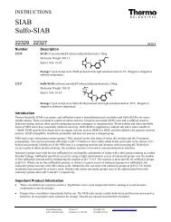
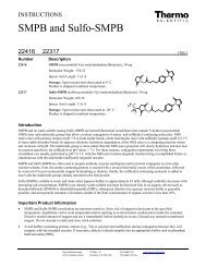
46628 46629 46630<br />
Number Description<br />
46628 Krypton <strong>Protein</strong> <strong>Stain</strong> (10X), 20 ml, sufficient reagent to stain up to 4 mini gels<br />
46629 Krypton <strong>Protein</strong> <strong>Stain</strong> (10X), 100 ml, sufficient reagent to stain up to 20 mini gels<br />
46630 Krypton <strong>Protein</strong> <strong>Stain</strong> (10X), 500 ml, sufficient reagent to stain up to 100 mini gels<br />
Excitation Wavelength: 520 nm<br />
Emission Wavelength: 580 nm<br />
1829.0<br />
Storage: Upon receipt store at 4C. Product shipped at ambient temperature.<br />
Introduction<br />
Thermo Scientific Krypton <strong>Protein</strong> <strong>Stain</strong> enables sensitive fluorescent staining of proteins separated by 1-D or 2-D SDS-<br />
PAGE. The stain is sensitive down to 0.25 ng using the standard protocol, or down to 2 ng using the rapid 30 minute<br />
protocol. The Krypton <strong>Protein</strong> <strong>Stain</strong> is supplied as a 10X stock solution that is diluted with water before use. This proteinspecific<br />
stain allows band visualization using a variety of fluorescence imaging systems. The optimal imaging systems are<br />
laser-based fluorescence scanners capable of exciting and detecting at 520 nm and 580 nm, respectively; however, filteredbased<br />
CCD camera systems are also effective. The Krypton <strong>Protein</strong> <strong>Stain</strong> has a linear quantitative range of three to four<br />
orders of magnitude and is compatible with mass spectrometry analysis.<br />
Important Product Information<br />
For best results, equilibrate the Krypton <strong>Protein</strong> <strong>Stain</strong> to room temperature and dilute to 1X immediately before use. The<br />
1X stain solution may be stored up to seven days at 4°C with minimal loss of sensitivity. A white cloudy precipitate may<br />
form when diluting cold 10X Krypton <strong>Protein</strong> <strong>Stain</strong>, but the precipitate rapidly dissolves when the 1X solution is mixed.<br />
<br />
Use sufficient volumes of stain, fixing and destain solutions to completely cover the gel and allow it to float freely.<br />
Typically, 35-50 ml is sufficient for an 8 8 cm gel and 75-100 ml is sufficient for a 13 9 cm gel.<br />
Additional Materials Required<br />
Gel Fixing Solution: 40% ethanol (v/v), 10% (v/v) acetic acid in ultrapure water<br />
Destaining Solution: 5% (v/v) acetic acid in ultrapure water<br />
Standard Procedure for <strong>Stain</strong>ing Gels<br />
The standard procedure allows band visualization in ~2 hours, 40 minutes with sensitivity down to 0.25 ng.<br />
1. Equilibrate the Krypton <strong>Protein</strong> <strong>Stain</strong> (10X) to room temperature and dilute 10-fold with ultrapure water. Prepare just<br />
enough reagent for the gel(s) being stained.<br />
2. Remove the gel(s) from the gel cassette or plates. Place gel in a clean tray with a sufficient volume of Gel Fixing<br />
Solution to immerse the gel. Cover the tray, and place it on a rocker or shaker and gently agitate for 30 minutes.<br />
3. Decant the fixing solution. Add more fixing solution and agitate gently for another 30 minutes.<br />
4. Carefully decant the fixing solution. To remove residual solution from the gel, add ultrapure water to the tray and agitate<br />
the gel for 5 minutes.<br />
5. Carefully decant the water and add a sufficient volume of 1X Krypton <strong>Protein</strong> <strong>Stain</strong> to immerse gel. Cover the tray with<br />
aluminum foil to minimize light exposure. Place tray on a shaker and agitate gel for 1 hour. <strong>Stain</strong>ing for 2 hours to<br />
overnight may improve band development for some proteins.<br />
<strong>Pierce</strong> Biotechnology PO Box 117 (815) 968-0747 www.thermo.com/pierce<br />
3747 N. Meridian Road Rockford, lL 61105 USA (815) 968-7316 fax
6. Carefully decant the stain solution. Add the Destaining Solution, cover the tray and agitate gently for 5 minutes.<br />
7. Remove the Destaining Solution and replace with an equal volume of ultrapure water. Gently agitate for 15 minutes.<br />
8. Carefully decant water and replace it with more ultrapure water. Agitate the gel gently for 15 minutes.<br />
9. For best results, detect bands using visible laser-based imagers equipped with a 532 nm laser light source. Although the<br />
optimum emission filter is 580 nm, 600 nm filters are also compatible. The gel can be imaged on any platform with the<br />
respective excitation and emission filters.<br />
Rapid Procedure for <strong>Stain</strong>ing Gels<br />
The rapid procedure allows band visualization in 30 minutes with sensitivity down to 2 ng.<br />
1. Equilibrate the Krypton <strong>Protein</strong> <strong>Stain</strong> (10X) to room temperature and dilute 10-fold with ultrapure water. Prepare just<br />
enough reagent for the gel(s) being stained.<br />
2. Remove the gel(s) from the gel cassette or plates. Place gel in a clean tray with a sufficient volume of Gel Fixing<br />
Solution to immerse the gel. Cover the tray, place it on a rocker or shaker and gently agitate for 5 minutes.<br />
3. Decant the fixing solution from the tray and add new fixing solution. Gently agitate tray for 5 minutes.<br />
4. Carefully decant the fixing solution. To remove residual solution from the gel, add ultrapure water to the tray and agitate<br />
the gel for 1 minute.<br />
5. Carefully decant the water and add a sufficient volume of 1X Krypton <strong>Protein</strong> <strong>Stain</strong> to immerse the gel. Cover the tray<br />
with aluminum foil to minimize light exposure. Place tray on a shaker and gently agitate for 15 minutes.<br />
6. Carefully decant the staining solution. Add a sufficient volume of ultrapure water and gently agitate for 3 minutes.<br />
7. For best results, detect bands using visible laser-based imagers equipped with a 532 nm laser light source. Although the<br />
optimum emission filter is 580 nm, 600 nm filters are also compatible. The gel can be imaged on any platform with the<br />
respective excitation and emission filters.<br />
Troubleshooting<br />
Problem Possible Cause Solution<br />
Bands or spots are Imaging system malfunction Check instrument manual for troubleshooting<br />
not visible<br />
No proteins in the gel<br />
Verify that there is protein in the gel by staining with another<br />
method (e.g., Imperial <strong>Protein</strong> <strong>Stain</strong>, Product No. 24615)<br />
Wrong filter sets selected Check excitation and emission settings to confirm the correct<br />
filter sets are being used<br />
Related Thermo Scientific Products<br />
25200-25244 Precise <strong>Protein</strong> Gels, see catalog or web site for a complete listing<br />
28398 BupH Tris-HEPES-SDS Running Buffer Packs, 10 packs<br />
28378 BupH Tris-Glycine-SDS Running Buffer Packs, 40 packs<br />
89871 In-Gel Tryptic Digestion Kit<br />
89865 2-D Sample Prep for Soluble <strong>Protein</strong>s Kit<br />
89866 2-D Sample Prep for Insoluble <strong>Protein</strong>s Kit<br />
24615 Imperial <strong>Protein</strong> <strong>Stain</strong>, 1 L<br />
24612 SilverSNAP ® <strong>Stain</strong> Kit II<br />
U.S. Patent Pending on Krypton <strong>Protein</strong> <strong>Stain</strong> Technology<br />
<strong>Pierce</strong> Biotechnology PO Box 117 (815) 968-0747 www.thermo.com/pierce<br />
3747 N. Meridian Road Rockford, lL 61105 USA (815) 968-7316 fax<br />
2
This product (“Product”) is warranted to operate or perform substantially in conformance with published Product specifications in effect at the time of sale,<br />
as set forth in the Product documentation, specifications and/or accompanying package inserts (“Documentation”) and to be free from defects in material and<br />
workmanship. Unless otherwise expressly authorized in writing, Products are supplied for research use only. No claim of suitability for use in applications<br />
regulated by FDA is made. The warranty provided herein is valid only when used by properly trained individuals. Unless otherwise stated in the<br />
Documentation, this warranty is limited to one year from date of shipment when the Product is subjected to normal, proper and intended usage. This<br />
warranty does not extend to anyone other than the original purchaser of the Product (“Buyer”).<br />
No other warranties, express or implied, are granted, including without limitation, implied warranties of merchantability, fitness for any particular<br />
purpose, or non infringement. Buyer’s exclusive remedy for non-conforming Products during the warranty period is limited to replacement of or<br />
refund for the non-conforming Product(s).<br />
There is no obligation to replace Products as the result of (i) accident, disaster or event of force majeure, (ii) misuse, fault or negligence of or by Buyer, (iii)<br />
use of the Products in a manner for which they were not designed, or (iv) improper storage and handling of the Products.<br />
Current versions of product instructions are available at www.thermo.com/pierce. For a faxed copy, call 800-874-3723 or contact your local distributor.<br />
© 2010 Thermo Fisher Scientific Inc. All rights reserved. Unless otherwise indicated, all trademarks are property of Thermo Fisher Scientific Inc. and its<br />
subsidiaries. Printed in the USA.<br />
<strong>Pierce</strong> Biotechnology PO Box 117 (815) 968-0747 www.thermo.com/pierce<br />
3747 N. Meridian Road Rockford, lL 61105 USA (815) 968-7316 fax<br />
3