Thermo Scientific Pierce Protein Assay Technical Handbook Version 2
Thermo Scientific Pierce Protein Assay Technical Handbook Version 2
Thermo Scientific Pierce Protein Assay Technical Handbook Version 2

Create successful ePaper yourself
Turn your PDF publications into a flip-book with our unique Google optimized e-Paper software.
<strong>Thermo</strong> <strong>Scientific</strong> <strong>Pierce</strong> 660nm <strong>Protein</strong> <strong>Assay</strong><br />
<strong>Thermo</strong> <strong>Scientific</strong> <strong>Pierce</strong> 660nm <strong>Protein</strong> <strong>Assay</strong>s<br />
Rapid, reproducible and colorimetric.<br />
Accurate protein concentration measurements are required to<br />
study many biochemical processes. Although there are several<br />
methods for quantifying proteins, colorimetric or chromogenic<br />
methods remain popular because of their relative simplicity and<br />
speed. The most commonly used dye-binding protein assay is the<br />
Bradford assay, 1 which is based on coomassie dye binding to<br />
proteins. The Bradford assay, however, is prone to inaccuracy<br />
from its typical non-linear standard curves. Moreover, the assay<br />
is not compatible with samples containing detergents at<br />
commonly used concentrations. The <strong>Pierce</strong> 660nm <strong>Protein</strong> <strong>Assay</strong><br />
is highly reproducible, rapid and more linear than the Bradford<br />
method. Furthermore, it is compatible with commonly used<br />
detergents and reducing agents.<br />
Highlights:<br />
• Accurate results – standard curves are more linear than the<br />
Bradford method<br />
• Versatile – compatible with commonly used detergents<br />
and reducing agents and with samples lysed in Laemmli<br />
sample buffer<br />
• Fast – single reagent with a simple mix-and-read assay<br />
• Flexible – available in test tube and microplate formats<br />
• Economical – use small volumes of valuable samples: 10µL<br />
in microplate and 100µL in standard procedures<br />
• Convenient – room temperature storage means no waiting<br />
for the reagent to warm-up before use<br />
Every protein assay has limitations depending on the application<br />
and the specific protein sample analyzed. The most useful<br />
features to consider when choosing a protein assay are sensitivity<br />
(lower detection limit), compatibility with common substances<br />
in samples (e.g., detergents, reducing agents, chaotropic agents,<br />
inhibitors, salts and buffers), standard curve linearity and<br />
protein-to-protein variation. Current methods for the colorimetric<br />
determination of protein concentration in solution include the<br />
Coomassie Blue G-250 dye-binding assay, 1 Biuret method, 2 the<br />
Lowry method, 3 the bicinchoninic acid (BCA) assay 4 and colloidal<br />
gold protein assay. 5<br />
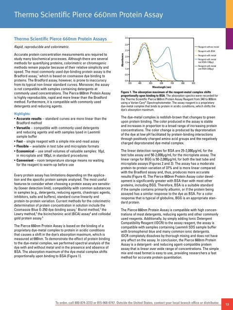
The <strong>Pierce</strong> 660nm <strong>Protein</strong> <strong>Assay</strong> is based on the binding of a<br />
proprietary dye-metal complex to protein in acidic conditions<br />
that causes a shift in the dye’s absorption maximum, which is<br />
measured at 660nm. To demonstrate the effect of protein binding<br />
to the dye-metal complex, we performed spectral analysis of the<br />
dye with and without metal and in the presence and absence of<br />
BSA. The absorption maximum of the dye-metal complex shifts<br />
proportionally upon binding to BSA (Figure 1).<br />
Absorbance<br />
3.5<br />
3.0<br />
2.5<br />
2.0<br />
1.5<br />
1.0<br />
0.5<br />
Reagent without metal<br />
Reagent with BSA<br />
Reagent with metal<br />
Reagent with metal<br />
and BSA (100µg)<br />
Reagent with metal<br />
and BSA (200µg)<br />
0.0<br />
300 400 500 600 700 800 900<br />
Wavelength (nm)<br />
Figure 1. The absorption maximum of the reagent-metal complex shifts<br />
proportionally upon binding to BSA. The absorption spectra were recorded for<br />
the <strong>Thermo</strong> <strong>Scientific</strong> <strong>Pierce</strong> 660nm <strong>Protein</strong> <strong>Assay</strong> Reagent from 340 to 800nm<br />
using a Varian Cary ® Spectrophotometer. The assay reagent is a proprietary<br />
dye-metal complex that binds to protein in acidic conditions, which shifts the<br />
dye’s absorption maximum.<br />
The dye-metal complex is reddish-brown that changes to green<br />
upon protein binding. The color produced in the assay is stable<br />
and increases in proportion to a broad range of increasing protein<br />
concentrations. The color change is produced by deprotonation<br />
of the dye at low pH facilitated by protein-binding interactions<br />
through positively charged amino acid groups and the negatively<br />
charged deprotonated dye-metal complex.<br />
The linear detection ranges for BSA are 25-2,000µg/mL for the<br />
test tube assay and 50-2,000µg/mL for the microplate assay. The<br />
linear range for BGG is 50-2,000µg/mL for both the test tube and<br />
microplate assays (Figures 2 and 3). The assay has a moderate<br />
protein-to-protein variation of 37% and is more linear compared<br />
with the Bradford assay and, thus, produces more accurate<br />
results (Figure 4). The <strong>Pierce</strong> 660nm <strong>Protein</strong> <strong>Assay</strong> color development<br />
is significantly greater with BSA than with most other<br />
proteins, including BGG. Therefore, BSA is a suitable standard<br />
if the sample contains primarily albumin, or if the protein being<br />
assayed has a similar response to the dye as BSA. For a color<br />
response that is typical of globulins, BGG is an appropriate standard<br />
protein.<br />
The <strong>Pierce</strong> 660nm <strong>Protein</strong> <strong>Assay</strong> is compatible with high concentrations<br />
of most detergents, reducing agents and other commonly<br />
used reagents. Additionally, by simply adding Ionic Detergent<br />
Compatibility Reagent (IDCR) to the assay reagent, the assay is<br />
compatible with samples containing Laemmli SDS sample buffer<br />
with bromophenol blue and many common ionic detergents.<br />
IDCR completely dissolves by thorough mixing and does not have<br />
any affect on the assay. In conclusion, the <strong>Pierce</strong> 660nm <strong>Protein</strong><br />
<strong>Assay</strong> is a detergent- and reducing agent-compatible protein<br />
assay that is linear over wide range of concentrations. The simple<br />
mix-and-read format is easy to use, providing researchers a fast<br />
method for accurate protein quantitation.<br />
To order, call 800-874-3723 or 815-968-0747. Outside the United States, contact your local branch office or distributor.<br />
13