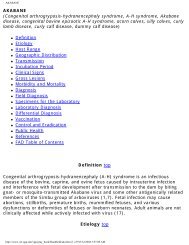
AAT | AFRICAN ANIMAL TRYPANOSOMIASIS
AAT | AFRICAN ANIMAL TRYPANOSOMIASIS
AAT | AFRICAN ANIMAL TRYPANOSOMIASIS

Create successful ePaper yourself
Turn your PDF publications into a flip-book with our unique Google optimized e-Paper software.
<strong>AAT</strong> | <strong>AFRICAN</strong> <strong>ANIMAL</strong> <strong>TRYPANOSOMIASIS</strong><br />
<strong>AFRICAN</strong> <strong>ANIMAL</strong> <strong>TRYPANOSOMIASIS</strong><br />
(Nagana, Tsetse Disease, Tsetse Fly Disease)<br />
●<br />
●<br />
●<br />
●<br />
●<br />
●<br />
●<br />
●<br />
●<br />
●<br />
●<br />
●<br />
●<br />
●<br />
●<br />
●<br />
●<br />
●<br />
●<br />
●<br />
Definition<br />
Etiology<br />
Host Range<br />
Geographic Distribution<br />
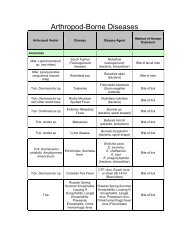
Transmission<br />
Incubation Period<br />
Pathogenesis<br />
Clinical Signs<br />
Gross Lesions<br />
Diagnosis<br />
Field Diagnosis<br />
Specimens for the Laboratory<br />
Control and Eradication<br />
Vector Control<br />
Chemotherapy and Chemoprophylaxis<br />
Immunization<br />
Trypanotolerance<br />
Public Health<br />
References<br />
FAD Table of Contents<br />
Definition top<br />
African animal trypanosomiasis (<strong>AAT</strong>) is a disease complex caused by tsetse-flytransmitted<br />
Trypanosoma congolense, T. vivax, or T. brucei brucei, or<br />
simultaneous infection with one or more of these trypanosomes. African animal<br />
trypanosomiasis is most important in cattle but can cause serious losses in pigs,<br />
camels, goats, and sheep. Infection of cattle by one or more of the three African<br />
animal trypanosomes results in subacute, acute, or chronic disease characterized<br />
by intermittent fever, anemia, occasional diarrhea, and rapid loss of condition and<br />
often terminates in death. In southern Africa the disease is widely known as<br />
nagana, which is derived from a Zulu term meaning "to be in low or depressed<br />
spirits"— a very apt description of the disease.<br />
Etiology top<br />
http://www.vet.uga.edu/vpp/gray_book/Handheld/aat.htm (1 of 12)3/5/2004 4:07:23 AM
<strong>AAT</strong> | <strong>AFRICAN</strong> <strong>ANIMAL</strong> <strong>TRYPANOSOMIASIS</strong><br />
African animal trypanosomiasis is caused by protozoa in the family<br />
Trypanosomatidae genus Trypanosoma. T. congolense resides in the subgenus<br />
Nannomonas, a group of small trypanosomes with medium-sized marginal<br />
kinetoplasts, no free flagella, and poorly developed undulating membranes. In<br />
east Africa, T. congolense is considered to be the single most important cause of<br />
<strong>AAT</strong>. This trypanosome is also a major cause of the disease in cattle in west Africa.<br />
Sheep, goats, horses, and pigs may also be seriously affected. In domestic dogs,<br />
chronic infection often results in a carrier state.<br />
T. vivax is a member of the subgenus Duttonella, a group of trypanosomes with<br />
large terminal kinetoplasts, distinct free flagella, and inconspicuous undulating<br />
membranes. T. vivax is a large (18-26 µm long) monomorphic organism that is<br />
very active in wet-mount blood smears. Cattle, sheep, and goats are primarily<br />
affected. Although this organism is considered to be less pathogenic for cattle than<br />
T. congolense, it is nevertheless the most important cause of <strong>AAT</strong> in west African<br />
cattle. This trypanosome readily persists in areas free of tsetse flies (for example,<br />
in Central and South America and in the Caribbean), where it is transmitted<br />
mechanically by biting flies or contaminated needles, syringes, and surgical<br />
instruments.<br />
T. brucei brucei resides in the subgenus Trypanozoon. T. b. brucei is an extremely<br />
polymorphic typanosome occurring as short, stumpy organisms without flagella,<br />
long slender organisms with distinct flagella, and intermediate forms that are<br />
usually flagellated. Horses, dogs, cats, camels and pigs are very susceptible to T.<br />
b. brucei infection. Infection of cattle, sheep, goats and sometimes pigs results in<br />
mild or chronic infection. This last observation, although widely accepted, has<br />
been called into question by Moulton and Sollod (13), who cite evidence that this<br />
organism is widespread in east and west Africa and that it can cause serious<br />
disease and high mortality in cattle, sheep, and goats.<br />
Host Range top<br />
Cattle, sheep, goats, pigs, horses, camels, dogs, cats, and monkeys are<br />
susceptible to <strong>AAT</strong> and may suffer syndromes ranging from subclinical mild or<br />
chronic infection to acute fatal disease. Rats, mice, guinea pigs, and rabbits are<br />
useful laboratory species.<br />
More than 30 species of wild animals can be infected with pathogenic<br />
trypanosomes, and many of these remain carriers of the organisms. Ruminants<br />
are widely known to be active reservoirs of the trypanosomes. Wild Equidae, lions,<br />
leopards, and wild pigs are all susceptible and can also serve as carriers of<br />
trypanosomes.<br />
http://www.vet.uga.edu/vpp/gray_book/Handheld/aat.htm (2 of 12)3/5/2004 4:07:23 AM
<strong>AAT</strong> | <strong>AFRICAN</strong> <strong>ANIMAL</strong> <strong>TRYPANOSOMIASIS</strong><br />
Geographic Distribution top<br />
The tsetse-fly-infested area of Africa extends from the southern edge of the<br />
Sahara desert (lat. 15 ° N.) to Angola, Zimbabwe, and Mozambique (lat. 20 ° S.).<br />
Of the three African animal trypanosomes, only T. vivax occurs in the Western<br />
Hemisphere in at least 10 countries in the Caribbean and South and Central<br />
America<br />
Transmission top<br />
In Africa, the primary vector for T. congolense, T. vivax, and T. b. brucei is the<br />
tsetse fly. These trypanosomes replicate in the tsetse fly and are transmitted<br />
through tsetse fly saliva when the fly feeds on an animal. The three main species<br />
of tsetse flies for transmission of trypanosomes are Glossina morsitans, which<br />
favors the open woodland of the savanna; G. palpalis, which prefers the shaded<br />
habitat immediately adjacent to rivers and lakes; and G. fusca, which favors the<br />
high, dense forest areas. Trypanosomiasis is also mechanically transmitted by<br />
tsetse and other biting flies through the transfer of blood from one animal to<br />
another. The most important mechanical vectors are flies of the genus Tabanus,<br />
but Haematopota, Liperosia, Stomoxys, and Chrysops flies have also been<br />
implicated. In Africa, both T. vivax and T. b. brucei have spread beyond the<br />
"tsetse fly belts" (20), where transmission is principally by tabanid and<br />
hippoboscid flies.<br />
The vector for T. vivax in the Western Hemisphere remains unknown, but several<br />
species of hematophagous (especially tabanid and hippoboscid) flies are believed<br />
to serve as mechanical vectors.<br />
Incubation Period top<br />
The incubation period for T. congolense varies from 4 to 24 days; for T. vivax,<br />
from 4 to 40 days; and for T. b. brucei, from 5 to 10 days.<br />
Pathogenesis top<br />
Initial replication of trypanosomes is at the site of inoculation in the skin; this<br />
causes a swelling and a sore (chancre). Trypanosomes then spread to the lymph<br />
nodes and blood and continue to replicate. T. congolense localizes in the<br />
endothelial cells of small blood vessels and capillaries. T. b. brucei and T. vivax<br />
localize in tissue. Antibody developed to the glycoprotein coat of the trypanosome<br />
kills the trypanosome and results in the development of immune complexes.<br />
http://www.vet.uga.edu/vpp/gray_book/Handheld/aat.htm (3 of 12)3/5/2004 4:07:23 AM
<strong>AAT</strong> | <strong>AFRICAN</strong> <strong>ANIMAL</strong> <strong>TRYPANOSOMIASIS</strong><br />
Antibody, however, does not clear the infection, for the trypanosome has genes<br />
that can code for many different surface-coat glycoproteins and change its surface<br />
glycoprotein to evade the antibody. Thus, there is a persistent infection that<br />
results in a continuing cycle of trypanosome replication, antibody production,<br />
immune complex development, and changing surface-coat glycoproteins.<br />
Immunologic lesions are significant in trypanosomiasis, and it has been suggested<br />
that many of the lesions (e.g., anemia and glomerulonephritis) in these diseases<br />
may be the result of the deposition of immune complexes that interfere with, or<br />
prevent, normal organ function. The most significant and complicating factor in<br />
the pathogenesis of trypanosomiasis is the profound immunosuppression that<br />
occurs following infection by these parasites. This marked immunosuppression<br />
lowers the host's resistance to other infections and thus results in secondary<br />
disease, which greatly complicates both the clinical and pathological features of<br />
trypanosomiasis.<br />
Clinical Signs top<br />
Because simultaneous infections with more than one trypanosome species are<br />
very common (18), and simultaneous infection with trypanosomes and other<br />
hemoparasites (Babesia spp., Theileria spp., Anaplasma spp., and Ehrlichia spp.)<br />
frequently occurs, it is difficult to conclude which clinical signs are attributable to a<br />
given parasite. Few adequately controlled studies have been made, and thus a<br />
"typical" clinical response to each trypanosome is difficult to reconstruct. What<br />
follows is a summation of the syndromes observed in field and experimental cases<br />
of trypanosomiasis caused by each of the three African animal trypanosomes.<br />
The cardinal clinical sign observed in <strong>AAT</strong> is anemia. Within a week of infection<br />
with the hematic trypanosomes (T. congolense and T. vivax) there is usually a<br />
pronounced decrease in packed cell volume, hemoglobin, red blood cell, and white<br />
blood cell levels, and within 2 months these may drop to below 50 percent of their<br />
preinfection values. Also invariably present are intermittent fever, edema and loss<br />
of condition (Fig. 2). Abortion may be seen, and infertility of males and females<br />
may be a sequel. The severity of the clinical response is dependent on the species<br />
and the breed of affected animal and the dose and virulence of the infecting<br />
trypanosome. Stress, such as poor nutrition or concurrent disease, plays a<br />
prominent role in the disease process, and under experimental conditions, where<br />
stress may be markedly reduced, it is difficult to elicit clinical disease.<br />
T. congolense is a hematic trypanosome found only in the blood vessels of the<br />
animals it infects. It does not localize and multiply outside blood vessels. Infection<br />
with T. congolense may result in peracute, acute, or chronic disease in cattle,<br />
http://www.vet.uga.edu/vpp/gray_book/Handheld/aat.htm (4 of 12)3/5/2004 4:07:23 AM
<strong>AAT</strong> | <strong>AFRICAN</strong> <strong>ANIMAL</strong> <strong>TRYPANOSOMIASIS</strong><br />
sheep, goats, horses, and camels. Pigs often develop a milder disease; chronic<br />
disease is common in dogs. The incubation period is followed by intermittent<br />
febrile episodes, depression, lethargy, weakness, loss of condition, anemia,<br />
salivation, lacrimation, and nasal discharge. As the disease progresses, loss of<br />
condition and hair color changes from black to metallic brown are seen. The back<br />
is often arched and the abdomen "tucked up." Accelerated pulse and jugular<br />
pulsation occur and breathing is difficult. Anemia is a prominent sign. Early in the<br />
infection, the organisms are readily demonstrable in blood smears, but, as the<br />
disease progresses to its acute and chronic forms, organisms are most readily<br />
demonstrated in lymph node smears.<br />
T. vivax has a variable incubation period, and, although it is considered to be less<br />
virulent for cattle than T. congolense, mortality rates of over 50 percent can occur.<br />
There seems to be a marked variation in the virulence of different strains of T.<br />
vivax, but it remains the most important cause of trypanosomiasis of cattle,<br />
sheep, and goats in west Africa. It causes mild disease in horses and chronic<br />
disease in dogs. T. vivax is often difficult to find in blood smears and can also be<br />
demonstrated in lymph node smears.<br />
T. brucei brucei has a relatively short incubation period and causes severe to fatal<br />
infection in horses, camels, dogs, and cats. It usually causes mild, chronic, or<br />
subclinical disease in cattle, sheep, goats, and pigs. A febrile response occurs in<br />
the horse 4-14 days after infection. This is followed by recurrent febrile reactions.<br />
The heartbeat and respiration may be accelerated, and loss of condition and<br />
weakness are seen, whereas the appetite remains good. Progressive anemia and<br />
icterus, and edema of the ventral regions, especially the male genitalia, are<br />
characteristic. The organisms are not always easily perceived in blood smears and<br />
are best demonstrated in tissue smears or sections, (e.g., lymph nodes). Infected<br />
animals die in a few weeks or several months, depending on the virulence of the<br />
strain of T. b. brucei.<br />
The marked immunosuppression resulting from trypanosome infection lowers the<br />
host's resistance to other infections and causes in secondary disease, which<br />
greatly complicates both the clinical and pathological features of trypanosomiasis.<br />
Gross Lesions top<br />
No pathognomonic change is seen in <strong>AAT</strong>. Anemia, edema, and serous atrophy of<br />
fat are commonly observed. Subcutaneous edema is particularly prominent and is<br />
usually accompanied by ascites, hydropericardium, and hydrothorax. The liver<br />
may be enlarged, and edema of lymph nodes is often seen in the acute disease,<br />
but they may be reduced in size in the chronic disease. The spleen and lymph<br />
http://www.vet.uga.edu/vpp/gray_book/Handheld/aat.htm (5 of 12)3/5/2004 4:07:23 AM
<strong>AAT</strong> | <strong>AFRICAN</strong> <strong>ANIMAL</strong> <strong>TRYPANOSOMIASIS</strong><br />
nodes may be swollen, normal, or atrophic. Necrosis of the kidneys and heart<br />
muscle and subserous petechial hemorrhages commonly occur. Gastroenteritis is<br />
common, and focal polioencephalomalacia may be seen. A localized lesion<br />
(chancre) may be noted at the site of fly bite, especially in goats. The anemic<br />
blood changes are anisocytosis, poikilocytosis, polychromasia, and punctate<br />
basophilia. All, some, or none of the above may be seen.<br />
The lesions caused by the trypanosomes in susceptible host species vary<br />
considerably, depending on the species and strain of trypanosome and the species<br />
and breed of host animal affected. The hematic trypanosomes (T. congolense and<br />
T. vivax) cause injury to the host mainly by the production of severe anemia,<br />
which is accompanied in the early stages of the disease by leukopenia and<br />
thrombocytopenia. In the terminal stages of the disease caused by the hematic<br />
trypanosomes, focal polioencephalomalacia probably results from ischemia due to<br />
massive accumulation of the parasites in the terminal capillaries of the brain.<br />
The lesions resulting from T. b. brucei (a tissue parasite) are remarkably different<br />
from those seen with the hematic trypanosomes. Anemia is an important lesion,<br />
but much more dramatic are the inflammation, degeneration, and necrosis<br />
resulting from cellular invasion of various organs. Marked proliferative changes<br />
reflecting immunologic response are observed in most body tissues.<br />
Field Diagnosis top<br />
Diagnosis top<br />
Trypanosomiasis should be suspected when an animal in an endemic area is<br />
anemic and in poor condition. Confirmation depends on the demonstration of the<br />
organism in blood or lymph node smears.<br />
In the early phases of infection, especially with T. vivax and T. congolense, the<br />
parasite can readily be observed by microscopic examination of a wet-mount of<br />
blood slides. Thick blood films and stained with Giemsa are also a good technique<br />
(Fig. 1), but in thin fixed blood films, which are favored for species identification,<br />
the parasites may be hard to demonstrate. When parasitemia is low, smears of<br />
buffy coat (obtained by microhematocrit centrifugation) can be useful for<br />
demonstration of the parasites. Because T. congolense tends to associate with the<br />
erythrocytes, it is essential that buffy coat and adjacent erythrocytes be included<br />
in the smear to ensure demonstration of the parasite.<br />
Stained lymph node smears are a very good method for diagnosis, especially for<br />
T. vivax and T. b. brucei. In chronic T. congolense infection, the parasites localize<br />
http://www.vet.uga.edu/vpp/gray_book/Handheld/aat.htm (6 of 12)3/5/2004 4:07:23 AM
<strong>AAT</strong> | <strong>AFRICAN</strong> <strong>ANIMAL</strong> <strong>TRYPANOSOMIASIS</strong><br />
in the microcirculation of the lymph nodes and in other capillary beds, allowing<br />
diagnosis by examination of lymph node smears or smears made with blood<br />
collected from the ear. Early in infection, blood smears are optimal for the<br />
demonstration of T. congolense.<br />
These conventional techniques of microscopic examination for the presence of<br />
trypanosomes are still widely used, but newer and far more sensitive methods are<br />
beginning to supplant them. The antigen-detecting enzyme-linked immunosorbent<br />
assay is extremely sensitive for the detection of trypanosomiasis in cattle and<br />
goats (12, 25), and species-specific DNA probes have been shown to detect<br />
simultaneous infection of cattle with T. vivax, T. b. brucei, and T. congolense when<br />
conventional methods revealed only single infections (18).<br />
Specimens for the Laboratory top<br />
To perform the preceding and more sensitive procedures, the following specimens<br />
should be submitted to the laboratory from several animals: serum, blood with the<br />
anticoagulant EDTA, dried thin and thick blood smears, and smears of needle<br />
lymph node biopsies.<br />
Vector Control top<br />
Control and Eradication top<br />
Fly eradication and drug prophylaxis are the only effective trypanosomiasis control<br />
methods now available. Several approaches to fly control have been used with<br />
varying degrees of success.<br />
Discriminative bush clearing, extensively used in early tsetse fly eradication<br />
campaigns, has been locally useful because it eliminates the breeding places of<br />
the tsetse. But, to be completely effective, bush clearing requires ecologically<br />
unacceptable destruction of vast areas of brush and forest. It is still a useful<br />
procedure when used locally in conjunction with other control methods.<br />
Game elimination, and thus elimination of the main source of bloodmeals for the<br />
tsetse, was used in early eradication campaigns.<br />
This was an ineffective and wasteful procedure.<br />
Application of the sterile male technique (as used in screwworm eradication in the<br />
United States) received considerable attention in the 1980's. Early problems with<br />
http://www.vet.uga.edu/vpp/gray_book/Handheld/aat.htm (7 of 12)3/5/2004 4:07:23 AM
<strong>AAT</strong> | <strong>AFRICAN</strong> <strong>ANIMAL</strong> <strong>TRYPANOSOMIASIS</strong><br />
breeding of the male flies have been overcome, and field trials have been done in<br />
both east and west Africa to determine the effectiveness of this approach in vector<br />
control. In limited trials, this procedure has reduced fly populations.<br />
Ground and aerial spraying with insecticides and the use of synthetic pyrethroids<br />
on cattle have lowered fly densities in some areas, but widespread use would<br />
require considerable international cooperation and expense. Widespread<br />
application of insecticide has the tremendous disadvantage of also eradicating<br />
many other arthropods, several of which are desirable. The recent introduction of<br />
odor-baited targets impregnated with insecticides is proving promising as a means<br />
of reducing the tsetse fly.<br />
Chemotherapy and Chemoprophylaxis top<br />
The use of drugs for the prevention and treatment of trypanosomiasis has been<br />
important for many decades, but the rapidity with which the trypanosomes have<br />
developed resistance to each drug introduced has tremendously complicated this<br />
approach to controlling the disease. In spite of this, some of the older<br />
chemoprophylactic drugs such as the quinapyramine derivatives Antrycide and<br />
Antrycide Prosalt are still used and give effective protection against T. b. brucei<br />
infection in horses, camels, and cattle for up to 3 months. The drug pyrithidium<br />
bromide (Prothidium and AD2801) is useful in the prophylaxis of T. vivax and T.<br />
congolense infections in cattle, sheep, and goats and can give protection for up to<br />
6 months. The most widely used of the newer chemoprophylactic drugs (and also<br />
the least expensive) is isometamidium chloride (26). This drug, in use for over 20<br />
years and sold under the trade names Samorin, Trypamidium, and M&B 4180A, is<br />
excellent for the prophylaxis of all three African animal trypanosomes, and gives<br />
protection for 3-6 months. The development of resistance to this drug has been<br />
reported in both east and west Africa. Homidium bromide has also been found to<br />
be an effective chemophrophylactic drug in Kenya, and the newly introduced<br />
arsenical Cymelarsan is effective in treatment of T. b. brucei infection.<br />
A very widely used chemotherapeutic drug is diminazine aceturate (Berenil), which<br />
is effective against all three African animal trypanosomes. The isometamidium<br />
drugs are also excellent chemotherapeutic agents as are the quaternary<br />
ammonium trypanocides Antrycide, Ethidium and Prothidium.<br />
Although extensively used in trypanosomiasis control, chemoprophylaxis is an<br />
expensive, time-consuming, and thus unsatisfactory long-term solution to the<br />
problem of African animal trypanosomiasis.<br />
Immunization top<br />
http://www.vet.uga.edu/vpp/gray_book/Handheld/aat.htm (8 of 12)3/5/2004 4:07:23 AM
<strong>AAT</strong> | <strong>AFRICAN</strong> <strong>ANIMAL</strong> <strong>TRYPANOSOMIASIS</strong><br />
No vaccine is currently available for African animal trypanosomiasis.<br />
Trypanotolerance top<br />
It has long been recognized that certain breeds of African cattle are considerably<br />
more resistant to African trypanosomiasis that others. This is especially true of the<br />
west African short-horned cattle (Muturu, Baoule, Laguna, Samba, and Dahomey)<br />
and the N'Dama, which is also of west Africa. These cattle have existed in the<br />
region for over 5,000 years. Susceptibility studies have shown the N'Dama to be<br />
the most resistant breed followed by the smaller west African short-horned cattle,<br />
but the large and more recently introduced Zebu is the most susceptible (15). The<br />
mechanisms of trypanotolerance have been extensively studied, and it is now well<br />
established that trypanotolerance has a genetic basis (13, 17). Trypanotolerance<br />
in sheep and goats has also been described, but the mechanisms of the tolerance<br />
phenomenon have not been defined.<br />
Public Health top<br />
The three <strong>AAT</strong> trypanosomes are considered to be nonpathogenic for humans. T.<br />
b. brucei, although not causing human disease, is closely related to T. b.<br />
gambiense and T. b. rhodesiense. The latter is the cause of human sleeping<br />
sickness, a very debilitating and often fatal disease considered to be of major<br />
public health significance in 36 sub-Saharan countries of west, central, and east<br />
Africa with 50 million people at risk (18). In west and central Africa, a chronic<br />
form of human sleeping sickness is caused by T. b. gambiense, which uses<br />
humans as its major host but also infects pigs. In east and southern Africa, T. b.<br />
rhodensiense is the cause of a much more acute form of human sleeping sickness.<br />
This trypanosome also infects cattle, bushbuck (Tragelaphus scriptus), and<br />
probably many other wild animals that may serve as reservoirs of the parasite.<br />
GUIDE TO THE LITERATURE top<br />
1. ANOSA, V.O., LOGAN-HENFREY, L.L., and SHAW, M.K. 1992. A light and<br />
electron microscopic study of changes in blood and bone marrow in acute<br />
hemorrhagic Trypanosoma vivax infection in calves. Vet. Pathol., 29:33-45<br />
2. ASHCROFT, M.D., BURTT, E., and FAIRBAIRN, H. 1959. The experimental<br />
infection of some African wild animals with Trypanosoma rhodesiense, T. brucei,<br />
and T. congolense. Ann. Trop. Med. Parasitol., 53:147-161<br />
3. DOLAN, R. B. 1987. Genetics and trypanotolerance. Parasit. Today 3:137-143.<br />
http://www.vet.uga.edu/vpp/gray_book/Handheld/aat.htm (9 of 12)3/5/2004 4:07:23 AM
<strong>AAT</strong> | <strong>AFRICAN</strong> <strong>ANIMAL</strong> <strong>TRYPANOSOMIASIS</strong><br />
4. EPSTEIN, H. 1971. The Origin of the Domestic Animals of Africa, Vols. 1 and 2.<br />
New York: Africana.<br />
5. FINELLE, P. 1973. African animal trypanosomiasis. World Animal Review, 7:1-6<br />
and 8:24-27.<br />
6. GOODING, R.H. 1992. Genetic variation in tsetse flies and implications for<br />
trypanosomiasis. Parasit. Today 8:92-95.<br />
7. KOBAYASHI, A., TIZARD, I.R., and WOO, P.T.K. 1976. Studies on the anaemia<br />
in experimental African trypanosomiasis. II. The pathogenesis of the anemia in<br />
calves infected with Trypanosoma congolense. Am. J. Trop. Med. Hyg., 25:401-<br />
406.<br />
8. KUZOE, F.A.S. 1991. Perspectives in research and control of African<br />
trypanosomiasis. Ann. Trop. Med. Parasit., 85:33-41.<br />
9. LOGAN-HENFREY, L.L., GARDINER, P.R., and MAHMOUD, M.M. 1992. "Animal<br />
Trypanosomiasis in Subsaharan Africa." In Parasitic Protozoa, Vol. 2, J. Krier and<br />
J. Baker, Eds., Academic Press, pp. 157-276.<br />
10. LOSOS, G.J., and CHOUINARD, A. 1979. Pathogenicity of Trypanosomes.<br />
Ottawa: IDRC Press..<br />
11. LOSOS, G.J., and IKEDE, B.O. 1972. Review of the pathology of disease in<br />
domestic and laboratory animals caused by Trypanosoma congolense, T. vivax, T.<br />
brucei, T. rhodesiense, and T. gambiense. Vet. Pathol. 9 (Suppl):1-71.<br />
12. MASAKE, R.A., and NANTULYA, V.M. 1991. Sensitivity of an antigen-detecting<br />
enzyme immunoassay for diagnosis of Tyrypanosoma congolense infections in<br />
goats and cattle. J. Parasitol. 77:231-236.<br />
13. MOULTON, J.E.. and SOLLOD, A.E. 1976. Clinical, serological and pathological<br />
changes in calves with experimentally induced Typanosoma brucei infection. Am.<br />
J. Vet. Res., 37:791.<br />
14. MULLA, A.F., and PICKMAN, L.R. 1988. How do African game animals control<br />
trypanosome infections ? Parasit. Today 4:352-354.<br />
15. MURRAY, M., BARRY, J.D., MORRISSON, W.I., WILLIAMS, R.O., HIRUMI, H.,<br />
and ROVIS, L. 1979. A review of the prospects for vaccination in African<br />
http://www.vet.uga.edu/vpp/gray_book/Handheld/aat.htm (10 of 12)3/5/2004 4:07:23 AM
<strong>AAT</strong> | <strong>AFRICAN</strong> <strong>ANIMAL</strong> <strong>TRYPANOSOMIASIS</strong><br />
trypanosomiasis. World Animal Review, 32:913.<br />
16. MURRAY, M., MORRISON, W.I., MURRAY, P.K., CLIFFORD, D.J., and TRAIL, J.C.<br />
M. 1979. Trypanotolerance — A review. World Animal Review, 31:2-12.<br />
17. MURRAY, M., TRAIL, J.C.M., DAVIS, C.E., and BLACK, S.J. 1984. Genetic<br />
resistance to African trypanosomiasis. J. Inf. Dis. 149:311-319.<br />
18. NYEKO, J.H.P., OLE-MOIYOI, O.K., MAJIWA, P.A.O., OTIENO, L.H., and OCIBA,<br />
P.M. 1990. Characterization of trypanosome isolates from cattle in Uganda using<br />
species-specific DNA probes reveals predominance of mixed infections. Insect Sci.<br />
Applic. 11:271-280.<br />
19. ONAH, D.N. 1991. Porcine trypanosomiasis in Nigeria. Trop. Anim. Hlth. Prod.<br />
23:141-146.<br />
20. RODER, P.L., SCOTT, J.M., and PEGRAM, R.G. 1984. Acute Trypanosoma vivax<br />
infection of Ethiopian cattle in the apparent absence of tsetse. Trop. Anim. Hlth.<br />
Prod. 16:141-147.<br />
21. ROGERS, D.J., and RANDOLPH, S.E. 1991. Mortality rates and population<br />
density of tsetse flies correlated with satellite imagery. Nature 351:739-741.<br />
22. SPRIGGS, D.R. 1985. Antigenic variation in trypanosomes: Genomes in flux. J.<br />
Inf. Dis. 152:855-856.<br />
23. TIZARD, I.R., HOLMES, W.L., YORK, D.A., and MELLORS, A. 1977. The<br />
generation and identification of the hemolysin of Trypanosoma congolense.<br />
Experientia (Switzerland), 33:901-902.<br />
24. TIZARD, I., NIELSEN, K.H., SEED J.R., and HALL., J.E. 1978. Biologically<br />
active products from African trypanosomes. Microb. Rev., 42:661-681.<br />
25. TRAIL, J.C.M., DIETEREN, G.D.M., MAILLE, J.C., YANGARI, G., and NANTULYA,<br />
V.M. 1991. Use of antigen-detection enzyme immunoassays in assessment of<br />
trypanotolerance in N'Dama cattle. Acta Tropica 50:11-18.<br />
26. OGUNYEMI, O., and ILEMOBADE, A.A. 1989. Prophylaxis of African<br />
trypanosmiasis; A review of some factors that may influence the duration of<br />
isometamidium chloride prophylaxis. Vet. Bul. 59:1-4.<br />
27. SULIMAN, H.B., and FELDMAN, B.F.1989. Pathogenesis and aetiology of<br />
http://www.vet.uga.edu/vpp/gray_book/Handheld/aat.htm (11 of 12)3/5/2004 4:07:23 AM
<strong>AAT</strong> | <strong>AFRICAN</strong> <strong>ANIMAL</strong> <strong>TRYPANOSOMIASIS</strong><br />
anaemia in trypanosomiasis with special reference to T. brucei and T. evansi. Vet.<br />
Bull. 59:99-107.<br />
28. WELLS, E.A., RAMIREZ, L.E., and BETANCOURT, A. 1982. Trypanosoma vivax<br />
in Colombia: Interpretation of field results. Trop. Anim. Hlth. Prod., 14:141-150.<br />
29. WILLIAMS, D.J.L, NAESSENS, J., SCOTT, J.R., and McODIMBA, F.A. 1991.<br />
Analysis of peripheral leucocyte populations in N'Dama and Boran cattle following<br />
a rechallenge infection with Trypanosoma cogolense. Parasite Immunol. 13;171-<br />
185.<br />
C. J. Maré, B.V.Sc.. Ph.D., Veterinary Science/Microbiology, University of<br />
Arizona, Tuson, Az<br />
TOP | FAD Table of Contents<br />
http://www.vet.uga.edu/vpp/gray_book/Handheld/aat.htm (12 of 12)3/5/2004 4:07:23 AM