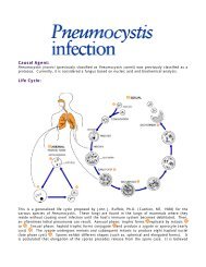
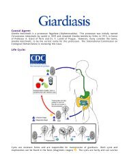
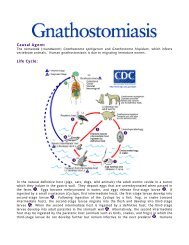
Causal Agents: Life Cycle:
Causal Agents: Life Cycle:
Causal Agents: Life Cycle:

Create successful ePaper yourself
Turn your PDF publications into a flip-book with our unique Google optimized e-Paper software.
to the pharynx, and are swallowed . The larvae reach the small intestine, where they reside and<br />
mature into adults. Adult worms live in the lumen of the small intestine, where they attach to the<br />
intestinal wall with resultant blood loss by the host . Most adult worms are eliminated in 1 to 2<br />
years, but longevity records can reach several years.<br />
Some A. duodenale larvae, following penetration of the host skin, can become dormant (in the<br />
intestine or muscle). In addition, infection by A. duodenale may probably also occur by the oral<br />
and transmammary route. N. americanus, however, requires a transpulmonary migration phase.<br />
Geographic Distribution:<br />
The second most common human helminthic infection (after ascariasis). Worldwide distribution,<br />
mostly in areas with moist, warm climate. Both N. americanus and A. duodenale are found in<br />
Africa, Asia and the Americas. Necator americanus predominates in the Americas and Australia,<br />
while only A. duodenale is found in the Middle East, North Africa and southern Europe.<br />
Clinical Features:<br />
Iron deficiency anemia (caused by blood loss at the site of intestinal attachment of the adult<br />
worms) is the most common symptom of hookworm infection, and can be accompanied by cardiac<br />
complications. Gastrointestinal and nutritional/metabolic symptoms can also occur. In addition,<br />
local skin manifestations ("ground itch") can occur during penetration by the filariform (L3) larvae,<br />
and respiratory symptoms can be observed during pulmonary migration of the larvae.<br />
Laboratory Diagnosis:<br />
Microscopic identification of eggs in the stool is the most common method for diagnosing<br />
hookworm infection. The recommended procedure is as follows:<br />
1. Collect a stool specimen.<br />
2. Fix the specimen in 10% formalin.<br />
3. Concentrate using the formalin–ethyl acetate sedimentation technique.<br />
4. Examine a wet mount of the sediment.<br />
Where concentration procedures are not available, a direct wet mount examination of the specimen<br />
is adequate for detecting moderate to heavy infections. For quantitative assessments of infection,<br />
various methods such as the Kato-Katz can be used.<br />
Diagnostic Findings<br />
• Microscopy<br />
• Morphologic comparison with other intestinal parasites<br />
Examination of the eggs cannot distinguish between N. americanus and A. duodenale. Larvae can<br />
be used to differentiate between N. americanus and A. duodenale, by rearing filariform larvae in a<br />
fecal smear on a moist filter paper strip for 5 to 7 days (Harada-Mori). Occasionally, it may be<br />
necessary to distinguish between the rhabditiform larvae (L2) of hookworms and those of<br />
Strongyloides stercoralis.<br />
Treatment:<br />
In countries where hookworm is common and reinfection is likely, light infections are often not<br />
treated. In the United States, hookworm infections are generally treated with albendazole*.<br />
Mebendazole* or pyrantel pamoate* can also be used.<br />
* This drug is approved by the FDA, but considered investigational for this purpose.