HEMORRHAGIC SEPTICEMIA
HEMORRHAGIC SEPTICEMIA
HEMORRHAGIC SEPTICEMIA

You also want an ePaper? Increase the reach of your titles
YUMPU automatically turns print PDFs into web optimized ePapers that Google loves.
<strong>HEMORRHAGIC</strong> <strong>SEPTICEMIA</strong><br />
<strong>HEMORRHAGIC</strong> <strong>SEPTICEMIA</strong><br />
●<br />
●<br />
●<br />
●<br />
●<br />
●<br />
●<br />
●<br />
●<br />
●<br />
●<br />
●<br />
●<br />
●<br />
●<br />
●<br />
●<br />
●<br />
●<br />
●<br />
Definition<br />
Etiology<br />
Host Range<br />
Geographic Distribution<br />
Transmission<br />
Incubation Period<br />
Clinical Signs<br />
Gross Lesions<br />
Morbidity and Mortality<br />
Diagnosis<br />
Field Diagnosis<br />
Specimens for the Laboratory<br />
Laboratory Diagnosis<br />
Differential Diagnosis<br />
Treatment<br />
Vaccination<br />
Control and Eradication<br />
Public Health<br />
References<br />
FAD Table of Contents<br />
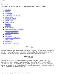
Definition top<br />
Classical hemorrhagic septicemia is a particular form of pasteurellosis caused by<br />
Pasteurella multocida and manifested by an acute and highly fatal septicemia<br />
mainly in susceptible cattle and water buffaloes.<br />
The name hemorrhagic septicemia is used rather loosely in some countries to<br />
include pneumonic pasteurellosis (shipping or transport fever), a disease caused<br />
mainly by P. haemolytica, although various serotypes of P. multocida are<br />
occasionally involved. Although the morbidity of pneumonic pasteurellosis of cattle<br />
can be high, the mortality rate is much less than that of hemorrhagic septicemia.<br />
Etiology top<br />
Hemorrhagic septicemia is caused by two serotypes of P. multocida; namely, B:2<br />
and E:2. The letter denotes the capsular antigen as determined originally by the<br />
http://www.vet.uga.edu/vpp/gray_book/Handheld/hes.htm (1 of 8)3/5/2004 4:15:01 AM
<strong>HEMORRHAGIC</strong> <strong>SEPTICEMIA</strong><br />
indirect hemagglutination test of Carter (5), and the numeral 2 stands for the<br />
somatic or O antigen as determined by the agar gel diffusion precipitin test<br />
developed by Heddelston and associates (17). This somatic antigen 2 is the<br />
equivalent to the 6 in the classification of Namioka and associates. In a new<br />
classification, Pasteurella multocida strains causing most pasteurella infections,<br />
including hemorrhagic septicemia, are called P. multocida subspecies multocida.<br />
Host Range top<br />
Cattle and water buffaloes are the principal hosts of hemorrhagic septicemia, and<br />
it is widely considered that buffaloes are the more susceptible. The disease is<br />
thought to be endemic in one large herd of North American range bison; however,<br />
epidemics appear to be rare. In the United States, the disease has been confirmed<br />
only in American bison in 1912, 1922, and 1965. The P. multocida isolant from the<br />
1922 outbreak, a serotype B:2, is maintained in the USDA culture collection as a<br />
reference strain. Although outbreaks of hemorrhagic septicemia have been<br />
reported in sheep and swine, it is not a frequent or significant disease. Cases have<br />
been reported in deer, elephants and yaks. There is as yet no evidence of a<br />
reservoir of infection outside the principal hosts: cattle, water buffaloes, and<br />
bison.<br />
Geographic Distribution top<br />
Hemorrhagic septicemia in epidemic form is a disease mainly of cattle and water<br />
buffaloes either maintained separately or together. Radical changes in weather,<br />
including the advent of monsoons, debility caused by seasonal levels of low<br />
nutrition, and the pressure of work (draft animals) are related to the explosive<br />
occurrences of the disease in certain parts of the world. Southeast Asia, where<br />
such conditions often coincide, is the area of highest incidence. The disease occurs<br />
in the Middle East and Africa where the environmental circumstances and<br />
predisposing conditions are not as clearly defined as in Southeast Asia. As in Asia,<br />
the disease is frequently associated with the rainy season and poor physical<br />
condition.<br />
Hemorrhagic septicemia was recognized in Japan as a specific disease of cattle<br />
caused by particular strains of Pasteurelia as early as 1923. Since 1926, the<br />
disease has been controlled, and the last recorded case in cattle in Japan occurred<br />
in 1952.<br />
The B:2 serotype has been recovered from hemorrhagic septicemia in countries of<br />
Southern Europe, the Middle East, and Southeast Asia, including China. This same<br />
serotype has been reported from Egypt and the Sudan. The E:2 serotype has been<br />
http://www.vet.uga.edu/vpp/gray_book/Handheld/hes.htm (2 of 8)3/5/2004 4:15:01 AM
<strong>HEMORRHAGIC</strong> <strong>SEPTICEMIA</strong><br />
recovered from hemorrhagic septicemia occurring in Egypt, the Sudan, the<br />
Republic of South Africa, and several other African countries. There is no report of<br />
either serotype being recovered from Australia, New Zealand, and countries of<br />
South and Central America.<br />
There is no evidence that the disease has spread from carrier bison in the Western<br />
United States to neighboring cattle. Given the conditions in which hemorrhagic<br />
septicemia occurs in endemic areas (e.g., primitive husbandry practices, low<br />
country plains, and well-defined dry and wet seasons), it seems unlikely that the<br />
disease will reach epidemic proportions in the United States.<br />
Transmission top<br />
The disease is spread by direct and indirect contact (fomites). The source of the<br />
infection is infected animals or carriers. The carrier state may be greater than 20<br />
percent shortly after an outbreak, but within 6 weeks the rate is usually less than<br />
5 percent. The causal agent does not survive for more than 2 to 3 weeks in the<br />
soil or on pastures. Close herding and wetness, as occurs during the rainy season,<br />
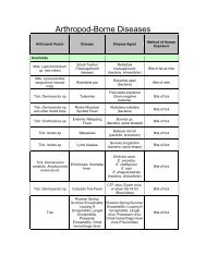
appear to contribute to spread. There is no evidence that biting arthropods are<br />
significant vectors.<br />
Incubation Period top<br />
The influence of extrinsic factors in the development of the clinical pasteurelloses,<br />
and particularly in hemorrhagic septicemia, has been noted by many workers.<br />
When favorable circumstances for the growth and multiplication of P. multocida in<br />
the animal body occur, severe septicemia develops within a few hours. However,<br />
the organisms may be harbored for varying periods in a small percentage of<br />
carrier animals without any clinical sign. The perpetuation of the disease from year<br />
to year or season to season is generally attributed to the carrier state. The<br />
immune status of the animal is thought to influence the severity of the disease.<br />
Cattle or buffalo artificially inoculated subcutaneously with lethal doses<br />
(approximately 20,000 bacilli) show clinical signs within a few hours and succumb<br />
within 18 to 30 hours.<br />
Clinical Signs top<br />
The majority of cases in cattle and buffalo are acute or peracute with death<br />
occurring from 6 to 24 hours after the first recognized signs. In a few outbreaks,<br />
animals may survive as long as 72 hours. Dullness, reluctance to move, and<br />
elevated temperature are the first signs. Following these signs, salivation and<br />
http://www.vet.uga.edu/vpp/gray_book/Handheld/hes.htm (3 of 8)3/5/2004 4:15:01 AM
<strong>HEMORRHAGIC</strong> <strong>SEPTICEMIA</strong><br />
nasal discharge appear, and edematous swellings are seen in the pharyngeal<br />
region and then spread to the ventral cervical region and brisket. Visible mucous<br />
membranes are congested, and respiratory distress is soon followed by collapse<br />
and death. Recovery, particularly in buffaloes, is rare. Chronic manifestations of<br />
hemorrhagic septicemia do not appear to occur.<br />
Gross Lesions top<br />
Widely distributed hemorrhages, edema, and general hyperemia are the most<br />
obvious tissue changes observed in infected animals. In almost all cases there is<br />
an edematous swelling of the head, neck, and brisket region (Fig. 63). Incision of<br />
the edematous swellings reveals a coagulated serofibrinous mass with strawcolored<br />
or blood-stained fluid. This edema, which distends tissue spaces, is also<br />
found in the musculature (Fig. 64). There are subserosal petechial hemorrhages<br />
throughout the animal, and blood-tinged fluid is frequently found in the thoracic<br />
and abdominal cavities. Petechiae may be found scattered throughout some<br />
tissues and lymph nodes, particularly the pharyngeal and cervical nodes, which<br />
are also swollen and often hemorrhagic. Pneumonia is not usually extensive nor is<br />
gastroenteritis. Cases that are atypical in regard to throat swelling (absent) and<br />
pneumonia (extensive) are occasionally seen.<br />
Morbidity and Mortality top<br />
Husbandry, weather and immunity affect morbidity. In endemic areas, from 10 to<br />
50 percent of the cattle or buffalo populations acquire solid immunity through<br />
exposure or subclinical infection. Close herding and wetness predispose to an<br />
increased morbidity. Most animals that develop clinical signs die.<br />
Diagnosis top<br />
Field Diagnosis top<br />
In countries where hemorrhagic septicemia is endemic, it is usually readily<br />
diagnosed — particularly if there is a history of previous outbreaks and a failure to<br />
vaccinate. When a small number of animals are affected, diagnosis may be more<br />
difficult. This could be the case if hemorrhagic septicemia were to occur in the<br />
United States. In endemic areas, the rapid course, usual high herd incidence, and<br />
the appearance of edematous swellings in the throat, cervical, and parotid regions<br />
is highly suggestive.<br />
Specimens for the Laboratory top<br />
http://www.vet.uga.edu/vpp/gray_book/Handheld/hes.htm (4 of 8)3/5/2004 4:15:01 AM
<strong>HEMORRHAGIC</strong> <strong>SEPTICEMIA</strong><br />
From an animal with typical signs, the organism can be isolated from heparinized<br />
blood, affected tissue, liver, lung, kidney, and spleen. All samples should be<br />
collected aseptically. Samples should be kept cool and shipped on wet ice as soon<br />
as possible. Swabs in transport media, ribs, and tips of ears are sometimes<br />
submitted from remote areas in developing countries.<br />
Laboratory Diagnosis top<br />
Isolation of a small gram-negative rod or coccobacillus in pure or nearly pure<br />
culture with the general colonial appearance of a Pasteurella species from an<br />
animal with typical signs is grounds to suspect hemorrhagic septicemia. If there<br />
has been postmortem decomposition with the presence of extraneous bacteria, the<br />
inoculation of mice and rabbits with blood or suspensions of tissues will facilitate<br />
recovery of the pasteurellae of hemorrhagic septicemia in pure or nearly pure<br />
culture. Both mice and rabbits are highly susceptible to the two serotypes B:2 and<br />
E:2. Definitive diagnosis depends upon the identification of the cultures as P.<br />
multocida and the subsequent identification of serotype B:2 or E:2. Because<br />
several different serotypes of P. multocida that do not produce classical<br />
hemorrhagic septicemia occur in cattle, it is necessary to serotype the isolate. The<br />
National Veterinary Services Laboratories, Ames, IA should be contacted regarding<br />
the serotyping of suspected hemorrhagic septicemia strains of P. multocida.<br />
Serologic procedures for the detection of specific antibody are not used in<br />
diagnosis.<br />
Differential Diagnosis top<br />
The sudden death seen with peracute and acute hemorrhagic septicemia must be<br />
differentiated from that due to lightning, snakebites, blackleg, rinderpest, and<br />
anthrax.<br />
Treatment top<br />
The onset and course of the disease are generally rapid and leave little time for<br />
antimicrobial therapy. However, several of the sulfonamides and antibiotics such<br />
as penicillin and the tetracyclines can be used successfully in the early stages. In<br />
some outbreaks in Southeast Asia, animals with elevated temperatures are<br />
isolated and treated intravenously with a soluble sulfonamide.<br />
Vaccination top<br />
http://www.vet.uga.edu/vpp/gray_book/Handheld/hes.htm (5 of 8)3/5/2004 4:15:01 AM
<strong>HEMORRHAGIC</strong> <strong>SEPTICEMIA</strong><br />
The most efficacious immunizing agent has been the oil-adjuvant vaccine prepared<br />
from the appropriate serotype. Vaccine of this type is more slowly absorbed and<br />
produces a longer-lasting immunity than do regular and alum-precipitated-type<br />
bacterins. The oil-adjuvant bacterin has the advantage of requiring only one dose<br />
annually, but it has the disadvantages of being difficult to syringe and occasionally<br />
produces a marked local reaction. A live vaccine prepared from a fallow deer strain<br />
of P. multocida has shown considerable promise with protection for as long as a<br />
year. This strain, serotype B:3,4, is closely related immunologically to serotype<br />
B:2 but is less virulent.<br />
Control and Eradication top<br />
In endemic areas the only practical ways to protect animals are by an organized<br />
program of vaccination and maintenance of animals in as good a condition as<br />
possible. When favorable conditions for outbreaks are known to recur periodically,<br />
such preventive measures can be carried out in advance, and the potential<br />
consequences of the disease will thus be lessened.<br />
Public Health top<br />
There is as yet no authenticated report of human infections due to serotypes B:2<br />
and E:2. However, because other serotypes of P. multocida can cause a variety of<br />
human infections, precautions should be taken to minimize exposure to the<br />
hemorrhagic septicemia varieties of P. multocida.<br />
GUIDE TO THE LITERATURE top<br />
1. ANONYMOUS. 1991. Proceedings of the Fourth International Workshop on<br />
Haemorrhagic Septicaemia, Kandy, Sri Lanka. Bangkok, Thailand:FAO/APHCA<br />
Publication. Food and Agricultural Organization of the United Nations.<br />
2. BAIN, R.V.S., De ALWIS, M.C.L., CARTER, G.R., and GUPTA, B.K. 1982.<br />
Haemorrhagic septicemia, FAO Animal Production and Health Paper 33, Food and<br />
Agriculture Organization of the United Nations, Rome.<br />
3. CARTER, G.R. 1967. Pasteurellosis: Pasteurelia multocida and Pasteurella<br />
hemolytica. Adv. Vet. Sci., 11:321-379.<br />
4. CARTER, G.R., and CHENGAPPA, M.M. 1981. Identification of types B and E<br />
Pasteurella multocide by counter-immunoelectrophoresis. Vet. Rec., 108:145-146.<br />
http://www.vet.uga.edu/vpp/gray_book/Handheld/hes.htm (6 of 8)3/5/2004 4:15:01 AM
<strong>HEMORRHAGIC</strong> <strong>SEPTICEMIA</strong><br />
5. CARTER, G.R. 1984. Serotyping Pasteurelia multocida. Methods in Microbiol.,<br />
16:247-258.<br />
6. CARTER, G.R., and De ALWIS, M.C.L. 1989. Haemorrhagic Septicaemia. In<br />
Pasteurella and Pasteurellosis, C. Adlam, and J.M. Rutter,eds., London:Academic<br />
Press, pp. 131-160.<br />
7. CARTER, G.R., and CHENGAPPA, M.M. 1991. Rapid presumptive identification of<br />
type B Pasteurella multocida from haemorrhagic septicaemia. Vet. Rec., 128:526.<br />
8. CARTER, G.R., MYINT, A., VAN KHAR, R., and KHIN, A. 1991. Immunization of<br />
cattle and buffaloes with live haemorrhagic septicaemia vaccine. Vet. Rec.,<br />
128:203.<br />
9. De ALWIS, M.C.L. 1981. Mortality among cattle and buffaloes in Sri Lanka due<br />
to haemorrhagic septicemia. Trop. Anim. Hlth. Prod., 13:195-202.<br />
10. De ALWIS, M.C.L. 1984. Haemorrhagic septicaemia in cattle and buffaloes.<br />
Rev. Sd. Tech. Off. Int. Epiz., 3:707-730.<br />
11. GOUCHENOUR, W.S. 1924. Haemorrhagic septicemic studies. J. Am. Vet. Med.<br />
Assoc., 65:433-445.<br />
12. HEDDLESTON, K.L., and GALLAGHER, J.E. 1969. Septicemic pasteurellosis<br />
(Hemorrhagic septicemia) in the American bison. A serologic survey. Bull. Wildl.<br />
Dis. Assoc., 5:207-207.<br />
13. HEDDLESTON, K.L., GALLAGHER, J.E., and REBERS, P.A. 1972. Fowl Cholera:<br />
Gel diffusion precipitin test for serotyping Pasteurelia multocida from avian<br />
species. Avian Dis., 16:925-936.<br />
14. HEDDLESTON, K.L., RHOADES, D.R., and REBERS, P.A. 1967. Experimental<br />
pasteurellosis; Comparative studies on Pasteurella multocida from Asia, Africa,<br />
and North America. Am. J. Vet. Res., 28:1003-1012.<br />
15. HIRAMUNE, T., and De ALWIS, M.C.L. 1982. Haemorrhagic septicemia carrier<br />
status of cattle and buffaloes in Sri Lanka. Trop. Hlth. Prod., 14:91-92.<br />
16. MYINT, A., CARTER G.R., and JONES, T.O. 1987. Prevention of haemorrhagic<br />
septicaemia with a live vaccine. Vet. Rec., 120:500-502.<br />
17. NAMIOKA, S., and BRUNER, D. W. 1963. Serological studies on Pasteurelia<br />
http://www.vet.uga.edu/vpp/gray_book/Handheld/hes.htm (7 of 8)3/5/2004 4:15:01 AM
<strong>HEMORRHAGIC</strong> <strong>SEPTICEMIA</strong><br />
multocida., IV. Type distribution of the organisms on the basis of their capsule and<br />
O groups. Cornell Vet., 53:41-53.<br />
18. PERREAU, P. 1961. Contribution a 1'etude immunologique de Pasteurella<br />
multocida. Rev. D'Elevage et de Med. Vet. Des Pays Tropicaux., 14:245-256.<br />
19. RHOADES, K.R., HEDDLESTON, K.L., and REBERS, P.A. 1967. Experimental<br />
hemorrhagic septicemia: Gross and microscopic lesions resulting from acute<br />
infections and from endotoxin administration. Can. J. Comp. Med., 31:226-233.<br />
20. RIMLER, R.B. 1978. Coagglutination test for identification of Pasteurella<br />
multocida associated with hemorrhagic septicemia. J. Clin. Microbiol., 8:214<br />
R.G.R. Carter, D.V.M., D.V.Sc., Professor Emeritus, Department of Pathobiology,<br />
Virginia-Maryland Regional College of Veterinary Medicine, Virginia Tech,<br />
Blacksburg. VA.<br />
http://www.vet.uga.edu/vpp/gray_book/Handheld/hes.htm (8 of 8)3/5/2004 4:15:01 AM