This Issue Complete PDF - Pelviperineology
This Issue Complete PDF - Pelviperineology
This Issue Complete PDF - Pelviperineology
You also want an ePaper? Increase the reach of your titles
YUMPU automatically turns print PDFs into web optimized ePapers that Google loves.
PELGIK REKONSTRUKTIF CERRAHI ve INKONTINANS DERNEGI - 2005<br />
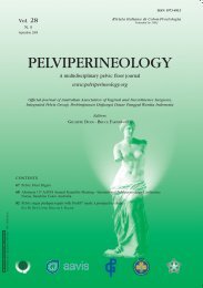
Vol. 29 - N. 4<br />
December 2010<br />
Rivista Italiana di Colon-Proctologia<br />
Founded in 1982<br />
ISSN 1973-4891<br />
A multidisciplinary pelvic floor journal<br />
‘Taxe Perçue’ ‘Tassa Riscossa’ - Padova C.M.P.<br />
Poste Italiane s.p.a.<br />
Spedizione in Abb. Post. - 70% - DCB Padova<br />
Contents<br />
99 Editorial<br />
100 Tethered vagina syndrome: cure of severe involuntary urinary<br />
loss by skin graft to the bladder neck area of vagina<br />
Klaus Goeschen, andrei Müller-FunoGea, Peter Petros<br />
103 Arc to Arc minisling 1999: a critical analysis of concept<br />
and technology<br />
Paulo PalMa<br />
106 Two year outcome data on efficacy and quality of life<br />
following mesh augmented vaginal reconstruction<br />
adaM s. holzberG, Peter s. FinaMore, Krystal hunter,<br />
ricardo caraballo, Karolynn t. echols<br />
110 Common genitourinary fistulae at a referral hospital<br />
in Saudi Arabia<br />
ahMed h al-badr, ola t Malabary, abdullah n al-Jasser,<br />
Valerie a ziMMerMan<br />
113 Diagnosis and management of adult female stress urinary<br />
incontinence. Summary of the guidelines for clinical practice<br />
from the French College of Gynaecologist and<br />
Obstetricians (CNGOF)<br />
GreGory trioPon, renaud de tayrac, Pierre Mares<br />
116 Development of a third generation surgical technique for<br />
mesh repair for pelvic organ prolapse using a lightweight<br />
monofilament polypropylene mesh. A preliminary report of<br />
efficacy and safety<br />
bruce Farnsworth<br />
123 The neuropelveology: a new speciality in medicine?<br />
Marc PossoVer<br />
aavis
Why?<br />
PSYLLOGEL<br />
fibra<br />
In case of...<br />
... Constipation,<br />
Hemorrhoids,<br />
Pelvic floor diseases.<br />
The most natural way to promote,<br />
restore and maintain regularity.<br />
Psyllium is the strongest natural dietary fiber for promoting<br />
regularity and supporting benefits overall health.<br />
Psyllium has low fermentation; this gel provides lubrication<br />
that facilitates propulsion of colon contents and<br />
produces a stool that is bulkier and more moist.<br />
AVAILABLE IN THE TASTES:<br />
Red orange Strawberry Lemon tea Cocoa Vanilla<br />
PSYLLOGEL ® fibra<br />
The leader in psyllium fiber 99% purity.<br />
Informations reserved to the doctors and pharmacists<br />
IN PHARMACY<br />
NATHURA SRL - Tel. +39 0522 865464 - www.nathura.com - nathura@nathura.com<br />
UNI EN ISO 9001:2008 QUALITY MANAGEMENT SYSTEM CERTIFIED BY CERTIQUALITY
Vol. 29<br />
N. 4<br />
December 2010<br />
Rivista Italiana di Colon-Proctologia<br />
Founded in 1982<br />
PELVIPERINEOLOGY<br />
A multidisciplinary pelvic floor journal<br />
www.pelviperineology.org<br />
Editors<br />
Giuseppe Dodi, Colorectal Surgeon, Italy<br />
Bruce Farnsworth, Gynaecologist, Australia<br />
Associate Joint Managing Editor<br />
Florian Wagenlehner, Urologist, Germany<br />
Co-Editors<br />
Nucelio Lemos, Gynaecologist, Brazil<br />
Akin Sivaslioglu, Urogynecologist, Turkey<br />
Editorial Board<br />
Burghard Abendstein, Gynaecologist, Austria<br />
Roberto Angioli, Gynaecologist, Italy<br />
Jacques Beco, Gynaecologist, Belgium<br />
Cornel Petre Bratila, Gynaecologist, Romania<br />
Klaus Goeschen, Urogynaecologist, Germany<br />
Daniele Grassi, Urologist, Italy<br />
Dirk G. Kieback, Gynaecologist, Germany<br />
Filippo LaTorre, Colorectal Surgeon, Italy<br />
Bernhard Liedl, Urologist, Germany<br />
Menahem Neuman, Urogynaecologist, Israel<br />
Oscar Contreras Ortiz, Gynaecologist, Argentina<br />
Paulo Palma, Urologist, Brazil<br />
Francesco Pesce, Urologist, Italy<br />
Peter Petros, Urogynecologist, Australia<br />
Richard Reid, Gynaecologist, Australia<br />
Giulio Santoro, Colorectal Surgeon, Italy<br />
Marco Soligo, Gynaecologist, Italy<br />
Jean Pierre Spinosa, Gynaecologist, Switzerland<br />
Michael Swash, Neurologist, UK<br />
Vincent Tse, Urologist, Australia<br />
Richard Villet, Urogynaecologist, France<br />
Pawel Wieczorek, Radiologist, Poland<br />
Rui Zhan, Urogynaecologist, P.R. China<br />
Carl Zimmerman, Gynaecologist, USA<br />
Official Journal of the: International Society for <strong>Pelviperineology</strong><br />
(the former Australasian Association of Vaginal and Incontinence Surgeons)<br />
International Pelvic Floor Dysfunction Society<br />
Pelvic Reconstructive Surgery and Incontinence Association (Turkey)<br />
Perhimpunan Disfungsi Dasar Panggul Wanita Indonesia<br />
Romanian Uro-Gyn Society<br />
Editorial Office: Enrico Belluco, Maurizio Spella<br />
c/o Clinica Chirurgica 2 University of Padova, 35128, Padova, Italy<br />
e-mail: editor@pelviperineology.org<br />
Quarterly journal of scientific information registered at the Tribunale di Padova, Italy n. 741 dated 23-10-1982<br />
Editorial Director: Giuseppe Dodi<br />
Printer “Tipografia Veneta” Via E. Dalla Costa, 6 - 35129 Padova - e-mail: info@tipografiaveneta.it
Editorial<br />
About us<br />
Le Meridien Hotel in Vienna, Austria was the venue for the latest AAVIS Annual Scientific Meeting<br />
and International <strong>Pelviperineology</strong> Conference, held on September 19th-21st 2010. The meeting was a<br />
tremendous success with a high quality scientific program and wonderful opportunity for fellowship and<br />
social interaction. The full scientific content of the meeting can now be viewed via webcast which is available<br />
through the AAVIS website at www.aavis.org.<br />
At the meeting it was decided to change the name of the society to reflect the changes that have occurred<br />
in recent years. AAVIS is now a true multidisciplinary and international society. The new name will be the<br />
International Society for <strong>Pelviperineology</strong>. There will soon be a new website to reflect this change Plans<br />
are now underway for the next International <strong>Pelviperineology</strong> Conference which will be held in Sydney<br />
during the second half of 2011. Further information will be available as soon as the program and venue are<br />
finalized.<br />
The Annual General Meeting of the International Society for Pelviperieology (formerly AAVIS) was held in<br />
Vienna and new Office Bearers were elected for 2011 as follows<br />
President: Professor Giuseppe Dodi, Colorectal Surgeon, Padova, Italy<br />
Vice President: Dr Bruce Farnsworth, Gynaecologist, Sydney, Australia<br />
Treasurer: Dr Jeff Tarr, Gynaecologist, Buderim, Australia<br />
Secretary: Dr Vincent Tse, Urologist, Sydney, Australia.<br />
The new Committee looks forward to working with you over the next 12 months<br />
Bruce Farnsworth<br />
ANNOUNCEMENT<br />
Dear Reader,<br />
<strong>Pelviperineology</strong> is a quarterly journal open access in the web.<br />
In 2010 www.pelviperineology.org and www.pelviperineologia.it have been visited over<br />
100.000 times. Some printed copies of the journal are distributed free by sponsors.<br />
If you wish to be sure to receive all issues of the printed journal, unless this is provided by your<br />
scientific society, please send to subscriptions@pelviperineology.org your surname, name, full<br />
postal address and speciality, and pay a yearly subscription fee (€ 30,00) to the Integrated Pelvic<br />
Group via Paypal account (see the instructions in the website www.pelviperineology.org).<br />
99
Original article<br />
Tethered vagina syndrome: cure of severe involuntary urinary<br />
loss by skin graft to the bladder neck area of vagina<br />
KLAUS GOESCHEN, 1 ANDREI MÜLLER-FUNOGEA, 2 PETER PETROS 3<br />
1<br />
Kvinno Center Hannover, Germany<br />
2<br />
EUREGIO-Pelvic-floor-Unit MZ StaedteRegion Aachen, Germany<br />
3<br />
University of Western Australia<br />
Abstract: Background The tethered vagina syndrome is an iatrogenic condition caused by scar-induced tightness in the bladder neck area of the<br />
vagina. The classical symptom is commencement of uncontrolled urine leakage as soon as the patient’s foot touches the floor on getting out of<br />
bed in the morning. With this condition, the bladder works like a watering can, due to loss of elasticity in the bladder neck area. <strong>This</strong> situation is<br />
somewhat similar to “motor detrusor instability”, and so is considered as being incurable. 1990 Petros described a new strategy for treatment.<br />
The first step is to free all scar tissue from urethra and bladder neck, the second to increase the tissue in the bladder neck area of vagina, thereby<br />
restoring elasticity. Aim: To test the efficacy and safety of three procedures which aim to restore elasticity in the bladder neck area of vagina.<br />
Methods: Between Jan. 2001 and Dec. 2009 we performed a plastic operation in the bladder neck area of vagina, “I-plasty” in 13 patients,<br />
a free skin graft in 21 patients, and a bulbo-cavernosus muscle-fat-skin-flap-operation from the labium majus in 85 patients. Results: At 6<br />
month review, the cure rate (Urine loss
Tethered vagina syndrome: cure of severe involuntary urinary loss by skin graft to the bladder neck area of vagina<br />
Fig. 3 – I-plasty operation A vertical incision is made in the bladder<br />
neck area of vagina. The vagina and urethra are extensively<br />
mobilized off the adjoining tissues and pelvic side wall. The incision<br />
is sutured horizontally, thus introducing fresh tissue to the site.<br />
Fig. 1 – The Zone of Critical Elasticity (ZCE) ZCE1 = ZCE at<br />
rest; ZCE2 = ZCE during effort or micturition. Adequate vaginal<br />
elasticity at ZCE allows the oppositely acting urethral (U) and<br />
bladder neck (BN) closure mechanisms to operate. F1 represents<br />
the forward acting vector which stretches the vaginal hammock<br />
forwards to close the distal urethra (“urethral closure mechanism”).<br />
F2 stretches the proximal urethra backwards and downwards<br />
against the pubourethral ligament “PUL”,to close it (“bladder neck<br />
closure mechanism”). A scar at ZCE “tethers” the oppositely acting<br />
muscle vector forces, so that on application of a strong prolonged<br />
force, such as occurs on getting up out of bed in the morning,<br />
F2 overcomes F1, and the posterior wall of the urethra is pulled<br />
open, exactly as occurs during micturition. Coughing exerts a short<br />
sharp force. If there is just sufficient elasticity remaining at ZCE,<br />
F1 and F2 may be able to operate separately, so no urine lost on<br />
coughing. However, if the vagina just behind the scar is gently<br />
stretched backwards by Allis forceps, all the residual elasticity<br />
is removed from ZCE, and urine is now lost on coughing. PCM<br />
= m.pubococcygeus; LP=mlevator plate; LMA=m.longitudinal<br />
muscle of the anus. F2 represents the resultant force of the LP/<br />
LMA vectors.<br />
up of a large gap. Care was taken to effect haemostasis. A<br />
full thickness skin graft approximately 6x4 cm was taken<br />
from the lower abdominal wall. After removal of underlying<br />
fat the graft was applied to the bladder base using several<br />
‘quilting sutures’. The graft was then trimmed as necessary,<br />
and sutured to the adjacent vaginal skin with interrupted 00<br />
Vicryl.<br />
“Skin-on” Martius flap graft (figure 5). In 85 patients<br />
the large gap after scar dissection was covered with a<br />
bulbocavernosus-muscle-fat-skin-flap from the labium<br />
majus. A 5x3 cm ellipse of vulval skin was created over the<br />
labium majus and transferred with underlying fat and muscle<br />
through a tunnel into the dissected area. The tunnel must<br />
be sufficiently large to avoid constriction of the vascular<br />
pedicle. The graft was attached to the adjacent vaginal skin.<br />
RESULTS<br />
The cure rates (Urine loss
K. Goeschen - A. Müller-Funogea - P. Petros<br />
Fig. 5 – Martius skin graft . The graft is brought through a hole<br />
in the lateral vaginal wall. The skin is sutured to the edges of the<br />
vagina. The wound from the site of the graft is to the left. <strong>This</strong> is<br />
sutured with subcuticular 00 Dexon sutures<br />
syndrome’. The ‘tethered vagina syndrome’ 1 is conceptually<br />
similar to “motor detrusor instability”, in that the urine loss<br />
is massive and uncontrolled. As the mechanism for opening<br />
out the posterior urethral wall is mechanical, urgency is<br />
frequently not found with this condition. The cause is<br />
iatrogenically induced fibrosis in the bladder neck area of<br />
the vagina. It is far more common in regions where surgeons<br />
are taught to remove significant amounts of vaginal skin<br />
during vaginal repairs.<br />
The explanation for cure of this condition by restoration<br />
of elasticity in this area may be explained by reference to<br />
a previously described hypothesis 5,6 (figure 1): there are<br />
separate urethral and bladder neck closure mechanisms. In<br />
the former, forward vectors stretch the underlying vagina<br />
on each side to close the urethra from behind. In the latter,<br />
backward/downward vectors stretch the proximal vagina<br />
and bladder base backwards and downwards to close off<br />
the bladder neck. Adequate elasticity is required for these<br />
separate movements. If fibrosis occurs at this critical point<br />
then the opportunity for independent movement is lost and<br />
the stronger posterior force overcomes the weaker anterior<br />
force. As a result the urethra is forced open (figure 1).<br />
Often there is very little stress incontinence. The reason<br />
is that cough creates short sharp fast switch contractions,<br />
and there may be just sufficient elasticity at ZCE to prevent<br />
urine leakage on coughing. Getting out of bed in the<br />
morning stretches ZCE far more as the pelvic floor contracts<br />
to support all the intra-abdominal organs. The classical<br />
symptom is commencement of uncontrolled urine leakage<br />
as soon as the patient’s foot touches the floor. Often there<br />
is no urgency, as the cause is mechanical: a scar at ZCE<br />
‘tethers’ the more powerful backward forces ‘F2’ (figure 1)<br />
to the weaker forward forces ‘F1’, so the bladder is pulled<br />
open as in micturition.<br />
To cure this condition the aim must be to restore elasticity<br />
in the bladder neck area of the vagina, the ‘zone of critical<br />
elasticity’ (ZCE), so that ‘F1’ and ‘F2’ can act independently<br />
of each other. As a first step, it is essential to dissect the<br />
vagina from the bladder neck and urethra, and to free all<br />
scar tissue from urethra, bladder neck and pubic bones<br />
(‘urethrolysis’).<br />
There must be no scar tissue anchoring the bladder neck<br />
to the pelvic side wall.<br />
The second step is to bring fresh tissue to the bladder<br />
neck area of the vagina to restore elasticity, and prevent new<br />
scar creation in this area. Our results demonstrate that the<br />
I-plasty operation cures less than one fourth of the patients.<br />
Therefore we decided not to continue with this method in<br />
cases where there is obvious tissue deficit. It is still the<br />
simplest technique but only indicated if there is no tissue<br />
deficit. The I-plasty works very well in patients where the<br />
cause is excessive bladder neck elevation, for example, after<br />
a Burch colposuspension. If there is a severe shortage of<br />
tissue or a large gap after dissection, this defect has to be<br />
covered with a skin graft or a flap.<br />
The results with free skin graft are much better than with I-<br />
plasty, but a cure rate of about 50% is still not convincing. A<br />
free graft is problematical because there is no blood supply.<br />
Therefore up to one third may not ‘take’, or the graft may<br />
shrink excessively.<br />
The bulbocavernosus-flap operation is technically more<br />
challenging, but brings its own blood supply. <strong>This</strong> is in our<br />
opinion the explanation for the high cure rate. Using this<br />
technique it is very important not to compromise the blood<br />
supply of the graft. Therefore the pedicle must be thick<br />
enough to prevent too much compression to the vessels in<br />
the pedicle, and the space created in the lateral vaginal wall<br />
for passage of the graft must be adequate.<br />
CONCLUSION<br />
The results appear to sustain the hypothesis that adequate<br />
tissue elasticity is required for the separate function of the<br />
bladder and urethral the closure mechanisms. 5,6 Application<br />
of a muscle-fat-flap to the zone of critical elasticity after scar<br />
dissection restores the tissue elasticity in the bladder neck<br />
area of vagina and continence in about 80% of patients.<br />
REFERENCES<br />
1. Petros PE, Ulmsten U, The tethered vagina syndrome, post<br />
surgical incontinence and I-plasty operation for cure. Act Obstet<br />
Gynecol Scand 1990; 69: 63-67; Suppl 153<br />
2. Petros PE, Reconstructive Pelvic Floor Surgery According to<br />
the Integral Theory. In: Petros PE, The Female Pelvic Floor.<br />
Springer Heidelberg 2006; 135-141; Chapter 4<br />
3. Petros PE, The integral theory system: A simplified clinical<br />
approach with illustrative case histories <strong>Pelviperineology</strong> 2010;<br />
29: 37-51<br />
4. Abrams P, Cardozo L, Fall M, Griffiths G, Rosier P, Ulmsten U,<br />
van Kerrebroeck P, Victor A, Wein A, The Standardization of<br />
Terminology of Lower Urinary Tract Function: Report from the<br />
Standardisation Sub-Committee of the International Continence<br />
Society. Neurourology and Urodynamics 2002; 21:167-178.<br />
5. Petros PE and Ulmsten U, An Integral Theory of Female Urinary<br />
Incontinence, Acta Obst Gynecol Scand 1990; 1-79; Suppl 153<br />
6. Petros PE, Ulmsten U, An Integral Theory and its Method for the<br />
Diagnosis and Management of Female Urinary Incontinence.<br />
Scand J Urol Nephrol 1993; 1-93; Suppl 153<br />
Correspondence to:<br />
Klaus Goeschen, Hildesheimer<br />
Str. 34-40, 30169 Hannover,<br />
Germany<br />
E-mail: goeschen@carpe-vitam.info<br />
102
Original article<br />
Arc to Arc minisling 1999: a critical analysis<br />
of concept and technology<br />
PAULO PALMA<br />
Chief Professor of Urology, UNICAMP, Brazil<br />
Abstract: The aim is to critically review the Arc to Arc minisling (Palma´s technique), a less invasive midurethral sling using bovine<br />
pericardium as the sling material. Methods: The Arc to Arc minisling, using bovine pericardium was the first published report of a minisling,<br />
in 1999. The technique was identical to the “tension-free tape” operation, midline incision and disssection of the urethra. The ATFP (white<br />
line) was identidied by blunt dissection, and the minisling was sutured to the tendineous arc on both sides with 2 polypropylene 00 sutures.<br />
Results: The initial results were encouraging, with 9/10 patients cured at the 6 weeks post-operative visit. However, infection and extrusion of<br />
the minisling resulted in sling extrusion and removal, with 5 patients remaining cured at 12 months . Critical analysis and conclusion: The Arc<br />
to Arc minisling was a good concept, but failed because of the poor technology available at that time. Further research using new materials<br />
and better technology has led to new and safer alternatives for the management of stress Urinary Incontinence.<br />
Key words: Urinary stress incontinence; Arc to Arc minisling, Bovine pericardium<br />
IntrOdUctIOn<br />
The understanding of stress urinary incontinence (SUI)<br />
pathophysiology has consistently improved over the past<br />
decade, and has resulted in the development of many<br />
surgical techniques. Based on the Integral Theory, Petros<br />
and Ulmsten proposed the tension-free vaginal tape (TVT).<br />
According to this theory a midurethral tape can stabilize<br />
the urethra during straining without modifying the urethral<br />
mobility. 1, 2 Despite the good cure rate reported for TVT,<br />
major complications as injuries to bowel and major blood<br />
vessels have been described. 3<br />
As an alternative to the TVT procedure, the transobturator<br />
tape (TOT) technique was developed by Delorme in 2001,<br />
to reduce the perioperative complications related to the<br />
penetration in the retropubic space. 4 Several short-term<br />
studies reported high cure rates and low complication rates<br />
for TOT, and discussed the mechanism responsible for<br />
the success of this treatment based only on preoperative<br />
urodynamic findings and postoperative clinical examination,<br />
uroflowmetry and the cough test. The continence rate with<br />
the transobturator approach has been similar to those<br />
obtained with the transvaginal retropubic approach. 5 Most<br />
of the described complications are related to the blind nature<br />
of these procedures. 6<br />
The aim of this paper is to report the initial results and<br />
complications of the Arc to Arc minisling (ATAM); then to<br />
critically analyse the ATAM technique, the materials used, 7<br />
and finally, to compare and contrast the ATAM as regards<br />
subsequent minislings .<br />
catheter-balloon was placed above the anal sphincter to obtain<br />
abdominal pressure. The test included water cystometry,<br />
Valsalva leak point pressure (VLPP) assessment, which was<br />
performed with a intravesical volume of 200ml and Valsalva<br />
maneuvers, and pressure-flow study.<br />
The stress test was positive in all patients. Patients who<br />
presented involuntary detrusor contractions during bladder<br />
filling or Maximum flow (Qmax) less than 15ml/s and/or<br />
post void residual urine of more than 20% of the volume<br />
voided were excluded from the study but those with irritative<br />
symptoms without urodynamically proven involuntary<br />
contractions were included. Although urodynamically<br />
proven detrusor instability does not have a significant effect<br />
on surgical outcome, this decision was based on the concept<br />
regarding the postoperative improvement of sensory<br />
urgency, as described previously.<br />
Follow-up was performed at 1, 6 and 12 months. At<br />
these recalls, the patients were questioned about presence<br />
of spontaneous voiding, involuntary urinary leakage, lower<br />
urinary tract symptoms, vaginal and suprapubic pain,<br />
and underwent stress test. The patients were considered<br />
subjectively dry in the absence of incontinence, improved,<br />
when the incontinence episodes were less than once in two<br />
weeks and when incontinence episodes were superior to once<br />
a week the patients were recorded as subjective failures.<br />
Surgical technique<br />
The procedure is performed with the patient in the<br />
lithotomy position. A 18F Foley catheter is introduced for<br />
safety. A inverted U vaginal incision is made at the level<br />
MAterIALs And MethOds<br />
Patients<br />
An open prospective non-randomized clinical trial<br />
involving SUI patient was conducted after receiving the<br />
approval of the Hospital Ethics Committee. Ten patients<br />
(mean age –58 years) underwent the Arc to Arc minisling<br />
(ATAM) procedure for SUI. The procedures were performed<br />
between March 1997 and October 1998.<br />
Study design<br />
All patients were given a routine work-up for incontinence,<br />
including history, physical examination, stress test and<br />
urodynamic investigation. Urodynamic evaluation was<br />
performed with 2 urethral catheters (one 10F for filling and<br />
another 4F for bladder pressure measurement). A rectal 4F<br />
<strong>Pelviperineology</strong> 2010; 29: 103-105 http://www.pelviperineology.org<br />
Fig. 1 – An inverted U shape incision is made and a metzenbaum<br />
scisors is used to dissect the vaginal wall (original illustrations).<br />
103
P. Palma<br />
Fig. 2 – Digital identification of the Arcus tendineous fascia pelvis<br />
(AtFP).<br />
of the blader neck. The vaginal wall is dissected from the<br />
underlying periurethral fascia, bilaterally to the inferior<br />
ramus of the pubic bone. The urethra is identified and a<br />
small perforation of the endopelvic fascia was made at the<br />
border of the ascending ramus of the pubic bone bilaterally<br />
(fig 1).<br />
Next, the surgeon´s index finger is introduced in the<br />
Retzius space towards the obturator internus muscle in order<br />
to identify the white line (fig 2).<br />
Once the white line is identified, 2 polypropylene 00<br />
stitches are placed in the tendinous arc at both sides (fig 3).<br />
Than a minisling of bovine pericardium 6 cm long and 2<br />
cm width is used to create a ATAM, providing back board<br />
support to the urethra (fig. 4).<br />
The vaginal incision is closed in the usual manner and and<br />
a Foley catheter is left in place overnight.<br />
resULts<br />
There were no vascular or visceral lesions, nor urinary<br />
retention.<br />
Nine out of 10 patients were cured of the incontinence<br />
at the first post operative month. After 2 months 2 patients<br />
presented infection of the minisling that were removed,<br />
late complications included 3 more patients that presented<br />
extrusion of the sling at 6 month. The remaining five<br />
patients did well and were continent after 12 months. All but<br />
one patient that has the minisling removed were incontinent<br />
account for 50% of good results after one year follow-up.<br />
dIscUssIOn<br />
The understanding of physiopatological concepts of stress<br />
urinary incontinence has consistently improved over the last<br />
years and their applications have lead to the development of<br />
many surgical techniques.<br />
In the past decade minimally invasive synthetic slings,<br />
such as TVT, have become the preferred technique, replacing<br />
Fig. 3 – sutures placed in the white line (AtFP).<br />
Fig. 4 – suburethral minisling anchored to the obturator internus<br />
muscles at the level of tendinous arc bilaterally.<br />
the Burch colposuspension for the treatment of stress urinary<br />
incontinence. 8<br />
Various factors have contributed to the popularization of<br />
slings, among them, the fact that needle suspensions have<br />
not stood the test of time, together with the various paradigm<br />
changes and the evolution of biomaterials. 1<br />
Synthetic slings present several advantages over<br />
autologous slings.<br />
Harvesting the graft, a time consuming step of the<br />
conventional technique is eliminated along with its related<br />
morbidity and a well standardized procedure is obtained.<br />
Besides it may be performed under local anesthesia as an<br />
outpatient procedure. Not to mention less post-operative<br />
pain and shorter seek leave. 2<br />
On the other hand synthetic slings brought about<br />
new complications related to the tape and even fatal<br />
complications. 3<br />
As an alternative to the TVT procedure, the transobturator<br />
tape (TOT) technique was developed by Delorme in 2001.<br />
<strong>This</strong> procedure reduces per operative complications related<br />
to the penetration in the retropubic space. 4 Several shortterm<br />
studies reported high cure rates and low complication<br />
rates for TOT.<br />
But, as with any form of surgery, adverse events can<br />
occur, and the surgeon should be aware of the common<br />
complications that can accompany sling surgery, and how<br />
to best manage them. 3<br />
The most common complication reported with sling<br />
surgery is bladder perforation during needle passage.<br />
Bladder perforation usually occurs on the side opposite the<br />
surgeon’s dominant hand, and with greater frequency in<br />
patients undergoing repeat procedures.<br />
Many studies report an incidence of bladder perforation<br />
of between 1–15%, and an average perforation rate of 5%.<br />
Management of bladder perforation includes recognition of<br />
the injury during cystoscopy, withdrawal and repositioning<br />
of the needle and a Foley catheter for 24 to 48 hours.<br />
Transobturator sling, on the other hand present a lower<br />
rate of bladder and urethral injury during the needle passage,<br />
which generally occurs in less than 1% of patients, usually<br />
during the learning curve of the procedure.<br />
Bleeding is another important complication and can occur<br />
mainly during needle passage. Bleeding upon entry into the<br />
retropubic space can be difficult to manage, as exposure of<br />
the perivesical venous plexus is difficult.<br />
Care must be taken during lateral replacement of needles<br />
to avoid injuring the external iliac vein for vascular injuries<br />
are usually caused by excessive lateral passage of the<br />
needle.<br />
Despite the good results described worldwide, with<br />
cure rates of more than 80% of the cases, some major<br />
complications like bowel, vascular injuries and deaths were<br />
described. 3<br />
Most of the described major complications are related<br />
104
Arc to Arc minisling 1999: a critical analysis of concept and technology<br />
to the blind nature of these procedures. 6 In fact, reducing<br />
needles diameter alone was not enough to overcome these<br />
problems that occurred even with experimented surgeons.<br />
In an attempt to reduce major complications, mainly<br />
deaths, anatomical reconstruction of the urethral support<br />
placing a low- tension suburethral tape anchored to the<br />
obturator internus muscles bilaterally at the level of the<br />
tendineous arc, the Tissue Fixation System (TFS) were<br />
described. 6 By doing so, bowl lesions and major vessels<br />
injury are avoided.<br />
A decade ago, we used this good principle, but poor<br />
technology in biomaterials at that time, lead to less than<br />
optimal results due to an unacceptable extrusion rate.<br />
Insisting in the principle of restoring the urethropelvic<br />
ligament, we used the porcine small intestine submucosa<br />
(SIS) in 25 patients in 2001. 9<br />
Long term results with arc to arc minisling using swine<br />
intestine submucosa, produced 60% of good results after six<br />
years follow-up. 10<br />
Although the concept was good and the biomaterial<br />
improved, the absence of an appropriate anchoring system<br />
and delivery instruments were a major drawback to its<br />
widespread use.<br />
The first commercially available kit, was the Tissue<br />
Fixation System (TFS) described by Petros. <strong>This</strong> kit<br />
contained two polypropylene anchors and a multifilament<br />
mesh. Preliminary report disclosed similar cure rates<br />
and fewer complications when compared to TOT. 6 <strong>This</strong><br />
preliminary studies reported no pain, mesh exposure,<br />
vascular or visceral complications. No doubt a remarkable<br />
achievement.<br />
Long term follow-up with TFS disclosed good cure rates<br />
after 3 years (11) and good technology available today<br />
allowed for using the TFS System to perform uterus esparing<br />
procedures as well (12) .<br />
Many other devices are available now, some of them<br />
depending on mesh integration for the fixation, like TVT-<br />
Secur and therefore presenting until 60% of failure in the<br />
first post-operative year. 13<br />
Primary fixation of mini slings is a key issue for success,<br />
and our experimental data disclosed that Ophira and TFS<br />
presents the best primary fixation when copared to others<br />
minisling. 14<br />
But needless to say that even minimally invasive<br />
procedures require a learn period and failure is an important<br />
complication as well.<br />
At this point in time, all we can say is that after many years<br />
or research and development we now have good concepts<br />
and good technology.<br />
cOncLUsIOn<br />
Minisling are here to stay and evidences are being<br />
building to determine its indications in the surgeon´s<br />
armamentarium.<br />
reFerences<br />
1. Petros, P, Ulmsten U. An integral theory and its method for<br />
the diagnosis and management of female urinary incontinence.<br />
Scand. J. Urol. Nephrol 153: 1-93,<br />
2. Ulmsten U, Henriksson L, Johnson P, Varhos G. An ambulatory<br />
surgical procedure under local anesthesia for treatment of<br />
female urinary incontinence. Int Urogynecol J. 1993; 7:81-86<br />
3. Deng DY,Rutman M, Raz S, Rodrigues L. Presentation and<br />
management of major complications of midurethral slings:<br />
Are complications under-reported? Neurourol Urodyn 2007;<br />
26(1)46-52<br />
4. Delorme E. La bandellette trans-obturatrice: un procede miniinvasif<br />
pour traiter l’incontinence urinaire d’effort de la femme.<br />
Progrès en Urologie 2001; 1:1306-13<br />
5. Palma P, Riccetto c, Herrmann V, Dambros M, Thiel M,<br />
Bandiera S, Netto N, jr. Transobturator safyre is as effective<br />
as the transvaginal procedure. Int Urogynecol J 2005; 16: 487-<br />
491<br />
6. Petros PE, Richardson P (2005) Midurethral Tissue Fixation<br />
System sling- a Micromethod for cure of stress incontinence-<br />
Preliminary Report. Aust NZ J Obstet Gyn; 45: 372-375<br />
7. Palma PCR. “Sling” tendineovaginal de pericárdio bovino.<br />
Experiência inicial. J. Bras. Ginec 1999; 109:93-97<br />
8. Palma P. A Requiem to the Burch. Int Urigynecol J Pelvic Floor<br />
Dysfunct 2007; 18(6):589-90<br />
9. Palma PCR, Riccetto CLZ, Herrmann V, Dambros M, Mesquita<br />
R, Netto NR Jr Tendinous vaginal support (T.V.S.) using the<br />
porcine small intestine submucosa (SIS): a promising anatomical<br />
approach for urinary stress incontinence. J. Urol 2001; 165: 5<br />
(A).<br />
10. Palma P, Riccetto C, Fraga R, Martins M, Reges R, Oliveira M,<br />
Rodrigues-Netto N jr Long term follow-up of the tendinous<br />
urethral support: na anatomical approach for stress urinari<br />
Incontinence. Actas Urol Esp 2007; 31(7):759-62<br />
11. Petros PE, Richardson PA. Midurethral tissue fixation system<br />
(TFS) sling for cure of stress incontinence-3 year results. Int<br />
Urogynecol J Pelvic Floor Dysfunct 2008;19 (6):869-71<br />
12. Inoue H, Sekiguchi Y, Kohata Y, Satono Y, Hishikawa K,<br />
Tominaga T, Oobashi M. Tissue Fixation System (TFS) to repair<br />
uterovaginal prolapsed with uterine preservation: a preliminary<br />
report on perioperative complications and safety. J Obstey<br />
Gynaecol Res 2009; 35(2):346-53<br />
13. Cornu JN, Sèbe P, Peyrat L, Ciofu C, Cussenot O, Haab F.<br />
Midterm prospective evaluation of TVT-Secur reveals high<br />
failure rate, Eur Urol Apr 23, 2010 [Epub ahead of print]<br />
14. Palma P, Siniscalchi R, Riccetto C, Maciel L, Miyaoka, Bigozzi<br />
M, Dal Fabro L: Primary fixation of mini sling: a comparative<br />
study “in vivo”. Actas Urol Esp, 2010 [Epub ahead of print]<br />
Correspondence to:<br />
Prof. Paulo Palma<br />
Coordenador Escola Superior de Urologia, SBU<br />
Presidente_Eleito da CAU<br />
Prof. Titular & Chefe Disciplina Urologia-UNICAMP, Brazil<br />
ppalma@uol.com.br<br />
105
Original article<br />
Two year outcome data on efficacy and quality of life<br />
following mesh augmented vaginal reconstruction<br />
ADAM S. HOLZBERG 1 , PETER S. FINAMORE 1 , KRYSTAL HUNTER 2 ,<br />
RICARDO CARABALLO 1 , KAROLYNN T. ECHOLS 1<br />
1<br />
Department of Obstetrics and Gynecology, Division of Female Pelvic Medicine and Reconstructive Surgery, Cooper University Hospital UMDNJ-RWJMS, Camden, NJ.<br />
2<br />
Biostatistics Group, Cooper University Hospital UMDNJ-RWJMS, Camden, NJ.<br />
Abstract: Objective: To evaluate quality of life 2 years following mesh augmented vaginal reconstructive surgery. Methods: Patients who<br />
underwent a mesh augmented vaginal reconstructive surgery during an 18 month period were invited to participate. Subjects filled out validated<br />
quality of life questionnaires (PFDI, PFIQ and PISQ), underwent POP-Q examination and were asked if they would have the surgery over again<br />
and if they would recommend it to a friend. Results: Eighty-one patients underwent a mesh augmented repair; 38 (46.9%) consented to return<br />
for follow-up. The average length of follow-up was 25 +/- 6 months. The QOL measures showed improvement comparing pre-operative to<br />
post-operative scores (PFDI: 239.2 vs. 26.5; PFIQ: 152.2 vs. 4.8). Eighty-four percent said they would have the surgery again and 95% would<br />
recommend it to a friend. Conclusion: We found an overall improvement in patients’ quality of life, subjective and objective outcome 2 year<br />
post-operative, following mesh augmented vaginal reconstruction.<br />
Key Words: Mesh, Prolapse, Quality of Life, Vaginal reconstruction<br />
INTRODUCTION<br />
A woman has an 11% lifetime risk for pelvic organ<br />
prolapse and a third of patients who undergo corrective<br />
surgery have repeat procedures. 1 Methods of repair vary<br />
greatly and there is limited evidence to help guide surgeons<br />
to determine which techniques have better outcomes. The<br />
high rates of failure with traditional colporrhaphy 2 have<br />
led to the use of graft materials to augment pelvic floor<br />
reconstruction. <strong>This</strong> has led to the debate as to what graft<br />
material is best? To help answer this question one has to<br />
look at both objective outcomes as identified by the surgeon<br />
as well as patient perception regarding success of the surgery<br />
and improvement in their quality of life. Our study presented<br />
here evaluates the objective, subjective and quality of life<br />
outcomes for a single surgeon’s use of synthetic mesh over<br />
an eighteen month period for the correction of pelvic organ<br />
prolapse.<br />
MATERIALS AND METHODS<br />
After institutional review board approval, a cohort of<br />
subjects who underwent polypropylene mesh augmented<br />
vaginal reconstruction between June 2005 and December<br />
2006 were asked to participate in the study. Vaginal<br />
reconstructive surgeries included any prolapse repair of the<br />
anterior, posterior or apical compartment using mesh. Based<br />
on the practice patterns of the primary surgeon, this included<br />
the use of polypropylene mesh in one of two ways. The<br />
graft was positioned in the appropriate compartment(s) in<br />
the vagina and secured utilizing suture or tension free mesh<br />
arms brought through the obturator foramen or ischiorectal<br />
fossa to achieve surgical correction of the prolapse. Our<br />
“traditional” anterior repairs included the use of a 10 X 15<br />
cm polypropylene mesh (Polyform, Boston Scientific Corp.,<br />
Boston MA or Pelvitex, Bard Corp., Atlanta, GA) cut in a<br />
trapezoidal fashion, anchored to the arcus tendineus fascia<br />
pelvis from the level of the ischial spines to the bladder<br />
neck. The alternative technique for anterior repair utilizes<br />
a prefabricated piece of polypropylene mesh with arms as<br />
described above using what is commonly referred to as a<br />
“lift kit” (Avaulta Anterior, Bard Corp., Atlanta, GA). Our<br />
“traditional” posterior repairs included the use of a 10 X 15<br />
cm polypropylene mesh (Polyform, Boston Scientific Corp.,<br />
Boston MA or Pelvitex, Bard Corp., Atlanta, GA) cut in a “top<br />
hat” like fashion with the 15 cm side of the mesh anchored to<br />
106<br />
the sacrospinous ligaments bilaterally and the distal portion<br />
of the mesh anchored to the levator fascia laterally and the<br />
rectovaginal septum distally. The alternative technique for<br />
posterior repair utilizes a posterior “lift kit” placed in the<br />
ischiorectal fossa as previous described (Avaulta Posterior,<br />
Bard Corp., Atlanta, GA).<br />
Each patient underwent a pelvic exam with prolapse<br />
staging utilizing the Pelvic Organ Prolapse Quantification<br />
scale (POP-Q) 3 pre-operatively, at 3 months and an<br />
average of 25 +/- 6 months post-operatively. At the initial<br />
pre-operative and 2 year post-operative visits, patients<br />
filled out validated questionnaires. Pre-operative and postoperative<br />
questionnaires included the long and short form<br />
versions, respectively of the Pelvic Floor Distress Inventory<br />
(PFDI) and the Pelvic Floor Impact Questionnaire (PFIQ). 4<br />
Both of these questionnaires contain 3 domains assessing<br />
prolapse, colorectal and urinary dysfunction. At the 2 year<br />
follow-up visit patients additionally filled out the Prolapse<br />
and Incontinence Sexual Function Questionnaire short<br />
form (PISQ-12). 5 Subjective evaluation was based on three<br />
questions asked to the patients at an average of 25+/- 6<br />
months post-op: 1) Would you do the surgery all over again?<br />
2) Would you recommend the surgery to a friend? 3) In<br />
terms of your prolapse how do you feel; 1: markedly worse,<br />
2: worse, 3: same, 4: improved, 5: markedly improved?<br />
Patients who did not return for participation in the study,<br />
were contacted by telephone and were specifically asked<br />
questions 1 and 2. These two questions were chosen because<br />
of their ability to have a concise yes or no answer.<br />
A retrospective chart review was performed to collect the<br />
following data for analysis: patient demographics, POP-<br />
Q results, PFDI and PFIQ scores, post-operative physical<br />
examination findings, additional surgical interventions and<br />
length of follow-up. Independent T test, Fisher Exact test<br />
and Pearson Chi Square test were used to determine if there<br />
was any demographic data associated with surgical failure.<br />
Mean, median and standard deviations were calculated for<br />
objective and subjective data.<br />
RESULTS<br />
Eighty-one patients, during the study period had a mesh<br />
augmented vaginal repair. Demographic data for all patients<br />
are listed in Table 1. Thirty-eight patients (46.9%) consented<br />
to return for this study and were included in the analysis. Of<br />
these 38 patients, the mean age at the time of surgery was<br />
<strong>Pelviperineology</strong> 2010; 29: 106-109 http://www.pelviperineology.org
Two year outcome data on efficacy and quality of life following mesh augmented vaginal reconstruction<br />
Table 1 – Demographics for all 81 Patients<br />
Age (yrs) (Mean (St Dev) / Range) 58 (10) 38-84<br />
BMI kg/m 2 (Mean (St Dev) / Range) 29.1 (5.5) 19.7-50.1<br />
Parity (Mean (St Dev) / Range) 3.3 (1.8) 0-10<br />
Tobacco users (N / %) 18 22<br />
Premenopausal (N / %) 17 21<br />
Postmenopausal (N / %) 64 79<br />
HRT (N / %) 8 10<br />
Vaginal estrogen only 3 4<br />
Oral estrogen only 2 2.5<br />
Estrogen patch 1 1.2<br />
Vaginal and oral estrogen 2 2.5<br />
Race (N / %)<br />
Not recorded in chart 30 37<br />
White 40 49<br />
African American 3 4<br />
Hispanic 8 10<br />
Diabetes Mellitus 9 11<br />
Previous Hysterectomy 24 30<br />
Previous prolapse or incontinence<br />
procedure<br />
13 16<br />
Urodynamics (UDTs)<br />
No UDTs pre-op 14 17<br />
UDTs pre-op 67 83<br />
Detrusor Overactivity Pre-op 15 18.5<br />
Pre-op Stress Incontinence by UDT 37 46<br />
BMI – Body mass index; HRT – Hormone replacement therapy<br />
59 +/- 9.9 years. Most patients were Caucasian (84%), postmenopausal<br />
(84%), and did not have a hysterectomy prior<br />
to the vaginal reconstructive procedure (68%). The average<br />
length of follow-up was 25 +/- 6 months. Five patients had<br />
an anterior polypropylene mesh augmented repair only: 4<br />
were performed using the Avaulta Anterior lift kit and 1<br />
was performed using a Polyform mesh suture based repair.<br />
Seven patients underwent a posterior polypropylene mesh<br />
augmented repair only: 4 were performed using the Avaulta<br />
Posterior lift kit and three using a Polyform mesh suture<br />
based repair. Twenty-six patients had a combined anterior<br />
and posterior polypropylene mesh augmented repair:<br />
seventeen patients had a combined graft augmented suture<br />
based repair: 13 were performed with Polyform and 4 were<br />
with Pelvitex. Of the remaining 9 patients, Avaulta Anterior<br />
and Posterior lift kit was placed in 8 patients and 1 patient<br />
had an anterior repair with Polyform and a site-specific<br />
posterior repair.<br />
The mean and median pre-operative, three month postoperative<br />
and two year post-operative POP-Q points of the<br />
thirty-eight patients seen for follow up are found in Table 2.<br />
Of the 3 patients with stage 3 prolapse, 2 had undergone a<br />
posterior repair only and presented with a stage 3 anterior<br />
prolapse at their latest follow-up visit. For the purposes<br />
of our analysis these 2 patients were counted as having<br />
recurrent prolapse although the initial defect repair was in<br />
a different compartment. Two of these three patients with<br />
stage 3 prolapse said they would have the same surgery<br />
again and the other patient said she was unsure.<br />
All of the eleven patients who had stage 2 recurrent<br />
prolapse were found to have the defect in the anterior<br />
compartment. One patient had stage 2 prolapse in both<br />
anterior and posterior compartments. Another patient<br />
initially underwent a posterior repair and was found to have<br />
stage 2 anterior prolapse at her two year follow-up visit. She<br />
was also added as another patient with recurrent prolapse. In<br />
response to our subjective quality of life measures nine of<br />
these subjects said they would have the surgery again, one<br />
said she would not have the surgery again and one said she<br />
was unsure. All fourteen of the patients considered to have<br />
recurrent prolapse (defined as greater than or equal to stage<br />
2 at their 2 year follow-up visit) said they would recommend<br />
the surgery to a friend. In all subjects with stage 2 prolapse<br />
at follow-up the greatest point of descent on the POP-Q was<br />
an Aa of -1. There was a trend suggesting that those who had<br />
recurrent prolapse or were our surgical failures were more<br />
likely to have had previous urogynecologic procedures.<br />
(p=0.052) There were no other associations with surgical<br />
“failure” (Table 3). The information in table 3 is not stable<br />
due to the small sample size.<br />
Of the forty-three patients who did not participate in<br />
the study, twenty-one were able to be reached by phone.<br />
Twenty stated they would have the surgery again and<br />
would recommend it to a friend. Table 4 demonstrates the<br />
mean and median scores from the quality of life surveys<br />
of the patients who followed-up 2 years post-op. Twentyseven<br />
out of thirty-eight patients filled out the long form<br />
version of the PFDI and PFIQ pre-operatively and thirtyfive<br />
of thirty-eight patients filled out the short version of<br />
these questionnaires post-operatively. The median preoperative<br />
PFDI and PFIQ was 256.7 and 143.9 (long form)<br />
respectively and post-operatively 29.1 and 4.8 (short form)<br />
respectively. <strong>This</strong> demonstrates an overall improvement<br />
in quality of life symptoms. The PISQ-12 was filled out<br />
by twenty-four patients post-operatively with results seen<br />
in Table 4. Twelve were not sexually active at the time of<br />
follow-up and two did not complete the survey. We did not<br />
have pre-operative PISQ-12 scores.<br />
There were two subjects who underwent additional surgery<br />
for recurrent prolapse during the two year follow-up period.<br />
There was one mesh erosion found in the thirty eight patients<br />
(2.6%) who followed up at two years. Eighty four percent<br />
of these patients said they would have the surgery again and<br />
95% would recommend the surgery to a friend. The median<br />
score for satisfaction was 5: markedly improved.<br />
Table 2 – POP-Q points for pre-operative, 3 month post-operative and 2 year follow-up visit (for patients who had long term follow-up<br />
(N=38).<br />
POP Q Points<br />
Pre-op<br />
3 mos Post-op<br />
2 year Follow-up<br />
Mean(std dev)/Median<br />
Mean(std dev)/ Median<br />
Mean (std dev)/ Median<br />
Aa 0.89(1.89) 1 -2.72(0.61) -3 -1.92(1.24) -2<br />
Ba 1.39(2.36) 1.5 -2.66(0.75) -3 -1.47(1.18) -2<br />
Ap -1.53(1.47) -2 -2.95(0.23) -3 -2.89(0.31) -3<br />
Bp -1.45(1.52) -2 -2.95(0.22) -3 -2.82(0.46) -3<br />
C -3.04(4.17) -4.75 -7.08(3.20) -7.5 -6.30(1.39) -6<br />
D -6.33(1.74) -6.5 -9.40(1.14) -9 -7.00(1.00) -7<br />
TVL 9.79(0.84) 9.75 9.21(1.02) 9 8.22(1.18) 8<br />
GH 4.42(1.19) 4.25 2.78(0.63) 3 3.20(0.72) 3<br />
PB 3.73(1.18) 3.5 4.46(0.74) 4.75 4.14(0.79) 4<br />
107
A. S. Holzberg - P. S. Finamore - K. Hunter - R. Caraballo - K. T. Echols<br />
Table 3 – Associations with Surgical Failure<br />
Stage of Prolapse<br />
at 2 Year Visit *<br />
P-Value<br />
0 or 1 (N=24) >2 (N=14)<br />
Race<br />
Caucasian (N/%) 21 (87.5) 11 (78.6)<br />
African American (N/%) 0 (0.0) 2 (14.3)<br />
Hispanic (N/%) 3 (12.5) 1 (7.1) 0.153<br />
Age (yrs) (Mean) 57.33 62.57 0.114<br />
BMI (kg/m 2 ) (Mean) 27.94 29.71 0.325<br />
BMI<br />
Obese (BMI >30) (N/%) 7 (29.2) 7 ((50.0)<br />
Not Obese (BMI500 ml<br />
Yes (N/%) 4 (17.4) 3 (21.4)<br />
No (N/%) 19 (82.6) 11 (78.6) 1.000<br />
* Stage 2 or greater considered recurrent prolapse or surgical<br />
failure<br />
BMI – Body Mass Index<br />
EBL – Estimated Blood Loss at time of reconstruction<br />
DISCUSSION<br />
In surgery for pelvic organ prolapse, there is increasing<br />
evidence in support of the use of mesh when correcting<br />
pelvic floor defects. 66-11 <strong>This</strong> management is supported<br />
by a recent Cochrane Review reporting a higher risk of<br />
recurrent prolapse after anterior colporrhaphy than after<br />
mesh repairs. 72 The availability of “lift kits” has resulted<br />
in more surgeons performing mesh augmented repairs for<br />
pelvic organ prolapse. <strong>This</strong> study demonstrates an overall<br />
improvement in the quality of life and outcome variables<br />
two years post-operative following mesh augmented<br />
vaginal reconstruction in a busy urogynecology practice.<br />
Many complications can occur from the use of mesh in the<br />
vagina. These complications include sexual dysfunction,<br />
de novo stress urinary incontinence or fecal incontinence,<br />
voiding dysfunction, pain, failure, and reoperation risk. 12-17<br />
Any of these complications can affect a person’s quality of<br />
life. A recent warning by the Food and Drug Administration<br />
describes many of these risks (http://www.fda.gov/cdrh/<br />
safety/102008-surgicalmesh.html). Subjective and objective<br />
data in this study demonstrate an overall improvement in<br />
quality of life following these procedures and the majority of<br />
Table 4 – Results of Questionnaires Pre-operatively and at 2 year<br />
Follow-up visit for the Patients who had long term follow-up<br />
PFDI<br />
PFIQ<br />
PISQ<br />
12<br />
N=24*<br />
POPDI-6<br />
Mean (std<br />
dev)/Median<br />
CRADI-8<br />
Mean (std<br />
dev)/Median<br />
UDI-6<br />
Mean (std<br />
dev)/Median<br />
Total Mean<br />
(std dev)/<br />
Median<br />
UIQ-7<br />
Mean (std<br />
dev)/Median<br />
CRAIQ-7<br />
Mean (std<br />
dev)/Median<br />
POPIQ-7<br />
Mean (std<br />
dev)/Median<br />
Total Mean<br />
(std dev)/<br />
Median<br />
Pre-op N=27<br />
(Long Forms)<br />
2 yr follow<br />
N=35<br />
(Short Forms)<br />
103.8(57.2) 1Œ07.7 14.0(21.0) 8.3<br />
72.0(67.8) 60.1 14.8(18.2) 7.85<br />
89.5(65.2) 72.2 18.9(21.0) 8.3<br />
265.3(159.7) 256.7 48.5(50.8) 29.1<br />
108.4(91.5) 98.35 7.9(12.8) 0<br />
57.3(81.3) 21 9.2(23.3) 0<br />
57.0(81.8) 20.4 3.9(9.8) 0<br />
222.6(214.7) 143.9 21.3(37.6) 4.8<br />
Not Done<br />
Not<br />
Done<br />
88.1(19.6) 92.9<br />
*12 patients were not sexually active at time of 2 year f/up and 3<br />
did not complete any surveysŒ<br />
patients would do their procedure again with the knowledge<br />
of their experience since the surgery. These results infer<br />
that these risks are likely minimal. Previous studies have<br />
defined failure as a POP-Q staging of 2 or greater. Stage 2<br />
prolapse is defined as any point between -1 and +1, relative<br />
to the hymenal ring. In our study, all patients with Stage 2<br />
prolapse at follow-up had no point of descent greater than -1.<br />
Most of these patients were unaware of any recurrence and<br />
were pleased with the results of their surgery based on the<br />
subjective questions put forth to them. <strong>This</strong> might lead us to<br />
redefine failure from a subjective point as opposed to a purely<br />
objective one. Of the three patients with stage 3 recurrent<br />
prolapse, two of them had a failure in the compartment not<br />
operated on at the time of their initial surgery. It is often a<br />
struggle in the field of urogynecology to decide whether to<br />
prophylactically repair an otherwise asymptomatic defect.<br />
Although these numbers are small, this might lead us to<br />
consider repairing even minor defects in compartments<br />
opposite to those which appear to be causing the patients<br />
complaints. More evidence is needed in this area. Some of<br />
the limitations of this study include the retrospective nature<br />
of our data as well as the limited percentage of patients who<br />
followed up at two years. Although our rate of return was<br />
comparable and acceptable compared to other studies we<br />
would have liked to have seen a greater long term follow up<br />
rate. Other limitations include the varying brands of mesh<br />
as well as different techniques employed to perform these<br />
repairs. Our practice now uses the short form versions of the<br />
PFDI and PFIQ and thus made it difficult to show an exact<br />
comparison of data secondary to the use of the long form<br />
versions used previously. Unfortunately, at the time of this<br />
study, the PISQ-12 surveys were not filled out by our patients<br />
pre-operatively. It is now our practice to include this survey<br />
in our pre-operative packet distributed to all patients at their<br />
initial office visit. We can not make any assumptions with<br />
regards to patients’ change in sexual function following graft<br />
108
Two year outcome data on efficacy and quality of life following mesh augmented vaginal reconstruction<br />
augmented repairs. However, we can say that other similar<br />
studies have demonstrated similar results for the overall<br />
PISQ-12 score as our study. 18, 19 The strength of our study is<br />
its long term follow-up after the use of polypropylene mesh<br />
for a single surgeon in vaginal reconstruction. Subjective<br />
questions and objective validated questionnaires along with<br />
other outcome variables demonstrate overall satisfaction<br />
and efficacy.<br />
REFERENCES<br />
1. Olsen AL, Smith VJ, Bergstrom JO, Colling JV, Clark AL.<br />
Epidemiology of surgically managed pelvic organ prolapse and<br />
urinary incontinence. Obstet Gynecol 1997;89:501-6.<br />
2. Maher C, Baessler K, Glazener CM, Adams EJ, Hagen S.<br />
Surgical management of pelvic organ prolapse in women.<br />
Cochrane Database Syst Rev CD004014 2007.<br />
3. Bump RC, Mattiason A, Bo K, Brubaker LP, De Lancey JO,<br />
Klaeskov P, et al. The standardization of terminology of female<br />
pelvic organ prolapse and pelvic floor dysfuction. Am J Obstet<br />
Gynecol 1996;175:10-17.<br />
4. Barber MD, Walters MD, Bump RC, Short forms of two<br />
condition-specific quality-of-life questionnaires for women<br />
with pelvic floor disorders (PFDI-20 and PFIQ-7). Am J Obstet<br />
Gynecol 2005;193:103-13.<br />
5. Rogers RG, Kammerer-Doak, Villarreal A, Coates K, Qualls C.<br />
A new instrument to measure sexual function in women with<br />
urinary incontinence or pelvic organ prolapse. Am J Obstet<br />
Gynecol 2001;184:552-8.<br />
6. Hinoul P, Ombelet WU, Burger MP, Roovers JP. A prospective<br />
study to evaluate the anatomic and functional outcome of<br />
a transobturator mesh kit (prolift anterior) for symptomatic<br />
cystocele repair. J Minim Invasive Gynecol 2008;15(5):615-<br />
20.<br />
7. de Tayrac R, Devoldere G, Renaudie J, Villard P, Guilbaud<br />
O, Eglin G,et al. Prolapse repair by vaginal route using a new<br />
protected low-weight polypropylene mesh: 1-year functional<br />
and anatomical outcome in a prospective multicentre study. Int<br />
Urogynecol J Pelvic Floor Dysfunct 2007;18(3):251-6.<br />
8. De Vita D, Araco F, Gravante G, Sesti F, Piccione E. Vaginal<br />
reconstructive surgery for severe pelvic organ prolapses: a<br />
‘uterine-sparing’ technique using polypropylene prostheses. Eur<br />
J Obstet Gynecol Reprod Biol 2008;139(2):245-5.<br />
9. Nieminen K, Hiltunen R, Heiskanen E, Takala T, Niemi K,<br />
Merikari M, Heinonen PK. Symptom resolution and sexual<br />
function after anterior vaginal wall repair with or without<br />
polypropylene mesh. Int Urogynecol J Pelvic Floor Dysfunct<br />
2008;19(12):1611-6.<br />
10. Rutman MP, Deng DY, Rodriguez LV, Raz S. Repair of vaginal<br />
vault prolapse and pelvic floor relaxation using polypropylene<br />
mesh. Neurourol Urodyn 2005;24(7):654-8.<br />
11. de Tayrac R, Picone O, Chauveaud-Lambling A, Fernandez H.<br />
A 2-year anatomical and functional assessment of transvaginal<br />
rectocele repair using a polypropylene mesh. Int Urogynecol J<br />
Pelvic Floor Dysfunct. 2006;17(2):100-5.<br />
12. Boyles SH, McCrery R. Dyspareunia and mesh erosion after<br />
vaginal mesh placement with a kit procedure. Obstet Gynecol<br />
2008;111:969-75.<br />
13. Bako A, Dhar R. Review of synthetic mesh-related complications<br />
in pelvic floor reconstructive surgery. Int Urogynecol J Pelvic<br />
Floor Dysfunct 2008; epub ahead of print.<br />
14. De Ridder D. Should we use meshes in the management of<br />
vaginal prolapse? Cur Opin Urol 2008;18:377-82.<br />
15. Caquant F, Collinet P, Debodinance P, et al. Safetly of trans<br />
vaginal mesh procedure: Retrospective study of 684 patients. J<br />
Obstet Gynaecol Res 2008;34:449-56.<br />
16. Jia X, Glazener C, Mowatt G, et al. Efficacy and safety of using<br />
mesh or graft in surgery for anterior and/or posterior vaginal<br />
wall prolapse: systematic review and meta-analysis. BJOG<br />
2008;115:1350-61.<br />
17. Natale F, La Penna C, Padoa A, Agostini M, De Simone E,<br />
Cervigni M. A prospective, randomized, controlled study<br />
comparing Gynemesh, a synthetic mesh and Pelvicol, a<br />
biologic graft, in the surgical treatment of recurrent cystocele.<br />
Int Urogynecol J Pelvic Floor Dysfunct. 2008; epub ahead of<br />
print.<br />
18. Novi JM, Bradley CS, Mahmoud NN, Morgan MA, and Arya<br />
LA. Sexual function in women after rectocele repair with acellular<br />
porcine dermis graft vs site-specific rectovaginal fascia repair.<br />
Int Urogynecol J Pelvic Floor Dysfunct. 2007;18(10):1163-9.<br />
19. Thakar R, Chawla S, Scheer I, Barrett G, Sultan AH. Sexual<br />
function following pelvic floor surgery. Int J Gynaecol Obstet.<br />
2008 Aug;102(2):103-4.<br />
Correspondence to:<br />
Adam Holzberg<br />
6012 Piazza at Main Street Voorhees,<br />
NJ 08043, (856)325-6622(Office), (856)325-6522 (Fax),<br />
Email: holzberg-adam@cooperhealth.edu<br />
109
Original article<br />
Common genitourinary fistulae at a<br />
referral hospital in Saudi Arabia<br />
AHMED H AL-BADR, 1 OLA T MALABARY, 2 ABDULLAH N AL-JASSER, 3 VALERIE A ZIMMERMAN 4<br />
1<br />
Consultant and Chairman of Urogynecology and Pelvic Reconstructive Surgery, Women Specialized Hospital, King Fahad Medical City (KFMC), Riyadh, KSA. Formerly,<br />
consultant of Obstetrics and Gynecology, Security Forces Hospital (SFH), Riyadh, KSA<br />
2<br />
Resident in Obstetrics and Gynecology, McGill University, Montreal, Canada. Formerly, resident of Obstetrics and Gynecology, SFH, Riyadh, KSA<br />
3<br />
Consultant of Urology and Chairman, Department of Surgery, SFH, Riyadh, KSA<br />
4<br />
Research and Publication Office, KFMC<br />
Abstract: Objective: To evaluate genitourinary fistulae cases, including factors, management, and outcome. Material and Methods: A<br />
retrospective chart review of 10 genitourinary fistulae cases at a referral hospital. Results: Ten patients: 4 vesicouterine fistulae (VUF), 4<br />
vesicocervical fistulae (VCF), and 2 vesicovaginal fistulae (VVF). All VUF were complications of cesarean section (CS). Three VCF were<br />
secondary to cesarean subtotal hysterectomy, and one was subsequent to CS. One VVF was a complication of hysterectomy, and the other was<br />
secondary to a road traffic accident. Three VUF cases underwent surgical repair and one had fulguration. One VCF underwent surgical repair,<br />
and 3 had conservative management. One VVF underwent surgical repair, and one underwent fulguration. All patients were asymptomatic<br />
during follow up. Conclusion: The majority of cases were CS related, one was post gynecologic surgery, and one was related to an external<br />
injury. None were complications of prolonged or instrumental delivery. All cases were cured.<br />
Key Words: Fistula, Genitourinary, Saudi Arabia, Urinary incontinence.<br />
INTRODUCTION<br />
Genitourinary fistulae can be classified anatomically,<br />
etiologically, 1 or by surgical size. 2 It is generally accepted<br />
that surgical fistulae occur after unrecognized incision in<br />
urinary structures, pressure necrosis, devascularization,<br />
or a combination of these mechanisms. 3 Obstetric fistulae<br />
occur because of damage during the course of prolonged<br />
labor, instrumentation during delivery, or as consequences<br />
of cesarean section (CS). 3 Historically, most vesicovaginal<br />
fistulae (VVF) were the result of birth trauma; accordingly,<br />
they remain the major urinary fistulae and the most common<br />
cause of urinary incontinence in many underdeveloped<br />
nations. 4 In developed nations, genitourinary fistulae<br />
usually occur as rare complications of gynecologic or other<br />
pelvic surgery, or of radiotherapy. 5 In North America, for<br />
example, the most common cause of VVF is injury to the<br />
bladder during hysterectomy, 6 and the risk is reported as<br />
being more than 1% after radical surgery and radiotherapy<br />
for gynecologic malignancies. 7 Although vesicouterine<br />
fistula (VUF) is an uncommon condition comprising only<br />
1% to 4% of all urogenital fistulae, its prevalence has been<br />
rising in recent decades 8 because of the increased incidence<br />
of CS. VUF may develop immediately after a CS, or may<br />
not be observed until the late puerperium. 9 Management of<br />
genitourinary fistula is primarily surgical. 6 Nevertheless,<br />
conservative treatment or cystoscopic fulguration have met<br />
with some success. 9,10 The goal of our study was to determine<br />
the number of genitourinary fistula cases in our facility over<br />
10 years, evaluate their causes, and assess their management<br />
and outcome.<br />
MATERIALS AND METHODS<br />
<strong>This</strong> was a retrospective review of all cases of genitourinary<br />
fistula between January 1995 and May 2006 at the Security<br />
Forces Hospital (SFH), Riyadh, Kingdom of Saudi Arabia<br />
(KSA). SFH is a referral hospital with approximately 500<br />
beds. Through the medical coding system, all cases of genital<br />
fistula, urinary fistula, VVF, vesicocervical fistula (VCF),<br />
and VUF were identified and reviewed, whether they were<br />
admitted under the care of the Obstetrics and Gynecology<br />
or Urology Departments. Cases of rectovaginal fistula and<br />
other unrelated fistulae were excluded. Research committee<br />
110<br />
approval was obtained prior to data collection. Both vaginal<br />
and abdominal repair were done in layers after excising<br />
the fistula tract. Abdominal repair was accompanied by<br />
interposition of an omental graft.<br />
RESULTS<br />
Ten cases of genitourinary fistula were identified during<br />
the study period: 4 cases of VUF, 4 cases of VCF, and 2<br />
cases of VVF. Four cases were diagnosed by cystogram,<br />
3 by cystoscopy, and one by hysterosalpingiogram. <strong>This</strong><br />
information was missing from 2 files.<br />
All VUF cases were complications of CS (Table 1).<br />
The reasons for doing CS included: elective CS with 5<br />
previous CS (n=2, one with placenta previa), 3 previous<br />
CS (n=1), and one patient with 2 previous CS followed by<br />
vaginal delivery, and then a failed trial of vaginal delivery,<br />
which ended by emergency CS. Three of 4 post CS cases<br />
underwent surgical repair through an abdominal approach,<br />
and were kept on urethral catheterization for 9 to 11 days<br />
postoperatively. All 3 cases were asymptomatic at their last<br />
follow up from 7 months to 3 years after repair. The fourth<br />
case was initially treated with urethral catheterization for<br />
2 months, but the patient continued leaking after catheter<br />
removal. Accordingly, cystoscopic fulguration of the tract<br />
was done, followed by urethral catheterization for 2 weeks,<br />
after which the patient became dry and remained so 10 years<br />
after repair.<br />
Three of the 4 VCF cases were post emergency cesarean<br />
subtotal hysterectomy due to placenta accreta causing<br />
uncontrolled bleeding, and one was post emergency CS.<br />
In the 3 post emergency subtotal hysterectomy cases,<br />
CS was done as an elective repeated CS. One case had a<br />
recognized bladder injury that was subsequently repaired<br />
surgically. Two of the cases were treated by transurethral<br />
and suprapubic catheterization for 3 to 4 weeks, after which<br />
they were dry. The third patient declined any intervention,<br />
including catheterization. On her follow up visits up to 7<br />
months after surgery, she continued to be dry. The post<br />
CS patient was treated by surgery through an abdominal<br />
approach followed by urethral catheterization for 10 days,<br />
after which the patient became dry, remaining so at a 7-<br />
month follow up visit. One of the 2 VVF cases was post<br />
hysterectomy due to uterine fibroid. The patient underwent<br />
<strong>Pelviperineology</strong> 2010; 29: 110-112 http://www.pelviperineology.org
Common genitourinary fistulae at a referral hospital in Saudi Arabia<br />
Table 1 – Genitourinary Fistula Cases<br />
Management Incident Type Parity Age Case No.<br />
Abdominal repair CS VUF P6+1 37 1<br />
Abdominal repair CS VUF P3+1 36 2<br />
Abdominal repair CS VUF P6+2 41 3<br />
Cystoscopic fulguration;<br />
“Failed catheterization”<br />
CS VUF P4+0 37 4<br />
Bladder catheterization CS VCF P4+0 28 5<br />
Conservative management;”Patient<br />
refused any intervention”<br />
Cesarean subtotal hysterectomy VCF P4+6 38 6<br />
Bladder catheterization Cesarean subtotal hysterectomy VCF P3+1 25 7<br />
Abdominal repair Cesarean subtotal hysterectomy VCF P5+0 33 8<br />
Abdominal repair Hysterectomy VVF P1+0 52 9<br />
Trans-vaginal fulguration RTA VVF P0 28 10<br />
VUF: vesico-uterine fistula - CS : cesarean section - VCF: vesico-cervical fistula - VVF: vesico-vaginal fistula - RTA: road traffic accident<br />
surgical repair through an abdominal approach, followed<br />
by suprapubic catheterization for 10 days, and then urethral<br />
catheterization for 13 days. Subsequently, the patient was<br />
completely dry through 6 months of follow up. The other<br />
case was post road traffic accident (RTA), and had multiple<br />
pelvic fractures and a vaginal hematoma ended by a VVF.<br />
<strong>This</strong> was treated by trans-vaginal fulguration, after which<br />
the patient became dry, and remained so at a 4-year follow<br />
up. Surgical repair of fistulae was done abdominally in all<br />
cases (5/5) because of location of the fistula and surgeons’<br />
expertise and preferences. Cases were operated on by 3<br />
surgeons: 2 urologists, and one urogynecologist.<br />
DISCUSSION<br />
The incidence of VUF is increasing worldwide because of<br />
the increase in CS. 9 All 4 of our VUF cases were secondary<br />
to CS, similar to a study from Spain, where over a period<br />
of 25 years, 5 out of 6 (83%) VUF cases were secondary to<br />
CS. 11 In another series of 15 cases of VUF occurring over<br />
7 years from Benin, 14 were related to CS. 12<br />
Surgery is the mainstay and definitive treatment of<br />
VUF, although spontaneous healing occurred in 5% of<br />
cases. 9 In our series, conservative management was<br />
tried unsuccessfully in one case of VUF, after which the<br />
patient underwent successful cystoscopic fulguration<br />
without additional treatment. A similar successful case of<br />
cystoscopic fulguration was reported; however, hormonal<br />
amenorrhea was induced prior to fulguration. 10 Four of our<br />
cases (40%) were the rare VCF, occurring as complications<br />
of elective CS, 3 of which required concomitant subtotal<br />
hysterectomy. Only one required surgical treatment after<br />
conservative management failed. Interestingly, one patient<br />
became asymptomatic with no intervention. Only 2 of our<br />
cases (20%) were VVF, which were secondary to an RTA<br />
and hysterectomy. <strong>This</strong> paucity of cases is similar to reports<br />
from developed countries, 5 and unlike the situation in many<br />
developing countries like Nigeria, where 889 cases of VVF<br />
over 7 years were found to be complications of labor and<br />
delivery. 13 Another report from Pakistan showed that over<br />
7 years, 27 out of 32 cases of VVF were secondary to<br />
obstetrical trauma, out of which the success rate for surgical<br />
repair through the abdominal route was 100%, and through<br />
the transvaginal route was 80%. Repair was attempted<br />
through transvaginal fulguration in one case in that series, in<br />
which it failed. 14 Although one of our VVF cases was treated<br />
surgically, one was successfully treated by transvaginal<br />
fulguration. A study of 15 VVF cases with sizes less than<br />
3.5 mm in the United States revealed 73% of the patients had<br />
complete resolution after fulguration of the fistulae, either<br />
cystoscopically or vaginally. 15 Unfortunately, the size of<br />
fistulae in our study was not mentioned in any of the charts<br />
or radiology reports. Another study reported 4 cases of<br />
VVF treated successfully using conservative management,<br />
which involved simple bladder drainage for periods ranging<br />
from 19 to 54 days. 16 A review by Haferkamp et al showed<br />
that surgical repair of VVF using transvaginal, transvesical,<br />
and transperitoneal approaches have similar results. They<br />
emphasize that adequate surgical exposure and mobilization<br />
of the bladder and vagina, successful excision of the fistula<br />
tract, tension-free closure, and placement of an interposition<br />
flap, when applicable, are essential components of successful<br />
repair. 17<br />
The majority of our cases (80%) were related to CS or its<br />
complications (cesarean subtotal hysterectomy). <strong>This</strong> finding<br />
is unlike reports from developed or developing countries, in<br />
which fistulae were mainly related to gynecological surgeries<br />
or obstructed labor, respectively. 4,5 Also in contrast to the<br />
world literature, 18,19 VUF was noted to be more common in<br />
our population than VVF. <strong>This</strong> could be explained by the<br />
number of CS performed per year in our facility, which was<br />
approximately 900 cases out of 7000 deliveries per year,<br />
compared with hysterectomies, which were only 20 cases<br />
per year. The significant low frequency of hysterectomies<br />
in our hospital and our society can possibly be explained by<br />
the commitment to preserving fertility as much as possible,<br />
and the myth that hysterectomy can negatively affect the<br />
quality of sexual life.<br />
REFERENCES<br />
1. Drutz HP. Urinary fistulae. Obstet Gynecol Clin North Am.<br />
1989:16(4):911_21.<br />
2. Waaldijk K. Surgical classification of obstetric fistulae. Int J<br />
Gynecol Obstet. 1995:49(2):161-3.<br />
3. Drutz H P, Baker K R. Vesicovaginal Fistula. In: Drutz H,<br />
Herschorn S, Diamant NE (eds) Female Pelvic Medicine and<br />
Reconstructive Surgery. London: Springer; 2003:471-72p.<br />
4. Minassian VA, Drutz HP, Al-Badr A. Urinary incontinence as a<br />
worldwide problem. Int J Gynecol Obstet. 2003:(82)327-338.<br />
5. Cortesse A, Colau A. Vesicovaginal fistula. Ann Urol.<br />
2004:38(2):52_66.<br />
6. Hadley Hr. Vesicovaginal fistula. Curr Urol Rep. 2002:3(5):401_<br />
7.<br />
7. Angioli R, Penalver M, Muzii L, Mendez L, Mirhashemi R,<br />
Bellati F, Croce C, Panici Pb. Guidelines of how to manage<br />
vesicovaginal fistula. Crit Rev Oncol Hematol. 2003:48(3):295_<br />
304.<br />
8. Shing KY, Tak YL. Vesicouterine Fistula: An Updated Review.<br />
Int Urogynecol J. 1998:9:252_256<br />
9. Porcaro AB, Zicari M, Zecchini Antoniolli S, Pianon R, Monaco<br />
111
A.H. Al-Badr - O. T. Malabary - A. N. Al-Jasser - V. A. Zimmerman<br />
C, Migliorini F, Longo M, Comunale L. Vesicouterine fistulae<br />
following cesarean section: report on a case, review and update<br />
of the literature. Int Urol Nephrol. 2002:34(3):335_44<br />
10. Tarhan F, Erbay E , Penbegul N , Kuyumcuglu U . Minimal<br />
invasive treatment of vesicouterine fistula: a case report. Int<br />
Urol Nephrol. 2006:(39)791-3.<br />
11. Bonillo Garcia MA, Pacheco Bru JJ, Palmero Marti JL, Alapont<br />
Alacreu JM, Alonso Gorrea M, Arlandis Guzman S, Jimenez<br />
Cruz JF. Vesicouterine fistula. Our experience of 25 years. Actas<br />
Urol Esp. 2003:27(9):707_12.<br />
12. Hodonou R, Hounnasso PP, Biaou O, Akpo C. Vesicouterine<br />
fistula: report on 15 cases at Cotonou University Urology Clinic.<br />
Prog Urol. 2002:12(4):641_5.<br />
13. Wall LL, Karshima JA, Kirschner C, Arrowsmith SD. The<br />
obstetric vesicovaginal fistula: characteristics of 899 patients<br />
from Jos, Nigeria. Am J Obstet Gynecol. 2004:190(4):1011_9.<br />
14. Khan Rm, Raza N, Jehanzaib M, Sultana R. Vesicovaginal<br />
fistula: an experience of 30 cases at Ayub Teaching Hospital<br />
Abbottabad. J Ayub Med Coll Abbottabad. 2005:17(3):48_50.<br />
15. Stovsky MD, Ignatoff JM, Blum MD, Nanninja JB, O’Conner<br />
VJ, Kursh ED. Use of electrocoagulation in the treatment of<br />
vesicovaginal fistulae. J Urol. 1994:152(5 pt 1):1443_4.<br />
16. Davits RJ, Miranda SI. Conservative treatment of vesicovaginal<br />
fistulae by bladder drainage alone. Br J Urol. 1991:68(2):155_6.<br />
17. Haferkamp A, Wagener N, Buse S, Reitz A, Pfitzenmaier J, Hall<br />
Scheidt P, Hohenfellner M. Vesicovaginal Fistulae. Urologe A.<br />
2005:44(3):270_6.<br />
18. Rafique M. Genitourinary fistulas of obstetric origin. Int Urol<br />
Nephrol.2003:34(4):489-93.<br />
19. Navarro Sebastian FJ, Garcia Gonzalez JI, Castro Pita M, Diez<br />
Rodriguez JM, Arrizabalaga Moreno M, Manas Pelillo A, et<br />
al. Treatment approach for vesicogenital fistula. Retrospective<br />
analysis of our data. Urol Esp. 2003:27(7):530-7.<br />
Correspondence to:<br />
Ahmed Al-Badr,<br />
Department of Urogynecology & Pelvic Reconstructive Surgery,<br />
Woman Specialized Hospital, King Fahad Medical City, Riyadh,<br />
KSA<br />
PO Box: 59046, Riyadh 11525<br />
Phone: +966 (1) 2889999 ext 3030<br />
Fax: +966 (1) 2935613<br />
Email: ahmed@albadr.com<br />
112
Guidelines<br />
Diagnosis and management of adult female stress urinary<br />
incontinence. Summary of the guidelines for clinical practice from<br />
the French College of Gynaecologists and Obstetricians (CNGOF)<br />
GREGORY TRIOPON, RENAUD DE TAYRAC, PIERRE MARES<br />
Obstetrics and Gynaecology department, Carémeau University Hospital, Nimes, France<br />
Abstract: During the thirty third French College of Gynaecologists and Obstetricians (CNGOF) meeting, guidelines for good clinical practice<br />
(RPC) defined by the French High Authority for Health (HAS) were exposed about diagnosis and management of adult female urinary<br />
incontinence without any neurological pathology and, particularly with stress urinary incontinence. Guidelines had been established by a<br />
multidisciplinary committee’s work, directed by B Jacquetin, X Fritel, and A Fauconnier. 1 Methods, texts by the expert authors, synthesis of<br />
recommendations have already been published in French in a special number of the CNGOF journal. The intention of the French College of<br />
Gynaecologists and Obstetricians (CNGOF) was to improve previous recommandations, which had already been drawn up by several scientific<br />
societies, and by this way, to be complementary to these previous undertakings. <strong>This</strong> article is a summary of the French recommandations, and<br />
its aim is to give readers a clinical approach, and help them in their current and usual practices.<br />
Key words: Urinary incontinence, Stress urinary incontinence, TVT, TOT, Guidelines, Raccomandations<br />
DEFINITIONS OF URINARY INCONTINENCE<br />
Stress Urinary Incontinence (SUI) describes the complaint<br />
of involuntary urinary leakages during exertions, coughing or<br />
sneezing. It is divided into two groups: intrinsic sphincteric<br />
deficiency, or increased urethral mobility. 2<br />
Urge urinary incontinence (UUI or overactive bladder)<br />
is involuntary loss of urine, preceded or accompanied by a<br />
strong desire to void.<br />
Mixed urinary incontinence (MUI) is the association in<br />
variable proportions of SUI and UUI.<br />
ASSESSMENT OF FEMALE URINARY INCONTINENCE<br />
Clinical assessment of female urinary incontinence 3<br />
During examinations, you have to precise several points<br />
(symptoms) to try defining urinary incontinence type (even<br />
if there is not always a correspondence between urinary<br />
symptoms and real diagnosis), and checking severity of it:<br />
- Circumstances, frequency, and severity of leakages,<br />
- Urinary symptoms questionnaires: USP (urinary symptom<br />
profile), UDI-6 (Urogenital distress inventory-6), ICIQ<br />
(international consultation on incontinence questionnaire),<br />
MHU (mesure du handicap urinaire)…<br />
- A 3-days bladder diary,<br />
- Quality of Life (QOL) questionnaires, such as general ones<br />
(SF 36…), or specific ones (IIQ, Contilife, Ditrovie, …)<br />
- pad-test<br />
In case of UUI (urge incontinence, nocturia, frequency),<br />
a bladder diary is recommended. It is not specified in this<br />
guidelines but that is probably not necessary to precise that<br />
an urinary tract infection must be eliminated as first line.<br />
In case of SUI, several facts are pointed:<br />
- Cough test proves SUI. It Shows loss of urines and confirm<br />
SUI. It is recommended before all type of SUI surgery. If<br />
the test is negative, you can repeat it particularly in an upstanding<br />
position.<br />
- Urethral mobility can be assessed by examination,<br />
observation, sub urethral manoeuvers, and Q-Tip test. The<br />
best method to check urethral mobility has not yet been<br />
established.<br />
- Post-void residual urines (less than 50 ml) and functional<br />
bladder capacity measurements (up to 400 ml) are<br />
performed during urodynamic investigations.<br />
<strong>Pelviperineology</strong> 2010; 29: 113-115 http://www.pelviperineology.org<br />
Urodynamic investigations (UDI)<br />
UDI includes a uroflowmetry with post-void residual<br />
urine measurement, a cystometry with detrusor pressure<br />
flow study, and an urethral profile including Maximal<br />
Closure Urethral Pressure (MCUP) and Valsalva Leak Point<br />
Pressure (VLPP) measurements. 4<br />
UDI’s indications are:<br />
- all urinary incontinences before surgical treatment,<br />
- all recurrent urinary incontinence,<br />
- all POP previous surgical treatment’s failures, in<br />
association with urinary incontinence.<br />
UDI prescription is not needed before pelvic floor<br />
rehabilitation for urinary incontinence management.<br />
In case of pure SUI (without urge symptoms), UDI<br />
is not necessary before surgery if clinical assessment is<br />
complete (standardised questionnaire, cough test, bladder<br />
diary, establishment of post-void residual volume) and with<br />
concordant results.<br />
A low pre-operative flow rate (measured during<br />
uroflowmetry) is associated with a higher risk of<br />
postoperative voiding dysfunction (after sub urethral tape<br />
placement).<br />
Urinary sphincter deficiency (defined with a decreased<br />
MCUP or VLPP during UDI) is not a decisive prognostic<br />
factor for the result of a sub-urethral tape procedure.<br />
Others investigations?<br />
Others investigations are not recommended, with these<br />
guidelines, to improve your diagnosis or prior to perform<br />
a SUI surgery. Therefore, is there any place for ultrasound,<br />
and particularly bladder exam with ultrasound, to verify its<br />
normality (lithiasis, polyps…), in case of UUI. We probably<br />
have to remain that ultrasound can be usefull for a few cases,<br />
and preoperatively. Furthermore, MRI and cystography are<br />
not needed for urinary incontinence assessment.<br />
HOW TO TREAT A FEMALE SUI?<br />
Conservative treatment of female SUI<br />
- Treatment of female SUI with lower urinary tract<br />
rehabilitation.<br />
Pelvic floor muscle training (PFMT) is recommended<br />
first to treat SUI or MUI (PFMT seems to give better<br />
results than vaginal electrostimulation). Bladder training is<br />
113
G. Triopon - R. De Tayrac - P. Mares<br />
recommended first in cases of UUI, or MUI with predominant<br />
urge symptoms.<br />
- Oestrogen.<br />
Currently, studies don’t allow us to establish an optimum<br />
method of administration, dosage and type of oestrogen for<br />
prevention or treatment of urinary incontinence. Vaginal<br />
oestrogen treatment improves urge incontinence and<br />
frequency. Oral oestrogen treatment is not recommended<br />
for treatment or prevention of SUI. 5<br />
Vaginal oestrogen treatment can be used in postmenopausal<br />
women to improve urge incontinence or frequency.<br />
- Duloxetine.<br />
Objective data (24-hours pad-tests) don’t demonstrate any<br />
superiority for duloxetine in comparison with placebo. By<br />
this way, in France, duloxetine is not recommended at first<br />
line.<br />
- Hygiene and dietary measures.<br />
For overweight patients, loss of weight improves urinary<br />
incontinence, and dietary measures as well as physical<br />
exercices can be proposed.<br />
Surgical treatment at first line for female SUI<br />
Procedures<br />
Among many surgical procedures described to treat SUI,<br />
sub-urethral tape (retropubic or transobturator route) is the<br />
technique recommended at first line due to the easier and<br />
shorter postoperative course than Burch colposuspension. It<br />
is performed under local, locoregional or general anaesthesia.<br />
It can be placed on a one-day surgery or a traditional<br />
hospitalisation (depending on patient’s and surgeon’s<br />
preferences). And postoperative course is less expensive<br />
with sub-urethral tapes compared with colposuspensions by<br />
laparotomy or laparoscopy. 6<br />
Concerning sub urethral tapes :<br />
- ascending retropubic route gives better results in terms<br />
of continence than transobturator route in case of urinary<br />
sphincter deficiency,<br />
- transobturator routes from inside to outside or from<br />
outside to inside give similar results,<br />
- concerning sub-urethral tape procedures, both retropubic<br />
and transobturator routes give advantages, and it does not<br />
allow us to recommend a preferred route,<br />
- Place of mini-slings (in order to treat SUI) is not established<br />
because of the absence of any comparative studies,<br />
- Urinary sphincter deficiency is not a contra-indication for<br />
sub-urethral tape surgery.<br />
What about risks?<br />
The French college (CNGOF) offers (on line) an<br />
information letter for patients undergoing a SUI surgery. 7<br />
Main intra-operative complications of sub urethral tapes<br />
are:<br />
- Urinary tract injuries,<br />
- vaginal sulcus tract injuries, (greater risk of vaginal<br />
perforation with transobturator route, particularly in case<br />
of passage from outside to inside compared with inside to<br />
outside route)<br />
- bowel injuries,<br />
- bladder injuries. (frequency of bladder injury is higher<br />
with retropubic than transobturator route)<br />
Main postoperative complications of suburethral tapes<br />
are:<br />
- urinary retention,<br />
- urinary tract infection,<br />
- urge incontinence,<br />
- pain,<br />
- vaginal, bladder or urethral erosion (erosion rates are<br />
greater with transobturator route than retropubic route).<br />
Postoperatively, it is recommended to assess the quality<br />
of voiding function in order to screen eventual bladder<br />
retention. (urinary post-void residual measurements)<br />
Surgical treatment at second line for female SUI<br />
These French recommandations don’t give readers any<br />
explanations about recurrent SUI treatment, or complex<br />
SUI. In fact, in our practice, it can be a second sub urethral<br />
tape placement, an adjustable continence therapy next to<br />
the bladder neck (ACT, manufactured and disbributed by<br />
AMS®), trans or peri-urethral injections, artificial urinary<br />
sphincter, which are, for most of these procedures, intrinsic<br />
sphincter deficiency treatments.<br />
Surgical treatment for UUI<br />
These guidelines don’t treat UUI surgical treatments<br />
and their indications (botulic toxin, sacral nerve<br />
neuromodulation).<br />
PARTICULAR CIRCUMSTANCES<br />
Urinary incontinence during or after pregnancy<br />
Events of vaginal childbirth have no impact on the<br />
appearance or persistence of urinary incontinence during the<br />
postnatal period or later. 8<br />
At long term, birth by caesarian section doesn’t seem to<br />
reduce the risk of SUI, and so it is not a good way to prevent<br />
postnatal urinary incontinence.<br />
Pregnant women who already underwent a sub-urethral<br />
tape placement, frequency of postnatal urinary incontinence<br />
is not significantly reduced with a birth performed by<br />
caesarean section.<br />
Postnatal perineal rehabilitation including PFMT with a<br />
therapist (midwife or physiotherapist) decreases prevalence<br />
of urinary incontinence at short term (one year after birth)<br />
compared with councils on selfmade pelvic floor exercises.<br />
However, at long term, efficacy of this postpartum<br />
rehabilitation is not established.<br />
Pelvic floor rehabilitation during pregnancy improves<br />
urinary incontinence during pregnancy, and until 3 months<br />
in postpartum time. But, it doesn’t appear to treat it with a<br />
long drop, at long term.<br />
During pregnancy or immediate postnatal time, the first<br />
treatment to perform, in order to treat a SUI, is pelvic floor<br />
rehabilitation (PFMT), and so there is no place for other<br />
medical or surgical treatment at first line.<br />
Urinary incontinence in elderly women<br />
Before deciding any treatment in elderly women, it is<br />
recommended to screen for urinary tract infection (using<br />
a strip test), to make a bladder diary, to measure post-void<br />
urinary residual volume, and to search after triggering factors<br />
(such as confusion syndrome, polymedication, excessive<br />
diuresis, reduced mobility or terminal constipation). 9<br />
It is recommended to search after main vulnerability signs<br />
too: age over 85, polymedication, deteriorated cognitive<br />
functions, depression, undernutrition, neurosensorial<br />
problems, postural instability, lack of physical exercise, loss<br />
of independence, and social isolation. Elderly women who<br />
are heavily dependent from the cognitive and/or physical<br />
point of view should be managed by nursing methods:<br />
programmed voids, physical mobilisation and activity, use<br />
of suitable palliatives, regulation of bowel function.<br />
Anticholinergics are effective for urge incontinence or<br />
MUI in women aged over 65. Anticholinergics may cause<br />
cognitive deterioration in elderly patients who did not<br />
suffer from it before. Prescription of an anticholinergic in<br />
an elderly woman must be monitored about appearance<br />
of deterioration in brain functions, constipation, urinary<br />
voiding dysfunction, or restricted food intake.<br />
114
Diagnosis and management of adult female stress urinary incontinence<br />
Urinary incontinence and genital prolapse<br />
Genital prolapse may be associated with SUI, urge<br />
incontinence, and obstructive urinary symptoms. Urge<br />
incontinence or obstruction symptoms disappear in half of<br />
cases as soon as prolapse is cured.<br />
Prolapse may occur SUI from 20% to 70% of cases<br />
according studies recorded. In case of genital prolapse<br />
without SUI, POP’s correction with pessary reveals lesser<br />
rates of subsequent SUI, than with a speculum. Pessary test<br />
had also been used to predict postoperative continence result<br />
of prolapse surgery. But, the predictive value of the pessary<br />
test used in this cases is unclear, and it is not recommended<br />
to use it systematically. 10<br />
In order to find out an associated SUI before POP repair<br />
treatment, cough test is recommended and allow surgeons<br />
to identify patients who could need an associated urinary<br />
tract procedure during the same time of surgery. In this case,<br />
suburethral tape placement, during the same time of POP<br />
repair by the vaginal route, reduces the risk of postoperative<br />
SUI. In other cases, (no symptomatic or no occult SUI),<br />
there is no indication for a surgical procedure to prevent<br />
incontinence.<br />
In case of genital prolapse surgery in a woman who also<br />
presents symptomatic or occult stress urinary incontinence,<br />
associate surgical procedure for continence is a decision<br />
depending of SUI severity, risk factors, technique chosen<br />
and potential undesirable effects.<br />
REFERENCES<br />
1. A. Fauconnier, X. Fritel. Méthodes et organisation [Methods and<br />
organization]. J Gynecol Obstet Biol Reprod 2009;38:S135-40.<br />
2. D. Faltin. Épidémiologie et définition de l’incontinence urinaire<br />
féminine [Epidemiology and definition of female urinary<br />
incontinence]. J Gynecol Obstet Biol Reprod 2009;38:S142-8.<br />
3. R. de Tayrac, V. Letouzey, G. Triopon, L. Wagner, P. Costa.<br />
Diagnostic et évaluation clinique de l’incontinence urinaire<br />
féminine. Gynecol Obstet Biol Reprod. 2009; suppl vol.38 : S<br />
153-65<br />
4. A. Dompeyre, C.Pizzoferrato. Examen urodynamique et<br />
incontinence urinaire féminine non neurologique. Gynecol<br />
Obstet Biol Reprod. 2009; suppl vol.38: S 166-73<br />
5. J. Kerdraon, P. Denys Traitement conservateur de l’incontinence<br />
urinaire d’effort de la femme. Gynecol Obstet Biol Reprod.<br />
2009; suppl.38: S 174-81<br />
6. P. Debodinance, J.F. Hermieu, J.-P. Lucot Traitement chirurgical<br />
de première intention de l’incontinence urinaire d’effort de la<br />
femme. Gynecol Obstet Biol Reprod. 2009, suppl.38 : S 182-<br />
200<br />
7. G. Bader, M. Koskas Complications des bandelettes sousurétrales<br />
dans la chirurgie de l’incontinence urinaire d’effort<br />
de la femme. Gynecol Obstet Biol Reprod. 2009, suppl.38 : S<br />
201-11<br />
8. X. Deffieux Incontinence urinaire et grossesse. Gynecol Obstet<br />
Biol Reprod. 2009; suppl.38: S 212-31<br />
9. N. Michel-Laaengh Incontinence urinaire de la femme âgée.<br />
Gynecol Obstet Biol Reprod. 2009; suppl. 38 : S 232-8<br />
10. B. Fatton, C. Nadeau Incontinence urinaire et prolapsus génital.<br />
Gynecol Obstet Biol Reprod. 2009; suppl.38 : S 239-48<br />
Correspondence to:<br />
Grégory Triopon, MD<br />
Obstetrics and Gynaecology Department<br />
Carémeau University Hospital,<br />
Place du Professeur Robert Debré<br />
30900 Nimes, France<br />
+33466683216<br />
+33643117262<br />
Fax:+33466683459<br />
E-mail: gtriopon@hotmail.com<br />
115
Original article<br />
Development of a third generation surgical technique for mesh<br />
repair for pelvic organ prolapse using a lightweight monofilament<br />
polypropylene mesh. A preliminary report of efficacy and safety<br />
BRUCE FARNSWORTH<br />
Director, Centre for Pelvic Reconstructive Surgery, Sydney Adventist Hospital, Sydney, Australia<br />
Abstract: A comprehensive 3 level repair technique for prolapse is presented with preliminary results of efficacy and safety after short term<br />
follow up of 42 patients.<br />
Key words: POP; Genital prolapse; Lightweight macroporous polypropylene mesh; Comprehensive 3 level repair.<br />
INTRODUCTION<br />
As a result of audit and evaluation of existing techniques<br />
of pelvic reconstruction over 10 years (1997-2007), a<br />
third generation technique of prolapse repair with mesh<br />
has evolved into a standardized technique. Other mesh<br />
techniques have been associated with a high success rate<br />
and good functional results but erosion rates associated with<br />
defective healing have been reported at 5-10% and higher<br />
in the first three months after surgery using a number of<br />
commercially available mesh kits 2,3,4 and slings. 5,6 Reports<br />
of the significant advantages of light weight wide pore<br />
monofilament meshes in hernia and other surgery led to the<br />
adoption of this material.<br />
MATERIALS AND METHODS<br />
A total of 42 patients were followed prospectively as part<br />
of an ongoing quality assurance program. Ethics committee<br />
approval was obtained to report the outcomes of all patients<br />
undergoing reconstructive surgery on the condition that<br />
patient anonymity was preserved.<br />
All patients underwent surgery using a specialized<br />
prosthesis (CR Mesh) developed at the Centre for Pelvic<br />
Reconstructive Surgery and manufactured by Agency for<br />
Medical Innovation (AMI GmbH, Feldkirch, Austria). The<br />
author performed all surgery using a standardized technique<br />
documented in the Appendix 1.<br />
TECHNIQUE<br />
The CR Mesh technique involves comprehensive<br />
reconstruction of all three levels of pelvic support.<br />
Level 1 support is provided by an independent suspension<br />
of the cervix or vaginal vault, using a monofilament non<br />
absorbable Prolene suture which is attached to the proximal<br />
end of the sacrospinous ligament using a specialized<br />
instrument, either the AMI Suture Instrument or the AMI<br />
I-Stitch device.<br />
Level 2 support involves reattachment of the fascia to the<br />
adjacent levator and obturator complex by the passage of<br />
transobturator and translevator slings. Separate distal slings<br />
are also able to recreate the Level 3 support of the perineum<br />
or bladder neck.<br />
Demographic data of the 42 patients are shown below<br />
(Table 1).<br />
All patients operated with this procedure underwent full<br />
clinical assessment including pelvic organ quantification<br />
(POPQ) examination and a 3 dimensional pelvic ultrasound.<br />
Each patient prior to surgery and again 3 months after surgery<br />
completed a series of quality of life questionnaires. Ongoing<br />
follow-up is planned with annual review and quality of life<br />
assessment.<br />
116<br />
Table 1: Demographic data.<br />
Age: 39 – 86 (Mean age 67)<br />
Previous repair surgery 28 patients<br />
Previous hysterectomy 13 patients<br />
All 42 patients in this study presented with a significant<br />
pelvic organ prolapse (POPQ stage 3 or 4). Three patients<br />
also required a hysterectomy for other pathology and two<br />
patients had a co-existing rectal prolapse.<br />
Table 2: Procedures performed<br />
Anterior CR Mesh 12 patients<br />
Posterior CR Mesh 9 patients<br />
Anterior and Posterior CR Mesh 21 patients<br />
RESULTS<br />
Surgery was successfully completed in all 42 patients.<br />
The range of follow-up was 4 – 15 weeks. There were no<br />
significant intraoperative complications. No patients needed<br />
blood transfusion and there was no evidence of any surgical<br />
morbidity. No patient suffered from any bladder or urethral<br />
injury. There was no incidence of bowel injury.<br />
Initial postoperative assessment showed restoration<br />
of apical support in all 42 patients. Two patients also had<br />
resolution of their rectal prolapse. One patient reported a<br />
slight deterioration of anterior wall support at 3 months<br />
postop in association with mild stress incontinence. There<br />
was no incidence of early mesh erosion or defective<br />
healing. All patients reported significant resolution of<br />
the symptoms of prolapse. Three patients, two of which<br />
required a concurrent hysterectomy complained of severe<br />
postoperative pain and in one patient this was still present<br />
after 6 weeks. No assessment was made of sexual function<br />
at this stage due to the short length of follow-up. <strong>This</strong> aspect<br />
will be reported in future reviews. Two patients reported denovo<br />
stress incontinence.<br />
DISCUSSION<br />
The early outcomes of 42 patients who underwent CR<br />
Mesh implantation show a reduction in the incidence of<br />
mesh erosion and no early evidence of defective vaginal<br />
wound healing.<br />
Ongoing data collection and quality of life assessments<br />
after longer follow up will clarify long-term outcomes and<br />
facilitate the planning of comparative trials. Early results<br />
indicate that the combination of this technique together with<br />
<strong>Pelviperineology</strong> 2010; 29: 116-122 http://www.pelviperineology.org
A preliminary report of efficacy and safety<br />
the new lightweight low density macroporous CR mesh will<br />
result in at least equal outcomes for patients and a significant<br />
reduction in mesh related complications.<br />
REFERENCES<br />
1. Farnsworth B. Suspended mesh reconstruction for the treatment<br />
of total vaginal prolapse. Presentation AAVIS Annual Scientific<br />
meeting Noosa Queensland 1 st August 2009<br />
2. Prospective study of the Perigee system for the management<br />
of cystocoeles--medium-term follow up. Aust N Z J Obstet<br />
Gynaecol. 2008;48:427-32.<br />
3. Farnsworth B, Parodi M. Total Vaginal Reconstruction with<br />
Polypropylene Mesh. Objective and Functional Outcome<br />
Assessment. Int Urogynecol J 2005; 16: Supplement 2 Number<br />
55<br />
4. Fatton B, Amblard J, Debodinance P, Cosson M, Jacquetin B.<br />
Transvaginal repair of genital prolapse: preliminary results of a<br />
new tension-free vaginal mesh (Prolift technique)--a case series<br />
multicentric study. Int Urogynecol J Pelvic Floor Dysfunct.<br />
2007;18:743-52.<br />
5. Farnsworth B. Posterior IVS (Infracoccygeal Sacropexy) for<br />
Vaginal Vault Prolapse. A Preliminary Report of Efficacy and<br />
Safety. Int Urogynecol J 2002; 13:4-8<br />
6. Farnsworth B. Posterior IVS for vault suspension: a reevaluation.<br />
<strong>Pelviperineology</strong> 2007; 26:70-72.<br />
7. Informal meeting of CR Mesh surgeons, Modena, Italy<br />
September 27 th 2008<br />
8. Reported data. Sydney Adventist Hospital Pelvic Surgery<br />
Complications meetings (2004-2007)<br />
<strong>This</strong> paper was first presented at the AMI Pelvic Surgery<br />
Symposium at the 10 th Annual AAVIS Scientific Meeting,<br />
International <strong>Pelviperineology</strong> Congress, Westin Excelsior Resort,<br />
Venice Lido on Friday 3 rd October 2008<br />
APPENDIX 1<br />
A.M.I. ADVANCED PELVIC FLOOR REPAIR SYSTEM<br />
using CR-Mesh and AMI suture instrument<br />
DETAILED PROCEDURE INSTRUCTIONS (version 2.1)<br />
These detailed step by step instructions describe a total vaginal<br />
reconstruction procedure using a CR-Mesh prosthesis in both<br />
anterior and posterior vaginal compartments including restoration<br />
of Level 1 apical support using the AMI Suture instrument.<br />
Prior to commencing surgery an intra-operative assessment is<br />
made and the surgeon decides whether anterior, middle and posterior<br />
compartments are to be repaired. The procedure is performed<br />
through an anterior, posterior or both incisions. If a hysterectomy<br />
has been performed then the vault epithelium should be left intact<br />
for at least an anterior-posterior length of 4cm.<br />
STEP 1: ANTERIOR INFILTRATION<br />
Dilute normal saline 100-200mls in Local Anaesthetic ( e.g.,<br />
xylocaine 1%) +/- adrenalin.<br />
Pull down the cervix to visualize the anterior vaginal wall and<br />
infiltrate with the saline mixture. Choose a point half way between<br />
the vault and the cervix and inject 20 mls at a depth of 2-4 mm. The<br />
subcutaneous tissues should expand evenly in every direction. If<br />
the infiltration seems to expand locally only at the site of infiltration<br />
then the needle needs to be a little deeper. Inject another 20-40ml<br />
at about the same point. Inject the fluid centrally so that it spreads<br />
laterally and hydrodissects under the fascia in all directions.<br />
STEP 2: ANTERIOR INCISION<br />
<strong>This</strong> is a full thickness vertical incision initially 2-3 cm long in<br />
the central portion of the anterior vaginal wall. The incision is deep<br />
enough to reach the dark clear layer under the fascia created by the<br />
hydro-dissection The incision is extended distally to the transverse<br />
vaginal sulcus at the level of the bladder neck and proximally to a<br />
point 2cm from the cervix or vault.<br />
The anterior vaginal incision is further developed by reflecting the<br />
central tissue overlying the bladder away from the skin edge. <strong>This</strong><br />
done by starting the lateral dissection at the midpoint of the incision.<br />
Do one side at a time. The surgeon’s assistant or nurse holds the<br />
epithelial edge laterally on each side with two Alliss or Littlewood<br />
forceps. The surgeon holds the central tissues medially with his<br />
forceps and extends the lateral dissection to the sulcus on each side<br />
and proximally to the cervix or vault . Distally, the bladder neck<br />
also needs to be dissected free from the overlying epithelium but at<br />
this stage the dissection is limited to the superficial space under the<br />
skin to avoid penetrating the venous plexus in each vaginal sulcus<br />
until the last possible moment in order to reduce potential bleeding.<br />
STEP 3: POSTERIOR INFILTRATION<br />
Pull the cervix up to visualize the posterior vaginal wall and<br />
infiltrate at a point 3-5cm distal to the posterior cervix in a similar<br />
manner to the anterior vaginal wall.<br />
STEP 4: POSTERIOR INCISION<br />
Start the incision 3-4cm distal to the cervix at the site of<br />
infiltration and extend the incision towards the perineum once the<br />
correct layer under the fascia is identified and entered. Be wary<br />
of an enterocoele close to the cervix. As the incision reaches the<br />
perineum be careful of causing any rectal damage as the rectum<br />
may be caught up in the scar tissue associated with previous<br />
perineal repairs or episiotomies. Do not use blunt dissection to<br />
reflect the rectum off the perineal body.<br />
The proximal end of the posterior vaginal incision should be<br />
1-2cm from the cervix or 2-3cm from the vaginal vault. At each<br />
end of the incision dissection is performed to provide good access<br />
to the back of the cervix, the perineal body and distal levator<br />
muscles. <strong>Complete</strong> this dissection at this time as it will be difficult<br />
to complete once the mesh is in place.<br />
STEP 5: PREPARE THE CERVICAL RING<br />
The cervical ring is the central support structure of the upper<br />
vagina. If the uterus is absent then a cervical structure has to be<br />
recreated as described below.<br />
A. UTERUS PRESENT<br />
Place a single 2/0 monofilament polypropylene suture in the<br />
anterior and posterior cervix under the skin edge at the cervical end<br />
of each vaginal incision. Ensure that a good strong bite of cervical<br />
connective tissue is included in these sutures. Each suture is then<br />
held by a clamp which can be placed to rest temporarily on the<br />
suprapubic area. These sutures should be long enough to not fall<br />
down into the operative site.<br />
B. UTERUS ABSENT<br />
The epithelium and fascia of the vault is preserved for an anteroposterior<br />
length of at least 3cm. <strong>This</strong> enables a central structure<br />
to be created which is made up of a body of fascial tissue behind<br />
the intact vault held together by three double 2/0 monofilament<br />
polypropylene sutures which are passed from the anterior to<br />
posterior vaginal incision using a large Mayo needle.<br />
The six sutures are passed carefully from the anterior vaginal<br />
incision to the posterior vaginal incision without button holing the<br />
vault or damaging any viscera. The three pairs of sutures are tied<br />
above and below the vault to firmly hold the fascia behind the vault.<br />
The first pair of sutures are tied in the midline and then the two<br />
lateral pairs are tied 1-2cm on each side of the midline.<br />
It is impossible to recreate the cervical ring in this way if the<br />
fascia behind the vault is broken down to link the anterior and<br />
posterior incisions. Ensure adequate tissue is left to facilitate<br />
this step. Care is also taken not to tie these sutures too tight and<br />
compromise the tissue held in the sutures or bunch up the overlying<br />
epithelium excessively.<br />
Once the three pairs of sutures are tied above and below the vault<br />
all six clamps holding these sutures are placed out of the way on<br />
the suprapubic area.<br />
STEP 6: OPEN THE PARARECTAL SPACE<br />
First ensure that the central rectal tissues under the posterior<br />
incision have been dissected away from the lateral vagina and<br />
adequate access to the back of the cervix and the perineal body has<br />
117
B. Farnsworth<br />
been achieved. The lateral dissection should extend to the sulcus<br />
on each side.<br />
<strong>This</strong> dissection is completed by getting an assistant to hold part<br />
of the edge of the posterior vaginal incision using two Allis clamps<br />
held apart and retracting the central tissues with a pair of forceps<br />
held by the surgeon. The Allis clamps are moved around the skin<br />
edge in segments until the lateral dissection is completed. By lifting<br />
up the central tissues with a forcep while the assistant provides<br />
counter traction using two Allis forceps the correct plane can be<br />
seen and usually the yellow fat associated with an enterocoele is<br />
identified in the pararectal tissues.<br />
Use a single index finger to probe the upper lateral aspect of<br />
the dissection on each side aiming in the general direction of the<br />
ischial spine which should be palpable through the fascial tissues<br />
overlying the pelvic sidewall. In some cases the fascia falls away<br />
easily with this digital examination and the ischial spine can then<br />
be felt clearly together with the arcus ligament above and the<br />
sacrospinous ligament below. Remember that with the patient lying<br />
on her back in the lithotomy position the arcus and sacrospinous<br />
ligaments will be vertical in their orientation above and below the<br />
ischial spine respectively.<br />
If this dissection is not straightforward and relatively easy<br />
then use the index finger to sweep up and down over the lateral<br />
sacrospinous ligament and the arcus ligament until a defect is<br />
created in the fascia.<br />
If this also proves to be unsuccessful then introduce a pair of<br />
scissors into the upper lateral aspect of this dissection on each<br />
side and open the fascia with the scissors by using a push-openwithdraw<br />
technique. Do not cut tissue with the scissors. Use the<br />
index finger to open up the fascial defect created by the scissors.<br />
Sometimes opening this fascia can be quite difficult. Resist<br />
the temptation to use excessive force or sharp dissection. Rather,<br />
repetitive push-open-withdraw technique with scissors should be<br />
effective. Sometimes counter-traction using 2-3 clamps on the skin<br />
edge will facilitate this dissection.<br />
STEP 7: CREATE THE POSTERIOR APICAL<br />
ATTACHMENTS<br />
The apical attachments are created using 2/0 monofilament<br />
polypropylene sutures placed in position using the AMI Suture<br />
instrument.<br />
Apical attachments are placed on each side in the medial posterior<br />
aspect of the sacrospinous ligament immediately adjacent to the<br />
sacrum/coccyx. <strong>This</strong> placement is only possible with a narrow<br />
instrument such as the AMI Suture Instrument which can access<br />
this medial position on the sacrospinous ligament.<br />
TECHNIQUE OF SUTURE ATTACHMENT USING AMI<br />
SUTURE INSTRUMENT<br />
The AMI Suture instrument is loaded with a 2/0 monofilament<br />
polypropylene suture. Once the surgeon is confident that he has<br />
adequate access to the attachment point an index finger is placed in<br />
position on the ligament through the incision and the loaded suture<br />
device is slid along this pre-positioned finger. Once the surgeon can<br />
feel the suture instrument is in the correct position the index finger<br />
is used to push the instrument tip into the body of the ligament.<br />
The assistant steadies the handle of the suture instrument and the<br />
surgeon uses his free hand to release the suture from the holding<br />
point on the handle. During this time the surgeon keeps pressure<br />
on the tip of the instrument with his index finger so that it does<br />
not become dislodged from the ligament prior to completing the<br />
fixation.<br />
The handle of the suture instrument is then slowly closed until<br />
a click is felt which indicates the suture has been harpooned. The<br />
ring pull handle is then slowly withdrawn until it is locked in the<br />
second position.<br />
Only then can the index finger holding the tip of the instrument<br />
on to the ligament be released. The suture instrument then has to<br />
be lifted forward to disengage the tip from the ligament without<br />
dislodging the suture from the instrument.<br />
Once it is withdrawn the suture can be released from the second<br />
position on the instrument and the suture is pulled through and<br />
held with a large clip. Pull firmly on the suture to ensure that<br />
the attachment is strong. It is better for the suture to break or the<br />
tissue to give way at this stage when the attachment can be easily<br />
repeated.<br />
The posterior sutures are then clipped to the drapes with a distinctive<br />
large clamp. These are the only sutures in the procedure<br />
that need to be specially identified.<br />
Notes:<br />
1. There is a potential for damage to the suture when using the<br />
AMI Suture instrument and this is more of a problem for some<br />
surgeons due to their technique.<br />
a. Always ensure that the suture has been released from the<br />
handle before closing the device as any tension in the suture could<br />
lead to the suture being cut.<br />
b. Use the instrument smoothly and slowly. Do not close the<br />
instrument rapidly or it might also cut the suture at this point.<br />
c. When placing the suture in the instrument do not centre the<br />
suture. Attach the suture to the tip of the AMI Suture instrument<br />
away from the midpoint of the suture so as to ensure that any<br />
point of potential weakness or damage to the suture is not at the<br />
midpoint.<br />
d. After placing the suture in position around the ligament tie it<br />
gently without tension to ensure that if one of the threads was to<br />
break the other will still be able to be used (The Bath Knot).<br />
e. Some surgeons would prefer to use a 2/0 braided polyester to<br />
place the suture and then use the braided polyester to pull the 2/0<br />
monofilament polypropylene into position.<br />
Standard practice for apical attachments:<br />
A. UTERUS PRESENT<br />
In the presence of the uterus two apical attachments are performed<br />
on each side through the posterior incision and there is NO apical<br />
attachment through the anterior incision.<br />
These sutures will eventually pass through the cervix to be tied<br />
anterior to the cervix so that the cervix is pulled back to the sacrum<br />
and uterine retroversion is corrected.<br />
These sutures are clipped to the drapes with large clamps.<br />
Make sure that this is always done using DISTINCTIVE LARGE<br />
CLAMPS to signify that these attachments are through the posterior<br />
vaginal incision. The two attachments on each side are made to the<br />
same area on the ligament, i.e., the medial posterior surface of the<br />
ligament.<br />
B. UTERUS ABSENT<br />
When the uterus is absent two separate apical attachments are<br />
performed on each side of the pelvis with one passing through the<br />
posterior vaginal incision and one through the anterior vaginal<br />
incision. These sutures will later be attached separately to the<br />
anterior and posterior CR Mesh on each side.<br />
Again, it is critical that these sutures are distinguished from each<br />
other by always placing a distinctive large clamp on the posterior<br />
apical attachments and a small clamp on the anterior apical<br />
attachment as once the mesh has been pulled into position it will be<br />
difficult to identify which suture is which.<br />
Once these sutures are in place they are clipped to the drapes on<br />
either side to keep them away from the operative field.<br />
NOTE: Vault or cervical attachments are lying freely on the<br />
suprapubic area whilst the apical attachments are clipped laterally<br />
to the drapes.<br />
STEP 8: OPEN THE PARAVESICAL SPACE<br />
<strong>This</strong> dissection is not performed until the steps above have been<br />
completed due to the possibility of bleeding from the venous plexus<br />
when entering the paravesical space lateral to the paravaginal<br />
sulcus.<br />
By delaying this step until this point in the procedure the<br />
potential for bleeding is reduced. <strong>This</strong> bleeding could potentially<br />
slow down the previous steps and lengthen the operation. Bleeding<br />
due to damage to the paravaginal venous plexus is usually easily<br />
controlled once the anterior mesh is pulled into place.<br />
Each side of the dissection is done separately. The assistant<br />
grasps the edge of the vaginal incision with two Allis forceps and a<br />
Briesky or similar retractor is used to inspect the vaginal sulcus and<br />
ensure that it is not buttonholed.<br />
The dissection is behind the fascia initially (in the vagina) but<br />
passes through the fascia at the pelvic side wall as the sulcus is<br />
reached. It is at this point that bleeding is more likely to occur.<br />
The lateral dissection should always be commenced midway<br />
between the urethra and the vaginal apex. It is initially sharp<br />
118
A preliminary report of efficacy and safety<br />
dissection behind the fascia and this is facilitated by hydrodissection.<br />
The fascia is breached to enter the paravesical space with scissors<br />
using a push-open-withdraw technique.<br />
Once the the paravesical space is opened the dissection needs<br />
to be extended along the incision to allow a Sims speculum to<br />
be placed between the vaginal incision and the bladder (into the<br />
paravesical space) to enable inspection of the pelvic side wall. The<br />
lateral support tissue of the urethra is reflcted medially by extending<br />
the dissection anteriorly with blunt dissection pushing the urethra<br />
medially and sharp dissection cutting laterally.<br />
The ischial spine, arcus ligament and sacrospinous ligament(SSL)<br />
are identified and the SSL is explored through the anterior incision<br />
to facilitate attachment of the anterior apical suspension sutures.<br />
(As mentioned above these anterior apical sutures are only inserted<br />
when there is no uterus).<br />
STEP 9: PREPARATION OF BLADDER NECK AND<br />
ANTERIOR CERVIX<br />
The anterior vaginal dissection is completed by preparing the<br />
bladder neck and anterior cervix for subsequent placement of the<br />
CR Mesh.<br />
The bladder neck is prepared by dissecting free the lateral supports<br />
of the upper urethra using blunt and sharp dissection as described<br />
above. <strong>This</strong> should be done until the surgeon’s index finger can<br />
pass lateral to the urethra and easily feel the inner surface of the<br />
obturator fossa at the level of the clitoris without being restricted<br />
by a band of connective tissue lateral to the urethra.<br />
The anterior cervix has already been prepared to accept the<br />
anterior mesh and this task is completed by ensuring that there is<br />
good access to the strong fibrous tissue of the anterior cervix with a<br />
flap of epithelium of at least 1cm to cover the attachment point.<br />
STEP 10: PRELIMINARY STEPS TO ATTACH<br />
CR-MESH<br />
Prior to attaching the CR Mesh to the anterior and posterior<br />
vaginal apex preparation is completed so as to enable the attachment<br />
to be done smoothly without confusion or suture entanglement.<br />
Before commencing check that the following have been done:<br />
1. Anterior and posterior vaginal incisions have been completed<br />
with preparation of the bladder neck and perineum.<br />
2. Cervical ring has been prepared for attachment of the CR Mesh<br />
with pre-placement of sutures. <strong>This</strong> is either a single 2/0 Prolene<br />
suture in the front and back of the cervix or three pairs of 2/0<br />
Prolene sutures tied around the central fascia behind the vault.<br />
3. Four apical attachment sutures are in position with correct<br />
clamps used for identification. Distinctive large clamps are<br />
placed on the apical attachments through the posterior vaginal<br />
incision while smaller clamps identify any apical attachments<br />
through the anterior vaginal incision.<br />
4. The operative field is made tidy with removal of extraneous<br />
instruments, suture material and needle holders and their return<br />
to the scrub sister.<br />
5. At this point drapes can be refreshed and gloves changed if<br />
necessary.<br />
STEP 11: PLACEMENT OF POSTERIOR CR MESH<br />
First unpack and unfold the CR Mesh. Identify the proximal and<br />
distal ends as well as the FRONT and BACK of the mesh. The<br />
proximal sling passes through the mesh and attaches laterally to the<br />
back of the mesh with a nylon holding suture.<br />
The mesh is positioned by the surgeon 4-5 cm in front of the<br />
vagina and held in the anatomical position by the assistant(s) who<br />
hold the proximal corner of the mesh on each side. The proximal<br />
1-2 cm of the mesh is folded over to create a double layer.<br />
The back of the mesh is orientated to face the rectum while the<br />
front of the mesh faces the vaginal lumen.<br />
The Mesh is held by the assistant(s) until attached to the cervix<br />
or vault. It is important that they not release the Mesh until this<br />
attachment is complete.<br />
The CR Mesh is attached differently depending on the presence<br />
or absence of a cervix. The following sections are potentially the<br />
most confusing and difficult parts of this procedure.<br />
A. UTERUS PRESENT<br />
First a single Allis forcep is attached to the skin edge of the<br />
posterior incision on each side 2cm lateral to the midline and<br />
these two clips are allowed to fall down behind the mesh. They<br />
will be important in a subsequent step but for now they sit in the<br />
background.<br />
The surgeon first passes the solitary 2/0 monofilament<br />
polypropylene suture which has been pre-positioned in the posterior<br />
aspect of the cervix through the proximal end of the mesh in the<br />
midline 2-3mm from the edge of the mesh. The posterior cervical<br />
suture passes through this point from the FRONT of the mesh to the<br />
BACK. A clamp is used to hold the suture behind the mesh and this<br />
clamp is also allowed to fall down behind the mesh which is held<br />
in position by the assistant(s). The assistant(s) must not release the<br />
Mesh at this point.<br />
Both of the pre-positioned apical (medial sacrospinous)<br />
attachment sutures are then passed through the two layers of the<br />
posterior CR Mesh from the BACK of the mesh to the FRONT<br />
2-3 mm from the folded proximal edge of the mesh and about 1cm<br />
lateral to the central cervical attachment suture that is already being<br />
held by the clamp hanging behind the mesh in the midline. Try to<br />
ensure that each suture of the apical attachments passes through a<br />
different hole in the mesh.<br />
NOTE: The cervical attachment sutures and the apical<br />
sacrospinous attachment sutures pass through the proximal part of<br />
the CR Mesh in opposite directions.<br />
A large Mayo needle is then used to pass the two pairs of apical<br />
attachment sutures through the cervix from back to front on each<br />
side. These sutures emerge through the anterior cervix and a LARGE<br />
DISTINCTIVE clip is placed on the end of each pair of sutures. All<br />
four apical sutures should be positioned from the back of the mesh<br />
through the mesh then through the cervix to emerge through the<br />
anterior incision. Throughout this procedure the assistant(s) have<br />
continued to hold the posterior mesh in position.<br />
The proximal end of the mesh is lifted up to lie flush with the<br />
posterior cervix and the midline suture is tied firmly from behind<br />
to lock the central upper (proximal) folded edge of the mesh to the<br />
cervix in the midline.<br />
The assistant(s) can now release the corners of the mesh and<br />
carefully lift up and hold laterally the two pre-positioned Allis<br />
clamps holding the skin edge of the posterior incision 2cm lateral<br />
to the midline on either side.<br />
The surgeon can then add a 2/0 Vicryl or similar suture to secure<br />
the mesh to the fascia under the skin over the posterior cervix<br />
lateral to the apical attachments that pass through the cervix to<br />
emerge anteriorly.<br />
The posterior aspect of the cervix will be secured to the posterior<br />
mesh by the following:<br />
1. the midline suture which has been tied firmly to hold the proximal<br />
edge of the posterior CR Mesh to the posterior cervix.<br />
2. the two pairs of apical attachment sutures which pass from<br />
behind the posterior CR Mesh on each side to emerge 2-3 mm<br />
from the proximal edge of the mesh and 1cm lateral to the<br />
midline and then pass through the body of the cervix to emerge<br />
from the anterior aspect of the lower cervix.<br />
3. a fascial attachment suture lateral to the above sutures which<br />
also holds the proximal edge of the mesh onto the posterior<br />
cervix or fascia<br />
The apical sutures which emerge through the anterior cervix are<br />
clipped but not tied.<br />
Once this attachment is complete the surgeon should ensure that<br />
the main body of the mesh is lying flat and the two lateral slings are<br />
also lying flat in the anatomical position.<br />
B. UTERUS ABSENT<br />
First a single Allis forcep is attached to the skin edge of the<br />
posterior incision on each side 2cm lateral to the midline and these<br />
two clips are allowed to fall down behind the mesh. Once again,<br />
they will be important in a subsequent step but for now they sit in<br />
the background.<br />
The surgeon first passes the central 2/0 monofilament<br />
polypropylene suture which has been prepositioned in the fascia<br />
behind the vault through the proximal end of the mesh in the<br />
midline 2-3mm from the edge of the mesh. The posterior central<br />
vault suture passes through this point from the FRONT of the mesh<br />
to the BACK. A clamp is used to hold the suture and this clamp is<br />
119
B. Farnsworth<br />
allowed to fall down behind the mesh which is held in position by<br />
the assistant(s).<br />
The assistant(s) must now hold the Mesh in position without<br />
releasing it until it is firmly secured to the vault.<br />
The lateral vault sutures are now also passed through the mesh<br />
from FRONT to BACK approximately 1cm on each side of the<br />
central suture. All three clamps holding these sutures are allowed<br />
to fall down behind the mesh.<br />
The prepostioned POSTERIOR apical attachment sutures (one<br />
on each side) are then passed through the posterior CR Mesh from<br />
BACK to FRONT about 2-3 mm from the folded proximal edge of<br />
the mesh and just lateral to the most lateral vault attachment suture.<br />
Note that the clamps holding the vault attachments will hang down<br />
behind the Posterior CR Mesh while the two apical attachments will<br />
lie on the front of the mesh so they can later be tied in the vaginal<br />
lumen. Try to ensure that each of the posterior apical attachment<br />
sutures passes through a different hole in the mesh.<br />
Ensure that the large clamp used to identify each of the posterior<br />
apical attachment sutures is repositioned on the end of the sutures<br />
once they have been passed through the mesh.<br />
The proximal end of the mesh is then lifted up by the assistant(s)to<br />
lie flush with the posterior vault and the vault sutures are tied firmly<br />
from behind to lock the upper (proximal) folded edge of the mesh<br />
onto the posterior aspect of the vault in the midline.<br />
The assistant(s) can now release the corners of the mesh and<br />
carefully lift up and hold laterally the two pre-positioned Allis<br />
clamps holding the skin edge of the posterior incision 2cm lateral<br />
to the midline on each side.<br />
The surgeon can then add a 2/0 Vicryl or similar suture to<br />
secure the mesh to the fascia behind the skin lateral to the apical<br />
attachments.<br />
At the end of this part of the procedure there should be the<br />
following attachments on the posterior vault:<br />
• the three sutures which have been tied firmly to hold the mesh to<br />
the posterior vault.<br />
• the single pair of posterior apical attachment sutures which pass<br />
from behind the mesh on each side to emerge 2-3 mm from the<br />
proximal edge of the mesh 1cm lateral to the midline and then<br />
lie on top of the posterior CR Mesh and can be identified as<br />
posterior by the attachment of a large clip.<br />
• a fascial attachment suture lateral to the apical sutures<br />
The apical sutures which emerge through the posterior mesh are<br />
clipped but not tied at this point.<br />
Once this attachment is complete the surgeon should ensure that<br />
the main body of the mesh is lying flat and the two lateral slings are<br />
also lying flat in the anatomical position.<br />
STEP 12: PLACEMENT OF ANTERIOR CR-MESH<br />
The CR Mesh is removed from sterile packaging and unfolded<br />
so that the FRONT and BACK of the mesh can be identified. The<br />
mesh is held by the assistant(s) in the anatomical position with the<br />
BACK of the mesh facing the bladder and the FRONT of the mesh<br />
facing the vaginal lumen.<br />
A. UTERUS PRESENT<br />
First the central anterior cervical attachment suture is passed<br />
through the edge of the mesh in the midline passing from the<br />
FRONT to the BACK of the mesh.<br />
Both pairs of apical attachment sutures that emerge through the<br />
anterior cervix are now passed from the BACK to the FRONT of<br />
the anterior CR Mesh and the LARGE DISTINCTIVE clips are<br />
allowed to fall down in front of the mesh.<br />
The central suture is tied firmly to hold the mesh onto the cervix<br />
in the midline. The apical sutures are left hanging down in the<br />
vaginal lumen.<br />
Once the central suture has been secured the assistant(s) can let<br />
go of the corners of the mesh and then lift up the two pre-positioned<br />
Allis forceps to facilitate placement of any lateral fascial sutures<br />
using 2/0 Vicryl or similar suture.<br />
B. UTERUS ABSENT<br />
The mesh is orientated with the FRONT of the mesh facing the<br />
vaginal lumen and held by the assistant(s) in the manner described<br />
above.<br />
The three pairs of sutures attached to the anterior vault are passed<br />
through the mesh from FRONT to BACK. The anterior apical<br />
attachment sutures which are identified by the presence of SMALL<br />
CLAMPS are passed from the BACK of the mesh adjacent to the<br />
vault attachment sutures. They are then able to hang down below<br />
the mesh in the vaginal lumen.<br />
The mesh is moved close to the cervix and the three anterior vault<br />
sutures are tied pulling the proximal end of the anterior CR Mesh<br />
into position attached to the vault. Once in position the assistant(s)<br />
can release the mesh and carefully hold and lift up the two Allis<br />
forceps attached to the skin edge on either side. Further sutures to<br />
fix the mesh to the fascial tissue can then be placed in position if<br />
necessary.<br />
At all times through this part of the procedure the surgeon should<br />
ensure that the mesh is not twisted and maintains its anatomical<br />
position. The assistant(s) must concentrate on holding the mesh<br />
still because if they release the mesh at this stage it may be difficult<br />
to orientate and could slow the procedure down considerably.<br />
At the end of this step both anterior and posterior mesh should<br />
be lying flat over the perineum with the four apical sutures lying<br />
between the two meshes and able to be identified by their different<br />
clamps.<br />
STEP 13: PLACEMENT OF PROXIMAL<br />
TRANSOBTURATOR SLINGS<br />
The proximal transobturator slings are placed in position without<br />
moving the mesh from its resting position at the end of the previous<br />
step.<br />
First make a small vertical skin incision 1cm above the ischial<br />
tuberosity on each side.<br />
Next, check that the Sims speculum and small Briesky retractor<br />
are sitting on the perineal table ready to be placed in position.<br />
Attach the pulling suture of the first sling to be positioned onto a<br />
needle holder and also rest it on the perineal table.<br />
Hold the A.M.I. TVA tunneller with both hands and pass it<br />
through the proximal medial aspect of the obturator fossa. <strong>This</strong><br />
is done by holding it in a horizontal position and sliding the tip<br />
into the subcutaneous tissue and then for 1-2cm onto the pelvic<br />
bone before lifting the handle 90 degrees to penetrate the obturator<br />
fossa. At this point transfer the alternative index finger into the<br />
paravesical incision and guide the TVA tunneller medially through<br />
the arcus ligament 1cm anterior to the ischial spine.<br />
The assistant surgeon then holds the TVA tunneller so that the<br />
surgeon can carefully place the Sims speculum into the paravesical<br />
dissection and then under the tip of the tunneller. The Briesky<br />
retractor is then placed in position to retract the bladder medially<br />
and ensure good vision of the tunneller tip. Using the pre-positioned<br />
needle holder the pulling suture of the appropriate sling is carefully<br />
attached to the end of the tunneller and the suture pulled through<br />
the obturator fossa as far as possible without disrupting the mesh.<br />
Once the sling is in position cut the pulling suture off the end of<br />
the sling but do not cut the sling or the protective plastic cover at<br />
this stage.<br />
The same procedure is then repeated on the other side.<br />
STEP 14: PLACEMENT OF THE PROXIMAL<br />
TRANSLEVATOR SLINGS<br />
Before placing the translevator slings the anterior and posterior<br />
CR Mesh and associated clamps and sutures have to be carefully<br />
lifted up out of the way and onto the suprapubic area. First remove<br />
any retractors or speculums from the vagina. Lift the anterior CR<br />
Mesh up and lie it down on the suprapubic area starting with the<br />
distal sutures (level 3 attachments) then the four apical sutures,<br />
then the posterior mesh and its associated distal sutures, then finally<br />
the posterior proximal levator slings that are about to be placed in<br />
position.<br />
Make a small skin incision approximately 3cm lateral and 3cm<br />
posterior to the anus on each side. Use the A.M.I. TVA tunneller<br />
to pass through the ischiorectal fossa from below and with the<br />
alternative index finger resting on the inner surface of the levator<br />
muscle guide the tip of the tunneller through the levator 2cm medial<br />
and below the ischial spine. Attach the pulling suture and pull the<br />
translevator slings through as far as possible without moving the<br />
mesh.<br />
Cut the pulling suture off the end of each sling but do not cut the<br />
sling or protective plastic cover at this stage.<br />
120
A preliminary report of efficacy and safety<br />
STEP 15: SECURE THE UPPER VAGINAL<br />
ATTACHMENTS<br />
If necessary due to a deep narrow vagina it is sometimes<br />
appropriate to commence the posterior vaginal skin closure suture<br />
at this time as access to the posterior fornix may be difficult when<br />
everything is pulled up into position. Use a 2/0 Vicryl or equivalent<br />
suture for this purpose.<br />
Use a speculum to visualize the vaginal apex and then grasp the<br />
cervix or vault with an Allis forceps. Carefully push the vault back<br />
into the pelvis with this instrument. The assistant should make sure<br />
that the main body of the mesh does not get caught on anything<br />
during this process.<br />
Whilst holding the cervix or vault in the correct position carefully<br />
pull on the four upper vaginal slings to take up any laxity. DO NOT<br />
USE THE TRANSOBTURATOR OR TRANSLEVATOR SLINGS<br />
TO PULL THE CERVIX OR VAULT INTO POSITION.<br />
STEP 16: FIX APICAL SUTURES<br />
When these sutures are being tied ensure that the vaginal apex is<br />
held down into the pelvis by using either a speculum or retractor at<br />
all times. <strong>This</strong> ensures that the vaginal length is maximized.<br />
A. UTERUS INTACT<br />
Place the speculum in the posterior fornix to ensure good vision.<br />
There are four apical sutures that need to be tied anterior to the<br />
cervix.<br />
First pull each suture into position in turn until the cervix is<br />
sitting nicely in its new position. Once all four sutures have been<br />
tensioned complete the job of securing them with at least 7 knots<br />
on each suture as monofilament polypropylene has a tendency to<br />
unravel and these attachments are critical to the success of the<br />
procedure.<br />
B. UTERUS ABSENT<br />
There are two posterior apical attachment sutures and two<br />
anterior. These sutures should be carefully pulled into position<br />
before tying with the two anterior sutures left relatively loose<br />
compared to the two posterior ones. Once again, tie all four sutures<br />
with at least 7 knots to avoid them unraveling.<br />
Note: The four apical suspension sutures are not meant to pull the<br />
vaginal vault or cervix up and attach it to the sacrospinous ligament.<br />
rather, they are designed to replace the uterosacral ligament and<br />
suspend the apex from its’ normal anatomical origin.<br />
Excessive tension on these attachments will increase the amount<br />
of postoperative pain and limit the mobility of the upper vagina.<br />
Once the four apical sutures are secure gently pull on the<br />
transobturator and translevator slings then cut off the distal ends of<br />
each sling half way along. Carefully remove the protective plastic<br />
sheaths but do not remove the nylon holding sutures at this time.<br />
The upper vagina is now secure.<br />
STEP 17: POSTERIOR MESH ADJUSTMENT<br />
During this part of the operation a large Briesky retractor should<br />
be used to hold the vagina open and hold the anterior mesh out of<br />
the way. <strong>This</strong> instrument will also ensure that the vaginal length is<br />
maximized when adjusting the mesh.<br />
First check that the perineal dissection performed early is<br />
adequate. Grasp the posterior skin edge 2cm lateral to the midline<br />
on each side and pull the vagina open to visualize the posterior<br />
mesh. Check the mesh has not been twisted or caught in any of the<br />
earlier sutures.<br />
If only one assistant is available secure the two Allis forceps<br />
holding the vaginal epithelium with towel clips. Prepare a 2/0<br />
PDS suture to secure the mesh to the midline and hold one side of<br />
the distal end of the mesh. The assistant surgeon holds a retractor<br />
deeply into the posterior fornix to maximize the vaginal length<br />
with one hand and the other side of the distal mesh with the other.<br />
The surgeon can then cut the distal mesh in the midline until he<br />
reaches the perineal body where the mesh is secured with a single<br />
2/0 PDS suture in the midline. Once this suture has been placed<br />
in position the assistant can remove the anterior retractor unless it<br />
is still needed to keep the anterior mesh away from the operative<br />
field.<br />
STEP 18: PERINEAL SLINGS<br />
The divided posterior CR Mesh distal to the attachment to the<br />
perineum in the midline forms two slings which pass posteriorly<br />
through the perineum, around the anus and emerge from the same<br />
skin incision as the proximal translevator sling<br />
The pulling suture of the distal mesh on each side is attached to<br />
the TVA Tuneller and this instrument is then passed laterally at the<br />
distal end of the levator muscle adjacent to the perineal body for<br />
a maximum distance of 1cm. It is then turned posteriorly to pass<br />
lateral to the anus and exit the skin through the same incision as the<br />
proximal translevator sling. <strong>This</strong> is done on each side.<br />
When passing the TVA tunneller posteriorly on each side be sure<br />
to remain very superficial, just under the skin, as the rectal artery<br />
and vein cross the path of this instrument adjacent to the anus but<br />
at a deeper level.<br />
The perineal slings are pulled through and if necessary lateral<br />
holding sutures are added at the edge of the mesh in the vagina to<br />
ensure that the mesh lies smoothly and does not curl up or move to<br />
a different position. Cut off the excess mesh and the pulling suture<br />
from each distal sling to leave 2-3cm of mesh on each side.<br />
STEP 19: BLADDER NECK<br />
Place an Allis forcep on the skin edge on either side of the bladder<br />
neck to lift up the anterior vaginal skin incision so as to visualize<br />
the bladder neck and paraurethral tissues. Make sure a speculum<br />
or Briesky retractor is placed in position posteriorly to hold the<br />
vaginal apex deep back into the pit of the sacrum while any anterior<br />
vaginal measurement and adjustment of the mesh is performed.<br />
Ask your assistant(s) to hold the distal corners of the anterior<br />
CR Mesh while you cut the mesh in the midline to reach the<br />
bladder neck. The midpoint and length can be marked by placing<br />
a small clamp on the mesh to identify this point. Use a 2/0 PDS<br />
or equivalent suture to secure the mesh to the paraurethral tissues<br />
close to the bladder neck on either side.<br />
STEP 20: DISTAL TRANSOBTURATOR SLINGS<br />
First ensure that there is adequate dissection lateral to the bladder<br />
neck and that a finger placed through the incision between the mesh<br />
and the vaginal side wall adjacent to the bladder neck can reach the<br />
inner surface of the obturator foramen.<br />
The TOA Universal tunneller is used with an outside in approach<br />
to enter the vagina from a skin incision at a point 1cm medial to the<br />
skin fold at the level of the clitoris.<br />
Hold the TOA Universal tuneller in a vertical position with the<br />
tip inside the skin incision. Use the free hand to place an index<br />
finger on the undersurface of the obturator membrane by passing<br />
it through the vaginal incision lateral to the mesh. Place the thumb<br />
of the same hand on top of the TOA tuneller and use it to feel the<br />
tunneller passing adjacent to the bone to enter the upper medial<br />
aspect of the obturator.<br />
Use the other hand holding the handle of the tunneller to move<br />
the handle 45 degrees lateral and then rotate the tunneller onto the<br />
tip of the index finger inside the vaginal incision then guide the<br />
needle out into the vagina. Be careful not to perforate the vaginal<br />
epithelium in the sulcus by keeping close contact with the tip of the<br />
needle at all times.<br />
Attach the correct pulling suture and hold the mesh in the correct<br />
alignment to facilitate it pulling through the obturator successfully.<br />
<strong>Complete</strong> the procedure on the contralateral side.<br />
STEP 21: ADJUSTMENT OF DISTAL<br />
TRANSOBTURATOR SLINGS<br />
The distal transobturator slings are attached to the bladder neck<br />
and are similar to a traditional bladder neck sling but they are<br />
suspended from the medial anterior aspect of the obturator foramen<br />
rather than retropubically.<br />
These slings can be adjusted using a coaptation test, once they<br />
have been pulled into position. Excessive tension may result in<br />
voiding difficulty, although this is usually temporary.<br />
STEP 22: VAGINAL SKIN CLOSURE<br />
The anterior vaginal incision is closed with interrupted 2/0 Vicryl<br />
sutures. The posterior vaginal incision is closed with a 2/0 Vicryl<br />
continuous suture.<br />
121
B. Farnsworth<br />
STEP 23: REMOVE HOLDING SUTURES<br />
Nylon holding sutures are removed from the four proximal<br />
slings.<br />
STEP 24: TRIM EXCESS SLING AND MESH<br />
All slings and mesh are cut off at the skin. Push the skin down<br />
before cutting. Lift the skin edge up after cutting each sling or<br />
mesh extension to ensure there is no residual prosthesis close to the<br />
surface within each incision.<br />
STEP 25: EXTERNAL SKIN CLOSURE<br />
External skin closure is performed using 2/0 Vicryl sutures or<br />
Steristrips or Skin adhesive.<br />
STEP 26: CYSTOSCOPY<br />
Cystoscopy should be performed if there is any suspicion of<br />
bladder trauma or unexplained haematuria. Some surgeons prefer<br />
to perform cystoscopy as a routine.<br />
At cystoscopy check the following<br />
• Integrity of the bladder.<br />
• Presence or absence of any signs of obstructed voiding.<br />
• Ureteric orifices with normal flow<br />
• Urethra<br />
STEP 27: CATHETER AND PACK<br />
Place a Size14 Silastic catheter in the bladder and connected to<br />
a drainage bag at the end of the procedure. <strong>This</strong> catheter is left on<br />
free drainage for 2 days after the surgery.<br />
A vaginal pack or bandage soaked in a suitable cream or fluid<br />
such as Betadine, Hibitane or oestrogen is placed in the vagina at<br />
the end of the procedure.<br />
STEP 28: RECTAL EXAMINATION<br />
Check the rectal mucosa and sacrospinous attachments at the end<br />
of the procedure.<br />
Correspondence to:<br />
Dr Bruce Farnsworth<br />
Director, Centre for Pelvic Reconstructive Surgery<br />
Sydney Adventist Hospital<br />
185 Fox Valley Road<br />
WAHROONGA 2076<br />
Tel: +61 2 94738555<br />
Fax: +61 2 94738559<br />
122
Focus<br />
The neuropelveology: a new specialty in medicine?<br />
MARC POSSOVER<br />
Department for Surgical Gynecology & Neuropelveology, Hirslanden Clinic, Zürich<br />
InTROduCTIOn<br />
The pelvis contains not only different organs such as the<br />
bladder, rectum or genital organs, but also pelvic nerves.<br />
After the central nervous system and spinal cord, no other<br />
part of the body contains so many and such important<br />
nerves: pelvic nerves are not only involved in sexuality,<br />
voiding and storage pelvic organs and locomotion but also<br />
in the transport of all sensitive information’s generated in<br />
the lower limbs and pelvis to the central nervous system.<br />
Pelvic nerves damages lead therefore to pelvic visceral<br />
dysfunctions, problems with locomotion and different kinds<br />
of pain. unfortunately no specialty deals electively with the<br />
pathologies of the pelvic nerves and plexuses!<br />
PElVIC nERVES ARE OMITTEd In MEdICInE<br />
Reports about pelvic nerves damages secondary to<br />
surgical and obstetrical procedures are rare in literature.<br />
<strong>This</strong> is surprising if one considers how many invasive<br />
procedures in proximity to the pelvic nerves are performed<br />
every day over the world and how many pelvic pathologies<br />
do exist which could potentially induce a compression,<br />
entrapment or invasion of the pelvic nerves. Incidences<br />
of pelvic nerves pathologies are widely underestimated<br />
obviously because of lack of awareness that such lesions<br />
may exist, lack of diagnosis and acceptance, declaration and<br />
report of such lesions. The same phenomenon of “incidence<br />
underestimation” is observed with all neurogenic and nonneurogenic<br />
pelvic nerves and plexuses pathologies.<br />
The most probable reasons for omission of the pelvic<br />
nerves in medicine are the complexity of the pelvic nerve<br />
system, the difficulties of etiologic diagnosis and - probably<br />
the main reason - the limitations of access to the pelvic<br />
nerves for neurophysiologic explorations and neurosurgical<br />
treatments. Neurosurgical procedures techniques are well<br />
established in nerve lesions of the upper limb but pelvic<br />
retroperitoneal areas and surgeries to the pelvic nerves are still<br />
unusual for neurosurgeons. Few open-surgical approaches<br />
to the sacral plexus have been described by neurosurgeons<br />
for treatment for traumatic pelvic plexopathies, but these<br />
approaches are laborious and invasive, offer only limited<br />
access to the different pelvic areas and expose patients to<br />
risk of severe vascular complications. Techniques of nerves<br />
neuromodulation to control pelvic pain syndromes and<br />
dysfunctions are for the same reasons, limited to spinal cord<br />
and sacral nerves roots stimulation that restrict considerably<br />
their indications and effectiveness.<br />
lAPAROSCOPy EnAblES PElVIC nEuROfunCTIOnAl<br />
SuRgERy…<br />
All these limitations of access to the pelvic nerves and<br />
plexuses can now be overcome with the laparoscopy:<br />
development of video endoscopy and microsurgical<br />
instruments enables good access to all areas in the<br />
retroperitoneal pelvic space, providing the necessary visibility<br />
with magnification of the structures and possibility to work<br />
with appropriate instruments (figure 1). 1 using laparoscopic<br />
exposure and sparing of the motor autonomic nerves of the<br />
pelvic organs, postoperative functional morbidities can be<br />
avoided successfully in radical pelvic surgery. 2 Therefore<br />
iatrogenic dysfunctions such as bladder retention, chronic<br />
<strong>Pelviperineology</strong> 2010; 29: 123-124 http://www.pelviperineology.org<br />
constipation, urinary and faecal incontinence or sexual<br />
dysfunctions cannot longer been accepted as a fatality that<br />
patients must accept as the “price for a optimal procedure”<br />
, but must be considered complications that can be avoided<br />
by using selective nerve sparing techniques, without<br />
compromising the radicality of the procedure. In pelvic<br />
pain and/or dysfunctions due to pelvic nerves damages<br />
secondary to pelvic surgeries or pelvic pathologies,<br />
laparoscopy allows for an exact morphologic, etiologic and<br />
functional exploration of the pelvic nerves that can results<br />
in an effective treatment. Classical neurosurgical procedures<br />
such as nerve decompression and reconstruction are feasible<br />
in optimal surgical conditions through this way. Moreover,<br />
laparoscopic surgery is the gold standard for treatment of<br />
etiologies such as nerve endometriosis or vascular/fibrotic<br />
tissue/surgical material nerve entrapment. 3 The laparoscopy<br />
is therefore the essential and logical step in the management<br />
of pelvic nerve pathologies that must be indicated as soon as<br />
possible, before the nerve damage becomes irreversible and<br />
before the process of “pain chronification” starts.<br />
... ThAT OPEnS A nEw ThERAPEuTIC wAy fOR A<br />
lARgE nuMbER Of PATIEnTS ...<br />
laparoscopy is also the only technique that enables<br />
selective placements of electrodes to all pelvic nerves<br />
and plexus. <strong>This</strong> technique of Laparoscopic Implantation<br />
Of Neuroprothesis also called “lIOn procedure”,<br />
enable both, a morphologic and functional exploration<br />
of the nerves before the decision of implantation, and a<br />
selective placement under optimal vision of electrodes in<br />
direct contact to the nerves. 4 The use of multiple channel<br />
electrodes enables stimulation of different pelvic nerves<br />
and plexuses at the same time and with a broad variation<br />
and combination of electrical currents. So the sacral plexus<br />
lIOn procedure permits to control most of pain syndromes<br />
and dysfunctions of pelvic organs (chronic pelvic pain,<br />
sacral radiculopathies, pudendal/gluteal/genital pain,<br />
urgency syndrome, bladder hyperactivity and/or retention,<br />
urinary and faecal incontinence...) and of the lower limbs<br />
(phantom and residual post-amputation pain, spasticity and<br />
spasms, muscle atrophy...). <strong>This</strong> evolution also presents new<br />
therapeutic options in the management of patients suffering<br />
from neurogenic pathologies of the peripheral (multiple<br />
sclerosis, polyneuropathies, neuromas..) and of the central<br />
nervous system (multiple sclerosis, Parkinson syndromes,<br />
stroke...).<br />
...And A REVOluTIOn fOR SPInAl CORd InjuREd<br />
PEOPlES<br />
for spinal cord injured people, since a complete biological<br />
cure is unlikely to be developed in the near future, electrical<br />
devices are still required to restore functions. The lIOn<br />
procedure enables implantation of electrodes to the different<br />
pelvic nerves involved in pelvic functions and locomotion.<br />
In this way, pudendal neuromodulation enables relaxation<br />
of the bladder during filling phase and micturition when<br />
desired, letting patients free from catheterization. The<br />
sciatic neuromodulation allows for control of spasticity of<br />
the lower extremities by muscle training that constitute in<br />
combination with the electrical induced skin blood flow<br />
123
M. Possover<br />
areas, which usually have nothing in common: knowledge<br />
in neurology, pelvic neuro-anatomy, pathologies of the<br />
pelvic organs and training in laparoscopic (neuro-)surgery<br />
are mandatory. nonetheless, because of the huge number<br />
of patients who may profit from these new developments,<br />
omission of the pelvic nerves in medicine is not any longer<br />
acceptable. <strong>This</strong> evolution will require more exchange of<br />
ideas between clinical physicians and basic researchers and<br />
should encourage young physicians to involve energy and<br />
work in fields of clinical and experimental surgery.<br />
fig. 1 – laparoscopic exposure of the left sacral plexus Sn: sciatic<br />
nerve – lST: lumbosacral trunk – S: sacral nerve root.<br />
improvement, an optimal prophylaxis against decubitus<br />
lesions. 5 blockade of the knees in extension by femoral<br />
stimulation with stabilization of the pelvis by concomitant<br />
sciatic stimulation enable lower paraplegics (Th7-Th12) to<br />
recover an autonomic alternative locomotion. In spina bifida<br />
children’s, the lIOn procedure offers a unique method for<br />
controlling pelvic floor dysfunction using selective pelvic<br />
nerve stimulation and bypassing the points of anatomic<br />
abnormalities and scar tissue due to previous dorsal surgeries.<br />
laparoscopic neuro-navigation is an essential Technique in<br />
this pathology since it grants an exact functional exploration<br />
and cartography of the pelvic nerves allowing for a more<br />
selective stimulation adapted to specific nerve damages.<br />
All these new aspects are the results of pioneering work<br />
which has been resumed under the term “neuropelevology”.<br />
<strong>This</strong> new specialty in medicine focuses on the prevention,<br />
diagnosis and treatments of pathologies of the pelvic nerves<br />
and plexuses. The dilemma is that all knowledge required for<br />
this approach is dispersed into completely different speciality<br />
REfEREnCES<br />
1. Possover M. laparoscopic exposure and electrostimulation of<br />
the somatic and autonomous pelvic nerves: a new method for<br />
implantation of neuroprothesis in paralysed patients? journal<br />
gynecological Surgery – Endoscopy, Imaging, and Allied<br />
Techniques 2004; 1: 87-90<br />
2. Possover M, Quakernack j, Chiantera V. The “lAnn-technique“<br />
to reduce the postoperative functional morbidity in laparoscopic<br />
radical pelvic surgery. j Am Coll Surg 2005; 6:913-7<br />
3. Possover M. laparoscopic management of endopelvic etiologies<br />
of pudendal pain in 134 consecutive patients. j urol 2009; 181:<br />
1732-1736<br />
4. Possover M. The laparoscopic approach to control intractable<br />
pelvic neuralgia: from laparoscopic pelvic neurosurgery to the<br />
lIOn technique. Clin j Pain 2007; 23: 821-825<br />
5. Possover M, Schurch b, henle KP. new pelvic nerves stimulation<br />
strategy for recovery bladder functions and locomotion in<br />
complete paraplegics. neurourol urodyn Published Online<br />
2010, june 29<br />
Correspondence to:<br />
Prof. dr. med. Marc Possover, Md, Phd<br />
director of department for<br />
Surgical gynecology & neuropelveology<br />
hirslanden Clinic, witellikerstrasse 40, Ch – 8032 Zürich<br />
Phone: +41(0)443872830<br />
Fax: +41(0)443872831<br />
Email: Marc.Possover@hirslanden.ch<br />
124
2nd Biennial Meeting<br />
New technologies in<br />
colorectal surgery<br />
June 15th-18th, 2011 - Torino, Italy<br />
President: Francis Seow-Choen<br />
Honorary President: Mario Pescatori<br />
for info: ectamed@gmail.com, www.ectamed.org<br />
FREE COURSES and FREE JOURNAL for ECTA members<br />
JOIN THE Mediterranean Society of Coloproctology<br />
AND GET FREE COURSES<br />
MSCP residential courses free only for MSCP members<br />
Please fill the membership form and send it to the Treasurer: Dr. S. Ramuscello<br />
E-mail: ocme.chirurgia1@ulss12.ve.it<br />
www.mscp-online.org
M. Possover<br />
InSTRuCTIOnS fOR AuThORS<br />
<strong>Pelviperineology</strong> publishes original papers on clinical and experimental<br />
topics concerning the diseases of the pelvic floor in the fields<br />
of urology, gynaecology and Colo-Rectal Surgery from a multidisciplinary<br />
perspective. All submitted manuscripts must adhere strictly<br />
to the following Instructions for Authors.<br />
The manuscript and illustrations must be mailed in three separate<br />
copies printed on A4 size paper with double spacing. In addition an<br />
electronic copy in Word for Windows format (example.doc) or rich<br />
text format (example.rtf) must be sent with the manuscript or emailed<br />
separately to one of the joint editors. Images must be in jPEg format<br />
(.jpg) with a definition not less than 300 dpi.<br />
Submission address: Manuscripts and letters can be sent to one<br />
of the joint editors:<br />
Prof g. dodi, dept Surgery, Policlinico university of Padova,<br />
Italy; Padova, e-mail: giuseppe.dodi@unipd.it<br />
dr b farnsworth, PO box 1094, wahroonga 2076 Australia e-<br />
mail: drbruce505@yahoo.com.au<br />
Prof. f. wagenlehner, Clinic for urology, justus-liebig-university,<br />
Rudolf-buchheim Str. 7, 35385 giessen, germany, e-mail:<br />
wagenlehner@aol.com<br />
Address for correspondence with the Authors: full details of<br />
postal and e-mail addresses of the author(s) should accompany each<br />
submitted manuscript.<br />
Responsibility of the Authors: <strong>Pelviperineology</strong> takes no responsibility<br />
for the Authors’ statements. The manuscripts, once accepted,<br />
become property of the journal and cannot be published elsewhere<br />
without the written permission of the journal <strong>Pelviperineology</strong>. All<br />
manuscripts must carry the following statement that must be signed<br />
by all the Authors: “the Authors transfer the property of the copyright<br />
to the journal <strong>Pelviperineology</strong>, in case their contribution ‘xyz’<br />
will be published”. They must also make a written statement that the<br />
submitted article is original and has never been submitted for publication<br />
to any other journal, nor has it ever been published elsewhere,<br />
except as an Abstract or as a part of a lecture, review, or thesis.<br />
Evaluation and review of the manuscripts: All manuscripts are<br />
evaluated by a scientific committee and/or by two or more experts<br />
anonymously. Only manuscripts that strictly adhere to these Instructions<br />
for Authors will be evaluated. Contributions are accepted on<br />
the basis of their importance, originality, validity and methodology.<br />
Comments of Peer Reviewers may be forwarded to the Author(s) in<br />
cases where this is considered useful. The Author(s) will be informed<br />
whether their contribution has been accepted, refused, or if it has been<br />
returned for revision and further review. The Editors review all manuscripts<br />
prior to publication to ensure that the best readability and brevity<br />
have been achieved without distortion of the original meaning.<br />
Reprints: Request forms for reprints are mailed to the Author(s)<br />
with the proofs.<br />
Preparation of the manuscript: The manuscript should be typed<br />
with double spacing and generous margins. Each page must be numbered<br />
including the title page.<br />
Abbreviations should only be used when a lengthy term is repeated<br />
frequently. words must appear in full initially with the abbreviation<br />
in brackets. All measurements must be expressed in SI units. drugs<br />
must be described by their generic names. If research papers include<br />
a survey a copy of the questions must be supplied.<br />
Each of the following sections must start on a new page: 1) Title,<br />
Summary and Key Words, 2) Introduction, 3) Materials and Methods,<br />
4) Results, 5) Discussion, 6) References, 7) Tables, 8) Legends.<br />
Title page: The title page must contain: 1) the title of the article;<br />
2) full name and family name, institution for each of the Authors; 3)<br />
full name and full address and e-mail of the Author responsible for<br />
the correspondence; 4) any grants, pecuniary interests or financial<br />
support of the Authors.<br />
Summary/Abstract: The summary must not exceed 250 words<br />
and should possibly follow the format below: 1. a sentence indicating<br />
the problem and the objective of the study; 2. one or two sentences<br />
reporting the methods; 3. a short summary on the results, detailed<br />
enough to justify the conclusions. Avoid writing “the results are presented”<br />
or “… discussed”; 4. a sentence with the conclusions.<br />
Key words: below the summary, 2 to 5 key words must be<br />
listed.<br />
Introduction: Clearly state the objective of the study. give only<br />
strictly relevant references and don’t review extensively their topics.<br />
Methods: Clearly explain the methods and the materials in detail<br />
to allow the reader to reproduce the results.<br />
Results: Results must be presented in a logic sequence with text,<br />
tables and illustrations. All data in the tables and figures must not be<br />
repeated in text. underline or summarize only the most important<br />
observation.<br />
Discussion: Emphasize only the new and most important aspects<br />
of the study and their conclusions.<br />
Acknowledgments: Mention only those that give a substantial<br />
contribution.<br />
References: References in the text must be numbered in the order<br />
of citation. References in text, tables and legends must be identified<br />
with Arabic numerals in superscript. The style of references and<br />
abbreviated titles of journals must follow that of Index Medicus or<br />
one of the examples illustrated below:<br />
1) Article from a Journal (Index Medicus):<br />
a) Standard:<br />
MacRae hM, Mcleod RS. Comparison of haemorrhoid treatment<br />
modalities: a metanalysis. dis Colon Rectum 1995; 38: 687-94.<br />
Court fg, whiston Rj, wemyss-holden SA, dennison AR, Maddern<br />
gj. bioartificial liver support devices: historical perspectives.<br />
AnZ j Surg 2003; 73: 793-501.<br />
or:<br />
Court fg, whiston Rj, wemyss-holden SA, et al. bioartificial<br />
liver support devices: historical perspectives. AnZ j Surg 2003; 73:<br />
793-501.<br />
b) Committees and Groups of Authors<br />
The Standard Task force, American Society of Colon and Rectal<br />
Surgeons: Practice parameters for the treatment of haemorrhoids.<br />
dis Colon Rectum 1993; 36: 1118-20.<br />
c) Cited paper:<br />
Treitz w. ueber einem neuen Muskel am duodenum des Menschen,<br />
uber elastiche Sehnen, und einige andere anatomische Verhaltnisse.<br />
Viertel Jarhrsxhrift Prar. Heilkunde (Prager) 1853; 1:<br />
113-114 (cited by Thomson wh. The nature of haemorrhoids. br j<br />
Surg 1975; 62: 542-52. and by: loder Pb, Kamm MA, nicholls Rj,<br />
et al. haemorrhoids: pathology, pathophysiology and aetiology. br j<br />
Surg 1994; 81: 946-54).<br />
2) Chapter from a book:<br />
Milson jw. haemorrhoidal disease. In: beck dE, wexner S, eds.<br />
fundamentals of Anorectal Surgery. 1 st ed. new york: Mcgraw-hill<br />
1992; 192-214.<br />
Tables: Each table must be typed on a separate page, numbered, and<br />
with a short title. Each table must be captioned and self explanatory.<br />
The layout should be as simple as possible with no shading or tinting.<br />
Illustrations: Only images relating to the text may be used. Illustrations<br />
should be professionally produced and of a standard suitable<br />
for reproduction in print. The name of the first author, the number<br />
of the figure and an arrow to indicate the top should be written on<br />
the back of each illustration, using a soft pencil. The identity of<br />
any individual in a photograph or illustration should be concealed<br />
unless written permission from the patient to publish is supplied.<br />
Each table and illustration must be cited in the text in consecutive<br />
order. Electronic submission of images must include identification<br />
of each image by number (e.g., 1.jpg, 2.jpg) in order of citation. The<br />
appropriate position in the text should be indicated in the margin of<br />
the manuscript.<br />
Legends must be typed on a separate page.<br />
Proof reading and correction of manuscripts: final proofs will<br />
be sent to the first Author and should be reviewed, corrected and<br />
returned within 7 days of receipt.<br />
128


