A multidisciplinary pelvic floor journal - Pelviperineology
A multidisciplinary pelvic floor journal - Pelviperineology
A multidisciplinary pelvic floor journal - Pelviperineology

Create successful ePaper yourself
Turn your PDF publications into a flip-book with our unique Google optimized e-Paper software.
PELGIK REKONSTRUKTIF CERRAHI ve INKONTINANS DERNEGI - 2005<br />
Vol. 29 - N. 2<br />
June 2010<br />
Rivista Italiana di Colon-Proctologia<br />
Founded in 1982<br />
ISSN 1973-4891<br />
A <strong>multidisciplinary</strong> <strong>pelvic</strong> <strong>floor</strong> <strong>journal</strong><br />
‘Taxe Perçue’ ‘Tassa Riscossa’ - Padova C.M.P.<br />
Poste Italiane s.p.a.<br />
Spedizione in Abb. Post. - 70% - DCB Padova<br />
Contents<br />
35 Editorial.<br />
Bernhard Liedl<br />
37 The integral theory system. A simplified clinical approach<br />
with illustrative case histories<br />
Peter E. Papa Petros<br />
52 TFS posterior sling improves overactive bladder, <strong>pelvic</strong> pain<br />
and abnormal emptying, even with minor prolapse.<br />
A prospective urodynamic study<br />
Peter Petros, Peter A. Richardson<br />
56 A prospective randomized controlled trial of the<br />
transobturator tape and tissue fixation system minisling<br />
in 80 patient with stress urinary incontinence - 3 year results<br />
A. Akin Sivaslioglu, Eylem Unlubilgin, Serpil Aydogmus,<br />
Emine Celen, Ismail Dolen<br />
60 Interstitial cystis (painful bladder syndrome) may,<br />
in some cases, be a referred pain from the<br />
uterosacral ligaments<br />
Peter Petros<br />
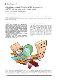
62 Cure of haemorroids following a TFS posterior sling<br />
and TFS perineal body repair - a case report<br />
Giancarlo Paradisi, Peter Petros<br />
aavis
Why?<br />
PSYLLOGEL<br />
fibra<br />
In case of...<br />
... Constipation,<br />
Hemorrhoids,<br />
Pelvic <strong>floor</strong> diseases.<br />
The most natural way to promote,<br />
restore and maintain regularity.<br />
Psyllium is the strongest natural dietary fiber for promoting<br />
regularity and supporting benefits overall health.<br />
Psyllium has low fermentation; this gel provides lubrication<br />
that facilitates propulsion of colon contents and<br />
produces a stool that is bulkier and more moist.<br />
AVAILABLE IN THE TASTES:<br />
Red orange Strawberry Lemon tea Cocoa Vanilla<br />
PSYLLOGEL ® fibra<br />
The leader in psyllium fiber 99% purity.<br />
Informations reserved to the doctors and pharmacists<br />
IN PHARMACY<br />
NATHURA SRL - Tel. +39 0522 865464 - www.nathura.com - nathura@nathura.com<br />
UNI EN ISO 9001:2008 QUALITY MANAGEMENT SYSTEM CERTIFIED BY CERTIQUALITY
Vol. 29<br />
N. 2<br />
June 2010<br />
Rivista Italiana di Colon-Proctologia<br />
Founded in 1982<br />
PELVIPERINEOLOGY<br />
A <strong>multidisciplinary</strong> <strong>pelvic</strong> <strong>floor</strong> <strong>journal</strong><br />
www.pelviperineology.org<br />
Editors<br />
Giuseppe Dodi, Colorectal Surgeon, Italy<br />
Bruce Farnsworth, Gynaecologist, Australia<br />
Associate Joint Managing Editor<br />
Florian Wagenlehner, Urologist, Germany<br />
Co-Editors<br />
Nucelio Lemos, Gynaecologist, Brazil<br />
Akin Sivaslioglu, Urogynecologist, Turkey<br />
Editorial Board<br />
Roberto Angioli, Gynaecologist, Italy<br />
Jacques Beco, Gynaecologist, Belgium<br />
Cornel Petre Bratila, Gynaecologist, Romania<br />
Daniele Grassi, Urologist, Italy<br />
Filippo LaTorre, Colorectal Surgeon, Italy<br />
Bernhard Liedl, Urologist, Germany<br />
Menahem Neuman, Urogynaecologist, Israel<br />
Oscar Contreras Ortiz, Gynaecologist, Argentina<br />
Paulo Palma, Urologist, Brazil<br />
Francesco Pesce, Urologist, Italy<br />
Peter Petros, Gynaecologist, Australia<br />
Richard Reid, Gynaecologist, Australia<br />
Giulio Santoro, Colorectal Surgeon, Italy<br />
Marco Soligo, Gynaecologist, Italy<br />
Jean Pierre Spinosa, Gynaecologist, Switzerland<br />
Michael Swash, Neurologist, UK<br />
Vincent Tse, Urologist, Australia<br />
Richard Villet, Urogynaecologist, France<br />
Pawel Wieczorek, Radiologist, Poland<br />
Rui Zhang, Urogynaecologist, P.R. China<br />
Carl Zimmerman, Gynaecologist, USA<br />
Official Journal of the: Australian Association of Vaginal and Incontinence Surgeons<br />
International Pelvic Floor Dysfunction Society<br />
Pelvic Reconstructive Surgery and Incontinence Association (Turkey)<br />
Perhimpunan Disfungsi Dasar Panggul Wanita Indonesia<br />
Romanian Uro-Gyn Society<br />
Editorial Office: Enrico Belluco, Maurizio Spella<br />
c/o Clinica Chirurgica 2 University of Padova, 35128, Padova, Italy<br />
e-mail: editor@pelviperineology.org<br />
Quarterly <strong>journal</strong> of scientific information registered at the Tribunale di Padova, Italy n. 741 dated 23-10-1982<br />
Editorial Director: Giuseppe Dodi<br />
Printer “Tipografia Veneta” Via E. Dalla Costa, 6 - 35129 Padova - e-mail: info@tipografiaveneta.it
The Organising Committee is pleased to invite you to attend the<br />
3 rd International <strong>Pelviperineology</strong> Congress<br />
12 th Annual AAVIS Scientific Meeting<br />
The Organising Committee is pleased to invite you to attend the<br />
19 th – 21 st September 2010 - Vienna Austria<br />
AAVIS Annual Scientific Meeting<br />
This year the meeting is to be held at Meridian hotel, Vienna, Austria<br />
Sunday 19 th September<br />
16.00 <strong>Pelviperineology</strong> Editorial Board meeting<br />
18.30 Welcome cocktail party<br />
Monday 20 th September<br />
08.30 WORKSHOP 1 ANATOMY<br />
08.30 WORKSHOP 2 ULTRASOUND<br />
10.00 Coffee break<br />
10.30 WORKSHOP 3 INTEGRAL THEORY<br />
13.00 Lunch<br />
PROVISIONAL<br />
• to be held<br />
PROGRAM<br />
in •<br />
Vienna, Austria<br />
19th-21st September 2010<br />
13.30 Coffee break<br />
16.00 MULTIDISCIPLINARY PLENARY SESSION 1<br />
Tuesday 21 st September<br />
This year the meeting is to be held at<br />
Meridian hotel, Vienna, 08.30 Austria LIVE SURGERY 1<br />
10.00 Coffee break<br />
10.30 LIVE SURGERY CONTINUES 2<br />
13.00 Lunch<br />
14.00 MULTIDISCIPLINARY PLENARY SESSION 2<br />
15.30 Coffee break<br />
Program highlights 16.00 include: MULTIDISCIPLINARY PLENARY SESSION 3<br />
14.00 SYMPOSIUM 1 FAECAL INCONTINENCE<br />
17.30 AAVIS Annual general meeting<br />
14.00 SYMPOSIUM 2 MALE INCONTINENCE Plenary sessions<br />
19.00 Conference dinner<br />
Expert panel discussions<br />
Specialised workshops and symposia<br />
Live surgery will include prolapse and<br />
incontinence procedures<br />
AAVIS is a <strong>multidisciplinary</strong> <strong>pelvic</strong> <strong>floor</strong> society founded in AUSTRALIA IN 1997.<br />
www.aavis.org<br />
For further information contact Bruce Farnsworth drbruce505@yahoo.com.au<br />
Australian Association Vaginal Incontinence Surgeons
Editorial<br />
bernhard liedl<br />
President ICOPF (International Collaboration of Pelvic Floor Surgeons)<br />
Almost all modern urogynecological surgery has been inspired by 2 major scientific discoveries. In the 1990s, an<br />
entirely new theory, the “Integral Theory”, by Petros (Australia) and Ulmsten (Sweden) 1,2 , proposed that bowel and<br />
bladder problems originate mainly from damaged vaginal ligaments, not from the bladder or bowel itself.<br />
The second discovery by Petros and Papadimitriou 4 , was a method for repairing these ligaments by creation of<br />
artificial ligaments. This had the effect of converting major operations with large incisions to relatively minor procedures<br />
performed through “keyhole incisions”.<br />
Application of these twin discoveries has revolutionized the treatment of stress incontinence, with more than 1,500,000<br />
operations performed world-wide to date. The experience of myself and other surgeons world-wide, confirms that this<br />
method can cure or improve many other conditions besides stress incontinence, for instance, prolapse, and symptoms<br />
such as urgency, nocturia, <strong>pelvic</strong> pain, bowel and bladder incontinence 5-7 .<br />
Many of these conditions were previously considered incurable.<br />
It takes many years for a radical change in thinking to become widely known, especially when it involves a whole<br />
new approach to diagnosis and treatment.<br />
This presentation, “A simplified clinical approach” has been especially prepared for those readers who are not<br />
familiar with the Integral Theory System. It begins with the basic anatomy of the structures. Then, step by step, the<br />
reader is introduced to the ligaments and other connective tissue structures, and their key role in <strong>pelvic</strong> <strong>floor</strong> function,<br />
dysfunction, diagnosis and surgery. A number of practical clinical examples outline in detail how to diagnose the site<br />
of connective tissue damage, and correction thereof, using the minimally invasive “keyhole” surgical procedures which<br />
derive from the Integral Theory System.<br />
Some original articles based on the Integral System follow: 1.) 3 year follow-up data on an RCT of 80 patients<br />
between the TFS midurethral sling and a TOT approach; 2) a case report of hemorrhoid cure following TFS sling<br />
applications to the uterosacral ligaments and perineal body for cure of large rectocoele; 3.) symptom cure of <strong>pelvic</strong> pain,<br />
nocturia, abnormal emptying, urgency and idiopathic fecal incontinence in 67 patients who had a posterior TFS sling;<br />
4.) an interesting challenge to the theory’s prediction that many symptoms of <strong>pelvic</strong> pain, vulvodynia, and interstitial<br />
cystitis may be referred pain from the uterosacral ligaments. Three patients with documented Interstitial Cystitis<br />
reported temporary disappearance of urethral and cervical tenderness, vulval hypersensitivity and lower abdominal pain<br />
following local anesthetic injection into the region of the uterosacral ligaments.<br />
It is hoped that these presentations will stimulate researchers to go one step beyond presentation of observational<br />
data, and to reference their findings against the Integral Theory System’s predictions, so as to test the Theory for truth<br />
or falsity.<br />
REFERENCES<br />
1. Petros P E, Ulmsten U, An Integral Theory of female urinary incontinence. Acta Obstetricia et Gynecologica Scandinavica,<br />
Supplement153, Vol 69, (1990), 1-79.<br />
2. Petros P E, Ulmsten U, An Integral Theory and its Method, for the Diagnosis and Management of female urinary incontinence,<br />
Scandinavian Journal of Urology and Nephrology (1993) - Vol 27 Supplement No 153 – 1-93.<br />
3. Petros P E, The International Continence Society and Integral Theory systems for management of the incontinent female -a comparative<br />
analysis. <strong>Pelviperineology</strong> 2007; 26: 25-29<br />
4. Petros P E, Ulmsten U, Papadimitriou J, The Autogenic Neoligament procedure: A technique for planned formation of an artificial neoligament.<br />
Acta Obstetricia et Gynecologica Scandinavica, Supplement 153, Vol 69, (1990), 43-51.<br />
5. Petros P E, New ambulatory surgical methods using an anatomical classification of urinary dysfunction improve stress, urge, and<br />
abnormal emptying, Int. J. Urogynecology (1997) vol 8, 5: 270-278.<br />
6. Abendstein B, Petros PE, Richardson PA Ligamentous repair using the Tissue Fixation System confirms a causal link between damaged<br />
suspensory ligaments and urinary and fecal incontinence, 2008, J. <strong>Pelviperineology</strong>, 27;114-117.<br />
7. Abendstein B, Brugger BA, Furtschegger A, Rieger M, Petros PE, . Role of the uterosacral ligaments in the causation of rectal<br />
intussusception, abnormal bowel emptying, and fecal incontinence-a prospective study, (2008), J. <strong>Pelviperineology</strong>, 27;118-121.<br />
Correspondence to:<br />
Bernhard Liedl<br />
Chefarzt Urogenitale Chirurgie<br />
Beckenbodenzentrum München, Denningerstr. 44, D-81679 München<br />
E-mail: liedl@bbzmuenchen.de<br />
Phone: +49-89-92794-1522<br />
35
TOUGH<br />
ON HAEMORRHOIDS<br />
GENTLE<br />
ON<br />
YOUR PATIENTS<br />
A.M.I. Recto Anal Repair (RAR®) combines<br />
HAL (Haemorrhoidal Artery Ligation)<br />
with a pexy for prolapsing mucosa in<br />
one procedure. The result is a safe and<br />
gentle treatment that’s effective even<br />
for higher grades of haemorrhoids. And<br />
thousands of satisfied patients to prove it.<br />
Take a look at www.ami.at<br />
or contact us at info@ami.at
Integral Theory<br />
The Integral Theory System.<br />
A simplified clinical approach with illustrative case histories<br />
PETER E. PAPA PETROS (1,2)<br />
(1)<br />
University of Western Australia<br />
Abstract: The integral theory: states that prolapse and <strong>pelvic</strong> <strong>floor</strong> symptoms such as urinary stress, urge, abnormal bowel and bladder emptying,<br />
and some forms of <strong>pelvic</strong> pain, mainly arise, for different reasons, from laxity in the vagina or its supporting ligaments, a result of altered<br />
connective tissue. Normal Function: The organs are suspended by ligaments against which muscles contract to open or close the outlet tubes,<br />
urethra and anus. These ligaments fall naturally into a 3 zone classification, anterior, middle, and posterior.<br />
Dysfunction: Damaged ligaments weaken the force of muscle contraction, causing prolapse and abnormal bladder and bowel symptoms<br />
Diagnosis: A pictorial diagnostic algorithm relates specific symptoms to damaged ligaments in each zone.<br />
Treatment: In mild cases, new <strong>pelvic</strong> <strong>floor</strong> muscle exercises based on a squatting principle strengthen the natural closure muscles and their<br />
ligamentous insertions, thereby improving the symptoms predicted by the Theory. With more severe cases, polypropylene tapes applied through<br />
“keyhole” incision using special instruments reinforce the damaged ligaments, restoring structure and function.<br />
Problems which can be potentially addressed by application of the Integral System: Urinary stress incontinence; Urinary urge incontinence;<br />
Abnormal bladder emptying; Faecal incontinence and “obstructed evacuation” (“constipation’); Pelvic pain, and some types of vulvodynia and<br />
interstitial cystitis; Organ prolapse.<br />
Key words: Integral Theory; diagnosis; minisling; ligaments; connective tissue; pictorial algorithm.<br />
INTRODUCTION<br />
The Integral Theory states that prolapse and most <strong>pelvic</strong><br />
<strong>floor</strong> symptoms such as urinary stress, urge, abnormal bowel<br />
and bladder emptying, and some forms of <strong>pelvic</strong> pain,<br />
mainly arise, for different reasons, from laxity in the vagina<br />
or its supporting ligaments, a result of altered connective<br />
tissue. 1-5 Birth related laxity, fig. 1, compounded by ageing,<br />
are the principal causes of ligament laxity.<br />
The Integral Theory has evolved into the Integral Theory<br />
System, which applies the damaged ligament theory to<br />
• Function – the role of competent suspensory ligaments in<br />
organ support and function.<br />
• Dysfunction-how damaged ligaments upset the<br />
musculoelastic control mechanism to cause prolapse and<br />
abnormal bowel and bladder symptoms.<br />
• Diagnosis- how to diagnose which damaged ligaments<br />
are causing which prolapse and which symptoms.<br />
• Treatment - in mild cases, new <strong>pelvic</strong> <strong>floor</strong> muscle exercises<br />
based on a squatting principle strengthen the natural<br />
closure muscles and their ligamentous insertions. With<br />
more severe cases, polypropylene tapes applied through<br />
“keyhole” incision using special instruments, reinforce the<br />
damaged ligaments, restoring structure and function.<br />
PART 1<br />
THE DYNAMIC ANATOMY OF NORMAL FUNCTION<br />
Bladder, bowel and uterus<br />
Fig. 2 is a schematic view of the bladder, bowel and<br />
uterus with the woman in a sitting position. The organs<br />
are storage containers. The bladder stores urine, the uterus<br />
the foetus, and the rectum faeces. Each organ is connected<br />
to the outside by a tube, the urethra, which is about 4cm<br />
long, vagina, which is 10-12cm long, and the anus, about 4<br />
cm long. The menstrual blood and foetus pass through the<br />
vagina. Urine and faeces pass through the urethra and anus.<br />
Muscles compress these tubes to close them, and stretch<br />
them open for emptying.<br />
The importance of suspensory ligaments<br />
“Problems of bladder, bowel, prolapse, and some types<br />
of <strong>pelvic</strong> pain, mainly originate from the vaginal ligaments,<br />
not from the organs themselves”- Integral Theory 1996.<br />
<strong>Pelviperineology</strong> 2010; 29: 37-51 http://www.pelviperineology.org<br />
Fig. 1 - Birth-related laxity The diagram shows the baby’s head<br />
severely stretching ligaments and other tissues in and outside the<br />
vagina. This may cause various degrees of looseness, prolapse of the<br />
bladder and bowel, and urine and bowel incontinence. Fundamental<br />
to any surgical treatment is the approximation of laterally displaced<br />
tissues, and the strengthening of damaged suspensory ligament(s).<br />
The bladder sits on top of the vagina, and is partly attached<br />
to it. Muscles pull against the ligaments to close or open the<br />
urethra. Therefore loose ligaments may weaken the muscle<br />
contraction to cause problems with closure (incontinence)<br />
Fig. 2 - The organs and their outlet tubes.<br />
37
P.E.P. Petros<br />
Fig. 3 - Unsuspended ligaments have no shape, strength or<br />
function.<br />
or opening (evacuation of urine). Figure 3 indicates what<br />
the vagina, bladder, and bowel would look like with no<br />
ligaments to suspend them, a blob of tissue, with no form,<br />
no structure, no strength, and no function.<br />
A ligament is like a thick cord in a suspension bridge, fig.<br />
4. In fact, the vagina is suspended exactly like a suspension<br />
bridge, with the ligaments above, and the muscles (arrows)<br />
below. The muscle forces (arrows) contract against the<br />
suspensory ligaments to give the bridge form and strength.<br />
The organs, fig. 5, are suspended from above by the<br />
vaginal ligaments - exactly like a suspension bridge. All<br />
the ligaments are attached to the vagina and/or uterus. The<br />
vagina supports the bladder situated above it, and the rectum<br />
situated below it, so anything which damages the vaginal<br />
structure can also affect the bladder and rectum.<br />
Separating the lower end of the vagina from the rectum<br />
is a solid mass of tissue, the perineal body (PB) complex<br />
which is about 4 cm long. If this is damaged, the rectum may<br />
bulge forwards into the vagina as a rectocoele.<br />
The uterus is an anchoring point for the ligaments -<br />
it needs to be preserved where possible<br />
The role of the uterus in maintaining the structure, fig. 6,<br />
and function of the <strong>pelvic</strong> <strong>floor</strong> is greatly underestimated.<br />
Some doctors routinely recommend removal of the uterus<br />
during surgery for prolapse. It is preferable to retain the<br />
uterus wherever possible, as many important ligaments<br />
are attached to it. During the menopause, the ovaries cease<br />
production of oestrogen. Since oestrogen is essential for<br />
maintaining the strength of the ligaments, the detrimental<br />
effects of hysterectomy on prolapse and incontinence become<br />
especially evident after the menopause. Hysterectomy<br />
reduces the blood supply to the cardinal and uterosacral<br />
ligaments, weakening them further. All these factors<br />
Fig. 5 - Four ligaments suspend the organs from above like a<br />
suspension bridge. The perineal body (PB) supports the organs<br />
from below. CL=cardinal ligament.<br />
predispose to prolapse, and development of posterior zone<br />
symptoms. 6<br />
The <strong>pelvic</strong> muscles (dark red), fig.7, have a dual function,<br />
organ support, and opening and opening and closure of<br />
urethra and anorectum. They extend from the coccyx to the<br />
pubic bone, and contract to support the vagina, bladder and<br />
bowel from below. The red arrows indicate the directions<br />
where the muscles contract, backwards to open these organs,<br />
forwards to close them.<br />
An external striated muscle opening and closure<br />
7&8, 9-17<br />
mechanism, fig 8.<br />
Put simplistically, when the muscles pull backwards<br />
(blue arrows), the urethra and anus are pulled open, vastly<br />
decreasing intracavity resistance to the 4 th power, so that the<br />
woman can quickly and easily evacuate her urine and faeces;<br />
when the muscles pull forwards* (black arrows), the urethra<br />
and anus are closed by a vast increase in resistance to the 4 th<br />
power. Normally all the organs, even the vagina, are kept in<br />
the closed position by slow-twitch muscle contraction.<br />
* The closure mechanism is a little more complex than that depicted<br />
in fig. 8, involving a distal and proximal mechanism for closure of the<br />
urethral and anal tubes. 7-10<br />
How damaged ligaments may cause incontinence or<br />
emptying disorders. We saw from the suspension bridge<br />
diagram, that the muscles pull against the ligaments.<br />
Fig. 4 - The vagina is suspended from above, like a suspension<br />
bridge, with the ligaments above, and the muscles (arrows) below.<br />
PS=pubic symphysis;S=sacrum; PUL=pubourethral ligament;<br />
ATFP=arcus tendineus fascia pelvis; USL=uterosacral ligament.<br />
Fig. 6 - The uterus functions like the keystone of an arch. Remove<br />
the arch, and the whole structure is put at risk of a downward<br />
collapse.<br />
38
The integral theory system<br />
Fig. 7 - The muscles support the organs, vagina, bladder, and bowel<br />
from below, and also, open and close them by 3 external directional<br />
muscle forces (arrows).<br />
So if the suspensory ligaments are loose, the muscle<br />
strength weakens, and may not be able to keep the bladder<br />
or bowel emptying tubes closed. As a consequence of<br />
this, a patient may feel a leakage of urine, wind or faeces,<br />
“incontinence”. Another related condition is failure to close<br />
the vaginal tube, so water may enter the vagina during<br />
swimming, or complain of vaginal flatus. If the damaged<br />
ligaments do not allow the muscles to open these same<br />
emptying tubes, a patient may have to strain to empty her<br />
bladder or bowel, “evacuation disorder” or “emptying<br />
disorder”.<br />
A Symphony Orchestra<br />
The vagina, bladder, bowel, muscles, and ligaments,<br />
fig. 9, are like instruments in an orchestra*. The brain is<br />
the conductor, and ensures that all the instruments work<br />
harmoniously to produce the right music. Every instrument<br />
in the orchestra has a specific task. Damage to even one<br />
instrument will affect the performance. The brain directs<br />
the orchestra to open the bladder (and bowel) or to close<br />
it. Depending on what structure is damaged, the bladder<br />
may not be able to close properly, and the patient leaks<br />
(“incontinence”); or she may not be able empty her bladder,<br />
or she may have both problems. Nerves at the base of the<br />
bladder (‘N’ in the diagram which follows) sense when the<br />
Fig. 9 - The cortex of the brain gives directions for closure (C), and<br />
opening (O). Nerves “N”, at the base of the bladder sense when<br />
the bladder is full, and send impulses to the brain. Depending on<br />
the situation, the brain sends directions either for closure (C), or<br />
opening (O). Like instructions from the orchestra conductor, these<br />
directions, “C” and “O”, engage all the muscles, nerves, ligaments<br />
and tissues required for each function. The Pons, a lower part of the<br />
brain, works as a co-ordinating station.<br />
bladder is full, and send impulses to the brain. These are<br />
perceived as urgency, a desire to go to the toilet.<br />
* I am grateful to Dr Alfons Gunnemann for the orchestra analogy<br />
The brain and its nerves- a sophisticated feedback system<br />
The brain works like the computer at a big telephone<br />
exchange. Think of the nerves as telephone wires going out<br />
to the bladder, the vagina and bowel. These organs have<br />
sensors which send signals posterior to the brain via another<br />
set of nerves, to inform it as to what is happening. The<br />
brain receives and processes these signals, and depending<br />
on what is required, sends out orders via a series of nerves.<br />
Most of this co-ordination occurs in “automatic mode”. The<br />
patient is not aware of what is happening. Sometimes, a<br />
patient may actually instruct the brain. For example, if it is<br />
inconvenient to empty the bladder or bowel, the muscles can<br />
be pulled upwards to close off the urethral and anal tubes.<br />
Pushing down assist emptying the urine and faeces. During<br />
intercourse the vagina can be narrowed by pulling the<br />
muscles upwards. This action grips the penis, and increases<br />
the sensation for both partners.<br />
PART 2<br />
DYSFUNCTION - the role of lax ligaments in<br />
the causation of symptoms and prolapse<br />
Fig. 8 - An external striated muscle opening and closure mechanism.<br />
The red lines represent the <strong>pelvic</strong> muscles. Fibromuscular extensions<br />
from these muscle fibres loop around the urethra and anorectum to<br />
activate closure and opening.<br />
The structure of ligaments<br />
A ligament is a complicated contractile structure which<br />
needs to be both elastic and strong, and have the ability to<br />
contract or relax according to whether the urethra and anus<br />
are being closed or opened. It relies on its collagen content<br />
for strength, elastin for flexibility, smooth muscle for<br />
contractility, and nerves to co-ordinate all these functions.<br />
39
P.E.P. Petros<br />
Fig. 13 - A rectocoele bulging out of the vagina during straining.<br />
Fig. 10 - The baby’s head (circles) may damage the ligaments and<br />
vaginal tissues to varying degrees as it descends through the vagina<br />
to cause stress incontinence’1’, cystocoele ‘2’, uterine/apical<br />
prolapse ‘3’, and rectocoele ‘4’. PUL=pubourethral ligament;<br />
ATFP=arcus tendineus fascia pelvis; USL=uterosacral ligament.<br />
Not shown are cardinal ligament (Middle Zone) and Perineal Body<br />
(Posterior zone).<br />
Fig. 14 - A uterus chronically bulging out of the vagina. The white<br />
areas are caused by chronic friction.<br />
Fig. 11 - This diagram illustrates the cystocoele “2”, rectocoele<br />
“4”and uterus”3”, all pushing into the vagina, as “lumps”, like<br />
a glove turning inside out. All are caused by looseness in the<br />
suspensory ligaments and their associated tissues.<br />
Fig. 12 - A cystocoele bulging out of the vaginal entrance during<br />
straining.<br />
Fig. 15 - Pictorial Diagnostic Algorithm. The anterior (pubourethral)<br />
and posterior (uterosacral) ligaments are in purple. The middle<br />
ligaments (ATFP& cardinal) are not shown in this diagram. There<br />
are 3 columns, one for each ligament group and the symptoms<br />
and prolapses (lumps) associated with damage to these ligaments.<br />
Labelling is ‘front’ and ‘back’ instead of ‘anterior’ and ‘posterior’.<br />
40
The integral theory system<br />
Collagen fibres work like the steel rods in cement. Single<br />
collagen fibres are “glued” together to give ligaments<br />
strength. The elastin content gives them elasticity. It is the<br />
change in collagen which is the ultimate cause of prolapse<br />
and incontinence.<br />
How ligaments are stretched and damaged during<br />
pregnancy and labour<br />
As we saw from the suspension bridge diagram, the<br />
muscles pull against the ligaments which support the bridge.<br />
If the ligaments are stretched and loosened during childbirth,<br />
as in fig. 1, a patient may develop a prolapse, a “dragging”<br />
pain low in the abdomen, bladder symptoms, for example<br />
urgency, frequency, nocturia, or even problems with bowel<br />
emptying or faecal incontinence.<br />
Commencing 6 months before childbirth, the “glue”<br />
between the collagen rods begins to soften in response to<br />
hormones from the placenta (“afterbirth”). This explains<br />
the onset of bladder, bowel, and pain symptoms at this<br />
time. Some 24-48 hours before delivery, however, this<br />
softening accelerates, and the collagen rods lose 95% of their<br />
strength. 18 During delivery, the baby’s head greatly stretches<br />
these collagen rods. Of course, the rods “re-glue” soon after<br />
delivery, but often they “re-glue” in a loose and extended<br />
position. Neither the ligaments nor the muscles can now<br />
work properly, and this may lead to prolapse of the uterus,<br />
cystocoele, rectocoele, and a wide range of bladder, bowel<br />
and <strong>pelvic</strong> pain symptoms (fig. 11). Women who have had<br />
Caesarian sections may also become incontinent, but they<br />
have less stretching, and therefore, fewer problems than<br />
vaginal delivery patients. Loose ligaments may occur in<br />
women who have never had children. Such women are born<br />
with loose ligaments, or they may have a congenital defect in<br />
their collagen. All these conditions are potentially curable by<br />
creation of artificial ligaments, as will be explained later.<br />
The effect of age and menopause<br />
Both collagen and elastin deteriorate markedly after the<br />
menopause, and this explains the vast increase in prolapse<br />
and incontinence which occurs after this event. A partly<br />
damaged ligament which is only just functioning before<br />
the menopause, may lose enough collagen after it, to<br />
“give way”. This looseness may result in organ prolapse,<br />
abnormal symptoms, or both. The oestrogen hormone<br />
replacement therapy (“HRT”) helps to slow down the<br />
degeneration of collagen. HRT can be applied vaginally, or<br />
by patches, creams, tablets, injections. Though oestrogens<br />
may be associated with an apparent increased risk of<br />
breast cancer, this has to be weighed against their benefits,<br />
prevention of osteoporosis, hip fractures, thickening of<br />
the vaginal wall in sexually active patients, even perhaps<br />
prevention of heart disease. Vaginally inserted oestrogen<br />
tablets are considered safe, as they act locally, and so are<br />
advantageous for every post-menopausal woman, even<br />
those of the most mature age.<br />
Site of damage and its consequences<br />
The circles in fig. 10 represent the baby’s head descending<br />
through the vagina during childbirth. This may stretch and<br />
loosen the ligaments (thick grey lines) in the 3 zones of the<br />
vagina. These are, Anterior Zone (meatus to bladder neck),<br />
Middle Zone, (bladder neck to cervix), and the Posterior<br />
Zone (cervix to perineal body). The numbers 1-4 indicate<br />
damage to specific ligaments which may cause<br />
1. Stress incontinence<br />
2. Cystocoele<br />
3. Prolapse of the uterus<br />
4. Rectocoele.<br />
All may occur to varying degrees in the same patient.<br />
It is evident that a head descending down a vulnerable<br />
vaginal canal, fig. 10, is unlikely to damage just one single<br />
structure. All structures will be damaged to a greater or<br />
lesser extent. This explains appearance of a cystocoele, for<br />
example, months or years after apparently successful surgery<br />
for prolapse of the uterus. Further prolapse can occur in<br />
perhaps 30-50% of cases after a successful vaginal repair.<br />
The problem is that once the vaginal tissues are damaged,<br />
it is difficult to fully repair them. It is like repairing frayed<br />
cloth. The surgeon repairs one area, only to see it give way<br />
in another area. That is why we have to create artificial<br />
ligaments by using tapes.<br />
A cystocoele “balloons” out from above, fig. 12. The cause<br />
is damage to the middle ligaments (ATFP and/or cardinal<br />
ligaments) and the anterior wall of the vagina.<br />
A rectocoele, fig. 13, balloons out from below. Separating<br />
the lower end of the vagina from the rectum is the perineal<br />
body, a major supporting structure, as it occupies 50% of<br />
the posterior vaginal wall. A rectocoele, may be caused<br />
by damage to the uterosacral ligaments (high rectocoele),<br />
and/or rectovaginal fascia & perineal bodies (mid & low<br />
rectocoele.<br />
Uterine prolapse, fig. 14, is caused by damage to the<br />
cardinal and uterosacral ligaments.<br />
A perspective on organ prolapse<br />
The organs bulge to varying degrees. Clearly a severe<br />
prolapse, such as the uterine prolapse above, requires<br />
treatment. If the bulge is minor, and there are no associated<br />
symptoms, there is no need for treatment. However, a patient<br />
may have severe symptoms which may require treatment,<br />
even though the prolapse is minor.<br />
When are these symptoms and prolapses problematical?<br />
If a patient answers “yes” to one of the following, she has<br />
a problem.<br />
1. You lose urine during exertion or coughing.<br />
2. You can‘t “hold on” and you wet yourself.<br />
3. You can’t empty your bladder properly.<br />
4. You have bowel soiling.<br />
5. You feel a lump in your vagina (prolapse)<br />
6. You have lower abdominal or <strong>pelvic</strong> pain.<br />
How serious is the problem?<br />
Assessment by the patient. This question “how serious is<br />
my problem” is not so easy to answer, as symptoms vary,<br />
and patients’ perceptions vary. A simple rule is to seek help<br />
if it is interfering with the patient’s quality of life. If the<br />
problem is mild and not bothersome, no action is required.<br />
Assessment by the clinic. The doctor has a different<br />
perspective, a) to assess which ligaments have been damaged,<br />
and b) to assess the seriousness of the problem. An accurate<br />
assessment is paramount. This will vary according to the<br />
clinic, but at a minimum, use of the Pictorial Diagnostic<br />
Algorithm, examination to assess ligamentous damage in<br />
the 3 zones, and “simulated operations”(e.g. midurethral<br />
anchoring during coughing in patients with urinary stress<br />
incontinence or urge symptoms).<br />
Symptoms – what they mean<br />
A symptom is a warning bell from the brain that something<br />
is wrong with some part of the body. As regards the <strong>pelvic</strong><br />
<strong>floor</strong>, many bladder and bowel symptoms are secondary to<br />
damage in one or more related ligaments, not from the organ<br />
itself. The challenge is to find which ligaments are causing<br />
the problem.<br />
41
P.E.P. Petros<br />
The Diagnostic Algorithm indicates which ligaments are<br />
causing symptoms and prolapse<br />
The Diagnostic Algorithm which follows is a simplified<br />
version of that published in the textbook, “The Female Pelvic<br />
Floor” 2 nd Edition (2006), Springer, Heidelberg. To use this<br />
diagram, a tick is placed in every column which describes a<br />
patient’s symptoms, and the diagram will indicate the zone<br />
of damage, anterior, middle, or posterior ligaments.<br />
How to use the Diagnostic Algorithm. Simply tick every<br />
column which describes a symptom. One needs to tick all<br />
the relevant columns for symptoms such as urgency and<br />
emptying, which may occur in more than one column. In<br />
such cases, other associated symptoms which are more<br />
specific, will help to guide the diagnosis.<br />
Definitions for Symptoms<br />
• Stress incontinence Urine loss on effort, such as coughing,<br />
exercise.<br />
• Abnormal emptying Inability to empty the bladder or<br />
abnormal flow.<br />
• Urgency An uncontrollable desire to pass urine.<br />
• Frequency Going more than 8 times a day to the toilet to<br />
pass urine.<br />
• Nocturia Getting up twice or more per night to pass urine.<br />
• Faecal incontinence Uncontrolled soiling from the<br />
bowel.<br />
• Obstructed defaecation or constipation Difficulty in<br />
emptying the bowel.<br />
• Pelvic pain. Pain in the lower abdomen, lower posterior,<br />
or during intercourse. Some types of vulvodynia and<br />
interstitial cystitis are often associated with <strong>pelvic</strong> pain.<br />
Symptoms occur in groups – an aid to diagnosis<br />
For example, urgency symptoms are indicated in all 3<br />
columns. Symptom grouping is the only way we can deduce<br />
which column (ligament) is causing the urgency. Fortunately,<br />
urgency almost always occurs in tandem with at least one<br />
other symptom.<br />
Characteristics of pain* caused by posterior<br />
ligament looseness<br />
• Low abdominal ‘dragging’ pain usually unilateral, often<br />
right-sided<br />
• Low sacral pain (pain near the tailbone)<br />
• Pain on deep penetration with intercourse<br />
• Low abdominal ache the next day after intercourse<br />
• Tiredness<br />
• Irritability<br />
• Pain worsens during the day, and is relieved by lying<br />
down<br />
• Pain is reproduced on pressing the cervix or the posterior<br />
wall of the vagina if a patient has had a hysterectomy.<br />
* There is growing evidence that some types of introital<br />
hypersensitivity (‘vulvodynia’) and perhaps even some types of<br />
bladder pain (‘interstitial cystitis) may be part of the posterior zone<br />
symptom complex in figure 15, nocturia, urgency and abnormal bladder<br />
emptying.<br />
Characteristics of ‘vulvodynia’<br />
A burning pain over the entrance of the vagina and<br />
anus, with extreme sensitive to touch. This condition is<br />
often associated with dragging lower abdominal pain and<br />
sometimes painful bladder conditions.<br />
Characteristics of bladder emptying difficulty<br />
Typical symptoms are a slow stream, starting and<br />
stopping, dribbling after micturition has been completed, a<br />
feeling that the bladder has not emptied. Often such patients<br />
have chronic urinary infections.<br />
Characteristics of faecal incontinence<br />
Typical symptoms, in order of severity, are uncontrolled<br />
wind loss, liquid soiling, solid faecal soiling. There are two<br />
main categories, patients with faecal incontinence caused by<br />
an anal sphincter torn at childbirth, and another where no<br />
obvious cause can be found. The anal sphincter constricts<br />
the lower part of the anus. It is what a patient feels when<br />
she contracts her muscles to delay bowel emptying. Where<br />
no obvious cause can be found, it is called “idiopathic<br />
incontinence”. It is “idiopathic incontinence” which is<br />
potentially curable by reconstructing the anterior or posterior<br />
ligaments.<br />
Characteristics of lumps (prolapse) in the vagina<br />
Initially, these only appear during straining. The three<br />
main causes of such ‘lumps’ are from the bladder (cystocoele<br />
“2”) uterus (“3”) and bowel (rectocoele “4”), fig. 15. These<br />
can only be accurately diagnosed by a vaginal examination,<br />
as not all lumps are accompanied by symptoms. Where<br />
symptoms accompany the prolapse, the symptoms may<br />
give an indication of where the problem is. For example if a<br />
patient has a lump plus nocturia, <strong>pelvic</strong> pain and urgency, it<br />
is highly likely that she has weak posterior ligaments, as per<br />
the Diagnostic Diagram, fig. 15.<br />
New time efficient <strong>pelvic</strong> <strong>floor</strong> exercises<br />
strengthen muscles and ligaments<br />
In 1995, we first conceived another approach to <strong>pelvic</strong><br />
<strong>floor</strong> exercises. We knew from our ultrasound studies that<br />
the traditional Kegel methods were NOT addressing the<br />
posterior closure muscles, which stretch, rotate, and close<br />
the proximal urethra against the pubourethral ligament.<br />
Our ultrasound studies had demonstrated that squatting<br />
exercises the very same muscles which close the urethra<br />
during coughing. We also reasoned that strengthening a<br />
<strong>pelvic</strong> muscle would also strengthen the ligament against<br />
which it contracted, and we knew from the surgery, that it<br />
was ligament weakness which was causing the incontinence<br />
problems.<br />
We therefore added squatting exercises to the traditional<br />
Kegel programme. Our target group of patients were those<br />
with symptoms which were bothersome, but not sufficiently<br />
to require surgery. The results were dramatic. This patient<br />
group reported a more than 60% improvement in such<br />
symptoms as urgency, nocturia, <strong>pelvic</strong> pain, and abnormal<br />
bladder emptying. The most interesting observation,<br />
however, was that those patients who were cured, did not<br />
need to remember to contract their <strong>pelvic</strong> <strong>floor</strong> in advance.<br />
They coughed, and did not leak. 19-21<br />
A major problem with <strong>pelvic</strong> <strong>floor</strong> exercises is that<br />
women with young families and jobs, simply do not have<br />
time to perform them regularly. Even with our highly<br />
motivated group, the dropout rate was 50%. Because of<br />
this, we concluded that the programme required re-analysis.<br />
We knew that the slow-twitch muscle fibres were the prime<br />
contributors to continence. Sitting on a “fitball” instead of<br />
a chair, is a very simple and effective exercise technique,<br />
as it requires a balanced upright position, with co-ordinated<br />
contraction of abdominal, back, and <strong>pelvic</strong> <strong>floor</strong> muscles.<br />
Unlike all traditional Kegel exercises which require attention<br />
and time, sitting on a “fitball” requires no extra time to be set<br />
aside during the day. We have found that the “fitball’ method<br />
applied alone was well accepted, and results seemed equally<br />
effective in the short-term.<br />
42
The integral theory system<br />
Fig. 16 - Sitting on a rubber “fitball” instead of a chair strengthen<br />
all the <strong>pelvic</strong> slow-twitch muscles, and their ligamentous<br />
attachments.<br />
Surgery based on the Integral Theory System<br />
“Tension-free” techniques Beginning in the late 1980s<br />
an entirely new surgical method for stress incontinence<br />
was introduced. Polypropylene tapes were placed around<br />
the middle part of the urethra (best known as the“TVT’<br />
operation) to reinforce the pubourethral ligaments, then a<br />
little later, the posterior ligaments (infracoccygeal sacropexy,<br />
“PIVS”). 5 This method, now known as the “tension-free<br />
tape” technique, has revolutionized the treatment of stress<br />
incontinence (SI) and prolapse surgery. The operations are<br />
conducted via 1cm incisions in the abdominal skin just above<br />
the pubic bone, groin or perineum. There is minimal pain,<br />
hospital stay is reduced to 1 or 2 days, and patients generally<br />
have few problems passing urine after the operation. The<br />
cure rate is high in the longer term. Later variations of these<br />
operations, include the transobturator (TOT) approach for<br />
SI (very successful), and the addition of mesh sheets to the<br />
TOT and PIVS techniques (not so successful). The only<br />
significant problem with all tape/mesh implant operations,<br />
was partial or total rejection of the tape/mesh. More recently,<br />
an even less invasive method, the “minisling” has been<br />
introduced to address incontinence and organ prolapse. 22<br />
Fig. 18 - An example of ‘next generation’nonstretch monofilament<br />
tape. Such tapes are purpose knitted, not cut from sheets, with finer<br />
fibrils, and less weight per unit area. They do not fragment, and are<br />
less likely to surface in the wound.<br />
Minislings- a new horizon for stress incontinence, and<br />
repair of cystocoele, rectocoele, and prolapse of the uterus.<br />
The TFS, fig 17, was the first minisling. It was applied in late<br />
2003 to a patient with stress incontinence and uterovaginal<br />
prolapse. Since 2006, there has been a profusion of other<br />
minislings introduced for cure of stress incontinence, for<br />
example, TVT-Secur, Mini Arc, Ophira, and many others.<br />
Because the TFS is a tensioned sling, it can also address<br />
not only prolapse, but also, many symptoms from the<br />
Pictorial Diagnostic Algorithm (fig. 15), including urgency,<br />
nocturia, abnormal emptying and <strong>pelvic</strong> pain.<br />
Like other minislings, the TFS uses only small sections of<br />
monofilament tape (fig. 18), so it causes less tissue irritation.<br />
It uses a bioengineering principle similar to that of a buttressed<br />
cathedral ceiling (fig. 19) for cystocoele and rectocoele repair.<br />
It avoids the spaces between rectum, bladder and vagina,<br />
and so it is not subject to the adhesive complications seen<br />
with large mesh. As with all polypropylene implantations,<br />
the main complication is rejection of the tape by the body’s<br />
immune mechanisms. However, this occurs only in a small<br />
percentage of patients, as only very small segments of tape<br />
are used, and the anchor prevents “slippage” into the wound,<br />
a major cause of erosion.<br />
Only a tensioned minisling can reliably improve symptoms<br />
Essential to cure of posterior zone symptoms with the<br />
posterior “tension-free” sling was restoration of tissue<br />
tension by approximation of laterally displaced tissues. 5<br />
With the infracoccygeal sacropexy (“posterior IVS”),<br />
this could only be done with a suture, which was neither<br />
sufficiently strong, nor reliable.<br />
The TFS minisling, 22-26 was designed to precisely<br />
reconstruct and tension the 5 main structures which support<br />
the organs, pubourethral, ATFP, cardinal, uterosacral<br />
ligaments, and perineal body, fig. 20, and to approximate<br />
laterally displaced tissues.<br />
PART 3<br />
ILLUSTRATIVE CASE HISTORIES<br />
The following illustrative case histories are taken from the<br />
files of the Kvinno Centre, Perth Western Australia, the first<br />
clinic in the world to apply the Integral Theory System.<br />
Fig. 17 - TFS applicator ‘Ap”. ‘A’ is the soft tissue anchor which<br />
sits on the saddle ‘S’. The tape ‘T’ is a ‘next generation” non-stretch<br />
macroporous monofilament polypropylene tape. The polypropylene<br />
tape passes through the unidirectional ‘trapdoor’ at the base of the<br />
anchor. This one-way system of tightening brings the laterally<br />
displaced ligaments and fascia towards the midline.<br />
ANTERIOR ZONE DAMAGE<br />
In this section, we give a series of typical case reports from<br />
patients who came to the Clinic with particular problems<br />
which mainly derive from front (pubourethral) ligament<br />
looseness, in particular, stress incontinence. We also discuss<br />
other less typical problems.<br />
43
P.E.P. Petros<br />
Fig. 19 - The TFS works like a buttressed cathedral ceiling<br />
structure. The pillars (bone) provide the anchoring point for the<br />
beams (tapes), which in turn provide sufficient support for the<br />
weaker plaster board (vagina). Like a wire suspension bridge,<br />
tensioned TFS tapes provide a much stronger support than meshes<br />
which have a tendency to sag.<br />
Stress incontinence (leaking during coughing) is the main<br />
symptom for front ligament looseness.<br />
Mrs CYL,was 55 years old, and she had had 3 normal<br />
deliveries. The ultrasound showed that her bladder and<br />
urethra became one large funnel when she coughed, and<br />
the urine just ran out. The Diagnostic Diagram confirmed<br />
that the damage was in the front ligament column, fig. 21.<br />
Maximal Urethral Closure Pressure was 16cm H2O.<br />
“Mixed” stress and urge incontinence from<br />
front ligament looseness<br />
Mrs JC, was a 38 year para 2. She had stress incontinence,<br />
and she also wet with urgency 2-3 times a day before she<br />
arrived at the toilet. She had been refused surgery for her<br />
stress incontinence because a urodynamic test had shown<br />
an “unstable bladder”. With reference to the Diagnostic<br />
Diagram, fig. 23, it was evident that she had specific middle<br />
or posterior zone symptoms, indicating that her pubourethral<br />
ligament (PUL) was probably causing both problems, stress<br />
and urge. This was confirmed with a “simulated operation”,<br />
gently pressing an instrument upwards on one side in the<br />
position of PUL at midurethra, just behind the pubic bone.<br />
This controlled her urine loss on coughing, and greatly<br />
diminished her urge symptoms. She was cured of both stress<br />
and urge with a polypropylene sling placed around the<br />
middle of her urethra to strengthen the front ligament.<br />
Fig. 21 - Anterior Zone defect The tick indicates only front ligament<br />
damage. A TFS polypropylene tape was inserted through a very<br />
small incision in the vagina to repair the front ligament, fig 22<br />
(23,24). The patient went home the next day entirely dry.<br />
Bedwetting from childhood caused by a lax front ligament<br />
Miss M was 25 years old, para 0. She had wet her bed<br />
as a child. Bedwetting cleared at puberty, but she still wet<br />
with coughing and exercise, and with urge. Ultrasound<br />
demonstrated bladder neck rotation, indicating that the<br />
front ligament (pubourethral) was loose. Her bladder<br />
symptoms were cured with a TFS sling which reinforced<br />
her pubourethral ligaments.<br />
Fig. 20 - The TFS minisling (Tissue Fixation System) a new<br />
approach to surgery for prolapse. It works by approximating<br />
laterally displaced tissues, and by reinforcing the 4 suspensory<br />
ligaments of the vagina, PUL (pubourethral), ATFP, cardinal (CL),<br />
uterosacral (USL) and also, the perineal body (PB).<br />
Fig. 22 - The “mini” or “micro” sling is inserted exclusively from<br />
the vagina. It avoids most complications of “tension-free” slings.<br />
44
The integral theory system<br />
Bedwetting from childhood and faecal incontinence<br />
caused by a lax front ligament<br />
In contrast, Miss G, 18 years old, had continued wetting<br />
wet her bed since childhood, and also had stress urinary<br />
and faecal incontinence. On examination, her urine loss<br />
was controlled by gentle pressure upwards in the vagina,<br />
applied just behind the pubic bone. Transperineal ultrasound<br />
demonstrated rotation of bladder neck, indicative of a<br />
loose anterior (pubourethral) ligament. At the 6 week postoperative<br />
visit, all symptoms were cured, and there was a<br />
remarkable transformation in the patient’s psychological<br />
state.<br />
Stress faecal incontinence caused by a lax front ligament<br />
Mrs T, 45years old, came to see us because she lost urine<br />
and solid faeces on coughing. Again, symptom grouping<br />
gave us the clue that her symptoms originated from<br />
pubourethral ligament damage. Her assessment indicated<br />
she had damaged front ligaments, which was successfully<br />
addressed with a polypropylene midurethral sling. 27<br />
Comment<br />
The bowel works in a similar way to the bladder.<br />
If a ligament is loose, the muscles which close the<br />
bowel cannot work properly, and the patient may<br />
leak wind, fluid or solid faeces<br />
MIDDLE ZONE DAMAGE (cystocoele)<br />
In this section, a series of typical case reports is presented<br />
from patients who came to our Clinic with particular<br />
problems which mainly derive from middle ligament<br />
Fig. 24 - Anterior Zone Defect Stress and faecal incontinence.<br />
Grouping of SI symptoms with FI, and absence of other posterior<br />
zone sympotms indicates anterior ligament damage.<br />
Fig. 23 - Anterior Zone Defect Stress and Urgency in Mrs JC’s case<br />
are both most likely caused by damage to the front ligament. Because<br />
urge symptoms may derive from all 3 zones, all 3 spaces are ticked.<br />
Diagnosis of anterior zone defect was made by deduction using the<br />
presence of SI and absence of other zone-specific symptoms.<br />
looseness. Mostly patients with a cystocoele, only complain<br />
of a “lump” in the vagina. However, they sometimes have<br />
symptoms of urgency and difficulty in emptying the bladder,<br />
and chronic bladder infections.<br />
Urge incontinence caused by cystocoele occurring<br />
after prolapse repair<br />
Mrs DV was 53 years old. She had had a successful repair<br />
of the uterosacral ligaments for prolapse of the uterus 12<br />
months earlier. She came to see us, stating that her symptoms<br />
had reappeared in the past few weeks.<br />
It was noted from Mrs DV’s questionnaire, that the nocturia<br />
and <strong>pelvic</strong> pain she had 12 months ago, remained cured, fig.<br />
25. When she was examined, it was evident that her posterior<br />
ligaments (uterosacrals)were intact. There was no prolapse<br />
of the uterus. However, a cystocoele was seen just inside the<br />
vaginal entrance on straining. Her urgency symptoms were<br />
relieved by gently supporting the cystocoele, indicating that<br />
this was the cause of her urge symptoms. The cystocoele<br />
was cured by TFS ATFP and cardinal ligament operation, fig<br />
26. Relief of urgency, and improved bladder emptying were<br />
reported immediately after the surgery. 22<br />
Comment Mrs DV is a good example of what<br />
happens in patients with damaged ligaments. In up<br />
to 30% of cases, repairing one part of the vagina can<br />
be followed by another lump or symptom appearing<br />
weeks, months or even years later.<br />
Recurrent or chronic cystitis – its relationship to<br />
abnormal emptying, cystocoele and prolapse of the uterus<br />
Whilst there are many causes of cystitis, this presentation<br />
concerns patients who have recurrent cystitis because they<br />
45
P.E.P. Petros<br />
Fig. 26 - The ceiling joist principle for cystocoele repair. Schematic<br />
view into the anterior wall of the vagina. The horizontal tape<br />
provides structural support to the proximal half of the anterior<br />
vaginal wall and recreates the cervical ring. The vertical U-sling<br />
joins with existing ATFP structures to provide structural support to<br />
the distal half of the vagina.<br />
Fig. 25 - Middle Zone Defect The ticks indicated a middle or<br />
posterior defect for Mrs DV. The presence of a cystocoele, absence<br />
of a prolapse of the uterus and its specific symptoms, nocturia and<br />
<strong>pelvic</strong> pain, indicated it was a middle ligament problem.<br />
cannot empty their bladder adequately due to damaged<br />
ligaments in the middle or posterior parts of the vagina.<br />
In the author’s experience, cystocoele, and prolapse of the<br />
uterus are major correctable causes of abnormal emptying<br />
and chronic bladder infections. 5<br />
Other causes of recurrent cystitis<br />
Anything which irritates the inside of the bladder, such<br />
as a polyp, a bladder stone, or penetration of a plastic<br />
mesh after a surgical procedure for incontinence can cause<br />
recurrent cystitis. The mesh can cause irritation per se, or<br />
become calcified into a stone. Inserting a cystoscope into the<br />
bladder is the best method for diagnosing such a problem.<br />
Other causes of abnormal emptying?<br />
Anything which interrupts the messages from the brain<br />
may cause this problem. One cause which is often stated is<br />
diabetes. However, in the author’s experience, many patients<br />
labelled as “diabetic neuropathy” in fact had damaged<br />
uterosacral ligaments which prevented the opening muscles<br />
from working properly. Such patients had accompanying<br />
symptoms such as nocturia, urgency and <strong>pelvic</strong> pain, as<br />
per the Pictorial Diagnostic Algorithm Diagram, fig. 15,<br />
and were able to be cured. A much rarer cause of abnormal<br />
emptying, multiple sclerosis, cannot be cured, and often<br />
requires intermittent self-catheterization.<br />
Severe wetting on getting out of bed in the morning caused<br />
by excessive scarring from previous surgery “tethered<br />
vagina” – a hitherto unrecognised problem<br />
Mrs EM, 68 years old, was referred with a history of<br />
worsening incontinence over the previous 2 years. She had<br />
had 4 previous operations for prolapse and incontinence<br />
some years earlier. She offered the cardinal symptom of this<br />
condition “ my bladder empties uncontrollably immediately<br />
my foot touches the <strong>floor</strong> on getting out of bed in the<br />
morning.” She also lost urine on standing up from a chair,<br />
or bending down.<br />
On examination, she did NOT lose urine during coughing,<br />
a common feature of this condition. The large amount of urine<br />
measured with a 24 hour pad test validated the seriousness<br />
of this lady’s problem. There was very little movement of<br />
her bladder neck during straining with ultrasound testing,<br />
consistent with the thick scarring observed in the bladder<br />
neck area of her vagina. This scarring immobilized the<br />
muscles and ligaments needed to close the urethra, hence<br />
the name, “tethered vagina”. A skin graft placed in this<br />
area, fig. 28, restored elasticity and vastly improved her<br />
incontinence.<br />
Comment The “tethered vagina” syndrome is still not<br />
a well-recognized condition. It is entirely iatrogenic,<br />
and is caused by excessive scarring from previous<br />
Fig. 27 - The diagram shows how a cystocoele “2”, droops<br />
downwards in a sac, preventing it from emptying. The urine pool<br />
gets infected over time, leading to chronic cystitis.<br />
46
The integral theory system<br />
Fig. 28 - Martius skin graft applied to the bladder neck area of the<br />
vagina subsequent to extensive freeing of the urethra and vagina<br />
from adhesions to the pubic bone and each other in a case of<br />
“tethered vagina syndrome”.<br />
surgeries. It is called the “tethered vagina syndrome”<br />
because dense scar tissue in the vagina “tethers”<br />
the muscles, and prevents them from closing the<br />
urethral tube. This condition was not previously<br />
recognized as originating from a scarred vagina. It<br />
was thought to originate from the bladder itself, and<br />
was (still is, by many!) treated with drugs, which, of<br />
course, cannot succeed, as the cause is mechanical.<br />
Treatment involves restoration of elasticity in the<br />
bladder neck area of vagina, using some sore of skin<br />
graft. Restoration of continence following skin graft<br />
surgery is the ultimate proof of the Integral Theory .<br />
POSTERIOR ZONE DEFECTS<br />
In this section, some typical case reports from patients<br />
are presented, from problems which mainly derive from<br />
posterior (uterosacral) ligament looseness.<br />
Structural and functional consequences of laxity<br />
in the uterosacral ligaments<br />
The problems associated with posterior (uterosacral)<br />
ligament damage are usually far more complicated and<br />
serious, than those seen with damaged anterior or middle<br />
ligaments. Sometimes patients complain of just a “lump”<br />
in the vagina without accompanying symptoms. However,<br />
symptoms such as <strong>pelvic</strong> pain, nocturia, and abnormal<br />
emptying are found as accompaniments to the prolapse.<br />
However, these symptoms may occur without significant<br />
prolapse.<br />
Laxity in the uterosacral ligaments associated with<br />
uterovaginal prolapse nocturia, urgency,<br />
abnormal emptying and <strong>pelvic</strong> pain<br />
Mrs LM, 53 years old, stated “ I get up 4-5 times a night.<br />
I find this very tiring, as I have to work next day. I have a<br />
dragging pain on the right side which can be quite distracting<br />
by the end of the day. I am always going to the toilet at<br />
work. My urine dribbles away after I stand up, and I often<br />
wet the toilet seat. I have problems with bladder infections”<br />
Mrs LM had symptoms typical of looseness in the<br />
posterior ligaments, fig. 29. When we examined her, we<br />
noted that she had significant prolapse of the uterus, but<br />
it was not protruding. A TFS “minisling” was inserted to<br />
reinforce the damaged uterosacral ligaments. The advantage<br />
of the TFS method is that it can precisely tighten the vaginal<br />
membrane to prevent sensitive nerve endings from firing off<br />
at a low bladder volume. It is a very minimal technique,<br />
and is performed entirely from the vagina. Mrs LM required<br />
only an overnight stay in hospital, and she returned to work<br />
in 7 days. When reviewed at 9 months, she was getting up<br />
only once per night to empty her bladder. She said that her<br />
low abdominal pain was still present, but was 90% better.<br />
Her bladder emptying also was not entirely cured, but had<br />
improved significantly, and she had not had any bladder<br />
infections since the operation.<br />
The posterior TFS “minisling” operation is performed<br />
entirely from the vagina, which makes it minimally invasive<br />
and less painful than other sling procedures which pierce the<br />
skin. It has a one-way tightening system, so it can restore<br />
the tension in the ligaments (white arrows). Adequate tissue<br />
tension is required to support the nerves which cause pain<br />
and urgency symptoms. Without restoring the tension, it is<br />
unlikely that such symptoms can be cured.<br />
Comment on the causation of urgency, nocturia and<br />
pain by damaged ligaments<br />
Strong uterosacral ligaments are required to support<br />
the pain fibres which run inside them, and to anchor<br />
Fig. 29 - Posterior Zone Defect Ticks in the posterior column are<br />
typical for symptoms from damaged uterosacral ligaments. Ticks<br />
are inserted in all column for urge and emptying. Grouping of<br />
symptoms indicates either a middle or posterior defect. Absence of<br />
a cystocoele on vaginal examination confirmed a posterior defect.<br />
47
P.E.P. Petros<br />
the muscle forces which stretch the vagina to<br />
support the nerves and volume receptors at bladder<br />
base. Loose tissues will not support the pain nerves,<br />
which “droop” and fire off, sending signals of pain to<br />
the brain. Like a trampoline with damaged springs,<br />
loose ligaments will not allow the muscles to stretch<br />
the vagina. The bladder nerves are unsupported, and<br />
fire off prematurely causing urge and frequency.<br />
Pain during intercourse and bowel problems<br />
caused by posterior ligament looseness.<br />
Mrs R M was a 47 year old para 2. She stated “I always<br />
have urgency to empty my bowel, but I am also frequently<br />
constipated. I get up 3-4 times a night to pass urine. I have<br />
problems emptying my bladder. My worst problem is<br />
that I can’t have sex any more. Almost every time I have<br />
intercourse, my bowels open.<br />
Reference to her Diagnostic Diagram, fig. 29, indicated<br />
that most of Mrs RM’s bladder problems problems may<br />
have been caused by damaged posterior ligaments. On<br />
examination, however, there was very minimal prolapse.<br />
This was consistent with the Theory, which states that major<br />
symptoms may be caused by minimal prolapse, and a high rate<br />
of improvement (up to 80-%) was possible with a posterior<br />
sling. No predictions were made for the urge to empty her<br />
bowel, and her constipation. She was advised these could be<br />
due to many other causes, so we were reluctant to predict<br />
cure for these symptoms. A posterior TFS “minisling” was<br />
inserted to repair the posterior ligaments. Mrs RM was<br />
discharged the next day with very little pain, and she went to<br />
work the following week. She attended with her husband for<br />
the post-operative visit. Smiling and confident, she reported<br />
cure of all her bowel symptoms, and a major improvement<br />
in her other symptoms.<br />
Comment on losing faeces during intercourse<br />
This patient was a challenge to us, as some of her<br />
symptoms were not the typical symptoms seen in the<br />
Diagnostic Diagram. We had encountered women<br />
opening their bladder during intercourse before, but<br />
never their bowel. In such cases, we rely on the other<br />
typical symptoms to guide us as to which ligaments<br />
have been damaged, and on the guiding principle of<br />
this type of surgery, “repair the structure, and you<br />
will repair the function”.<br />
Comment on pain with intercourse<br />
Earlier we discussed how a loose ligament will not<br />
Fig 30 - The TFS posterior minisling repairs and tightens the<br />
posterior ligaments (arrows) without penetrating the skin of the<br />
buttocks.<br />
support the pain fibres. As the penis thrusts into the<br />
posterior part of the vagina, it will cause pain if it<br />
stretches the unsupported nerve fibres.<br />
Severe Pelvic pain caused by uterosacral<br />
ligament looseness.<br />
Mrs D was a 34 year old para2. She attended with severe<br />
pain in the right side of her abdomen. Some years previously,<br />
she had previously attended a London hospital which had<br />
developed an international reputation using psychological<br />
tests to prove that such pain was psychological in origin.<br />
Mrs D had read widely on the subject of pain. Her facial<br />
expression indicated a person who was guarded. Her face lit<br />
up after she answered positively to the following questions,<br />
as she knew, that we knew, what her problem was.<br />
“Do you have pain on deep penetration with intercourse?<br />
“Do you get up more than twice per night to pass urine?<br />
“Do you have problems emptying your bladder?<br />
“Do you have urgency?”<br />
Positive answers to at least some symptoms other than pain<br />
are required before we can predict that the pain is caused by<br />
damage to the posterior ligaments. There are, after all, many<br />
other causes of chronic <strong>pelvic</strong> pain in the 30 plus age group,<br />
for example, endometriosis, infection in the Fallopian tubes,<br />
problems with large intestine, to name just a few.<br />
This is what she said one week after her pain was cured by<br />
a small operation which tightened her posterior ligaments. 28<br />
“I was almost suicidal after interminable attacks of pain<br />
on my right side. It has now been a week since the operation,<br />
and I feel like a rabbit that has been released from a trap.<br />
My mind keeps scanning up and down my body searching<br />
for the pain which for so long has been my centre and<br />
focus.”<br />
The operation, fig. 31, was simple, and it was performed<br />
entirely under local anaesthesia. A 3 cm incision was made<br />
in the vagina behind the cervix. Two sutures (green lines)<br />
were inserted to tighten the ligaments (white arrows).<br />
Comment<br />
This condition, severe <strong>pelvic</strong> pain caused by loose<br />
posterior ligaments, is still not well recognized by<br />
the majority of gynaecologists. The operation, fig.<br />
31, unfortunately has a significant recovery rate, as<br />
it approximates damaged tissue to damaged tissue.<br />
Insertion of a polypropylene sling gave a higher<br />
symptomatic cure rate, by creating a collagenous reinforcement,<br />
better able to support the unmyelinated<br />
nerve endings.<br />
Vulvodynia – pain and burning at the entrance to the<br />
vagina caused by posterior ligament looseness.<br />
Mrs P was 49 years old para3 with chronic <strong>pelvic</strong> pain<br />
diagnosed as having a psychological cause. Her General<br />
Practitioner, an empathetic and caring man rang the doctor<br />
before she arrived, and asked that we “handle her very<br />
carefully”, as she was severely disturbed psychologically,<br />
that this was the reason for her pain, and “there was nothing<br />
anyone could do for her”. The first impression of this<br />
lady did indeed fit the description of her GP. Her face was<br />
contorted, she spoke rapidly and with obvious anxiety. She<br />
had visited many specialists over the years for her pain.<br />
She had undergone several diagnostic laparoscopies, even<br />
a hysterectomy, and had attended a pain clinic. None of<br />
these treatments had helped her pain. The consensus from<br />
other specialists as reported to the GP, was that her problem<br />
was psychological. Her replies to the questionnaire gave the<br />
first hint that this woman may have a physical cause for her<br />
problem, damage to her posterior ligaments. She woke 6<br />
times per night to empty her bladder (nocturia), wore pads<br />
48
The integral theory system<br />
Fig. 31 - Approximation of uterosacral ligaments A small 3cm<br />
transverse incision into the vagina just below the cervix gave access<br />
for tightening her loose posterior ligaments (white arrows).<br />
continually as she wet 6 times per day, and had difficulties<br />
emptying her bladder. She also had faecal incontinence. We<br />
asked her if she had told her General Practitioner about her<br />
bladder and bowel problems. She said she had only consulted<br />
him about the burning pain around her vagina and anus. She<br />
said that her vagina was so tender, that she couldn’t have<br />
sexual intercourse, and sometimes had problems sitting.<br />
Examination revealed a prolapse of the posterior part of her<br />
vagina. The entrance to the vagina was hypersensitive- she<br />
recoiled when gently tested with a cotton bud, the classical<br />
test for “vulvodynia” (pain at the entrance of the vagina).<br />
We did not claim that we could cure this lady’s pain, as<br />
there are many other causes for <strong>pelvic</strong> pain. Nevertheless,<br />
it was explained that her vaginal prolapse needed to be<br />
fixed, and that there was a strong possibility that some of<br />
her symptoms would also improve with a sling inserted<br />
into the posterior part of her vagina, a fairly minor day-care<br />
29, 30<br />
procedure.<br />
The first thing we noticed at the 6 week post-operative<br />
visit was the absence of tension in her face. She was smiling<br />
and calm. Her pain was gone, as was her urgency and faecal<br />
incontinence. Her nocturia had reduced to 2 per night, and<br />
her bladder emptying was “60% improved”.<br />
A diagnosis that a patient’s pain is of psychological origin<br />
is not entirely unreasonable. Any type of chronic pain is<br />
sufficient to unsettle even the most rational person, and such<br />
patients do become “psychologically disturbed”. But this<br />
disturbance is usually secondary to the pain.<br />
Comment<br />
We do not claim that all vulvodynia patients have this<br />
causation. However, if other symptoms of posterior<br />
ligament looseness such as nocturia, abnormal<br />
bladder emptying, and urgency are grouped with the<br />
vulvodynia, there is a strong possibility that this pain<br />
can be improved in many patients with a posterior<br />
sling for repair of the posterior ligaments.<br />
Hysterectomy for lower posterior ache and <strong>pelvic</strong> pain<br />
caused by posterior ligament looseness.<br />
Mrs JMK developed chronic lower posterior pain and<br />
pain with intercourse after a difficult forceps delivery of<br />
her second child 50 years ago, when she was 27 years old.<br />
The pain worsened after the birth of her third child 3 years<br />
later. The pain was constant and debilitating, and she also<br />
had heavy menstrual bleeds. By the age of 35, the pain<br />
had worsened sufficiently to require consultation with a<br />
specialist gynaecologist. He told her that she needed to have<br />
a hysterectomy. By the time Mrs JMK was 65 years old, the<br />
chronic <strong>pelvic</strong> pain and low abdominal ache had returned.<br />
She had developed prolapse of the vagina and bladder, with<br />
significant bladder symptoms, urgency and nocturia. The<br />
return of symptoms can be attributed to age-related loss of<br />
collagen, and weakening of the posterior ligaments, a longterm<br />
problem in patients who have had hysterectomy.<br />
Abnormal emptying and chronic bladder infection caused<br />
by looseness in the posterior ligaments<br />
Mrs KB, a 32 year para 0 flight attendant, had a long<br />
history of inability to empty her bladder, and chronic bladder<br />
infections, dating back to her teenage years. She came to us<br />
because the infections were becoming more frequent, and<br />
were affecting her ability to work on long flights. Her situation<br />
had reached a stage where she felt forced to consider leaving<br />
her profession. She was diagnosed as having congenitally<br />
weak posterior ligaments. She did not respond to our <strong>pelvic</strong><br />
<strong>floor</strong> regime, and she requested surgical reconstruction<br />
of the ligaments. We agreed, having advised her that she<br />
may need a caesarian section if she fell pregnant, as any<br />
vaginal delivery could disrupt her operation. Her bladder<br />
returned to normal emptying immediately after the surgery,<br />
and she reported no further bladder infections even 10 years<br />
afterwards<br />
Comment on abnormal bladder emptying in the<br />
younger woman Congenitally weak posterior<br />
ligaments must always be considered as a cause of<br />
abnormal bladder emptying in the younger woman,<br />
as these women do not generally have a cystocoele.<br />
Increased difficulty in emptying the bladder at period<br />
time in such women is highly suggestive that the<br />
cause is looseness in the posterior ligaments. Other<br />
symptoms such as <strong>pelvic</strong> pain, urgency and nocturia<br />
are frequently present, and these may become<br />
worse during period time. Though not helpful with<br />
Mrs KB, good results in young women have been<br />
achieved at our Clinic by encouraging such patients<br />
to “squat” instead of bending, and to sit on a fitball<br />
at work instead of a chair. These exercises work by<br />
strengthening the <strong>pelvic</strong> muscles and ligaments.<br />
An 87 year old woman not able to pass urine, requiring self<br />
-catheterisation caused by posterior ligament looseness.<br />
There is a prevalence of this condition in Nursing Homes.<br />
Many patients require indwelling catheters.<br />
Mrs R was 87 years old, and weighed 90kg. She had had a<br />
hysterectomy 40 years earlier. For some years she needed to<br />
self-catheterize 3-4 times a day, as she could not pass urine<br />
adequately. She had large residual volumes (the amount<br />
retained in the bladder after passing urine). On testing, we<br />
confirmed she also had severe incontinence, with a large<br />
measured urine loss over a 24 hour period. She had 3 rd<br />
degree prolapse of the vagina. We inserted a posterior sling,<br />
performed a rectocoele repair and perineal body repair. She<br />
passed urine immediately after the surgery. Her nocturia,<br />
previously 5 times per night, reduced to twice per night.<br />
Comment on how age causes ligament looseness and<br />
bladder emptying difficulties<br />
The tissues of the vagina and its supporting ligaments<br />
may loosen considerably with age. The effect of<br />
this is that many older women, especially those in<br />
Nursing Homes, cannot empty their bladder, and<br />
they require indwelling catheters. These catheters are<br />
a major cause of chronic cystitis, as they introduce<br />
bacteria. We have returned many women to normal<br />
micturition by reconstructing the posterior ligaments<br />
and tightening the neighbouring tissues.<br />
49
P.E.P. Petros<br />
Faecal incontinence, “constipation” and bleeding<br />
caused by caused by posterior ligament looseness.<br />
Mrs DMV, 63 year old para4, complained of a lump in<br />
the vagina. She had a 2 nd degree prolapse of the uterus,<br />
urgency, and nocturia. She also had faecal incontinence,<br />
constipation, fig.32, and occasional bleeding from the<br />
bowel. She had been investigated for the bleeding by a<br />
specialist who found no evidence of bowel cancer. In the<br />
early days where the Diagnostic Algorithm was applied,<br />
we sometimes encountered patients with symptoms whose<br />
origin we did not fully understand. One example of this is<br />
the constipation, faecal incontinence and bleeding which<br />
had afflicted this lady. Faecal incontinence was not part of<br />
the Diagnostic Diagram at that time. In all such cases, we<br />
followed the principles of the Theory, “ repair the structure,<br />
and you will improve the symptoms”. We repaired the<br />
prolapse of the uterus by reinforcing the posterior ligaments<br />
with a polypropylene sling. Mrs DMV had a good result. Her<br />
faecal incontinence was cured, her constipation improved,<br />
31, 32<br />
and her bleeding disappeared.<br />
Explanation for Mrs DMV’s improvement in her<br />
“constipation” and bleeding. The reason for improvement<br />
in this lady’s “constipation” and bleeding was discovered<br />
some years later by an Austrian colleague, Dr Abendstein. 31<br />
With reference to the diagram fig. 32, it is evident that the<br />
uterus, vagina, and rectum (bowel) are suspended by the<br />
uterosacral ligaments, like the apex of a tent. If the guy rope<br />
of the tent (posterior ligament) is loose, the wall of the tent<br />
will sag inwards, in this case, the wall of the vagina and<br />
rectum. It is difficult to have a normal bowel motion when<br />
the bowel walls are loose and sagging into the cavity of the<br />
bowel. Furthermore, the sagging wall of the rectum (bowel)<br />
caused congestion of the veins close to the cavity similar to<br />
what happens with a haemorrhoid. This congestion caused<br />
bleeding into the bowel cavity from time to time.<br />
The tape lifted up and tightened the attachments of the<br />
uterus, vagina and rectum to the tail bone in the same way<br />
a guy rope does to the apex of a tent. This tightened the<br />
tissues, restored the function and improved her symptoms.<br />
Bowel emptying problems cured by repair of posterior<br />
ligaments and perineal body<br />
Mrs VCD, 46 years, presented with a rectocoele, faecal<br />
incontinence and difficulty with emptying her bowel, fig.<br />
32. She stated, “every time I need to open my bowels, I have<br />
to press my fingers into the posterior wall of my vagina so<br />
I can empty. I find the necessary hygiene after completion<br />
quite unpleasant.” In Mrs VCD’s case, we found that her<br />
perineal body had been stretched very thinly, and the muscle<br />
bellies of this structure had been pushed to the side, allowing<br />
the rectocoele to protrude into the vagina as a sac containing<br />
faeces. That is why she had to press into the lower part of<br />
her vagina to facilitate evacuation.<br />
Her posterior ligaments were repaired with a sling, and her<br />
perineal body was repaired in the traditional way, without<br />
a sling. The rectocoele and bowel evacuation difficulty<br />
were initially cured, but recurred within 6 months, as did<br />
requirement to assist evacuation by pressing her fingers into<br />
the posterior wall of her vagina. The faecal incontinence<br />
remained cured, and we attributed this to the continuing<br />
action of the posterior sling. The rectocoele and lax perineal<br />
body were repaired by the TFS (Tissue Fixation System).<br />
adjustable tape technique, fig. 33. Bowel evacuation<br />
returned to normal, and the rectocoele remained cured at her<br />
2year review.<br />
Comment Why a tape was necessary for repair of<br />
the perineal body There are really 2 perineal bodies<br />
joined together with a fibrous band. When these are<br />
Fig. 32 - Posterior Zone Defect Difficulty with bowel evacuation<br />
in a patient with a large rectocoele indicates perineal body damage<br />
added to posterior ligament damage. Both needed to be repaired.<br />
pushed aside by childbirth, the rectum protrudes into<br />
the vagina. The traditional method relies on suturing<br />
damaged tissue to damaged tissue under significant<br />
tension. This method is very painful, and is prone to<br />
recur. The TFS repair creates an artificial ligament to<br />
permanently join the laterally displaced bodies.<br />
Urinary urgency, <strong>pelvic</strong> pain and nocturia cured by Pelvic<br />
Floor Exercises<br />
Miss B, 31 years, gave this story. ‘I began to experience<br />
symptoms of urgency, <strong>pelvic</strong> pain and nocturia at the age<br />
of 25, sufficiently to seek medical advice. I saw 8 different<br />
specialists. I was given drugs to stop the bladder contracting.<br />
I spent a small fortune on herbal medicines. Nothing seemed<br />
to work”. Finally she contacted our Clinic through an<br />
intermediary. She worked overseas, and could not attend the<br />
Clinic for a formal assessment. We advised her how to use a<br />
large rubber fitball as a substitute for a chair, and to develop<br />
good <strong>pelvic</strong> <strong>floor</strong> habits such as squatting wherever possible,<br />
(instead of bending), erect posture, and exercise. The result<br />
was remarkable- virtually all her symptoms disappeared, and<br />
she remained cured at last contact 4 years later .<br />
Comment Non-surgical treatment of posterior<br />
ligament symptoms A vast improvement in symptoms<br />
such as <strong>pelvic</strong> pain, urgency, abnormal emptying<br />
and nocturia has been achieved in many patients<br />
attending our Clinic, by the regime utilized by Miss<br />
B, in particular, substituting a “fitball” for a chair,<br />
and squatting instead of bending. This regime is<br />
especially effective in the younger woman.<br />
50
The integral theory system<br />
Fig. 33 - Large rectocoele repair. Approximation of the perineal<br />
body (PB) and uterosacral ligaments (USL) by tightening the TFS,<br />
effectively blocks entry of the rectocoele into the vagina. A=anus;<br />
RVF=rectovaginal fascia; CL=cardinal ligament.<br />
Permission to publish<br />
The Editors grant permission for any individual, group or<br />
company to reproduce any text or document in this series<br />
of articles, with the sole condition that this article is quoted<br />
as a literature reference, and any diagram is appropriately<br />
acknowledged in the text.<br />
REFERENCES<br />
1. Petros P E, Ulmsten U. An Integral Theory of female urinary<br />
incontinence. Acta Obstetricia et Gynecologica Scandinavica,<br />
1990; Supplement 153, 69: 1-79.<br />
2. Petros P E, Ulmsten U. An Integral Theory and its Method, for<br />
the Diagnosis and Management of female urinary incontinence,<br />
Scandinavian Journal of Urology and Nephrology 1993; 27<br />
Supplement No 153 – 1-93.<br />
3. Petros P E, The International Continence Society and Integral<br />
Theory systems for management of the incontinent female -a<br />
comparative analysis. <strong>Pelviperineology</strong> 2007; 26: 25-29.<br />
4. Petros P E, Ulmsten U, Papadimitriou J. The Autogenic<br />
Neoligament procedure: A technique for planned formation of<br />
an artificial neo-ligament. Acta Obstetricia et Gynecologica<br />
Scandinavica. 199; Supplement 153, 69: 43-51.<br />
5. Petros P E, New ambulatory surgical methods using an anatomical<br />
classification of urinary dysfunction improve stress, urge, and<br />
abnormal emptying, Int. J. Urogynecology 1997; 8: 270-278.<br />
6. Petros P E, Influence of hysterectomy on <strong>pelvic</strong> <strong>floor</strong> dysfunction,<br />
Lancet 2000; 356: 1275.<br />
7. Petros P E, Ulmsten U. Role of the <strong>pelvic</strong> <strong>floor</strong> in bladder neck<br />
opening and closure: I muscle forces, Int. J. Urogynecol. and<br />
Pelvic Floor, 1997; l8: 74-80.<br />
8. Petros P E, Ulmsten U, Role of the <strong>pelvic</strong> <strong>floor</strong> in bladder neck<br />
opening and closure: II vagina. Int. J. Urogynecol. and Pelvic<br />
Floor, 1997; 8: 69-73.<br />
9. Petros PE, Swash M. The Musculoelastic Theory of anorectal<br />
function and dysfunction. <strong>Pelviperineology</strong>, 2008; 27: 89-93.<br />
10. Petros PE, Swash M. Directional muscle forces activate anorectal<br />
continence and defecation in the female, <strong>Pelviperineology</strong> 2008;<br />
27: 94-97.<br />
11. Petros PE, Swash M A. Direct test for the role of the pubourethral<br />
ligament in anorectal closure. <strong>Pelviperineology</strong> 2008; 27: 98.<br />
12. Petros PE, Swash M. Reflex contraction of the levator plate<br />
increases intra-anal pressure, validating its role in continence.<br />
<strong>Pelviperineology</strong> 2008; 27: 99.<br />
13. Petros PE, Swash M. Abdominal pressure increase during<br />
anorectal closure is secondary to striated <strong>pelvic</strong> muscle<br />
contraction. <strong>Pelviperineology</strong>, 2008; 27:100-101.<br />
14. Petros PE, Swash M. A prospective endoanal ultrasound study<br />
suggests that internal anal sphincter damage is unlikely to be a<br />
major cause of fecal incontinence. <strong>Pelviperineology</strong> 2008; 27:<br />
102.<br />
15. Petros PE, Swash M. Correction of abnormal geometry and<br />
dysfunction by suspensory ligament reconstruction gives insights<br />
into mechanisms for anorectal angle formation. <strong>Pelviperineology</strong><br />
2008; 27:103-104.<br />
16. Petros PE, Swash M. Role of puborectalis muscle in anal<br />
continence- comments on original 4D <strong>pelvic</strong> ultrasound data<br />
from Chantarasorn & Dietz. <strong>Pelviperineology</strong>, 2008; 27;105.<br />
17. Petros PE, Swash M, Kakulas B. Stress urinary incontinence<br />
results from muscle weakness and ligamentous laxity in the<br />
<strong>pelvic</strong> <strong>floor</strong>. <strong>Pelviperineology</strong> 2008; 27:107-109.<br />
18. Rechberger T, Uldbjerg N & Oxlund H. Connective tissue<br />
changes in the cervix during normal pregnancy and pregnancy<br />
complicated by a cervical incompetence Obstets & Gynecol.<br />
1988; 71:563-567.<br />
19. Petros PE and Skilling PM. Pelvic <strong>floor</strong> rehabilitation according<br />
to the Integral Theory of Female Urinary Incontinence- First<br />
report, European J Obstetric & Gynecology and Reproductive<br />
Biology 2001; 94: 264-269.<br />
20. Petros PE and Skilling PM. The physiological basis of <strong>pelvic</strong><br />
<strong>floor</strong> exercises in the treatment of stress urinary incontinence. Br<br />
J Obstet Gynaecol 1999;106 :615-616.<br />
21. Skilling PM, Petros PE. Synergistic non-surgical management of<br />
<strong>pelvic</strong> <strong>floor</strong> dysfunction: second report. Int J Urogyne 2004; 15:<br />
106-110.<br />
22. Abendstein B, Petros PE, Richardson PA. Ligamentous repair<br />
using the Tissue Fixation System confirms a causal link between<br />
damaged suspensory ligaments and urinary and fecal incontinence.<br />
<strong>Pelviperineology</strong> 2008; 27: 114-117.<br />
23. Sivaslioglu AA, Unlubilgin E, Aydogmus S, Celen E, Dolen<br />
I. A Prospective Randomized Comparison of Transobturator<br />
Tape and Tissue Fixation System Minisling in the Treatment of<br />
Female Stress Urinary Incontinence: 3 Year Results, 2010, J.<br />
<strong>Pelviperineology</strong> (in press).<br />
24. Sekiguchi Y, Kinjyo M, Inoue H, Sakata H, and Kubota<br />
Y. Outpatient mid urethral tissue fixation system sling for<br />
urodynamic stress urinary incontinence, The Journal of Urology<br />
2009; 182:2810-3.<br />
25. Petros PE Richardson PA. A 3 year follow-up review of uterine/<br />
vault prolapse repair using the TFS minisling. ANZJOG 2009;<br />
49: 439-440.<br />
26. Inoue H, Sekiguchi Y, Kohata Y, SatonoY, HishikawaK,<br />
TominagaT, and Oobayashi M. Tissue Fixation System (TFS)<br />
to repair uterovaginal prolapse with uterine preservation: A<br />
preliminary report on perioperative complications and safety J.<br />
Obstet. Gynaecol. Res. 2009; 35: 346–353.<br />
27. Hocking I. Experimental Study No. 9: Double incontinence,<br />
urinary and fecal, cured by surgical reinforcement of the<br />
pubourethral ligaments. <strong>Pelviperineology</strong> 2008; 27:110.<br />
28. Petros PE. Severe chronic <strong>pelvic</strong> pain in women may be caused<br />
by ligamentous laxity in the posterior fornix of the vagina, Aust<br />
NZ J Obstet Gynaecol. 1996; 36: 351-354.<br />
29. Petros PE and Bornstein J. Vulvar vestibulitis may be a<br />
referred pain arising from laxity in the uterosacral ligamentsa<br />
hypothesis based on 3 prospective case reports. ANZJO&G,<br />
2004; 44: 483–486.<br />
30. Bornstein J, Zarfati, D, Petros PEP. Causation of vulvar vestibulitis<br />
ANZJOG 2005; 45: 538–541.<br />
31. Abendstein B, Brugger BA, Furtschegger A, Rieger M, Petros<br />
PE, Role of the uterosacral ligaments in the causation of rectal<br />
intussusception, abnormal bowel emptying, and fecal incontinencea<br />
prospective study. <strong>Pelviperineology</strong> 2008; 27;118-121.<br />
32. Petros PE, Richardson PA. Fecal incontinence cure by surgical<br />
reinforcement of the <strong>pelvic</strong> ligaments suggests a connective<br />
tissue aetiology. <strong>Pelviperineology</strong> 2008; 27: 111-113.<br />
Correspondence to:<br />
Professor Peter Petros<br />
14A Osborne Pde - Claremont - WA 6010 - Australia<br />
Tel No : M 61-411 181 731<br />
Fax : 61-89-384 0176<br />
Email: kvinno@highway1.com.au<br />
51
Original article<br />
TFS posterior sling improves overactive bladder, <strong>pelvic</strong> pain<br />
and abnormal emptying, even with minor prolapse.<br />
A prospective urodynamic study<br />
PETER PETROS (1) , PETER a. RICHARDSON (2)<br />
(1)<br />
University of Western Australia<br />
(2)<br />
Gallier’s Hospital, Armadale, Western Australia<br />
Abstract: Objective To prospectively challenge the Integral Theory’s predictions that female patients with even with minor degrees of<br />
prolapse, and who have a symptomatic overactive bladder (urgency, nocturia and frequency), are surgically curable with uterosacral ligament<br />
repair. Methods The study group comprised 67 patients with >2nd degree prolapse, with a subgroup of 27 symptomatic patients with<br />
only 1st degree prolapse. All patients were assessed pre and post-operatively by questionnaire, 24-hr urinary diary, transperineal ultrasound,<br />
urodynamics, cough stress and 24-hr pad test. Uterine/apical prolapse were repaired as a day case using the TFS. The TFS (‘Tissue Fixation<br />
System”) comprises an adjustable polypropylene ‘mini sling’ applied entirely per vagina, and which can be accurately tightened to reinforce<br />
loose or damaged <strong>pelvic</strong> ligaments. Results: At median 9 months’ review, the percentage improvement rate in pre-operative dysfunctions<br />
for the total group is expressed as a percentage, with 1st degree in parenthesis: urinary frequency>10/day 63% (63%); nocturia >2/night 83%<br />
(76%); urge-incontinence >2/day 78% (76%); abnormal emptying, 73% (73%); <strong>pelvic</strong> pain, 86%(82%) fecal incontinence, 87% (100%).<br />
Conclusions: The posterior TFS sling is a promising minisling technique which appears to address symptoms such as urgency, nocturia,<br />
frequency, abnormal bladder emptying, and fecal incontinence, even in patients with minimal uterovaginal prolapse.<br />
Key words: TFS; Integral Theory; urge incontinence; overactive bladder; nocturia; <strong>pelvic</strong> pain; fecal incontinence.<br />
INTRODUCTION<br />
In the 1993 publication of the Integral Theory of<br />
Female Urinary Incontinence, the “posterior fornix<br />
syndrome”(posterior zone, figure 1) was described. It<br />
comprised symptoms of frequency, urgency, nocturia,<br />
<strong>pelvic</strong> pain, and abnormal bladder emptying. 1 Causation of<br />
this grouping of symptoms was attributed to laxity in the<br />
uterosacral ligaments (USL). According to this Theory, 1<br />
the anatomical rationale for symptom causation was that<br />
lax posterior ligaments prevented the posterior muscle<br />
forces from stretching the vaginal membrane to support the<br />
bladder base stretch receptors, “trampoline analogy”, fig. 1.<br />
Fundamental to this mechanism is the Theory’s interpretation<br />
of “Overactive Bladder” (OAB) as a prematurely<br />
activated, but otherwise normal micturition reflex. This<br />
was urodynamically demonstrated in 1993. 2 Significant<br />
improvement in such “posterior fornix” symptoms in patients<br />
with 2nd degree or greater prolapse following reinforcement<br />
of USLs with a posterior sling operation has been reported<br />
previously. 3,4 More recently, a less invasive posterior sling<br />
using the Tissue Fixation System (TFS) was described, 5<br />
fig. 2. The TFS allows precise tensioning of ligaments and<br />
fascia, consistent with the trampoline concept, fig. 1. There<br />
were 2 aims to this study. The primary aim was to challenge<br />
the theory’s prediction 1 that even a 1 st degree prolapse could<br />
cause major symptoms of urgency, nocturia and abnormal<br />
emptying. Twenty-eight patients fitted this description. A<br />
secondary aim was to test the efficacy of the new TFS sling<br />
in curing these conditions.<br />
PATIENTS AND METHODS<br />
Ethics Committee and Dept of Surgery Royal Perth<br />
Hospital approvals were obtained prior to the study.<br />
Pre-operative assessment<br />
All patients completed a 24 hour urinary diary, and a selfadministered<br />
questionnaire which was completed at home<br />
in the patient’s own time. 6 A cough stress test, and 24 hour<br />
pad test 6 were also administered. Symptoms derived from<br />
the questionnaire were ticked off in a diagnostic algorithm,<br />
52<br />
Fig. 1. – The Trampoline Analogy for OAB. Schematic<br />
representation. Muscle forces contract against the suspensory<br />
ligaments (“trampoline springs”) to stretch the vaginal membrane.<br />
Even damage to one ligament may prevent the trampoline membrane<br />
from stretching. A lax vaginal membrane may not be able to<br />
support sensitive stretch receptors “N”, so that these activate the<br />
micturition reflex prematurely. The patient senses this as urgency<br />
and frequency, and at night, nocturia. Minor damage may cause<br />
urgency, as this symptom is neurologically determined. Uterosacral<br />
ligaments (USL); pubourethral ligaments (PUL): Arcus Tendineus<br />
Fascia Pelvis (ATFP).<br />
indicating the zone of connective tissue damage, anterior,<br />
middle, posterior. In this classification, 7 the anterior zone<br />
extends from the external urethral meatus to bladder neck, the<br />
middle zone 4 from bladder neck to cervix or hysterectomy<br />
scar, and the posterior zone from apex to perineal body.<br />
Only patients with posterior zone defects were included in<br />
the study.<br />
Inclusion/exclusion criteria<br />
There were no exclusion criteria. The principal inclusion<br />
criterion for surgery was patients with 2 or more symptoms<br />
<strong>Pelviperineology</strong> 2010; 29: 52-55 http://www.pelviperineology.org
TFS posterior sling<br />
Fig. 2. – The Tissue Fixation System – a tensioned minisling. This is<br />
a photograph of the TFS anchor “A”, which sits in a saddle “S”, on<br />
an applicator “Ap”. T is a monofilament macropore polypropylene<br />
tape.<br />
of urgency, frequency, nocturia, abnormal emptying or<br />
<strong>pelvic</strong> pain. Of the 67 patients who underwent surgery,<br />
28 had only 1 st degree prolapse, plus symptoms such as<br />
urgency, nocturia or <strong>pelvic</strong> pain. Without symptoms, none<br />
of these patients would normally have been considered for<br />
surgery. The criteria for symptomatic improvement postoperatively<br />
are detailed below: nocturia: change in patients<br />
having >2 episodes per night; abnormal emptying: selfassessed<br />
improvement >80%, using a 0-100 scale expressed<br />
as a percentage; urge incontinence: change in the number<br />
of times a patient wet per day (threshold 2/day); frequency:<br />
change in patients having >10 episodes per day; <strong>pelvic</strong> pain<br />
and fecal incontinence: self-assessed improvement >85%,<br />
using a 0-100 scale expressed as a percentage; otherwise<br />
objective measures such as pad weights and residual urine<br />
measured by catheter were used.<br />
Fig. 3. – Posterior TFS sling. Perspective: View from above. The TFS<br />
anchors were inserted into the Uterosacral Ligaments (USL), and<br />
tightened to reduce apical or uterine prolapse. The arrows indicate<br />
that the remnants of USL and also the cardinal ligaments (CL) need<br />
to be approximated if the apex is wide, so as to prevent enterocele<br />
formation. Figure from <strong>Pelviperineology</strong>, by permission.<br />
Fig. 4. – In its simplest form, anorectal closure occurs when the<br />
backward muscle forces LP (levator plate) and LMA (longitudinal<br />
muscle of the anus 15 stretch the rectum like a rubber tube, around an<br />
anus anchored by m.puborectalis (PRM) contraction. On relaxation<br />
of PRM, LP/LMA vectors open out the anal canal for evacuation to<br />
occur (broken lines). The vector forces, LP/LMA contract against<br />
the uterosacral ligaments (USL). If USLs are loose, the closure<br />
mechanism is invalidated, and fecal incontinence may result.<br />
Figure from <strong>Pelviperineology</strong>, by permission.<br />
Objective assessment<br />
Vaginal examination, 7 involved inspection for damage to<br />
perineal body, posterior vaginal wall (rectovaginal fascia),<br />
and for the degree of prolapse of the vaginal vault or uterus.<br />
Transperineal ultrasound was performed by PP with a<br />
Toshiba 3.5 Mhz curvilinear probe to check organ position at<br />
rest and straining. Urodynamic testing was performed with a<br />
Neomedix System using Gaeltec microtransducers to assess<br />
for maximal urethral closure pressure, flow, residual urine,<br />
and ‘overactive bladder’. The patients were monitored at 6<br />
weeks, and at 3 monthly intervals thereafter using a 24 hour<br />
urinary diary, structured self-administered questionnaire,<br />
cough stress test, 24 hour pad test, transperineal ultrasound,<br />
and urodynamic investigation. Residual urine was assessed<br />
by catheter after the patient had voided.<br />
Surgical technique<br />
The posterior TFS sling consists of two polypropylene<br />
soft tissue anchors through which is inserted an adjustable<br />
polypropylene tape, fig. 2. A transverse full thickness<br />
vaginal incision was made 1cm below the hysterectomy<br />
scar, or cervical ring. The uterosacral ligament was<br />
identified with Allis forceps. A 4-5 cm channel was<br />
dissected immediately lateral to the uterosacral ligaments.<br />
Using a special applicator, the TFS anchors were inserted<br />
into the uterosacral ligaments, fig. 3. The tape was tightened<br />
via a one-way system at the base to reduce apical or uterine<br />
prolapse, figure 3. Coexisting rectocele was repaired in 5 in<br />
18 patients without vaginal tissue excision.<br />
The halfway classification system of Baden & Walker<br />
was used to classify apical or uterine prolapse: first degree<br />
descent to halfway along the posterior vaginal wall (n = 28),<br />
second degree almost to the introitus (n = 20), third degree<br />
to 2 cm beyond the introitus (n = 17), and fourth degree total<br />
eversion (n = 2).<br />
RESULTS<br />
A total of 67 posterior TFS operations were performed.<br />
The median age of the women was 65 years (range 35–87).<br />
Median parity was 3 (range 0–8), and median weight was<br />
71 kg (range 38–117 kg). Of the 67 patients, 28 had only 1 st<br />
degree prolapse, and 39 had 2 nd degree or greater prolapse.<br />
53
P. Petros - P.A. Richardson<br />
Table 1. – Sympton Outcome - 67 patients.<br />
Symptom change with surgery % cure in brackets<br />
Frequency Nocturia Urge Abnormal Pelvic<br />
Fecal >10/Day >2/night incontinence emptying pain<br />
Incontinence<br />
>2/Day<br />
n=23 n=27 n=47 n=36 n=53 n=46<br />
(87%) (63%) (83%) (78%) (73%) (86%)<br />
P
TFS posterior sling<br />
Fecal incontinence<br />
Utlilizing pre and postoperative evacuating proctograms,<br />
Abendstein 12 reported cure of fecal incontinence, obstructive<br />
defecation, <strong>pelvic</strong> pain, and xray proven anterior rectal wall<br />
intussusception. Abendstein proposed that the uterosacral<br />
ligament supports the anterior rectal wall much like the<br />
apex of a tent. The mechanism proposed for cure of fecal<br />
incontinence also involves the uterosacral ligaments. 13 The<br />
longitudinal muscle of the anus (LMA) and levator plate<br />
(LP) vectors stretch the rectum around an anus anchored<br />
by forward contraction of the puborectalis muscle (PRM),<br />
figure 4. Laxity in the uterosacral ligaments (USL), the<br />
anchoring point of LMA, may inactivate this closure<br />
mechanism causing fecal incontinence. 13 In a study similar to<br />
that of Abendstein, it was demonstrated that in patients with<br />
fecal incontinence, abnormal anorectal pressures, delayed<br />
pudendal nerve conduction times, and thinned internal anal<br />
sphincters had no negative impact on the results of surgery. 14<br />
Fecal incontinence was cured in 83% of patients by repair of<br />
the uterosacral and/or pubourethral ligaments.<br />
CONCLUSIONS<br />
The posterior TFS sling is a promising minisling<br />
technique which appears to address symptoms such as<br />
urgency, nocturia, frequency, abnormal emptying, and fecal<br />
incontinence, even in patients with minimal uterovaginal<br />
prolapse.<br />
REFERENCES<br />
1. Petros PE, Ulmsten U. An Integral Theory and its Method for<br />
Diagnosis and Management of Female Urinary Incontinence.<br />
Scand J Urol Nephrol. 1993; 27 (Suppl. No 153, 1-93.<br />
2. Petros PE & Ulmsten U. Bladder instability in women: A<br />
premature activation of the micturition reflex. Neurourology<br />
and Urodynamics 1993;12: 235-239.<br />
3. Petros PE. New ambulatory surgical methods using an<br />
anatomical classification of urinary dysfunction improve stress,<br />
urge and abnormal emptying. Int Urogynecol J. 1997; 8:270-7.<br />
4. Farnsworth BN. Posterior intravaginal slingplasty<br />
(infracoccygeal sacropexy) for severe posthysterectomy vaginal<br />
vault prolapse, a preliminary report on efficacy and safety. Int<br />
Urogynecol J. 2002; 13:4-8.<br />
5.- Petros PEP, Richardson PA, The midurethral TFS sling- a<br />
‘micro-method’ for cure of stress incontinence- preliminary<br />
report AANZJOG; 2005; 45: 372–375.<br />
6. Petros PEP, Appendix , in The Female Pelvic Floor, Function,<br />
Dysfunction and Management, according to the Integral Theory,<br />
2nd edition, Springer Heidelberg: 2006; 194-199.<br />
7. Petros PEP, Ch 2-4, Dynamic Anatomy, Diagnosis of Connective<br />
Tissue damage, Surgery, in The Female Pelvic Floor, Function,<br />
Dysfunction and Management, according to the Integral Theory,<br />
2nd edition, Springer Heidelberg. 2006; 48-138.<br />
8. Abrams P, Blaivas J, Stanton S and Andersen J. Standardisation<br />
of Terminology of Lower Urinary Tract Function. Scand J. Urol<br />
Nephrol., 1988; Suppl.No 114, 1- 25.<br />
9. Neuman M, Lavy Y, Posterior Intra-Vaginal Slingplasty (PIVS)<br />
for the treatment of vaginal apex prolapse: medium term results<br />
of the 140 operations with a novel procedure, Eur J Obstet<br />
Gynecol Reprod Biol. 2008;140:230-3.<br />
10. Pilsgaard K. Mouritsen L. Follow-up after repair of vaginal<br />
vault prolapse with abdominal colposacropexy, Acta Obstet<br />
Gynecol Scand. 1999; 78:66-70.<br />
11. Everaerts W, Gevaert T, Nilius B, De Ridder D, On the Origin<br />
of Bladder Sensing: Tr(i)ps in urology, Nerourol. Urodynam.<br />
2008; 27:264-273.<br />
12. Abendstein B, Brugger BA, Furtschegger A, Rieger M .<br />
Role of the uterosacral ligaments in the causation of rectal<br />
intussusception, abnormal bowel emptying, and fecal<br />
incontinence-a prospective study. <strong>Pelviperineology</strong>, 2008; 27:<br />
118-121.<br />
13. Petros PE, Swash M The Musculoelastic Theory of anorectal<br />
function and dysfunction, J. <strong>Pelviperineology</strong>. 2008; 27: 89-93.<br />
14. Petros PE, Richardson PA Fecal incontinence cure by surgical<br />
reinforcement of the <strong>pelvic</strong> ligaments suggests a connective<br />
tissue aetiology. <strong>Pelviperineology</strong> 2008: 27:111-113.<br />
15. Macchi V, Porzionato A, Stecco C, Benettazzo F, Stecco A,<br />
Parenti A, Dodi G, De Caro R, Histo-topographic study of the<br />
longitudinal anal muscle. <strong>Pelviperineology</strong>. 2007; 26: 30-32.<br />
Correspondence to:<br />
Professor Peter Petros<br />
14A Osborne Pde - Claremont<br />
WA 6010 - Australia<br />
Tel No : M 61-411 181 731<br />
Fax : 61-89-384 0176<br />
E-mail: kvinno@highway1.com.au<br />
55
Original article<br />
A prospective randomized controlled trial of the transobturator<br />
tape and tissue fixation system minisling in 80 patient<br />
with stress urinary incontinence - 3 year results<br />
A. Akin Sivaslioglu (1) , Eylem Unlubilgin (2)<br />
Serpil Aydogmus (1) , Emine Celen (1) , Ismail Dolen (2)<br />
(1)<br />
Ankara Atatürk Education and Research Hospital, Clinic of Gynecology and Obstetrics, Urogynecology Division,Turkey<br />
(2)<br />
Ankara Zubeyde Hanim Women’s and Maternity Teaching and Research Hospital, Clinic of Urogynecology, Turkey<br />
Abstract: INTRODUCTION: We aimed to perform a randomised controlled trial (RCT) comparing the efficiency of transobturator tape<br />
(TOT) operation against an adjustable minisling, the tissue fixation system (TFS), in the treatment of urodynamically proven stress urinary<br />
incontinence. METHODS: The trial comprised 80 patients with urodynamically proven stress urinary incontinence. The patients were randomly<br />
allocated to either TOT group (group I) or TFS group (group II). Total follow period was 36 months. The objective cure rates in TFS group and<br />
TOT group were 90% and 84%, respectively, and the difference was statistically significant (p
Transobturator tape and tissue fixation system<br />
Fig. 1. – Study flow chart.<br />
the tape inserted at the level of the clitoris. The TFS (TFS<br />
Surgical Adelaide SA) consists of two anchors attached to<br />
an adjustable sling (fig.2). It consists of an 11mm X 4mm<br />
anchor with 4 prongs, with a one-way trapdoor at its base<br />
which allows tightening of the laterally displaced fascia. The<br />
tape is macroporous, non-stretch, 7.5 mm wide, individually<br />
knitted from 80 micron monofilament threads.<br />
Technique of TFS<br />
A small channel was made between the vagina and urethra<br />
to perforate the urogenital diaphragm (perineal membrane),<br />
exactly like the first part of a TVT. The anchors were inserted<br />
into the inferior surface of the pubovaginalis muscles,<br />
immediately behind the urogenital diaphragm (fig. 3). The<br />
tape was tightened over an 18 gauge rigid Foley catheter<br />
until it touched, but did not indent, the urethra, fig. 2. The<br />
TOT also had a non-stretch 10 mm wide monofilament tape,<br />
and was performed with a standard “outside in” protocol.<br />
The patients were reevaluated at 2 weeks, 3 months, 6<br />
months, 1 year, 2 years and at 3 years. The postoperative<br />
assessment was done by a senior surgeon of the<br />
urogynecology clinic who did not take part in the operations.<br />
The first author was not involved in the follow up process.<br />
Fig. 2. – The polypropylene anchor (A) is held in a saddle (S) on an<br />
applicator (Ap). A macroporous polypropylene tape (T) is threaded<br />
through the base of the anchor to provide a one way tightening<br />
system.<br />
57
A. A. Sivaslioglu - E. Unlubilgin - S. Aydogmus - E. Celen - I. Dolen<br />
To have 80% power to detect this difference and to limit<br />
the chance of type I error to 5%, the number of patients<br />
for each group was estimated to be 36. We decided to add<br />
approximately 10% to this concerning loss to follow up.<br />
Therefore, the total sample size was calculated to be 80<br />
patients (40 in each arm) (α=0.1).<br />
All the data were recorded using standard forms. We<br />
used SPSS 11.5 (Statistical Package for Social Sciences)<br />
for Windows for statistical analysis. We gave the results<br />
as mean±standard deviation and as numbers and percents.<br />
The Student’s t test and Fischer Exact test were used where<br />
appropriate.<br />
For all comparisons, p < 0.05 was considered statistically<br />
significant.<br />
Fig. 3. – The TFS midurethral sling. The sling is inserted just<br />
behind the perineal membrane (PM) into the undersurface of the<br />
pubovaginalis muscle (pubococcygeus and puborectalis), below<br />
the Space of Retzius.<br />
Our primary outcome measure was objective cure rate<br />
after 3 years of antiincontinence surgery. Secondary outcome<br />
measures were duration of procedures, postoperative<br />
comfort of the patients (e.g groin pain) and quality of life<br />
(OoL) scores.<br />
If the CSPT was negative after the operation and the<br />
patient reported the restoration of urinary continence, then<br />
it was regarded as “objective cure”. If the patient reported<br />
the restoration of urinary incontinence but the supine cough<br />
stress test was positive, then it was regarded as “subjective<br />
cure”. If no change in the incontinence complaint after the<br />
operation then it was regarded as ‘’failure’’<br />
Total follow period was 36 months (36± 1 month).<br />
Table 1. – Patient characteristics.<br />
Variable TOT TFS p value<br />
n=38 n=39<br />
Age 51.5 ±12.5 54 ±13.6 0.6<br />
Body mass index 29.6 ± 2.7 28.7±3.1 0.7<br />
Parity 2.5±1.7 2.7±1.3 0.4<br />
Postmenopausal 27 (71%) 29(74%) 0.3<br />
Hormone replacemet therapy 5 (13%) 7(18%) 0.6<br />
Duration of SUİ (in years) 4.7±1.3 5.1± 0.3 0.5<br />
Mean+standard deviation and n (%)<br />
Table 2. – Operation duration, intra and postoperative complications<br />
Variable TOT TFS p value<br />
n=38 n=39<br />
Duration of procedure(min) 12±1 5±1 0.001<br />
Bladder injury 0 0 Not applicable<br />
Bleeding exceeding 100 ml. 0 0 Not applicable<br />
Urinary retention 2 (5%) 0 0.3<br />
Postop groin pain 12 (31.5%) 0 0.04<br />
Anchor displacement 0 1 Not<br />
(2.5%) applicable<br />
Mean ± standard deviation and n (%)<br />
RESULTS<br />
The groups were similar in terms of age, body mass index,<br />
parity, menopausal status, hormone replacement therapy,<br />
and the duration of SUI (Table 1).<br />
The analysis of the data showed that the duration of<br />
the operation was significantly shorter in the TFS group<br />
(5±1 min) compared to the TOT group (12±1min, p <<br />
0.001) (Table 2). No intraoperative complications such as<br />
bladder injury or/and bleeding exceeding 100 ml were seen.<br />
However, there were 2 urinary retentions in TOT group. In<br />
addition postoperative groin pain was significantly high in<br />
TOT group (12 patients) (Table 2).<br />
During the preoperative assessment period, the VLPP<br />
(cystometry) and cough stress pad tests were carried out.<br />
Both tests were also carried out at the 3 years follow up.<br />
There were no statistically significant difference between<br />
groups in terms of preoperative and postoperative CSPT<br />
values (Table 3). Whereas, there were statistically significant<br />
difference within groups in terms of preoperative and<br />
postoperative CSPT values (Table 4).<br />
Table 3. – Pre and postoperative assessment of and CSPT in both<br />
groups.<br />
Variable TOT TFS p value<br />
n=38 n=39<br />
Preop. CSPT (gr) 67±24 75±14 0.7<br />
Postop CSPT (gr) 0.46±0.5 0.75±0.2 0.4<br />
Table 4. – Pre and postoperative assessment of CSPT within<br />
groups<br />
preoperative Postoperative p value<br />
TOT Group<br />
CSPT(gm) 67±24 0.46±0.5 0.0002<br />
TFS Group<br />
CSPT(gm) 75±14 0.75±0.2 0.0001<br />
Table 5. – Cure rates after the operations<br />
Variable TOT TFS p value<br />
n=38 n=39<br />
Objective cure rate 84% (32 cases) 90% (35) 0.036<br />
Subjective cure rate 5% (2 cases) 2% (1) 0.812<br />
Failure 11% (4 cases) 8% (3) 0.674<br />
Table 6. – Pre and postoperative QoL(mean of scores) comparisons<br />
within groups<br />
preoperative Postoperative p value<br />
TOT Group<br />
Mean of QoL 14±7 4±1 0.006<br />
TFS Group<br />
Mean of QoL 15±6 3±1 0.005<br />
58
Transobturator tape and tissue fixation system<br />
For analysis of cure rates at the end of 3 years, 38 patients<br />
were available for the TOT group and 39 patients were<br />
available for the TFS group (fig. 1). The objective cure rate,<br />
subjective cure rate and failure rate in TFS group were 90%<br />
(35 cases), 2% (1 case) and 8% (3 cases), respectively. The<br />
objective cure rate, subjective cure rate and failure rate in<br />
TOT group were 84% (32 cases), 5% (2 cases), and 11%<br />
(4 cases), respectively (Table 5). There was statistically<br />
significant difference between groups in terms of objective<br />
cure rate (p>0.05).<br />
There were no statistically significant difference between<br />
groups in terms of the mean of the preoperative QoL scores<br />
(p>0.05). However, statistically significant differences were<br />
obtained when the preoperative and postoperative mean of<br />
QoL scores were compared within groups (p
Original article<br />
Interstitial cystitis (painful bladder syndrome) may,<br />
in some cases, be a referred pain from the uterosacral ligaments<br />
Peter PETROS<br />
University of Western Australia<br />
Abstract: OBJECTIVE: To directly test the hypothesis that painful bladder syndrome/interstitial cystitis may be a referred pain from the<br />
uterosacral ligaments. Background to the study: Previous studies have demonstrated high statistical relationships between painful<br />
bladder syndrome/interstitial cystitis, and symptoms such as low abdominal pain, dyspareunia, nocturia, urgency, abnormal bladder emptying.<br />
These symptoms first described as part of the “posterior fornix syndrome” in 1993, are potentially curable by surgical reinforcement of the<br />
uterosacral ligaments. Methods: Five ml of xylocaine 1% local anesthetic solution were injected just lateral to the position of the uterosacral<br />
ligaments of 3 patients with symptoms of painful bladder syndrome/interstitial cystitis, all of whom had vascular glomerular changes in the<br />
bladder wall vessels observed during cystoscopy and bladder distension. All 3 patients also had symptoms of vulvodynia, low abdominal pain,<br />
dyspareunia, nocturia, urgency, and abnormal bladder emptying. Results: The abdominal, urethral, introital and cervical tenderness and pain<br />
which were demonstrated objectively in all 3 patients immediately before the intervention, disappeared entirely, or were substantially improved,<br />
within 5 minutes of the injection. Conclusions: Though only 3 patients were tested, the association of painful bladder syndrome/interstitial<br />
cystitis vulvodynia, lower abdominal pain, nocturia, urgency and abnormal emptying symptoms suggests a possible link with previous studies,<br />
where such symptoms improved following surgical tensioning of the uterosacral ligaments. The hypothesis concerning this group of IC patients<br />
awaits this crucial test.<br />
INTRODUCTION<br />
In 1993, as part of the second exposition of the Integral<br />
Theory, the “Posterior Fornix Syndrome” was described,<br />
an apparently unrelated group of symptoms, comprising<br />
urgency, nocturia, abnormal emptying, frequency, low<br />
abdominal pain and deep dyspareunia, arising from laxity<br />
in the uterosacral ligaments. 1 A high cure rate was obtained<br />
in a group of patients with these symptoms by surgically<br />
tightening the uterosacral ligaments. 1 These data were<br />
subsequently confirmed by use of a posterior polypropylene<br />
sling to reinforce the uterosacral ligaments. 2-4 In a group of<br />
67 patients who had a posterior TFS sling operation for the<br />
“Posterior Fornix Syndrome”, 4 the cure rate was as follows,<br />
chronic <strong>pelvic</strong> pain 86% (n=46), nocturia, 83% (n= 47) urge<br />
incontinence 78% (n=36), abnormal emptying 73% (n=56).<br />
In 2005, local anaesthetic injection into the uterosacral<br />
ligaments in 10 consecutive patients caused disappearance<br />
of introital hypersensitivity. This indicated that vulvodynia<br />
may also be a referred pain from the uterosacral ligaments 5 .<br />
In the late 1990’s 3 patients with posterior fornix symptoms,<br />
and also painful bladder syndrome (Interstitial Cystitis,<br />
“IC”) reported more than an 80% improvement of these<br />
symptoms following posterior ligament reconstruction<br />
with polypropylene tapes. Although the results from these<br />
three cases were not in themselves sufficient to draw any<br />
conclusions, they were instrumental in initiating a survey<br />
of known IC cases. With the assistance of Sandy McNicol,<br />
secretary of the Canadian Interstitial Cystitis Society, a<br />
structured questionnaire was distributed to members. Of<br />
136 questionnaires completed in the year 2000, besides<br />
their <strong>pelvic</strong> pain, 130 women had two or more symptoms<br />
such as urgency, nocturia, abnormal emptying consistent<br />
with posterior ligamentous laxity, 6 figure 1. In a recent<br />
retrospective study, Butrick et al 7 described 408 patients<br />
with Bladder Pain Syndrome/Interstitial Cystitis which<br />
were classified into 4 groups Interstitial Cystitis (n=157),<br />
Chronic Pelvic Pain (n=98), Vulvodynia/Dyspareunia<br />
(n=40) “Other” (n=113). Similar findings were found in<br />
all 4 subgroups: complaints of voiding dysfunction (70%),<br />
dyspareunia (54%), mean PUF (pain, urgency, frequency)<br />
score of 15.9 +/- 6.4, and a positive potassium sensitivity test<br />
in 83%. Urodynamics revealed a maximal urethral pressure<br />
of 131 cm of water and an abnormal uroflow in 80%.<br />
In another study 8 of 231 patients with painful bladder<br />
60<br />
syndrome/interstitial cystitis, 48% had symptoms and<br />
urodynamic evidence of bladder obstruction, as determined<br />
by a flow rate
Interstitial cystitis (painful bladder syndrome)<br />
vaginal fornix tenderness found pre-intervention all<br />
decreased to zero on a VAS scale.<br />
Discussion<br />
The amelioration of vulvodynia, lower abdominal pain,<br />
urethral tenderness, and cervical excitation pain following<br />
injection of local anaesthetic into the position of the<br />
uterosacral ligaments was emphatic. All 3 patients also<br />
had posterior fornix symptoms (fig. 1) nocturia, urgency,<br />
abnormal bladder emptying. The association of these<br />
posterior fornix symptoms with painful bladder syndrome<br />
(Interstitial Cystitis) in these patients and those from other<br />
studies 7,8 support (but do not finally prove) the hypothesis<br />
that these separate manifestations of pain may be referred<br />
pain from these ligaments.<br />
Another explanation for the disappearance of cervical,<br />
bladder and urethral tenderness, is that the nerve block<br />
injection into the uterosacral ligaments ligament blocked<br />
transmission of pain impulses from these organs.<br />
Frankenhauser’s plexus located just lateral to the uterosacral<br />
ligaments is an important pathway for pain. This explanation<br />
cannot account for the disappearance of lower abdominal<br />
pain in all 3 patients with the LA injection, nor the cure<br />
of chronic lower abdominal and cervical excitation pain<br />
following surgical tensioning of lax uterosacral ligaments, 1-4<br />
which indicate cause and effect. The hypothesis concerning<br />
bladder pain causation by lax uterosacral ligaments in this<br />
group of IC patients awaits this crucial test.<br />
Acknowledgments<br />
I acknowledge the generous assistance of Professor<br />
Mauro Cervigni and Professor Roberto Angioli provided for<br />
this study.<br />
Fig. 1 - Pictorial Diagnostic Algorithm The anterior (pubourethral)<br />
and posterior (uterosacral) ligaments are in purple. The middle<br />
ligaments (ATFP& cardinal) are not shown in this diagram. There<br />
are 3 columns, one for each ligament group and the symptoms<br />
and prolapses (lumps) associated with damage to these ligaments.<br />
Labelling is ‘front’ and ‘back’ instead of ‘anterior’ and ‘posterior’.<br />
the injection, her central pain had moved from a VAS (visual<br />
analogue scale) of 8 to 0, and her right-sided pain from a VAS<br />
of 9 to 4. Her vulval hypersensitivity, suburethral tenderness,<br />
and cervical excitation pain had all disappeared. There was<br />
only 1st degree prolapse on speculum examination, with<br />
separation of the uterosacral ligaments.<br />
Mrs AA aged 51 years, para 2, had a 10 year history<br />
of chronic bladder pain, left-sided abdominal pain, and<br />
dyspareunia. She had typical symptoms of the posterior<br />
fornix syndrome, nocturia x3-8 per night, urgency (but<br />
no incontinence) and abnormal bladder emptying. On<br />
examination she had tenderness on the left side of her lower<br />
abdomen, extreme sensitivity on palpating the urethra and<br />
the cervix. Within 5 minutes of the injection, her left sided<br />
pain had moved from a VAS) of 9 to 0. Her suburethral<br />
tenderness, and cervical excitation pain had all disappeared.<br />
On speculum examination, there was separation of the<br />
uterosacral ligaments, but no obvious uterine prolapse.<br />
Mrs ML aged 40 years, para 2, had undergone a<br />
hysterectomy 5 years earlier, and presented with left-sided<br />
pain, bladder tenderness, vulvodynia, which had worsened<br />
over the past 2 years. She also had nocturia x2 per night,<br />
hourly frequency, urgency (but no incontinence). On testing,<br />
she had hypersensitivity only on the left side of her introitus,<br />
and tenderness over the lower left side of her abdomen and<br />
vaginal fornix. Following the local anaesthetic injection,<br />
her left-sided pain decreased by an estimated 40% on a<br />
VAS scale. The suburethral tenderness, introital sensitivity,<br />
References<br />
1. Petros PE & Ulmsten U. The posterior fornix syndrome:<br />
a multiple symptom complex of <strong>pelvic</strong> pain and abnormal<br />
urinary symptoms deriving from laxity in the posterior fornix.<br />
Scandinavian Journal of Urology and Nephrology (1993) - Vol<br />
27 Supplement No 153 - PART IV: 89-93.<br />
2. Farnsworth BN Posterior Intravaginal Slingplasty (infraccocygeal<br />
sacropexy) for severe posthysterectomy vaginal vault prolapsea<br />
preliminary report, Int J Urogynecol 2002; 13:4-8.<br />
3. Abendstein B, Brugger BA, Furtschegger A, Rieger M, Petros<br />
PE. Role of the uterosacral ligaments in the causation of rectal<br />
intussusception, abnormal bowel emptying, and fecal incontinencea<br />
prospective study, J. <strong>Pelviperineology</strong> 2008; 27:118-121.<br />
4. Petros PEP, Richardson PA, The TFS posterior sling improves<br />
overactive bladder, <strong>pelvic</strong> pain and abnormal emptying,<br />
even with minor prolapse – a prospective urodynamic study,<br />
<strong>Pelviperineology</strong> 2010, in press.<br />
5 Bornstein J, Zarfati, D, Petros PEP, Causation of vulvar<br />
vestibulitis ANZJOG 2005, 45: 538–541.<br />
6 Petros PE, Interstitial Cystitis, CHAPTER 7 – Current and<br />
Emerging Res earch Issues, The Female Pelvic Floor 2 nd Edition,<br />
Petros PE, 2006, Springer Heidelberg, p 209.<br />
7 Butrick CW, Sanford D, Hou Q, Mahnken JD. Chronic<br />
<strong>pelvic</strong> pain syndromes: clinical, urodynamic, and urothelial<br />
observations. Int Urogynecol J Pelvic Floor Dysfunct.<br />
2009;20:1047-53.<br />
8. Cameron A1 Gajewski J Bladder Outlet Obstruction in Painful<br />
Bladder Syndrome/Interstitial Cystitis, Neurourology and<br />
Urodynamics 2009; 28:944–948.<br />
Correspondence to:<br />
Professor Peter Petros<br />
14A Osborne Pde - Claremont<br />
WA 6010 - Australia<br />
Tel No: M 61-411 181 731<br />
Fax: 61-89-384 0176<br />
E-mail: kvinno@highway1.com.au<br />
61
Case Report<br />
Cure of haemorrhoids following a TFS posterior sling<br />
and TFS perineal body repair - a case report<br />
GIANCARLO PARADISI (1) , PETER PETROS (2)<br />
(1)<br />
Department of Obstetrics and Gynecology, Università Cattolica del Sacro Cuore, Rome, Italy,<br />
(2)<br />
University of Western Australia<br />
Summary: Cure of 3rd degree prolapse, cystocele, rectocele and a bleeding haemorrhoids is reported following total <strong>pelvic</strong> <strong>floor</strong> reconstruction<br />
with TFS tensioned minisling surgery.<br />
This case illustrates the importance of a holistic approach to <strong>pelvic</strong> organ disorders, repairing the organ’s structural components, rather than<br />
traumatic excision of organ parts<br />
Case Report<br />
Mrs FA, aged 43 years, para3, BMI 22, presented with a<br />
feeling of heaviness, a uterus extruding from the vagina, and<br />
a history of haemorrhoids and bleeding for several years.<br />
She had regular periods with slight menstrual loss, normal<br />
ovaries, uterus and endometrium on ultrasound examination.<br />
There was no history or symptom of urinary dysfunction.<br />
Urodynamic testing indicated normal cystometry, normal<br />
urine flow, normal cough stress test at 400 ml, and a maximal<br />
urethral closure pressure of 156cm H2O.<br />
On clinical examination, prolapse according to the Baden<br />
Walker Score was Cystocele II°,<br />
Urethrocele II-III° with urethral hypermobility,<br />
Hysterocele III°, Rectocele I° with a thin bulging. POPQ<br />
Score (ICS) was : 3Aa, 3C, 1Bp<br />
Assessment of ligamentous damage with<br />
‘simulated operations’ in the OR<br />
Allis forceps applied 2cm superolaterally to the cervix,<br />
in the position of the cardinal ligaments and when<br />
approximated, restored the cystocele entirely, indicating<br />
that a cardinal ligament TFS would be sufficient.<br />
Application and approximation of Allis forceps to the<br />
vagina 2cm posterolateral to the cervix totally restored<br />
the uterine prolapse. The perineum was extremely lax,<br />
and the bulge created by digital stretching anteriorly was<br />
equivalent to a 3rd degree prolapse . Approximation of the<br />
laterally displaced perineal bodies significantly reduced the<br />
rectocele.<br />
Fig. 1. – The sites of TFS minisling reconstruction. This is a 3D view of the <strong>pelvic</strong> organs and ligaments from above and behind. The<br />
numbers signify which structures are supported by the TFS tapes. 1 TFS midurethral sling (pubourethral ligament); 2 TFS U-Sling (ATFP<br />
and pubocervical fascia); 3 TFS high cystocoele repair (cardinal ligament cervical ring); 4 TFS uterine prolapse/apical repair (uterosacral<br />
ligaments); 5 TFS perineal body repair (m.deep transversus perinei).<br />
62<br />
<strong>Pelviperineology</strong> 2010; 29: 62-63 http://www.pelviperineology.org
Case report<br />
Surgery<br />
The uterine prolapse, cystocele and rectocele were<br />
repaired entirely by precise application of the TFS tapes<br />
in the position of the damaged ligaments, Nos 4,3&5, in<br />
fig1, without tissue excision, as described previously 1,2 . The<br />
surgical operations were all performed by the first author,<br />
some for the first time, with the assistance of the 2nd author.<br />
Total operating time including discussion, and ‘simulated<br />
operations’ to assess which ligaments were damaged was<br />
150 minutes. Total blood loss was 200ml. At the end of the<br />
operation, a deep normal axial restoration of anatomy was<br />
noted for the anterior vaginal wall, uterus, and posterior<br />
vaginal wall, with complete disappearance of cystocoele<br />
and rectocele. The postoperative changes to the perineal<br />
anatomy are self-evident, figs 2&3.<br />
Discussion<br />
This case emphasizes the need for a holistic view of <strong>pelvic</strong><br />
<strong>floor</strong> dysfunctions. We were able to cure the haemorrhoids<br />
as part of a total <strong>pelvic</strong> <strong>floor</strong> reconstruction. “‘restoration of<br />
form (structure) leads to restoration of function’.”- Integral<br />
Theory 3 . This case also sheds light on a wider issue, the<br />
futility of painful and sometimes hazardous excision of<br />
major segments of an organ, in this case rectum, when a<br />
minor, less damaging procedure not only is enough, but also<br />
restores other dysfunctions such as hemorrhoids.<br />
We attribute hemorrhoid cure to upward and backward<br />
stretching of the anterior wall of rectum by the posterior<br />
sling, a restoration which also eliminates the varicosities<br />
and venous return of rectal wall veins which we believe<br />
are the ultimate cause of hemorrhoid formation. Using pre<br />
Fig. 3 – Haemorrhoids post-operative. There was minimal pain,<br />
and the patient was discharged on the second post-operative day.<br />
and postoperative evacuating proctograms, Abenstein et al. 4<br />
demonstrated cure of anterior rectal wall intussusception with<br />
a posterior sling, but did not report cure of hemorrhoids..<br />
The concept of ‘simulated operations’ in effect brings<br />
together laterally displaced structures in real time. This<br />
manoeuvre diminishes the herniation, and restores muscle<br />
tension on the structures, allowing the surgeon to more<br />
accurately decide which structures to repair.<br />
References<br />
1. Petros PEP, Chapter 4,Surgery,in “The female Pelvic Floor”,<br />
2nd Edition, Springer Heidelberg, 2006, pp 112-159.<br />
2. Petros PE Richardson PA. A 3 year follow-up review of uterine/<br />
vault prolapse repair using the TFS minisling, RANZJOG 2009;<br />
49; 4. 439-440 (2).<br />
3. Petros PEP, Chapter 1, Overview, in “The female Pelvic Floor”,<br />
2nd Edition, Springer Heidelberg, 2006, p 7.<br />
4. Abendstein B, Petros P. E. P, Richardson P. A., Goeschen K,<br />
Dodero D. The surgical anatomy of rectocele and anterior rectal<br />
wall intussusception. Int Urogynecol. J 2008; 19: 513-7.<br />
Fig. 2 – Haemorrhoids pre-operative.<br />
Correspondence to:<br />
Email addresses:<br />
giancarlo.paradisi@tin.it - kvinno@highway1.com.au<br />
Website: www.integraltheory.org<br />
63